review article airway management of the patient with...
TRANSCRIPT

Review ArticleAirway Management of the Patient with Maxillofacial Trauma:Review of the Literature and Suggested Clinical Approach
Michal Barak,1 Hany Bahouth,2 Yoav Leiser,3 and Imad Abu El-Naaj4
1Department of Anesthesiology, Rambam Health Care Campus, and the Ruth and Bruce Rappaport Faculty of Medicine,Technion-Israel Institute of Technology, 31069 Haifa, Israel2Trauma Center & Emergency Surgery, Department of General Surgery, Rambam Health Care Campus, 31096 Haifa, Israel3Department of Oral and Maxillofacial Surgery, Rambam Health Care Campus, 31096 Haifa, Israel4Department of Oral and Maxillofacial Surgery, Baruch Padeh Medical Center, Affiliated to the Faculty of Medicine ofBar-Ilan University, Poriya, 15208 Tiberias, Israel
Correspondence should be addressed to Michal Barak; m [email protected]
Received 14 December 2014; Accepted 10 February 2015
Academic Editor: Kamil Toker
Copyright © 2015 Michal Barak et al. This is an open access article distributed under the Creative Commons Attribution License,which permits unrestricted use, distribution, and reproduction in any medium, provided the original work is properly cited.
According to the Advanced Trauma Life Support recommendations for managing patients with life-threatening injuries, securingthe airway is the first task of a primary caregiver. Airway management of patients with maxillofacial trauma is complex and crucialbecause it can dictate a patient’s survival. Securing the airway of patients with maxillofacial trauma is often extremely difficultbecause the trauma involves the patient’s airway and their breathing is compromised. In these patients, mask ventilation andendotracheal intubation are anticipated to be difficult. Additionally, someof these patientsmaynot yet have been cleared of a cervicalspine injury, and all are regarded as having a full stomach and having an increased risk of regurgitation and pulmonary aspiration.The requirements of the intended maxillofacial operation may often preclude the use of an oral intubation tube, and alternativemethods for securing the airway should be considered before the start of the surgery. In order to improve the clinical outcome ofpatients withmaxillofacial trauma, cooperation betweenmaxillofacial surgeons, anesthesiologists, and trauma specialists is needed.In this review, we discuss the complexity and difficulties of securing the airway of patients with maxillofacial trauma and presentour approach for airway management of such patients.
1. Introduction
The patient with maxillofacial trauma presents serious chal-lenges for the physician because airway management in thesepatients can be complicated by their injury.The first challengeis to secure the airway for sufficient and effective breathingand/or ventilation. When planning to secure the airway, thephysician has to consider several aspects: (a) the nature of thetrauma and its effect on the airways, (b) potential difficultiesin mask ventilation or endotracheal intubation, (c) possibletrauma of the cervical spine, (d) the risk of regurgitation andaspiration of gastric contents, (e) significant bleeding thatprecludes view of airway anatomy and may cause circulatorydeterioration, and (f) the type of maxillofacial operationthat is to be done and whether the oral cavity needs tobe empty for performing the procedure and closed with
maxilla-mandibular fixation (MMF) at the end of surgery.The time available for deciding on and then performingthe optimal method in order to secure the airway under aparticular set of circumstances is often short because thepatient’s condition can deteriorate quickly.
In this review we will describe and discuss the variousstages of airwaymanagement of the patient withmaxillofacialtrauma and how each stage contributes to comprehensive,safe, and practical airway management of these patients.
2. Maxillofacial Trauma and Airway Injuries
Safe and optimal airway management of the patient withmaxillofacial trauma requires appreciation of the nature ofthe trauma. There are several maxillofacial injuries that
Hindawi Publishing CorporationBioMed Research InternationalVolume 2015, Article ID 724032, 9 pageshttp://dx.doi.org/10.1155/2015/724032
2 BioMed Research International
require immediate treatment, especially in acute upper air-way compromise and/or when profuse hemorrhage occurs.According to Hutchison et al. [1], there are six specificsituations associated with maxillofacial trauma, which canadversely affect the airway.
(1) Posteroinferior displacement of a fractured maxillaparallel to the inclined plane of the base of the skullmay block the nasopharyngeal airway.
(2) A bilateral fracture of the anterior mandible maycause the fractured symphysis and the tongue to slideposteriorly and block the oropharynx in the supinepatient.
(3) Fractured or exfoliated teeth, bone fragments, vom-itus, blood, and secretions as well as foreign bodies,such as dentures, debris, and shrapnel, may block theairway anywhere along the oropharynx and larynx.
(4) Hemorrhage from distinct vessels in open wounds orsevere nasal bleeding from complex blood supply ofthe nose may also contribute to airway obstruction.
(5) Soft tissue swelling and edema which result fromtrauma of the head and neck may cause delayedairway compromise.
(6) Trauma of the larynx and trachea may cause swellingand displacement of structures, such as the epiglottis,arytenoid cartilages, and vocal cords, thereby increas-ing the risk of cervical airway obstruction.
A high index of suspicion, a meticulous physical exam-ination, and close observation of the patient may assist inthe early detection of such situations and facilitate properand timely management in order to avoid future compli-cations. Once airway management has been completed andhemorrhage is controlled at all sites, the patient should havea computerized tomography (CT) scan of the head andneck with i.v. contrast material, in order to demonstrate thevascular structures surrounding the injury sites and providedetailed information on the type and extent of the trauma,for definitive management of bone and soft tissue injuries.The imaging and the definitive maxillofacial operation maybe deferred until all life- and/or organ-threatening injurieshave been properly managed.
3. Early Airway Maintenance
According to the Advanced Trauma Life Support (ATLS)recommendations for managing patients who sustained life-threatening injuries, airway maintenance with cervical spineimmobilization is the first priority [2]. The loss of an airwaymay be lethal and can occur faster than the loss of the abilityto breathe or the onset of circulatory problems. Thus, life-saving intervention should begin with airway management,when required [2–4]. In fact, the most common critical careerrors that contribute to the death of trauma patients arerelated to airway and respiratory management [5]. Airwaymanagement problems are not confined to the early stagesof the “triage process” or to the resuscitation of the patient.Morbidity and mortality of in-hospital trauma patients often
result from critical care errors, with airway managementbeing themost common [5, 6]. Gruen et al. studied the causesof death of 2594 trauma patients in order to identify theerror patterns which contributed to inpatient deaths [6].Theyfound that 16% of inpatient deaths were caused by failure tointubate or failure to secure or protect the airway.
Thefirst action in the process of early airwaymanagementis preoxygenation, which may prolong the time interval upto hypoxemic state. Effective preoxygenation of the lungsincreases oxygen content in the functional residual capacitywhich is the principal oxygen store during apnea. Since thetime for achieving airway control before onset of dangerouslevels of hypoxemia is critical, preoxygenation is crucial and isto be carried out as much as possible, using a nonrebreathingmask. In some patients preoxygenation is unfeasible dueto the maxillofacial trauma itself, and hypoxemia is to beexpected.
Endotracheal intubation is the gold standard procedure tosecure the airway in trauma patients. It is to be performed viathe oral route with a rapid sequence induction and a manualin-line stabilization maneuver, in order to decrease the riskof pulmonary aspiration and take into account a potentialcervical spine (C-spine) injury [2]. However, endotrachealintubation is expected to be difficult in amaxillofacial traumapatient. The challenge in performing the intubation arisesmainly from a difficulty in viewing the vocal cords usingconventional direct laryngoscope. The oral cavity, pharynx,and larynx may be filled with blood, secretions, soft tissue,and bone fragments, all of which preclude a good view of thevocal cords.
Regarding mask ventilation, mask ventilation is prob-lematical in the patient with maxillofacial trauma becausethe oral cavity and/or oropharynx’s anatomy could be dis-arranged by the trauma and/or blocked by bleeding. Thus,the ventilation mask cannot be properly fitted to the face foreffective mask ventilation. Furthermore, an injured airwaymay prevent efficient air transfer from the mask to the lungs.
In addition to the problem of anticipated difficult intu-bation and difficult mask ventilation, several other factorsmay aggravate the scenario: the risk of regurgitation andaspiration, the potential C-spine injury, the patient who isstarved for air and may already be hypoxemic, could also beuncontrollable and combative, and lack of experience of theprimary care provider.
3.1. Full Stomach. Like all trauma patients, the patient withmaxillofacial trauma must be assumed to have a “full stom-ach” because digestion stopswhen the traumaoccurred. Sincesuch patients often bleed from the upper airway, blood isswallowed and accumulates in the stomach. Accordingly,the risk of regurgitation and aspiration is high. In order todiminish such risks, evacuating the contents of the stom-ach through the nasogastric tube before proceeding withairway management is recommended. However, insertion ofa nasogastric tube in a confused, uncooperative, sometimesintoxicated patient who has sustained a facial injury may, byitself, trigger vomiting. In addition, it is relatively contraindi-cated in cases with a possible fracture of the base of skull.
BioMed Research International 3
Formerly it was accustomed to use Sellick’s maneuver [7], inorder to reduce the risk of pulmonary aspiration.The Sellick’smaneuver is a technique in which the esophagus is occludedby applying pressure on the cricoid cartilage. Over the yearsSellick’s maneuver, which is also called cricoid pressure, hasbeen incorporated into “rapid sequence induction” (RSI).Although Sellick’s maneuver and RSI are widely used, themaneuver may significantly hamper endotracheal intubationbecause the laryngeal view is worsened [8, 9]. In addition,its efficacy in preventing aspiration is questionable [10], andin some cases it may lead to ruptured esophagus. Thus, theapplication of cricoid pressure as prophylaxes for aspirationin trauma patients is no longer indicated [11].
3.2. C-Spine Injury. A patient with a supraclavicular injury isconsidered to have a C-spine injury, until proven otherwiseby imaging [12, 13]. Since a complete C-spine clearance maytake several hours and sometimes days to achieve, the patientmust be fitted with a neck collar for cervical spine immo-bilization. At the time of intubation, the anesthesiologist’sassistant performs “in-line stabilization” in order to supportthe head and neck in place and prevent neck movementthroughout the procedure [14]. However, several studiesindicate that direct laryngoscopy and intubation are unlikelyto cause clinically significant neck movements. On the otherhand, “in-line stabilization” may not always immobilize theinjured segments effectively. In addition, “in-line stabiliza-tion” worsens the laryngoscopic view which may, in turn,worsen the outcome in traumatic brain injury by delayingendotracheal intubation and causing hypoxia [15, 16]. Usinga video laryngoscope, instead of a conventional laryngoscopewith a Macintosh blade, may be beneficial for intubatingpatients whose neck position needs to be in a neutral positionand their cervical spine requires immobilization [17–19].Neck movements during laryngoscopy using a conventionalMacintosh laryngoscope has been compared to that using theGlideScope video laryngoscope [18] and the Truview PCDlaryngoscope [19]. The results of the two studies found thatthe number of neck movements is reduced when using thevideo laryngoscopes for endotracheal intubation.
3.2.1. Maxillofacial Bleeding. In patients with major max-illofacial trauma, severe uncontrolled bleeding is possible,especially in trauma that involves more than two thirdsof the face, “panfacial trauma.” Since the head and neckregion is abundantly vascularized, severe life-threateningbleeding may occur during isolated facial trauma [20, 21].The hemorrhage affects the patient’s condition and prognosisin several ways: (a) blood in the oral cavity often excludesmask ventilation, (b) it may preclude good view of airwayanatomy, thusmaking intubation very difficult, (c) significanthemorrhage may cause circulatory compromise that maybe fatal, (d) coagulation may deteriorate due to massiveblood transfusion, and (e) the surgical field conditions duringbleeding are less than optimal for operating. Management ofthe patient includes volume replacement and local controlof the bleeding with packing, ligation, or, in selected cases,arterial embolization [22, 23].
3.3. Emergency Situations. Managing the airway in an emer-gent situation poses additional difficulty because the time toaccomplish the task is short and the patient’s condition maydeteriorate quickly. Both decision-taking and performanceare diminished at such times. The performance of urgentor emergent intubation is associated with remarkably highcomplication rates, which may exceed 20% [24, 25]. Thesehigh rates are due to several factors, which include repeatedintubation attempts, the need to perform direct laryngoscopywithout muscle relaxation, and the lack of experience ofthe operator. The main complications that may occur atthat time are hypoxemia, aspiration, esophageal intubation,esophageal tear, alterations in the heart rate, new onsetcardiac dysrhythmias, and cardiac arrest.
3.4. Personnel Experience. In emergency situations, the careof acute trauma patients is provided by individuals whoare often not experienced, the “inverse care law” [26]. Theresponsibility for acute airway management often falls intothe hands of nonanesthesiologists [27, 28]. In their multicen-ter analysis of 8937 intubations in the emergency department,Walls et al. [28] reported that anesthesiologists performedonly 3% of the intubations, and the remaining 97% of theintubations were performed by emergency physicians (87%)and physicians from other specialties (10%). In order toimprove the clinical outcome of patients with maxillofacialtrauma,we believe that themost experienced personnel in thehospital should be tasked with airway management of suchpatients.
4. Approach to the Airway of the Patient withMaxillofacial Trauma
4.1. Airway Evaluation and Preparation. Airway evaluationof a patient with maxillofacial trauma should be donethoroughly and as quickly as possible because the patient’s air-way is compromised. Additionally, the attending physiciansshould become familiar with all details of the trauma andidentify the difficulties involved in order to choose the bestapproach for managing the patient’s airway [29, 30]. Teamwork between the surgeons, the anesthesiologists, and thetrauma specialists is necessary for managing the patient.
At this time we ask the following questions.
(i) Is the patient conscious? If so, the use of sedatives oranalgesics should be done cautiously, if at all, becausethe airway can be lost following injudicious use ofsuch drugs [31].
(ii) Is the patient breathing spontaneously? If so, pre-oxygenation is mandatory. There is time to arriveat the hospital and manage the airway under thebest conditions, with the best equipment and by themost experienced personnel. Failed attempts at endo-tracheal intubation by inexperienced or nonexpertindividuals could cause rapid deterioration in thepatient’s condition. According to the American Soci-ety of Anesthesiologists (ASA) Practice Guidelinesfor management of the difficult airway, spontaneous
4 BioMed Research International
breathing should be preserved in patients with antic-ipated difficult endotracheal intubation [32].
(iii) Is the patient hypoxemic? If preoxygenation is possi-ble and effective in improving patient’s oxygenationthen it is to be done with a face mask. If preoxygena-tion is not possible then ventilation is to be pursuedat that time by the caretakers, according to theircapability and equipment.
(iv) What is the extent, the details, and the anatomy of theinjury? Are the bony structures of the face involved?In cases of massive injuries, mask ventilation may beimpossible, while injury limited to the soft tissuesmayenable mask ventilation [33].
(v) For quick and easy identification of factors that maypredispose difficult intubation or ventilation, onemayuse the LEMONassessment [33, 34].The componentsof this assessment are as follows: look externallyto detect difficult airway predictors, such as shortneck and evaluate mouth opening and thyromentaldistance, Mallampati class, obstruction of the upperairway that may be noticed by stridor, and neckmobility. If one or more of the components aredegraded then difficulty in airway control is to beexpected.
(vi) Is there a limitation of mouth opening? If so, is painthe cause of the limitation and can the mouth beopened wider after analgesia? The answers to thesequestions depend, among other things, on whetherthere is the clinical or radiological evidence of atemporomandibular joint (TMJ) injury. If the limi-tation of mouth opening is caused by a TMJ injury,sedation will not improve mouth opening and mayeven worsen the scenario.
(vii) Are there additional predictors for difficult endo-tracheal intubation, such as obesity? In their studyof 1377 intubations in the emergency departmentpatients, Gaither et al. identified C-spine immobility,blood or vomitus in the airway, airway edema, facialor neck injury, and obesity as predictors of difficultendotracheal intubation [35].
(viii) What are the requirements of the upcoming max-illofacial surgery? Does the oral cavity need to becompletely free of anymedical devices for performingthe surgery?
As with all situations of difficult airway management,the staff should be notified and prepared. The patientshould be transferred as quickly as possible to a dedicatedlocation, in the emergency department or the operatingrooms, where the best equipment and conditions are availablefor performing endotracheal intubation. That location is tobe equipped with all available airway management tools,including laryngoscopes of various types and sizes, videolaryngoscopes, fiber-optic devices, and surgical devices forcricothyroidotomy, according to the published guidelines’difficult airway equipment list [36]. In addition, high-flowsuction unit, high pressure blood heaters and transfusers, and
resuscitation equipment are to be prepared and ready whenthere is a call.
4.2. Airway Management Devices. There are numerous air-way management devices; however, only an endotrachealtube or tracheostomy tube is considered to be definitive whenapplied. As stated earlier, not having an unobstructed viewof the vocal cords of the patient with maxillofacial traumais the main obstacle for performing successful endotrachealintubation in such patients. Numerous airway devices andstrategies have been developed to overcome this obstacle.Some devices, such as the flexible fiber-optic bronchoscope(FOB), enable an indirect view of the vocal cords. Otherdevices, such as the laryngealmask airway (LMA) or the dou-ble lumen esophageal-tracheal Combitube, can be insertedblindly and do not require view of the vocal cords by anymeans. Another option for endotracheal intubation of apatient with maxillofacial injury is to place an LMA andthen pass an endotracheal intubation tube through the LMA.The final option is the surgical one: to establish a directaccess to the trachea by performing a cricothyroidotomy ora tracheotomy.
Since this review is a limited scope review, we choseto discuss several airway devices that are beneficial in themanagement of the patient with a maxillofacial trauma.
4.3. Airway Devices That Enable an Indirect View of theVocal Cords
4.3.1. The FOB. Although performing fiber-optic intubationunder local anesthesia for achieving successful endotrachealintubation is one of the recommended methods in situationswhere airway management is difficult [32], the use of FOB issomewhat impractical in patients with maxillofacial trauma.Blood, vomitus, and secretions in the patient’s airway maypreclude vision by fiber-optic instruments, and accomplish-ing effective local anesthesia in the injured regions is difficult.Furthermore, the patient’s cooperation is essential for such anapproach, and this cooperation is not easy to obtain in thetrauma patient.
4.3.2.The Video Laryngoscope. The video laryngoscope, suchas GlideScope video laryngoscope, enables an indirect viewof the epiglottis and the vocal cords [37]. The successful useof a video laryngoscope relies on a good view of the innerairway, which is precluded in the trauma patient by bloodand secretions.Accordingly, the use of a video laryngoscope isnot better than that of FOB. However, the video laryngoscopemay be useful in selected patients with soft tissue swellingat the base of the tongue, and in those patients in whomdisruption of the normal anatomy precludes locating theepiglottis.
4.4. Blindly Placed AirwayManagement Devices. Supraglotticairway devices (SAD), such as the LMA and its several diversevariations, are very important devices for managing thedifficult airway [32]. For airway management of the traumapatient, the SAD is placed blindly in the oropharynx and its

BioMed Research International 5
successful placement requires minimal experience [38–40].However, SADs do not provide a definitive airway and canbe displaced when the patient with an SAD is moved andtransferred. In addition, patients suffering from facial traumaoften have minimal space in the mouth, which complicatesthe use of supraglottic airway device. This restricts the use ofthese devices in some cases. Thus, it is not a final airway toolfor managing trauma patients, especially for trauma patientthat requires maxillofacial surgery, where the oral cavity isto be empty. However, a SAD is an ideal rescue device forventilating a patient until the definitive airway is achieved,as has been repeatedly proven in combat casualties and manyother trauma victims [41–43]. When the definitive surgery isto be performed, the SADmay be replaced by an endotrachealtube [44] or, alternatively, into a tracheostomy.
The Combitube is another airway management devicethat is inserted blindly into the oropharynx. In a patientwith a maxillofacial trauma, the use of the Combitube mayresult in additional damage to the upper airway. Furthermore,insertion of Combitube can be associated with serious injuryto the upper airway and digestive tract, such as esophageallaceration and perforation, tongue edema, vocal cord injury,tracheal injury, aspiration pneumonitis, and pneumomedi-astinum [45].
4.5. The Surgical Airway. The surgical airway is consideredto be the last option in airway management; however, inpatient with facial trauma sometimes it is the best solution.To be prepared well, a qualified surgeon should stand onsite during conventional airway management in order to beimmediately in charge. Performing a cricothyroidotomy ortracheotomy under local anesthesia is a lifesaving procedurein selected patients in the “cannot intubate, cannot ventilate”situation [32, 46–48]. Surgical creation of an airway is asafe method for securing the airway when the procedure isdone by an experienced surgeon. However, this approach hasits drawbacks: it carries a 6% rate of complications such ashemorrhage or pneumothorax, in an elective scenario [49].This procedure can be difficult to perform in an urgent oremergent situation [50, 51] and procedure can occasionallybe fatal [52]. When a tracheotomy is carried out under localanesthesia, it is uncomfortable or even painful for the patient,who may already experiencing severe pain and anxiety. Forthe operator, especially the less experienced one, it may beextremely stressful [53, 54] and, as a rule, the procedureis best performed by the team’s surgeon rather than theanesthesiologist.
Of the two surgical procedures, there seems to be apropensity for doing a tracheotomy rather than a cricothy-roidotomy. In their retrospective analysis of 4312 emergentairways, Dillon et al. found that only 34 patients (0.008%)required emergency surgical access, and of these 34 patientsa tracheotomy was done in 24 and a cricothyroidotomy wasdone in 10 patients [55]. This preference may be attributed tothe higher failure risk of cricothyroidotomy [56]. Althoughemergency surgical access is not frequently used, the surgicalairway may be the route of choice when the maxillofacial
Figure 1: A patient with maxillofacial trauma being ventilatedthrough tracheostomy.
trauma is extensive and the patient requires postoperativemechanical ventilation and MMF (Figure 1).
4.6. The Conventional Direct Laryngoscopy. Direct laryn-goscopy using a conventional laryngoscope is a simple andstraightforward method for securing the airway of a patientandmay be successful when done by an experienced operator.However, the risk of losing the airway is high, and hemo-dynamic side effects sometimes occur [57]. Considering therisk of a failed endotracheal intubation, direct laryngoscopyshould be reserved for selected slim patients with goodsurface anatomyof the neck, where urgent cricothyroidotomyor tracheotomy is feasible when necessary, and an ear, nose,and throat specialist is ready to perform the surgical airway.
5. Preparing the Patient forMaxillofacial Surgery
The maxillofacial surgery is done after stabilization of thepatient; the radiographic tests were performed, and all theinjuries were identified. In some patients, the surgery isperformed at the same time as the surgery on other injuredorgans. Operating on patients with a maxillofacial traumaand especially those with a severe complex comminutedpanfacial fractures is quiet challenging for the surgeon. Thesurgeon has to perform fracture reduction, repair soft tissueinjuries, and restore the occlusion. In order to facilitateoptimal operating conditions and to achieve a proper pre-traumatic figuration and function, the occlusion has to bemaintained and checked at all times during the surgery.At the end of the surgery the mouth is to be set closedwith MMF [33]. These surgical requirements preclude theuse of oral endotracheal tube. In cases when MMF is notrequired, an oral tube may be suitable. The choice of anairway device that will be used during the operation is to beagreed upon by the surgeon who is familiar with the plannedprocedure, including possible intraoperative change of planand potential postoperative complications.
6 BioMed Research International
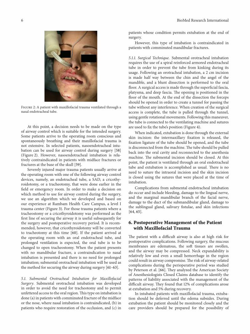
Figure 2: A patient with maxillofacial trauma ventilated through anasal endotracheal tube.
At this point, a decision needs to be made on the typeof airway control which is suitable for the intended surgery.Some patients arrive to the operating room conscious andspontaneously breathing and their maxillofacial trauma isnot extensive. In selected patients, nasoendotracheal intu-bation can be used for airway control during surgery [58](Figure 2). However, nasoendotracheal intubation is rela-tively contraindicated in patients with midface fractures orfractures at the base of the skull [59].
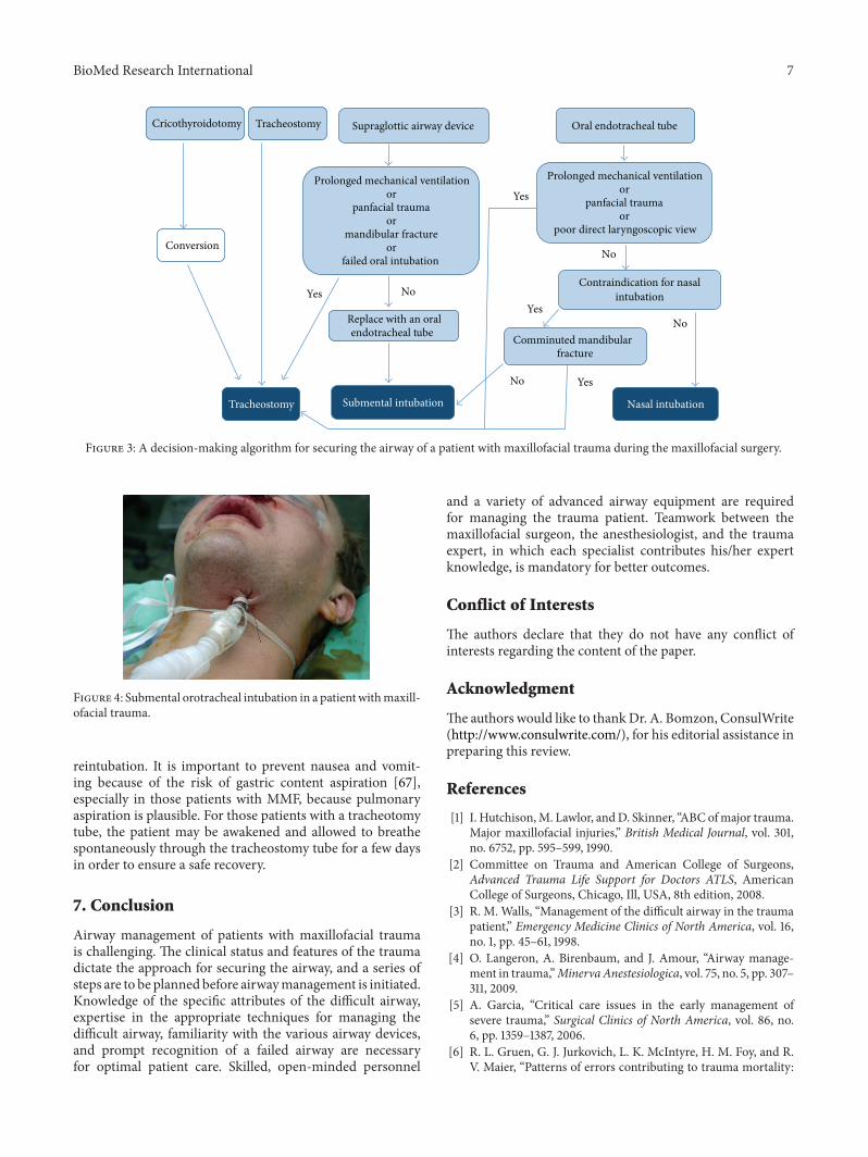
Severely injured major trauma patients usually arrive atthe operating room with one of the following airway controldevices, namely, an endotracheal tube, a SAD, a cricothy-roidotomy, or a tracheotomy, that were done earlier in thefield or emergency room. In order to make a decision onwhich method to use for airway control during the surgery,we use an algorithm which we developed and based onour experience at Rambam Health Care Campus, a level Itrauma center (Figure 3). For those trauma patients where atracheostomy or a cricothyroidotomy was performed as thefirst line of securing the airway it is useful subsequently forthe surgery and postoperative recovery period. It is recom-mended, however, that cricothyroidotomy will be convertedto tracheotomy at this time [60]. If the patient arrived atthe operating room with an oral endotracheal tube, andprolonged ventilation is expected, the oral tube is to bechanged to open tracheostomy. When the patient presentswith no mandibular fracture, a contraindication to nasalintubation is presented and there is no need for prolongedintubation; submental orotracheal intubation will be used asthe method for securing the airway during surgery [61–63].
5.1. Submental Orotracheal Intubation for MaxillofacialSurgery. Submental orotracheal intubation was developedin order to avoid the need for tracheotomy and to permitunfettered access to the oral region.This type on intubation isdone (a) in patients with comminuted fracture of themidfaceor the nose, where nasal intubation is contraindicated, (b) inpatients who require restoration of the occlusion, and (c) in
patients whose condition permits extubation at the end ofsurgery.
However, this type of intubation is contraindicated inpatients with comminuted mandibular fractures.
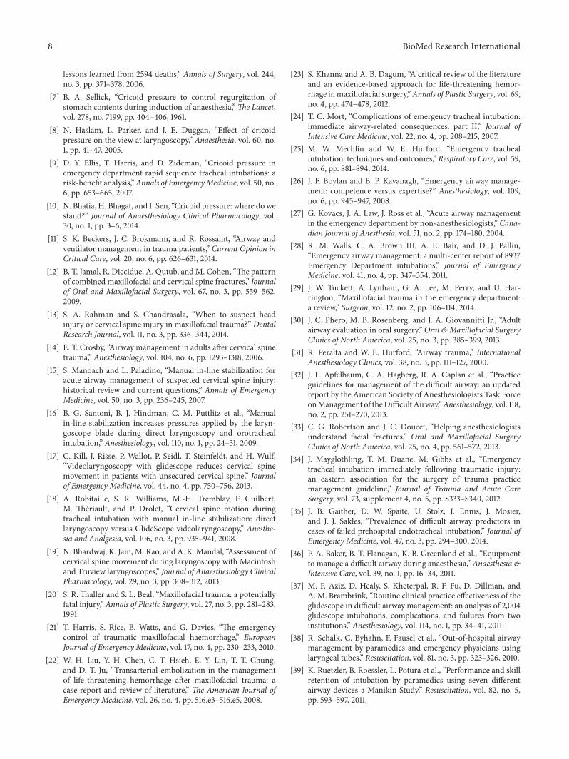
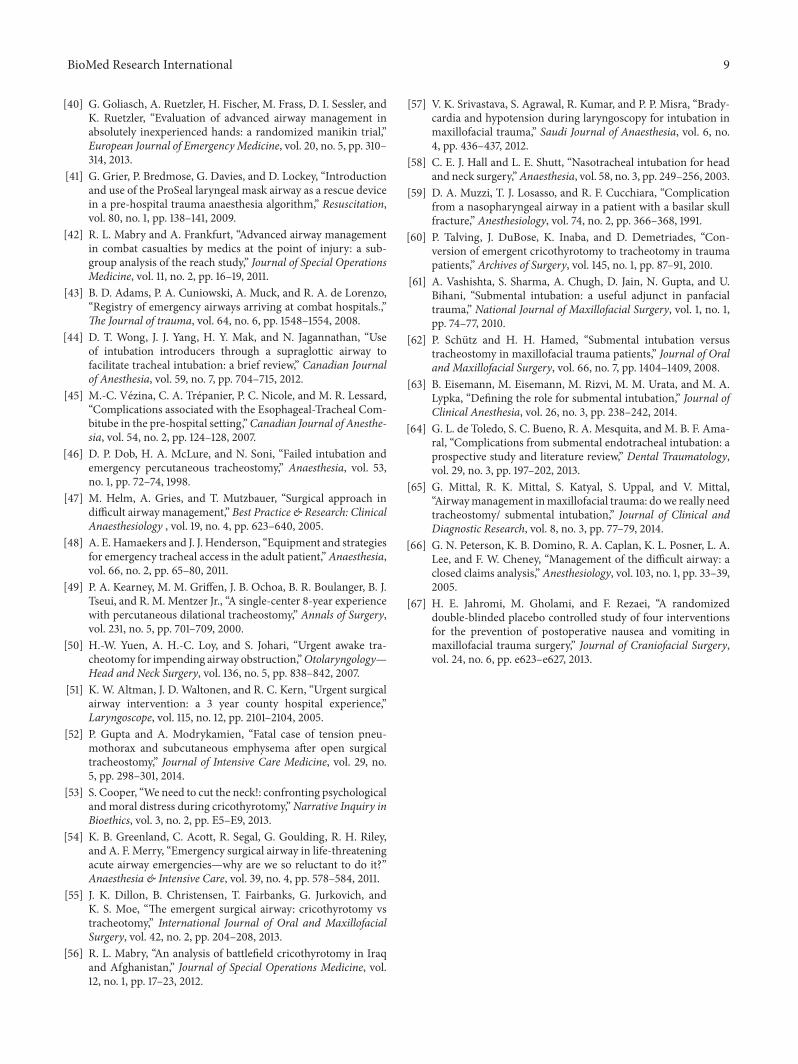
5.1.1. Surgical Technique. Submental orotracheal intubationrequires the use of a spiral reinforced armored endotrachealtube in order to prevent the tube from kinking during itsusage. Following an orotracheal intubation, a 2 cm incisionis made half way between the chin and the angel of themandible, and a blunt dissection is performed to the oralfloor. A surgical access is made through the superficial fascia,platysma, and deep fascia. The opening is positioned in thefloor of the mouth. At the end of the dissection the forcepsshould be opened in order to create a tunnel for passing thetube without any interference. When creation of the surgicalaccess is complete, the tube is pulled through the tunnel,using gentle rotational movements. Following this maneuver,the tube is connected to the ventilating machine and suturesare used to fix the tube’s position (Figure 4).
When indicated, extubation is done through the externalskin incision: the intermaxillary fixation is released, thefixation ligature of the tube should be opened, and the tubeis disconnected from the machine.The tube should be pulledback into the oral cavity and reconnected to the anesthesiamachine. The submental incision should be closed. At thispoint, the patient is ventilated through an oral endotrachealtube and extubation is accomplished as usual. There is noneed to suture the intraoral incision and the skin incisionis closed using the sutures that were placed at the time ofintubation.
Complications from submental endotracheal intubationdo occur and include bleeding, damage to the lingual nerve,and the marginal mandibular branch of the facial nerve,damage to the duct of the submandibular gland, damage tothe sublingual gland, salivary fistulae, and skin infections[64, 65].
6. Postoperative Management of the Patientwith Maxillofacial Trauma
The patient with a difficult airway is also at high risk forpostoperative complications. Following surgery, the mucousmembranes are edematous, the soft tissues are swollen,and the airway may be compressed. Neck expandability isrelatively low and even a small hemorrhage in the regioncould result in airway compromise.The risk of airway-relatedcomplications during the perioperative period was studiedby Peterson et al. [66]. They analyzed the American Societyof Anesthesiologists Closed Claims database to identify thepatterns of liability associated with the management of thedifficult airway. They found that 12% of complications aroseat extubation and 5% during recovery.
In intubated patients with maxillofacial trauma, extuba-tion should be deferred until the edema subsides. Duringextubation the patient should be monitored closely and thecare providers should be prepared for the possibility of
BioMed Research International 7
TracheostomyCricothyroidotomy Supraglottic airway device Oral endotracheal tube
intubation
Prolonged mechanical ventilationor
panfacial traumaor
mandibular fractureor
failed oral intubation
Tracheostomy Submental intubation
endotracheal tube
Conversion
Prolonged mechanical ventilationor
panfacial traumaor
Nasal intubation
Yes
Yes
Yes
Yes
No
No
No
No
fracture
Contraindication for nasal
Replace with an oral
poor direct laryngoscopic view
Comminuted mandibular
Figure 3: A decision-making algorithm for securing the airway of a patient with maxillofacial trauma during the maxillofacial surgery.
Figure 4: Submental orotracheal intubation in a patientwithmaxill-ofacial trauma.
reintubation. It is important to prevent nausea and vomit-ing because of the risk of gastric content aspiration [67],especially in those patients with MMF, because pulmonaryaspiration is plausible. For those patients with a tracheotomytube, the patient may be awakened and allowed to breathespontaneously through the tracheostomy tube for a few daysin order to ensure a safe recovery.
7. Conclusion
Airway management of patients with maxillofacial traumais challenging. The clinical status and features of the traumadictate the approach for securing the airway, and a series ofsteps are to be planned before airwaymanagement is initiated.Knowledge of the specific attributes of the difficult airway,expertise in the appropriate techniques for managing thedifficult airway, familiarity with the various airway devices,and prompt recognition of a failed airway are necessaryfor optimal patient care. Skilled, open-minded personnel
and a variety of advanced airway equipment are requiredfor managing the trauma patient. Teamwork between themaxillofacial surgeon, the anesthesiologist, and the traumaexpert, in which each specialist contributes his/her expertknowledge, is mandatory for better outcomes.
Conflict of Interests
The authors declare that they do not have any conflict ofinterests regarding the content of the paper.
Acknowledgment
The authors would like to thankDr. A. Bomzon, ConsulWrite(http://www.consulwrite.com/), for his editorial assistance inpreparing this review.
References
[1] I. Hutchison,M. Lawlor, andD. Skinner, “ABC ofmajor trauma.Major maxillofacial injuries,” British Medical Journal, vol. 301,no. 6752, pp. 595–599, 1990.
[2] Committee on Trauma and American College of Surgeons,Advanced Trauma Life Support for Doctors ATLS, AmericanCollege of Surgeons, Chicago, Ill, USA, 8th edition, 2008.
[3] R. M. Walls, “Management of the difficult airway in the traumapatient,” Emergency Medicine Clinics of North America, vol. 16,no. 1, pp. 45–61, 1998.
[4] O. Langeron, A. Birenbaum, and J. Amour, “Airway manage-ment in trauma,”MinervaAnestesiologica, vol. 75, no. 5, pp. 307–311, 2009.
[5] A. Garcia, “Critical care issues in the early management ofsevere trauma,” Surgical Clinics of North America, vol. 86, no.6, pp. 1359–1387, 2006.
[6] R. L. Gruen, G. J. Jurkovich, L. K. McIntyre, H. M. Foy, and R.V. Maier, “Patterns of errors contributing to trauma mortality:
8 BioMed Research International
lessons learned from 2594 deaths,” Annals of Surgery, vol. 244,no. 3, pp. 371–378, 2006.
[7] B. A. Sellick, “Cricoid pressure to control regurgitation ofstomach contents during induction of anaesthesia,”The Lancet,vol. 278, no. 7199, pp. 404–406, 1961.
[8] N. Haslam, L. Parker, and J. E. Duggan, “Effect of cricoidpressure on the view at laryngoscopy,” Anaesthesia, vol. 60, no.1, pp. 41–47, 2005.
[9] D. Y. Ellis, T. Harris, and D. Zideman, “Cricoid pressure inemergency department rapid sequence tracheal intubations: arisk-benefit analysis,”Annals of EmergencyMedicine, vol. 50, no.6, pp. 653–665, 2007.
[10] N. Bhatia, H. Bhagat, and I. Sen, “Cricoid pressure: where do westand?” Journal of Anaesthesiology Clinical Pharmacology, vol.30, no. 1, pp. 3–6, 2014.
[11] S. K. Beckers, J. C. Brokmann, and R. Rossaint, “Airway andventilator management in trauma patients,” Current Opinion inCritical Care, vol. 20, no. 6, pp. 626–631, 2014.
[12] B. T. Jamal, R. Diecidue, A. Qutub, andM. Cohen, “The patternof combined maxillofacial and cervical spine fractures,” Journalof Oral and Maxillofacial Surgery, vol. 67, no. 3, pp. 559–562,2009.
[13] S. A. Rahman and S. Chandrasala, “When to suspect headinjury or cervical spine injury in maxillofacial trauma?” DentalResearch Journal, vol. 11, no. 3, pp. 336–344, 2014.
[14] E. T. Crosby, “Airway management in adults after cervical spinetrauma,” Anesthesiology, vol. 104, no. 6, pp. 1293–1318, 2006.
[15] S. Manoach and L. Paladino, “Manual in-line stabilization foracute airway management of suspected cervical spine injury:historical review and current questions,” Annals of EmergencyMedicine, vol. 50, no. 3, pp. 236–245, 2007.
[16] B. G. Santoni, B. J. Hindman, C. M. Puttlitz et al., “Manualin-line stabilization increases pressures applied by the laryn-goscope blade during direct laryngoscopy and orotrachealintubation,” Anesthesiology, vol. 110, no. 1, pp. 24–31, 2009.
[17] C. Kill, J. Risse, P. Wallot, P. Seidl, T. Steinfeldt, and H. Wulf,“Videolaryngoscopy with glidescope reduces cervical spinemovement in patients with unsecured cervical spine,” Journalof Emergency Medicine, vol. 44, no. 4, pp. 750–756, 2013.
[18] A. Robitaille, S. R. Williams, M.-H. Tremblay, F. Guilbert,M. Theriault, and P. Drolet, “Cervical spine motion duringtracheal intubation with manual in-line stabilization: directlaryngoscopy versus GlideScope videolaryngoscopy,” Anesthe-sia and Analgesia, vol. 106, no. 3, pp. 935–941, 2008.
[19] N. Bhardwaj, K. Jain, M. Rao, and A. K. Mandal, “Assessment ofcervical spine movement during laryngoscopy with Macintoshand Truview laryngoscopes,” Journal of Anaesthesiology ClinicalPharmacology, vol. 29, no. 3, pp. 308–312, 2013.
[20] S. R. Thaller and S. L. Beal, “Maxillofacial trauma: a potentiallyfatal injury,”Annals of Plastic Surgery, vol. 27, no. 3, pp. 281–283,1991.
[21] T. Harris, S. Rice, B. Watts, and G. Davies, “The emergencycontrol of traumatic maxillofacial haemorrhage,” EuropeanJournal of Emergency Medicine, vol. 17, no. 4, pp. 230–233, 2010.
[22] W. H. Liu, Y. H. Chen, C. T. Hsieh, E. Y. Lin, T. T. Chung,and D. T. Ju, “Transarterial embolization in the managementof life-threatening hemorrhage after maxillofacial trauma: acase report and review of literature,” The American Journal ofEmergency Medicine, vol. 26, no. 4, pp. 516.e3–516.e5, 2008.
[23] S. Khanna and A. B. Dagum, “A critical review of the literatureand an evidence-based approach for life-threatening hemor-rhage inmaxillofacial surgery,”Annals of Plastic Surgery, vol. 69,no. 4, pp. 474–478, 2012.
[24] T. C. Mort, “Complications of emergency tracheal intubation:immediate airway-related consequences: part II,” Journal ofIntensive Care Medicine, vol. 22, no. 4, pp. 208–215, 2007.
[25] M. W. Mechlin and W. E. Hurford, “Emergency trachealintubation: techniques and outcomes,” Respiratory Care, vol. 59,no. 6, pp. 881–894, 2014.
[26] J. F. Boylan and B. P. Kavanagh, “Emergency airway manage-ment: competence versus expertise?” Anesthesiology, vol. 109,no. 6, pp. 945–947, 2008.
[27] G. Kovacs, J. A. Law, J. Ross et al., “Acute airway managementin the emergency department by non-anesthesiologists,” Cana-dian Journal of Anesthesia, vol. 51, no. 2, pp. 174–180, 2004.
[28] R. M. Walls, C. A. Brown III, A. E. Bair, and D. J. Pallin,“Emergency airway management: a multi-center report of 8937Emergency Department intubations,” Journal of EmergencyMedicine, vol. 41, no. 4, pp. 347–354, 2011.
[29] J. W. Tuckett, A. Lynham, G. A. Lee, M. Perry, and U. Har-rington, “Maxillofacial trauma in the emergency department:a review,” Surgeon, vol. 12, no. 2, pp. 106–114, 2014.
[30] J. C. Phero, M. B. Rosenberg, and J. A. Giovannitti Jr., “Adultairway evaluation in oral surgery,” Oral &Maxillofacial SurgeryClinics of North America, vol. 25, no. 3, pp. 385–399, 2013.
[31] R. Peralta and W. E. Hurford, “Airway trauma,” InternationalAnesthesiology Clinics, vol. 38, no. 3, pp. 111–127, 2000.
[32] J. L. Apfelbaum, C. A. Hagberg, R. A. Caplan et al., “Practiceguidelines for management of the difficult airway: an updatedreport by the American Society of Anesthesiologists Task ForceonManagement of theDifficult Airway,”Anesthesiology, vol. 118,no. 2, pp. 251–270, 2013.
[33] C. G. Robertson and J. C. Doucet, “Helping anesthesiologistsunderstand facial fractures,” Oral and Maxillofacial SurgeryClinics of North America, vol. 25, no. 4, pp. 561–572, 2013.
[34] J. Mayglothling, T. M. Duane, M. Gibbs et al., “Emergencytracheal intubation immediately following traumatic injury:an eastern association for the surgery of trauma practicemanagement guideline,” Journal of Trauma and Acute CareSurgery, vol. 73, supplement 4, no. 5, pp. S333–S340, 2012.
[35] J. B. Gaither, D. W. Spaite, U. Stolz, J. Ennis, J. Mosier,and J. J. Sakles, “Prevalence of difficult airway predictors incases of failed prehospital endotracheal intubation,” Journal ofEmergency Medicine, vol. 47, no. 3, pp. 294–300, 2014.
[36] P. A. Baker, B. T. Flanagan, K. B. Greenland et al., “Equipmentto manage a difficult airway during anaesthesia,” Anaesthesia &Intensive Care, vol. 39, no. 1, pp. 16–34, 2011.
[37] M. F. Aziz, D. Healy, S. Kheterpal, R. F. Fu, D. Dillman, andA. M. Brambrink, “Routine clinical practice effectiveness of theglidescope in difficult airway management: an analysis of 2,004glidescope intubations, complications, and failures from twoinstitutions,” Anesthesiology, vol. 114, no. 1, pp. 34–41, 2011.
[38] R. Schalk, C. Byhahn, F. Fausel et al., “Out-of-hospital airwaymanagement by paramedics and emergency physicians usinglaryngeal tubes,” Resuscitation, vol. 81, no. 3, pp. 323–326, 2010.
[39] K. Ruetzler, B. Roessler, L. Potura et al., “Performance and skillretention of intubation by paramedics using seven differentairway devices-a Manikin Study,” Resuscitation, vol. 82, no. 5,pp. 593–597, 2011.
BioMed Research International 9
[40] G. Goliasch, A. Ruetzler, H. Fischer, M. Frass, D. I. Sessler, andK. Ruetzler, “Evaluation of advanced airway management inabsolutely inexperienced hands: a randomized manikin trial,”European Journal of Emergency Medicine, vol. 20, no. 5, pp. 310–314, 2013.
[41] G. Grier, P. Bredmose, G. Davies, and D. Lockey, “Introductionand use of the ProSeal laryngeal mask airway as a rescue devicein a pre-hospital trauma anaesthesia algorithm,” Resuscitation,vol. 80, no. 1, pp. 138–141, 2009.
[42] R. L. Mabry and A. Frankfurt, “Advanced airway managementin combat casualties by medics at the point of injury: a sub-group analysis of the reach study,” Journal of Special OperationsMedicine, vol. 11, no. 2, pp. 16–19, 2011.
[43] B. D. Adams, P. A. Cuniowski, A. Muck, and R. A. de Lorenzo,“Registry of emergency airways arriving at combat hospitals.,”The Journal of trauma, vol. 64, no. 6, pp. 1548–1554, 2008.
[44] D. T. Wong, J. J. Yang, H. Y. Mak, and N. Jagannathan, “Useof intubation introducers through a supraglottic airway tofacilitate tracheal intubation: a brief review,” Canadian Journalof Anesthesia, vol. 59, no. 7, pp. 704–715, 2012.
[45] M.-C. Vezina, C. A. Trepanier, P. C. Nicole, and M. R. Lessard,“Complications associated with the Esophageal-Tracheal Com-bitube in the pre-hospital setting,”Canadian Journal of Anesthe-sia, vol. 54, no. 2, pp. 124–128, 2007.
[46] D. P. Dob, H. A. McLure, and N. Soni, “Failed intubation andemergency percutaneous tracheostomy,” Anaesthesia, vol. 53,no. 1, pp. 72–74, 1998.
[47] M. Helm, A. Gries, and T. Mutzbauer, “Surgical approach indifficult airway management,” Best Practice & Research: ClinicalAnaesthesiology , vol. 19, no. 4, pp. 623–640, 2005.
[48] A. E. Hamaekers and J. J. Henderson, “Equipment and strategiesfor emergency tracheal access in the adult patient,” Anaesthesia,vol. 66, no. 2, pp. 65–80, 2011.
[49] P. A. Kearney, M. M. Griffen, J. B. Ochoa, B. R. Boulanger, B. J.Tseui, and R. M. Mentzer Jr., “A single-center 8-year experiencewith percutaneous dilational tracheostomy,” Annals of Surgery,vol. 231, no. 5, pp. 701–709, 2000.
[50] H.-W. Yuen, A. H.-C. Loy, and S. Johari, “Urgent awake tra-cheotomy for impending airway obstruction,”Otolaryngology—Head and Neck Surgery, vol. 136, no. 5, pp. 838–842, 2007.
[51] K. W. Altman, J. D. Waltonen, and R. C. Kern, “Urgent surgicalairway intervention: a 3 year county hospital experience,”Laryngoscope, vol. 115, no. 12, pp. 2101–2104, 2005.
[52] P. Gupta and A. Modrykamien, “Fatal case of tension pneu-mothorax and subcutaneous emphysema after open surgicaltracheostomy,” Journal of Intensive Care Medicine, vol. 29, no.5, pp. 298–301, 2014.
[53] S. Cooper, “We need to cut the neck!: confronting psychologicaland moral distress during cricothyrotomy,”Narrative Inquiry inBioethics, vol. 3, no. 2, pp. E5–E9, 2013.
[54] K. B. Greenland, C. Acott, R. Segal, G. Goulding, R. H. Riley,and A. F. Merry, “Emergency surgical airway in life-threateningacute airway emergencies—why are we so reluctant to do it?”Anaesthesia & Intensive Care, vol. 39, no. 4, pp. 578–584, 2011.
[55] J. K. Dillon, B. Christensen, T. Fairbanks, G. Jurkovich, andK. S. Moe, “The emergent surgical airway: cricothyrotomy vstracheotomy,” International Journal of Oral and MaxillofacialSurgery, vol. 42, no. 2, pp. 204–208, 2013.
[56] R. L. Mabry, “An analysis of battlefield cricothyrotomy in Iraqand Afghanistan,” Journal of Special Operations Medicine, vol.12, no. 1, pp. 17–23, 2012.
[57] V. K. Srivastava, S. Agrawal, R. Kumar, and P. P. Misra, “Brady-cardia and hypotension during laryngoscopy for intubation inmaxillofacial trauma,” Saudi Journal of Anaesthesia, vol. 6, no.4, pp. 436–437, 2012.
[58] C. E. J. Hall and L. E. Shutt, “Nasotracheal intubation for headand neck surgery,”Anaesthesia, vol. 58, no. 3, pp. 249–256, 2003.
[59] D. A. Muzzi, T. J. Losasso, and R. F. Cucchiara, “Complicationfrom a nasopharyngeal airway in a patient with a basilar skullfracture,” Anesthesiology, vol. 74, no. 2, pp. 366–368, 1991.
[60] P. Talving, J. DuBose, K. Inaba, and D. Demetriades, “Con-version of emergent cricothyrotomy to tracheotomy in traumapatients,” Archives of Surgery, vol. 145, no. 1, pp. 87–91, 2010.
[61] A. Vashishta, S. Sharma, A. Chugh, D. Jain, N. Gupta, and U.Bihani, “Submental intubation: a useful adjunct in panfacialtrauma,” National Journal of Maxillofacial Surgery, vol. 1, no. 1,pp. 74–77, 2010.
[62] P. Schutz and H. H. Hamed, “Submental intubation versustracheostomy in maxillofacial trauma patients,” Journal of Oraland Maxillofacial Surgery, vol. 66, no. 7, pp. 1404–1409, 2008.
[63] B. Eisemann, M. Eisemann, M. Rizvi, M. M. Urata, and M. A.Lypka, “Defining the role for submental intubation,” Journal ofClinical Anesthesia, vol. 26, no. 3, pp. 238–242, 2014.
[64] G. L. de Toledo, S. C. Bueno, R. A. Mesquita, and M. B. F. Ama-ral, “Complications from submental endotracheal intubation: aprospective study and literature review,” Dental Traumatology,vol. 29, no. 3, pp. 197–202, 2013.
[65] G. Mittal, R. K. Mittal, S. Katyal, S. Uppal, and V. Mittal,“Airwaymanagement inmaxillofacial trauma: dowe really needtracheostomy/ submental intubation,” Journal of Clinical andDiagnostic Research, vol. 8, no. 3, pp. 77–79, 2014.
[66] G. N. Peterson, K. B. Domino, R. A. Caplan, K. L. Posner, L. A.Lee, and F. W. Cheney, “Management of the difficult airway: aclosed claims analysis,” Anesthesiology, vol. 103, no. 1, pp. 33–39,2005.
[67] H. E. Jahromi, M. Gholami, and F. Rezaei, “A randomizeddouble-blinded placebo controlled study of four interventionsfor the prevention of postoperative nausea and vomiting inmaxillofacial trauma surgery,” Journal of Craniofacial Surgery,vol. 24, no. 6, pp. e623–e627, 2013.

Submit your manuscripts athttp://www.hindawi.com
Stem CellsInternational
Hindawi Publishing Corporationhttp://www.hindawi.com Volume 2014
Hindawi Publishing Corporationhttp://www.hindawi.com Volume 2014
MEDIATORSINFLAMMATION
of
Hindawi Publishing Corporationhttp://www.hindawi.com Volume 2014
Behavioural Neurology
EndocrinologyInternational Journal of
Hindawi Publishing Corporationhttp://www.hindawi.com Volume 2014
Hindawi Publishing Corporationhttp://www.hindawi.com Volume 2014
Disease Markers
Hindawi Publishing Corporationhttp://www.hindawi.com Volume 2014
BioMed Research International
OncologyJournal of
Hindawi Publishing Corporationhttp://www.hindawi.com Volume 2014
Hindawi Publishing Corporationhttp://www.hindawi.com Volume 2014
Oxidative Medicine and Cellular Longevity
Hindawi Publishing Corporationhttp://www.hindawi.com Volume 2014
PPAR Research
The Scientific World JournalHindawi Publishing Corporation http://www.hindawi.com Volume 2014
Immunology ResearchHindawi Publishing Corporationhttp://www.hindawi.com Volume 2014
Journal of
ObesityJournal of
Hindawi Publishing Corporationhttp://www.hindawi.com Volume 2014
Hindawi Publishing Corporationhttp://www.hindawi.com Volume 2014
Computational and Mathematical Methods in Medicine
OphthalmologyJournal of
Hindawi Publishing Corporationhttp://www.hindawi.com Volume 2014
Diabetes ResearchJournal of
Hindawi Publishing Corporationhttp://www.hindawi.com Volume 2014
Hindawi Publishing Corporationhttp://www.hindawi.com Volume 2014
Research and TreatmentAIDS
Hindawi Publishing Corporationhttp://www.hindawi.com Volume 2014
Gastroenterology Research and Practice
Hindawi Publishing Corporationhttp://www.hindawi.com Volume 2014
Parkinson’s Disease
Evidence-Based Complementary and Alternative Medicine
Volume 2014Hindawi Publishing Corporationhttp://www.hindawi.com