reversed-phase high-performance liquid chromatography
TRANSCRIPT
9
2
Reversed-Phase High-Performance LiquidChromatography
Marie-Isabel Aguilar
1. IntroductionReversed-phase high-performance liquid chromatography (RP-HPLC)
involves the separation of molecules on the basis of hydrophobicity. The sepa-ration depends on the hydrophobic binding of the solute molecule from themobile phase to the immobilized hydrophobic ligands attached to the station-ary phase, i.e., the sorbent. A schematic diagram showing the binding of a pep-tide or a protein to a reversed-phase surface is shown in Fig. 1. The solutemixture is initially applied to the sorbent in the presence of aqueous buffers, andthe solutes are eluted by the addition of organic solvent to the mobile phase.Elution can proceed either by isocratic conditions where the concentration oforganic solvent is constant, or by gradient elution whereby the amount of organicsolvent is increased over a period of time. The solutes are, therefore, eluted inorder of increasing molecular hydrophobicity. RP-HPLC is a very powerfultechnique for the analysis of peptides and proteins because of a number of fac-tors that include: (1) the excellent resolution that can be achieved under a widerange of chromatographic conditions for very closely related molecules as wellas structurally quite distinct molecules; (2) the experimental ease with whichchromatographic selectivity can be manipulated through changes in mobilephase characteristics; (3) the generally high recoveries and, hence, high pro-ductivity; and (4) the excellent reproducibility of repetitive separations carriedout over a long period of time, which is caused partly by the stability of the sor-bent materials under a wide range of mobile phase conditions (1,2). However,RP-HPLC can cause the irreversible denaturation of protein samples therebyreducing the potential recovery of material in a biologically active form.
From: Methods in Molecular Biology, vol. 251, HPLC of Peptides and Proteins: Methods and ProtocolsEdited by: M.-I. Aguilar © Humana Press Inc., Totowa, NJ
CH02,9-22,14pgs 10/30/03 6:59 PM Page 9

The RP-HPLC experimental system for the analysis of peptides and proteinsusually consists of an n-alkylsilica-based sorbent from which the solutes areeluted with gradients of increasing concentrations of organic solvent such as ace-tonitrile containing an ionic modifier such as trifluoroacetic acid (TFA) (1,2).Complex mixtures of peptides and proteins can be routinely separated and lowpicomolar—femtomolar amounts of material can be collected for further charac-terization. Separations can be easily manipulated by changing the gradient slope,the operating temperature, the ionic modifier, or the organic solvent composition.
The extensive use of RP-HPLC for the purification of peptides, small polypep-tides with molecular weights up to 10,000, and related compounds of pharma-ceutical interest has not been replicated to the same extent for larger polypeptides
10 Aguilar
Fig. 1. Schematic representation of the binding of (A) a peptide and (B) a protein,to an RP-HPLC silica-based sorbent. The peptide or protein interacts with the immo-bilized hydrophobic ligands through the hydrophobic chromatographic contact region.
CH02,9-22,14pgs 10/30/03 6:59 PM Page 10
(molecular mass > 10 KDa) and globular proteins. The combination of thetraditionally used acidic buffering systems and the hydrophobicity of then-alkylsilica supports which can result in low mass yields or the loss of biolog-ical activity of larger polypeptides and proteins have often discouraged practi-tioners from using RP-HPLC methods for large-scale protein separations. Theloss of enzymatic activity, the formation of multiple peaks for compositionallypure samples, and poor yields of protein can all be attributed to the denaturationof protein solutes during the separation process using RP-HPLC (3–6).
RP-HPLC is extremely versatile for the isolation of peptides and proteinsfrom a wide variety of synthetic or biological sources and is used for both ana-lytical and preparative applications (1–2, see also Chs. 10–21). Analyticalapplications range from the assessment of purity of peptides following solid-phase peptide synthesis (see Ch. 14), to the analysis of tryptic maps of proteins.Preparative RP-HPLC is also used for the micropurification of protein fragmentsfor sequencing to large-scale purification of synthetic peptides and recombi-nant proteins. The complexity of the mixture to be chromatographed will dependon the nature of the source and the degree of preliminary clean-up that can beperformed. In the case of synthetic peptides, RP-HPLC is generally employedboth for the initial analysis and the final large-scale purification. The purifica-tion of synthetic peptides usually involves an initial separation on an analyti-cal scale to assess the complexity of the mixture followed by large-scalepurification and collection of the target product. A sample of the purified mate-rial can then be subjected to RP-HPLC analysis under the same or different elu-tion conditions to check for purity. The isolation of proteins from a biologicalcocktail derived from a tissue extract or biological fluid for example, oftenrequires a combination of techniques to produce a homogenous sample. HPLCtechniques are then introduced at the later stages following initial precipitation,clarification, and preliminary separations using soft gels.
The challenge facing the scientist who wishes to analyze and/or purify theirpeptide or protein sample by RP-HPLC is the selection of the initial separationconditions and subsequent optimization of the appropriate experimental para-meters. This chapter describes a standard method that can be used as an initialprocedure for the RP-HPLC analysis of a peptide sample, and then differentexperimental options available to achieve a high-resolution separation of a pep-tide or protein mixture using RP-HPLC are outlined in Subheading 4.
2. Materials2.1. Chemicals
1. Acetonitrile (CH3CN), HPLC grade.2. Milli-Q water.3. Trifluoroacetic acid (TFA).
RP-HPLC 11
CH02,9-22,14pgs 10/30/03 6:59 PM Page 11
2.2. Equipment and Supplies
1. HPLC solvent delivery system with binary gradient capability and a UV detector.2. Reversed-phase octadecylsilica (C18) column (see Note 1) (4.6 mm id (internal
diameter) × 250 mm length (see Note 2), 5 µm particle size, 300 Å pore size (seeNote 3).
3. C18 guard column.4. Solvent filtration apparatus equipped with a 0.22-µm Teflon filter.5. Sample filters, 0.22 µm porosity.6. Buffer A: 0.1% (v/v) TFA in water (see Note 4).7. Buffer B: 100% CH3CN containing 0.1% (v/v) TFA (see Note 5).
3. Methods3.1. Sample Preparation
Dissolve 1 mg of sample in 1 mL of Buffer A. If there is some undissolvedmaterial, filter the sample through a 0.22-µm filter.
3.2. Solvent Preparation
Filter all solvents through a 0.22-µm filter before use. This removes partic-ulates that could block solvent lines or the column and also serves to degassthe solvent. If the HPLC instrument is not installed with on-line degassing capa-bility, check with your instrument requirements to assess whether furtherdegassing is required.
3.3. Column Equilibration and Blank Run
1. Connect the guard and the column to the solvent delivery system according to theHPLC system requirements and equilibrate under the following initial conditions.a. Solvent: 100% Buffer Ab. Flow rate: 1 mL/min (see Note 6)c. Detection wavelength: 215 nm (see Note 7)D. Temperature: Ambient (see Note 8)
2. Once a stable baseline is obtained, inject 10 µL of Milli-Q water (either manuallyor via an automatic injector). It is generally advisable to perform two blank runsto ensure proper equilibration of the column.
3.4. Sample Injection and Analysis
Once a stable baseline is obtained, inject 10 µL of the sample (either man-ually or via an automatic injector) and use a linear gradient from 0 to 100%buffer B over 30 min to elute the sample (see Note 9).
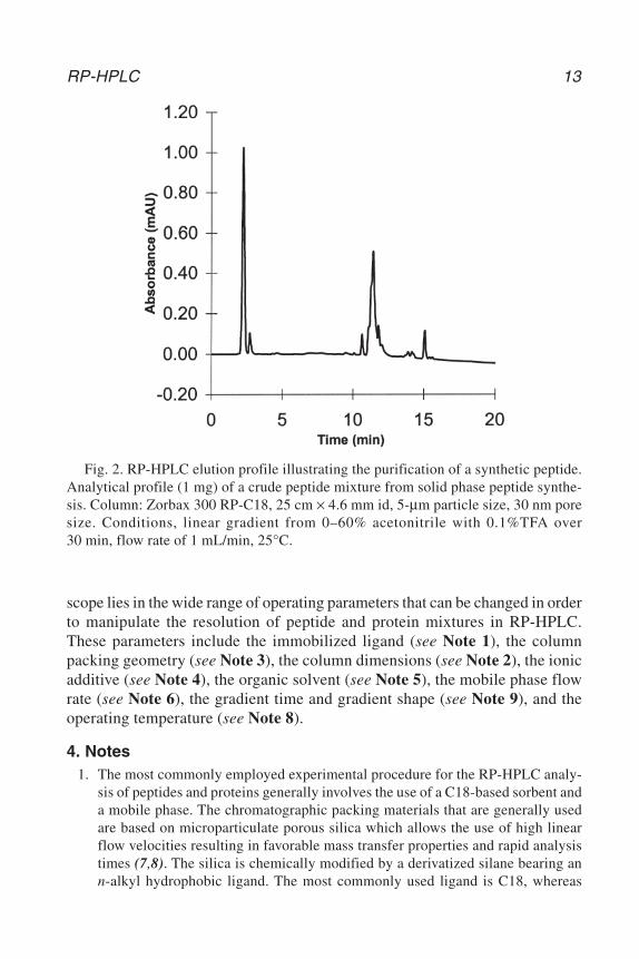
Figure 2 shows a typical chromatogram of a crude synthetic peptide. Thelarge majority of components should normally elute within the gradient time.Thus, each individual method is relatively straightforward to perform. The
12 Aguilar
CH02,9-22,14pgs 10/30/03 6:59 PM Page 12
scope lies in the wide range of operating parameters that can be changed in orderto manipulate the resolution of peptide and protein mixtures in RP-HPLC.These parameters include the immobilized ligand (see Note 1), the columnpacking geometry (see Note 3), the column dimensions (see Note 2), the ionicadditive (see Note 4), the organic solvent (see Note 5), the mobile phase flowrate (see Note 6), the gradient time and gradient shape (see Note 9), and theoperating temperature (see Note 8).
4. Notes1. The most commonly employed experimental procedure for the RP-HPLC analy-
sis of peptides and proteins generally involves the use of a C18-based sorbent anda mobile phase. The chromatographic packing materials that are generally usedare based on microparticulate porous silica which allows the use of high linearflow velocities resulting in favorable mass transfer properties and rapid analysistimes (7,8). The silica is chemically modified by a derivatized silane bearing ann-alkyl hydrophobic ligand. The most commonly used ligand is C18, whereas
RP-HPLC 13
Fig. 2. RP-HPLC elution profile illustrating the purification of a synthetic peptide.Analytical profile (1 mg) of a crude peptide mixture from solid phase peptide synthe-sis. Column: Zorbax 300 RP-C18, 25 cm × 4.6 mm id, 5-µm particle size, 30 nm poresize. Conditions, linear gradient from 0–60% acetonitrile with 0.1%TFA over30 min, flow rate of 1 mL/min, 25°C.
CH02,9-22,14pgs 10/30/03 6:59 PM Page 13
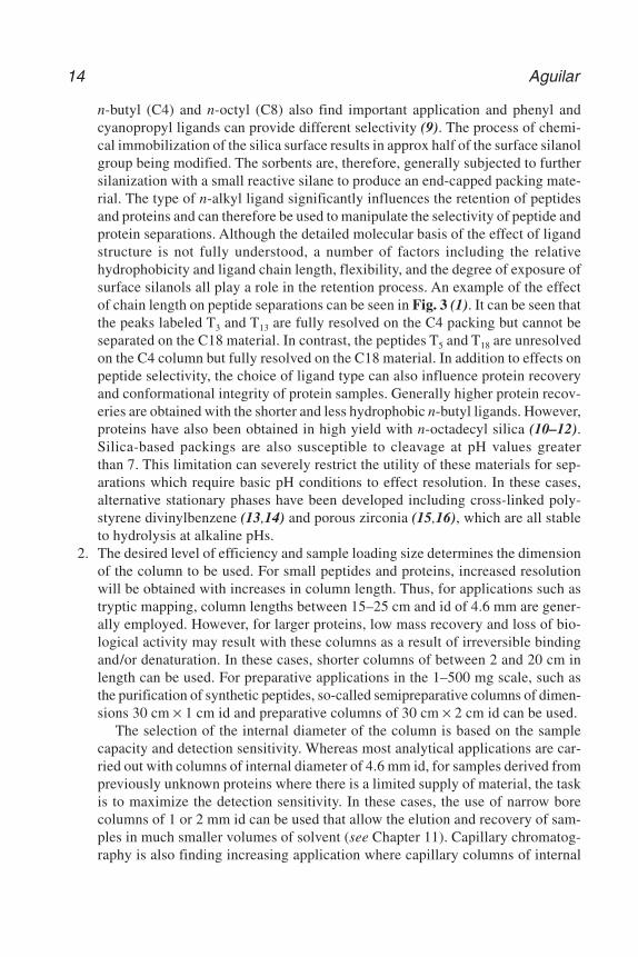
n-butyl (C4) and n-octyl (C8) also find important application and phenyl andcyanopropyl ligands can provide different selectivity (9). The process of chemi-cal immobilization of the silica surface results in approx half of the surface silanolgroup being modified. The sorbents are, therefore, generally subjected to furthersilanization with a small reactive silane to produce an end-capped packing mate-rial. The type of n-alkyl ligand significantly influences the retention of peptidesand proteins and can therefore be used to manipulate the selectivity of peptide andprotein separations. Although the detailed molecular basis of the effect of ligandstructure is not fully understood, a number of factors including the relativehydrophobicity and ligand chain length, flexibility, and the degree of exposure ofsurface silanols all play a role in the retention process. An example of the effectof chain length on peptide separations can be seen in Fig. 3 (1). It can be seen thatthe peaks labeled T3 and T13 are fully resolved on the C4 packing but cannot beseparated on the C18 material. In contrast, the peptides T5 and T18 are unresolvedon the C4 column but fully resolved on the C18 material. In addition to effects onpeptide selectivity, the choice of ligand type can also influence protein recoveryand conformational integrity of protein samples. Generally higher protein recov-eries are obtained with the shorter and less hydrophobic n-butyl ligands. However,proteins have also been obtained in high yield with n-octadecyl silica (10–12).Silica-based packings are also susceptible to cleavage at pH values greaterthan 7. This limitation can severely restrict the utility of these materials for sep-arations which require basic pH conditions to effect resolution. In these cases,alternative stationary phases have been developed including cross-linked poly-styrene divinylbenzene (13,14) and porous zirconia (15,16), which are all stableto hydrolysis at alkaline pHs.
2. The desired level of efficiency and sample loading size determines the dimensionof the column to be used. For small peptides and proteins, increased resolutionwill be obtained with increases in column length. Thus, for applications such astryptic mapping, column lengths between 15–25 cm and id of 4.6 mm are gener-ally employed. However, for larger proteins, low mass recovery and loss of bio-logical activity may result with these columns as a result of irreversible bindingand/or denaturation. In these cases, shorter columns of between 2 and 20 cm inlength can be used. For preparative applications in the 1–500 mg scale, such asthe purification of synthetic peptides, so-called semipreparative columns of dimen-sions 30 cm × 1 cm id and preparative columns of 30 cm × 2 cm id can be used.
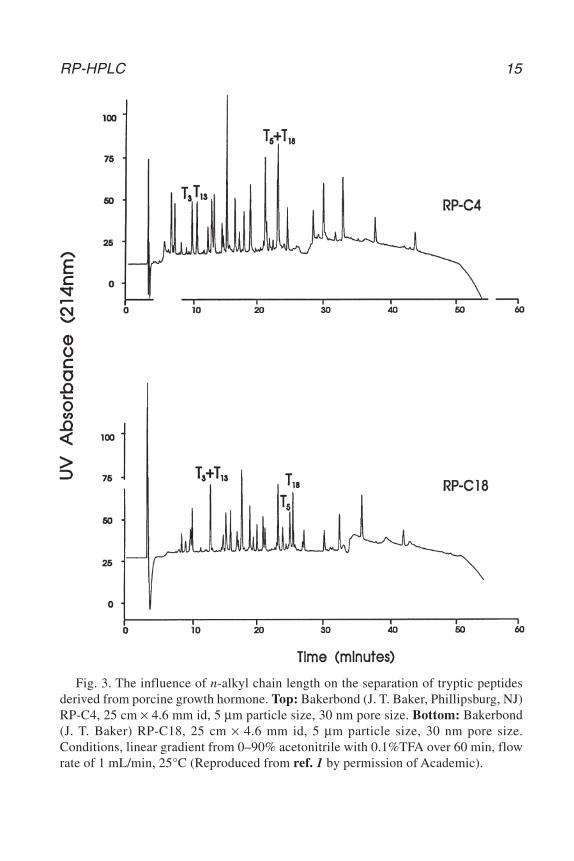
The selection of the internal diameter of the column is based on the samplecapacity and detection sensitivity. Whereas most analytical applications are car-ried out with columns of internal diameter of 4.6 mm id, for samples derived frompreviously unknown proteins where there is a limited supply of material, the taskis to maximize the detection sensitivity. In these cases, the use of narrow borecolumns of 1 or 2 mm id can be used that allow the elution and recovery of sam-ples in much smaller volumes of solvent (see Chapter 11). Capillary chromatog-raphy is also finding increasing application where capillary columns of internal
14 Aguilar
CH02,9-22,14pgs 10/30/03 6:59 PM Page 14
RP-HPLC 15
Fig. 3. The influence of n-alkyl chain length on the separation of tryptic peptidesderived from porcine growth hormone. Top: Bakerbond (J. T. Baker, Phillipsburg, NJ)RP-C4, 25 cm × 4.6 mm id, 5 µm particle size, 30 nm pore size. Bottom: Bakerbond(J. T. Baker) RP-C18, 25 cm × 4.6 mm id, 5 µm particle size, 30 nm pore size.Conditions, linear gradient from 0–90% acetonitrile with 0.1%TFA over 60 min, flowrate of 1 mL/min, 25°C (Reproduced from ref. 1 by permission of Academic).
CH02,9-22,14pgs 10/30/03 6:59 PM Page 15
diameter between 0.2–0.4 mm and column length of 15 cm result in the analysisof femtomole of sample (see Chapter 10). The effect of decreasing column inter-nal diameter on detection sensitivity is shown in Fig. 4 for the analysis of lysozymeon a C18 material packed into columns of 4.6, 2.1, and 0.3 mm id (17).
3. The geometry of the particle in terms of the particle diameter and pore size, is alsoan important feature of the packing material. Improved resolution can be achievedby decreasing the particle diameter and the most commonly used range of parti-cle diameters for analytical scale RP-HPLC is 3–5 µm. There are also examplesof the use of nonporous particles of smaller diameter (18). For preparative scaleseparations, 10–20 µm particles are utilized. The pore size of RP-HPLC sorbentsis also an important factor that must be considered. For peptides, the pore size gen-erally ranges between 100–300 Å depending on the size of the peptides. Porousmaterials of ≥300 Å pore size are necessary for the separation of proteins, as the
16 Aguilar
Fig. 4. Effect of column internal diameter on detector sensitivity. Column: BrownleeRP-300 C8 (7 µm particle size, 30 nm pore size), 3 cm × 4.6 mm id and 10 cm × 2.1 mmid (Applied Biosystems) and 5 cm × 0.32 mm id. Conditions: linear gradient from0–60% acetonitrile with 0.1% TFA over 60 min, 45°C. Flow rates, 1 mL/min, 200 µLl/min,and 4 µL/min for the 4.6, 2.1, and 0.32 mm id columns, respectively. Sample loadings,lysozyme, 10 µg, 4 µg, and 0.04 µg for the 4.6, 2.1, and 0.32 mm id columns, respec-tively. (Reproduced from ref. 17 by permission of Elsevier Science, copyright 1992.)
CH02,9-22,14pgs 10/30/03 6:59 PM Page 16
solute molecular diameter must be at least one-tenth the size of the pore diame-ter to avoid restricted diffusion of the solute and to allow the total surface area ofthe sorbent material to be accessible. The development of particles with6000–8000 Å pores with a network of smaller pores of 500–1000 Å has alsoallowed very rapid peptide and protein separations to be achieved (19,20).
4. RP-HPLC is generally carried out with an acidic mobile phase, with TFA the mostcommonly used additive because of its volatility. However, for high sensitivityapplications, the amount of TFA in buffer B can be adjusted downward becausephosphoric acid, perchloric acid, formic acid, hydrochloric acid, acetic acid, andheptaflourobutyric acid have also been used (21–23). Alternative additives suchas nonionic detergents can be used for the isolation of more hydrophobic proteinssuch as membrane proteins (24, see Chapter 22).
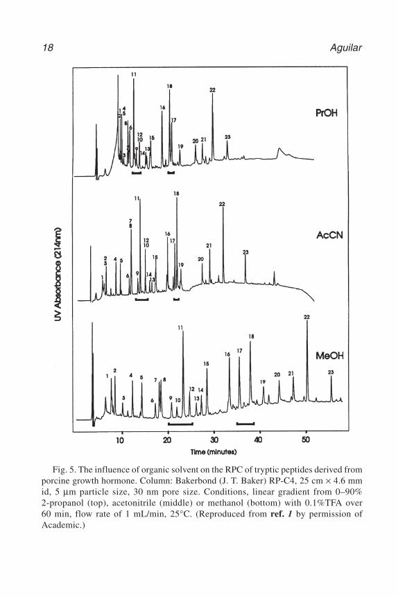
5. One of the most powerful characteristics of RP-HPLC is the ability to manipulatesolute retention and resolution through changes in the composition of the mobilephase. In RP-HPLC, peptide and protein retention is a result of multisite interac-tions with the ligands. The practical consequence of this is that high resolutionisocratic elution of peptides and proteins can rarely be achieved as the experimentalwindow of solvent concentration required for their elution is very narrow. Mixturesof peptides and proteins are therefore routinely eluted by the application of agradient of increasing organic solvent concentration. The three most commonlyemployed organic solvents in RP-HPLC are acetonitrile, methanol, and2-propanol, which all exhibit high optical transparency in the detection wave-lengths used for peptide and protein analysis. Acetonitrile provides the lowest vis-cosity solvent mixtures and 2-propanol is the strongest eluent. An example of theinfluence of organic solvent is shown in Fig. 5 where changes in selectivity canbe observed for a number of peptide peaks in the tryptic map. In addition to theeluotropic effects, the nature of the organic solvent can also influence the con-formation of both peptides and proteins and will, therefore, have an additionaleffect on selectivity through changes in the structure of the hydrophobic contactregion. In the case of proteins, this may also impact on the level of recovery ofbiologically active material.
6. The typical experiment with an analytical scale column would utilize flow ratesranging between 0.5–2.0 mL/min. With microbore columns (1–2 mm id) flow ratesof 50–250 µL/min are used, whereas for capillary columns of 0.2–0.4 mm id, flowrates of 1–4 µL/min are applied. At the preparative end of the scale with columnsof 10–20 mm id, flow rates ranging between 5–20 mL/min are required.
7. Detection of peptides and proteins in RP-HPLC, generally involves detectionbetween 210 and 220 nm, which is specific for the peptide bond, or at 280 nm,which corresponds to the aromatic amino acids tryptophan and tyrosine. The use ofphotodiode array detectors can enhance the detection capabilities by the on-lineaccumulation of complete solute spectra. The spectra can then be used to identifypeaks specifically on the basis of spectral characteristics and for the assessmentof peak purity (24–26). In addition, second derivative spectroscopy can provideinformation on the conformational integrity of proteins following elution (27,28)
RP-HPLC 17
CH02,9-22,14pgs 10/30/03 6:59 PM Page 17
18 Aguilar
Fig. 5. The influence of organic solvent on the RPC of tryptic peptides derived fromporcine growth hormone. Column: Bakerbond (J. T. Baker) RP-C4, 25 cm × 4.6 mmid, 5 µm particle size, 30 nm pore size. Conditions, linear gradient from 0–90%2-propanol (top), acetonitrile (middle) or methanol (bottom) with 0.1%TFA over60 min, flow rate of 1 mL/min, 25°C. (Reproduced from ref. 1 by permission ofAcademic.)
CH02,9-22,14pgs 10/30/03 7:00 PM Page 18
RP-HPLC 19
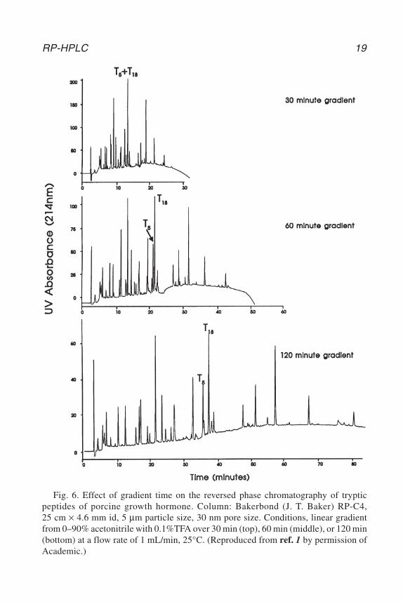
Fig. 6. Effect of gradient time on the reversed phase chromatography of trypticpeptides of porcine growth hormone. Column: Bakerbond (J. T. Baker) RP-C4,25 cm × 4.6 mm id, 5 µm particle size, 30 nm pore size. Conditions, linear gradientfrom 0–90% acetonitrile with 0.1%TFA over 30 min (top), 60 min (middle), or 120 min(bottom) at a flow rate of 1 mL/min, 25°C. (Reproduced from ref. 1 by permission ofAcademic.)
CH02,9-22,14pgs 10/30/03 7:00 PM Page 19
8. The operating temperature can also be used to manipulate resolution. Althoughthe separation of peptides and proteins is normally carried out at ambient tem-perature, solute retention in RP-HPLC is influenced by temperature throughchanges in solvent viscosity. In addition to this, peptide and protein conformationcan also be manipulated by temperature. Changes in temperature can therefore alsobe used to manipulate the structure and retention of peptide mixtures. For pep-tides, it has been shown that secondary structure can actually be enhanced throughbinding to the hydrophobic sorbent (29). In the case of proteins that are to be sub-jected to further chemical analysis and thus where recovery of a biologicallyactive protein is not essential, increasing temperature can be used to modulateretention via denaturation of the protein structure (3–6). However, if the efficientrecovery of both mass and biological activity is of paramount importance, the useof elevated temperatures is not an option.
9. The choice of gradient conditions will depend on the nature of the molecules ofinterest. The influence of gradient time on the separation of a series of tryptic pep-tides proteins is shown in Fig. 6 (1). Generally the use of longer gradient timesprovides improved separation. However, these conditions also increase the resi-dence time of the peptide or protein solute at the sorbent surface, which may thenresult in an increase in the degree of denaturation.
References
1. Aguilar, M. I. and Hearn, M. T. W. (1996) High resolution reversed phase high per-formance liquid chromatography of peptides and proteins. Meth. Enzymol. 270,3–26.
2. Mant, C. T. and Hodges, R. S. (1996) Analysis of peptides by high performanceliquid chromatography. Meth. Enzymol. 271, 3–50.
3. Purcell, A. W., Aguilar, M. I., and Hearn, M. T. W. (1995) Conformational effectsin the RP-HPLC of polypeptides. II: The role of insulin A and B chains in the chro-matographic behaviour of insulin. J. Chromatogr. 711, 71–79.
4. Richards, K. L., Aguilar, M. I., and Hearn, M. T. W. (1994) The effect of proteinconformation on experimental bandwidths in RP-HPLC. J. Chromatogr. 676,33–41.
5. Oroszlan, P., Wicar, S., Teshima, G., Wu, S.-L., Hancock, W. S., and Karger, B. L.(1992) Conformational effects in the reversed phase chromatographic behaviourof recombinant human growth hormone (rhGH) and N-methionyl recombinanthuman growth hormone (met-hGH). Anal. Chem. 64, 1623–1631.
6. Lin S. and Karger B. L. (1990) Reversed phase chromatographic behaviour of pro-teins in different unfolded states. J. Chromatogr. 499, 89–102.
7. Unger, K. K. (1990) Silica as a support, in HPLC of Biological Macromolecules:Methods and Applications (Gooding, K. M. and Regnier, F. E., eds.), Dekker, NewYork, pp. 3–24.
8. Henry, M. (1991) Design requirements of silica-based matrices for biopolymerchromatography. J. Chromatogr. 544, 413–443.
20 Aguilar
CH02,9-22,14pgs 10/30/03 7:00 PM Page 20

9. Zhou, N. E., Mant, C. T., Kirkland J. J., and Hodges R. S. (1991) Comparison ofsilica-based cyanopropyl and octyl reversed phase packings for the separation ofpeptides and proteins. J. Chromatogr. 548, 179–193.
10. Moy, F. J., Li, Y.-C., Rauenbeuhler, P., Winkler, M. E., Scheraga, H. A., andMontelione, G. T. (1993) Solution structure of human type-α transforming growthfactor determined by heteronuclear NMR spectroscopy and refined by energy min-imisation with restraints. Biochemistry 32, 7334–7353.
11. Chlenov, M. A., Kandyba, E. I., Nagornaya, L. V., Orlova, I. L., and Volgin, Y. V.(1993) High performance liquid chromatography of human glycoprotein hor-mones. J. Chromatogr. 631, 261–267.
12. Welinder, B. S., Sorenson, H. H., and Hansen, B. (1986) Reversed-phase highperformance liquid chromatography of insulin. Resolution and recovery in rela-tion to column geometry and buffer components. J. Chromatogr. 361, 357–363.
13. Welinder, B. S. (1991) Use of polymeric reversed-phase columns for thecharacterisation of polypeptides extracted from human pancreata. II. Effect of thestationary phase. J. Chromatogr. 542, 83–99.
14. Amersham Pharmacia Biotech. Source 5RPC ST 4.6/150 High PerformanceReversed Phase Chromatgoraphy Data File 18-1132-36, 1999; AmershamPharmacia Biotech. Purification of Amyloid-β 1-42 at High pH Using a PolymerStationary Phase Application Note 18-1130-92, 1998.
15. Wirth, H.-J., Eriksson, K.-O., Holt, P., Aguilar, M. I., and Hearn, M. T. W. (1993)Ceramic based particles as chemically stable chromatographic supports.J. Chromatogr. 646, 129–141.
16. Sun, L. and Carr, P. W. (1995) Chromatography of proteins using polybutadiene-coated zirconia. Anal. Chem. 67, 3717–3721.
17. Moritz, R. L. and Simpson, R. J. (1992) Application of capillary reversed phasehigh performance liquid chromatography to high sensitivity protein sequenceanalysis. J. Chromatogr. 599, 119–130.
18. Jilge, G., Janzen, R., Unger, K. K., Kinkel, K. N., and Hearn, M. T. W. (1987)Evaluation of advanced silica packings for the separation of biopolymers by highperformance liquid chromatography. III. Retention and selectivity of proteins andpeptides in gradient elution on non-porous monodisperse 1.5 µm reversed phasesilicas. J. Chromatogr. 397, 71–80.
19. Paliwal, S. K., deFrutos, M., and Regnier, F. E. (1996) Rapid separations of pro-teins by liquid chromatography. Meth. Enzymol. 270, 133–151.
20. Premstaller, A., Oberacher, H., Walcher, W., et al. (2001) High-performance liquidchromatography-electrospray ionization mass spectrometry using monolithic cap-illary columns for proteomic studies. Anal. Chem. 73, 2390–2396.
21. Young, P. M. and Wheat, T. E. (1990) Optimisation of high performance liquidchromatographic peptide separations with alternative mobile and stationary phases.J. Chromatogr. 512, 273–281.
22. Poll, D. J. and Harding, D. R. K. (1989) Formic acid as a milder alternative totrifluoroacetic acid and phosphoric acid in two dimensional peptide mapping.J. Chromatogr. 469, 231–239.
RP-HPLC 21
CH02,9-22,14pgs 10/30/03 7:00 PM Page 21
23. Thevenon, G. and Regnier, F. E. (1989) Reversed-phase liquid chromatography ofproteins with strong acids. J. Chromatogr. 476, 499–511.
24. Mant, C. T. and Hodges, R. S. (1991) The effects of anionic ion-pairing reagentson peptide retention in reversed phase chromatography, in High Performance LiquidChromatography of Peptides and Proteins: Separation, Analysis and Conformation(Mant, C. T. and Hodges, R. S., eds.), CRC, Boca Raton, FL, pp. 327–341.
25. Welling, G. W., Van der Zee, R., and Welling-Wester, S. (1987) Column liquidchromatography of integral membrane proteins. J. Chromatogr. 418, 223–243.
26. Frank, J., Braat, A. and Duine, J. A. (1987) Assessment of protein purity bychromatography and multiwavelength detection. Anal. Biochem. 162, 65–73.
27. Nyberg, F., Pernow, C., Moberg, U., and Eriksson, R. B. (1986) High performanceliquid chromatography and diode array detection for the identification of peptidescontaining aromatic amino acids in studies of endorphin-degrading activity inhuman cerebrospinal fluid. J. Chromatogr. 359, 541–551.
28. Rozing, G. P. (1996) Diode array detection. Meth. Enzymol. 270, 201–234.29. Lazoura, E., Maidonis, J., Bayer, E., Hearn, M. T. W., and Aguilar, M. I. (1997)
The conformational analysis of NPY-[18-36] analogues at hydrophobic surfaces.Biophys. J. 72, 238–246.
22 Aguilar
CH02,9-22,14pgs 10/30/03 7:00 PM Page 22
http://www.springer.com/978-0-89603-977-3