reverse segond fracture and associated knee injuries: a ... · pdf filereverse segond fracture...
TRANSCRIPT

ble at ScienceDirect
Acta Orthopaedica et Traumatologica Turcica 50 (2016) 587e591
Contents lists availa
Acta Orthopaedica et Traumatologica Turcica
journal homepage: https: / /www.elsevier .com/locate/aott
Reverse Segond fracture and associated knee injuries: A case reportand review of 13 published cases
Ozkan Kose b, Selahattin Ozyurek a, *, Adil Turan b, Ferhat Guler b
a Aksaz Military Hospital, Department of Orthopaedic Surgery, Mugla, Turkeyb Department of Orthopaedics and Traumatology, Antalya Education and Research Hospital, Turkey
a r t i c l e i n f o
Article history:Received 28 March 2014Received in revised form28 April 2014Accepted 18 June 2014Available online 7 October 2016
Level of evidence: Level V.
Keywords:Segond fractureReverse Segond fracturePosterior cruciate ligament rupturePosterior cruciate ligament avulsion fracture
* Corresponding author.E-mail address: [email protected] (S. OzyurekPeer review under responsibility of Turkish Asso
Traumatology.
http://dx.doi.org/10.1016/j.aott.2016.08.0171017-995X/© 2016 Turkish Association of Orthopaedic(http://creativecommons.org/licenses/by-nc-nd/4.0/).
a b s t r a c t
Reverse Segond fracture is originally described as an indirect radiographic clue for a specific injurycomplex of the knee joint that includes posterior cruciate ligament (PCL) rupture and medial meniscaltear. Herein, we describe a case with reverse Segond fracture associated with PCL avulsion fractureinstead of PCL rupture. According to current literature review, reverse Segond fracture is not onlyassociated with PCL and medial meniscal injuries, but also frequently associated with anterior cruciateligament (ACL) and medial collateral ligament (MCL) injuries. Furthermore, medial meniscus and PCLmay remain intact.© 2016 Turkish Association of Orthopaedics and Traumatology. Publishing services by Elsevier B.V. This isan open access article under the CC BY-NC-ND license (http://creativecommons.org/licenses/by-nc-nd/
4.0/).
Introduction
Segond fracture is a small avulsion fracture from the lateralaspect of the proximal tibia just below the level of the tibial plateau,best demonstrated on anteroposterior (AP) view of the knee. Themechanism of injury is internal rotation of the tibia associated withvarus stress on a flexed knee that creates tension on the lateralcapsule and lateral capsular ligament, also called anterolateral lig-ament (ALL).1,2 This, in turn, causes an avulsion fracture at theinsertion of ALL on the lateral plateau. This innocent lookingavulsion fracture is frequently associated with anterior cruciateligament (ACL) rupture and lateral capsular or lateral meniscal tear,resulting in chronic anterolateral knee instability.3
In 1997, Hall and Hochman described a reverse Segond-typefracture, also called medial Segond fracture, affecting medialtibial plateau.4 The mechanism of this injury and the constellationof radiographic findings are the reverse of that seen in the classicalSegond fracture. The avulsion fracture of the medial tibial plateau iscaused by the valgus stress and external rotation of a flexed knee.
).ciation of Orthopaedics and
s and Traumatology. Publishing se
Hall and Hochman proposed that this radiographic finding isanother radiographic clue for a specific injury complex and asso-ciated with rupture of the posterior cruciate ligament (PCL) andmedial meniscus tears.4
To the best of our knowledge, a total of 13 cases have been re-ported in current English literature after the description of reverseSegond fracture (Table 1). In contrast to original description, eachindividual case had variety of concomitant knee injuries that raisesa question whether reverse Segond fracture represents a specificinjury complex of the knee. Herein, we describe one further casewith reverse Segond fracture associated with PCL avulsion fractureinstead of PCL rupture which is only reported once previously incurrent English literature.5 We also reviewed all previously pub-lished cases of reverse Segond fracture and discussed its de-mographic and clinical characteristics, imaging findings, andtreatment options.
Case report
A 20-year-old female was admitted to the emergency depart-ment after sustaining a motorcycle accident. On presentation shewas unable to weight bear and there was slight knee effusionwithout ecchymosis or deformity. On physical examination, shewas keeping her knee in slight flexion and knee range of motion
rvices by Elsevier B.V. This is an open access article under the CC BY-NC-ND license
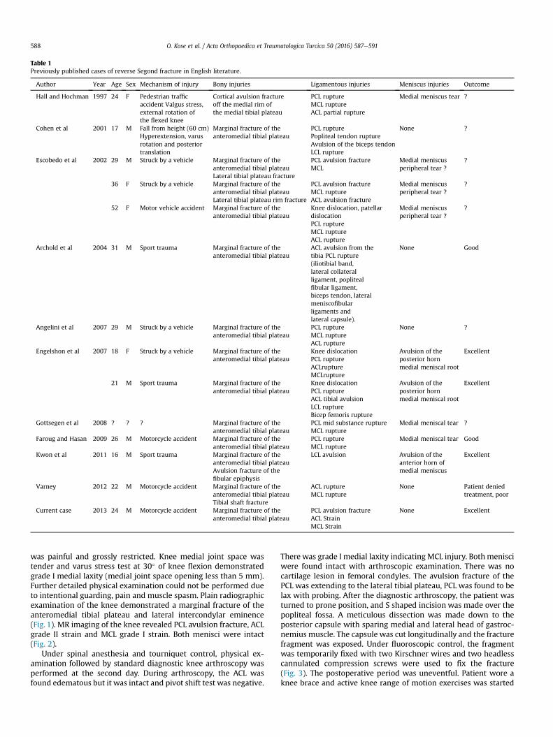
Table 1Previously published cases of reverse Segond fracture in English literature.
Author Year Age Sex Mechanism of injury Bony injuries Ligamentous injuries Meniscus injuries Outcome
Hall and Hochman 1997 24 F Pedestrian trafficaccident Valgus stress,external rotation ofthe flexed knee
Cortical avulsion fractureoff the medial rim ofthe medial tibial plateau
PCL ruptureMCL ruptureACL partial rupture
Medial meniscus tear ?
Cohen et al 2001 17 M Fall from height (60 cm)Hyperextension, varusrotation and posteriortranslation
Marginal fracture of theanteromedial tibial plateau
PCL rupturePopliteal tendon ruptureAvulsion of the biceps tendonLCL rupture
None ?
Escobedo et al 2002 29 M Struck by a vehicle Marginal fracture of theanteromedial tibial plateauLateral tibial plateau fracture
PCL avulsion fractureMCL
Medial meniscusperipheral tear ?
?
36 F Struck by a vehicle Marginal fracture of theanteromedial tibial plateauLateral tibial plateau rim fracture
PCL avulsion fractureMCL ruptureACL avulsion fracture
Medial meniscusperipheral tear ?
?
52 F Motor vehicle accident Marginal fracture of theanteromedial tibial plateau
Knee dislocation, patellardislocationPCL ruptureMCL ruptureACL rupture
Medial meniscusperipheral tear ?
?
Archold et al 2004 31 M Sport trauma Marginal fracture of theanteromedial tibial plateau
ACL avulsion from thetibia PCL rupture(iliotibial band,lateral collateralligament, poplitealfibular ligament,biceps tendon, lateralmeniscofibularligaments andlateral capsule).
None Good
Angelini et al 2007 29 M Struck by a vehicle Marginal fracture of theanteromedial tibial plateau
PCL ruptureMCL ruptureACL rupture
None ?
Engelshon et al 2007 18 F Struck by a vehicle Marginal fracture of theanteromedial tibial plateau
Knee dislocationPCL ruptureACLruptureMCLrupture
Avulsion of theposterior hornmedial meniscal root
Excellent
21 M Sport trauma Marginal fracture of theanteromedial tibial plateau
Knee dislocationPCL ruptureACL tibial avulsionLCL ruptureBicep femoris rupture
Avulsion of theposterior hornmedial meniscal root
Excellent
Gottsegen et al 2008 ? ? ? Marginal fracture of theanteromedial tibial plateau
PCL mid substance ruptureMCL rupture
Medial meniscal tear ?
Faroug and Hasan 2009 26 M Motorcycle accident Marginal fracture of theanteromedial tibial plateau
PCL ruptureMCL rupture
Medial meniscal tear Good
Kwon et al 2011 16 M Sport trauma Marginal fracture of theanteromedial tibial plateauAvulsion fracture of thefibular epiphysis
LCL avulsion Avulsion of theanterior horn ofmedial meniscus
Excellent
Varney 2012 22 M Motorcycle accident Marginal fracture of theanteromedial tibial plateauTibial shaft fracture
ACL ruptureMCL rupture
None Patient deniedtreatment, poor
Current case 2013 24 M Motorcycle accident Marginal fracture of theanteromedial tibial plateau
PCL avulsion fractureACL StrainMCL Strain
None Excellent
O. Kose et al. / Acta Orthopaedica et Traumatologica Turcica 50 (2016) 587e591588
was painful and grossly restricted. Knee medial joint space wastender and varus stress test at 30� of knee flexion demonstratedgrade I medial laxity (medial joint space opening less than 5 mm).Further detailed physical examination could not be performed dueto intentional guarding, pain and muscle spasm. Plain radiographicexamination of the knee demonstrated a marginal fracture of theanteromedial tibial plateau and lateral intercondylar eminence(Fig. 1). MR imaging of the knee revealed PCL avulsion fracture, ACLgrade II strain and MCL grade I strain. Both menisci were intact(Fig. 2).
Under spinal anesthesia and tourniquet control, physical ex-amination followed by standard diagnostic knee arthroscopy wasperformed at the second day. During arthroscopy, the ACL wasfound edematous but it was intact and pivot shift test was negative.
There was grade I medial laxity indicating MCL injury. Both menisciwere found intact with arthroscopic examination. There was nocartilage lesion in femoral condyles. The avulsion fracture of thePCL was extending to the lateral tibial plateau, PCL was found to belax with probing. After the diagnostic arthroscopy, the patient wasturned to prone position, and S shaped incision was made over thepopliteal fossa. A meticulous dissection was made down to theposterior capsule with sparing medial and lateral head of gastroc-nemius muscle. The capsule was cut longitudinally and the fracturefragment was exposed. Under fluoroscopic control, the fragmentwas temporarily fixed with two Kirschner wires and two headlesscannulated compression screws were used to fix the fracture(Fig. 3). The postoperative period was uneventful. Patient wore aknee brace and active knee range of motion exercises was started
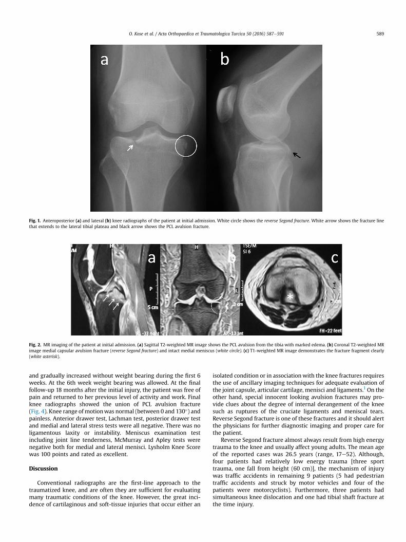
Fig. 1. Anteroposterior (a) and lateral (b) knee radiographs of the patient at initial admission. White circle shows the reverse Segond fracture. White arrow shows the fracture linethat extends to the lateral tibial plateau and black arrow shows the PCL avulsion fracture.
Fig. 2. MR imaging of the patient at initial admission. (a) Sagittal T2-weighted MR image shows the PCL avulsion from the tibia with marked edema. (b) Coronal T2-weighted MRimage medial capsular avulsion fracture (reverse Segond fracture) and intact medial meniscus (white circle). (c) T1-weighted MR image demonstrates the fracture fragment clearly(white asterisk).
O. Kose et al. / Acta Orthopaedica et Traumatologica Turcica 50 (2016) 587e591 589
and gradually increased without weight bearing during the first 6weeks. At the 6th week weight bearing was allowed. At the finalfollow-up 18 months after the initial injury, the patient was free ofpain and returned to her previous level of activity and work. Finalknee radiographs showed the union of PCL avulsion fracture(Fig. 4). Knee range ofmotionwas normal (between 0 and 130�) andpainless. Anterior drawer test, Lachman test, posterior drawer testand medial and lateral stress tests were all negative. There was noligamentous laxity or instability. Meniscus examination testincluding joint line tenderness, McMurray and Apley tests werenegative both for medial and lateral menisci. Lysholm Knee Scorewas 100 points and rated as excellent.
Discussion
Conventional radiographs are the first-line approach to thetraumatized knee, and are often they are sufficient for evaluatingmany traumatic conditions of the knee. However, the great inci-dence of cartilaginous and soft-tissue injuries that occur either an
isolated condition or in associationwith the knee fractures requiresthe use of ancillary imaging techniques for adequate evaluation ofthe joint capsule, articular cartilage, menisci and ligaments.1 On theother hand, special innocent looking avulsion fractures may pro-vide clues about the degree of internal derangement of the kneesuch as ruptures of the cruciate ligaments and meniscal tears.Reverse Segond fracture is one of these fractures and it should alertthe physicians for further diagnostic imaging and proper care forthe patient.
Reverse Segond fracture almost always result from high energytrauma to the knee and usually affect young adults. The mean ageof the reported cases was 26.5 years (range, 17e52). Although,four patients had relatively low energy trauma [three sporttrauma, one fall from height (60 cm)], the mechanism of injurywas traffic accidents in remaining 9 patients (5 had pedestriantraffic accidents and struck by motor vehicles and four of thepatients were motorcyclists). Furthermore, three patients hadsimultaneous knee dislocation and one had tibial shaft fracture atthe time injury.
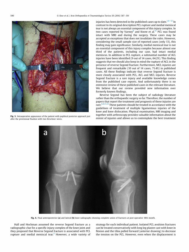
Fig. 3. Intraoperative appearance of the patient with popliteal posterior approach justafter the provisional fixation with two Kirschner wires.
Fig. 4. Final anteroposterior (a) and lateral (b) knee radiographs showing complete union of fractures at post-operative 18th month.
O. Kose et al. / Acta Orthopaedica et Traumatologica Turcica 50 (2016) 587e591590
Hall and Hochman assumed the reverse Segond fracture as aradiographic clue for a specific injury complex of the knee joint andthey proposed that Reverse Segond fracture is associated with PCLrupture and medial meniscal tear.3 However, a wide variety of
injuries has been detected in the published cases up to date.1,4e12 Incontrast to its original description PCL rupture andmedial meniscaltear is not always an essential component of this injury complex. Intwo cases reported by Varney8 and Know et al.11 PCL was foundintact with MRI and during the surgery. These cases may beaccepted as exceptions that does not invalidate the rules. However,considering the small sample size of reported cases (only 13), thisfinding may gain significance. Similarly, medial meniscal tear is notan essential component of this injury complex because almost onethird of the patients, including our case, had intact medialmeniscus. In addition to PCL rupture, a substantial number of ACLinjuries have been identified (9 out of 14 cases, 64.2%). This findingsuggests that we should also keep in mind the rupture of ACL in thepresence of reverse Segond fracture. Furthermore, MCL injuries arefrequent and remarkable (10 out of 14 cases, 71.4%) in publishedcases. All these findings indicate that reverse Segond fracture ismore closely associated with PCL, ACL and MCL injuries. ReverseSegond fracture is a rare injury and available knowledge comesfrom the published case reports. And unfortunately there is noextensive review of these published cases in the relevant literature.We believe that our review provided new information overformerly known findings.
Reverse Segond has been the subject of radiology literaturerather than the orthopaedic surgery so far. Therefore, the number ofpapers that report the treatment and prognosis of these injuries arerare.6,9,11,12 These patients should be treated in accordance with theguidelines of treatment of multiple ligamentous injuries of theknee and knee dislocation. Physical examination, MR imaging andtogether with arthroscopy provides valuable information about theextent of injuries and allows us to contemplate the best treatment
strategy for each individual patient. Isolated PCL avulsion fracturescan be treated conservativelywith long leg plaster cast with knee inflexion and the tibia pulled forward (anterior drawing) to decreasethe tension on the PCL. However, even when the displacement is

O. Kose et al. / Acta Orthopaedica et Traumatologica Turcica 50 (2016) 587e591 591
minimal, the risk of nonunion and malunion leads many surgeonsto consider surgical treatment.13e15 PCL incompetency and insta-bility result with functional disability and osteoarthritis of the kneein long term.16 On the other hand several authors reported excel-lent functional outcomes with ligamentous reconstruction andproper rehabilitation.13e15,17e20 Several surgical fixation techniquesincluding both open and arthroscopic methods have beendescribed for the treatment of PCL avulsion fractures.13,18,19 In ourcase, there was PCL avulsion fracture instead of PCL rupture andavulsed bone fragmentwas relatively big in size.We preferred to fixthe fragment with open posterior approach. In standard posteriorapproach in order to visualize the posterior capsule and allanatomic structures and to provide a wide surgical exposure,medial and lateral head of gastrocnemius muscle is cut. But wepreferred the modified posterior approach and did not cutgastrocnemius and used fluoroscopy to localize the place of screwscorrectly.20 Popliteal dissection should be done carefully and neu-rovascular bundle should be identified and protected. A cannulatedand compressive screw makes the operation easier. In most cases,small bony avulsion fractures do not need to be addressed at themedial side. However, if the fragment is large and compromisinganteromedial knee stability and meniscal function, fixation of thefragment is advocated. In current literature, Angelini et al andKnow et al fixed the avulsion fracture with a single corticalscrew.10,11 In our case, the fragment was very small and MCL wasintact therefore we managed medial sided injury conservatively.The avulsion fracture was united at the final follow-up in ourpatient.
Conclusions
In conclusion, reverse Segond fracture is an important indirectradiographic sign for multiple ligamentous and intraarticular in-juries of the knee joint. MR imaging should be performed to visu-alize the extent of all injuries in these patients. Although initialdescription of reverse Segond fracture included only PCL andmedial meniscal tears, reverse Segond fracture may also be asso-ciated with ACL and MCL injuries.
References
1. Gottsegen CJ, Eyer BA, White EA, Learch TJ, Forrester D. Avulsion fractures ofthe knee: imaging findings and clinical significance. Radiographics. 2008;28:1755e1770.
2. Claes S, Vereecke E, Maes M, Victor J, Verdonk P, Bellemans J. Anatomy of theanterolateral ligament of the knee. J Anat. 2013;223:321e328. http://dx.doi.org/10.1111/joa.12087.
3. Goldman AB, Pavlov H, Rubenstein D. The Segond fracture of the proximaltibia: a small avulsion that reflects major ligamentous damage. AJR Am JRoentgenol. 1988;151:1163e1167.
4. Hall FM, Hochman MG. Medial Segond-type fracture: cortical avulsion off themedial tibial plateau associated with tears of the posterior cruciate ligamentand medial meniscus. Skelet Radiol. 1997;26:553e555.
5. Escobedo EM, Mills WJ, Hunter JC. The “reverse Segond” fracture: associationwith a tear of the posterior cruciate ligament and medial meniscus. AJR Am JRoentgenol. 2002;178:979e983.
6. Archbold HA, Sloan S, Nicholas R. A tibial plateau fracture in a knee dislocation:a subtle sign of major ligamentous disruption. Injury. 2004;35:945e947.
7. Cohen AP, King D, Gibbon AJ. Impingement fracture of the anteromedial tibialmargin: a radiographic sign of combined posterolateral complex and posteriorcruciate ligament disruption. Skelet Radiol. 2001;30:114e116.
8. Varney JB. Reverse Segond fracture without PCL injury. Radiol Case Rep. 2012;7:537.
9. Engelsohn E, Umans H, Difelice GS. Marginal fractures of the medial tibialplateau: possible association with medial meniscal root tear. Skelet Radiol.2007;36:73e76.
10. Angelini FJ, Malavolta EA, D'Elia CO, P�ecora JR, Hernandez AJ, Camanho GL.Avulsion fracture of the medial tibial plateau (reverse Segond injury). ActaOrtop Bras. 2007;15:169e170.
11. Kwon OS, Park MJ, Tjoumakaris FP. Medial and lateral segond fractures in askeletally immature patient: a radiographic marker for the multiply injuredknee. Orthopedics. 2011;34:e772ee775.
12. Faroug R, Hasan A. Reverse Segond fracture: a case report. Inj Extra. 2009;40:109e111.
13. Torisu T. Avulsion fracture of the tibial attachment of the posterior cruciateligament: indications and results of delayed repairs. Clin Orthop. 1979;143:107e114.
14. Wajsfisz A, Makridis KG, Van Den Steene JY, Djian P. Fixation of posteriorcruciate ligament avulsion fracture with the use of a suspensory fixation. KneeSurg Sports Traumatol Arthrosc. 2012;20:996e999.
15. Hoogervorst P, Gardeniers JW, Moret-Wever S, van Kampen A. Pseudo-arthrosis repair of a posterior cruciate ligament avulsion fracture. Knee SurgSports Traumatol Arthrosc. 2010;18:1612e1616.
16. Lee BK, Nam SW. Rupture of posterior cruciate ligament: diagnosis and treat-ment principles. Knee Surg Relat Res. 2011;23:135e141.
17. Zhang X, Cai G, Xu J, Wang K. A minimally invasive postero-medial approachwith suture anchors for isolated tibial avulsion fracture of the posterior cru-ciate ligament. Knee. 2013;20:96e99.
18. Chen SY, Cheng CY, Chang SS, et al. Arthroscopic suture fixation for avulsionfractures in the tibial attachment of the posterior cruciate ligament. Arthros-copy. 2012;28:1454e1463.
19. Bali K, Prabhakar S, Saini U, Dhillon MS. Open reduction and internal fixation ofisolated PCL fossa avulsion fractures. Knee Surg Sports Traumatol Arthrosc.2012;20:315e321.
20. Nicandri GT, Klineberg EO, Wahl CJ, Mills WJ. Treatment of posterior cruciateligament tibial avulsion fractures through a modified open posterior approach:operative technique and 12- to 48-month outcomes. J Orthop Trauma. 2008;22:317e324.