retrospective analysis of 26 complete-arch implant ... · complete-arch implant-supported...
TRANSCRIPT

CLINICAL SCIENCE
aPrivate pracbDepartmentcPrivate pracdAggregate P
THE JOURNA
Retrospective analysis of 26 complete-arch implant-supportedmonolithic zirconia prostheses with feldspathic porcelain
veneering limited to the facial surface
Pietro Venezia, MD, DDS,a Ferruccio Torsello, DDS, PhD,b Raffaele Cavalcanti, DDS, PhD,c andSalvatore D’Amato, MD, DDSd
ABSTRACTStatement of problem. Monolithic zirconia prostheses on teeth or implants have been proposed inrecent years as a potential treatment. To date, limited data regarding the outcomes of theseprostheses have been presented and are mainly based on limited sample size and short-termfollow-up. Data on complete-arch monolithic zirconia prostheses are relatively scarce.
Purpose. The purpose of this retrospective study was to evaluate the clinical performances of 26implant-supported, complete-arch, monolithic zirconia restorations with facial feldspathic porcelainveneers for the rehabilitation of completely edentulous patients.
Material and methods. All patients’ charts from 2 private practices from 2010 to 2013 werereviewed. Patients rehabilitated with a complete-arch implant-supported monolithic zirconiaprostheses were included in the study. Several parameters were recorded so as to evaluate theoutcome of these rehabilitations: implant survival and success rates, prosthesis survival rate,interproximal bone loss, periimplant probing depth, and bleeding on probing. The number andtype of prosthetic complications were also recorded. Data were analyzed with descriptive statistics.
Results. Eighteen patients were treated with a total of 26 complete-arch fixed prostheses. Themean follow-up time was 20.9 months (SD 13.6; range, 10 to 36 months). In total, 154 implantswere placed supporting 309 retainers and pontics. The implant survival rate was 100% and thesuccess rate was 94.8%. Mean bone loss was 0.66 mm (SD 0.59 mm). Mean probing depth was3.4 mm (SD 0.92 mm). Bleeding on probing was positive in 19% of probing sites. The prosthesissurvival rate was 100%.
Conclusions. The results of this retrospective evaluation showed that monolithic zirconia restora-tions with facial porcelain veneer provided satisfactory clinical performance and suggest that theserehabilitations are a viable treatment option for completely edentulous patients. (J Prosthet Dent2015;-:---)
Complete-arch implant-supported monolithic zirconiarehabilitation with facial por-celain veneering could be aviable treatment for com-pletely edentulous individuals.
Several restorative mate-rials may be used to fabricatecomplete-arch implant-supportedfixed prostheses. Metal ceramicrestorations fabricated with avariety of alloys have beenwidely used and have demon-strated good outcomes.1 Metalacrylic resin, implant-supported,complete fixed dental prosthe-ses have also been suggestedas an option for rehabilitatingedentulous patients but haveshown an increased incidenceof mechanical complicationswhen compared with othermaterials.2,3
Zirconia frameworks havebecome popular in prostho-
dontics in the last 15 years because of their mechanicalproperties and the possibility of being produced ac-cording to a digital work flow. These prostheses, whenveneered with porcelain, have shown promising successtice, Bari, Italy.of Periodontics and Prosthodontics, Eastman Dental Hospital, Rome, Italytice, Bari, Italy.rofessor, Multidisciplinary Department of Medicine and Dentistry, Unit of M
L OF PROSTHETIC DENTISTRY
rates.4 Although these rehabilitations have beenreported to be safe and effective, their use has alsobeen associated with some complications, especiallyporcelain chipping.5-9 Short-term clinical data suggest
.
axillo-Facial Surgery, Second University of Naples, Naples, Italy.
1

Figure 1. Frontal view after implant healing period.
Figure 2. Frontal view with fixed complete-arch implant-supportedinterim prosthesis.
Figure 3. Phases of digital project. (Left) Scan of interim restorationsafter 6 months of use. (Right) Digital design of maxillary framework.Space for veneering porcelain was obtained by digital “carving” ofinterim restoration scan.
2 Volume - Issue -
that zirconia fixed dental prostheses may serve as analternative to metal ceramic in the anterior and posteriordentition.10
The use of monolithic zirconia restorations, poten-tially veneered with a limited amount of feldspathicporcelain in nonfunctional areas, have therefore beenproposed because of the reduced incidence of fracturesand lower cost.11 Monolithic zirconia restorations seemto cause less wear of the opposing dentition than feld-spathic porcelains and seem to have a better fit comparedto porcelain-veneered zirconia prostheses.12-14 The use ofmonolithic zirconia is increasing but is supported by onlylimited evidence.15,16 The purpose of this clinical caseseries was to report on the outcome of complete-archimplant-supported monolithic zirconia prostheses withfacial feldspathic porcelain veneers.
MATERIAL AND METHODS
All the records of edentulous patients treated in one orboth arches in 2 private practices from 2010 to 2013were reviewed. The study was conducted accordingto Italian laws and regulations. Patients rehabilitatedwith a complete-arch, single-piece, implant-supportedmonolithic zirconia restoration with facial porcelainveneers were selected.
Framework design inclusion criteria were thefollowing: in the posterior areas (premolars and molars),the zirconia frameworks represented the entire restora-tions, with the exception of facial surfaces that wereveneered with porcelain for esthetic purposes. In theanterior areas (incisors and canines), the zirconiaframeworks were designed in 2 different ways: in 6 pa-tients (11 prostheses), the palatal and lingual surfaces ofthe maxillary and mandibular frontal teeth were made ofzirconia, while the incisal and facial aspects of themaxillary and mandibular teeth were veneered withporcelain; in 12 patients (15 prostheses), the zirconiaframeworks were extended to the incisal part of themaxillary and mandibular incisors, and porcelain wasonly applied to the facial surfaces.
THE JOURNAL OF PROSTHETIC DENTISTRY
The prostheses were luted with an adhesive cement(Panavia F 2.0; Kuraray) to prefabricated titanium abut-ments in order to have a titanium-to-titanium connec-tion at the implant level. All the frameworks werefabricated with computer-aided design/computer-aidedmanufacturing systems.
Patients who presented with total edentulism or withhopeless dentition in one or both arches were givenclinical and radiographic examinations. Oral hygiene in-structions were given, hopeless teeth were extracted, andscaling and root planing were carried out on theremaining teeth with good periodontal prognosis. Ifnecessary, periodontal surgery was performed afterscaling and root planing, and periodontal reevaluationwas performed at 2 months to redetermine the prognosisof each individual tooth ahead of potential implantplacement. At this stage, imaging of the available bonewas performed with 3-dimensional (3D) systems,including computed tomography or cone beamcomputed tomography. Five to 7 implants were placed ineach edentulous arch by experienced surgeons. Thefollowing implants were used: bone level or tissue level(SLActive; Institut Straumann AG) and bone level-typeimplants (Osseotite; Biomet 3i or Ti-Unite; NobelBiocare).
Venezia et al
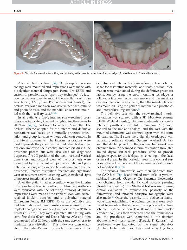
Figure 4. Zirconia framework after milling and sintering with zirconia protection of incisal edges. A, Maxillary arch. B, Mandibular arch.
- 2015 3
After implant healing (Fig. 1), pickup impressioncopings were mounted and impressions were made witha polyether material (Impregum Penta; 3M ESPE) andcustom impression trays (open tray technique). A face-bow record was used to mount the maxillary cast in anarticulator (SAM 3; Sam Präzisionstechnik GmbH), theocclusal vertical dimension was determined with estheticand phonetic tests, and the mandibular cast was moun-ted with the maxillary cast.17-22
In all patients a fixed, interim, screw-retained pros-thesis was fabricated, inserted by tightening the screws to20 Ncm (Fig. 2), and used for at least 6 months. Theocclusal scheme adopted for the interim and definitiverestorations was based on a mutually protected articu-lation and group function without balancing contacts inthe lateral movements. The interim restorations wereused to provide the patient with a fixed rehabilitation thatnot only improved the esthetics and comfort during theprosthetic phases but were also used for diagnosticpurposes. The 3D position of the teeth, occlusal verticaldimension, and occlusal wear of the prosthesis weremonitored by the patient (subjective esthetic and pho-netic evaluations) and clinician (visual examination of theprosthesis). Interim restoration fractures and significantwear or recurrent screw loosening were considered signsof incorrect functional articulation.
After the patient had uneventfully worn the interimprosthesis for at least 6 months, the definitive prostheseswere fabricated with the following protocol: definitiveimpressions were made at the implant level with screw-retained implant transfers and a polyether material(Impregum Penta; 3M ESPE). Once the definitive casthad been fabricated, new transfers were screwed on theimplant analogs and connected with acrylic resin (PatternResin; GC Corp). They were separated after setting withextra fine disks (Diamond Discs; Edenta AG) and thenreconnected after 24 hours with the same acrylic resin tominimize resin distortion.23 This index was then evalu-ated in the patient’s mouth to verify the accuracy of the
Venezia et al
definitive cast. The vertical dimension, occlusal scheme,space for restorative materials, and tooth position infor-mation were maintained during the definitive prosthesisfabrication by using the cross-mounting technique asfollows: a facebow record was made and the maxillarycast mounted on the articulator; then the mandibular castwas mounted using the patient’s interim fixed prosthesesand interocclusal registrations.24
The definitive cast with the screw-retained interimrestoration was scanned with a 3D laboratory scanner(D700; Wieland Dental), titanium abutments for screw-retained prostheses (Institut Straumann AG) weresecured to the implant analogs, and the cast with themounted abutments was scanned again with the same3D scanner. The 2 scans were digitally overlapped withlaboratory software (Dental System; Wieland Dental),and the digital project of the zirconia framework wasobtained from the scanned interim restoration through alimited digital cut-back procedure done to provideadequate space for the feldspathic veneering in the facialor incisal areas. In the posterior areas, the occlusal sur-faces obtained by the scan of the interim restoration werenot modified (Fig. 3).
The zirconia frameworks were then fabricated fromthe CAD files (Fig. 4) and milled from disks of yttrium-stabilized zirconia (Sagemax Zr; Sagemax BioceramicsInc) obtained from powder by cold isostatic pressing(Tosoh Corporation). The Sheffield test was used duringclinical evaluation to evaluate the passivity of theframeworks, and intraoral periapical radiographs werealso made.25 Once the passivity of the zirconia frame-works was established, the occlusal contacts were eval-uated to maintain the same mutually protected occlusalscheme. Feldspathic porcelain (E-max Ceram; IvoclarVivadent AG) was then veneered onto the frameworks,and the prostheses were cemented to the titaniumabutments with resin cement (Panavia F; Kuraray). Allprostheses were fabricated by the same laboratory(Apulia Digital Lab, Bari, Italy) and according to a
THE JOURNAL OF PROSTHETIC DENTISTRY

Figure 5. A-C, Representative zirconia prostheses on definitive cast afterporcelain veneering.
Figure 6. Frontal view of representative prostheses.
Figure 7. Representative panoramic radiograph of prostheses.
4 Volume - Issue -
1-piece, screw-retained design (Fig. 5).26-35 During de-livery (Figs. 6, 7), the prosthetic screws were tightened to20 Ncm, and the screw channels were filled with aninterim resin material (Telio Cs Inlay; Ivoclar VivadentAG); 1 month after delivery, the patients were recalled,and the prostheses were inspected to visualize eventualporcelain and/or framework cracks or chippings. Afterreevaluating the occlusion, the prosthetic screws weretightened to 35 Ncm and their access holes filled withpolytetrafluoroethylene tape (800 Golden Band; AW
THE JOURNAL OF PROSTHETIC DENTISTRY
Chesterton Co); they were then sealed with compositeresin (Tetric EvoCeram Bulk Fill; Ivoclar Vivadent AG).
The patients were recalled every 6 months for hygieneand clinical examinations, and periapical radiographswere made once a year to monitor crestal bone levels.Implant success rates were evaluated according to thecriteria of Buser et al36: absence of persistent subjectivecomplaints (pain, foreign body sensation, and/ordysesthesia), absence of periimplant infection with sup-puration, absence of mobility, and absence of continuousradiolucency around the implant. In addition to theaforementioned criteria, implants were considered tosurvive if they showed crestal bone resorption less than 2mm, with a probing depth less than 5 mm and with nobleeding on probing. Interproximal bone loss wasmeasured on follow-up periapical radiographs relative tothe implant platform and calculated from baseline, whichwas considered as the time of definitive prosthesis de-livery. Periimplant probing depth and bleeding onprobing, the survival rate of the prosthesis, and thenumber and type of prosthetic complications were alsorecorded at the follow-up visit and reported withdescriptive statistics.
Venezia et al

Table 1. Prostheses provided
PatientNo.
TreatedArches
No. ofImplants
IncisalMarginAnteriorTeeth Opposing Arch
No. ofProstheticElements Cantilever
CantileverElements
ProstheticComplications
1 Both 6 Maxillary,7 mandibular
Ceramic Monolithic zirconia 14+14 Y both mandibularsecond molars
N
2 Maxillary 6 maxillary Zirconia Natural teeth+monolithiczirconia FPD
12 N Minor chipping maxillaryleft canine
3 Both 5 maxillary,7 mandibular
Ceramic Monolithic zirconia 12+13 Y both maxillaryfirst molars
N
4 Maxillary 7 maxillary Zirconia Natural teeth 12 N N
5 Both 6 maxillary,6 mandibular
Zirconia Monolithic zirconia 12+12 Y maxillary rightfirst molar
N
6 Both 6 sup, 6 mandibular Zirconia Monolithic zirconia 12+12 N N
7 Maxillary 6 maxillary Zirconia Natural teeth 10 N N
8 Maxillary 6 maxillary Zirconia Natural teeth+monolithiczirconia FPD
12 N N
9 Maxillary 6 maxillary Zirconia Natural teeth 12 N N
10 Maxillary 5 maxillary Zirconia Natural teeth 10 Y N
11 Both 6 maxillary,6 mandibular
Ceramic Monolithic zirconia 12+12 N N
12 Maxillary 5 maxillary Ceramic Natural teeth 12 N N
13 Both 6 maxillary,6 mandibular
Ceramic Monolithic zirconia 11+13 Y mandibular leftsecond molar
Minor chipping, maxillaryright central incisor
14 Maxillary 6 maxillary Zirconia Natural teeth+PFM FPD 12 N N
15 Both 5 maxillary,7 mandibular
Ceramic Monolithic zirconia 10+12 Y both mandibularfirst molars
Minor chipping maxillaryleft lateral incisor
16 Maxillary 6 maxillary Zirconia Natural teeth 12 N N
17 Mandibular 4 mandibular Zirconia PFM 10 Y both mandibularfirst molars
N
18 Both 6 maxillary,6 mandibular
Zirconia Monolithic zirconia 12+12 N N
Total
18 Patients 26 archestreated
154 implants 11 ceramic15 zirconia
299 elements 10 cantileverunits
3 minor porcelainchippings
PFM, porcelain fused to metal; FPD, fixed partial denture.
Table 2.Descriptive statistics of measured parameters
CharacteristicCrestal BoneResorption
ProbingDepth
Bleeding onProbing
Mean 0.66 3.40
SD 0.59 0.92 Present 19%
Median 0.5 3 Absent 81%
95% confidenceinterval
0.59-0.72 3.3-3.5
Min 0 2
Max 2.5 7
- 2015 5
RESULTS
Eighteen patients were treated for a total of 26 complete-arch fixed prostheses, with 9 of them receiving maxillaryprostheses, 1 of them receiving a mandibular prosthesis,and 8 of them receiving prostheses in both arches. Themean follow-up time was 20.9 months (SD, 13.6; range,10 to 72 months).
A total of 154 implants were placed supporting 26restorations with 309 prosthetically replaced teeth, suchas pontics and retainers. In 6 out of 26 prostheses, distalcantilever extensions were included either unilaterally orbilaterally for a total of 10 pontics, with no more than 1pontic for each cantilever.
Table 1 describes the implants and the restorations.Eleven prostheses were designed so as to have veneeredporcelain on the incisal margins of the anterior teeth and15 with zirconia incisal margins; consequently, 243 re-tainers and pontics presented with porcelain veneered onthe facial aspect, but not on the occlusal or incisal area,while 66 units (all in the anterior areas) had porcelainveneered on the facial and incisal aspects.
Venezia et al
No implants were lost, achieving a 100% survivalrate; crestal bone loss was, on average, 0.66 mm (SD0.59 mm). Eight out of 154 implants showed more than2 mm of crestal bone resorption, more than 5 mmprobing depth, and bleeding on probing and wereconsidered to be surviving, thus leading to a 94.8%implant success rate. Probing depth showed a meanvalue of 3.4 mm (SD 0.92 mm), and bleeding on probingwas positive in only 19% of probing sites (Table 2).Three porcelain veneered teeth had minor cohesive
THE JOURNAL OF PROSTHETIC DENTISTRY

6 Volume - Issue -
chipping of the veneering porcelain in 3 differentprostheses, while no frameworks exhibited fracture ofthe zirconia structure.
The porcelain chippings were located on a maxillarycentral incisor, a maxillary lateral incisor and a maxillarycanine in 3 different patients, but because of their limitedextension, they did not affect the esthetic and functionaloutcome of the rehabilitations. All 3 porcelain fracturesoccurred in frameworks with veneered porcelain on theincisal margins and were treated by intraoral adjustmentand polishing with low-speed porcelain polishing rotaryinstruments (prosthesis survival rate was 100%).
DISCUSSION
The current study reported the results obtained withmonolithic zirconia with facial porcelain veneer used for1-piece, complete-arch restorations. It has some limita-tions because of its retrospective design and because ofthe absence of a control group.
The authors’ choice of screw-retained prostheses wasbased on the desire to avoid cementation because it hasbeen demonstrated that cement remnants may be diffi-cult to remove and that they could lead to mucositis andperiimplantitis.26-28 In addition, screw-retained prosthe-ses are more easily retrievable than cemented ones, andthis may be an advantage in the treatment of eventualmechanical and biological complications. Indeed, theEuropean Association of Osseointegration consensusstatement recommends screw-retained frameworks inextensive implant-supported reconstructions.29
Implant survival and success rate demonstrated excel-lent results while the incidence of biological and prostheticcomplications was low and consistent with publishedliterature.30-32 The 1-piece, complete-arch design of theprostheses, with the distribution of occlusal forces onseveral implants and the passivation of the frameworksobtained by luting them onto prefabricated titaniumabutments, probably contributed to the absence of screwloosening during the follow-up period.
Two different designs were used by the authors: somepatients were treated with porcelain veneer of the incisalmargin in the anterior teeth, while the most recent pa-tients received a monolithic zirconia incisal margin, withveneering limited to nonfunctional areas. The designwith a veneered incisal margin was used because of thesupposedly better esthetic outcome of veneered porcelainwhen compared with zirconia. The veneered porcelain,however, represented the weak point of the system,which is consistent with published evidence of relativelyfrequent chipping in porcelain fused to zirconia restora-tions.5-9 In 3 out of 11 prostheses with porcelain on theincisal margins, minor chippings were found, with aprevalence of 27% of the prostheses and 4.5% of theteeth with veneered porcelain on the incisal margins.
THE JOURNAL OF PROSTHETIC DENTISTRY
Although these complications were easily treatedbecause of the small extent of the fractures, this was themain reason the authors used a prosthesis design withzirconia incisal margins in subsequent prostheses. Noporcelain fractures were found in any of the 15 pros-theses fabricated with this modified design.
Another issue relates to the use of 1-piece frame-works rather than a segmented approach. Although thereis no definitive evidence for the superiority of oneparticular design, some authors suggest that asegmented, multiple-piece framework might be used forits ease of retrievability and repair,33 even if this designusually requires the placement of an increased number ofimplants. In the present case series, the 1-piece designwas used because in most patients 5 or 6 implants wereused to support the complete-arch prostheses.
In a few patients (6 prostheses), cantilever ponticswere used, as no evidence exists regarding the detri-mental effect of a distal cantilever in implant-supportedrestorations if its extension is limited.34,35 Moreover, theuse of monolithic zirconia frameworks with facial por-celain veneering allows the connectors between theprosthetic elements to be more robust than in situationswith completely veneered porcelain, thus increasing theresistance of the frameworks to occlusal loading, espe-cially in the cantilever sites.
In the present study, 8 patients received 2 monolithiczirconia restorations with facial porcelain veneering inboth jaws, and 10 patients received the restoration in asingle jaw, with natural teeth in the opposing jaw. Adifferent neuromuscular perception during functioncould be expected in these 2 situations, and although nosignificant difference in any of the measured parameterswas found, this issue should be investigated in furtherstudies.
CONCLUSIONS
In this retrospective evaluation, monolithic zirconia res-torations with facial porcelain veneering provided satis-factory clinical results. The rehabilitations with incisalprotection in the anterior areas showed the best resultswith minimal biologic and mechanical complications.
Further studies are needed to validate these prom-ising results.
REFERENCES
1. Wittneben JG, Millen C, Brägger U. Clinical performance of screw- versuscement-retained fixed implant-supported reconstructions-a systematic re-view. Int J Oral Maxillofac Implants 2014;29(suppl):84-98.
2. Priest G, Smith J, Wilson MG. Implant survival and prosthetic complicationsof mandibular metal-acrylic resin implant complete fixed dental prostheses.J Prosthet Dent 2014;1:466-75.
3. Purcell BA, McGlumphy EA, Holloway JA, Beck FM. Complications inmandibular metal-resin implant-fixed complete dental prostheses: a 5- to 9-year analysis. Int J Oral Maxillofac Implants 2008;23:847-57.
4. Al-Amleh B, Lyons K, Swain M. Clinical trials in zirconia: a systematic re-view. J Oral Rehabil 2010;37:641-52.
Venezia et al

- 2015 7
5. Ishibe M, Raigrodski AJ, Flinn BD, Chung KH, Spiekerman C, Winter RR.Shear bond strengths of pressed and layered veneering ceramics to high-noble alloy and zirconia cores. J Prosthet Dent 2011;106:29-37.
6. Kim HJ, Lim HP, Park YJ, Vang MS. Effect of zirconia surface treatments onthe shear bond strength of veneering ceramic. J Prosthet Dent 2011;105:315-22.
7. Saito A, Komine F, Blatz MB, Matsumura H. A comparison of bond strengthof layered veneering porcelains to zirconia and metal. J Prosthet Dent2010;104:247-57.
8. Guess PC, Att W, Strub JR. Zirconia in fixed implant prosthodontics. ClinImplant Dent Relat Res 2012;14:633-45.
9. Konstantinidis IK, Jacoby S, Rädel M, Böning K. Prospective evaluation ofzirconia based tooth- and implant-supported fixed dental prostheses: 3-yearresults. J Dent 2015;43:87-93.
10. Raigrodski AJ, Hillstead MB, Meng GK, Chung KH. Survival and complica-tions of zirconia-based fixed dental prostheses: a systematic review.J Prosthet Dent 2012;107:170-7.
11. Guess PC, Schultheis S, Bonfante EA, Coelho PG, Ferencz JL, Silva NR. All-ceramic systems: laboratory and clinical performance. Dent Clin North Am2011;55:333-52.
12. Kim MJ, Oh SH, Kim JH, Ju SW, Seo DG, Jun SH, Ahn JS, Ryu JJ. Wearevaluation of the human enamel opposing different Y-TZP dental ceramicsand other porcelains. J Dent 2012;40:979-88.
13. Stober T, Bermejo JL, Rammelsberg P, Schmitter M. Enamel wear caused bymonolithic zirconia crowns after 6 months of clinical use. J Oral Rehabil2014;41:314-22.
14. Karl M, Graef F, Wichmann M, Krafft T. Passivity of fit of CAD/CAM andcopy-milled frameworks, veneered frameworks, and anatomically contoured,zirconia ceramic, implant-supported fixed prostheses. J Prosthet Dent2012;107:232-8.
15. Sadid-Zadeh R, Liu PR, Aponte-Wesson R, O’Neal SJ. Maxillary cementretained implant supported monolithic zirconia prosthesis in a full mouthrehabilitation: a clinical report. J Adv Prosthodont 2013;5:209-17.
16. Rojas-Vizcaya F. Full zirconia fixed detachable implant-retained restorationsmanufactured from monolithic zirconia: clinical report after two years inservice. J Prosthodont 2011;20:570-6.
17. Silverman MM. Accurate measurement of vertical dimension by phoneticsand the closest speaking centric space. Part I. Dent Digest 1951;57:261-5.
18. Pound E. Cross arch splinting vs premature extractions. J Prosthet Dent1966;16:1058-68.
19. Pound E. Let /S/ be your guide. J Prosthet Dent 1977;38:482-9.20. Rivera-Morales WC, Mohl ND. Variability of closest speaking space
compared with interocclusal distance in dentulous subjects. J Prosthet Dent1991;65:228-32.
21. Rivera-Morales WC, Mohl ND. Relationship of occlusal vertical dimension tothe health of the masticatory system. J Prosthet Dent 1991;65:547-53.
22. Gross MD, Ormianer Z. A preliminary study on the effect of occlusal verticaldimension increase on mandibular postural rest position. Int J Prosthodont1994;7:216-26.
23. Ercoli C, Geminiani A, Feng C, Lee H. The influence of verification jig onframework fit for nonsegmented fixed implant-supported complete denture.Clin Implant Dent Relat Res 2012;14(suppl 1):e188-95.
24. Calesini G, Bruschi GB, Scipioni A, Micarelli C. Il montaggio crociato inprotesi ancorata ad impianti osteontegrati. Quint Int 1996;8-9:435-42.
25. Jemt T. Three-dimensional distortion of gold alloy casting and welded tita-nium frameworks. Measurements of the precision of fit between completed
Venezia et al
implant prostheses and the master cast in routine edentulous situations.J Oral Rehabil 1995;22:557-64.
26. Agar JR, Cameron SM, Hughbanks JC, Parker MH. Cement removal fromrestorations luted to titanium abutments with simulated subgingival margins.J Prosthet Dent 1997;78:43-7.
27. Wilson TG Jr. The positive relationship between excess cement and peri-implant disease: a prospective clinical endoscopic study. J Periodontol2009;80:1388-92.
28. Gotfredsen K, Wiskott A; Working Group 4. Consensus report-reconstructions on implants. The Third EAO Consensus Conference, 2012.Clin Oral Implants Res 2012;23(suppl 6):238-41.
29. Sailer I, Mühlemann S, Zwahlen M, Hämmerle CHF, Schneider D.Cemented and screw-retained implant reconstructions: a systematic reviewof the survival and complication rates. Clin Oral Impl Res 2012;23(suppl 6):163-201.
30. Berglundh T, Persson L, Klinge B. A systematic review of the incidence ofbiological and technical complications in implant dentistry reported in pro-spective longitudinal studies of at least 5 years. J Clin Periodontol2002;29(suppl 3):197-212.
31. Jung RE, Zembic A, Pjetursson B, Zwahlen M, Thoma D. Systematic reviewof the survival rate and the incidence of biological, technical and estheticcomplications of single crowns on implants reported in longitudinal studies ofat least 5 years. Clin Oral Impl Res 2012;23(suppl 6):2-21.
32. Pjetursson B, Thoma D, Jung R, Zwahlen M, Zembic A. A systematic reviewof the survival and complication rates of implant supported fixed dentalprostheses (FDPs) after a mean observation period of at least 5 years. ClinOral Impl Res 2012;23(suppl 6):22-38.
33. Cordaro L, Torsello F. Soft tissue conditioning by immediate restoration ofimmediately placed implants in full-arch rehabilitation: the double provi-sional technique. Eur J Esthet Dent 2006;1:216-29.
34. Romeo E, Lops D, Margutti E, Ghisolfi M, Chiapasco M, Vogel G. Implant-supported fixed cantilever prostheses in partially edentulous arches. A seven-year prospective study. Clin Oral Implants Res 2003;14:303-11.
35. Wennstrom J, Zurdo J, Karlsson S, Ekestubbe A, Gröndahl K, Lindhe J. Bonelevel change at implant-supported fixed partial dentures with and withoutcantilever extension after 5 years in function. J Clin Periodontol 2004;31:1077-83.
36. Buser D, Mericske-Stern R, Bernard JP, Behneke A, Behneke N, Hirt HP,et al. Long-term evaluation of nonsubmerged ITI implants. Part I: an 8-yearlife table analysis of a prospective multi-center study with 2359 implants. ClinOral Implants Res 1997;8:161-72.
Corresponding author:Dr Pietro VeneziaStudio di Odontoiatria Specialistica “Cavalcanti & Venezia”V. G. Posca, 15Bari (BA), 70124ITALYEmail: [email protected]
AcknowledgmentsThe authors thank Pasquale Lacasella, Francesco Grieco, and Leonardo Gallo fortheir invaluable help in the laboratory procedures.
Copyright © 2015 by the Editorial Council for The Journal of Prosthetic Dentistry.
THE JOURNAL OF PROSTHETIC DENTISTRY