retrocaval ureter - jumdc · retrocaval ureter or paracaval ureter is a rare congenital abnormality...
TRANSCRIPT
JUMDC Vol. 2, Issue 1, Jan-Jun 2011 39
Case Report
RETROCAVAL URETER 1Muhammad Akram Malik, 2Muhammad Sohail, 3Sarah Ramzan, 4Muhammad Hussain 1Associate Professor, Department of Urology, UMDC, Faisalabad. 2Registrar, Department of Urology, MTH, Faisalabad. 3House Officer, MTH, Faisalabad. 4Consultant Surgeon, THQ Hospital, Jaranwala. ABSTRACT Retrocaval ureter or paracaval ureter is a rare congenital abnormality in which ureter passes behind the inferior vena cava from medial to lateral side, causing obstruction which leads to hydronephrosis and lumbar pain. Intravenous urography, retrograde pyelography, CT, and MRI are main diagnostic investigations. Surgical intervention is required in most of cases. We present a case of 24 year old male which presented with right flank pain. Diagnosis was conformed by IVU and retrograde D-J Stenting. Exploration of ureter, its transaction and end to end anastomosis was done anterior to right ureter. INTRODUCTION Retrocaval ureter or paracaval ureter is a rare embryological anomaly involving venous system and consequently altering the course of ureter. Normally ureter crosses over the inferior vena cava (IVC) at this bifurcation. Ureter in retrovacal position passes behind the IVC from medially to lateral. Cardinal veins are considered to be the basic abnormality in which right subcardinal vein forms the main portion of IVC ventral to the ureter instead of right supra cardinal vein. Consequently the ureter winds behind the IVC from medial to lateral instead of lying lateral to it.1 Intravenous Urography (IVU) and retrograde uretro pyelograpy are the basic diagnostic investigations. On IVU there may be hydronephrosis of the right kidney, dilatation of the upper 1/3rd of ureter an S-shaped curve of the ureter and on oblique view ureter hugging the lumbar spine.2 Retrograde pydograghy reveals medial displacement of undialated lower ureter beyond the midline. Usual presentation of patients are right flank pain, recurrent infections and hematuria.
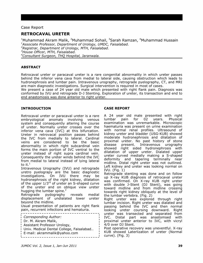
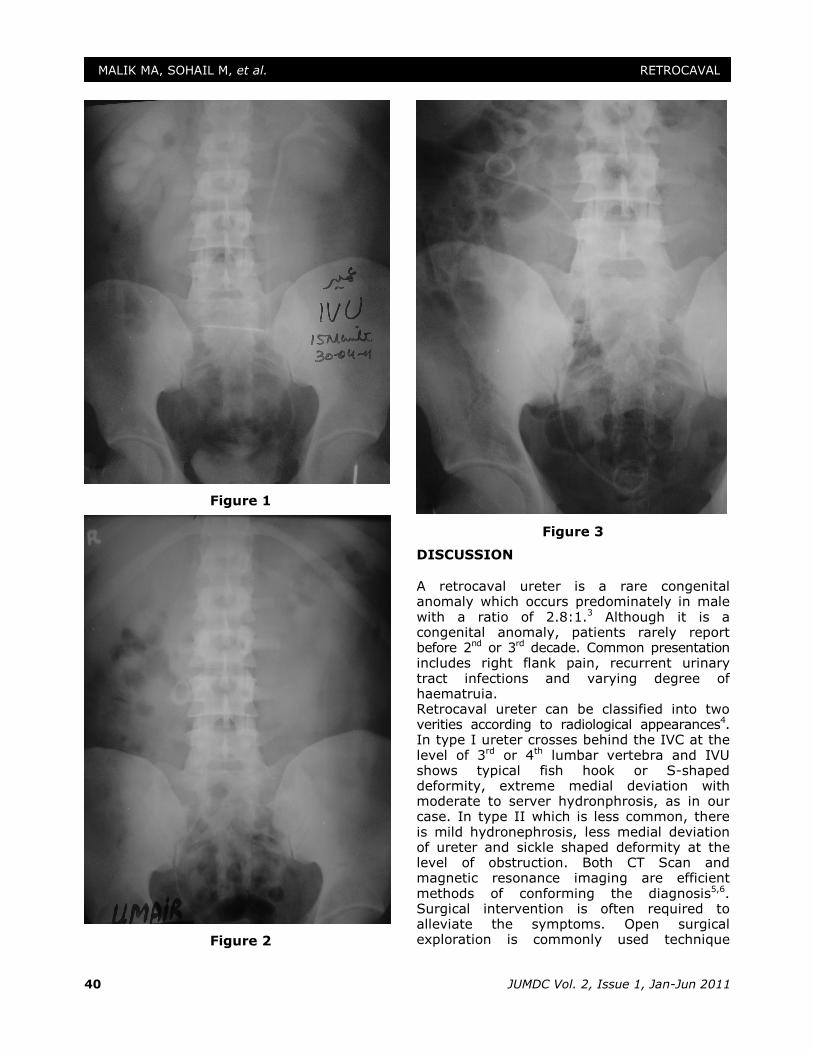
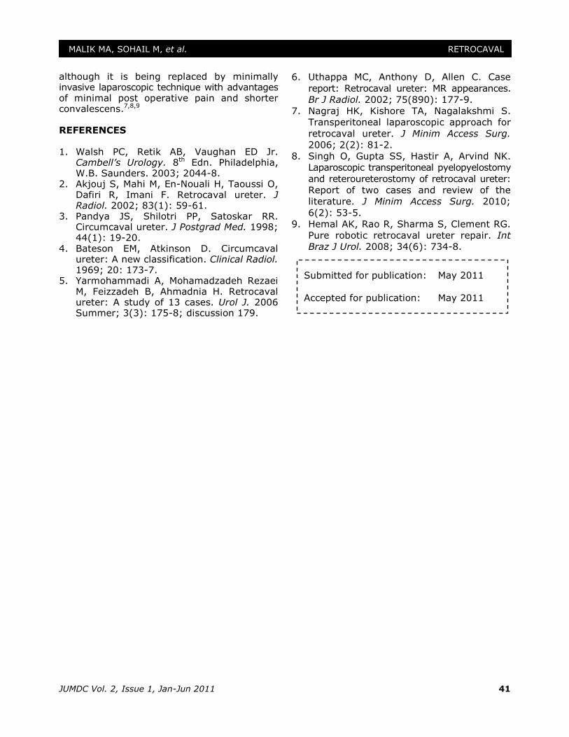
CASE REPORT A 24 year old male presented with right lumbar pain for 02 years. Physical examination was unremarkable. Microscopic haematuria was present on urine examination with normal renal profiles. Ultrasound of kidney ureter and bladder (USG-KUB) showed moderate hydronephrosis and dilatation of proximal ureter. No past history of stone disease present. Intravenous urography showed right sided hydronephrosis with dilatation of upper ureter. Dialated upper ureter curved medially making a S-shaped deformity and tapering terminally near midline. Distal right ureter was not outlined. Left kidney and ureter was looking normal on IVU. (Fig. 1) Retrograde stenting was done and on follow up X-ray KUB diagnosis of retrocaval ureter was confirmed. On X-ray KUB right ureter with double J-Stent (DJ Stent), was going toward midline and from midline crossing towards right kidney obliquely at the level of the lumbar vertebra. (Fig. 2) Right ureter was explored through right lumbar incision. Right ureter was dialated and passing behind the IVC and then normal looking ureter coursing downward. Right ureter was transected and separated from IVC. Distal part was anastomsed with proximal ureter anterior to IVC, with vicryl 4/0 over DJ Stent. Post operative recovery was uneventful. X-ray KUB showed Laterlization of ureter (Normal curve). (Fig. 3)
Corresponding Author:
Dr. M. Akram Malik,
Assistant Professor Urology,
Univ. Medical Dental College, Faisalabad.. E-mail: [email protected]
JUMDC Vol. 2, Issue 1, Jan-Jun 2011 40
MALIK MA, SOHAIL M, et al. RETROCAVAL
Figure 1
Figure 2
Figure 3
DISCUSSION A retrocaval ureter is a rare congenital anomaly which occurs predominately in male with a ratio of 2.8:1.3 Although it is a congenital anomaly, patients rarely report before 2nd or 3rd decade. Common presentation includes right flank pain, recurrent urinary tract infections and varying degree of haematruia. Retrocaval ureter can be classified into two verities according to radiological appearances4. In type I ureter crosses behind the IVC at the level of 3rd or 4th lumbar vertebra and IVU shows typical fish hook or S-shaped deformity, extreme medial deviation with moderate to server hydronphrosis, as in our case. In type II which is less common, there is mild hydronephrosis, less medial deviation of ureter and sickle shaped deformity at the level of obstruction. Both CT Scan and magnetic resonance imaging are efficient methods of conforming the diagnosis5,6. Surgical intervention is often required to alleviate the symptoms. Open surgical exploration is commonly used technique
JUMDC Vol. 2, Issue 1, Jan-Jun 2011 41
MALIK MA, SOHAIL M, et al. RETROCAVAL
although it is being replaced by minimally invasive laparoscopic technique with advantages of minimal post operative pain and shorter convalescens.7,8,9
REFERENCES 1. Walsh PC, Retik AB, Vaughan ED Jr.
Cambell’s Urology. 8th Edn. Philadelphia, W.B. Saunders. 2003; 2044-8.
2. Akjouj S, Mahi M, En-Nouali H, Taoussi O, Dafiri R, Imani F. Retrocaval ureter. J Radiol. 2002; 83(1): 59-61.
3. Pandya JS, Shilotri PP, Satoskar RR. Circumcaval ureter. J Postgrad Med. 1998; 44(1): 19-20.
4. Bateson EM, Atkinson D. Circumcaval ureter: A new classification. Clinical Radiol. 1969; 20: 173-7.
5. Yarmohammadi A, Mohamadzadeh Rezaei M, Feizzadeh B, Ahmadnia H. Retrocaval ureter: A study of 13 cases. Urol J. 2006 Summer; 3(3): 175-8; discussion 179.
6. Uthappa MC, Anthony D, Allen C. Case report: Retrocaval ureter: MR appearances.
Br J Radiol. 2002; 75(890): 177-9.
7. Nagraj HK, Kishore TA, Nagalakshmi S.
Transperitoneal laparoscopic approach for
retrocaval ureter. J Minim Access Surg.
2006; 2(2): 81-2.
8. Singh O, Gupta SS, Hastir A, Arvind NK. Laparoscopic transperitoneal pyelopyelostomy
and reteroureterostomy of retrocaval ureter:
Report of two cases and review of the
literature. J Minim Access Surg. 2010;
6(2): 53-5.
9. Hemal AK, Rao R, Sharma S, Clement RG.
Pure robotic retrocaval ureter repair. Int
Braz J Urol. 2008; 34(6): 734-8.
Submitted for publication: May 2011
Accepted for publication: May 2011