retinal neuroprotective effect of sirtuins
TRANSCRIPT
Central JSM Ophthalmology
Cite this article: Mimura T, Noma H, Funatsu H, Kondo A, Matsubara M (2014) Retinal Neuroprotective Effect of Sirtuins. JSM Ophthalmol 2(1): 1016.
*Corresponding authorTatsuya Mimura, Department of Ophthalmology, Tokyo Women’s Medical University Medical Center East, 2-1-10 Nishiogu, Arakawa-ku, Tokyo, 116-8567 Japan. Tel: 81 33810-1111 (Ex7765); Fax: 81 33894-0282; E-mail:
Submitted: 23 February 2014
Accepted: 03 March 2014
Published: 07 March 2014
ISSN: 2333-6447
Copyright© 2014 Mimura et al.
OPEN ACCESS
Review Article
Retinal Neuroprotective Effect of SirtuinsTatsuya Mimura1*, Hidetaka Noma2, Hideharu Funatsu3, Aki Kondo1 and Masao Matsubara1
1Department of Ophthalmology, Tokyo Women’s Medical University Medical Center, Japan2Department of Ophthalmology, Hachioji Medical Center, Tokyo Medical University, Japan3Department of Ophthalmology, Yachiyo Medical Center, Tokyo Women’s Medical University, Japan
IntroductIonistone deacetylases (HDACs) are enzymes that deacetylate
histones, but also act on certain non-histone substrates. Class III HDACs, which are known as sirtuins, catalyze deacetylation of the acetyl-lysine residues of histones using nicotinamide adenine dinucleotide (NAD+) as a cofactor. Silent information regulator 2 (Sir2) was the first gene of the sirtuin family to be discovered. Sir2 shows a high level of evolutionary conservation and is an important regulator of senescence, cell differentiation, stress tolerance, metabolism, and cancer in several organisms. Sirtuins have been suggested to have a role in aging [1,2], calorie restriction [1-10], and inflammation. Overexpression of Sir2 prolongs the lifespan of various organisms, whereas deletion or mutation of Sir2 leads to a shorter lifespan [11-13]. Seven human Sir2 homologues (sirtuins) have been identified to date, and these are designated as SIRT1 to SIRT7 [14-15]. Sirtuins are also important in preventing age-related ocular diseases [16]. In this review, we focus on the retinal neuroprotective effect of sirtuins.
EnzymatIc actIvIty of SIrtuInSSirtuins carry out deacetylation via a two-step reaction that
consumes NAD+ and releases nicotinamide (NAM), O-acetyl- adenosine diphosphate (ADP) -ribose (AADPR), and the deacetylated substrate [17-21]. Sirtuin activity is regulated by the intracellular [NAD]/[NADH] ratio and responds to changes of cellular metabolism [22-25]. NAD+ is an activator of sirtuins,
while nicotinamide and NADH are inhibitors. Sirtuins can catalyze deacetylation or ADP-ribosylation reactions, with both of these reactions involving cleavage of NAD+ as a cofactor and the production of nicotinamide (NAM). Five sirtuins (SIRT1, SIRT2, SIRT3, SIRT5, and SIRT7) catalyze deacetylation of the lysine residues of their target proteins, using NAD+ as cofactor and releasing nicotinamide along with the production of 2’-O-acetyl-ADP ribose [25-27]. In contrast, SirT4 and SirT6 catalyze ADP-ribosylation which involves transfer of an ADP-ribosyl moiety to the substrate [26,28].
Sirtuins have a highly conserved core domain that contains a catalytic domain and an NAD+-binding site [29]. Human SirT2 is composed of two globular domains, one of which is large, while the other is small. The large domain contains an inverted classical open α/β Rossmann-fold, six β-strands that form a parallel β-sheet, and six -helices, while the small domain is composed of a helical module and a zinc-binding module. The active site is located at the interface between the large and small domains, along with a binding site for NAD+. The NAD+-binding pocket can be divided into three spatially distinct regions, which are the A site showing affinity for adenine–ribose, the B site with affinity for nicotinamide–ribose, and the C site that binds NAD+. In the presence of an acetyl-lysine substrate, the NAD+-bound B site undergoes a conformational change that brings nicotinamide into proximity with the C site so that it can be cleaved. The ADP
Keywords•Review•SIRT1•Retina•Resveratrol
Abstract
In this paper, we review current knowledge about the retinal neuroprotective effect of sirtuins. The sirtuins are highly conserved nicotinamide adenine dinucleotide (NAD+)-dependent histone deacetylases that are involved in mammalian diseases of aging. The human genome encodes seven different sirtuins (SIRT1-7). SIRT1 is localized in the nucleus and cytoplasm of the cells making up all normal ocular structures, including the retina. Age-related macular degeneration (AMD) is a typical age-related condition due to the lifelong accumulation of molecular damage caused by reactive oxygen species (ROS). SIRT1 can decrease ROS levels and promotes cell survival under oxidative stress. Upregulation of SIRT1 has a protective effect against retinal degeneration in animal models. Resveratrol is a polyphenolic SIRT1 activator that has been shown to increase the lifespan and to protect various organs against aging, including oxidative stress-induced retinal damage. Anti-aging therapy with resveratrol could be an attractive treatment option for age-related macular degeneration.
Central
Mimura et al. (2014)Email:
JSM Ophthalmol 2(1): 1016 (2014) 2/4
ribose product then returns to the B site, allowing deacetylation to occur. The C site is the binding site for free nicotinamide. At high concentrations, nicotinamide can occupy the site and block the conformational change of NAD+ [30].
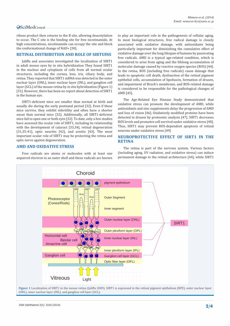
rEtInal dIStrIbutIon and rolE of SIrtuInSJaliffa and associates investigated the localization of SIRT1
in adult mouse eyes by in situ hybridization They found SIRT1 in the nucleus and cytoplasm of cells from all normal ocular structures, including the cornea, lens, iris, ciliary body, and retina. They reported that SIRT1 mRNA was detected in the outer nuclear layer (ONL), inner nuclear layer (INL), and ganglion cell layer (GCL) of the mouse retina by in situ hybridization (Figure 1) [31]. However, there has been no report about detection of SIRT1 in the human eye.
SIRT1-deficient mice are smaller than normal at birth and usually die during the early postnatal period [32]. Even if these mice survive, they exhibit infertility and often have a shorter snout than normal mice [32]. Additionally, all SIRT1-deficient mice fail to open one or both eyes [32]. To date, only a few studies have assessed the ocular role of SIRT1, including its relationship with the development of cataract [33,34], retinal degeneration [31,35-41], optic neuritis [42], and uveitis [43]. The most important ocular role of SIRT1 may be protecting the retina and optic nerve against degeneration.
amd and oxIdatIvE StrESS Free radicals are atoms or molecules with at least one
unpaired electron in an outer shell and these radicals are known
to play an important role in the pathogenesis of cellular aging. In most biological structures, free radical damage is closely associated with oxidative damage, with antioxidants being particularly important for diminishing the cumulative effect of oxidative damage over the long lifespan of humans by passivating free radicals. AMD is a typical age-related condition, which is considered to arise from aging and the lifelong accumulation of molecular damage caused by reactive oxygen species (ROS) [44]. In the retina, ROS (including free radicals) cause damage that leads to apoptotic cell death, dysfunction of the retinal pigment epithelial cells, accumulation of lipofuscin, formation of drusen, and impairment of Bruch’s membrane, and ROS-related damage is considered to be responsible for the pathological changes of AMD [45].
The Age-Related Eye Disease Study demonstrated that oxidative stress can promote the development of AMD, while antioxidants and zinc supplements delay the progression of AMD and loss of vision [46]. Oxidatively modified proteins have been detected in drusen by proteomic analysis [47]. SIRT1 decreases ROS levels and promotes cell survival under oxidative stress [48]. Thus, SIRT1 may prevent ROS-dependent apoptosis of retinal neurons under oxidative stress [49]
nEuroprotEctIvE EffEct of SIrt1 In thE rEtIna
The retina is part of the nervous system. Various factors (including aging, UV radiation, and oxidative stress) can induce permanent damage to the retinal architecture [44], while SIRT1
pigment epithelium
Outer Segment
Inner segment
Outer nuclear layer (ONL)
Outer plexiform layer (OPL)
Inner nuclear layer (INL)
Inner plexiform layer (IPL)
Ganglion cell layer (GCL)
Optic fiber layer (OFL)
Cone Cone Cone
Rod Rod
Horizontal cell
Amacrine cell
Photoreceptor(Cones/Rods)
Bipolar cell
Ganglion cell
SIRT1
Choroid
Vitreous Light
figure 1 Localization of SIRT1 in the mouse retina (Jaliffa 2009). SIRT1 is expressed in the retinal pigment epithelium (RPE), outer nuclear layer (ONL), inner nuclear layer (INL), and ganglion cell layer (GCL).
Central
Mimura et al. (2014)Email:
JSM Ophthalmol 2(1): 1016 (2014) 3/4
appears to have a neuroprotective effect on the retina. SIRT1 is localized in most layers of the normal mouse retina (including the ONL, INL, GCL, and RPE) [31]. In SIRT1-deficient adult mice, multiple retinal cell layers are significantly thinner than in normal mouse eyes, while the inner and outer nuclear layers are disorganized [35]. The inner and outer photoreceptor cell segments are also difficult to detect in SIRT1-deficient adult mice, indicating that SIRT1 has an important role in ocular morphogenesis [35].
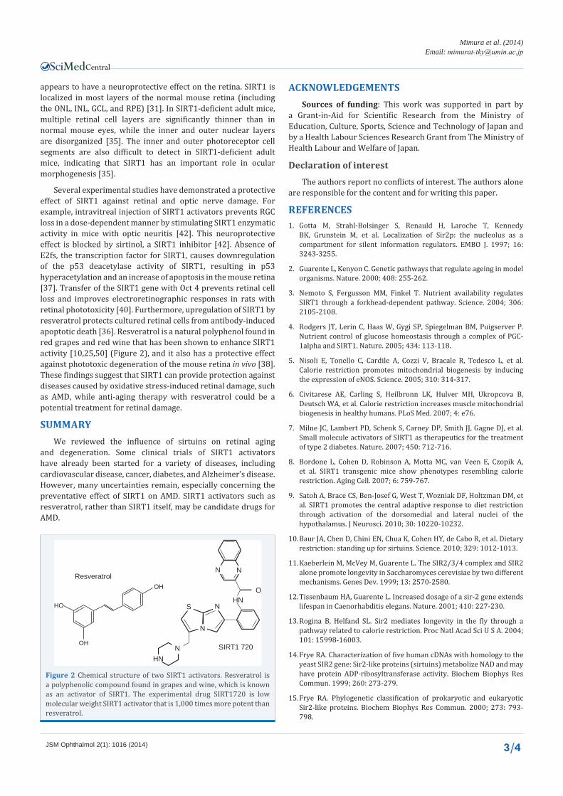
Several experimental studies have demonstrated a protective effect of SIRT1 against retinal and optic nerve damage. For example, intravitreal injection of SIRT1 activators prevents RGC loss in a dose-dependent manner by stimulating SIRT1 enzymatic activity in mice with optic neuritis [42]. This neuroprotective effect is blocked by sirtinol, a SIRT1 inhibitor [42]. Absence of E2fs, the transcription factor for SIRT1, causes downregulation of the p53 deacetylase activity of SIRT1, resulting in p53 hyperacetylation and an increase of apoptosis in the mouse retina [37]. Transfer of the SIRT1 gene with Oct 4 prevents retinal cell loss and improves electroretinographic responses in rats with retinal phototoxicity [40]. Furthermore, upregulation of SIRT1 by resveratrol protects cultured retinal cells from antibody-induced apoptotic death [36]. Resveratrol is a natural polyphenol found in red grapes and red wine that has been shown to enhance SIRT1 activity [10,25,50] (Figure 2), and it also has a protective effect against phototoxic degeneration of the mouse retina in vivo [38]. These findings suggest that SIRT1 can provide protection against diseases caused by oxidative stress-induced retinal damage, such as AMD, while anti-aging therapy with resveratrol could be a potential treatment for retinal damage.
SummaryWe reviewed the influence of sirtuins on retinal aging
and degeneration. Some clinical trials of SIRT1 activators have already been started for a variety of diseases, including cardiovascular disease, cancer, diabetes, and Alzheimer’s disease. However, many uncertainties remain, especially concerning the preventative effect of SIRT1 on AMD. SIRT1 activators such as resveratrol, rather than SIRT1 itself, may be candidate drugs for AMD.
acknowlEdgEmEntSSources of funding: This work was supported in part by
a Grant-in-Aid for Scientific Research from the Ministry of Education, Culture, Sports, Science and Technology of Japan and by a Health Labour Sciences Research Grant from The Ministry of Health Labour and Welfare of Japan.
declaration of interest
The authors report no conflicts of interest. The authors alone are responsible for the content and for writing this paper.
rEfErEncES1. Gotta M, Strahl-Bolsinger S, Renauld H, Laroche T, Kennedy
BK, Grunstein M, et al. Localization of Sir2p: the nucleolus as a compartment for silent information regulators. EMBO J. 1997; 16: 3243-3255.
2. Guarente L, Kenyon C. Genetic pathways that regulate ageing in model organisms. Nature. 2000; 408: 255-262.
3. Nemoto S, Fergusson MM, Finkel T. Nutrient availability regulates SIRT1 through a forkhead-dependent pathway. Science. 2004; 306: 2105-2108.
4. Rodgers JT, Lerin C, Haas W, Gygi SP, Spiegelman BM, Puigserver P. Nutrient control of glucose homeostasis through a complex of PGC-1alpha and SIRT1. Nature. 2005; 434: 113-118.
5. Nisoli E, Tonello C, Cardile A, Cozzi V, Bracale R, Tedesco L, et al. Calorie restriction promotes mitochondrial biogenesis by inducing the expression of eNOS. Science. 2005; 310: 314-317.
6. Civitarese AE, Carling S, Heilbronn LK, Hulver MH, Ukropcova B, Deutsch WA, et al. Calorie restriction increases muscle mitochondrial biogenesis in healthy humans. PLoS Med. 2007; 4: e76.
7. Milne JC, Lambert PD, Schenk S, Carney DP, Smith JJ, Gagne DJ, et al. Small molecule activators of SIRT1 as therapeutics for the treatment of type 2 diabetes. Nature. 2007; 450: 712-716.
8. Bordone L, Cohen D, Robinson A, Motta MC, van Veen E, Czopik A, et al. SIRT1 transgenic mice show phenotypes resembling calorie restriction. Aging Cell. 2007; 6: 759-767.
9. Satoh A, Brace CS, Ben-Josef G, West T, Wozniak DF, Holtzman DM, et al. SIRT1 promotes the central adaptive response to diet restriction through activation of the dorsomedial and lateral nuclei of the hypothalamus. J Neurosci. 2010; 30: 10220-10232.
10. Baur JA, Chen D, Chini EN, Chua K, Cohen HY, de Cabo R, et al. Dietary restriction: standing up for sirtuins. Science. 2010; 329: 1012-1013.
11. Kaeberlein M, McVey M, Guarente L. The SIR2/3/4 complex and SIR2 alone promote longevity in Saccharomyces cerevisiae by two different mechanisms. Genes Dev. 1999; 13: 2570-2580.
12. Tissenbaum HA, Guarente L. Increased dosage of a sir-2 gene extends lifespan in Caenorhabditis elegans. Nature. 2001; 410: 227-230.
13. Rogina B, Helfand SL. Sir2 mediates longevity in the fly through a pathway related to calorie restriction. Proc Natl Acad Sci U S A. 2004; 101: 15998-16003.
14. Frye RA. Characterization of five human cDNAs with homology to the yeast SIR2 gene: Sir2-like proteins (sirtuins) metabolize NAD and may have protein ADP-ribosyltransferase activity. Biochem Biophys Res Commun. 1999; 260: 273-279.
15. Frye RA. Phylogenetic classification of prokaryotic and eukaryotic Sir2-like proteins. Biochem Biophys Res Commun. 2000; 273: 793-798.
Resveratrol
HO
OH
OH
SIRT1 720
OHN
HN
z Nz N
z N
z N
z S
N
figure 2 Chemical structure of two SIRT1 activators. Resveratrol is a polyphenolic compound found in grapes and wine, which is known as an activator of SIRT1. The experimental drug SIRT1720 is low molecular weight SIRT1 activator that is 1,000 times more potent than resveratrol.
Central
Mimura et al. (2014)Email:
JSM Ophthalmol 2(1): 1016 (2014) 4/4
16. Mimura T, Kaji Y, Noma H, Funatsu H, Okamoto S. The role of SIRT1 in ocular aging. Exp Eye Res. 2013; 116: 17-26.
17. Imai S, Armstrong CM, Kaeberlein M, Guarente L. Transcriptional silencing and longevity protein Sir2 is an NAD-dependent histone deacetylase. Nature. 2000; 403: 795-800.
18. Tanner KG, Landry J, Sternglanz R, Denu JM. Silent information regulator 2 family of NAD- dependent histone/protein deacetylases generates a unique product, 1-O-acetyl-ADP-ribose. Proc Natl Acad Sci U S A. 2000; 97: 14178-14182.
19. Landry J, Slama JT, Sternglanz R. Role of NAD(+) in the deacetylase activity of the SIR2-like proteins. Biochem Biophys Res Commun. 2000; 278: 685-690.
20. North BJ, Marshall BL, Borra MT, Denu JM, Verdin E. The human Sir2 ortholog, SIRT2, is an NAD+-dependent tubulin deacetylase. Mol Cell. 2003; 11: 437-444.
21. Denu JM. Linking chromatin function with metabolic networks: Sir2 family of NAD(+)-dependent deacetylases. Trends Biochem Sci. 2003; 28: 41-48.
22. Lin SJ, Defossez PA, Guarente L. Requirement of NAD and SIR2 for life-span extension by calorie restriction in Saccharomyces cerevisiae. Science. 2000; 289: 2126-2128.
23. Lin SJ, Kaeberlein M, Andalis AA, Sturtz LA, Defossez PA, Culotta VC, et al. Calorie restriction extends Saccharomyces cerevisiae lifespan by increasing respiration. Nature. 2002; 418: 344-348.
24. Lin SJ, Ford E, Haigis M, Liszt G, Guarente L. Calorie restriction extends yeast life span by lowering the level of NADH. Genes Dev. 2004; 18: 12-16.
25. Haigis MC, Sinclair DA. Mammalian sirtuins: biological insights and disease relevance. Annu Rev Pathol. 2010; 5: 253-295.
26. Milne JC, Denu JM. The Sirtuin family: therapeutic targets to treat diseases of aging. Curr Opin Chem Biol. 2008; 12: 11-17.
27. Donmez G, Guarente L. Aging and disease: connections to sirtuins. Aging Cell. 2010; 9: 285-290.
28. Yamamoto H, Schoonjans K, Auwerx J. Sirtuin functions in health and disease. Mol Endocrinol. 2007; 21: 1745-1755.
29. Alcain FJ, Villalba JM. Sirtuin inhibitors. Expert Opin Ther Pat. 2009; 19: 283-294.
30. Carafa V, Nebbioso A, Altucci L. Sirtuins and disease: the road ahead. Front Pharmacol. 2012; 3: 4.
31. Jaliffa C, Ameqrane I, Dansault A, Leemput J, Vieira V, Lacassagne E, et al. Sirt1 involvement in rd10 mouse retinal degeneration. Invest Ophthalmol Vis Sci. 2009; 50: 3562-3572.
32. McBurney MW, Yang X, Jardine K, Hixon M, Boekelheide K, Webb JR, et al. The mammalian SIR2alpha protein has a role in embryogenesis and gametogenesis. Mol Cell Biol. 2003; 23: 38-54.
33. Zheng T, Lu Y. Changes in SIRT1 expression and its downstream pathways in age-related cataract in humans. Curr Eye Res. 2011; 36: 449-455.
34. Lin TJ, Peng CH, Chiou SH, Liu JH, Lin-Chung-Woung, Tsai CY, et al. Severity of lens opacity, age, and correlation of the level of silent information regulator T1 expression in age-related cataract. J Cataract Refract Surg. 2011; 37: 1270-1274.
35. Cheng HL, Mostoslavsky R, Saito S, Manis JP, Gu Y, Patel P, et al. Developmental defects and p53 hyperacetylation in Sir2 homolog (SIRT1)-deficient mice. Proc Natl Acad Sci U S A. 2003; 100: 10794-10799.
36. Anekonda TS, Adamus G. Resveratrol prevents antibody-induced apoptotic death of retinal cells through upregulation of Sirt1 and Ku70. BMC Res Notes. 2008; 1: 122.
37. Chen D, Pacal M, Wenzel P, Knoepfler PS, Leone G, Bremner R. Division and apoptosis of E2f-deficient retinal progenitors. Nature. 2009; 462: 925-929.
38. Kubota S, Kurihara T, Ebinuma M, Kubota M, Yuki K, Sasaki M, et al. Resveratrol prevents light-induced retinal degeneration via suppressing activator protein-1 activation. Am J Pathol. 2010; 177: 1725-1731.
39. Ozawa Y, Kubota S, Narimatsu T, Yuki K, Koto T, Sasaki M, et al. Retinal aging and sirtuins. Ophthalmic Res. 2010; 44: 199-203.
40. Peng CH, Cherng JY, Chiou GY, Chen YC, Chien CH, Kao CL, et al. Delivery of Oct4 and SirT1 with cationic polyurethanes-short branch PEI to aged retinal pigment epithelium. Biomaterials. 2011; 32: 9077-9088.
41. Geng Y, Wang J, Liang J, Xu C, Zhi Y. Expression of Sirt1 and Sirt2 in injured optic retina of calorie restricted rats. Eye Sci. 2011; 26: 221-224.
42. Shindler KS, Ventura E, Rex TS, Elliott P, Rostami A. SIRT1 activation confers neuroprotection in experimental optic neuritis. Invest Ophthalmol Vis Sci. 2007; 48: 3602-3609.
43. Kubota S, Kurihara T, Mochimaru H, Satofuka S, Noda K, Ozawa Y, et al. Prevention of ocular inflammation in endotoxin-induced uveitis with resveratrol by inhibiting oxidative damage and nuclear factor-kappaB activation. Invest Ophthalmol Vis Sci. 2009; 50: 3512-3519.
44. Fletcher AE. Free radicals, antioxidants and eye diseases: evidence from epidemiological studies on cataract and age-related macular degeneration. Ophthalmic Res. 2010; 44: 191-198.
45. Winkler BS, Boulton ME, Gottsch JD, Sternberg P. Oxidative damage and age-related macular degeneration. Mol Vis. 1999; 5: 32.
46. Age-Related Eye Disease Study Research Group. A randomized, placebo-controlled, clinical trial of high-dose supplementation with vitamins C and E, beta carotene, and zinc for age-related macular degeneration and vision loss: AREDS report no. 8. Arch Ophthalmol. 2001; 119: 1417-1436.
47. Crabb JW, Miyagi M, Gu X, Shadrach K, West KA, Sakaguchi H, et al. Drusen proteome analysis: an approach to the etiology of age-related macular degeneration. Proc Natl Acad Sci U S A. 2002; 99: 14682-14687.
48. Hori YS, Kuno A, Hosoda R, Horio Y. Regulation of FOXOs and p53 by SIRT1 modulators under oxidative stress. PLoS One. 2013; 8: e73875.
49. Zhang F, Wang S, Gan L, Vosler PS, Gao Y, Zigmond MJ, et al. Protective effects and mechanisms of sirtuins in the nervous system. Prog Neurobiol. 2011; 95: 373-395.
50. Howitz KT, Bitterman KJ, Cohen HY, Lamming DW, Lavu S, Wood JG, et al. Small molecule activators of sirtuins extend Saccharomyces cerevisiae lifespan. Nature. 2003; 425: 191-196.
Mimura T, Noma H, Funatsu H, Kondo A, Matsubara M (2014) Retinal Neuroprotective Effect of Sirtuins. JSM Ophthalmol 2(1): 1016.
Cite this article