resveratrol-induced gene expression profiles in...
TRANSCRIPT

Resveratrol-Induced Gene Expression Profiles in HumanProstate Cancer Cells
Sunita B. Jones,1 Samuel E. DePrimo,1 Michael L. Whitfield,2 and James D. Brooks1
Departments of1Urology and
2Genetics, Stanford University School of Medicine, Stanford, California
Abstract
Objective: The transhydroxystilbene resveratrol is found athigh levels in red wine and grapes, and red wine consump-tion may be inversely associated with prostate cancer risk. Togain insights into the possible mechanisms of action ofresveratrol in human prostate cancer, we did DNA microarrayanalysis of the temporal transcriptional program induced bytreatment of the human prostate cancer cell line LNCaP withresveratrol.Methods: Spotted DNA microarrays containing over 42,000elements were used to obtain a global view of the effects ofresveratrol on gene expression. Prostate-specific antigen(PSA) and androgen receptor (AR) expression were deter-mined by Northern blot and immunoblot analyses. Cellproliferation was determined by the 3-(4, 5-dimethylthiazolyl-2)-2, 5-diphenyltetrazolium bromide assay and cell cycleanalysis by flow cytometry.
Results: We observed time-dependent expression changesin >1,600 transcripts as early as 6 hours after treatmentwith resveratrol. Most striking was the modulation of anumber of important genes in the androgen pathwayincluding PSA and AR. Resveratrol also down-regulatedexpression of cell cycle and proliferation-specific genesinvolved in all phases of the cell cycle, inducednegative regulators of proliferation, caused accumulationof cells at the sub-G1 and S phases of the cell cycle,and inhibited cell proliferation in a time- and dose-dependent manner.Conclusion: Resveratrol produces gene expression changesin the androgen axis and cell cycle regulators thatmay underlie its putative anticancer activities in pros-tate cancer. (Cancer Epidemiol Biomarkers Prev 2005;14(3):596–604)
Introduction
The most diagnosed cancer among men, prostate cancer willclaim an estimated 29,900 lives this year in the United Statesalone (1). Considerable effort has been devoted to detectingand treating localized prostate cancer, and limited progresshas been made in the treatment of recurrent or advanceddisease. Epidemiologic evidence and two intervention trialshave fueled interest in developing chemopreventive strategiesfor prostate cancer (http://www.crab.org/select/; ref. 2). Thusfar, selenium, vitamin E, lycopene, cruciferous vegetables, andantiandrogens have been proposed as potential prostate cancerchemopreventive agents (3, 4). The recent inverse associationof red wine intake with prostate cancer risk (5) led us towonder whether resveratrol, a polyphenol transhydroxystil-bene found at high levels in grapes and red wine, might exertbiological effects that protect against prostate cancer.
Resveratrol appears in substantial quantities in severalfoods. Red wine contains between 0.72 and 3.18 mg/Lresveratrol (6-8). Resveratrol is rapidly absorbed by the gutand shows excellent tissue bioavailability (9-14). The effects ofresveratrol in biological systems are wide-ranging, and severalstudies have shown that it can inhibit or modulate metabolicpathways, act as an anti-inflammatory agent or antioxidant,and block cell proliferation (15-21). In prostate cancer cell lines,resveratrol has been shown to block proliferation and possiblyact as a negative regulator of androgen pathways (22, 23).
DNA microarray technology has provided insights into themolecular taxonomy of human tumors as well as thetranscriptional underpinnings of the cell cycle, prostatecellular senescence, cellular response to stress, and androgenaction (24-32). To gain insights into the possible mechanisms ofaction of resveratrol in human prostate cancer, we did DNAmicroarray analysis of the temporal transcriptional programinduced by treatment of the human prostate cancer cell lineLNCaP with resveratrol. Based on these findings, we furtherinvestigated the effects of resveratrol on androgen pathwaysand the cell cycle.
Materials and Methods
Cell Culture and Treatments. The LNCaP cell line wasobtained from American Type Culture Collection (Manassas,VA) and maintained in RPMI medium with 10% fetal bovineserum (CDT; Hyclone Laboratories, Logan, UT) and penicil-lin/streptomycin (Mediatech, Herndon, VA) in an environ-ment of 95% air and 5% CO2 at 37jC. Upon reaching 75%confluence, cells were treated with either DMSO control orpurified resveratrol at several concentrations (LKT Laborato-ries, St. Paul, MN), dissolved in DMSO, and incubated forvarying lengths of time. Final concentration of DMSO in mediawas 0.01%. Total RNA was prepared from cells using TRIzol(Invitrogen Life Technologies, Carlsbad, CA).
Microarray Hybridizations and Data Analysis. StanfordHuman cDNA microarrays were used in this study. Eachmicroarray contains 42,000 elements representing f30,000UNIgene Clusters (32). Microarray hybridizations were doneas previously described (The Brown Lab: http://brownlab.stanford.edu; ref. 33). Briefly, 100 Ag total RNA from eachsample was reverse-transcribed and labeled with fluorescence-tagged nucleotides (Cy3-dUTP for the DMSO control samples;Cy5-dUTP for the resveratrol-treated samples). Pairs ofresveratrol-treated and DMSO control– labeled cDNA were
Received 5/31/04; revised 9/15/04; accepted 10/22/04.
The costs of publication of this article were defrayed in part by the payment of page charges.This article must therefore be hereby marked advertisement in accordance with 18 U.S.C.Section 1734 solely to indicate this fact.
Note: Department of Defense New Investigator Award DAMD17-98-1-8555 and Doris DukeClinician Scientist Award T98064.
M.L. Whitfield is currently in the Department of Genetics, Dartmouth Medical School,Hanover, NH 03755.
Supplementary data for this article are available at http://cebp.aacrjournals.org.
Requests for reprints: James D. Brooks, Department of Urology, Stanford University School ofMedicine, MC 5118, Stanford, CA 94305. Phone: 650-498-4464; Fax: 650-723-0765.E-mail: [email protected]
Copyright D 2005 American Association for Cancer Research.
Cancer Epidemiology, Biomarkers & Prevention596
Cancer Epidemiol Biomarkers Prev 2005;14(3). March 2005
on May 4, 2018. © 2005 American Association for Cancer Research. cebp.aacrjournals.org Downloaded from

mixed and hybridized to microarray slides for 14 to 18 hours at65jC. Microarrays were scanned with a GenePix microarrayscanner (Axon Instruments, Union City, CA) and analyzedwith GenePix software (34). Spots of insufficient quality wereexcluded from further analysis by visual inspection. All dataare stored in the Stanford Microarray Database at http://genome-www.stanford.edu/microarray. Only spots with afluorescent intensity in each channel >2.0-fold over back-ground were selected for further analysis; data points that didnot meet this criteria were excluded. Genes missing >20% oftheir data points were also excluded from further analysis.Genes were selected that exhibited a z2-fold change inexpression level relative to the control sample in at least twoexperiments. Application of different filtering and dataselection criteria resulted in highly reproducible gene cluster-ing patterns demonstrating the robustness of the data set. Allmicroarray data can be downloaded at http://genome-www.stanford.edu/microarray/ (35).
Northern Blot. Equal amounts of total RNA were resolvedon 1% agarose formaldehyde gel and transferred to a membrane(Hybond N+, Amersham Biosciences, Piscataway,NJ) andhybridized with 32P-labeled prostate-specific antigen (PSA)cDNA probe. After quantitation of PSA, the membrane wasstripped and rehybridized with a 32P-labeled h-actin probe tomonitor RNA sample loading and transfer efficiency.
PSA Quantitation. Six-centimeter culture dishes wereseeded with 7 � 105 cells/plate and allowed to adhere for18 hours. Resveratrol or DMSO was added as mentionedabove. For each time point, media was collected and secretedtotal PSA was measured (Immulite 2000, Diagnostic Products,Corp., Randolph, NJ) and normalized by cell density. Allexperiments were done in triplicate.
Western Blot. Protein extracts were resolved by SDS-PAGEand transferred to a membrane, blocked with 5% nonfat milkin TBS plus Tween 20 overnight at 4jC, and subsequentlyincubated with a 1:1,000 dilution rabbit polyclonal a-ARprimary antibody (Santa Cruz Technologies, Santa Cruz, CA)for 1 hour at room temperature. Bands were visualized with ananti-rabbit, horseradish peroxidase secondary antibody, and achemiluminescence probe (ECL kit, Amersham Biosciences)following manufacturer’s directions. Glyceraldehyde-3-phosphate dehydrogenase was used as the control for proteinloading and transfer efficiency (not shown).
3-(4,5-Dimethylthiazolyl-2)-2,5-Diphenyltetrazolium Bro-mide Assay. Inhibition of cell proliferation was determinedusing the 3-(4,5-dimethylthiazolyl-2)-2,5-diphenyltetrazoliumbromide (MTT) assay kit according to the manufacturer’sprotocol (ATCC). After treatment with 25, 75, and 150 Amol/Lresveratrol or DMSO alone, cells were harvested and assayedat 12, 24, 48, and 60 hours. Ten microliters MTT labelingreagent were added to each well and the plates incubated for4 hours followed by the addition of 100 AL solubilizationsolution. The cells were incubated for another 12 hours andabsorbance was measured with a microtiter plate reader(Thermoelectron Corporation, Franklin, MA) at a test wave-length of 540 nm and a reference wavelength of 620 nm. Theabsorbance was calculated as the difference between theabsorbance at the reference wavelength and that at the testwavelength. Percent inhibition was calculated as absorbance ofcontrol minus absorbance of drug-treated sample divided byabsorbance of control minus absorbance of blank times 100[I = (C � E) / (C � B) � 100].
Cell Cycle Analysis. The cell cycle distribution of LNCaPcells after treatment with resveratrol or DMSO alone wasmonitored by flow cytometry of propidium iodide–stainedcells. Briefly, 6 � 105 cells were plated in 10 cm culture dishes.Upon reaching 75% confluence, cells were treated with DMSO
or 25, 75, and 150 Amol/L resveratrol in DMSO. Adherent andfloating cells were collected, resuspended in PBS, fixed in 70%ethanol, stained with propidium iodide (20 Ag/mL), 30 Ag/ALRNase A, and incubated in the dark for 30 minutes. DNAcontent was measured using a FACScan instrument equippedwith a FACStation running CellQuest software (BectonDickinson, Franklin Lakes, NJ). All experiments were donein duplicate with similar results. Percentage of cells in eachphase of the cell cycle was quantified using FlowJo software(Tree Star, Inc., Ashland, OR).
Results
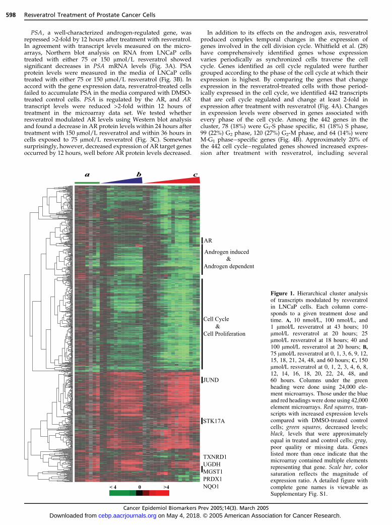
To gain insights into the potential mechanisms through whichresveratrol acts as a putative cancer preventive agent, we usedDNA microarrrays to assess global gene expression patterns ina prostate cancer cell line, LNCaP, after treatment withresveratrol. As a first test of whether resveratrol couldmodulate gene expression patterns, LNCaP cells were treatedwith 10 nmol/L, 100 nmol/L, 1 Amol/L, 10 Amol/L, 25 Amol/L,40 Amol/L, and 100 Amol/L resveratrol. Total RNA washarvested between 18 and 40 hours. These RNAs (labeled withthe Cy-5 fluorophore) were cohybridized with RNA from cellstreated in parallel with vehicle alone (labeled with Cy-3) onspotted cDNA microarrays containing 42,264 elements repre-senting f27,286 unique genes (estimated by UNIgeneclusters). Only minor gene expression changes were noted atlow doses (10 nmol/L-1 Amol/L); however, consistent, dose-dependent changes in gene expression were observed startingat 10 Amol/L that increased up to 100 Amol/L (Fig. 1;Supplementary Fig. S1).
Based on the observed expression changes in our dose-escalation experiments, we assessed the genome-wide patternsof gene expression induced after treatment with either 75 or 150Amol/L resveratrol in LNCaP cells at time points between 0 and60 hours after treatment (Fig. 1). We selected 1,656 transcriptswhose expression levels changed at least 2-fold from the controlsamples on at least two arrays. These 1,656 transcripts werefurther analyzed by hierarchical clustering, revealing groups ofgenes that varied both in their magnitude and temporal patternsof gene expression in a resveratrol-dependent manner. Sixhundred fourteen transcripts (37%) were induced and 1,044transcripts (63%) were repressed following treatment withresveratrol. Changes in transcript levels were detected as earlyas 1 hour in a few genes and was apparent in most genes by8 hours. Many transcripts represented named genes, althoughmost were poorly characterized, and 198 (12%) of the geneswere uncharacterized expressed sequence tags.
The human prostate cancer cell line LNCaP expresses theandrogen receptor (AR) and responds to androgen stimulation(36, 37). DNA microarrays have been used to characterize thetranscriptional program induced by treatment of LNCaP cellswith dihydrotestosterone and R1881, a synthetic androgenanalogue (31, 35, 38). Deprimo et al. (31) reported 567androgen-responsive genes and, of these, 517 showed aresponse to resveratrol of z2-fold over control in at least oneexperiment (Fig. 2) as early as 4 hours after resveratroltreatment and more than half of the transcripts were affectedreciprocally. Of the 412 genes that showed increased expres-sion after androgen treatment, 210 were down-regulated byresveratrol. These included genes involved in cell proliferation,apoptosis, polyamine biosynthesis, and many well-characterizedandrogen targets. Interestingly, a majority of these transcriptsshowed greater repression at 75 Amol/L than at 150 Amol/L.Of the 105 genes normally repressed by androgens, 92 (88%)were induced by resveratrol. A subset of genes (19%) wasinduced by both resveratrol and androgen and included genesinvolved in lipid metabolism, protein trafficking, vesicleformation, and stress response.
Cancer Epidemiology, Biomarkers & Prevention 597
Cancer Epidemiol Biomarkers Prev 2005;14(3). March 2005
on May 4, 2018. © 2005 American Association for Cancer Research. cebp.aacrjournals.org Downloaded from
PSA , a well-characterized androgen-regulated gene, wasrepressed >2-fold by 12 hours after treatment with resveratrol.In agreement with transcript levels measured on the micro-arrays, Northern blot analysis on RNA from LNCaP cellstreated with either 75 or 150 Amol/L resveratrol showedsignificant decreases in PSA mRNA levels (Fig. 3A). PSAprotein levels were measured in the media of LNCaP cellstreated with either 75 or 150 Amol/L resveratrol (Fig. 3B). Inaccord with the gene expression data, resveratrol-treated cellsfailed to accumulate PSA in the media compared with DMSO-treated control cells. PSA is regulated by the AR, and ARtranscript levels were reduced >2-fold within 12 hours oftreatment in the microarray data set. We tested whetherresveratrol modulated AR levels using Western blot analysisand found a decrease in AR protein levels within 24 hours aftertreatment with 150 Amol/L resveratrol and within 36 hours incells exposed to 75 Amol/L resveratrol (Fig. 3C). Somewhatsurprisingly, however, decreased expression of AR target genesoccurred by 12 hours, well before AR protein levels decreased.
In addition to its effects on the androgen axis, resveratrolproduced complex temporal changes in the expression ofgenes involved in the cell division cycle. Whitfield et al. (28)have comprehensively identified genes whose expressionvaries periodically as synchronized cells traverse the cellcycle. Genes identified as cell cycle regulated were furthergrouped according to the phase of the cell cycle at which theirexpression is highest. By comparing the genes that changeexpression in the resveratrol-treated cells with those period-ically expressed in the cell cycle, we identified 442 transcriptsthat are cell cycle regulated and change at least 2-fold inexpression after treatment with resveratrol (Fig. 4A). Changesin expression levels were observed in genes associated withevery phase of the cell cycle. Among the 442 genes in thecluster, 78 (18%) were G1-S phase specific, 81 (18%) S phase,99 (22%) G2 phase, 120 (27%) G2-M phase, and 64 (14%) wereM-G1 phase–specific genes (Fig. 4B). Approximately 20% ofthe 442 cell cycle–regulated genes showed increased expres-sion after treatment with resveratrol, including several
Figure 1. Hierarchical cluster analysisof transcripts modulated by resveratrolin LNCaP cells. Each column corre-sponds to a given treatment dose andtime. A, 10 nmol/L, 100 nmol/L, and1 Amol/L resveratrol at 43 hours; 10Amol/L resveratrol at 20 hours; 25Amol/L resveratrol at 18 hours; 40 and100 Amol/L resveratrol at 20 hours; B,
75 Amol/L resveratrol at 0, 1, 3, 6, 9, 12,15, 18, 21, 24, 48, and 60 hours; C, 150Amol/L resveratrol at 0, 1, 2, 3, 4, 6, 8,12, 14, 16, 18, 20, 22, 24, 48, and60 hours. Columns under the greenheading were done using 24,000 ele-ment microarrays. Those under the blueand red headings were done using 42,000element microarrays. Red squares, tran-scripts with increased expression levelscompared with DMSO-treated controlcells; green squares, decreased levels;black, levels that were approximatelyequal in treated and control cells; gray,poor quality or missing data. Geneslisted more than once indicate that themicroarray contained multiple elementsrepresenting that gene. Scale bar, colorsaturation reflects the magnitude ofexpression ratio. A detailed figure withcomplete gene names is viewable asSupplementary Fig. S1.
Resveratrol Treatment of Prostate Cancer Cells598
Cancer Epidemiol Biomarkers Prev 2005;14(3). March 2005
on May 4, 2018. © 2005 American Association for Cancer Research. cebp.aacrjournals.org Downloaded from

negative regulators of proliferation (PA26 , TSG101 , PCAF ,and HDAC3). Most genes (80%) showed decreased expressionafter resveratrol treatment by 8 hours and remained repressedover the remainder of the time course. Transcript levels weresuppressed over the entire time course and include many of thegenes necessary for all the basic processes required to duplicatea human cell (cell cycle control, DNA replication, spindleassembly, mitosis; Fig. 4B). Also seen were proapoptotic genesJUND , IPLA2 , TP53INP1 , BOK , PA26 , MDM2 , RRM2B ,PIGPC1 , SARS , PDCD4 , and STK17A (Fig. 1B) that wereinduced after treatment with 75 and 150 Amol/L resveratrol.
Because resveratrol produced large-scale changes in expres-sion of cell cycle–regulated genes, we tested whether resvera-trol affected LNCaP cell growth by using an MTT assay (ATCC).In agreement with the microarray data, resveratrol produced adose- and time-dependent inhibition of cell growth (Fig. 5).Resveratrol (25 Amol/L) inhibited cell growth by only 3% after24 hours and 16% by 60 hours, corresponding well with limitedeffects on gene expression seen at that dose. More dramaticgrowth inhibition was seen with 75 and 150 Amol/L doses thatproduce significant changes in gene expression of the cell cycle–regulated genes. Resveratrol (75 Amol/L) inhibited cell growthby 16% at 24 hours and 83% by 60 hours after treatment, whereas150 Amol/L inhibited growth by 53% at 24 hours and 86% by60 hours.
Flow cytometry was used to further characterize the effects ofresveratrol on cell growth. As expected, 25 Amol/L resveratrolproduced only minor changes in the cell cycle, with the portionof cells in S phase increasing from 12% in control cells to 18% intreated cells by 60 hours (Fig. 6). A much more dramatic effectwas evident at 75 and 50 Amol/L resveratrol, where disappear-ance of the G2-M peak and accumulation of cells in S phase wasobserved. By 48 hours, cells in S phase comprised 27% of thetotal when treated with 75 Amol/L resveratrol and 30% in 150Amol/L resveratrol-treated cells. A sub-G1 peak was observedin cells treated with 75 and 150 Amol/L resveratrol by 48 hours,although a corresponding sub-G1 peak was absent in cellstreated with 25 Amol/L resveratrol even after 60 hours. Thesefindings suggest that resveratrol induces apoptosis at highdoses and this may, in part, account for the significant effectsseen on MTT assay.
Discussion
The effects of resveratrol on the AR and androgen-responsivegenes seem to be complex. In LNCaP cells, resveratrol has beenshown to suppress secretion of PSA, although controversyexists as to whether this decrease is due to decreasedexpression of the AR or is independent of AR signaling
Figure 2. Expression levels of androgen-responsivegenes in LNCaP cells exposed to dihydrotestosterone,R1881, or resveratrol. Gene-expression changes inLNCaP treated with 1 nmol/L R1881 at 7, 9, 18, 24,50, and 72 hours; 10 nmol/L dihydrotestosterone at 18and 50 hours; 100 nmol/L dihydrotestosterone and 1Amol/L dihydrotestosterone at 24 hours; and androgendeprivation at 46 and 70 hours (A , controls); 10 nmol/L,100 nmol/L, and 1 Amol/L resveratrol at 43 hours;10 Amol/L resveratrol at 20 hours; 25 Amol/L resveratrolat 18 hours; 40 and 100 Amol/L resveratrol at 20 hours(B); 75 Amol/L resveratrol at 0, 1, 3, 6, 9, 12, 15, 18, 21,24, 48, and 60 hours (C); 150 Amol/L resveratrol at 0, 1,2, 3, 4, 6, 8, 12, 14, 16, 18, 20, 22, 24, 48, and 60 hours(D). Transcript levels exhibit time- and dose-dependentreciprocal changes between androgen and resveratrol inthe majority of the genes. Color bands and saturationscales are as in Fig. 1. A detailed figure with completegene names is viewable as Supplementary Fig. S2.
Cancer Epidemiology, Biomarkers & Prevention 599
Cancer Epidemiol Biomarkers Prev 2005;14(3). March 2005
on May 4, 2018. © 2005 American Association for Cancer Research. cebp.aacrjournals.org Downloaded from

pathways (23, 39). The gene expression data suggests that thedown-regulation of androgen-responsive genes is not solelybecause of decreased levels of the AR. Resveratrol treatmentproduced an early and sustained decreased expression ofmany androgen-responsive genes (KLK2, KLK3 , KLK4 , AIbZIP ,NKX3 , FKBP5 , TMEPAI) long before AR protein levels werediminished. Furthermore, the decreased expression of androgen-responsive genes occurred even at low doses of resveratrol,whereas the decreases in AR transcript levels occurred only atvery high doses. Finally, resveratrol did not oppose alltranscriptional changes induced by androgen. A subset of153 genes was up-regulated by both resveratrol and androgen.However, many of these genes, such as JUNB , HSP40 , SERP1 ,and STCH seem to reflect cellular stress. In LNCaP, androgentreatment is known to produce cellular stress by inducing anoxidative burst, and this stress pattern has been observed inother gene expression profiles (31, 40). Resveratrol treatmentundoubtedly places these cells under stress because theyundergo cell cycle arrest and, at higher doses, apoptosis.
How resveratrol might affect expression of androgen-regulated genes, aside from its effects on AR protein levels,is unclear. It is possible that resveratrol acts, in part, as anandrogen-receptor antagonist, or by blocking androgen-
signaling pathways downstream of AR. Resveratrol has beenshown to have partial agonist effects in estrogen-responsivemammary cancer cells and estrogens have been used to treatprostate cancer (41-43). This raises the possibility thatresveratrol exerts its effects through other steroid signalingpathways. Whether resveratrol acts directly as an antiandro-gen by binding to the AR, or indirectly through its estrogeniceffects, awaits further study. Other putative prostate cancer–preventive agents, including vitamin E, lycopene, and meth-ylselenic acid, an organic selenium compound, have beenreported to modulate androgen signaling in LNCaP cellsthrough unknown mechanisms (44-49).
We have found that resveratrol inhibits the proliferation ofLNCaP cells in a dose- and time-dependent manner. Cellgrowth arrest occurred in G1 and S phase of the cell cycle asshown by flow cytometry combined with a concomitantdecrease in the number of cells in G2-M (Fig. 6). Dramaticgene expression changes accompany this cell cycle arrest andare distributed throughout all phases of the cell cycle.Resveratrol suppressed transcript levels for genes involvedin cell cycle control, such as cyclins D , E , A , and B (Fig. 4).Negative regulators of proliferation, such as cyclins G1 and G2
and PA26 and HDAC3 (Fig. 1B), were induced in response toresveratrol and likely contribute to S-phase arrest. Therefore,resveratrol-induced growth arrest seems to be mediated by acomplex network of cell cycle regulatory genes. The resultsobserved here confirm other studies of gene expressiondemonstrating that cell cycle arrest results in decreasedexpression of the cell cycle–regulated genes (28). The cellcycle – regulated genes seem to be regulated such thatexpression occurs primarily when cells are dividing and notat any other time.
Considerable work has been published on resveratrol andits effect on the cell cycle and is consistent with the resultspresented here. Most studies suggest that resveratrol arrestcells in S phase and at the S/G2 transition. In a study usingHL-60 cells, resveratrol exposure resulted in accumulation ofcells at G1 and at the S phases with and an absence of the G2-M peak (20). This was attributed to an increase in the levels ofcyclins A and E along with accumulation of phosphorylatedcdc2. Hsieh et al. (50) reported that resveratrol induced NOsynthase in pulmonary epithelial cells with suppression of thecell cycle through the S and G2 phases. Cell cycle arrest wasaccompanied by a corresponding increase in the expression ofp53 and p21 and apoptosis. Inhibition in cell cycle progres-sion by resveratrol by inducing S-phase arrest was alsoreported in osteoblasts, breast, colon, and prostate cancer cells(51, 52). In breast cancer cells, resveratrol caused anaccumulation of cells in the S phase with a concomitantreduced expression of Rb and increased expression of p53and bcl-2 proteins (53). Resveratrol-mediated growth inhibi-tion and apoptosis in prostate cancer were observed inandrogen nonresponsive cell lines with a disruption in theG1-S phase transition (54) and S-phase arrest in androgenresponsive LNCaP cells (55).
Resveratrol has been reported to induce apoptosis viaseveral pathways including DNA damage (56, 57) throughp53-dependent and p53-independent pathways (16, 56-59)and by affecting lipoxygenase and cyclooxygenase activities(60). Narayanan et al. (61), using highly purified resveratroland a different microarray platform, reported induction ofapoptosis at 10 Amol/L in LNCaP that is correlated withmodulation of p53-mediated molecular targets. We seesimilar regulation of p53 genes, but only at relatively highlevels of resveratrol. In addition, we see apoptosis only at thehighest concentration used in our study. In our geneexpression data set, a few apoptosis genes were modulated,but not enough to elucidate pathways involved in apoptosis.It is, therefore, possible that critical apoptotic signals occurposttranscriptionally.
Figure 3. Effects of resveratrol on PSA and AR. A, Northern blotanalysis of PSA mRNA in LNCaP cells shows decreased PSAexpression over time. Equal loading was confirmed by rehybridiza-tion of the stripped membrane with radiolabeled h-actin (bottom). B,dose- and time-dependent inhibition of accumulation of secreted PSAin media of LNCaP cells. Culture medium was collected at indicatedtime intervals after addition of resveratrol (75 and 150 Amol/L) formeasurement of total PSA. PSA levels were normalized to celldensity. C, immunoblot analysis of AR protein levels in LNCaP cellsshows decreased expression after exposure to resveratrol (25, 75, and150 Amol/L). Equal loading was determined by glyceraldehyde-3-phosphate dehydrogenase immunoblotting (not shown).
Resveratrol Treatment of Prostate Cancer Cells600
Cancer Epidemiol Biomarkers Prev 2005;14(3). March 2005
on May 4, 2018. © 2005 American Association for Cancer Research. cebp.aacrjournals.org Downloaded from

The microarray results reported here provide additionalinsights into the mechanisms of action of this compoundin prostate cells. Resveratrol produced striking induction ofquinone reductase (NQO1) transcript levels. Quinonereductase is tightly regulated at the transcriptional level,and has served as a surrogate for phase 2 enzymeresponsiveness (62). Indeed, we observe coordinate induc-tion of other phase 2 enzymes (MGST1 , TXNRD1 , GSTA2 ,and PRDX1) and glutathione synthetic pathways (UGDH).Induction of phase 2 enzyme activity by resveratrol hasbeen reported in other model systems, but may beparticularly relevant to prostate cancer. From its earlieststages, human prostate cancers lose expression of a criticalcarcinogen defense enzyme, glutathione S-transferase-k or
GSTP1, because of extensive methylation of deoxycytidineresidues in the 5V-regulatory regions of the GSTP1 gene(63). Loss of GSTP1 could render prostate cells susceptibleto carcinogenesis by compromising their defenses againstendogenous or exogenous electrophilic mutagens. Compen-sation for loss of GSTP1 expression by induction of globalcarcinogen defenses could protect against the DNAdamage that contributes to prostate cancer initiation orprogression.
We recognize that our findings, whereas provocative, haveseveral limitations. We have analyzed comprehensively thegene expression changes induced in a single, androgen-sensitive prostate cancer cell line, LNCaP, and recognize thatthese findings should be validated in other model systems.
Figure 4. Expression of cell cycle and proliferation genes in LNCaP cells exposed to resveratrol. Color bands, saturation scales and treatmenttimes are the same as in Fig. 1. A, overview of the cluster diagram generated by querying microarray data using a gene list containing cellcycle–regulated genes. The full image of this cluster diagram is viewable as Supplementary Fig. S4. B, transcript profiles of selected genesinvolved in each phase of the cell cycle. Resveratrol treatments, color bands, and saturation scales are the same as in Fig. 1. The full image ofthis cluster diagram is viewable as Supplementary Fig. S4.
Cancer Epidemiology, Biomarkers & Prevention 601
Cancer Epidemiol Biomarkers Prev 2005;14(3). March 2005
on May 4, 2018. © 2005 American Association for Cancer Research. cebp.aacrjournals.org Downloaded from

An important factor in explaining the efficacy of resveratrolis the comparison of dosages used in vitro and in animalstudies, with dosages that can be expected to be clinicallyeffective in humans. Tissue bioavailability of resveratrol in
rat kidney has been reported at 77.75 ng/h/mL (14)following a single administration of red wine containing28.24 Ag resveratrol. Animal and human studies have shownthat resveratrol is rapidly absorbed in the gut, attaininghighest concentration in the blood in 1 hour, and itsaccumulation in organs vary (64-66). No information existsregarding resveratrol levels in the prostate tissue for anyspecies. In this study, we have shown changes in geneexpression at 10 to 100 Amol/L, physiologically attainablelevels in rats (64). However, the levels of resveratrol used in thisstudy that result in gene expression modulation in vitro may ormay not be attainable in man. At the very least, the relativelylow concentration of resveratrol in grapes or wine will likelymake it necessary for it to be given as a dietary supplement.Further clinical translational work is urgently needed to clarifythe relationship between serum concentrations and levelsachieved in the prostate and whether similar gene expressionchanges are observed in vivo.
Our data provides a global view of the potential mecha-nisms through which resveratrol may act in protecting againstprostate cancer and serves as a resource for future inves-tigations into its mechanisms of action. Resveratrol exertsantiandrogenic effects not strictly attributable to repression ofAR expression, inhibits the cell cycle, induces apoptosis, andup-regulates enzymes of carcinogen defense. This data setserves as a resource for understanding the effects of resveratrolin the prostate and as a potential source of biomarkers ofresponse in vivo .
Figure 5. Time course of resveratrol-induced inhibition of LNCaPcell proliferation determined using the MTT assay. Results areexpressed as a percent of inhibition in treated cells compared withcontrol cells treated with the vehicle DMSO only. Data representmeans for three separate experiments. *P < 0.05 compared withcontrol.
Figure 6. Fluorescence-activated cell sorting analysis of LNCaP cells after treatment with resveratrol. Cell cycle phase distributions werequantified by staining cells with propidium iodide. Results are expressed as percent of cells in G1, S, G2-M, and sub-G1 phase at each timepoint after exposure.
Resveratrol Treatment of Prostate Cancer Cells602
Cancer Epidemiol Biomarkers Prev 2005;14(3). March 2005
on May 4, 2018. © 2005 American Association for Cancer Research. cebp.aacrjournals.org Downloaded from

AcknowledgmentsWe thank Dr. Zijie Sun, Department of Urology, Stanford UniversitySchool of Medicine, for generous donation of PSA and AR probes,Dr. Manju Sharma for assistance with the assays and many usefuldiscussions, and Stanford Microarray Database and Core Facility forproviding arrays and technical support with data storage and retrieval.
References1. Jemal A, Tiwari RC, Murray T, et al. Cancer statistics 2004. CA Cancer J Clin
2004;54:8 – 29.2. Klein EA, Thompson IM. Update on chemoprevention of prostate cancer.
Curr Opin Urol 2004;14:143 – 9.3. Parnes HL, House MG, Kagan J, Kausal DJ, Lieberman R. Prostate cancer
chemoprevention agent development: the National Cancer Institute, Divi-sion of Cancer Prevention portfolio. J Urol 2004;171:S68 – 74; discussion S75.
4. DePrimo SE, Shinghal R, Vidanes G, Brooks JD. Prevention of prostatecancer. Hematol Oncol Clin North Am 2001;15:445 – 57.
5. Schoonen WM, Salinas CA, Kiemeney LA, Stanford LM. Alcohol consump-tion and risk of prostate cancer in middle-aged men. Int J Cancer 2005;113(1):133 – 40.
6. Wang Y, Catana F, Yang Y, Roderick R, van Breemen RB. An LC-MS methodfor analyzing total resveratrol in grape juice, cranberry juice, and in wine. JAgric Food Chem 2002;50:431 – 5.
7. Careri M, Corradini C, Elviri L, Nicoletti I, Zagnoni I. Direct HPLC analysisof quercetin and trans -resveratrol in red wine, grape, and winemakingbyproducts. J Agric Food Chem 2003;51:5226 – 31.
8. Celotti E, Ferrarini R, Zironi R, Conte LS. Resveratrol content of some winesobtained from dried Valpolicella grapes: Recioto and Amarone. J Chroma-togr A 1996;730:47 – 52.
9. Soleas GJ, Diamandis EP, Goldberg DM. Wine as a biological fluid: history,production, and role in disease prevention. J Clin Lab Anal 1997;11:287 – 313.
10. Soleas GJ, Yan J, Goldberg DM. Measurement of trans -resveratrol, (+)-catechin, and quercetin in rat and human blood and urine by gaschromatography with mass selective detection. Methods Enzymol 2001;335:130 – 45.
11. Bertelli A, Bertelli AA, Gozzini A, Giovannini L. Plasma and tissueresveratrol concentrations and pharmacological activity. Drugs Exp ClinRes 1998;24:133 – 8.
12. Bertelli AA, Giovannini L, Stradi R, Bertelli A, Tillement JP. Plasma, urineand tissue levels of trans - and cis-resveratrol (3,4V,5-trihydroxystilbene) aftershort-term or prolonged administration of red wine to rats. Int J Tissue React1996;18:67 – 71.
13. Bertelli AA, Giovannini L, Stradi R, Urien S, Tillement JP, Bertelli A. Kineticsof trans - and cis -resveratrol (3,4V,5-trihydroxystilbene) after red wine oraladministration in rats. Int J Clin Pharmacol Res 1996;16:77 – 81.
14. Bertelli AA, Giovannini L, Stradi R, Urien S, Tillement JP, Bertelli A.Evaluation of kinetic parameters of natural phytoalexin in resveratrol orallyadministered in wine to rats. Drugs Exp Clin Res 1998;24:51 – 5.
15. Jang M, Pezzuto JM. Cancer chemopreventive activity of resveratrol. DrugsExp Clin Res 1999;25:65 – 77.
16. Clement MV, Hirpara JL, Chawdhury SH, Pervaiz S. Chemopreventive agentresveratrol, a natural product derived from grapes, triggers CD95 signaling-dependent apoptosis in human tumor cells. Blood 1998;92:996 – 1002.
17. Miller NJ, Rice-Evans CA. Antioxidant activity of resveratrol in red wine.Clin Chem 1995;41:1789.
18. Subbaramaiah K, Chung WJ, Michaluart P, et al. Resveratrol inhibitscyclooxygenase-2 transcription and activity in phorbol ester-treated humanmammary epithelial cells. J Biol Chem 1998;273:21875 – 82.
19. Damianaki A, Bakogeorgou E, Kampa M, et al. Potent inhibitory action ofred wine polyphenols on human breast cancer cells. J Cell Biochem2000;78:429 – 41.
20. Ragione FD, Cucciolla V, Borriello A, et al. Resveratrol arrests the celldivision cycle at S/G2 phase transition. Biochem Biophys Res Commun1998;250:53 – 8.
21. Joe AK, Liu H, Suzui M, Vural ME, Xiao D, Weinstein IB. Resveratrolinduces growth inhibition, S-phase arrest, apoptosis, and changes inbiomarker expression in several human cancer cell lines. Clin Cancer Res2002;8:893 – 903.
22. Kampa M, Hatzoglou A, Notas G, et al. Wine antioxidant polyphenolsinhibit the proliferation of human prostate cancer cell lines. Nutr Cancer2000;37:223 – 33.
23. Hsieh TC, Wu JM. Grape-derived chemopreventive agent resveratroldecreases prostate-specific antigen (PSA) expression in LNCaP cells by anandrogen receptor (AR)-independent mechanism. Anticancer Res2000;20:225 – 8.
24. Perou CM, Sorlie T, Eisen MB, et al. Molecular portraits of human breasttumours. Nature 2000;406:747 – 52.
25. Garber ME, Troyanskaya OG, Schluens K, et al. Diversity of geneexpression in adenocarcinoma of the lung. Proc Natl Acad Sci U S A2001;98:13784 – 9.
26. Lossos IS, Alizadeh AA, Eisen MB, et al. Ongoing immunoglobulinsomatic mutation in germinal center B cell-like but not in activated B cell-like diffuse large cell lymphomas. Proc Natl Acad Sci U S A 2000;97:10209 – 13.
27. Stremmel C, Wein A, Hohenberger W, Reingruber B. DNA microarrays: anew diagnostic tool and its implications in colorectal cancer. Int J ColorectalDis 2002;17:131 – 6.
28. Whitfield ML, Sherlock G, Saldanha AJ, et al. Identification of genesperiodically expressed in the human cell cycle and their expression intumors. Mol Biol Cell 2002;13:1977 – 2000.
29. Schwarze SR, DePrimo SE, Grabert LM, Fu VX, Brooks JD, Jarrard DF. Novelpathways associated with bypassing cellular senescence in human prostateepithelial cells. J Biol Chem 2002;277:14877 – 83.
30. Gasch AP, Spellman PT, Kao CM, et al. Genomic expression programs in theresponse of yeast cells to environmental changes. Mol Biol Cell 2000;11:4241 – 57.
31. DePrimo SE, Diehn M, Nelson JB, et al. Transcriptional programs activatedby exposure of human prostate cancer cells to androgen. Genome Biol2002;3:RESEARCH0032.
32. DeRisi J, Penland L, Brown PO, et al. Use of a cDNA microarray to analysegene expression patterns in human cancer. Nat Genet 1996;14:457 – 60.
33. Chu S, DeRisi J, Eisen M, et al. The transcriptional program of sporulation inbudding yeast. Science 1998;282:699 – 705.
34. Eisen MB, Spellman PT, Brown PO, Botstein D. Cluster analysis and displayof genome-wide expression patterns. Proc Natl Acad Sci U S A 1998;95:14863 – 8.
35. Xu LL, Su YP, Labiche R, et al. Quantitative expression profile of androgen-regulated genes in prostate cancer cells and identification of prostate-specific genes. Int J Cancer 2001;92:322 – 8.
36. Gittes RF. Carcinoma of the prostate. N Engl J Med 1991;324:236 – 45.37. Wilding G. Endocrine control of prostate cancer. Cancer Surv 1995;23:43 – 62.38. Nelson PS, Clegg N, Arnold H, et al. The program of androgen-responsive
genes in neoplastic prostate epithelium. Proc Natl Acad Sci U S A 2002;99:11890 – 5.
39. Mitchell SH, Zhu W, Young CY. Resveratrol inhibits the expression andfunction of the androgen receptor in LNCaP prostate cancer cells. CancerRes 1999;59:5892 – 5.
40. Segawa T, Nau ME, Xu LL, et al. Androgen-induced expression ofendoplasmic reticulum (ER) stress response genes in prostate cancer cells.Oncogene 2002;21:8749 – 58.
41. Bhat KP, Lantvit D, Christov K, Mehta RG, Moon RC, Pezzuto JM.Estrogenic and antiestrogenic properties of resveratrol in mammary tumormodels. Cancer Res 2001;61:7456 – 63.
42. Gehm BD, McAndrews JM, Chien PY, Jameson JL. Resveratrol, apolyphenolic compound found in grapes and wine, is an agonist for theestrogen receptor. Proc Natl Acad Sci U S A 1997;94:14138 – 43.
43. Bowers JL, Tyulmenkov VV, Jernigan SC, Klinge CM. Resveratrol acts as amixed agonist/antagonist for estrogen receptors a and h. Endocrinology2000;141:3657 – 67.
44. Kim L, Rao AV, Rao LG. Effect of lycopene on prostate LNCaP cancer cellsin culture. J Med Food 2002;5:181 – 7.
45. Kotake-Nara E, Kushiro M, Zhang H, Sugawara T, Miyashita K, Nagao A.Carotenoids affect proliferation of human prostate cancer cells. J Nutr2001;131:3303 – 6.
46. Thompson TA, Wilding G. Androgen antagonist activity by the antioxidantmoiety of vitamin E, 2,2,5,7,8-pentamethyl-6-chromanol in human prostatecarcinoma cells. Mol Cancer Ther 2003;2:797 – 803.
47. Fleshner N, Fair WR, Huryk R, Heston WD. Vitamin E inhibits the high-fatdiet promoted growth of established human prostate LNCaP tumors innude mice. J Urol 1999;161:1651 – 4.
48. Israel K, Yu W, Sanders BG, Kline K. Vitamin E succinate induces apoptosisin human prostate cancer cells: role for Fas in vitamin E succinate-triggeredapoptosis. Nutr Cancer 2000;36:90 – 100.
49. Zhao H, Whitfield ML, Xu T, Botstein D, Brooks JD. Diverse effects ofmethylseleninic acid on the transcriptional program of human prostatecancer cells. Mol Biol Cell 2004;15:506 – 19.
50. Hsieh TC, Juan G, Darzynkiewicz Z, Wu JM. Resveratrol increases nitricoxide synthase, induces accumulation of p53 and p21(WAF1/CIP1), andsuppresses cultured bovine pulmonary artery endothelial cell prolifera-tion by perturbing progression through S and G2. Cancer Res 1999;59:2596 – 601.
51. Ulsperger E, Hamilton G, Raderer M, et al. Resveratrol pretreatmentdesensitizes AHTO-7 human osteoblasts to growth stimulation in responseto carcinoma cell supernatants. Int J Oncol 1999;15:955 – 9.
52. Sgambato A, Ardito R, Faraglia B, Boninsegna A, Wolf FI, CittadiniA. Resveratrol, a natural phenolic compound, inhibits cell prolifera-tion and prevents oxidative DNA damage. Mutat Res 2001;496:171 – 80.
53. Hsieh TC, Burfeind P, Laud K, et al. Cell cycle effects and control of geneexpression by resveratrol in human breast carcinoma cell lines with differentmetastatic potentials. Int J Oncol 1999;15:245 – 52.
54. Hsieh TC, Wu JM. Differential effects on growth, cell cycle arrest, andinduction of apoptosis by resveratrol in human prostate cancer cell lines.Exp Cell Res 1999;249:109 – 15.
55. Kuwajerwala N, Cifuentes E, Gautam S, Menon M, Barrack ER, Reddy GP.Resveratrol induces prostate cancer cell entry into S phase and inhibits DNAsynthesis. Cancer Res 2002;62:2488 – 92.
56. Katdare M, Jinno H, Osborne MP, Telang NT. Negative growth regulation ofoncogene-transformed human breast epithelial cells by phytochemicals.Role of apoptosis. Ann N Y Acad Sci 1999;889:247 – 52.
57. Katdare M, Osborne MP, Telang NT. Inhibition of aberrant proliferation and
Cancer Epidemiology, Biomarkers & Prevention 603
Cancer Epidemiol Biomarkers Prev 2005;14(3). March 2005
on May 4, 2018. © 2005 American Association for Cancer Research. cebp.aacrjournals.org Downloaded from

induction of apoptosis in pre-neoplastic human mammary epithelial cells bynatural phytochemicals. Oncol Rep 1998;5:311 – 5.
58. Surh YJ, Hurh YJ, Kang JY, Lee E, Kong G, Lee SJ. Resveratrol, anantioxidant present in red wine, induces apoptosis in human promyelocyticleukemia (HL-60) cells. Cancer Lett 1999;140:1 – 10.
59. Huang C, Ma WY, Goranson A, Dong Z. Resveratrol suppresses celltransformation and induces apoptosis through a p53-dependent pathway.Carcinogenesis 1999;20:237 – 42.
60. MacCarrone M, Lorenzon T, Guerrieri P, Agro AF. Resveratrol preventsapoptosis in K562 cells by inhibiting lipoxygenase and cyclooxygenaseactivity. Eur J Biochem 1999;265:27 – 34.
61. Narayanan BA, Narayanan NK, Re GG, Nixon DW. Differential expressionof genes induced by resveratrol in LNCaP cells: P53-mediated moleculartargets. Int J Cancer 2003;104:204 – 12.
62. Brooks JD, Paton VG, Vidanes G. Potent induction of phase 2 enzymes in
human prostate cells by sulforaphane. Cancer Epidemiol Biomarkers Prev2001;10:949 – 54.
63. Lee WH, Morton RA, Epstein JI, et al. Cytidine methylation ofregulatory sequences near the pi-class glutathione S-transferase geneaccompanies human prostatic carcinogenesis. Proc Natl Acad Sci U S A1994;91:11733 – 7.
64. Gescher AJ, Steward WP. Relationship between mechanisms, bioavailibility,and preclinical chemopreventive efficacy of resveratrol: a conundrum.Cancer Epidemiol Biomarkers Prev 2003;12:953 – 7.
65. Goldberg DM, Yan J, Soleas GJ. Absorption of three wine-relatedpolyphenols in three different matrices by healthy subjects. Clin Biochem2003;36:79 – 87.
66. Meng X, Maliakal P, Lu H, Lee MJ, Yang CS. Urinary and plasma levels ofresveratrol and quercetin in humans, mice, and rats after ingestion of purecompounds and grape juice. J Agric Food Chem 2004;52:935 – 42.
Resveratrol Treatment of Prostate Cancer Cells604
Cancer Epidemiol Biomarkers Prev 2005;14(3). March 2005
on May 4, 2018. © 2005 American Association for Cancer Research. cebp.aacrjournals.org Downloaded from

2005;14:596-604. Cancer Epidemiol Biomarkers Prev Sunita B. Jones, Samuel E. DePrimo, Michael L. Whitfield, et al. Prostate Cancer CellsResveratrol-Induced Gene Expression Profiles in Human
Updated version
http://cebp.aacrjournals.org/content/14/3/596
Access the most recent version of this article at:
Material
Supplementary
http://cebp.aacrjournals.org/content/suppl/2005/04/06/14.3.596.DC1
Access the most recent supplemental material at:
Cited articles
http://cebp.aacrjournals.org/content/14/3/596.full#ref-list-1
This article cites 65 articles, 22 of which you can access for free at:
Citing articles
http://cebp.aacrjournals.org/content/14/3/596.full#related-urls
This article has been cited by 4 HighWire-hosted articles. Access the articles at:
E-mail alerts related to this article or journal.Sign up to receive free email-alerts
Subscriptions
Reprints and
To order reprints of this article or to subscribe to the journal, contact the AACR Publications
Permissions
Rightslink site. (CCC)Click on "Request Permissions" which will take you to the Copyright Clearance Center's
.http://cebp.aacrjournals.org/content/14/3/596To request permission to re-use all or part of this article, use this link
on May 4, 2018. © 2005 American Association for Cancer Research. cebp.aacrjournals.org Downloaded from