respiratory system - napa valley college...
TRANSCRIPT

Respiratory System
Biol 105
Lecture 18
Chapter 14

Copyright © 2009 Pearson Education, Inc.
Outline - Respiratory System
I. Function of the respiratory system
II. Parts of the respiratory system
III. Mechanics of breathing
IV. Regulation of breathing
V. Disorders of the respiratory system

Copyright © 2009 Pearson Education, Inc.
Respiratory system Function
The function of the respiratory system is to
bring in oxygen to the body and remove
carbon dioxide.

Copyright © 2009 Pearson Education, Inc.
The Respiratory System
Figure 14.1
Breathing moves airin and out of the lungs.
External respiration
is the exchange ofoxygen and carbondioxide between thelungs and the blood.
Internal respiration isthe exchange of oxygenand carbon dioxidebetween blood and thebody tissues.
Gas transport
moves oxygen andcarbon dioxidebetween the lungsand the body tissues.
TissueGas diffusion Gas diffusion
Oxygen
transport
Carbon
dioxide
transportLungs

Copyright © 2009 Pearson Education, Inc.
This type of tissue covers and lines body parts
1. Connective
2. Epithelial
3. Muscle
4. Nervous
Copyright © 2009 Pearson Education, Inc.
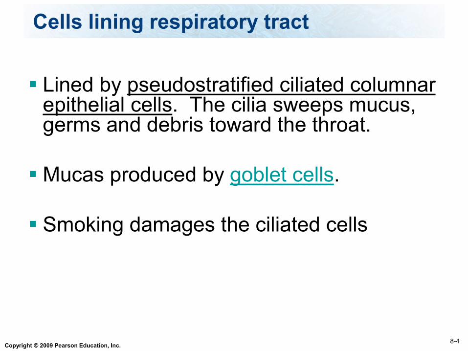
Lined by pseudostratified ciliated columnar epithelial cells. The cilia sweeps mucus, germs and debris toward the throat.
Mucas produced by goblet cells.
Smoking damages the ciliated cells
8-4
Cells lining respiratory tract
Copyright © 2009 Pearson Education, Inc.
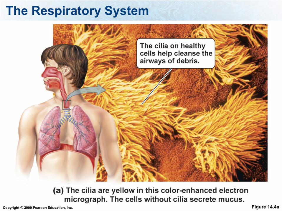
The Respiratory System
Figure 14.4a

Copyright © 2009 Pearson Education, Inc.
Ciliated cells in respiratory tract

Copyright © 2009 Pearson Education, Inc.
The Respiratory System
Figure 14.3

Copyright © 2009 Pearson Education, Inc.
The Respiratory System
Figure 14.2 (1 of 2)
Nasal cavity
• Produces mucus
• Filters, warms, and
moistens air
• Olfaction
Pharynx
• Passageway for
air and food
Sinuses
• Cavities in skull
• Lighten head
• Warm and moisten
air
Intercostal
muscles Diaphragm
• Muscle sheet between
chest and abdominal
cavities with a role in
breathing
UPPER RESPIRATORY
SYSTEM
RESPIRATORY
MUSCLES
• Cause breathing
• Filters, warms, andmoistens air
• Move ribs during
breathing

Copyright © 2009 Pearson Education, Inc.
The Respiratory System
Figure 14.2 (2 of 2)
Epiglottis
• Covers larynx during
swallowing
Bronchi
• Two branches of
trachea that conduct
air from trachea to
each lung
Bronchioles
• Narrow passageways
to conduct air from
bronchi to alveoli
Lungs
• Structures that contain
alveoli and air
passageways
• Allow exchange of
oxygen and carbon
dioxide between
atmosphere and blood
Alveoli
• Microscopic chambers
for gas exchange
Trachea
• Connects larynx with
bronchi leading to
each lung
• Conducts air to and
from bronchi
Larynx
• Air passageway
• Prevents food and drink
from entering lower
respiratory system
• Produces voice
LOWER RESPIRATORY
SYSTEM
• Exchanges gases

Copyright © 2009 Pearson Education, Inc.
Functions:
1. filter
2. warm
3. moisten the air entering the lungs
4. smell
8-3
1. Nasal cavity

Copyright © 2009 Pearson Education, Inc.
Parts of the nasal cavity:
Mucus membranes - secrete sticky mucus
to trap germs & debris.
Contains olfactory receptor cells for the
sense of smell
Sinuses – air filled cavities, warm and
moisten air
8-3
1. Nasal cavity

Copyright © 2009 Pearson Education, Inc.
Functions - is a passageway for air, liquids, and food. (swallowing begins here). Connects the nasal cavity to the esophagus and the larynx
Tonsils are found here – lymphatic tissue that protects against infection
8-4
2. Pharynx

Copyright © 2009 Pearson Education, Inc.
Functions1. Connects the pharynx to the
trachea
2. Contains vocal cords used to generate sound
3. Prevents food from entering lower respiratory tract
8-4
3. Larynx

Copyright © 2009 Pearson Education, Inc.
Structure made from cartilage
Epiglottis closes the trachea when swallowing
8-4
3. Larynx

Copyright © 2009 Pearson Education, Inc.

Copyright © 2009 Pearson Education, Inc.

Copyright © 2009 Pearson Education, Inc.
Windpipe held open by concentric rings of cartilage
Function – Connects the larynx to the bronchi.
8-4
4. Trachea

Copyright © 2009 Pearson Education, Inc.
Trachea leads to the bronchial tree: Bronchi (bronchus)
Bronchioles
Alveoli (alveolus).
8-4
Bronchial Tree

Copyright © 2009 Pearson Education, Inc.
The Respiratory System
Figure 14.7
Copyright © 2009 Pearson Education, Inc.
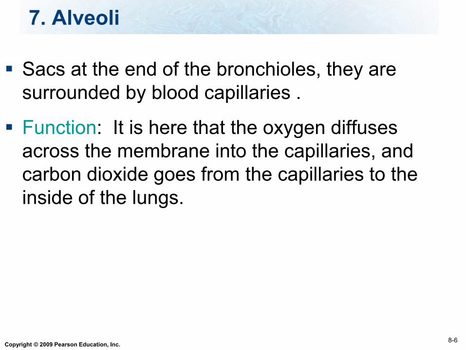
Sacs at the end of the bronchioles, they are
surrounded by blood capillaries .
Function: It is here that the oxygen diffuses
across the membrane into the capillaries, and
carbon dioxide goes from the capillaries to the
inside of the lungs.
8-6
7. Alveoli

Copyright © 2009 Pearson Education, Inc.
Alveoli
Lungs - have about 300 million alveoli
The structure of the alveoli increases
surface area of lung
For alveoli to function properly they are
coated with phospholipid molecules called
surfactant that keep them open

Copyright © 2009 Pearson Education, Inc.
Alveoli
Figure 14.8

Copyright © 2009 Pearson Education, Inc.
The Respiratory System
Table 14.1 (1 of 2)
Copyright © 2009 Pearson Education, Inc.
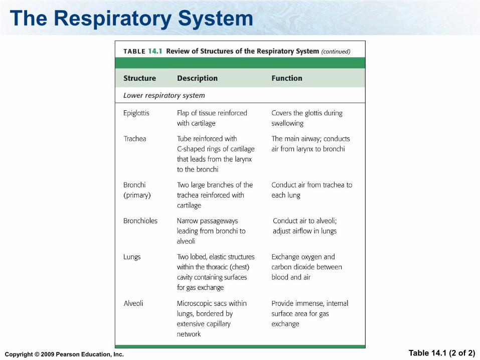
The Respiratory System
Table 14.1 (2 of 2)

Copyright © 2009 Pearson Education, Inc.
What cells secrete mucus
1. Cilliated columnar epi
2. goblet
3. Squamous epi
4. osteocytes

Copyright © 2009 Pearson Education, Inc.
The tube connecting the larynx to the primary
bronchi is
1. pharynx
2. trachea
3. bronchioles
4. alveoli
Copyright © 2009 Pearson Education, Inc.
Common passageway for air, food and drink
1. pharynx
2. trachea
3. bronchioles
4. alveoli
Copyright © 2009 Pearson Education, Inc.
Conduct air from the trachea to the bronchioles
1. pharynx
2. trachea
3. bronchi
4. alveoli

Copyright © 2009 Pearson Education, Inc.
Gas exchange takes place here
1. pharynx
2. trachea
3. bronchioles
4. alveoli

Copyright © 2009 Pearson Education, Inc.
Which cavity is the lung located in?
1. Abdominal
2. Pericardial
3. Pleural
4. Dorsal
Copyright © 2009 Pearson Education, Inc.
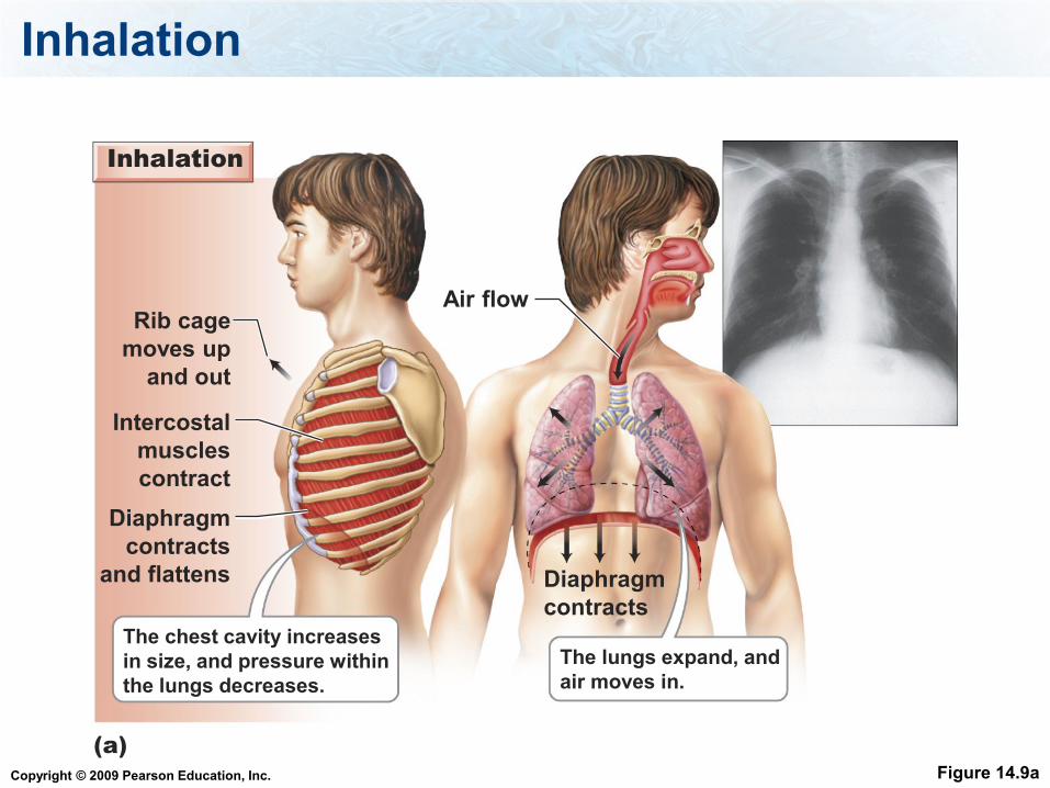
Inhalation
Figure 14.9a
The lungs expand, and
air moves in.
The chest cavity increases
in size, and pressure within
the lungs decreases.
Diaphragm
contracts
and flattens Diaphragm
contracts
Intercostal
muscles
contract
Rib cage
moves up
and out
Air flow
Inhalation
(a)
Copyright © 2009 Pearson Education, Inc.
Inhalation
When the diaphragm and intercostal muscles
contract, the volume of the thoracic cavity
increases, causing the pressure in the lungs to
decrease
Inhalation is also called inspiration

Copyright © 2009 Pearson Education, Inc.
Exhalation
Figure 14.9b
The lungs recoil,
and air moves out.
The chest cavity decreases
in size, and pressure
within the lungs increases.
Diaphragm
relaxes and
moves upward Diaphragm
relaxes
Intercostal
muscles relax
Rib cage
moves down
and inward
Air flow
Exhalation
(b)

Copyright © 2009 Pearson Education, Inc.
Exhalation
Exhalation = Expiration
When the same muscles relax, volume of
the thoracic cavity decreases, pressure in
the lungs increase

Copyright © 2009 Pearson Education, Inc.
Air Volumes
The volume of air inhaled or exhaled
during a normal breath is called the tidal
volume
Tidal volume is usually around 500 ml
The volume of air moved into and out of
the lungs is an indication of health

Copyright © 2009 Pearson Education, Inc.
Air Volumes
Inspiratory reserve volume = forced inhalation
volume
Expiratory reserve volume = forced exhalation
volume
Residual volume is the amount of air left in the
lungs after forced exhalation
Vital capacity is the amount of air brought in
and out of the lungs during forced breathing

Copyright © 2009 Pearson Education, Inc.
Air Volumes
Figure 14.10 (1 of 2)
6000
5000
4000
3000
2000
1000
0
Inspiratory
reserve
(forced
inhalation)
volumeVital
capacity
Expiratory reserve
(forced exhalation)
volume
Residual
volume
Tidal volume
Total
lung
capacity
Lung V
olu
me (m
l)

Copyright © 2009 Pearson Education, Inc.
Remember that O2 enters and CO2 leaves the
lungs = External respiration
Then O2 and CO2 is exchanged between the
blood vessels and tissues = Internal Respiration
This gas exchange is due to diffusion
8-14
Gas Exchanges in the Body

Copyright © 2009 Pearson Education, Inc.
Oxygen is transported on Hemoglobin.
When Oxygen is bound to hemoglobin, then it is
called Oxyhemoglobin
8-15
Oxygen Transport

Copyright © 2009 Pearson Education, Inc.
1. CO2 is transported dissolved in the plasma (10%)
2. CO2 is bound to hemoglobin (20%)
3. CO2 is converted to bicarbonate ions (70%)
8-14
Carbon Dioxide Transport

Copyright © 2009 Pearson Education, Inc.
CO2 + H2O H2CO3 H+ + HCO3-
8-14
Bicarbonate ions
Carbonic
anhydrase

Copyright © 2009 Pearson Education, Inc.
Diffusion of Gasses: Alveoli and Capillaries
Figure 14.11 (2 of 2)

Copyright © 2009 Pearson Education, Inc.
Diffusion of Gasses: Capillaries and Tissues
Figure 14.11 (1 of 2)

Copyright © 2009 Pearson Education, Inc.
Normally we breath 12 - 15 ventilations per
minute.
This rate is controlled by the medulla oblongata
region of the brain. Nerves transmit signal to the
diaphragm and muscles.
Chemoreceptors in the medulla oblongata and
arteries detect levels of CO2 and O2 in the blood,
controlling the rate and depth of breathing.
8-12
Regulation of Breathing

Copyright © 2009 Pearson Education, Inc.
Common cold
Flu
Pneumonia
Strep Throat
Tuberculosis
Bronchitis
Asthma
Emphysema
Lung Cancer
8-16
Respiratory Disorders

Copyright © 2009 Pearson Education, Inc.
Respiratory Disorders – Common Cold
The common cold - Caused by several types
of viruses.
Symptoms: runny nose, sore throat, sneezing,
nasal discharge
Treatment: rest and plenty of fluids
Prevention: wash your hands
Copyright © 2009 Pearson Education, Inc.
Respiratory Disorders - Flu
The flu is caused by the Influenza viruses but there are many variants of these viruses
Symptoms: Similar to colds but appear suddenly and more severe. Usually have fever and chills, may have muscle aches, headache, and weakness.
Treatment and prevention – same as cold
Can take drugs to ease symptoms and antiviral medications may ease symptoms
Copyright © 2009 Pearson Education, Inc.
Respiratory Disorders - Pneumonia
Pneumonia is an inflammation of the lungs that
causes fluid to accumulate in the alveoli,
reducing gas exchange
Usually caused by a viral or bacterial infection
Symptoms: fever, chills, chest pain, cough,
shortness of breath.
Treatment depends on cause – bacteria can be
treated with antibiotics.

Copyright © 2009 Pearson Education, Inc.
Respiratory Disorders Strep throat
Strep throat is caused by Streptococcus
bacteria
Can lead to rheumatic fever which can
damage heart and kidney disease
Symptoms: Sore throat accompanied by
swollen glands and fever
Treatment: antibiotics

Copyright © 2009 Pearson Education, Inc.
Respiratory Disorders - Tuberculosis
Tuberculosis is caused by a bacteria =
mycobacterium tuberculosis.
Bacteria spread through airborne transmission
Our body encapsulates the bacteria with a
fibrous capsule made of connective tissue to
try to protect itself, capsule is called tubercles
Copyright © 2009 Pearson Education, Inc.
Respiratory Disorders - Tuberculosis
Symptoms: similar to flu, weight loss, tired, dry
cough.
Treatment: Antibiotics must be taken for 6
months to 2 years – some people to stop early
– leads to antibiotic resistant strains of bacteria

Copyright © 2009 Pearson Education, Inc.
Respiratory Disorders - Bronchitis
Bronchitis is an inflammation of the mucous
membrane of the bronchi
Caused by viruses, bacteria, or chemical
irritation
Symptoms: Inflammation results in the
production of excess mucus, which triggers a
deep cough
Treatment: Depends on cause

Copyright © 2009 Pearson Education, Inc.
Respiratory Disorders - Asthma
The smooth muscles surrounding the
bronchi spasm – causing the bronchi to
constrict, making it hard to breathe
Causes and triggers: allergies, colds,
exercise, stress

Copyright © 2009 Pearson Education, Inc.
Respiratory Disorders - Emphysema
Emphysema is caused by the destruction of
alveoli, usually by smoking
Reduction in the surface area available for
gas exchange and the increased dead air
space results in shortness of breath
Treatment - no cure, can supplement with
oxygen and drugs can dilate airways.

Copyright © 2009 Pearson Education, Inc.
Respiratory Disorders - Emphysema
Figure 14.14
Copyright © 2009 Pearson Education, Inc.
Lung Cancer
Lung Cancer is the result of uncontrolled cell
division forms a tumor
The smoke irritates the lining of the bronchi.
The cilia that normally function to clear dust
and particles from the lungs are destroyed.
Often caused by inhaled carcinogens,
including those found in tobacco smoke.
Between 85 – 90% of lung cancer is from
smoking.

Copyright © 2009 Pearson Education, Inc.
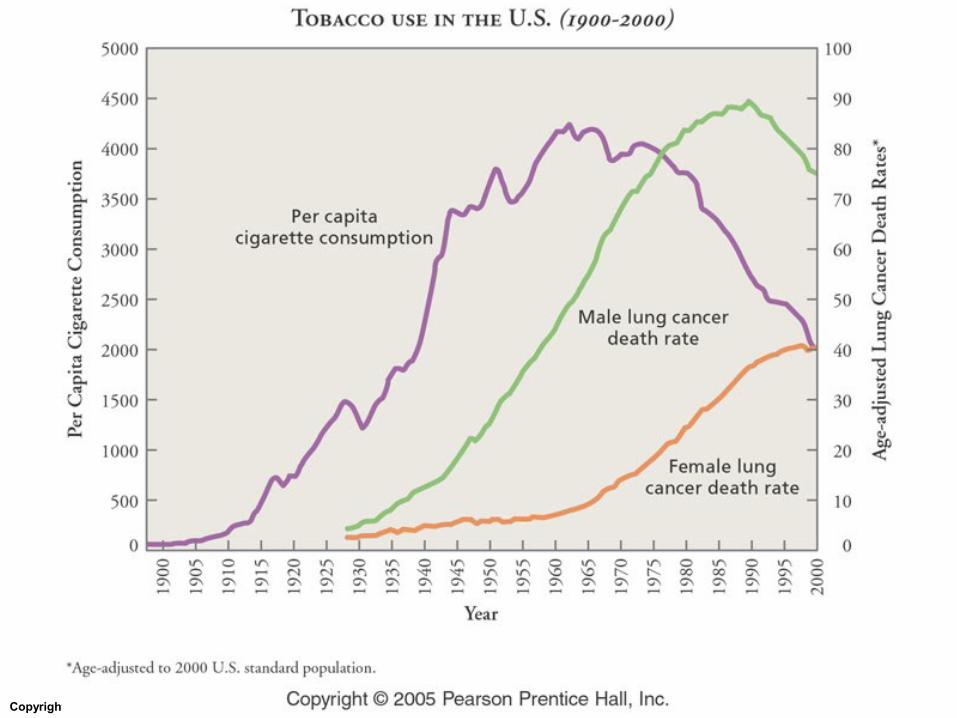
Lung cancer is more common in men, but as
more women are smoking, the rate of lung
cancer in women is rising. Women are more at
risk
Many compounds in the smoke are cancer
promoters, they trigger the progression of cancer
in cells.
8-16
Lung Cancer
Copyright © 2009 Pearson Education, Inc.
The 5-year survival rate is 13%. Smoking can
cause cancers of other parts of the respiratory
system.
8-16
Effects of Smoking

Copyright © 2009 Pearson Education, Inc.
Cigarette smoke contains CO, the fetal blood has
a higher affinity for CO than the mothers blood,
so CO builds up in the fetuses body.
Nicotine is also passed into the fetus, stimulating
the developing nervous system.
Men smoking can damage the DNA in their
sperm and pass genetic mutations to their
offspring.
8-16
Effects of Smoking on Pregnancy

Copyright © 2009 Pearson Education, Inc.

Copyright © 2009 Pearson Education, Inc.
Lung Cancer
Figure 14.15

Copyright © 2009 Pearson Education, Inc.
Copyright © 2009 Pearson Education, Inc.
Copyright © 2009 Pearson Education, Inc.
Copyright © 2009 Pearson Education, Inc.
What is the smoking policy on campus?
1. You can smoke
anywhere
2. You can smoke
anywhere outside
3. You can smoke at
designated spots
4. No smoking on campus

Copyright © 2009 Pearson Education, Inc.
Do you think smoking should be allowed on
campus?
1. Yes
2. No

Copyright © 2009 Pearson Education, Inc.
Do you smoke?
1. Never
2. Used to, but quit
3. Smoke, but trying to quit
4. Smoke

Copyright © 2009 Pearson Education, Inc.
Important Concepts
Read Ch 15
What is the function of the respiratory system?
What is the location and function of the all the
parts of the respiratory system?
What are the parts of the nasal cavity and their
functions?
What are the parts of the larynx and their
functions?
Copyright © 2009 Pearson Education, Inc.
Important Concepts
What cell types lines the trachea, what are
their functions, be able to discus how
smoking effects this tissue?
Where does the exchange of gases occur in
the lungs?
What cavity contains the lungs?
What controls the rate of breathing?
Be able to discuss the mechanics of
breathing?

Copyright © 2009 Pearson Education, Inc.
Important Concepts
How is oxygen carried in the blood?
How is carbon dioxide carried in the blood,
know all the ways, and the which is the
predominate mode? (You don’t need to know
the chemical equation of bicarbonate
formation)
What is the diaphragm and what is its
function?

Copyright © 2009 Pearson Education, Inc.
Important Concepts
Be able to discuss the disorders of the
respiratory system including the description,
symptoms, cause, and treatments.
How does smoking effect pregnancy?

Copyright © 2009 Pearson Education, Inc.
Definitions
Goblet cells, sinuses, epiglottis, surfactant,
diaphragm, intercostal muscles,
Inhalation/inspiration, exhalation/expiration
tidal volume, inspiratory reserve volume,
expiratory reserve volume, residual volume,
vital capacity, hemoglobin, oxyhemoglobin,
chemoreceptors, tubercles, antibiotic
resistant