respiratory system elkhatib
TRANSCRIPT

By M.elkhatib

Understanding the anatomy of the respiratory system
Understanding the physiology of the respiratory system and the differences in paediatrics
Understanding the mechanism of breathing and gas exchange
Clinical implications for paediatrics

Respiration is achieved through the mouth, nose, trachea, lungs, and diaphragm.
The primary function of the respiratory system is to supply the blood with oxygen in order for the blood to deliver oxygen to all parts of the body. The respiratory system does this through breathing. When we breathe, we inhale oxygen and exhale carbon dioxide. This exchange of gases is the respiratory system's means of getting oxygen to the blood.
Molecules of oxygen and carbon dioxide are passively exchanged, by diffusion, between the gaseous external environment and the blood. The blood exchange process occurs in the alveolar region of the lungs.

Oxygen enters the respiratory system through the mouth and the nose.
As the oxygen enters, it passes by the Olfactory nerve which allows you to smell.

The oxygen then passes through the larynx (where speech sounds are produced) and the trachea which is a tube that enters the chest cavity.
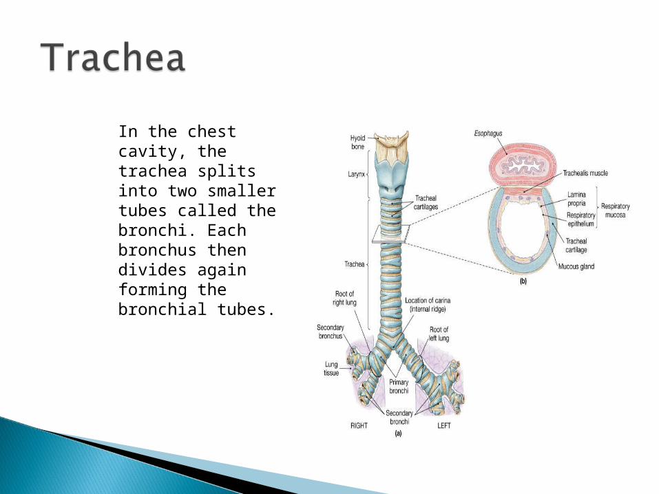
In the chest cavity, the trachea splits into two smaller tubes called the bronchi. Each bronchus then divides again forming the bronchial tubes.
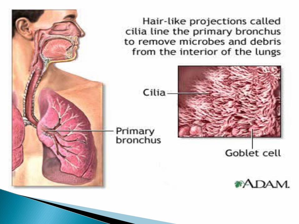
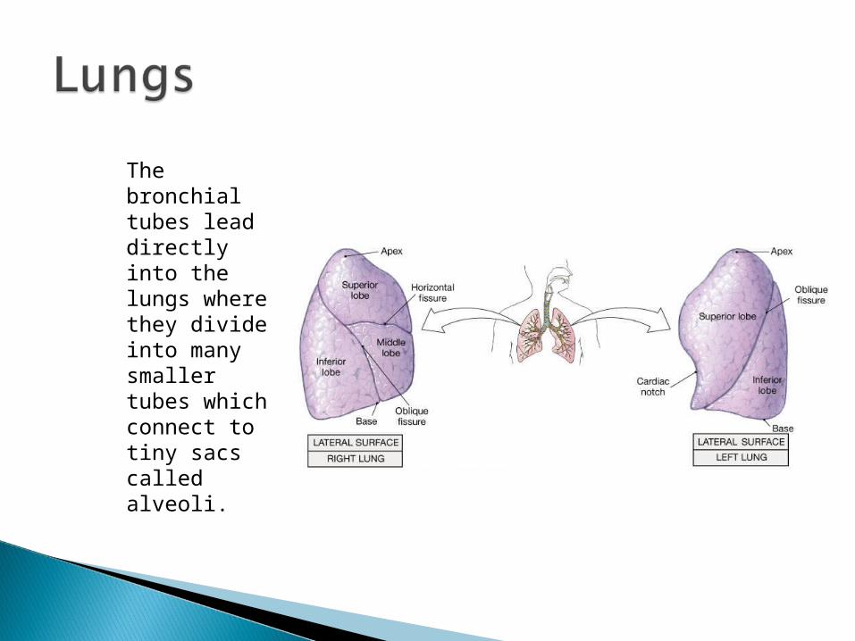
The bronchial tubes lead directly into the lungs where they divide into many smaller tubes which connect to tiny sacs called alveoli.

The average adult's lungs contain about 600 million of these spongy, air-filled sacs that are surrounded by capillaries. The inhaled oxygen passes into the alveoli and then diffuses through the capillaries into the arterial blood. Meanwhile, the waste-rich blood from the veins releases its carbon dioxide into the alveoli. The carbon dioxide follows the same path out of the lungs when you exhale.
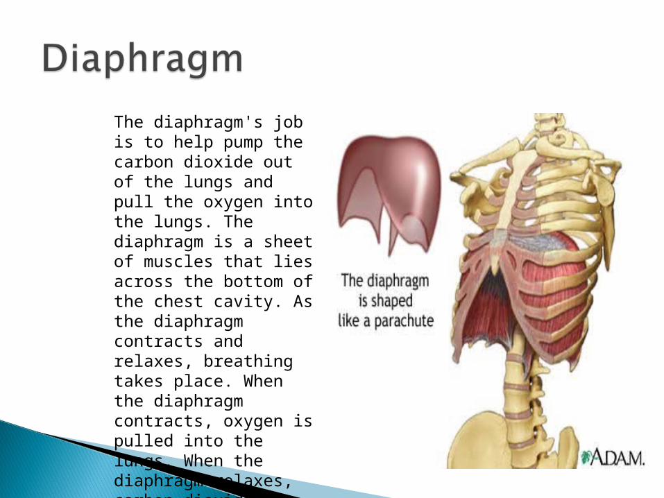
The diaphragm's job is to help pump the carbon dioxide out of the lungs and pull the oxygen into the lungs. The diaphragm is a sheet of muscles that lies across the bottom of the chest cavity. As the diaphragm contracts and relaxes, breathing takes place. When the diaphragm contracts, oxygen is pulled into the lungs. When the diaphragm relaxes, carbon dioxide is pumped out of the lungs

The respiratory system lies dormant in the human foetus during pregnancy.
At birth, the respiratory system becomes fully functional upon exposure to air, although some lung development and growth continues throughout childhood.
Pre-term birth can lead to infants with under-developed lungs. These lungs show incomplete development of the alveolar type II cells, cells that produce surfactant.
The lungs of pre-term infants may not function well because the lack of surfactant leads to increased surface tension within the alveoli.
Thus, many of the alveoli collapse, such that no gas exchange can occur within some or most regions of an infant's lungs, a condition termed respiratory distress syndrome (RDS)

Basic scientific experiments, carried out using cells from chicken lungs, support the potential for using steroids as a means of furthering development of type II alveolar cells. In fact, once a pre-mature birth is threatened, every effort is made to delay the birth, and a series of steroid shots is frequently administered to the mother during this delay in an effort to promote lung growth.
Surfactant is now produed and manufactured. It is administered to the pre-term infant after birth via an endotracheal tube. Sometimes more then one dose is required.

Inhalation is initiated by the diaphragm and supported by the external intercostal muscles. Normal resting respirations are 10 to 18 breaths per minute, with a time period of 2 seconds. During vigorous inhalation (at rates exceeding 35 breaths per minute), or in approaching respiratory failure, accessory muscles of respiration are recruited for support. These consist of sternocleidomastoid, platysma, and the scalene muscles of the neck. Pectoral muscles and latissimus dorsi are also accessory muscles.

Under normal conditions, the diaphragm is the primary driver of inhalation. When the diaphragm contracts, the ribcage expands and the contents of the abdomen are moved downward.
This results in a larger thoracic volume and negative pressure (with respect to atmospheric pressure) inside the thorax. As the pressure in the chest falls, air moves into the conducting zone. Here, the air is filtered, warmed, and humidified as it flows to the lungs.
During forced inhalation, as when taking a deep breath, the external intercostal muscles and accessory muscles aid in further expanding the thoracic cavity. During inhalation the diaphragm contracts.

Exhalation is generally a passive process; however, active or forced exhalation is achieved by the abdominal and the internal intercostal muscles. During this process air is forced or exhaled out.The lungs have a natural elasticity: as they recoil from the stretch of inhalation, air flows back out until the pressures in the chest and the atmosphere reach equilibrium.During forced exhalation, as when blowing out a candle, expiratory muscles including the abdominal muscles and internal intercostal muscles, generate abdominal and thoracic pressure, which forces air out of the lungs.


The major function of the respiratory system is gas exchange between the external environment and the circulatory system. This exchange facilitates oxygenation of the blood with a concomitant removal of carbon dioxide and other gaseous metabolic wastes from the circulation.
As gas exchange occurs, the acid-base balance of the body is maintained as part of homeostasis. If proper ventilation is not maintained, two opposing conditions could occur: respiratory acidosis, a life threatening condition, and respiratory alkalosis.

Upon inhalation, gas exchange occurs at the alveoli, the tiny sacs which are the basic functional component of the lungs. The alveolar walls are extremely thin (approx. 0.2 micrometres). These walls are composed of a single layer of epithelial cells (type I and type II epithelial cells) close to the pulmonary capillaries which are composed of a single layer of endothelial cells.
The close proximity of these two cell types allows permeability to gases and, hence, gas exchange. This whole mechanism of gas exchange is carried by the simple phenomenon of pressure difference. When the atmospheric pressure is low outside, the air from lungs flow out. When the air pressure is low inside, then the vice versa.
Primary function is to obtain oxygen for use by body's cells & eliminate carbon dioxide that cells produce
Pathway of air: nasal cavities (or oral cavity) > pharynx > trachea > primary bronchi (right & left) > secondary bronchi > tertiary bronchi > bronchioles > alveoli (site of gas exchange)
