respiratory support in neonates gcnc chw · therapy for any amount of time should have their spo....
TRANSCRIPT
Guideline No: 2007-0005 v4 Guideline: Respiratory Support in Neonates - GCNC - CHW
This document reflects what is currently regarded as safe practice. However, as in any clinical situation, there may be factors which cannot be covered by a single set of guidelines. This document does not replace the need for the application of clinical judgement to each individual presentation.
Approved by: SCHN Policy, Procedure and Guideline Committee Date Effective: 1st May 2019 Review Period: 3 years Team Leader: Nurse Educator Area/Dept: GCNC
Date of Publishing: 23 April 2019 3:59 PM Date of Printing: Page 1 of 35 K:\CHW P&P\ePolicy\Apr 19\Respiratory Support in Neonates - GCNC - CHW.docx This Guideline may be varied, withdrawn or replaced at any time.
RESPIRATORY SUPPORT IN NEONATES - GCNC - CHW
PRACTICE GUIDELINE ©
DOCUMENT SUMMARY/KEY POINTS
• All ventilated infants are fully monitored with alarm limits set appropriately and checked by two nurses at the beginning of each shift.
• Infants on any form of oxygen therapy require, at minimum, oxygen saturation monitoring in the form of pulse oximetry.
• Medical orders are required for infants on any form of ventilation or oxygen therapy. All changes in ventilation settings are recorded by the medical officer in the electronic medical record. No changes to ventilation should be made without a medical order.
• Oxygen/ventilation settings and orders are checked and documented by two nurses at shift commencement.
• Ideally hand ventilation is performed using a rebreathing bag with a pressure manometer.
• Need for suctioning of ETT is determined on an individual basis based on clinical assessment.
Key Performance Indicators
• All new and revised respiratory support orders are to be written and signed by the medical officer in the electronic medical record.
• Respiratory support checks are completed and documented each shift by nursing staff including review of equipment settings and oxygen
Pain assessments are completed and documented by nursing staff each shift whilst the infant is ventilated and 48 hours post intubation and cessation of analgesia/sedation.

Guideline No: 2007-0005 v4 Guideline: Respiratory Support in Neonates - GCNC - CHW
This document reflects what is currently regarded as safe practice. However, as in any clinical situation, there may be factors which cannot be covered by a single set of guidelines. This document does not replace the need for the application of clinical judgement to each individual presentation.
Approved by: SCHN Policy, Procedure and Guideline Committee Date Effective: 1st May 2019 Review Period: 3 years Team Leader: Nurse Educator Area/Dept: GCNC
Date of Publishing: 23 April 2019 3:59 PM Date of Printing: Page 2 of 35 K:\CHW P&P\ePolicy\Apr 19\Respiratory Support in Neonates - GCNC - CHW.docx This Guideline may be varied, withdrawn or replaced at any time.
CHANGE SUMMARY
• Document format restructured
• Management of transport equipment included
• References updated
READ ACKNOWLEDGEMENT
• All staff caring for patients that require some form of respiratory support or oxygen therapy are required to read and sign the revised policy.
Guideline No: 2007-0005 v4 Guideline: Respiratory Support in Neonates - GCNC - CHW
Date of Publishing: 23 April 2019 3:59 PM Date of Printing: Page 3 of 35 K:\CHW P&P\ePolicy\Apr 19\Respiratory Support in Neonates - GCNC - CHW.docx This Guideline may be varied, withdrawn or replaced at any time.
TABLE OF CONTENTS Documentation of Respiratory Orders ................................................................................... 4 Oxygen Administration, Monitoring & Documentation ..................................................... 4 Pulse Oximetry ...................................................................................................................... 5 CO2 monitoring .................................................................................................................... 6 End-tidal CO2 (ETCO2) Monitoring7 ....................................................................................... 6 Transcutaneous CO2 Monitoring ........................................................................................... 6 Airway Adjuncts .................................................................................................................. 7 Nasopharyngeal Airways/Tubes ............................................................................................ 7 Laryngeal Mask Airway (LMA) ............................................................................................... 8 Non-invasive Ventilation ..................................................................................................... 9 Nasal-Cannula Oxygen19,20, 21 ................................................................................................ 9 Humidified High Flow Nasal Cannula (HHFNC) ..................................................................... 9 Intubation and Endotracheal Tube Management ............................................................11 Intubation .............................................................................................................................11 Taping of endotracheal (ETT) tubes ..................................................................................12 Cuffed Endotracheal Tubes ...............................................................................................12 Suctioning an endotracheal tube ......................................................................................13 Suction Technique ................................................................................................................15 Open versus closed suction .................................................................................................15 Nasal and Oral Pharyngeal Suction ......................................................................................17 Management of the Ventilated Patient ..............................................................................17 Ventilation Modes ...............................................................................................................19 Volume Guarantee ...............................................................................................................19 High Frequency Oscillatory Ventilation ............................................................................21 Practical Management ..........................................................................................................22 Managing specific conditions with HFOV ..............................................................................24 Nursing Care ........................................................................................................................25 Troubleshooting during HFOV ..............................................................................................26 Weaning Ventilation and Extubation .................................................................................27 Continuous Positive Airway Pressure (CPAP) .................................................................30 Nursing Care ........................................................................................................................30 Feeds ...................................................................................................................................31 Documentation .....................................................................................................................32 CPAP Complications58-60 ......................................................................................................32 References ..........................................................................................................................33
Guideline No: 2007-0005 v4 Guideline: Respiratory Support in Neonates - GCNC - CHW
Date of Publishing: 23 April 2019 3:59 PM Date of Printing: Page 4 of 35 K:\CHW P&P\ePolicy\Apr 19\Respiratory Support in Neonates - GCNC - CHW.docx This Guideline may be varied, withdrawn or replaced at any time.
Defining Statement
Respiratory support may take the form of mechanical ventilation, continuous positive airway pressure (CPAP), humidified high flow nasal cannula (HHFNC) or oxygen therapy. Support can be given via an endo-tracheal tube (ETT), nasal cannula, nasal mask or face mask. The airway of the newborn infant is not fully mature and is susceptible to damage. Trauma can result from suctioning of the endo-tracheal tube, tube mobility, inadequate or excessive humidification, and inadequate or excessive levels of oxygen1,2 . Care is required in the application of respiratory support.
Documentation of Respiratory Orders Documentation of respiratory orders apply to all infants who are receiving supplemental oxygen and/or require respiratory support via an endotracheal tube, naso-pharyngeal tube or midline CPAP. All orders are to be written and signed by the medical officer or Nurse Practitioner under in the electronic medical record.
Component Documentation required
Intubation
In section ‘procedure – airway management’ in the electronic medical record:
• For initial and subsequent intubation, tube change or retaping the ETT date, tube size and position are documented
• Any problems incurred during the procedure
Ventilator Set-up Ventilator set up and circuit assembly or change are documented on the card on the ventilator
Ventilator settings (Including CPAP, HHFNC)
• All changes in ventilation settings are recorded in the electronic medical record
• The humidifier temperature, oxygen and ventilator settings are recorded on the respiratory flow chart each hour
Oxygen Oxygen therapy is ordered by the medical officer and documented in the electronic medical record
Oxygen Administration, Monitoring & Documentation
Supplemental oxygen is the most common drug prescribed in hospitals. It is provided to correct or prevent hypoxemia and hypoxia which can occur as a result of cardiovascular, metabolic, neurological or respiratory dysfunction3.
Infants receiving supplemental require an identified target range for SpO2 delivery, to ensure adequate tissue oxygenation without the complications of oxygen toxicity.
Premature infants are at increased risk of developing retinopathy of prematurity (ROP), bronchopulmonary dysplasia (BPD), and cerebral injury associated with hyperoxia and the

Guideline No: 2007-0005 v4 Guideline: Respiratory Support in Neonates - GCNC - CHW
Date of Publishing: 23 April 2019 3:59 PM Date of Printing: Page 5 of 35 K:\CHW P&P\ePolicy\Apr 19\Respiratory Support in Neonates - GCNC - CHW.docx This Guideline may be varied, withdrawn or replaced at any time.
length of time for oxygen therapy4. Extra precautions should be taken when delivering oxygen to this population3.
Safe Administration of Oxygen
All neonates receiving supplemental oxygen must have their oxygen levels monitored. Hourly checks and documentation:
• oxygen flow rate
• patency of tubing/equipment
Documentation for delivery of oxygen via any method must include
• Handover of shift equipment check documentation
• Oxygen saturation range
• Method, flow rate or percentage of supplemental oxygen administration
• Vital signs (RR, HR, SaO2) documented hourly
Additional assessment should include
• The chest assessed for bilateral chest movement and work of breathing including auscultation of air entry.
• Tolerance of the infant to supplemental oxygen administration during handling and after feeds.
Pulse Oximetry Infants and children who are at risk of either critical oxygen desaturation or hyper-oxygenation require monitoring to avoid complications arising from these episodes.
• The recommended range of SpO2 is 92–96%, unless a specifically indicated level is ordered by the Neonatologist or Cardiologist3.
• Neonates born at less than 32 weeks gestation who require supplemental oxygen therapy for any amount of time should have their SpO2 levels targeted to decrease morbidity associated with oxygen therapy5. Current evidence suggests Oxygen saturation targeting of 90-94%3,4
Patient and Carer Safety Considerations
• Set the alarm limits for the patient with an upper limit no greater than 99% to allow for weaning. Ensure alarm limits are checked each shift.
• When a monitor alarms, in addition to silencing the alarm the infant should be checked to establish if there is a reason for the alarm i.e. deterioration, sudden event, movement artefact or monitoring lead no longer in position
• Inaccurate readings occur when the patient is active or distressed due to anaemia, hyperbilirubinaemia, lipid infusion, inotropic drugs and poor peripheral perfusion.
• Shield the probe from phototherapy lights with Webrill to avoid an incorrect reading
• Re-site the probe at least every 4-6 hours to avoid pressure sores. In poorly perfused neonates this may need to occur more frequently.

Guideline No: 2007-0005 v4 Guideline: Respiratory Support in Neonates - GCNC - CHW
Date of Publishing: 23 April 2019 3:59 PM Date of Printing: Page 6 of 35 K:\CHW P&P\ePolicy\Apr 19\Respiratory Support in Neonates - GCNC - CHW.docx This Guideline may be varied, withdrawn or replaced at any time.
Caveat If a skin burn occurs remove probe. Check for cable damage, notify Medical Officer. An (iMMs) should be completed, including grading of the injury and referral to plastics considered.
CO2 monitoring
Monitoring of CO2 assesses the effectiveness of ventilatory strategies and is utilised to wean support, minimise iatrogenic lung disease and prevent hypocapnia4,5. Low arterial CO2 pressures (PaCO2) may contribute to the development of chronic lung disease and periventricular leukomalacia in neonates, whereas high levels may increase cerebral blood flow and the risk of intraventricular haemorrhage (IVH)5,6. Continuous CO2 monitoring is used as a guide to changes in the state of the lung, and can minimise the need for repeated arterial blood gas sampling.
There is a lack of evidence to specify targets for CO2 monitoring in ventilated neonates7. Tolerating higher partial pressure of carbon dioxide (pCO2) in mechanically ventilated, extremely low birthweight infants might reduce ventilator-induced lung injury and bronchopulmonary dysplasia6.
• A baseline correlation is established between the infant’s pCO2 (via blood sampling) and the ETCO2 to monitor the trend of the patient’s CO2.
• The recommended range for paCO2 is 35-45mmHg or that determined by the specific condition as guided by the Neonatologist.
• All infants requiring invasive respiratory support have their CO2 levels monitored.
• There are two methods for monitoring CO2 End-tidal or transcutaneous CO2 monitoring.
End-tidal CO2 (ETCO2) Monitoring7 • ETCO2 detectors measure exhaled CO2 via infrared spectroscopy displayed as mmHg9,
providing a continuous trace of the infant’s exhaled breath, known as a capnogram.
• ETCO2 monitoring is most accurate for patients with a regular respiratory rate (i.e. sedated/muscle-relaxed) with minimal/no leak around the ETT.
• ETCO2 monitoring may be affected by secretions or decreased tidal volume.
• ETCO2 monitoring is the default mode for CO2 monitoring in ventilated neonates
Instructions for Correlating ETCO2
Transcutaneous CO2 Monitoring Transcutaneous monitoring (TCM) provides a non-invasive method for monitoring oxygen tension in the newborn. A sensor is heated and when placed on the skin causes vasodilatation of the cutaneous vessels. This promotes the diffusion of oxygen/carbon dioxide from the arterial capillaries through the skin and is measured by electrochemical reduction. The signal is amplified and converted to a digital readout8.
• Recommended TcO2 range is determined by the specific condition.
• TcO2 monitoring is not considered default monitoring for oxygenation

Guideline No: 2007-0005 v4 Guideline: Respiratory Support in Neonates - GCNC - CHW
Date of Publishing: 23 April 2019 3:59 PM Date of Printing: Page 7 of 35 K:\CHW P&P\ePolicy\Apr 19\Respiratory Support in Neonates - GCNC - CHW.docx This Guideline may be varied, withdrawn or replaced at any time.
Indications for use
• May be used in neonates where a rapid change in CO2 is likely and a blood gas may not be utilised prior to implementing ventilation changes
• Conditions where TCCO2 monitoring may be beneficial include diaphragmatic hernia
• For infants receiving HFOV
• For infants where there is a risk of retinopathy of prematurity i.e. <28 week gestation
For guidelines to set up the equipment refer to the TC-CO2 monitoring section as the principles are the same.
Instructions for Correlating ETCO2
Airway Adjuncts
Neonates and infants breathe through the nose as the entire length of their tongue sits against the hard and soft airway occluding the passage of air through the mouth9. Combined with the high position of their epiglottis these factors increase the resistance of the oral airway and make them preferential nose breathers. Nasal obstruction can therefore lead to significant respiratory distress5.
Nasopharyngeal Airways/Tubes Airway adjuncts including Nasopharyngeal Tubes and Laryngeal Masks can be used in an emergency or over a longer period of time10,11. The nasopharyngeal (NP) tube bypasses upper airway obstruction at the level of the nose, naso-pharynx, or base of the tongue. The NP tube acts as a "splint” preventing the tongue from falling back on the posterior pharyngeal wall occluding the airway, therefore preventing airway obstruction, hypoxia and asphyxia12.
NP tubes are generally well tolerated by conscious infants and used in the management of congenital maxillofacial abnormalities, syndromic craniosynotosis, mid facial hypoplasia or to support the upper airway post-surgery10,12,13,.
Two types of NP Airways are used in GCNIC:
• PVC Nasopharyngeal Airways
• Modified Endotracheal tubes
Safety and Precautions
• Insertion of an NP Airway is a medical order directed by a neonatologist
• A Senior Medical Officer or Nurse Practitioner should insert the initial NPT
• Medical and nursing staff that have attended an airway management workshop may undertake routine changes
• PVC NP tubes are replaced every 48 hours or PRN if secretions are affecting tube patency when CPAP is in use and daily when no CPAP is used.
• Modified Endotracheal Tube NP airways are inserted during LBO’s via the ENT team and changed weekly

Guideline No: 2007-0005 v4 Guideline: Respiratory Support in Neonates - GCNC - CHW
Date of Publishing: 23 April 2019 3:59 PM Date of Printing: Page 8 of 35 K:\CHW P&P\ePolicy\Apr 19\Respiratory Support in Neonates - GCNC - CHW.docx This Guideline may be varied, withdrawn or replaced at any time.
• More frequent occlusions may occur initially following insertion of the tube due to insertion related trauma.
• Ensure to alternate the nostrils utilised during changes of tubes where possible to avoid unsightly deformation of the nostril.
• A spare NP airway of the same size is placed at the bedside.
Management
• Documentation of the tube type and size is recorded in the electronic medical record.
• Infants on nasopharyngeal tubes may have gaseous distension of the stomach and an intragastric tube is recommended.
• As the NPA is bypassing the route for natural humidification this may lead to thickened secretions that block the NPA. The patient should be assessed regularly to review if suction of the NPA is required.
• Check the skin around the nostrils frequently for blanching. Blanching leads to pressure injury/ skin breakdown around the alar rim it may indicative of an incorrect size.
• Medical staff or Nurse Practitioners are notified if there is any alteration in the neonate’s respiratory status.
Laryngeal Mask Airway (LMA) Laryngeal mask airways (LMA) are used as an alternative to intubation with an ETT tube if an airway is known to be difficult, where face mask ventilation is unsuccessful or when there have been multiple failed attempts at intubation15.
• The LMA once inserted can be attached either to a bag and mask apparatus or to a ventilator setup.
• Use of an LMA is considered a temporary measure as the airway is still unstable and unprotected from rising gastric fluids16. It may provide clinicians with time during management of a difficult airway until additional help arrives.
• Failure of the LMA occurs in <1% of children and can include leak, obstruction and intolerance to the LMA17.
Criteria for use of LMA
• Known difficult airway with previous difficult intubation or grade IV view where intubation attempts would be unsafe
• Failed intubation (3 attempts)
• Failure in face mask ventilation where help is not available immediately or where other methods of ventilation have failed
Patient and Carer Safety Considerations18
• Ensure a call for help to senior clinicians has been placed as LMA insertion is a temporary measure and a more stable airway is required long term
• An appropriate rebreathing bag with manometer and appropriate sized face mask must be available at the bedside.

Guideline No: 2007-0005 v4 Guideline: Respiratory Support in Neonates - GCNC - CHW
Date of Publishing: 23 April 2019 3:59 PM Date of Printing: Page 9 of 35 K:\CHW P&P\ePolicy\Apr 19\Respiratory Support in Neonates - GCNC - CHW.docx This Guideline may be varied, withdrawn or replaced at any time.
• Suction apparatus is checked and in working order. Suction catheters of appropriate size and a stethoscope are also available.
• The resuscitation trolley is prepared with relevant equipment – pre-cut adhesive tapes, lubricant and resuscitation drugs
• Ensure the correct size of LMA is available for the size of the baby (2 – 5kg = Size 1)
Technique for inserting LMA
Assessment
• Review chest wall movement, improvement in heart rate and oxygenation and the chest auscultated.
• Utilise a Pedicap to confirm position
Ongoing Management
• The tube remains in-situ until a long term airway can be obtained, which should be completed as soon as possible
• Review pain scores and analgesia requirements
Removal of the LMA
• Decision to cease use of the LMA should be made by the consultant in charge, once the infant no longer requires it, or just prior to insertion of a more long term airway.
• Deflate the mask
• Loosen and remove tapes
• Gently remove tube prior to exhalation
• Document removal and change of ventilation device in electronic medical records
Non-invasive Ventilation
Nasal-Cannula Oxygen19,20, 21 Oxygen delivery by nasal cannula (low flow) is used for infants who have reducing oxygen and flow requirements in Humidified High Flow Nasal Cannula.
• The maximum flow that may be delivered via nasal cannula is 2 litres per minute.
• Humidification is not required when administering oxygen via nasal cannula.
• Do not cut the cannula probes as the shape and length is designed for optimal oxygen delivery via the nares.
Technique to apply Nasal Cannula
Humidified High Flow Nasal Cannula (HHFNC) Breathing cool dry gases may result in mucosal damage, reduced ciliary motility, decreased mucous production, bronchospasm and nasal discomfort20. HHFNC is a high flow, humidified system that delivers high flow air or oxygen with 100% humidity heating and blending of titrated oxygen via nasal cannula to patients who are spontaneously breathing22. This

Guideline No: 2007-0005 v4 Guideline: Respiratory Support in Neonates - GCNC - CHW
Date of Publishing: 23 April 2019 3:59 PM Date of Printing: Page 10 of 35 K:\CHW P&P\ePolicy\Apr 19\Respiratory Support in Neonates - GCNC - CHW.docx This Guideline may be varied, withdrawn or replaced at any time.
emulates the balance of temperature and humidity that occurs in healthy lungs thus maintaining mucociliary clearance and decreases discomfort and irritation.
As a consequence of flow, HHFNC produces a degree of positive end expiratory pressure resulting in increased Functional Residual Capacity which leads to improved oxygenation and reduced work of breathing22, 23. Compared to CPAP, HHFNC allows easier access for the parents and facilitates interaction with the child23.
Instructions on sizing and HHFNC set up.
Indications for Humidified High Flow Nasal Cannula HHFNC may be utilised for neonates in GCNIC with22, 23
• Respiratory distress or oxygen requirement up to 50 % due to:
a) Bronchiolitis
b) Pneumonia
c) Post-extubation
• Weaning from CPAP
• Neonates >32 weeks gestation with apnoea of prematurity
• Neonates/infants with chronic lung disease
• Continuation of HHFNC therapy in newborns transferred from PICU and other units
Contraindications
• FiO2 requirement > 50% (unless indicated for specialist management i.e. cardiac patients in consultation with the cardiology team)
• Hypercapnia pCO2 > 65 mmHg
• Respiratory Acidosis pH<7.2
• Nasal Obstruction (e.g Choanal atresia)
• Life threatening hypoxia
• Use with caution in infants with gastro-intestinal obstruction or immediately following GI surgery
Nursing Care23
• The neonatologist/fellow on call is notified prior to commencement of HHFNC.
• Each hour observe the circuit, water level and prong position. Prong dislodgement will result in a loss of respiratory support.
• Check the nares and nasal septum for signs of pressure each shift and during cares, document in notes any redness or broken skin.
Caution HHFNC delivers positive pressure in a variable, relatively unpredictable and unregulated fashion24. Adverse clinical effects and changes in pulmonary function may result from HHFNC.

Guideline No: 2007-0005 v4 Guideline: Respiratory Support in Neonates - GCNC - CHW
Date of Publishing: 23 April 2019 3:59 PM Date of Printing: Page 11 of 35 K:\CHW P&P\ePolicy\Apr 19\Respiratory Support in Neonates - GCNC - CHW.docx This Guideline may be varied, withdrawn or replaced at any time.
• If the patient’s clinical state deteriorates escalation of respiratory support may be required.
• Increases to oxygen concentration and flow rate are ordered by medical staff and documented in the patient’s electronic medical record.
• Observe infant for abdominal distension. Aspirate air from abdomen when required.
• If at any stage the infant has saturations below 92% a medical officer or Nurse Practitioner is to be notified (N.B: Discuss appropriate range of acceptable SaO2 in patients with Cyanotic Heart Disease with medical officer prior to starting HHFNC).
• Duoderm at the nares and use of chin strap can lead to increased pressure delivery and are not recommended.
Failure Criteria Assess if HHFNC is adequate for clinical condition:
• O2 demand of more than 50% requires review my medical staff or Nurse Practitioner.
• Changes in oxygen requirement of more than 10% require review by the registrar/NP/fellow.
Rapid clinical deterioration
• Bag and mask breaths can be applied while the nasal cannula remains insitu.
• Exclude pneumothorax as a side effect of HHFNC.
• Check for nasal obstruction.
Weaning
• Weaning occurs when the neonate’s clinical condition is improving evidenced by decreased work of breathing, normal respiratory rate range (RR), normal cardiovascular parameters and O2 saturations between accepted target range of 92-96%
• Wean oxygen as required to maintain saturation range
• Wean flow as directed and tolerated by patient (normally 1L/day)
• Monitor SaO2, RR and WOB after each flow change and notify medical staff/NP of any change in respiratory and cardiovascular status
• When the total gas flow is reduced to 2 L/min and FiO2 is < 40% consider transfer to low flow nasal cannula oxygen.
For patients transferred to the ward varying levels of maximum HHFNC can be administered. For additional information regarding ward administration of HHFNC please refer to: SCHN Humidified High Flow Nasal Cannula Therapy Policy
Intubation and Endotracheal Tube Management
Intubation Intubation involves the insertion of an endotracheal tube through the mouth or nose into the trachea. Nasal tubes are the preferred route of intubation for infants in GCNC. An oral
Guideline No: 2007-0005 v4 Guideline: Respiratory Support in Neonates - GCNC - CHW
Date of Publishing: 23 April 2019 3:59 PM Date of Printing: Page 12 of 35 K:\CHW P&P\ePolicy\Apr 19\Respiratory Support in Neonates - GCNC - CHW.docx This Guideline may be varied, withdrawn or replaced at any time.
tracheal tube may be used for very low birth weight infants or those with cranio-facial abnormalities, in an emergency intubation or where nasal intubation is difficult, or where an appropriate size ETT will not pass down the nasal passages,. Intubation route will be determined by the clinician intubating the patient. Nasal tubes may be easier to secure than oral tubes, they can reduce excessive tube mobility and the risk of accidental extubation.
Patient and Carer Safety Considerations
• A rebreathing bag with manometer and appropriate sized face mask must be available at the bedside of ventilated neonates.
• Suction equipment is checked each shift and must be in working order. Suction catheters of appropriate size and a stethoscope should also be available at the bedside.
• The resuscitation trolley is placed at the bedside with relevant intubation equipment – appropriate size ETT, laryngoscope, Magill forceps and introducer, pre-cut adhesive tapes and lubricant.
• The size of the ETT selected in patient management is directed by medical staff or NP.
Intubation nursing role instructions
Taping of endotracheal (ETT) tubes
Correct taping of an ETT is essential to ensure tube safety and prevent accidental dislodgement. Taping of ETT is an accredited skill and must be undertaken by a staff member that has completed the NICU Skills accreditation package.
Staff should be familiar with the technique for:
• Taping an oral ETT
• Taping an nasal ETT
• Securing an ETT with an alternate device (Neobar©)
Instructions for taping an oral and nasal ETT
Instructions for securing an ETT with a Neobar
Cuffed Endotracheal Tubes
Previous concerns about cuffed endotracheal tubes in neonates causing sub-glottic stenosis are unfounded25. Cuffed ETTs have an advantage for some neonates requiring specific modes of ventilation25, 26. Cuffed ETTs are selected a half size smaller for than predicted for age to provide minimal cricoid pressure when the tube remains in-situ. Only cuffed tubes with a low pressure, high volume cuff (Kimberley Clark Microcuff©) should be used.
Potential advantages of cuffed ETT’s include25, 26
• Reduced gas leak during anaesthesia and while receiving NO Therapy
• Improved ventilation and non-invasive monitoring of ventilation (capnography)
• Preferred option for certain forms of ventilation like HFOV and VG
• Decreased airway trauma and less frequent ET Tube changes
Guideline No: 2007-0005 v4 Guideline: Respiratory Support in Neonates - GCNC - CHW
Date of Publishing: 23 April 2019 3:59 PM Date of Printing: Page 13 of 35 K:\CHW P&P\ePolicy\Apr 19\Respiratory Support in Neonates - GCNC - CHW.docx This Guideline may be varied, withdrawn or replaced at any time.
Insertion of cuffed ETT
• Cuffed ETT’s may be placed in operating theatre or electively in patients in GCNC.
• For patients returning from OT nursing staff should assess if the cuff is inflated and measure the cuff pressure as soon as possible.
• Medical staff or the NP should determine and document if the cuff is to remain inflated.
• The continued need for inflation of the cuff should be reviewed every shift by the medical team and is dependent on the respiratory status of the patient.
• The green connector will be replaced with a blue Portex connector so the in-line suction catheter can be attached and used.
• The anaesthetist will replace the connector prior to leaving the operating theatre the nurses accompanying the baby may need to remind them.
Day to Day Management of Inflated Cuffed ETT
• Suction oropharynx before any deflation of the cuff.
• When inflating the cuff, the minimum cuff volume necessary to reduce leak to an acceptable level must be used (minimum volume technique).
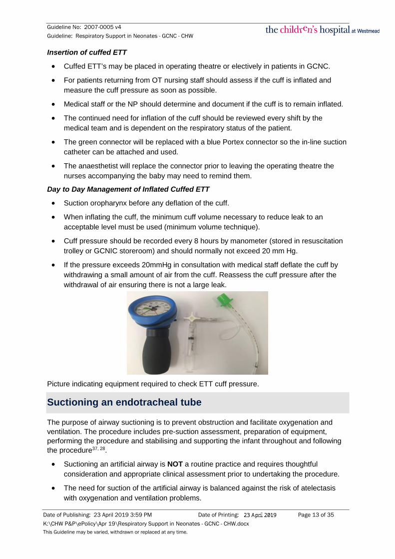
• Cuff pressure should be recorded every 8 hours by manometer (stored in resuscitation trolley or GCNIC storeroom) and should normally not exceed 20 mm Hg.
• If the pressure exceeds 20mmHg in consultation with medical staff deflate the cuff by withdrawing a small amount of air from the cuff. Reassess the cuff pressure after the withdrawal of air ensuring there is not a large leak.
Picture indicating equipment required to check ETT cuff pressure.
Suctioning an endotracheal tube
The purpose of airway suctioning is to prevent obstruction and facilitate oxygenation and ventilation. The procedure includes pre-suction assessment, preparation of equipment, performing the procedure and stabilising and supporting the infant throughout and following the procedure37, 28.
• Suctioning an artificial airway is NOT a routine practice and requires thoughtful consideration and appropriate clinical assessment prior to undertaking the procedure.
• The need for suction of the artificial airway is balanced against the risk of atelectasis with oxygenation and ventilation problems.
Guideline No: 2007-0005 v4 Guideline: Respiratory Support in Neonates - GCNC - CHW
Date of Publishing: 23 April 2019 3:59 PM Date of Printing: Page 14 of 35 K:\CHW P&P\ePolicy\Apr 19\Respiratory Support in Neonates - GCNC - CHW.docx This Guideline may be varied, withdrawn or replaced at any time.
• Performing the suction procedure whilst the infant is in a quiet behavioural state may result in less peripheral and cerebral circulatory instability
• The internal lumen of an ETT decreases substantially after a few days of intubation, due to the formation of biofilm, however suctioning should still only be performed when clinically indicated27.
• Mechanical ventilation with adequate humidification of the airway is necessary to ensure that secretions remain fluid and easily removed with short duration suction28.
• Suctioning can cause:
o Changes in lung dynamics resulting in hypoxemia and hypercapnoea.
o Hypoxemia that leads to cardiac arrhythmia
o Tissue trauma to the tracheal and/or bronchial mucosa
o Bronchospasm through irritation while suctioning.
o Microbial colonisation of the lower airway
o Changes to cerebral blood flow and intracranial pressure (specifically in ELBW infants)
o Pain and general discomfort
o Vagal nerve stimulation with associated bradycardia
All of these complications need to be considered and prepared for before suctioning
Patient and Carer Safety Considerations
• Suctioning is always considered a two person procedure. Supporting and containing the infant by a second nurse has been found to reduce the effects of discomfort and procedural pain caused by suctioning 27.
• Suctioning can be is associated with hypoxemia resulting in alterations in heart rate and blood pressure. The infant should be observed and given time for vital signs to return to baseline before considering passing the catheter a second time.
• Frequency of suctioning should be determined by the infant’s clinical condition and volume of secretions
• Intrapulmonary pressure changes generated by suction can be interpreted as a loss of lung volume 29.
• There is no evidence to determine the maximum time frame between suction in order to prevent ETT blockage, however there is some evidence that allowing 12 hours between suction does not result in an increased incidence of ETT occlusion, ventilator associated pneumonia or nosocomial infection 29.
• Infants who are muscle relaxed do not have the ability to cough and may be more difficult to assess for the presence of secretions, for these infants more frequent suctioning may be considered.

Guideline No: 2007-0005 v4 Guideline: Respiratory Support in Neonates - GCNC - CHW
Date of Publishing: 23 April 2019 3:59 PM Date of Printing: Page 15 of 35 K:\CHW P&P\ePolicy\Apr 19\Respiratory Support in Neonates - GCNC - CHW.docx This Guideline may be varied, withdrawn or replaced at any time.
Suction Technique The neonates underlying condition needs to be considered before suctioning. For example neonates with chronic lung disease or PPHN are extremely fragile while suctioned.
Pre-oxygenation
• Pre-oxygenation in general is not recommended particularly in preterm infants and neonates with cyanotic heart disease31.
• It is reasonable to consider an increase in the baseline oxygen by 10% - 20% for 30-60 seconds prior to suction if the infant is already hypoxemic or has demonstrated previous hypoxemia with suctioning 30, 31, 32.
Suction Depth34, 35
• All suctioning in GCNIC utilises the shallow suctioning technique – that is insertion of the catheter only to the tip of the ETT.
• When measuring the suction catheter prior to insertion, calculate the measurement of the ETT from the point of disconnection (including adaptor) and the entire length of the tube by using a visual marking. Use the template for in-line suction catheters.
• Withdraw catheter slightly before applying negative suction to ensure catheter tip is not abutting the mucosa.
For additional guidance on ETT suction utilise the ETT Suction Assessment Guide.
Catheter size
Catheter size will vary according to the internal diameter of the ETT. Catheter size has the greatest influence on lung volume loss due to airway obstruction by the suction catheter29. The catheter size should not exceed double of the size of the ETT tube e.g. double a size 4mm tube is 8, that is the maximum size suction catheter to be used in that size tube.
ETT Size Catheter Size
2.5 FG 5
3.0 & 3.5 FG 6 – 7 FG
4.0mm FG 8
Open versus closed suction • Closed suction technique results in an overall better physiological stability (especially in
infants < 1000 grams), with less desaturation and bradycardia 37, 38.
• The closed suction technique facilitates continuous mechanical ventilation and oxygenation during the suctioning event37. It may prevent lung de-recruitment associated with the use of open suction technique, particularly in patients at higher risk of desaturation, such as premature infants38.
• When there is evidence of acute lung injury (ALI), that is patients requiring high FiO2 and PEEP, closed suction should be considered38.
Guideline No: 2007-0005 v4 Guideline: Respiratory Support in Neonates - GCNC - CHW
Date of Publishing: 23 April 2019 3:59 PM Date of Printing: Page 16 of 35 K:\CHW P&P\ePolicy\Apr 19\Respiratory Support in Neonates - GCNC - CHW.docx This Guideline may be varied, withdrawn or replaced at any time.
• Closed suction systems support the continuation of ventilation throughout the procedure and importantly, PEEP is maintained.
• The closed system eliminates the risk splash injury to the clinician.
• Closed suction is the preferred suction method in GCNC.
• Open suction should only be considered following consultation with medical staff/NP/CNS
Suction pressure and duration
• The negative pressure required for suctioning neonates has been recommended to be between 10-15Kp29.
• On withdrawal suction should be continuous rather than intermittent to ensure effective removal of secretions.
• The duration of suctioning an endo-tracheal tube should be kept to a minimum48
• A maximum of 10 seconds is used as a guide for the insertion and removal of the catheter. The duration of negative suction should be no longer than 5 seconds in neonates29, 30, 31.
• One pass of the catheter is recommended with the need for further passes being assessed individually and should not exceed two.
Normal saline instillation
• Sterile normal saline should not be routinely36 instilled during ETT suctioning unless directed by a senior medical officer.
• If Normal saline use 0.1– 0.2mL/kg (using a 1mL syringe) is instilled at body temperature immediately prior to the insertion of the suction catheter.
In line suction management
• Note the size and position of the ETT, select the appropriate size inline catheter
• Ensure infant stability prior to attempting the procedure
• Attach the ventilator circuit to the larger of the two ports (perpendicular to the ETT).
• Assess ventilation to ensure ETT position unchanged
• In-line suction catheters should be changed every 72 hours.
Inline suction technique
ETT Suspected or actual Blockage
• If the endo-tracheal tube is blocked or is thought to be blocked call for assistance from medical and nursing staff as the infant may require re-intubation.
• Press the emergency button if the infant requires emergency resuscitation due to respiratory or cardiorespiratory arrest.
• Attempt to suction the ETT tube, if necessary remove the ETT (preferably after medical or Nurse Practitioner assessment) and manually ventilate the infant with a resuscitation bag and mask until a new tube has been inserted.
Guideline No: 2007-0005 v4 Guideline: Respiratory Support in Neonates - GCNC - CHW
Date of Publishing: 23 April 2019 3:59 PM Date of Printing: Page 17 of 35 K:\CHW P&P\ePolicy\Apr 19\Respiratory Support in Neonates - GCNC - CHW.docx This Guideline may be varied, withdrawn or replaced at any time.
Nasal and Oral Pharyngeal Suction Nasopharyngeal and oropharyngeal suction can be extremely stressful for the infant and should not be performed routinely. It is only undertaken when other less invasive techniques have proved unsuccessful, or where the secretions are causing physiological deterioration or distress to the infant.
Nasal and Oral Pharyngeal suction is considered a two person procedure in GCNIC.
• Infants intubated with an oral ETT tube may have copious oral secretions which require suctioning to avoid saliva loosening the tapes that secure the ETT.
• Naso-pharyngeal suction is an invasive procedure and can result in hypoxaemia, mucosal injury, vagal bradycardia, changes in intra-cerebral blood flow and behavioural distress for the infant.
• Repeated oral suction can result in oral aversion causing pain-related responses to oral stimuli or dysphagia.
• Catheters used for pharyngeal suction have a blind end and side hole, which may minimise damage to the soft tissues in the nasal/oral cavity.
• An 8FG catheter is recommended to remove the secretions adequately. • For thick tenacious oral secretions a short 12fg catheter can be utilised to remove
secretions from the mouth and shallow suction at the back of the pharynx • Suction pressure remains the same as for ETT suction at 10mmHg, the duration of oral
suction should not exceed 10-20 seconds to minimise distress for the infant Document the amount, consistency and colour of secretions, the number of catheter passes and patient tolerance in the electronic medical record
Management of the Ventilated Patient
Patient and Carer Safety Considerations
Ventilated infants:
• Require a nurse to be in immediate attendance at all times.
• Are nursed on a memory foam mattress
• Are fully monitored with the alarm limits set and checked by two nurses at each shift change.
• Settings should not be changed without a documented medical/NP order.
• Circuits are changed each week. Prior to connecting the circuit to the infant it is checked by an accredited staff member.
Care of ventilated infants
• The infant is positioned according to their clinical needs39 and changed based on their NSRAS score.
• All procedures, including suctioning, repositioning and weighing require two nurses, one of whom is experienced in caring for a ventilated infant.
Guideline No: 2007-0005 v4 Guideline: Respiratory Support in Neonates - GCNC - CHW
Date of Publishing: 23 April 2019 3:59 PM Date of Printing: Page 18 of 35 K:\CHW P&P\ePolicy\Apr 19\Respiratory Support in Neonates - GCNC - CHW.docx This Guideline may be varied, withdrawn or replaced at any time.
• The circuit temperature is pre-set on the humidifier by selecting invasive ventilation mode and is set at ± 40°C and rainout at –3 on the humidification chamber36. If there is excessive rainout in the circuit and this setting requires adjustment notify inhalation therapy or biomedical engineering40.
• Ensure the flow sensor is in an upright position to prevent accumulation of water at the sensor site
• Blood gases are undertaken as ordered by the neonatologist; or as indicated by the clinical status of the infant.
• Parents should be supported to have skin to skin cuddles as able
Precautions for muscle relaxed infants
• Muscle relaxed infants require monitoring at all times.
• Weighing muscle relaxed infants is at the specific request of the neonatologist and should be attended with caution.
• The parents of infants who are muscle relaxed need guidance regarding the effects of the muscle relaxant on their baby’s responses.
• Ensure adequate use of analgesics. The response to analgesia may be determined by changes in blood pressure and heart rate.
• A liquid film gel is instilled to both eyes regularly to prevent corneal abrasions.
• Bladder catheterisation is necessary if urine has not been passed for 8hrs.
Assessment of ventilated infants
• At the commencement and during each shift the adequacy of ventilation should be assessed by: o Symmetrical and synchronous chest movement o Equal air entry on chest auscultation o Setting and reviewing alarms and measurements for tidal and minute volumes o Heart rate o Skin perfusion and colour o CO2 trends o Oxygen saturation (SpO2) o Arterial blood gas (ABG) measurement o Reviewing Chest X-ray
• Observations are recorded hourly, monitored and assessed continuously.
• Variations in vital signs i.e. oxygen desaturation and bradycardia are documented in the electronic medical record and medical staff informed.
• The ETT tube is measured from nares/lips after repositioning the infant. The internal ETT position at the nares and vocal cord is recorded in the electronic medical record.
• Position the ventilator tubing to avoid kinks or twisting. The ETT and ventilator tubing should be positioned in a downward direction from the nose to avoid nostril pressure.

Guideline No: 2007-0005 v4 Guideline: Respiratory Support in Neonates - GCNC - CHW
Date of Publishing: 23 April 2019 3:59 PM Date of Printing: Page 19 of 35 K:\CHW P&P\ePolicy\Apr 19\Respiratory Support in Neonates - GCNC - CHW.docx This Guideline may be varied, withdrawn or replaced at any time.
• Ensure at all times wheel breaks are on for all beds and standalone ventilators.
• Mechanical ventilation is potentially a painful and stressful intervention leading to clinical instability41, 42. Some form of analgesia and/or sedation is prescribed to facilitate comfort for most ventilated infants. However, routine opioid infusions are not recommended and standard practice without appropriate pain assessment.
• A pain score assessment is completed to determine if the infant is receiving adequate analgesia-sedation for comfort and pain relief.
Managing Rainout in the ventilator circuit
If the ventilation circuit has excessive mobile condensation (rainout) resulting in auto-triggering auto-cycling remove excess water from the tubing without disconnecting from the patient, dry/clean or replace flow sensor and calibrate the flow sensor. Cover the tubing with foil, blanket and have as much of the tubing as possible in the bed so it is buffered by the radiant heater. Where possible position the ventilator away from the overhead ceiling air vents. If the problem reoccurs tap the humidifier at the inspiratory and expiratory tube points where water can collect. If the problem persists change the flow sensor cable and contact inhalation therapy.
Caveat
Changes to ventilator settings may be required to compensate for infant instability. At these times a ventilation order is not required. Medical staff is informed that changes were made and if the settings cannot be reduced a new order prescribed in the electronic medical record.
Ventilated infants should not be bathed in a tub unless they are clinically stable and it has been discussed with the NUM/IC and Neonatologist.
Ventilation Modes
Numerous ventilation modes are used in GCNIC for information relating to the specific modes and when they are utilised refer to: Ventilation Mode Information
Volume Guarantee Volume guarantee ventilation facilitates the use of a set minimum tidal volume while continuously adjusting for alterations in tidal volume generated by changes in resistance and compliance of the lung. Volume Guarantee Ventilation can be used on any neonate requiring mechanical ventilation43.
Advantages44, 45, 46, 47
• Auto-weaning of PIP as the lung compliance improves reducing barotrauma.
• Potential benefits including reduction in duration of ventilation, IVH and air leak.
• PCO2 maintains stable as continuous auto adjusting occurs.

Guideline No: 2007-0005 v4 Guideline: Respiratory Support in Neonates - GCNC - CHW
Date of Publishing: 23 April 2019 3:59 PM Date of Printing: Page 20 of 35 K:\CHW P&P\ePolicy\Apr 19\Respiratory Support in Neonates - GCNC - CHW.docx This Guideline may be varied, withdrawn or replaced at any time.
• Control of tidal volume helps avoiding hyper- and hypocapnoea with consequences of less alteration in cerebral blood flow.
Contraindications
Large air leak, if the leak is greater than 30 % do not use volume guarantee as the targeted volume cannot be achieved.
Principles of Operation
• Set a target expired VT (4-6mls/kg).
• The ventilator measures the VT for each inflation and automatically adjusts the PIP for the next breath triggered or un-triggered, aiming to deliver the VT around the set level.
Managing VG Components
Component Recommendations
Target TV • TV is double the anatomical dead space (2 -2.5ml/kg58) to
ensure adequate ventilation (4 – 6 mls/kg).
• TV > 8 mL/kg causes volutrauma45.
PIP Limit (Pmax)
• VG continuously alters the delivered PIP to achieve the set TV
• The set PIP needs to be high enough to allow fluctuations
• On the Babylog 8000 plus the Pinsp control sets the PIP limit.
• The Pmax is determined in consultation with the neonatologist.
• Start VG ventilation with a PIP limit (Pmax) of 5 – 10 cm H2O above previously set PIP.
• Adjust to t 5 – 10 cm H2O above the working PIP once established
Low TV alarms
• Low TV alarm occurs if the expired TV is <90% of the set TV.
• For frequent low TV alarms consider ETT leak, splinting, worsening lung mechanics, air leak, ETT tube blockage of dislocation.
Setting Trigger Sensitivity
• The flow-sensor trigger threshold should be set at its greatest sensitivity.
• If sensitivity is decreased, triggering is delayed and reduces synchrony between the baby’s inspiration and ventilator inflation, and increases work of breathing.
• Movement of water within the circuit is often misinterpreted by the flow sensor as the onset of a breath and
Guideline No: 2007-0005 v4 Guideline: Respiratory Support in Neonates - GCNC - CHW
Date of Publishing: 23 April 2019 3:59 PM Date of Printing: Page 21 of 35 K:\CHW P&P\ePolicy\Apr 19\Respiratory Support in Neonates - GCNC - CHW.docx This Guideline may be varied, withdrawn or replaced at any time.
inappropriately trigger inflation out of synchrony with the infant. The circuit must be kept free of condensed water.
Setting Ventilator Rate
• Volume targeting will only occur for the set rate.
• Spontaneous breaths in addition to the set rate in SIPPV or PC-AC are supported
Large Tidal Volumes
• During periods of crying, breathing hard or gasping, the spontaneous Tidal volume may exceed the set Tidal volume.
• VG permits infants to take large breaths but does not augment these inflations due to inbuilt safety features.
• The inflation stops and the expiration valve opens if inspired VT exceeds 130% of set TV.
Weaning from Ventilator (VG)
• The pressure required to deliver the tidal volume will automatically decrease as the baby’s lung compliance improves (autoweaning). Set Tidal volumes below 3.5 mL/kg are not recommended.
• Reference to the weaning ventilation protocol is recommended
Avoiding Over/Under-Ventilation Over ventilation Over ventilation may occur if the infant does not breathe above the ventilator (no triggering) or if the tidal volume is set too high and the PaCO2 is low the baby will not have a respiratory drive to sustain respiration.
Auto triggering due to water in the circuit or excessive air leak also causes over ventilation.
• Check for auto triggering
• Wean set tidal volume
Under ventilation If the infant has increased work of breathing consider:
• Possible air leak
• ETT tube obstruction
• Inadequate VT either due to inadequate maximum PIP (Too low to deliver desired PIP)
• Or infant requires higher VT due to lung mechanics
High Frequency Oscillatory Ventilation

Guideline No: 2007-0005 v4 Guideline: Respiratory Support in Neonates - GCNC - CHW
Date of Publishing: 23 April 2019 3:59 PM Date of Printing: Page 22 of 35 K:\CHW P&P\ePolicy\Apr 19\Respiratory Support in Neonates - GCNC - CHW.docx This Guideline may be varied, withdrawn or replaced at any time.
High frequency oscillatory ventilation (HFOV) is a type of mechanical ventilation that uses a constant distending pressure (MAP = mean airway pressure) with pressure variations oscillating around the MAP at very high rates (up to 900 cycles per minute).
Indications At present HFOV is only indicated as a rescue therapy in the following situations48:
1. Failure of conventional ventilation in the term infant (for example in PPHN, Meconium Aspiration Syndrome)
2. Air leak syndromes (pneumothorax, pulmonary interstitial emphysema)
3. Severe respiratory failure in term/preterm infant unresponsive to conventional ventilation or to reduce barotrauma when the apparent required conventional ventilator settings are deemed detrimentally high
4. Congenital diaphragmatic hernia where conventional ventilation has proven inadequate
Caution is required when HFOV is used as high airway pressure may result in impaired cardiac output and hypotension requiring inotropic support or volume expansion. If there is no improvement with HFOV, consider reverting to conventional ventilation.
Oxygenation49, 50
• Oxygenation is dependent on MAP and FiO2.
• MAP provides a constant distending pressure equivalent to CPAP.
• This inflates the lung to a constant and optimal lung volume maximising the area for gas exchange and preventing alveolar collapse.
Ventilation49, 50
• CO2 elimination is dependent on amplitude and to a lesser degree on frequency.
Haemodynamic Effects
HFOV may reduce cardiovascular function. The reduction in cardiac output is likely to be exacerbated by aggressive lung volume recruitment strategies and high mean airway pressures50.
Practical Management HFOV can be delivered by two ventilators the VN500 and Sensormedics 3100A. Inhalation therapy department will set up the circuits for both machines and complete the circuit tests, additional information regarding checking and set up of circuits can be found in the HFOV and VN500 resource folders.
Preparation
• Infant should be intubated with minimal/no leak around ETT, use of cuffed ETT is recommended
• Infant should have a naso-/orogastric tube insitu
• Arterial access available for ABG sampling and MABP monitoring
• TCO2 set up
Guideline No: 2007-0005 v4 Guideline: Respiratory Support in Neonates - GCNC - CHW
Date of Publishing: 23 April 2019 3:59 PM Date of Printing: Page 23 of 35 K:\CHW P&P\ePolicy\Apr 19\Respiratory Support in Neonates - GCNC - CHW.docx This Guideline may be varied, withdrawn or replaced at any time.
• Continuous saturation monitoring is essential, preductal and postductal if PPHN is present
• Hypotension should be actively treated
• Muscle relaxants are not indicated unless the baby’s respiratory effort is interfering with ventilation
• Analgesia is provided indicated by the baby’s neonatal pain score
The Neonatologist on call should personally supervise the initiation of HFOV Typical start up settings
• Bias flow: 20L/min (Sensormedics only)
• Ti: 33 %
• HZ: see specific conditions
• Paw: Start MAP at 1-3 cm H2O above mean airway pressure on conventional ventilator
• Increase pressure each 10 minutes by 1 cm H2O to achieve good oxygenation (recruitment strategy)
• Once FiO2 stabilising/improving decrease pressure until de-recruitment occurs. This will allow ventilation targeting to the minimum pressure required for inflation. Re-recruitment may be needed after any derecruitment.
• Maximum MAP that have been used are up to 25 cm H2O
• Paw limit: Set 2 cm above and below Paw (Sensormedics only)
• Power/Amplitude: Start at twice the mean airway pressure and adjust until you have got good chest wobble
After Commencement
• Observe the TcCO2 and measure blood gas 30 min after starting on HFOV
• A higher amplitude will lower the CO2
• Perform chest x-ray after 1 hour to ensure well expanded chest at 8 -10 posterior ribs and no air leak. Over-inflation is present if >10 ribs are visible and bulging of the lungs at the intercostal spaces. Under-inflation is present if there is a high diaphragm.
• Recheck ABG 30 min after each adjustment

Guideline No: 2007-0005 v4 Guideline: Respiratory Support in Neonates - GCNC - CHW
Date of Publishing: 23 April 2019 3:59 PM Date of Printing: Page 24 of 35 K:\CHW P&P\ePolicy\Apr 19\Respiratory Support in Neonates - GCNC - CHW.docx This Guideline may be varied, withdrawn or replaced at any time.
Adjustments
Oxygenation and ventilation are managed independently:
Poor Oxygenation Over Oxygenation Under Ventilation Over Ventilation
Increase FiO2 Decrease FiO2 Increase Amplitude Decrease Amplitude
Increase MAP*
(1-2cmH2O) Decrease MAP (1-2cmH2O)
Decrease Frequency**
(1-2Hz)
if Amplitude Maximal
Increase Frequency**
(1-2Hz) if Amplitude Minimal
Managing specific conditions with HFOV Condition Management strategies
Congenital Diaphragmatic Hernia
HFOV is introduced at a lower MAP due to the infant ventilating only one lung.
Do not exceed a MAP of 15 cm H2O.
Pulmonary Hypoplasia
These babies frequently fail to have a sustained response probably secondary to pulmonary hypertension and inadequate lung tissue to support gas exchange.
Set MAP 2 cm H2O above MAP when on conventional ventilation.
Frequency 10 -12 Hz. Amplitude to control CO2
Meconium Aspiration Syndrome
Set MAP to the equal MAP when on conventional ventilation.
Set Frequency at 6 – 10 HZ, dependent on CO2.
Use Amplitude to control CO2
Air Leak
Set MAP 1 cm H2O above MAP when on conventional ventilation.
Set Frequency 12 – 15 Hz dependent on CO2.
Use Amplitude to control CO2.
Term/Preterm Infant with RDS
Set MAP 2 cm H2O above MAP when on conventional ventilation.
Set Frequency 10 – 12 Hz.
Use Amplitude to control CO2.

Guideline No: 2007-0005 v4 Guideline: Respiratory Support in Neonates - GCNC - CHW
Date of Publishing: 23 April 2019 3:59 PM Date of Printing: Page 25 of 35 K:\CHW P&P\ePolicy\Apr 19\Respiratory Support in Neonates - GCNC - CHW.docx This Guideline may be varied, withdrawn or replaced at any time.
Weaning HFOV
• Lung volume should be maintained whilst weaning
• Decrease FiO2 < 0.4
• Decrease MAP by 1 cm H2O at a rate determined by fellow/consultant/NP
• At MAP 8 -10 cm H2O, extubate to CPAP or change to SIMV
Nursing Care Staff ratio
The recommended staff to patient ratio is 1:1 for infants receiving HFOV
Clinical management
• Visibly assess the chest symmetry each hour, including the range of chest wiggle from umbilicus to clavicle and during any clinical deterioration.
• Amplitude, Hz, FiO2 and MAP settings must be clearly documented by Medical staff or Nurse Practitioner in the electronic ventilation orders.
• Assess the infants neurological, behavioural and pain states.
• There is a potential for blood pressure drop; this is due to the increased intrathoracic pressure resulting in decreased venous return. Have volume and inotropes ready.
• Due to increased noise from the ventilator it is recommended to place ear shields over the neonate’s ears. It is also important to consider the influence of HFOV in the general environment and exposure of surrounding infants to noise. The brakes on the ventilator/open care system must always be on.
• The position of the bed may need to be altered when using the Sensormedics ventilator.
• Position the baby with their head towards the centre of the room versus traditional head to wall position to allows easier access and more movement of the ventilator.
• Accommodate space around equipment for easy access of staff in case of emergency.
• Check ETT landmarks hourly as the vibrating motion of the ventilator tubing may cause ETT movement.
• Ensure the baby’s bed is slightly higher than the ventilator to promote circuit drainage of rainout from humidification.
• Because of the unstable nature of the infant’s condition associated with the risk of poor tissue perfusion/hypoxia, the neonate is at greater risk for pressure areas. Memory foam mattresses must always be used.
• When repositioning the infant a minimum of three people are required.
• Do not disconnect the ventilator tubing during repositioning.
• Weighing infants whilst on HFOV should only be undertaken following instruction from the staff specialist and should occur with a medical officer present.
• The ventilator circuit the ventilator is changed weekly. This procedure should be undertaken during a period of relative stability for the neonate and requires a minimum

Guideline No: 2007-0005 v4 Guideline: Respiratory Support in Neonates - GCNC - CHW
Date of Publishing: 23 April 2019 3:59 PM Date of Printing: Page 26 of 35 K:\CHW P&P\ePolicy\Apr 19\Respiratory Support in Neonates - GCNC - CHW.docx This Guideline may be varied, withdrawn or replaced at any time.
of 2 nurses. The medical or NP team should be notified of the procedure and may need to offer assistance.
• A conventional ventilator is required at the bedside of the infant receiving HFOV via the Sensormedics ventilator. This is a safety precaution in the event of a ventilator failure.
• The filter in the circuit for the Sensormedics ventilator is changed every 48 hours.
Disconnection
Disconnection of the ventilator tubing is discouraged as it can cause alveolar collapse and loss of lung volume49,50. Recruitment of the lung prior to disconnection may take some time to achieve and require a transient increase in ventilator settings. Use of bag and mask ventilation should be limited to mechanical, electrical failure or severe deterioration of the infant.
Suctioning on HFOV (inline suctioning)
Suction is indicated for diminished chest wall movement (wiggle), elevated CO2 and /or worsening oxygenation suggesting airway or ET tube obstruction, or if there are visible/audible secretions in the airway.
• Avoid suctioning in the first 24 hours of HFOV, unless clinically indicated.
• In-line suctioning must be used.
• Suction passes should be limited to 10 – 15 sec
• For suction on the Sensormedics ventilator press the STOP button briefly while quickly inserting and withdrawing suction catheter (PEEP is maintained in the circuit)
Re-taping ETT
• The inflexible nature of the Sensormedics HFOV tubing can make re-taping the ETT difficult.
• In the event a re-tape is required leave the patient on HFOV.
• Ensure adequate sedation and positioning of the head for easy access to both sides.
• Minimum of two nursing staff is required for re-taping of ETT (one must be accredited).
• The Medical officer or NP should be notified of the procedure.
• Re-recruitment may be needed post suctioning
Troubleshooting during HFOV
Low PaO2
• Check ETT patency
• Check for chest movement
• Check for water in ETT
• Air leak?
• Suboptimal lung volume recruitment
• Over inflated lung
• Displaced tube

Guideline No: 2007-0005 v4 Guideline: Respiratory Support in Neonates - GCNC - CHW
Date of Publishing: 23 April 2019 3:59 PM Date of Printing: Page 27 of 35 K:\CHW P&P\ePolicy\Apr 19\Respiratory Support in Neonates - GCNC - CHW.docx This Guideline may be varied, withdrawn or replaced at any time.
High PaCO2
• Check ETT patency
• Air leak?
• Insufficient alveolar ventilation
• Chest wiggle
• Increase Amplitude; make sure the chest wall is moving
• Reduce oscillator frequency
Hypotension/Acidosis
• Over distended with venous return obstruction
• Reduce MAP
• Check for pneumothorax
• Consider the need for volume expansion and inotropes
Weaning Ventilation and Extubation
Limiting the duration of mechanical ventilation in neonates is crucial due to the increased mortality and morbidity associated with this life saving treatment51. There is limited evidence about the best way to perform an effective weaning process, thus historically the weaning course has been based upon the individual judgement of clinicians51. The implementation of a structured weaning protocol in GCNC led to a 30% reduction in total ventilation time.
Not all neonates will be eligible to use the weaning protocol please refer to the inclusion and exclusion criteria prior to use.
Prior to commencing weaning of a ventilated neonate a multidisciplinary assessment is performed by the primary nursing and medical staff caring for the neonate.
Weaning Protocol Optimisation of ventilation strategies
• To support the ventilation weaning process for neonates the following strategies are recommended:
• Aim for a target tidal volume of 4-6ml/kg
• Normalise acid base balance
• Ensure adequate pain relief to achieve a PAS <5
• Correlate etCO2 with pCO2 on arterial blood gas
Inclusion Criteria for the Weaning Protocol
• Acid base balance normal
• TV >4ml/kg
• Spontaneous breathing (above set rate)

Guideline No: 2007-0005 v4 Guideline: Respiratory Support in Neonates - GCNC - CHW
Date of Publishing: 23 April 2019 3:59 PM Date of Printing: Page 28 of 35 K:\CHW P&P\ePolicy\Apr 19\Respiratory Support in Neonates - GCNC - CHW.docx This Guideline may be varied, withdrawn or replaced at any time.
• FiO2 <0.40
• PAS <5
• >32 weeks gestation
• No surgery planned within the next 48 hours
• No primary respiratory disease
If all inclusion criteria are met commence weaning protocol (see below), if not review the optimisation of ventilation strategies and discuss with the neonatologist.
For infants that do not meet the weaning protocol criteria a specific weaning plan is developed by the neonatologist for the individual baby based upon their clinical condition, underlying pathophysiology and response to alterations in ventilation.
Pre-weaning checklist: - Correlate non-invasive CO2 monitoring to guide weaning - Set ventilator minute volume alarms
On this protocol ABGs are only indicated in the presence of clinical deterioration, for review of electrolyte and metabolic status and as otherwise indicated by the
Neonatologist
Conventional ventilation
(SIMV)
Volume guarantee
(VG)
Initial VG settings 4ml/kg and titrated accordingly
AND Reduce ventilator rate by 4 breaths every 4 hours until
ventilator rate < 20 is achieved
According to PAS consider reducing opioid therapy
with rate reduction
Evaluate PIP hourly and change accordingly to
maintain TV 4-6ml/kg
AND Reduce ventilator rate by 4 breaths every 4 hours until
ventilator rate < 20 is achieved
According to PAS consider reducing opioid therapy
with rate reduction
Clinical
deterioration- go back one step & initiate
clinical review
Extubation criteria: -Spontaneous breathing adequate
to sustain independent ventilation
-Fi02 <0.40 -PIP <20cm H20
If infant meets extubation criteria
review clinical considerations
before proceeding to extubation

Guideline No: 2007-0005 v4 Guideline: Respiratory Support in Neonates - GCNC - CHW
Date of Publishing: 23 April 2019 3:59 PM Date of Printing: Page 29 of 35 K:\CHW P&P\ePolicy\Apr 19\Respiratory Support in Neonates - GCNC - CHW.docx This Guideline may be varied, withdrawn or replaced at any time.
Clinical Considerations prior to extubation
• Patient has been NBM for minimum of 4 hours
• Stop enteral feeds and empty stomach contents via gastric tube
• Review respiratory effort and level of sedation
• Consult with nursing team leader and registrar regarding suitability for extubation
• Ensure neonatologist on call is aware of the plan for extubation
• Notify parents of the plan
If the above clinical considerations have been met proceed to extubation. If not review weaning and extubation plan with relevant medical staff consider delaying any further weaning, increasing ventilator support and move to an individualised plan for extubation.
Elective (planned) Extubation
Elective or planned ETT extubation can be undertaken by accredited nursing and staff medical staff in GCNC. Elective extubations should be undertaken at time when assistance can be readily provided including re-intubation if required. For additional information on elective extubations refer to the instruction guide.
Post Extubation Assessment
• Closely observe for any signs of increasing oxygen respiratory distress including: tachypnoea, increased work of breathing, colour changes, decreasing oxygen saturations requiring an increase in supplemental oxygen or CPAP pressure or stridor which may indicate upper airway obstruction
• Post extubation blood gases are completed at the discretion of medical staff or NP and are based on the patient’s clinical condition. If there is resolving lung disease transcutaneous CO2 monitoring and regular blood gases may also be required.
• Feeds can be recommenced following consultation with medical staff or NP post extubation
• Document the time of extubation, indicating if the extubation was to room air, oxygen, CPAP +/- oxygen and how it was tolerated in the patients electronic medical record
• Cardio-respiratory monitoring, oxygen saturation and the infant’s work of breathing should be monitored continuously following extubation.
• Post extubation enteral feeds can be commenced after 2 to 4 hours. If at 2 hours the infant is clinically stable and re-intubation is not likely in consultation with the neonatologist feeds can be recommenced.
Complications and causes of failed extubation include
- Apnoea - Bradycardia
- Hypoxia - Atelectasis
- Respiratory acidosis - Upper airway obstruction
- Laryngeal oedema - Subglottic stenosis

Guideline No: 2007-0005 v4 Guideline: Respiratory Support in Neonates - GCNC - CHW
Date of Publishing: 23 April 2019 3:59 PM Date of Printing: Page 30 of 35 K:\CHW P&P\ePolicy\Apr 19\Respiratory Support in Neonates - GCNC - CHW.docx This Guideline may be varied, withdrawn or replaced at any time.
- Respiratory distress - Haemodynamic instability
- Neurological compromise
Patient and Carer Safety Considerations
• Prior to extubation ensure the infant is physiologically stable if the infant was receiving sedation/analgesia, they must demonstrate regular and independent breathing above the ventilator rate for a prolonged period prior to extubation.
• Patient is nil by mouth for 4 hours prior to planned extubation.
• Empty the stomach contents via the intra-gastric tube prior to extubation to reduce the risk of aspiration when the ETT is removed.
• Extubation is carried out by medical staff or accredited nursing staff member.
• Medical staff/NUM/IC/NP should be informed of the intention to extubate an infant and be freely available.
Continuous Positive Airway Pressure (CPAP)
Neonates weaning from mechanical ventilation may be placed on nasal CPAP to provide respiratory support to decrease the work of breathing, assist in alveolar recruitment, prevent alveolar collapse and reduce apnoeas52. CPAP is delivered by midline nasal prongs or a single nasopharyngeal tube. Short nasal prongs have been found to be more effective than a single tube or prong due to reduced resistance and pressure that is delivered to both nares53,
54. CPAP has been shown to reduce the likelihood of respiratory failure and the need for re-intubation, particularly in preterm infants54, 55, 56. CPAP may be delivered by mask in infants with upper airway obstruction or apnoea. Home CPAP is supported and managed by the Respiratory Team.
Refer to the instruction guide for the application and sizing of Midline CPAP
Nursing Care Assessing for effectiveness
• Infants are continuously monitored SaO2, HR and RR.
• Attend to blood gases as requested by the medical or NP team.
• Look for signs of decreased work of breathing, improvement in oxygen saturation and blood gases as a measure of effectiveness of the therapy.
• Signs of increased work of breathing, tachycardia, bradycardia, rapid shallow breathing signal the need to re-evaluate the infant’s clinical condition.
Positioning the infant
• Consider requesting a second person to assist you in re-positioning the infant on CPAP.
• Excessive movement increases the risk of nasal septum irritation.
• Swaddling is helpful in minimising movement and drag on the CPAP tubing.

Guideline No: 2007-0005 v4 Guideline: Respiratory Support in Neonates - GCNC - CHW
Date of Publishing: 23 April 2019 3:59 PM Date of Printing: Page 31 of 35 K:\CHW P&P\ePolicy\Apr 19\Respiratory Support in Neonates - GCNC - CHW.docx This Guideline may be varied, withdrawn or replaced at any time.
• When possible nurse the infant prone with neck slightly extended as this position has been found to maintain the CPAP pressure in the pharynx and reduce fluctuations69.
• A pacifier may reduce pressure loss
Avoiding damage to the nasal airways
• A side effect of nasal prong CPAP is nasal septum irritation related to the length of time CPAP is used57, 58
• Septum injury is usually the result of a combination of friction, pressure, and excessive moisture.
• Assess the nose regularly during your shift by removing the prongs to relieve pressure (ensure supplementary oxygen is given if required) and note the size, shape and position of the nares. Check to see if they are symmetrical, stretched out or any skin breakdown present58. Consideration if these checks should be undertaken during caregiving (a period of additional infant activity/fatigue) should be undertaken.
• When assessing for skin breakdown check the nasal wall and septum as prongs have been found to cause damage to these sites.
• Gently compress the tip of the nose and check to see if the nares remain symmetrical, if they are asymmetrical the septum may be dislocated – in particular small premature infants58.
• If the secretions are thick, check the temperature and humidity level of the CPAP system.
• Ensure the face is dry before replacing the prongs.
• Document findings in the electronic medical record system and on NSRAS.
Comfort58, 59, 60
• The use of comfort measures such as swaddling, a pacifier, decreasing light and noise can aid in keeping the device from being displaced due to excessive movement by the baby.
Gastric distension
• Some infants receiving nasal CPAP will develop gastric distension and feed intolerance.
• It is important to distinguish between distension caused by swallowed air and that caused from more serious problems such as necrotising enterocolitis (NEC).
• Skin discolouration, absent bowel sounds, abdominal rigidity and systemic signs of illness are more likely to indicate NEC.
• Continuous or transpyloric feeds may be an option for infants with feed intolerance secondary to gastric distension60.
Feeds CAUTION: There is limited evidence available regarding safe feeding practices for neonates requiring continuous respiratory support.
• Infants receiving nasal prong CPAP may be started carefully on enteral feeds.
Guideline No: 2007-0005 v4 Guideline: Respiratory Support in Neonates - GCNC - CHW
Date of Publishing: 23 April 2019 3:59 PM Date of Printing: Page 32 of 35 K:\CHW P&P\ePolicy\Apr 19\Respiratory Support in Neonates - GCNC - CHW.docx This Guideline may be varied, withdrawn or replaced at any time.
• Consider starting with small intermittent feeds and leave the tube vented in between feeds.
• If four hourly aspirates are large or increasing, look for signs of abdominal distension and inform the doctor.
Documentation In the electronic medical record for patients requiring CPAP document;
• Time CPAP commenced
• Size of prongs or mask used.
• Level of CPAP delivered.
• Infant’s tolerance of treatment.
• O2 at commencement and again 15 minutes after settling infant.
• Arterial Blood Gas, if clinically indicated.
• Clinical signs of improvement/deterioration.
CPAP Complications58-60
If any of the listed complications occur notify the NUM/TL/Medical Officer/NP and contact the registrar:
• Pulmonary air leaks, such as pneumothorax.
• Gastric distension, therefore used with caution when there is abdominal pathology.
• Over distension of lungs.
• Nasal irritation/septum damage/forehead pressure necrosis.
• Obstruction to prongs.
• Bacterial colonization of the trachea.

Guideline No: 2007-0005 v4 Guideline: Respiratory Support in Neonates - GCNC - CHW
Date of Publishing: 23 April 2019 3:59 PM Date of Printing: Page 33 of 35 K:\CHW P&P\ePolicy\Apr 19\Respiratory Support in Neonates - GCNC - CHW.docx This Guideline may be varied, withdrawn or replaced at any time.
References 1. Shaffer TH, Wolfson MR, Panitch HB, Airway structure, function and development in health and disease.
Paediatric Anaesthesia 14(1):3-14, 2004. 2. Spence K, Barr P. Nasal versus oral intubation for mechanical ventilation of newborn infants. Cochrane
Database of Systematic Reviews 1999, Issue 2. Art. No.: CD000948. DOI: 10.1002/14651858.CD000948
3. Askie LM, Henderson-Smart DJ, Ko H. Restricted versus liberal oxygen exposure for preventing morbidity and mortality in preterm or low birth weight infants. Cochrane Database of Systematic Reviews 2009, Issue Art. No.: CD001077. DOI: 10.1002/14651858.CD001077.pub2.
4. Tin W, Gupta S, Optimum oxygen therapy in preterm babies, Archives of Disease in Childhood – Fetal & Neonatal Edition Vol92, No 2, 2007.
5. Woodgate PG, Davies MW. Permissive hypercapnia for the prevention of morbidity and mortality in mechanically ventilated newborn infants. Cochrane Database of Systematic Reviews 2001, Issue 2. Art. No.: CD002061. DOI: 10.1002/14651858.CD002061
6. Neurodevelopmental outcomes of extremely low birthweight infants randomised to different PCO2 targets: the PHELBI follow-up study Thome, Ulrich H; Genzel-Boroviczeny, Orsolya; Bohnhorst, Bettina; Schmid, Manuel; Fuchs, Hans; et al. Archives of Disease in Childhood. Fetal and Neonatal Edition; London Vol. 102, Iss. 5, (Sep 2017): F376. DOI:10.1136/archdischild-2016-311581
7. Suminthrra Naidu (2012) Is ETCO2 a predictor of PaCO2 in ventilated neonates on the Neonatal Intensive Care Unit?Working Papers in Health Sciences 1:1 ISSN 2051-6266 / 20120005
8. Rozycki HJ, Sysyn GD, Marshall MK, Malloy R, Wiswell TE. Mainstream End-tidal Carbon Dioxide Monitoring in the Neonatal Intensive Care Unit. Pediatrics 1998, 101 (4):648-653.
9. Thomas PE. Technology in the NICU: What’s the Latest on Carbon Dioxide Monitoring? Neonatal Network, 23(4):70-72.
10. Molloy EJ, Deakins K. Are carbon dioxide detectors useful in neonates? Archives of Disease in Childhood - Fetal and Neonatal Edition 2006, 91 (4) F295-F298.
11. Bhat YR, Abhishek N. Mainstream end-tidal carbon dioxide monitoring in ventilated neonates. Singapore Med J 2008, 49(3):199-203.
12. Kapoor, M.C & Rangachari V (2012) Airway management in neonates and infants with congenital airway lesions. J Anaesthisol in Clin Pharmocol. 2012. Jul-September: 28(3): 285-286
13. Resuscitation Council Ltd (2015) European Paediatric Life Support: TT Lithro Printers Ltd. 14. Tweedie D (2007) The Nasopharyngeal prong airway: An effective postoperative adjunct after
adenotonsillectomy for obstructive sleep apnoea in children. 15. Elective use of supraglottic airway devices for primary airway management in children with difficult
airways. Jagannathan N; Sequera-Ramos L; Sohn L; Wallis B; Shertzer A; Schaldenbrand K. British Journal of Anaesthesia. 112(4):742-8, 2014.
16. Supraglottic airway devices vs tracheal intubation in children: a quantitative meta-analysis of respiratory complications. Luce V; Harkouk H; Brasher C; Michelet D; Hilly J; Maesani M; Diallo T; Mangalsuren N; Nivoche Y; Dahmani S. Paediatric Anaesthesia. 24(10):1088-98, 2014.
17. Failure of the Laryngeal Mask Airway UniqueTM and ClassicTM in the pediatric surgical patient: a study of clinical predictors and outcomes. Mathis MR; Haydar B; Taylor EL; Morris M; Malviya SV; Christensen RE; Ramachandran SK; Kheterpal S. Anesthesiology. 119(6):1284-95, 2013.
18. Wade. T. 2014. Neonatal Resuscitation Program: Use of the Laryngeal Mask Airway http://www.tomwademd.net/neonatal-resuscitation-program-use-of-the-laryngeal-mask-airway/
19. Benaron DA, Benitz WE. Maximizing the stability of oxygen delivered via nasal cannula. Arch Pediatr Adolesc Med 1994;148:294–300.
20. Campbell EJ, Baker D, Crites-Silver P. Subjective effects of humidification of oxygen for delivery by nasal cannula. Chest 1988;93:289–93
21. Finer NN, Bates R, Tomat P. Low flow oxygen delivery via nasal cannula to neonates. Pediatr Pulmonol 1996;21:48–51
22. Shoemaker MT, Pierce MR, Yoder BA, Di Geronimo RJ. High flow nasal cannula versus nasal CPAP for neonatal respiratory disease: a retrospective study. Journal of Perinatology, 2007; 27:85–91
23. de Klerk A. Humidified High-Flow Nasal Cannula. Is It the New and Improved CPAP? Advances in Neonatal Care 2008;8:98-106
24. Locke R. Wolfson M. Shaffer T. Rubenstien S. & Greenspan J. Inadvertent administration of positive end-distending pressure during nasal cannula flow. Pediatrics, 1993; 91:135-138.
25. Timmerman k, Thomas JM (2010) Endotracheal tubes in paediatric anaesthesia: the cuffed versus uncuffed debate. S Afr J Anaesthesiol Analg. 16(3):8891

Guideline No: 2007-0005 v4 Guideline: Respiratory Support in Neonates - GCNC - CHW
Date of Publishing: 23 April 2019 3:59 PM Date of Printing: Page 34 of 35 K:\CHW P&P\ePolicy\Apr 19\Respiratory Support in Neonates - GCNC - CHW.docx This Guideline may be varied, withdrawn or replaced at any time.
26. Kimberley-Clark microcuff paediatric endotracheal tubes – product information (accessed 9.1.12). www.kca.com.au/healthcare/products/endotracheal-tubes-paediatric.htm
27. Morrow B, Futter MJ, Argent AC. Endotracheal Suctioning: from Principles to Practice. Intensive Care Medicine 30:1167-1174, 2004.
28. AARC Clinical Practice Guidelines. Endotracheal Suctioning of Mechanically Ventilated Patients With Artificial Airways. Respiratory Care, 55(6), 758-764, 2010
29. Spence, K, Gillies, D & Waterowrth, L. Deep versus shallow suction of endotracheal tubes in ventilated neonates & young infants (review). The Cochrane collaboration, 2009
30. Gardner, D.L & Shirland, L Evidenced-Based Guideline for Suctioning the Intubated Neonate & Infant, Neonatal Network, 2009, 28 (5), 281 – 302
31. Pritchard M, Flenady V, Woodgate P, Preoxygenation for tracheal suctioning in intubated, ventilated newborn infants. The Cochrane Database of Systematic Reviews 2005; (2): (ID #CD000427)(Oxford).
32. Clifton-Koeppel, R. Endotracheal Tube Suctioning in the Newborn: A review of the literature. Newborn and Infant Nursing Reviews, 2006, 6, 2, 94-99.
33. Hahn, M. 10 considerations for endotracheal suctioning. RT magazine, 2010, July, 1-5 34. Trevisanuto, D; Doglioni, N & Zanardo, V. The management of endotracheal tubes & nasal cannulae:
The role of nurses. Early Human Development, 2009, 85, 10, S85 – S87 35. Morrow, B.M. & Argent, A.C. A comprehensive review of pediatric endotracheal suctioning: Effects,
indications, & clinical practice. Pediatric Critical Care Medicine, 2008, 9, 5, 465-477 36. Paratz, J.D. & Stockton, K.A., Efficacy and safety of normal saline installation: A systematic review.
Physiotherapy, 95, 241-250, 2009 37. Woodgate PG, Flenady V, Tracheal suctioning without disconnection in intubated ventilated neonates.
The Cochrane Database of Systematic Reviews 2005; (2): (ID #CD003065)(Oxford):. 38. Hoellering, A.B., Copnell, B., Dargaville, P.A., Mills, J.F., Morley, C.J., & Tingay, D.G. Lung volume and
cardiorespiratory changes during open and closed endotracheal suction in ventilated newborn infants, 2008, Archives of Diseases in Children, Fetal and neonatal edition, 93, F436-F441.
39. Balaguer A, Escribano J, Roqué i Figuls M. Infant position in neonates receiving mechanical ventilation. Cochrane Database of Systematic Reviews 2006, Issue 4. Art. No.: CD003668. DOI: 10.1002/14651858.CD003668.pub2.
40. Davies MW, Dunster KR, Catwright DW, Inspired Gas Temperature in Ventilated neonates. Pediatric Pulmonology 30:50-54 (2004).
41. Carbajal R, Eble B, Anand KJS. Premedication for Tracheal Intubation in Neonates: Confusion or Controversy? Seminars in Perinatology 2007, 31 (5): 309-317.
42. Bellù R, de Waal KA, Zanini R. Opioids for neonates receiving mechanical ventilation. Cochrane Database of Systematic Reviews 2008, Issue 1. Art. No.: CD004212. DOI: 10.1002/14651858.CD004212.pub3.
43. Clark RH, Gerstmann DR, Jobe AH, Moffitt ST, Slutsky AS, Yoder Ba. Lung injury in neonates: causes , strategies for prevention, and long term consequences. J Pediatr 2001;139:478-486
44. Kezler M , Abubakar KM. Volume guarantee ventilation. Clin Perinatol 2007;34:107-116 45. Cheema IU, Sinha AK, Kepmpley ST, Ahluwalia JS. The impact of volume guarantee ventilation on
arterial carbon dioxide tension in newborn infants: a randomized controlled trial. Early Hum Dev2007;83:183-189
46. Dawson C Davies MW. Volume-targeted ventilation and arterial carbon dioxide in neonates. J Paediatrc Child Health 2005;41:518-521
47. Sandberg K, Sjoqvist BA, Hjalmarson O, Osson T Analysis of alveolar ventilation in the newborn. Arch Dis Child 1984;59:542-547
48. Bhuta T, Clark RH & Henderson-Smart DJ (2005) Rescue High Frequency oscillatory ventilation versus conventional ventilation for infants with severe pulmonary dysfunction born at term Cochrane Database of Systematic Reviews 2009
49. Henderson- Smart DJ, Cools F, Bhuta T, et al. Elective High frequency oscillatory ventilation versus conventional ventilation for acute pulmonary dysfunction in preterm infants. Cochrane Database Syst Rev 2007
50. Jane Pillow. High frequency oscillatory ventilation. Mechanism of gas exchange and lung mechanics. Crit Care Med 2005;33:135-141
51. Biban P, Gaffuri M, Spaggiari S, Silvagni D, Zaglia F & Santuz P. 2013. Weaning newborn infants from mechanical ventilation. Journal of Pediatric and Neonatal Individualized Medicine 3(2): 1-7.
52. Ho JJ, Subramaniam P, Henderson-Smart DJ, Davis PG. Continuous distending pressure for respiratory distress in preterm infants. Cochrane Database of Systematic Reviews 2002, Issue 2. Art. No.: CD002271. DOI: 10.1002/14651858.CD002271.
53. DePaoli AG, Davis PG, Faber B, Morely CJ. Devices and pressure sources for administration of nasal Continuous positive airway pressure (NCAP) in preterm neonates. Cochrane Database of Systematic

Guideline No: 2007-0005 v4 Guideline: Respiratory Support in Neonates - GCNC - CHW
Date of Publishing: 23 April 2019 3:59 PM Date of Printing: Page 35 of 35 K:\CHW P&P\ePolicy\Apr 19\Respiratory Support in Neonates - GCNC - CHW.docx This Guideline may be varied, withdrawn or replaced at any time.
Reviews: 2008; Issue 4. 54. Chowdhury O, Wedderburn CJ, Duffy D, Greenough A. CPAP Review. European journal of Pediatrics.
2012, 171(10):1441-8. 55. Fraser Askin D. Noninvasive Ventilation in the Neonate. J Perinatal Neonatal Nursing 2007, 21(4):349-
358. 56. Morely CJ, Davis PG. Continuous positive airway pressure: scientific and clinical rationale. Current
Opinion in Pediatrics, 2008, 20:119-124. 57. McCoskey, L. Nursing Care Guidelines for Prevention of Nasal Breakdown in Neonates Receiving Nasal
CPAP. Advances in Neonatal Care. 8(2):116-124, April 2008 58. Kugelman, A; Bar, A; Riskin, A; Chistyakov, I; Mor, F; Bader, D. Nasal respiratory support in premature
infants: short-term physiological effects and comfort assessment. Acta Paediatrica. 2008, 97(5):557-561. 59. Hogeling M, Fardin SR, Frieden IJ, Wargon O. Forehead pressure necrosis in neonates following
continuous positive airway pressure. Pediatric Dermatology. 2012, 29(1):45-48. 60. Aly H, badawy M, Tomerak RH, El-Kholy AA, Hamed AS. Tracheal coloniozation in preterm infants
supported with nasal continuous positive airway pressure. Pediatrics International. 2012, 54(3):356-360.
Copyright notice and disclaimer:
The use of this document outside Sydney Children's Hospitals Network (SCHN), or its reproduction in whole or in part, is subject to acknowledgement that it is the property of SCHN. SCHN has done everything practicable to make this document accurate, up-to-date and in accordance with accepted legislation and standards at the date of publication. SCHN is not responsible for consequences arising from the use of this document outside SCHN. A current version of this document is only available electronically from the Hospitals. If this document is printed, it is only valid to the date of printing.