respiratory research biomed central - springer · respiratory research ... therefore, simple...
TRANSCRIPT
BioMed CentralRespiratory Research
ss
Open AcceResearchUpper airway dynamics during negative expiratory pressure in apneic and non-apneic awake snorersA Ferretti1, P Giampiccolo2, S Redolfi3, S Mondini4, F Cirignotta4, A Cavalli1 and C Tantucci*3Address: 1Division of Pneumology, S. Orsola-Malpighi Hospital, Bologna, Italy, 2Pneumology Unit, ASL, Imola, Italy, 3Respiratory Medicine Unit, Department of Internal Medicine, University of Brescia, Italy and 4Neurology Unit, S. Orsola-Malpighi Hospital, University of Bologna, Italy
Email: A Ferretti - [email protected]; P Giampiccolo - [email protected]; S Redolfi - [email protected]; S Mondini - [email protected]; F Cirignotta - [email protected]; A Cavalli - [email protected]; C Tantucci* - [email protected]
* Corresponding author
AbstractBackground: The ability of negative expiratory pressure (NEP) technique to differentiatebetween awake snorers with and without obstructive sleep apnea-hypopnea (OSAH) wasinvestigated.
Methods: Forty-eight subjects with sleep disordered breathing (SDB) and 7 healthy subjects, asnon-snorer controls, underwent the NEP application of -5 and -7 cmH2O in the seated and supineposition during wakefulness, after performing a sleep study. The upper airway collapsibility wasassessed by computing the volume exhaled during the first 0.5 sec. (V,NEP0.5) and 1 sec. (V,NEP1)following the NEP start.
Results: Patients with severe (AHI ≥ 30) (n = 19) and mild-to-moderate (AHI <30 and >5) (n =15) OSAH had lower V,NEP0.5 (340 ± 88 ml) as compared to snorers (AHI ≤ 5) (n = 14) (427 ±101 ml; p < 0.01) and controls (n = 7) (492 ± 69 ml; p < 0.001) in the supine position with NEP -5cmH2O. Less significant differences among the different groups were observed for V,NEP0.5 in theseated position with NEP -5 cmH2O and in both positions with NEP -7 cmH2O (only OSAHpatients vs controls, p < 0.001). Similar results were obtained for V,NEP1 in either position by usingboth NEP -5 cmH2O and -7 cmH2O. In spite of this, a substantial overlapping of V,NEP0.5 andV,NEP1 between snorers and OSAH patients did not allow to identify a reliable diagnostic cut-offlevel. An inverse correlation with AHI was found for V,NEP0.5 in the supine position with NEP -5cmH2O (rs = -0.46, p < 0.05) in severe OSAH patients.
Conclusion: The awake OSAH patients exhibit values of V,NEP0.5 and V,NEP1 lesser than thoseof awake snorers. The NEP technique, however, appears to have a limited usefulness as clinical toolfor routine screening of the OSAH patients during wakefulness.
IntroductionAmong the mechanical factors that are believed to pro-mote obstructive sleep apnea/hypopnea (OSAH), the
increase in passive upper airway compliance, as assessedby the pharyngeal volume (area)-pressure relationship inthe absence of upper airway dilator muscle activity, has
Published: 30 March 2006
Respiratory Research 2006, 7:54 doi:10.1186/1465-9921-7-54
Received: 23 December 2005Accepted: 30 March 2006
This article is available from: http://respiratory-research.com/content/7/1/54
© 2006 Ferretti et al; licensee BioMed Central Ltd.This is an Open Access article distributed under the terms of the Creative Commons Attribution License (http://creativecommons.org/licenses/by/2.0), which permits unrestricted use, distribution, and reproduction in any medium, provided the original work is properly cited.
Page 1 of 10(page number not for citation purposes)

Respiratory Research 2006, 7:54 http://respiratory-research.com/content/7/1/54
been repeatedly emphasized [1-6]. This feature influencesfor a given transmural pressure the end-expiratory cross-sectional area at different levels of the upper airways andmay be crucial for the development of upper airway nar-rowing and/or closure at the onset of inspiration duringsleep, when the neural activation of upper airway dilatormuscles decreases [7,8]. Moreover, the patients sufferingfrom OSAH exhibited less negative (sometimes positive)closing (or critical) pressure of the passive upper airways(i.e. the pressure inside the upper airways when theyclose), as compared to sex, age and body mass indexmatched snorers and normal subjects [3,9-11]. Theincreased critical pressure that is considered to reflect ahigh extraluminal pressure has been ascribed in apneicpatients to structural abnormalities such as para-pharyn-geal fat deposits in obesity and/or reduced cross-section ofbony structures of the lower face in cranio-facial anoma-lies [12,13]. In fact, several observations suggest thateither obesity or cranio-facial anomalies would act toincrease the tissue pressure surrounding the pharyngealairway, thus favoring OSAH by reducing the transmuralpharyngeal pressure and making the upper airways easierto narrow for a given compliance. In addition, there iscompelling evidence that the upper airways have a smallerlumen during wakefulness [8,13,14] and sleep [3] inOSAH patients, who show an increase in the upper airwayresistance [15-18], often assuming an anterior-posteriorconfiguration of their major axis with a prevalent lateralnarrowing [8,19]. These factors tend to increase both thepharyngeal compliance, which is volume and shapedependent, and the closing pressure. Recently, pharyngealairway length has been found to be greater in OSAHpatients, possibly influencing its collapsibility [20,21].
Hence, several, concurrent, inter-related mechanisms(increased compliance, decreased transmural pressure,smaller size and greater length of the upper airways)might enhance the pharyngeal collapsibility in patientswith OSAH.
Therefore, simple assessment of upper airway mechanicsduring wakefulness could identify OSAH subjects andselect them for standard polysomnography. In normalawake subjects the application of small negative expira-tory pressure (NEP) transients at the onset of resting expi-ration does not elicit reflex activity of the genioglossus norchanges in upper airway resistance per se [22,23]. Underthese conditions, the flow dynamics at the beginning ofthe expiratory phase during NEP application are expectedto reflect the mechanical behavior of the pharyngeal air-way in a "quasi-passive" condition even during wakeful-ness. Accordingly, the aim of our study was i) toinvestigate if volume exhaled during early application ofNEP at the onset of quiet expiration at rest was different inOSAH patients, snorers and normal subjects, suggesting
different degrees of pharyngeal collapsibility among thesegroups and ii) if these differences could be used to distin-guish non-apneic from apneic snorers.
MethodsSubjectsIn a prospective, randomized study we investigated at theDivision of Pneumology of the S. Orsola-Malpighi Hospi-tal of Bologna the early expiratory flow dynamics after theapplication of a small (-5 to -7 cmH2O) negative pressureat the mouth in 48 awake male subjects coming from theNeurology Unit who had performed a polysomnographicstudy in the Sleep Center because of suspected sleep disor-dered breathing. We excluded those with obvious ana-tomical defects such as cranio-facial and/or severeotorino-laryngoiatric (ORL) abnormalities, or with neuro-logical and endocrine diseases known to be causally asso-ciated with SDB. Subjects affected by cardiac andrespiratory disorders capable of causing intra-thoracictidal expiratory flow limitation (EFL) were also excluded,as well as obese subjects with tidal intra-thoracic EFL ineither position. Subjects were not treated with drugs activeon CNS or suffered from chronic alcoholism. Among theenrolled subjects 34 resulted affected by obstructive sleepapnea-hypopnea (OSAH) and 14 were snorers withoutOSAH (Sn). Seven male subjects, non-apneic, non-snorer,as assessed by nocturnal polysomnography, wererecruited from the Hospital staff as controls. The studywas approved by the local Ethics Committee and aninformed consent was obtained from each subject.
Study designSleep studyAll subjects were examined at the Sleep Center performingan overnight polysomnographic study by recording thefollowing parameters: nasal pressure (by nasal cannula),oral flow (by thermistor), abdominal and rib cage move-ments (by piezo-sensors), oxygen saturation and heartrate (by finger oxymeter), snoring (by microphone), bodymovements and body posture. Respiratory events weredefined as obstructive apnea in the presence of nose andmouth airflow cessation for at least 10 sec with concomi-tant inspiratory efforts and as obstructive hypopnea in thepresence of discernable inspiratory airflow reduction withinspiratory efforts accompanied by a decrease of >3% inoxygen saturation. The results were expressed as thenumber of apnea and hypopnea per hour of sleep (apnea-hypopnea index, AHI) [24]. The subjects were categorizedaccording to AHI as non-apneic snorers (AHI ≤ 5) andsnorers with mild-to-moderate (AHI <30 and >5) orsevere (AHI ≥ 30) OSAH.
NEP testingSubsequently, the subjects were sent to the Division ofPneumology to evaluate the upper airway mechanics
Page 2 of 10(page number not for citation purposes)

Respiratory Research 2006, 7:54 http://respiratory-research.com/content/7/1/54
looking at the flow-time relationship in the early tidalexpiration during strict wakefulness. Expiratory flowdynamics was assessed during the application of a nega-tive expiratory pressure at the mouth (NEP technique).NEP was applied randomly at two different levels, i.e. -5cmH2O and -7 cmH2O, initially in the seated position andlater, 10 minutes after assuming the supine posture. Inboth positions and at both levels of negative pressure, atleast 5 NEP breath-tests were performed at intervals of 5–10 respiratory cycles, always when the patient hadresumed regular breathing according to the spirogramthat was continuously displayed on the computer moni-tor. In this respect, great care was placed to check the levelof the end-expiratory lung volume. The expiratory flowrecorded under each NEP application was measured in thefirst 0.5 and 1 sec from the onset of NEP administrationto compute by time integration the volume exhaled in
these time intervals, labeled hence fore V,NEP0.5 andV,NEP1, respectively (Fig. 1). For all subjects in each exper-imental condition (different posture and negative pres-sure levels) the mean value of V,NEP0.5 and V,NEP1 wascalculated, after discarding the highest and the lowestvalue, by averaging those obtained during at least 3acceptable NEP maneuvers. It should be noted that theNEP was applied unknown to the subject by a computerat the very onset of the tidal expiration. We also computedthe differences between V,NEP0.5 and V,NEP1 and the cor-responding volumes exhaled during preceding spontane-ous expirations (∆V,NEP0.5 and ∆V,NEP1) in the differentgroups of subjects. These measurements were performedin both positions and at the same different levels of NEPapplied, aiming to normalize in each subject the V,NEP0.5and V,NEP1 values for the baseline expiratory flows and
Supine tidal flow-volume curves (control and during NEP of -5 cmH2O) and corresponding expiratory flow-time curves (only during NEP) in representative subjects of the different groupsFigure 1Supine tidal flow-volume curves (control and during NEP of -5 cmH2O) and corresponding expiratory flow-time curves (only during NEP) in representative subjects of the different groups. The hatched areas under the flow measure the volume exhaled in the first 0.5 sec. (V,NEP0.5) and 1 sec. (V,NEP1) after NEP application.
Page 3 of 10(page number not for citation purposes)
Respiratory Research 2006, 7:54 http://respiratory-research.com/content/7/1/54
volumes. The physician who performed and assessed theNEP tests was blinded to the polysomnographic results.
Pulmonary function testingAll subjects underwent spirometric measurements using acomputerized system (Vmax 22; Sensor Medics, YorbaLinda, CA) in seated position. Slow vital capacity (VC)and three acceptable and reproducible maximal full flow/volume curves were obtained. Subjects inspired to TLCand then expired forcefully without an end-inspiratorypause to obtain forced vital capacity. The predicted valuesfor volumes and flows were those proposed by the Euro-pean Community for Coal and Steel [25].
Experimental NEP set-upIn both seated and supine position, all subjects wearingnose-clips breathed spontaneously room air through aflanged mouthpiece and a heated pneumotachograph(3700 series; Hans Rudolph, Kansas City, MO) connectedto a differential pressure transducer (Raytech DP55 ± 3cmH2O; Raytech Instruments, Vancouver, BC, Canada) tomeasure the flow. The pneumotachograph was linear overthe experimental flow range. Volume (V) was obtained byelectrical time integration of the flow signal. Pressure wasrecorded at the mouth (Pm) via a rigid polyethylene cath-eter (internal diameter = 1.7 mm) connected to a differen-tial pressure transducer (Raytech DP55 ± 100 cmH2O;Raytech Instruments). The pneumotachograph wasassembled in series to a Venturi device that created a neg-ative pressure in the circuit, whose magnitude could beprecisely fixed. The application of the negative pressuredid not affect the accuracy of the pneumotachographwhich measured a flow less than 1 ml/s when the systemwas switched on. The Venturi device was connected to asolenoid valve (Asco electrical valve, model 8262G208;Ascoelectric, Ontario, Canada) controlled by a computerand automatically activated when the expiratory flowreached a pre-set threshold value (i.e.: 50 ml/s) and aftera pre-set time delay (i.e.: 200 ms.) [26]. In all instances theNEP application was timed to last until the lung volumecorresponding to the end-expiratory lung volume of theprevious control breath was reached or for at least 1.3 sec.
The flow and pressure signals were amplified (AC bridgeAmplifier-ABC module; Raytech Instruments), filteredthrough a low-pass filter at 50 Hz, sent to an A/D con-verter (Direc Physiologic Recording System; RaytechInstruments) connected to an IBM personal computer andsampled at 200 Hz. Both digitized signals were displayedin real time on the computer screen together with the vol-ume signal. The tracings were continuously monitoredboth with respect to time and as flow/volume curves. Allsignals were calibrated independently and simultaneouslyrecorded on the hard disk of the computer and were usedfor subsequent analysis. Data analysis was performedusing data analysis software (Direc NEP, version 3.1;Raytech Instruments or Anadat, version 5.2; RHT-InfoDat;Montreal, Quebec, Canada).
Statistical analysisData are presented as mean ± standard deviation (SD). Toassess and verify the normal distribution of the data ineach group the Kolmogorov-Smirnov test was performed.Then, one-way ANOVA was used to compare data amonggroups both in seated and supine position and at differentnegative pressure and finally multiple comparisons, cor-rected by the Bonferroni method, were performedbetween groups, if allowed by the F-value. To assess differ-ences in V,NEP0.5 and V,NEP1 within groups betweenseated and supine posture and different levels of negativepressure a paired Student's test was applied. Correlationsbetween quantitative variables were performed using theSpearman's rank-order test. A p value less than 0.05 wasconsidered statistically significant. The receiver-operatingcharacteristic curves (ROC) were performed to assess sen-sitivity and specificity of V,NEP0.5 and V,NEP1 obtainedwith different levels of NEP in both positions to get opti-mal cut-offs.
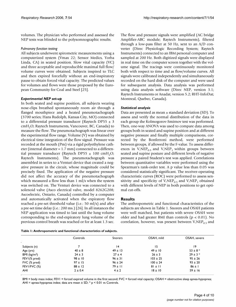
ResultsThe anthropometric and functional characteristics of thesubjects are shown in Table 1. Snorers and OSAH patientswere well matched, but patients with severe OSAH wereolder and had greater BMI than controls (p < 0.01). Nocorrelation, however, was present between V,NEP0.5 and
Table 1: Anthropometric and functional characteristics of subjects.
Controls Snorers OSAH, mild OSAH, severe
Subjects (n) 7 14 15 19Age (yrs) 40 ± 8 49 ± 13 49 ± 10 53 ± 8 *BMI (kg/m2) 24 ± 3 27 ± 4 26 ± 3 29 ± 3 *FEV1(% pred) 98 ± 10 96 ± 31 103 ± 25 95 ± 26FVC (% pred) 97 ± 15 96 ± 24 100 ± 24 93 ± 20FEV1/FVC (%) 88 ± 12 79 ± 11 81 ± 11 86 ± 1AHI 2 ± 0.4 4 ± 2 18 ± 10 59 ± 16
BMI = body mass index; FEV1 = forced expired volume in the first second; FVC = forced vital capacity; OSAH = obstructive sleep apnea-hypopnea; AHI = apnea-hypopnea index; data are mean ± SD; * p < 0.01 vs Controls.
Page 4 of 10(page number not for citation purposes)

Respiratory Research 2006, 7:54 http://respiratory-research.com/content/7/1/54
V,NEP1 and BMI in snorers and OSAH patients. None ofthe subjects had significant restrictive or obstructive venti-latory defect and exhibited tidal intrathoracic EFL in eitherposition during NEP application.
The values (mean ± SD) of V,NEP0.5 and V,NEP1, in bothpositions and NEP levels, are shown in Table 2. The indi-vidual V,NEP0.5 data in each group are shown in Fig. 2.Similar values of V,NEP0.5 and V,NEP1 were obtained insubjects with mild-to-moderate and with severe OSAH inall experimental conditions and were treated as a singlegroup for comparative analysis.
∆V,NEP0.5 and ∆V,NEP1 reflected exactly what was shownby V,NEP0.5 and V,NEP1with no additional advantage inorder to distinguish the different groups. Therefore, we
did not consider these time-consuming indices for subse-quent analysis.
Within each group V,NEP0.5 and V,NEP1were significantlyhigher with NEP -7 cmH2O than with NEP -5 cmH2O inboth positions, and with the same negative pressurehigher in the seated position than in the supine one (p <0.05 for controls, p < 0.01 for snorers and patients withOSAH).
The patients with OSAH consistently exhibited values ofV,NEP0.5 and V,NEP1 much lower than control subjects (p< 0.001), but had values of V,NEP0.5 significantly reducedas compared to snorers only with NEP -5 cmH2O in thesupine position (p < 0.01) (Fig. 2).
Individual values of V,NEP0.5 in seated and supine position at two NEP levels in patients with obstructive sleep apnea-hypopnea (OSAH) (circles, white for mild-to-moderate OSAH and black for severe OSAH), snorers (Sn) (triangles) and control (C) (squares)Figure 2Individual values of V,NEP 0.5 in seated and supine position at two NEP levels in patients with obstructive sleep apnea-hypop-nea (OSAH) (circles, white for mild-to-moderate OSAH and black for severe OSAH), snorers (Sn) (triangles) and controls (C) (squares). *p<0.01 vs C; **p<0.001 vs C; #p<0.05 vs Sn.
Page 5 of 10(page number not for citation purposes)
Respiratory Research 2006, 7:54 http://respiratory-research.com/content/7/1/54
The receiver operating characteristic (ROC) curves per-formed for V,NEP0.5 and V,NEP1 in both positions at thetwo different levels of NEP showed similar areas with thehighest value for V,NEP0.5 in the supine position usingNEP of -5 cmH2O (Fig. 3). Under these conditions, theoptimal cut-off V,NEP0.5 value of 393 ml had a sensitivityof 76% and a specificity of 74% to detect the presence ofOSAH with a likelihood ratio for positive results of 2.9.Accordingly, its positive and negative predictive value was84% and 64%, respectively.
No significant correlation between V,NEP0.5 (in thesupine position with NEP level of -5 cmH2O) and AHIwas observed in patients with OSAH (rs = -0.31, rs
2 = 0.10;95%IC = -0.59 – 0.04). However, taking into account onlythe patients with severe OSAH (AHI ≥ 30), a significantinverse correlation was found between V,NEP0.5 and AHI(p < 0.05; rs = -0.46, rs
2 = 0.21; 95%IC = -0.76 – -0.01) (Fig.4).
DiscussionThe present study indicates that during wakefulnessOSAH patients when compared to snorers and controlshave greater collapsibility of the upper airways which canbe easily assessed looking at the early expiratory flowdynamics after NEP application during tidal breathingand properly measured as V,NEP0.5 and V,NEP1. Suchmeasurements, however, are unable to distinguish on anindividual basis apneic from non-apneic snorers becauseof the overlapping of the V,NEP0.5 and V,NEP1 valuesbetween these groups of subjects. Nevertheless, our resultsprovide support to the idea that a high degree of the upperairway collapsibility promotes OSAH, even if OSAH mayseldom occur in subjects with normal upper airwaymechanics during wakefulness, suggesting the involve-ment of other pathogenetic factors.
Although our snorers had similar age, gender and BMIwithout obvious cranio-facial and ORL anomalies, lateralcephalometry or MRI studies of the pharynx were not per-formed, so we cannot exclude minor anatomic abnormal-ities in bony structure or soft tissue around the pharyngealairway in OSAH patients. Conversely, careful inspectionof the maximal and tidal expiratory flow-volume curvesallowed us to rule out the presence of intrathoracic expir-atory flow limitation during resting tidal breathing in allsubjects. Therefore, we are confident that our subjects hadno intrathoracic expiratory flow limitation which mighthave influenced the upper airway-related expiratory flowdynamics when NEP was applied.
The usefulness of the NEP method to assess the upper air-way collapsibility was previously tested in 16 awake sub-jects known to suffer from sleep-disordered breathing[27]. In contrast to snorers, all patients with OSAH (n = 8)
showed a substantial portion (>30%) of the expiratorytidal volume throughout the NEP application (-5 cmH2O,in the supine position) with lesser expiratory flow thanthe one recorded during the previous control tidal expira-tion.
Subsequently, in a group of 19 patients with OSAH whenNEP was applied (-5 and -10 cmH2O, in the supine posi-tion) the expiratory flow was reduced, when comparedwith the corresponding spontaneous expiratory flow, dur-ing a relevant part of the tidal expiration (>20%) in those(n = 13) with a higher mean apnea-hypopnea index (AHI)[28].
In these studies a significant correlation was foundbetween the percentage of the tidal volume during theNEP application with lower expiratory flow than duringthe spontaneous breathing and oxygen desaturation index(ODI), in the former, and ODI and AHI, in the latter[27,28]. Hence, the NEP method appeared suitable inorder to detect an increased pharyngeal collapsibility inpatients with OSAH during wakefulness and perhaps ableto predict the severity of OSAH.
Recently, using the same criteria, a large cohort of snoringsubjects was examined to assess the capacity of the NEPmethod to screen apneic from non-apneic subjects [29].In this study a sensitivity of 81.9% and specificity of69.1% in predicting OSAH was found when the expiratoryflow during NEP (-5 cmH2O in supine position) wasbelow that of the previous control expiration for ≥27.5%of the tidal volume. In addition, a significant correlationbetween NEP induced flow analysis and OSAH severity, asassessed by AHI, was found in the supine position using -5 cmH2O of NEP with a coefficient value (rs = 0.51) simi-lar to the one we obtained in the severe OSAH patients (rs= 0.46).
All of these studies, however, are based on the assumptionthat abnormal upper airway collapsibility is present or canbe identified only when the expiratory flow during NEPbecomes lower than the control one. Moreover, such find-ing has been erroneously taken as a marker of expiratoryflow limitation. In contrast, an increased pharyngeal col-lapsibility can also be reflected by a smaller increase ofexpiratory flow during NEP. We believe that this flow hasto be measured whether or not it is higher, lower or ini-tially higher and then lower (or vice versa) than the flowof the previous tidal expiration. Indeed, judging as abnor-mal (or quantifying the severity of) the upper airway col-lapsibility only by computing the percentage of the tidalvolume where the expiratory flow during NEP applicationbecomes lesser than the one exhibited in the previousexpiration is a poorly reliable tool. This is because suchmeasure is too dependent from the preceding control tidal
Page 6 of 10(page number not for citation purposes)

Respiratory Research 2006, 7:54 http://respiratory-research.com/content/7/1/54
breathing and because the expiratory flow profile is oftenerratic in the same subject during repeated NEP tests; in
addition, several apneic patients do not show such phe-nomenon constantly [27-29].
Table 2: Values of V,NEP0.5 and V,NEP1 at two different NEP levels in seated and supine position.
SEATED SUPINENEP -5 cmH2O -7 cmH2O -5 cmH2O -7 cmH2O
V,NEP0.5
Controls 559 ± 98 655 ± 113 492 ± 69 571 ± 96Snorers 457 ± 150 520 ± 147 427 ± 101 430 ± 134OSAH 363 ± 123* 419 ± 132** 340 ± 88** # 379 ± 113**
OSAH, m 402 ± 129 428 ± 157 353 ± 103 387 ± 108OSAH, s 332 ± 112 410 ± 112 329 ± 73 372 ± 119
V,NEP1
Controls 1036 ± 134 1128 ± 158 832 ± 95 920 ± 136Snorers 864 ± 246 930 ± 274 737 ± 194 755 ± 224OSAH 708 ± 189* 781 ± 210* 628 ± 136* 669 ± 177*
OSAH, m 732 ± 202 766 ± 230 625 ± 159 666 ± 178OSAH, s 688 ± 182 792 ± 199 629 ± 120 671 ± 180
OSAH = obstructive sleep apnea-hypopnea; m = mild; s = severe; data are mean ± SD; * p < 0.01 vs Controls; ** p < 0.001 vs Controls; # p < 0.01 vs Snorers
The receiver-operating characteristic (ROC) curves are shown for V,NEP0.5 values during NEP (-5 cmH2O) both in supine (continuous line) and seated (dashed line) position in 34 patients with OSAH and 21 subjects without OSAHFigure 3The receiver-operating characteristics (ROC) curves are shown for V,NEP0.5 values during NEP (-5 cmH2O) both in supine (continuous line) and seated (dashed line) position in 34 patients with OSAH and 21 subjects without OSAH. The area under the ROC curves reflects the ability of V,NEP0.5 to distinguish subjects without and with OSAH (AHI>5).
Page 7 of 10(page number not for citation purposes)

Respiratory Research 2006, 7:54 http://respiratory-research.com/content/7/1/54
In order to overcome these problems, recently Tamisierand coll. investigated a quantitative index correspondingto the ratio of the area under the expiratory flow/volumecurves between NEP (-5 and -10 cmH2O) and atmos-pheric pressure for the same tidal volume in awake sub-jects with sleep disordered breathing (SDB) and controlsubjects, both in supine and sitting position [30]. Theyfound that this index was significantly different betweencontrols and SDB subjects in all measurements, decreas-ing with the severity of the SDB. Moreover, in the supineposition when -5 cmH2O NEP was applied, a giventhreshold of this index had a positive predictive value of88.6% and a negative predictive value of 80% to screensubjects with SDB. The Authors concluded that the NEP-related quantitative index may be useful to detect abnor-mal upper airway collapsibility in awake subjects withSDB. However, some limits of this study are obvious suchas the lack of subjects with mild OSAH (AHI <15 and >5)and the age of the controls who were much younger (34 ±12 yrs) than the patients with OSAH. Furthermore, theapplication of NEP near end expiratory lung volume tendsto elicit reflex activation of genioglossus [22]. This canunpredictably influence the area under the final part ofthe expiratory flow/volume curve during NEP both incontrols and SDB subjects, affecting the quantitative indexused to assess the upper airway collapsibility.
In a very recent paper Insalaco and coworkers used thedrop of expiratory flow under NEP (∆V-NEP), expressedas percentage change of peak expiratory flow under NEP,as index of upper airway collapsibility to detect OSAH inpatients with sleep disordered breathing. Although this
index was a better indicator of OSAH severity when com-pared to the previous ones, they reported, at best, a deter-mination coefficient equal to 0.32 between the AHI and∆V-NEP [31], using NEP of -10 cmH2O in the supine posi-tion. An inherent problem with this approach is that ∆V-NEP does not take into account the duration of the expir-atory flow drop under the NEP application, while it is veryclear from the flow-time tracings given by the sameAuthors that this transient may last very differently withthe same percentage value of reduction.
By time-integration of the expiratory flow in the first 0.5and 1 sec after the application of a given level of NEP onecan easily calculate the expiratory volume exhaled in apreset time in a given body position during wakefulnessand use this parameter as an index of the mechanicalproperties of the upper airways at the onset of expirationwhen the genioglossus does not appear reflexively acti-vated [22]. Therefore, the novelty of this study is the utili-zation of a method which, still adopting the NEPtechnique, is more reliable to assess and measure theupper airway collapsibility because it is quantitative, andit is not influenced by the flow of the preceding tidal expi-ration and by the effect of neuromuscular factors.
V,NEP0.5 and V,NEP1 values were reduced in the supineposition at each level of applied NEP in all groups, likelyto reflect a posture-related increase in the upper airwayresistance [18,23,32,33]. Therefore, V,NEP0.5 and V,NEP1measurements appear to be influenced by the baselineexpiratory upper airway resistance which has been shownto be higher in OSAH patients, probably because of min-imal structural abnormalities (abnormal hyoid bone posi-tion and increase in soft pharyngeal tissues) [20] andrelated shape changes. It is conceivable that lowerV,NEP0.5 and V,NEP1 found in our OSAH patients may bepartly due to reduced baseline upper airway caliber which,on the other hand, is expected to increase the pharyngealcompliance and finally the upper airway collapsibility inthese subjects [21]. However, the expired volume in thefirst 0.5 or 1 sec was lower during NEP than during theprevious control expiration in either position in about15–18% of our apneic snorers. This never occurred insnorers and controls. This fact strongly suggests that inOSAH patients a brisk narrowing of upper airways is elic-ited by the sudden NEP application, the magnitude ofwhich is largely depending on the pharyngeal collapsibil-ity under the prevailing circumstances and substantiallyreflected by the V,NEP0.5 or V,NEP1 values. In line withthis reasoning, the early expiratory flow during NEP wasoften below the isovolume spontaneous expiratory flow,particularly in OSAH patients (see Fig. 1), as also shownin previous studies [22,27,28].
Relationship between AHI and V,NEP0.5 in the supine posi-tion during NEP (-5 cmH2O) in OSAH patients (white circles = mild-to-moderate OSAH; black circles = severe OSAH)Figure 4Relationship between AHI and V,NEP0.5 in the supine posi-tion during NEP (-5 cmH2O) in OSAH patients (white circles = mild-to-moderate OSAH; black circles = severe OSAH). The regression line refers only to severe OSAH patients.
Page 8 of 10(page number not for citation purposes)

Respiratory Research 2006, 7:54 http://respiratory-research.com/content/7/1/54
Under these experimental conditions, V,NEP0.5 was signif-icantly lower in apneic than in non-apneic snorers whenmeasured in the supine position utilizing the smallestlevel of NEP (i.e.: -5 cmH2O in our study). Thus, the lowerthe value of V,NEP0.5 (or V,NEP1), the higher the possibil-ity for snoring people to have OSAH. Indeed, according tothis method an increased pharyngeal collapsibility evenduring wakefulness affects the vast majority of snorerswho have OSAH. This information is obtained in a rapid,simple and non-invasive way without cooperation of thesubjects who can be studied when awake, repeatedly andin different body position. In this respect it has to bestressed that it is not necessary to control with regards tobaseline spontaneous tidal volumes and flows. Indeed,∆V,NEP0.5 and ∆V,NEP1 did not perform differently or bet-ter to distinguish between snorers and OSAH patientsthan V,NEP0.5 and V,NEP1.
Unfortunately, the ability to differentiate snorers with orwithout OSAH was not sufficient, at least within our capa-bilities, to recommend this technique and related param-eters as a reliable diagnostic tool to obviate sleep studiesor even to select subjects for polysomnographic evalua-tion. Lower levels of NEP (i.e.: -2 or -3 cmH2O), however,might be more useful for this purpose and deserve to betested in the future.
Three further comments need to be made. Firstly, gener-ally a high collapsibility of the upper airways does notseem sufficient to cause OSAH since several snorers with-out OSAH exhibited similarly reduced values of V,NEP0.5or V,NEP1. Secondly, other factors must influence theseverity of OSAH, as assessed by ODI and AHI, because nodifferent values of V,NEP0.5 (or V,NEP1) were found in anyposition or with different levels of NEP between mild-to-moderate and severe OSAH patients. Thirdly, some OSAHpatients have surprisingly high values of V,NEP0.5 (orV,NEP1) comparable to those of the controls, showing anormal upper airway collapsibility during wakefulness,and thus suggesting different state-related factors leadingto OSAH or a site of upper airway obstruction during sleeponly at naso-pharyngeal level which cannot be directlyassessed with this technique.
Finally, contrary to the opinion of the other Authors whoused the NEP technique to detect OSAH patients duringwakefulness [27-30], we have to stress that, although theresults obtained with V,NEP0.5 were similar or even betterthan the previous ones [27-30], whatever NEP-relatedparameter is adopted, presently this tool is not sufficientlycapable of revealing OSAH on an individual basis for clin-ical purpose.
In conclusion, the NEP technique when properly used ispotentially useful to study upper airway collapsibility in
patients with OSAH during wakefulness in order to betterunderstand its main mechanisms, to assess in the longterm the effects of various interventions, and possibly forselecting non-apneic snorers to follow up. On the otherhand, it cannot be recommended for routine OSAHscreening in awake snorers who should subsequently besubjected to sleep studies.
AbbreviationsOSAH = obstructive sleep apnea-hypopnea; AHI = apnea-hypopnea index; NEP = negative expiratory pressure; SDB= sleep disordered breathing; BMI = body mass index;V,NEP0.5 and V,NEP1 = exhaled volume in the first 0.5 and1 sec. after application of NEP; ROC = receiver operatingcharacteristic (curve)
References1. Isono S, Saeki N, Tanaka A, Nishino T: Collapsibility of passive
pharynx in patients with acromegaly. Am J Respir Crit Care Med1999, 160:64-68.
2. Isono S, Tanaka A, Sho Y, Konno A, Nishino T: Advancement ofthe mandible improves velopharyngeal airway patency. J ApplPhysiol 1995, 79:2132-2138.
3. Isono S, Remmers JE, Tanaka A, Sho Y, Sato J, Nishino T: Anatomyof pharynx in patients with obstructive sleep apnea and innormal subjects. J Appl Physiol 1997, 82:1319-1326.
4. Isono S, Shimada A, Utsugi M, Konno A, Nishino T: Comparison ofstatic mechanical properties of the passive pharynx betweennormal children and children with sleep-disordered breath-ing. Am J Respir Crit Care Med 1998, 157:1204-1212.
5. Liistro G, Stanescu D, Dooms G, Veriter C, Aubert-Tulkens G,Rodenstein D: Hypopharyngeal and neck cross-sectionalchanges monitored by inductive plethysmography. J ApplPhysiol 1990, 68:2649-2655.
6. Brown IG, Bradley TD, Phillipson EA, Zamel N, Hoffstein V: Pharyn-geal compliance in snoring subjects with and withoutobstructive sleep apnea. Am Rev Respir Dis 1985, 132:211-215.
7. Schwab RJ, Gefter WB, Hoffman EA, Gupta KB, Pack AI: Dynamicupper airway imaging during awake respiration in normalsubjects and patients with sleep disordered breathing. AmRev Respir Dis 1993, 74:1385-1400.
8. Schwab RJ, Gefter WB, Pack AI, Hoffman EA: Dynamic imaging ofthe upper airway during respiration in normal subjects. J ApplPhysiol 1993, 74:1504-1514.
9. Gleadhill IC, Schwartz AR, Schubert N, Wise RA, Permutt S, SmithPL: Upper airway collapsibility in snorers and in patients withobstructive hyperpnea and apnea. Am Rev Respir Dis 1992,143:1300-1303.
10. Smith PL, Wise RA, Gold AR, Schwartz AR, Permutt S: Upper air-way pressure-flow relationships in obstructive sleep apnea. JAppl Physiol 1988, 64:789-795.
11. Schwartz AR, Smith PL, Wise RA, Gold AR, Permutt S: Induction ofupper airway occlusion in sleeping individuals with subat-mospheric nasal pressure. J Appl Physiol 1988, 64:535-542.
12. Schwab RJ, Gupta KB, Gefter WB, Metzeger LJ, Hoffman EA, Pack AI:Upper airway and soft tissue anatomy in normal subjects andpatients with sleep disordered breathing. Significance of lat-eral pharyngeal walls. Am J Respir Crit Care Med 1995,152:1673-1689.
13. Watanabe T, Isono S, Tanaka A, Tanzawa H, Nishino T: Contribu-tion of body habitus and craniofacial characteristics to seg-mental closing pressures of the passive pharynx in patientswith sleep-disordered breathing. Am J Respir Crit Care Med 2002,165:260-265.
14. Haponik EF, Smith PL, Bohlman ME, Allen RP, Goldman SM, BleeckerER: Computerized tomography in obstructive sleep apnea.Correlation of airway size with physiology during sleep andwakefulness. Am Rev Respir Dis 1983, 127:221-226.
Page 9 of 10(page number not for citation purposes)
Respiratory Research 2006, 7:54 http://respiratory-research.com/content/7/1/54
Publish with BioMed Central and every scientist can read your work free of charge
"BioMed Central will be the most significant development for disseminating the results of biomedical research in our lifetime."
Sir Paul Nurse, Cancer Research UK
Your research papers will be:
available free of charge to the entire biomedical community
peer reviewed and published immediately upon acceptance
cited in PubMed and archived on PubMed Central
yours — you keep the copyright
Submit your manuscript here:http://www.biomedcentral.com/info/publishing_adv.asp
BioMedcentral
15. Rivlin J, Hoffstein V, Kalbfleisch J, McNicholas W, Zamel N, Bryan AC:Upper airway morphology in patients with idiopathicobstructive sleep apnea. Am Rev Respir Dis 1984, 129:355-360.
16. Anch AM, Remmers JE, Bunce H 3rd: Supraglottic airway resist-ance in normal subjects and patients with occlusive sleepapnea. J Appl Physiol 1982, 53:1158-1163.
17. Stauffer JL, Zwillich CW, Cadieux RJ, Bixler EO, Kales A, Varano LA,White DP: Pharyngeal size and resistance in obstructive sleepapnea. Am Rev Respir Dis 1987, 136:623-627.
18. Martin SE, Marshall I, Douglas NJ: The effect of posture on airwaycaliber with the sleep-apnea/hypopnea syndrome. Am J RespirCrit Care Med 1995, 152:721-724.
19. Rodenstein DO, Doom G, Thomas Y, Liistro G, Stanescu DC, CuleeC, Aubert-Tulkens G: Pharyngeal shape and dimension inhealthy subjects, snorers, and patients with obstructive sleepapnea. Thorax 1990, 45:722-727.
20. Verin E, Tardif C, Buffet X, Marie JP, Lacoume Y, Andrieu-Guitran-court J, Pasquis P: Comparison between anatomy and resist-ance of upper airway in normal subjects, snorers and OSASpatients. Respir Physiol 2002, 129:335-343.
21. Sforza E, Bacon W, Weiss T, Thibault A, Petiau C, Krieger J: Upperairway collapsibility and cephalometric variables in patientswith obstructive sleep apnea. Am J Respir Crit Care Med 2000,161:347-352.
22. Tantucci C, Mehiri S, Duguet A, Similowski T, Arnulf I, Zelter M, Der-enne JP, Milic-Emili J: Application of negative expiratory pres-sure during expiration and activity of genioglossus inhumans. J Appl Physiol 1998, 84:1076-1082.
23. Tantucci C, Duguet A, Ferretti A, Mehiri S, Arnulf I, Zelter M, Simi-lowski T, Derenne JP, Milic-Emili J: Effect of negative expiratorypressure on respiratory system flow resistance in awakesnorers and nonsnorers. J Appl Physiol 1999, 87:969-976.
24. American Academy of Sleep Medicine Task Force: Sleep-relatedbreathing disorders in adults: recommendation for syn-drome definition and measurement techniques in clinicalresearch. Sleep 1999, 22:667-689.
25. Quanjer PT, Tammeling GJ, Cotes JE, Fabbri LM, Matthys H, PedersonOF, Peslin R, Roca J, Sterk PG, Ulmer WT: Lung volume andforced ventilatory flows. Report Working Party Standardisa-tion of Lung Function Tests, European Community for Steeland Coal. Official Statement of the European RespiratorySociety. Eur Resp J Suppl 1993, 16:5-40.
26. Koulouris NG, Valta P, Lavoie A, Corbeil C, Chasse M, Braidy J, Milic-Emili J: A simple method to detect expiratory flow limitationduring spontaneous breathing. Eur Respir J 1995, 8:306-313.
27. Liistro G, Veriter C, Dury M, Aubert G, Stanescu D: Expiratoryflow limitation in awake sleep-disordered breathing subjects.Eur Respir J 1999, 14:185-190.
28. Verin E, Tardif C, Portier F, Similowski T, Pasquis P, Muir JF: Evi-dence for expiratory flow limitation of extrathoracic originin patients with obstructive sleep apnea. Thorax 2002,57:423-428.
29. Van Meerhaeghe A, Delpire P, Stenuit P, Kerkhofs M: Operatingcharacteristic of the negative expiratory pressure techniquein predicting obstructive sleep apnea syndrome in snoringpatients. Thorax 2004, 59:883-888.
30. Tamisier R, Wuyam B, Nicolle I, Pepin JL, Orliaguet O, Perrin CP,Levy P: Awake flow limitation with negative expiratory pres-sure in sleep disordered breathing. Sleep Medicine 2005,6:205-213.
31. Insalaco G, Romano S, Marrone O, Salvaggio A, Bonsignore G: A newmethod of negative expiratory pressure test analysis detect-ing upper airway flow limitation to reveal obstructive sleepapnea. Chest 2005, 128:2159-2165.
32. Jan MA, Marshall I, Douglas NJ: Effect of posture on upper airwaydimensions in normal human. Am J Respir Crit Care Med 1994,149:145-148.
33. Yildirim N, Fitzpatrick MF, Whyte KF, Jalleh R, Wightman AJ, DouglasNJ: The effect of posture on upper airway dimensions in nor-mal subjects and in patients with the sleeapnea/hyperpneasyndrome. Am Rev Respir Dis 1991, 144:845-847.
Page 10 of 10(page number not for citation purposes)