respiration - wikispaces · pdf file4 respiration • definition – release of energy...
TRANSCRIPT
1
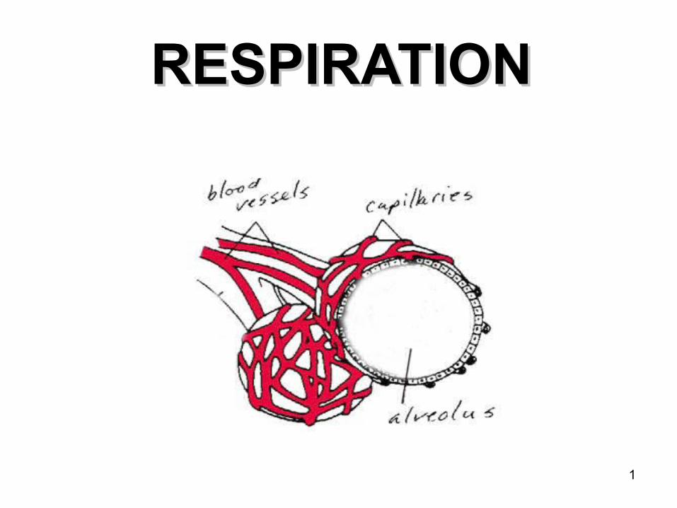
RESPIRATIONRESPIRATION
2
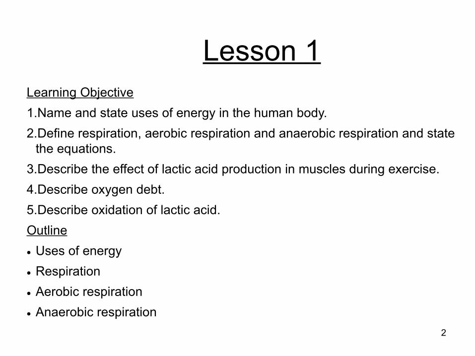
Lesson 1Learning Objective1.Name and state uses of energy in the human body.2.Define respiration, aerobic respiration and anaerobic respiration and state
the equations.3.Describe the effect of lactic acid production in muscles during exercise.4.Describe oxygen debt.5.Describe oxidation of lactic acid.Outline Uses of energy Respiration Aerobic respiration Anaerobic respiration

3
Why do humans respire?
• To produce energy• There are many uses of energy.
– All processes of 'staying alive'.– Muscle contraction– Protein synthesis– Cell division– Active transport– Growth – Reproduction – Transmission of nervous impulses– Maintenance of constant body temperature
4
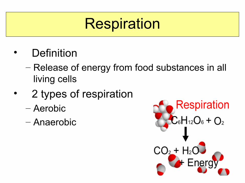
Respiration
• Definition– Release of energy from food substances in all
living cells• 2 types of respiration
– Aerobic– Anaerobic
5
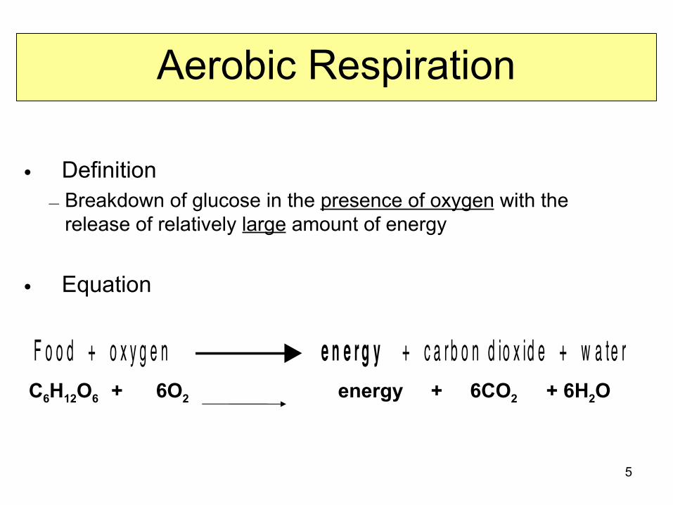
Aerobic Respiration
• Definition– Breakdown of glucose in the presence of oxygen with the
release of relatively large amount of energy
• Equation
C6H12O6 + 6O2 energy + 6CO2 + 6H2O
F o o d + o x y g e n e n e r g y + c a r b o n d io x id e + w a te r
6
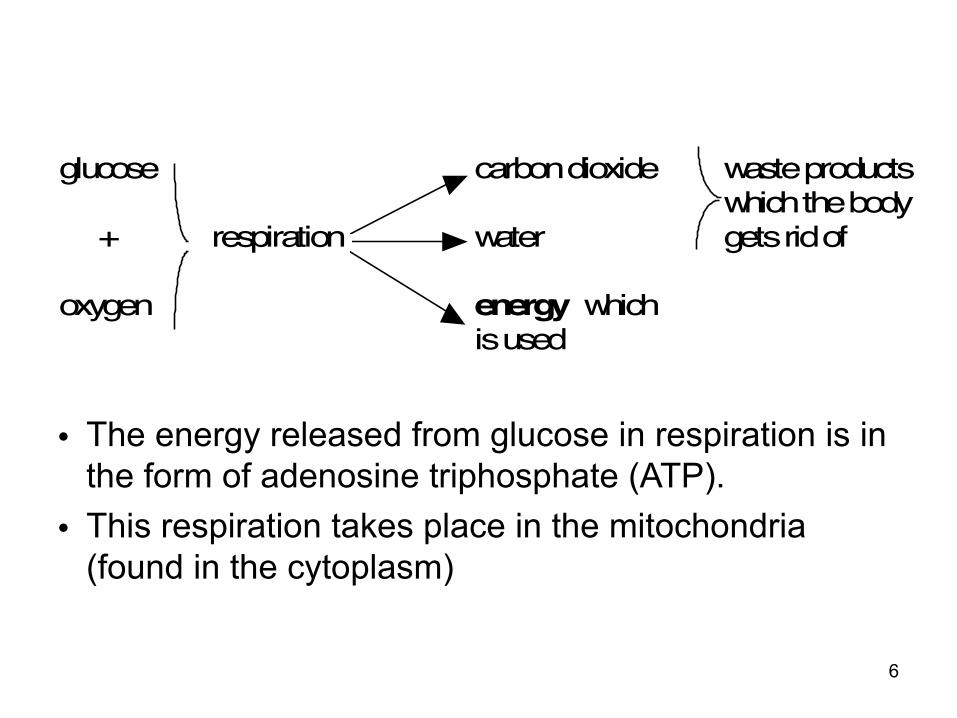
• The energy released from glucose in respiration is in the form of adenosine triphosphate (ATP).
• This respiration takes place in the mitochondria (found in the cytoplasm)
glucose carbon dioxide waste productswhich the body
+ respiration water gets rid of
oxygen energy whichis used
7
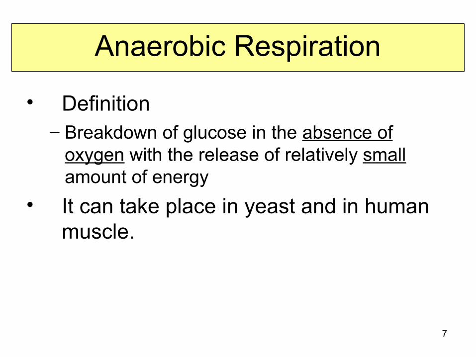
Anaerobic Respiration
• Definition– Breakdown of glucose in the absence of
oxygen with the release of relatively small amount of energy
• It can take place in yeast and in human muscle.
8
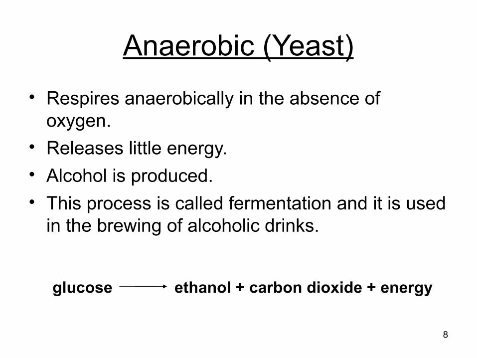
Anaerobic (Yeast)• Respires anaerobically in the absence of
oxygen.• Releases little energy.• Alcohol is produced.• This process is called fermentation and it is used
in the brewing of alcoholic drinks.
glucose ethanol + carbon dioxide + energy
9
Anaerobic (Muscle)
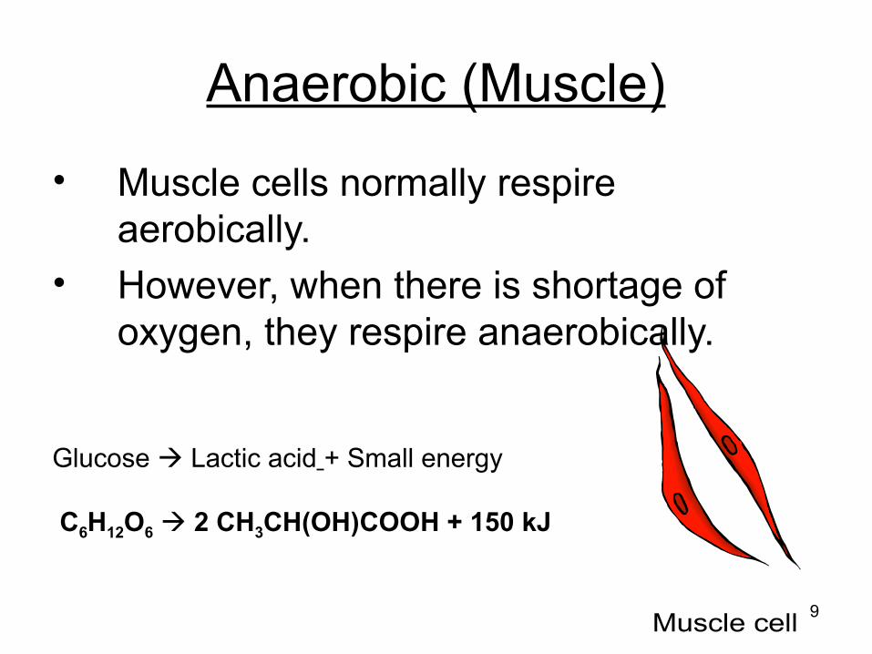
• Muscle cells normally respire aerobically.
• However, when there is shortage of oxygen, they respire anaerobically.
Glucose Lactic acid + Small energy
C6H12O6 2 CH3CH(OH)COOH + 150 kJ

10
Respiration during exercise
• Exercising requires energy.• When we do vigorous exercise, our
muscles need more energy than can be supplied by aerobic respiration.
• The extra energy needed comes from anaerobic respiration.
• This produces lactic acid.Glucose Lactic acid + Small energy
C6H12O6 2 CH3CH(OH)COOH + 150 kJ
11
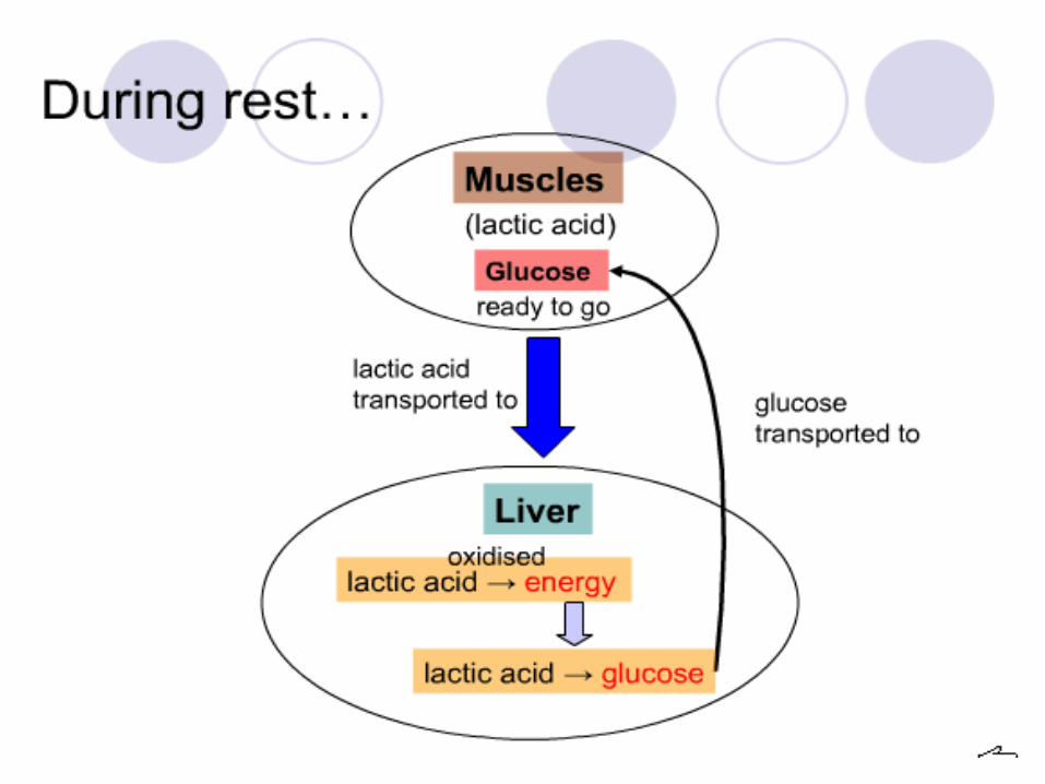
Lactic Acid• Lactic acid is produced when anaerobic respiration
occurs in human muscles.• Accumulation of lactic acid causes fatigue and muscular
pain. It forces the body to rest and recover.• During anaerobic respiration, human muscles lack
oxygen and is said to have an 'oxygen debt'.• This 'oxygen debt' need to be 'repaid' once oxygen is
available again. The oxygen is then used to remove lactic acid by oxidising it.
12
13
Exercise
1.Create a table to show the differences between aerobic and anaerobic respiration
2.Describe how lactic acid is removed to produce glucose.
14
Lesson 2Learning Objective1.Identify on diagrams and name the larynx, trachea, bronchi,
bronchioles, alveoli and associated capillaries.2.Describe the role of cilia and goblet cells.3.State the characteristics of alveoli and describe the role of exchange
surface of alveoli in gas exchange.
Outline How do we know organisms respire? The gaseous exchange system
– Air pathways
– Structures and functions
15
How do we know if organisms respire?
16
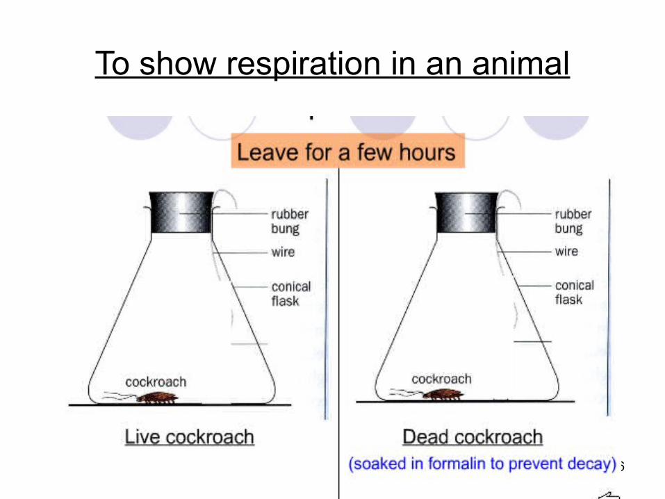
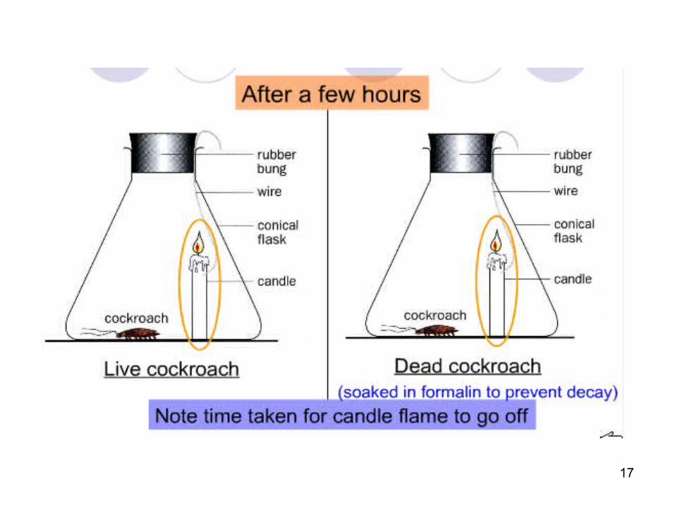
To show respiration in an animal
17
18
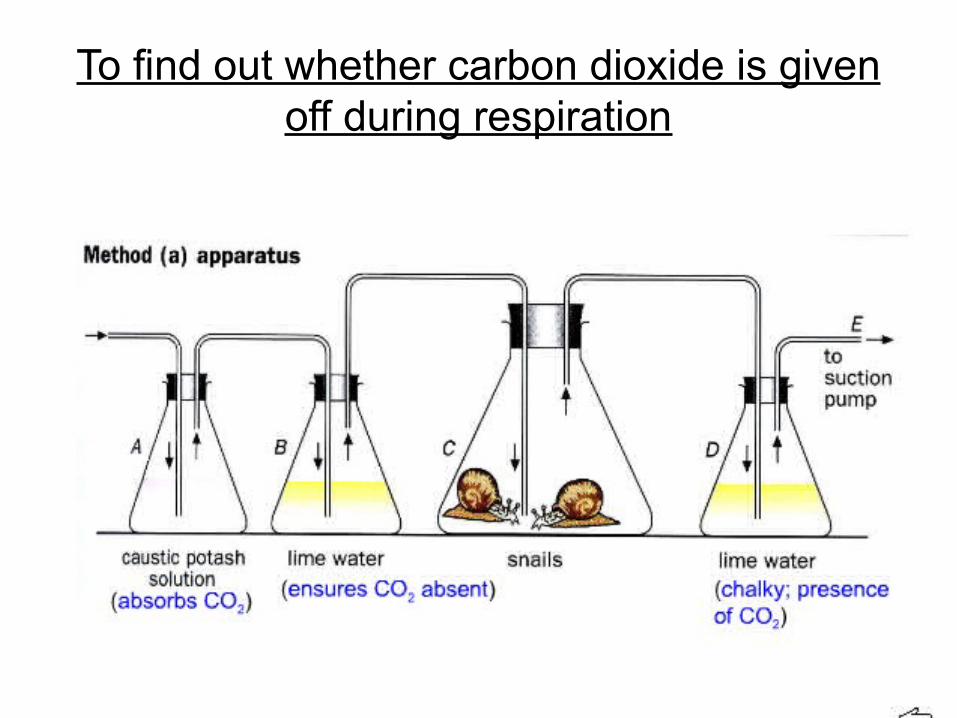
To find out whether carbon dioxide is given off during respiration

19
To find out whether carbon dioxide is given off during respiration
20
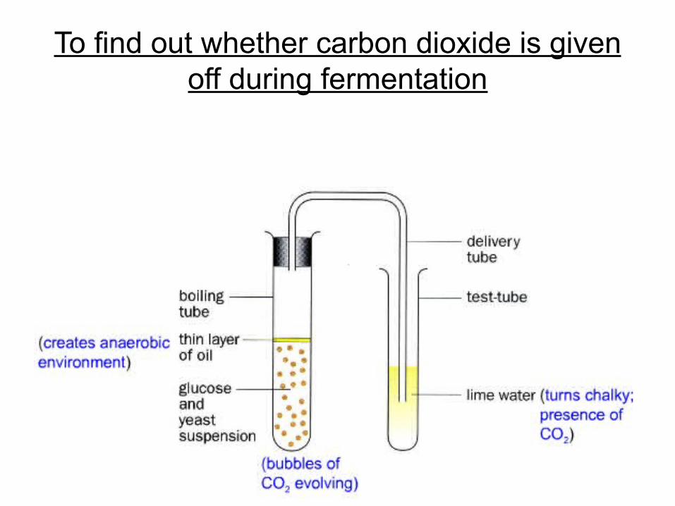
To find out whether carbon dioxide is given off during fermentation
21
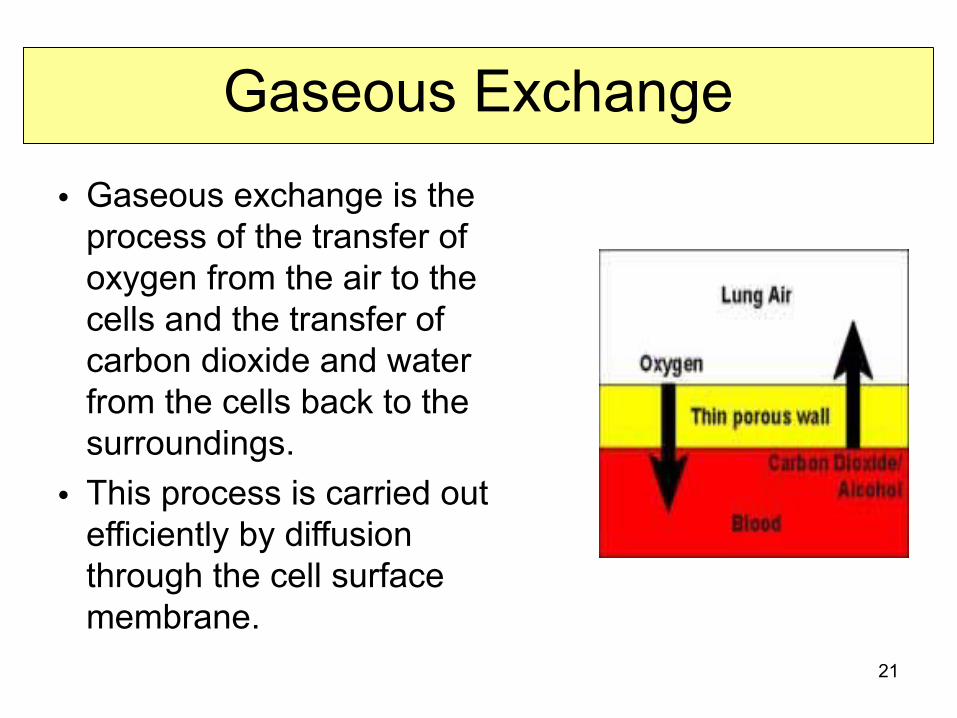
Gaseous Exchange• Gaseous exchange is the
process of the transfer of oxygen from the air to the cells and the transfer of carbon dioxide and water from the cells back to the surroundings.
• This process is carried out efficiently by diffusion through the cell surface membrane.
22
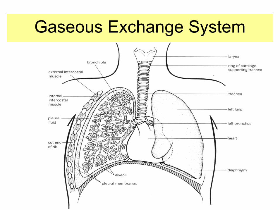
Gaseous Exchange System

23
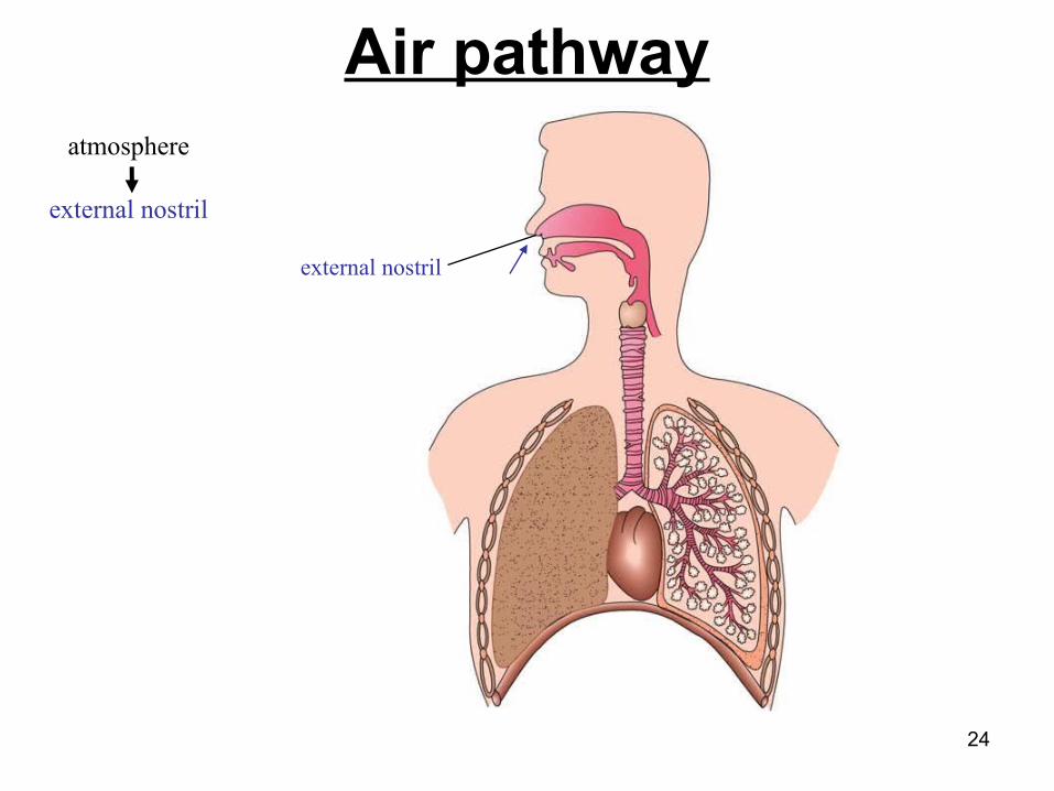
atmosphere
Air pathway
24
atmosphere
external nostril
external nostril
Air pathway
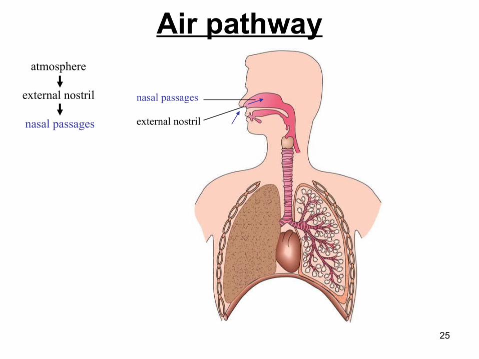
25
atmosphere
external nostril
nasal passages external nostril
nasal passages
Air pathway
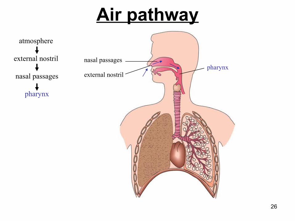
26
atmosphere
external nostril
nasal passages
pharynx
external nostrilpharynx
nasal passages
Air pathway
27
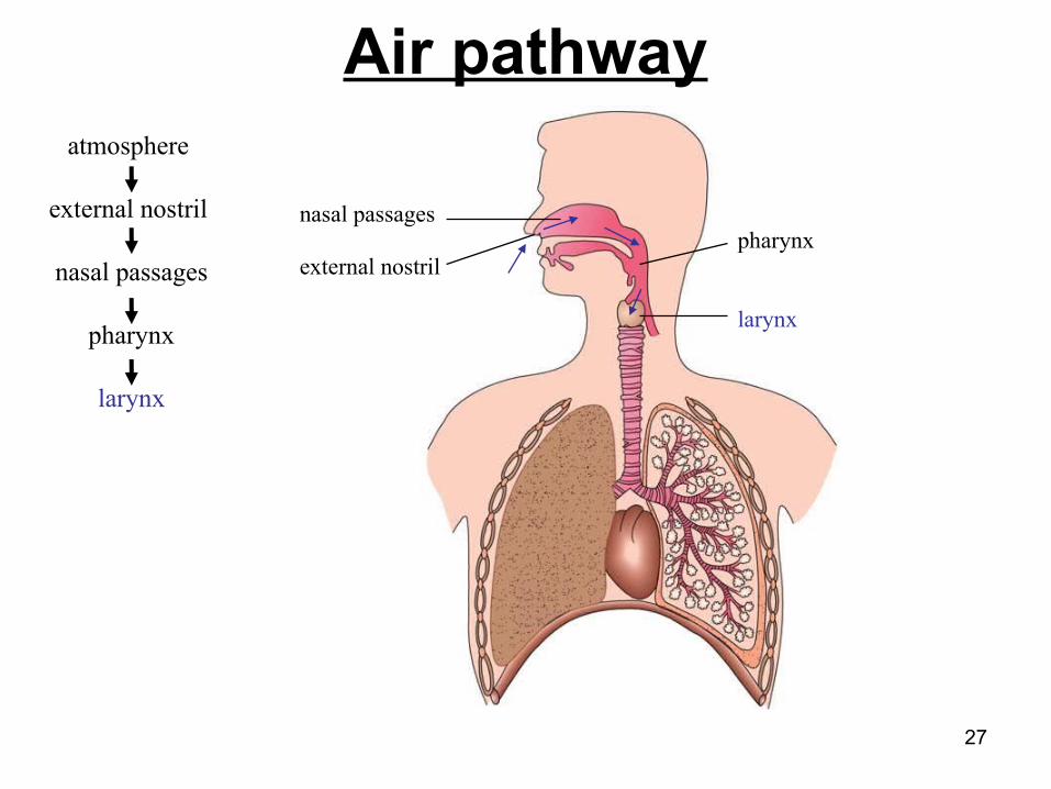
atmosphere
external nostril
nasal passages
pharynx
larynx
external nostril
larynx
pharynxnasal passages
Air pathway
28
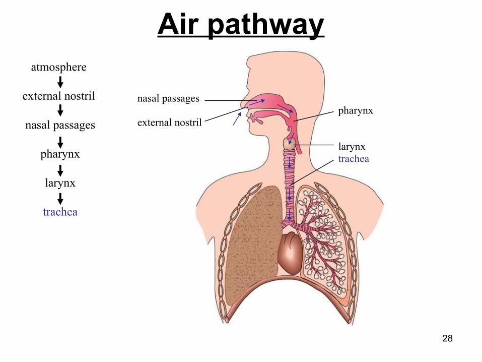
atmosphere
external nostril
nasal passages
pharynx
larynx
trachea
external nostril
trachealarynx
pharynxnasal passages
Air pathway

29
atmosphere
external nostril
nasal passages
pharynx
larynx
trachea
bronchi
external nostril
trachealarynx
pharynx
bronchi
nasal passages
Air pathway
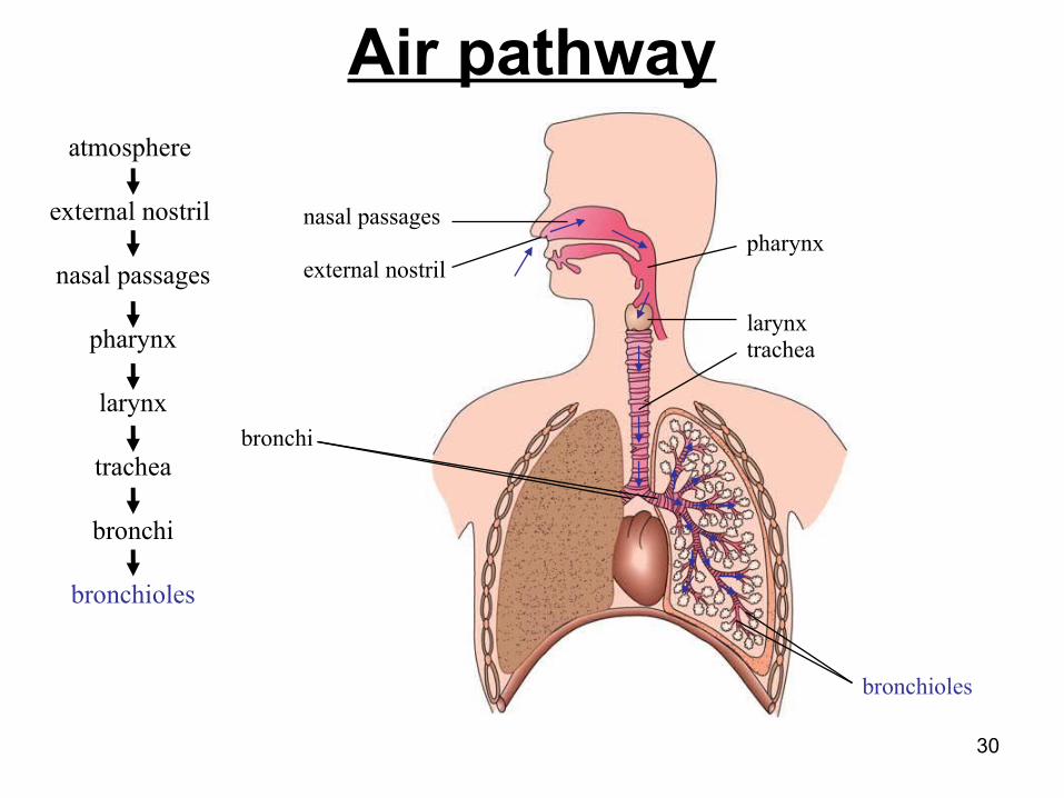
30
atmosphere
external nostril
nasal passages
pharynx
larynx
trachea
bronchi
bronchioles
external nostril
trachealarynx
pharynx
bronchi
bronchioles
nasal passages
Air pathway

31
atmosphere
external nostril
nasal passages
pharynx
larynx
trachea
bronchi
bronchioles
alveoli
external nostril
trachealarynx
pharynx
bronchi
bronchioles
cluster of alveoli (air sacs)
nasal passages
Air pathway
32
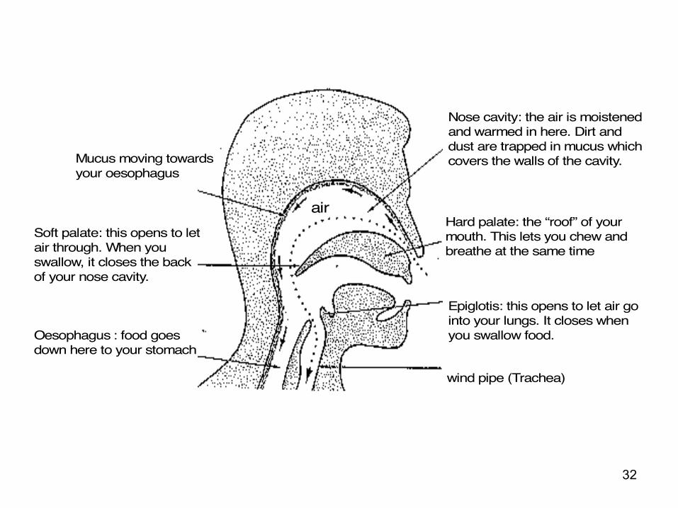
Nose cavity: the air is moistened and warmed in here. Dirt and dust are trapped in mucus which covers the walls of the cavity.
Hard palate: the “roof” of your mouth. This lets you chew and breathe at the same time
Epiglotis: this opens to let air go into your lungs. It closes when you swallow food.
wind pipe (Trachea)
air
Mucus moving towards your oesophagus
Soft palate: this opens to let air through. When you swallow, it closes the back of your nose cavity.
Oesophagus : food goes down here to your stomach

33
The Nose
• Air enters through two external nostrils (nares)
• Nasal passages are lined with hairs and moist mucous membrane.
• Advantages of breathing through nose:– Hairs trap dust and foreign particles, including bacteria– Air is warmed and moistened– Sensory cells in mucous membrane may detect
harmful chemical and produces mucus.
34
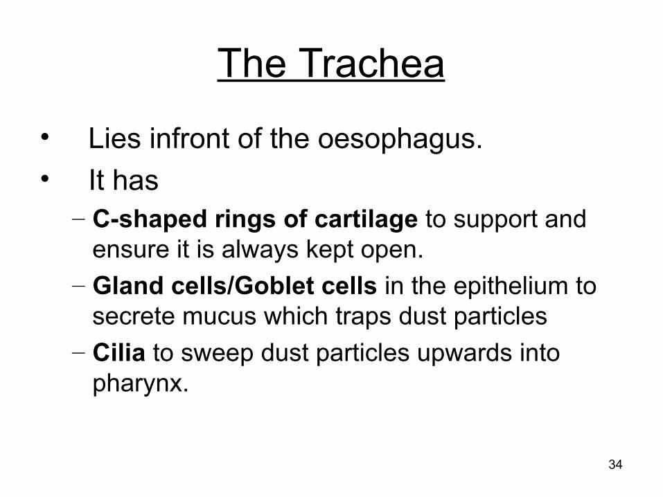
The Trachea
• Lies infront of the oesophagus.• It has
– C-shaped rings of cartilage to support and ensure it is always kept open.
– Gland cells/Goblet cells in the epithelium to secrete mucus which traps dust particles
– Cilia to sweep dust particles upwards into pharynx.

35
Gland cell
Nucleus
36
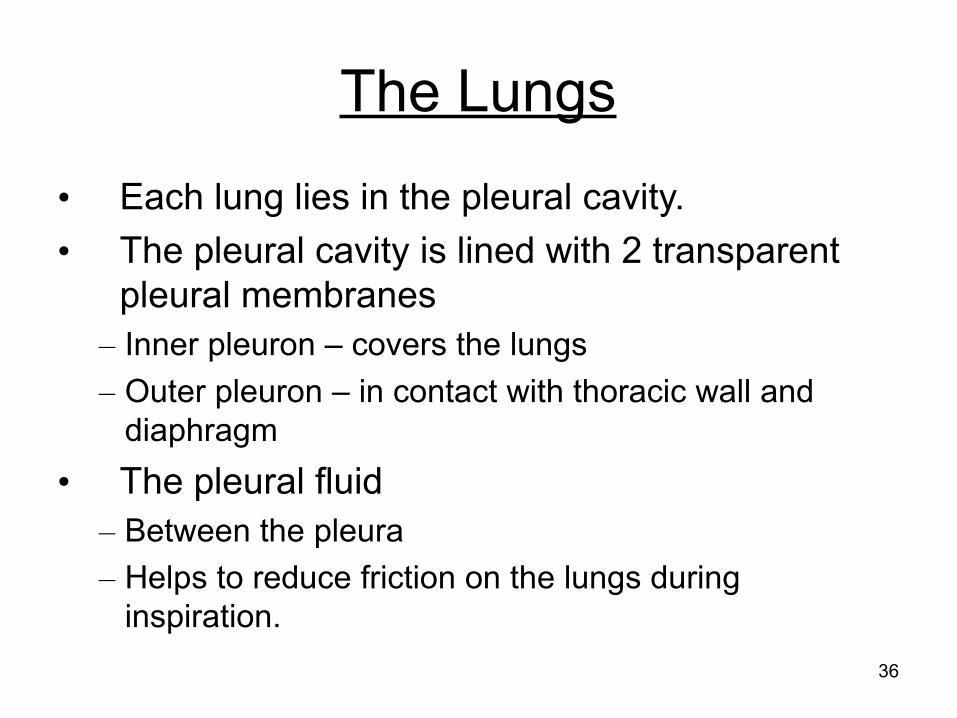
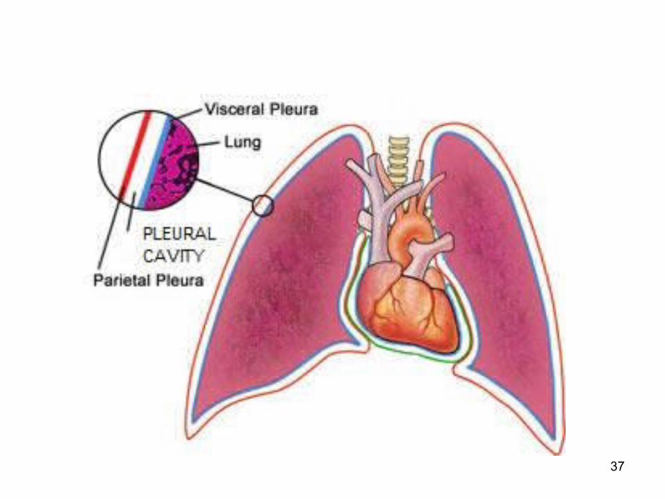
The Lungs
• Each lung lies in the pleural cavity.• The pleural cavity is lined with 2 transparent
pleural membranes– Inner pleuron – covers the lungs– Outer pleuron – in contact with thoracic wall and
diaphragm• The pleural fluid
– Between the pleura– Helps to reduce friction on the lungs during
inspiration.
37
38
Exercise
Explain how the nose and trachea help in preventing the entry of dust into the lungs. (4)

39
Lesson 3Learning Objective1.State the characteristics of alveoli and describe the
role of exchange surface of alveoli in gas exchange.
Outline The alveoli
– Characteristics– Gas exchange

40
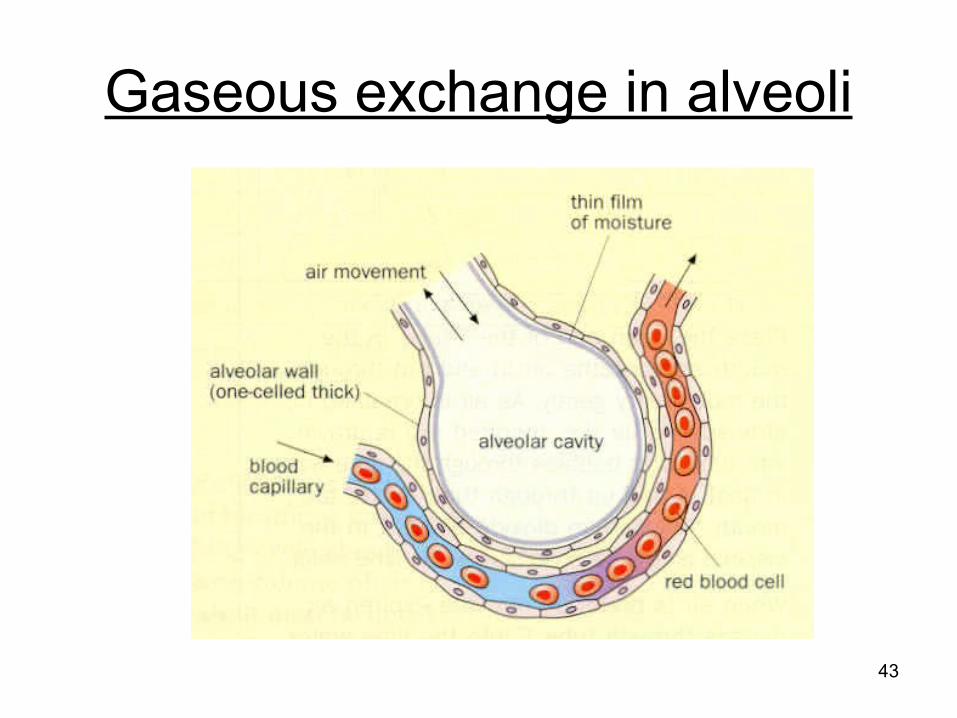
The Alveoli (singular: alveolus)
• The lower end of the trachea divides into 2 bronchi (singular: bronchus), one to each lung.
• Within the lungs, the bronchial tubes divide into smaller tubes – the bronchioles (*NO cartilage).
• Each bronchiole ends with many air sacs called alveoli.
41
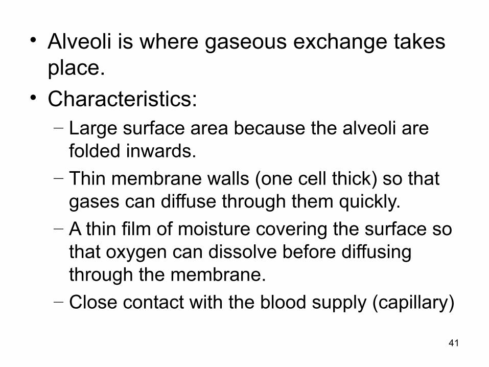
• Alveoli is where gaseous exchange takes place.
• Characteristics:– Large surface area because the alveoli are
folded inwards.– Thin membrane walls (one cell thick) so that
gases can diffuse through them quickly.– A thin film of moisture covering the surface so
that oxygen can dissolve before diffusing through the membrane.
– Close contact with the blood supply (capillary)
42
43
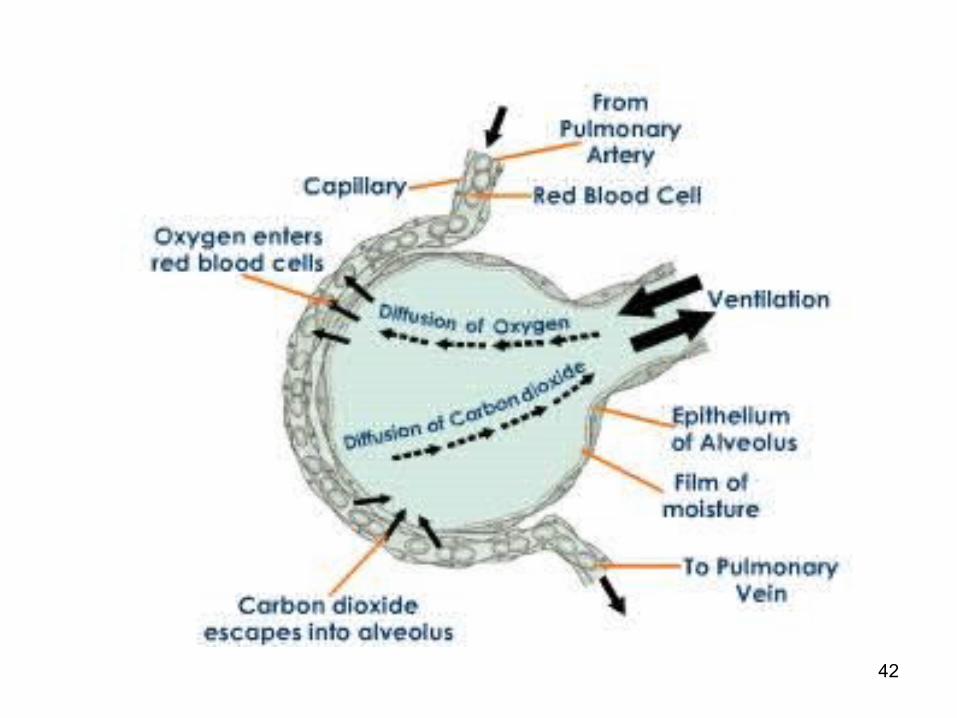
Gaseous exchange in alveoli
44
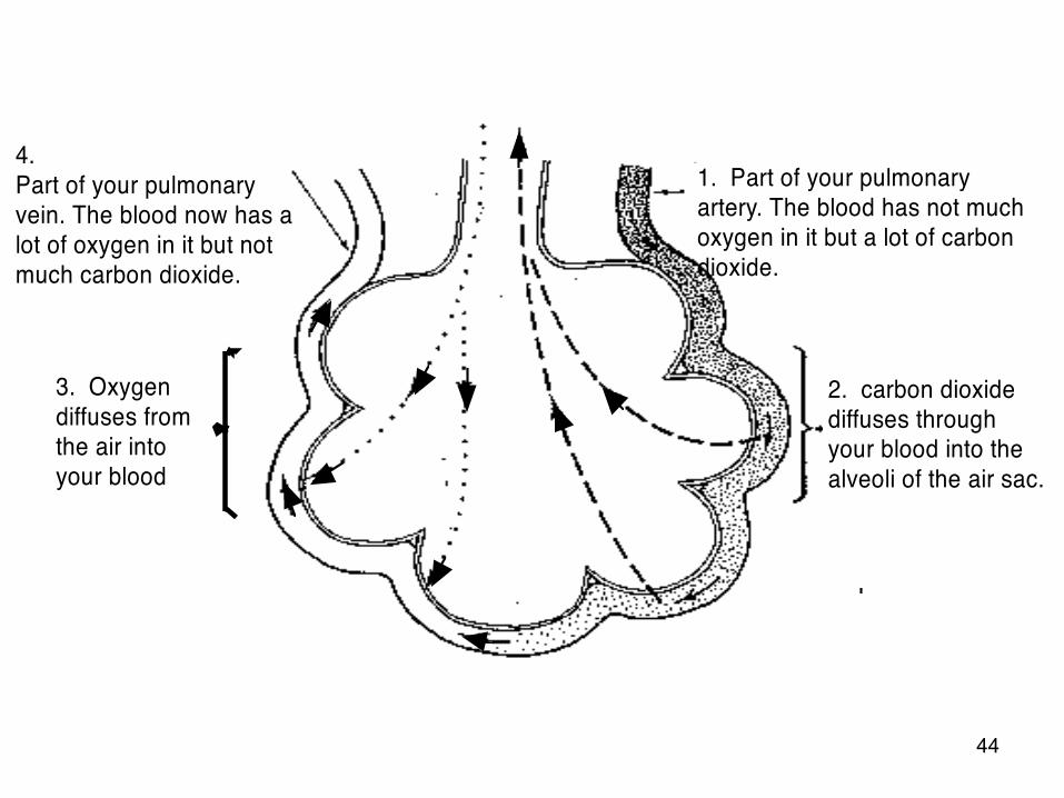
4.Part of your pulmonary vein. The blood now has a lot of oxygen in it but not much carbon dioxide.
1. Part of your pulmonary artery. The blood has not much oxygen in it but a lot of carbon dioxide.
2. carbon dioxide diffuses through your blood into the alveoli of the air sac.
3. Oxygen diffuses from the air into your blood
45
Oxygen• Alveolar air contains higher concentration of
oxygen than the blood.• Oxygen dissolves in the moisture lining and
diffuses into the blood capillaries.• Oxygen combines with haemoglobin to form
oxyhaemoglobin.
46
Carbon Dioxide• Tissue cells produce carbon dioxide during aerobic
respiration.• Carbon dioxide diffuses into the blood and enters red
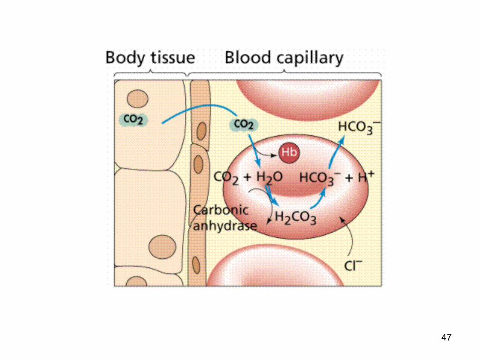
blood cells.• Carbon dioxide reacts with water to form carbonic acid.
This reaction is catalysed by carbonic anhydrase.• Carbonic acid breaks down into hydrogencarbonate ions
which diffuse out of the red blood cells into the plasma.
47
48
In The Lungs
• Hydrogencarbonate ions diffuse back into the red blood cells
• Converted into carbonic acid and then into water and carbon dioxide
• Carbon dioxide diffuses out of blood capillaries into the alveoli and out of the lungs

49
50
Exercise
What is the function of alveoli and how are they adapted to carry this function? (5)
51
Lesson 4Learning Objective1.Describe the mechanisms of breathing and
understand the changes in lung volume and pressure during breathing.
Outline Breathing mechanism
– Inhalation– Exhalation

52
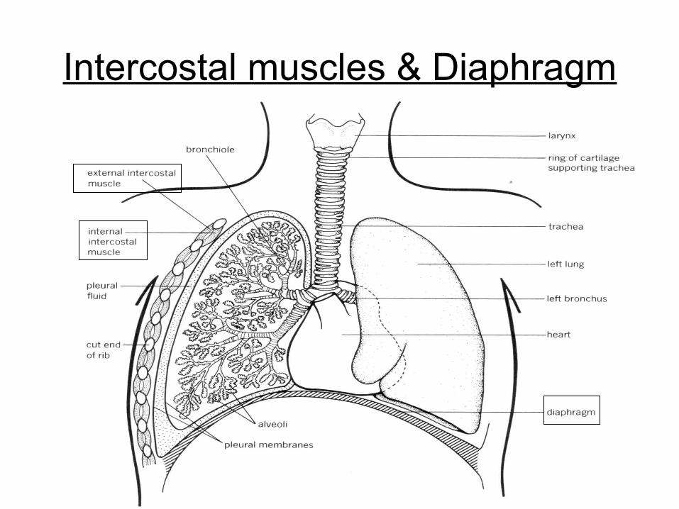
Chest (Thoracic) CavityDuring breathing, the lungs are inflated and deflated by the action of muscles. They are ...– Intercostal muscles (between the ribs)
• Internal intercostal and external intercostal– Control the movement of ribs
– Diaphragm (a sheet of muscle lying below the lungs, separating them from the abdomen)
The intercostal muscles and the diaphragm contract and relax, causing the volume of chest cavity to change.– Also changes the pressure within.
53
Intercostal muscles & Diaphragm
54
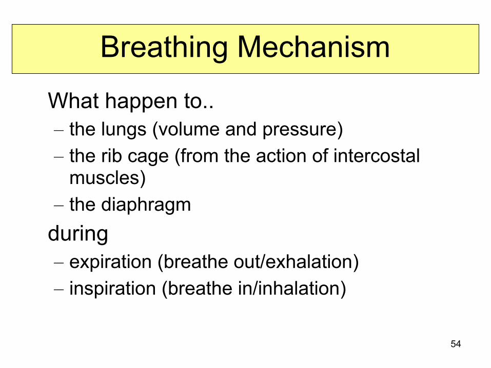
Breathing Mechanism
What happen to..– the lungs (volume and pressure)– the rib cage (from the action of intercostal
muscles)– the diaphragm
during– expiration (breathe out/exhalation)– inspiration (breathe in/inhalation)

55
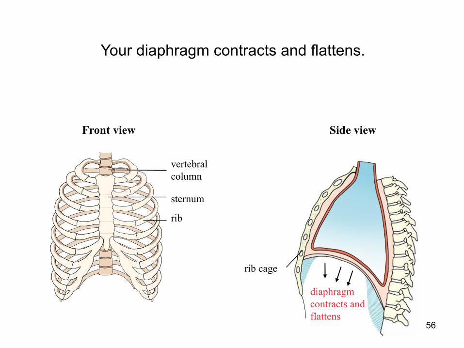
During Inspiration
rib
sternum
vertebral column
Front view Side view
When you breathe in or inspire, the following events take place:
rib cage
56
rib
sternum
vertebral column
Front view Side view
Your diaphragm contracts and flattens.
diaphragm contracts and flattens
rib cage
57
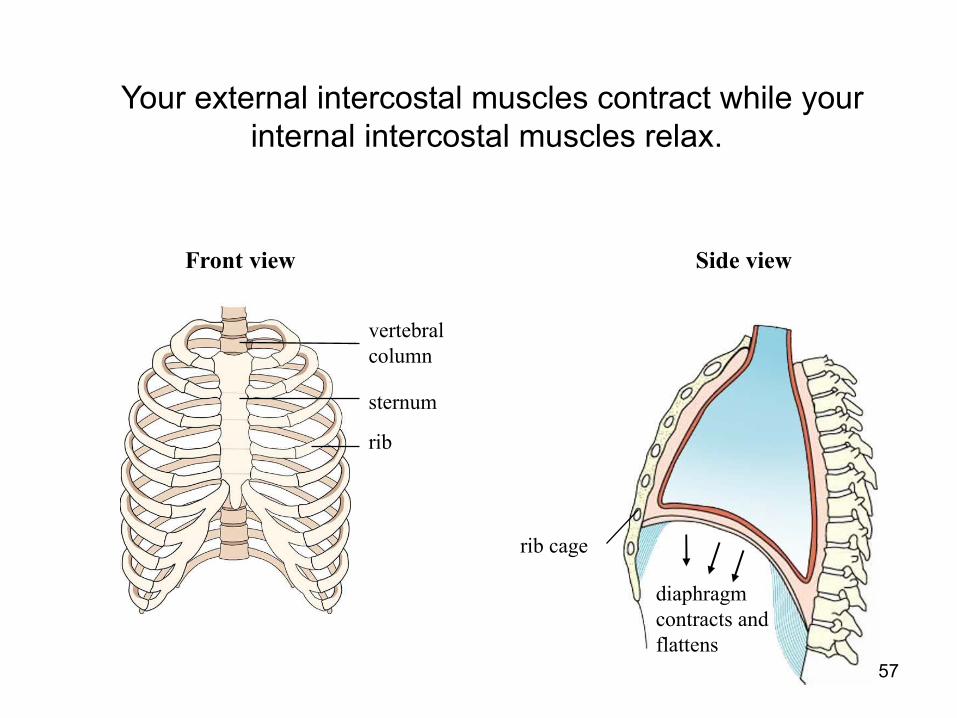
rib
sternum
vertebral column
Front view Side view
Your external intercostal muscles contract while your internal intercostal muscles relax.
rib cage
diaphragm contracts and flattens
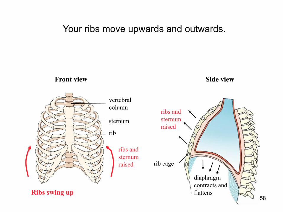
58
ribs and sternum raised
rib
sternum
vertebral column
Front view Side view
Ribs swing up
Your ribs move upwards and outwards.
rib cage
diaphragm contracts and flattens
ribs and sternum raised
59
ribs and sternum raised
rib
sternum
vertebral column
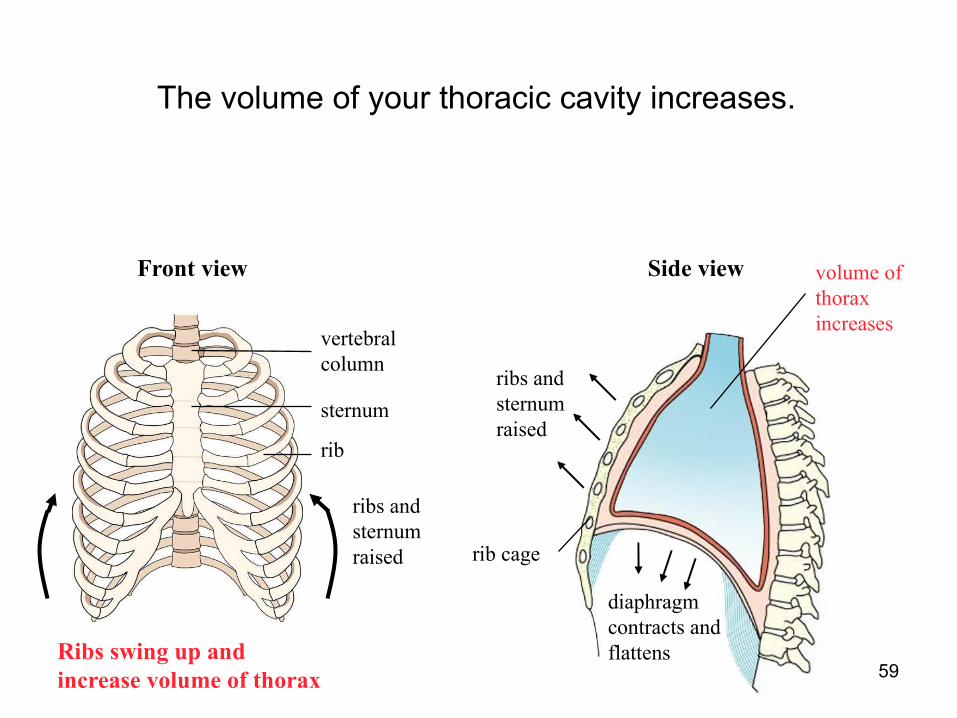
Front view Side view
Ribs swing up and increase volume of thorax
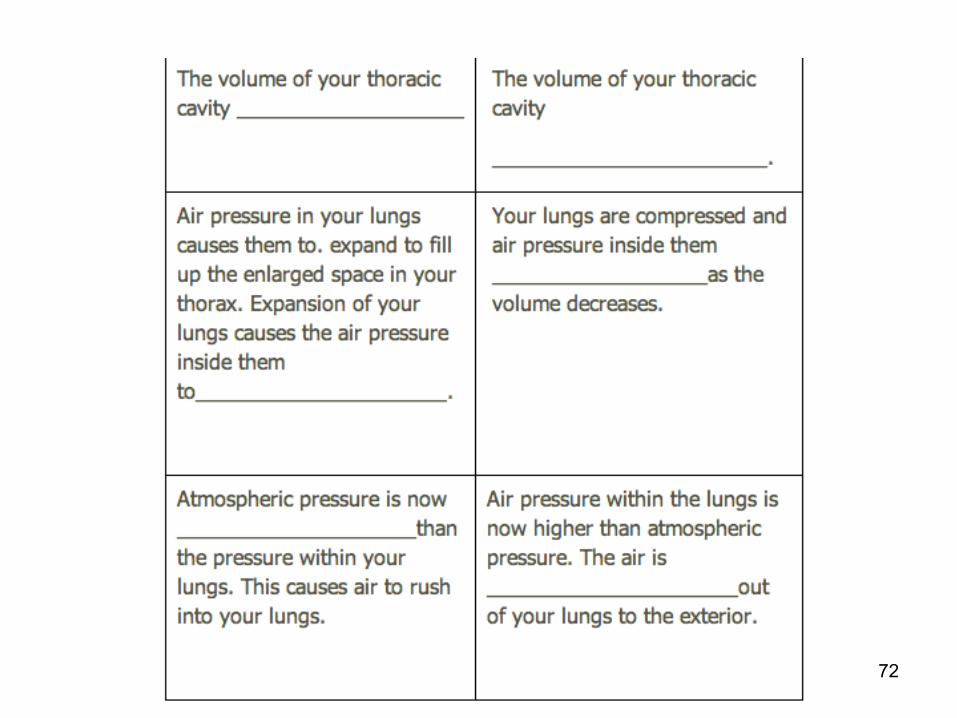
The volume of your thoracic cavity increases.
rib cage
diaphragm contracts and flattens
ribs and sternum raised
volume of thorax increases
60
ribs and sternum raised
rib
sternum
vertebral column
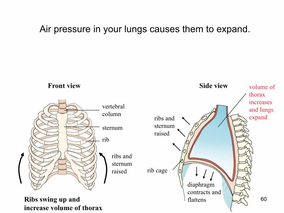
Front view
Ribs swing up and increase volume of thorax
Air pressure in your lungs causes them to expand.
Side view
rib cage
diaphragm contracts and flattens
ribs and sternum raised
volume of thorax increases and lungs expand
61
ribs and sternum raised
rib
sternum
vertebral column
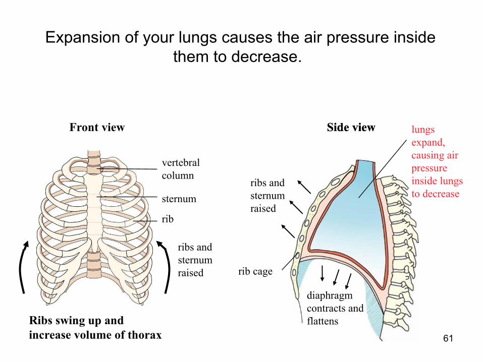
Front view Side view
Ribs swing up and increase volume of thorax
Expansion of your lungs causes the air pressure inside them to decrease.
Side view
rib cage
diaphragm contracts and flattens
ribs and sternum raised
lungs expand, causing air pressure inside lungs to decrease

62
ribs and sternum raised
rib
sternum
vertebral column
Front view
Ribs swing up and increase volume of thorax
Atmospheric pressure is now higher than the pressure within your lungs. This causes air to rush into your lungs.
rib cage
diaphragm contracts and flattens
ribs and sternum raised
lungs expand, causing air pressure inside lungs to decrease
air enters lungs
Side viewSide view

63
During ExpirationWhen you breathe out or expire, the following events take place:
rib
sternum
vertebral column
Side viewFront view
rib cage
64
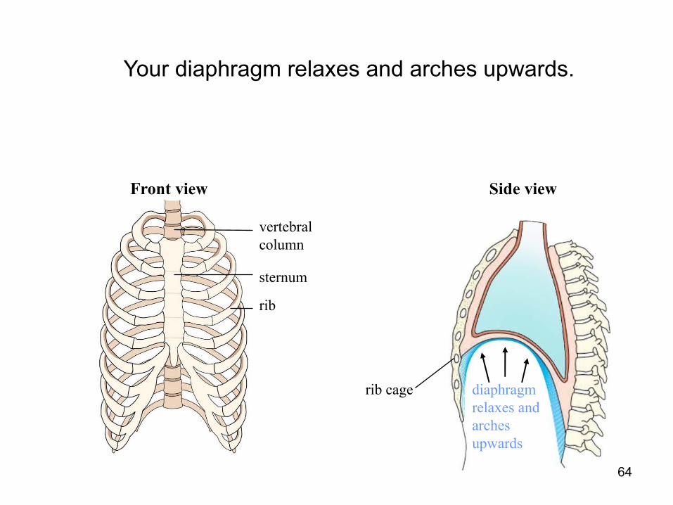
Your diaphragm relaxes and arches upwards.
rib
sternum
vertebral column
Side viewFront view
rib cage diaphragm relaxes and arches upwards
65
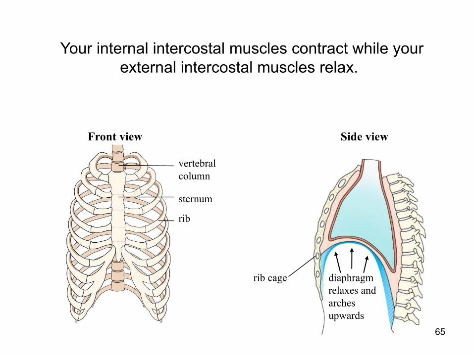
Your internal intercostal muscles contract while your external intercostal muscles relax.
rib
sternum
vertebral column
Side viewFront view
rib cage diaphragm relaxes and arches upwards

66
ribs and sternum raised
rib
sternum
vertebral column
Ribs swing down
Your ribs move downwards and inwards.
Front view Side view
rib cage diaphragm relaxes and arches upwards
ribs and sternum returned to original position

67
ribs and sternum raised
rib
sternum
vertebral column
Ribs swing down and decrease volume of thorax
The volume of your thoracic cavity decreases.
volume of thorax decreases
Front view Side view
rib cage diaphragm relaxes and arches upwards
ribs and sternum returned to original position
68
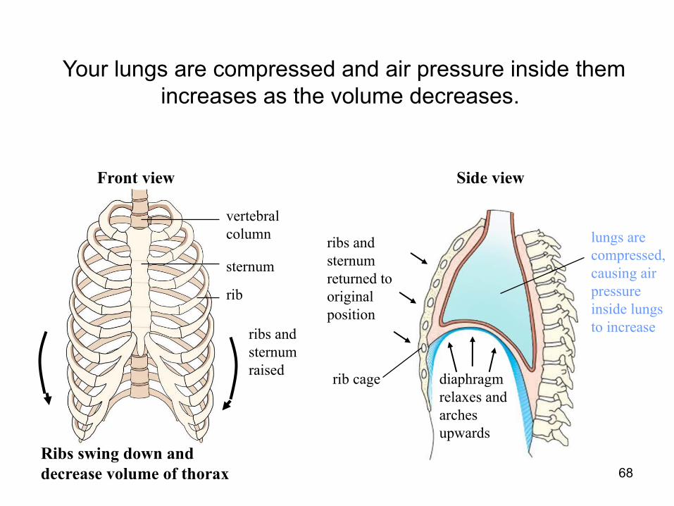
ribs and sternum raised
rib
sternum
vertebral column
Ribs swing down and decrease volume of thorax
Your lungs are compressed and air pressure inside them increases as the volume decreases.
Front view
lungs are compressed, causing air pressure inside lungs to increase
Side view
rib cage diaphragm relaxes and arches upwards
ribs and sternum returned to original position
69
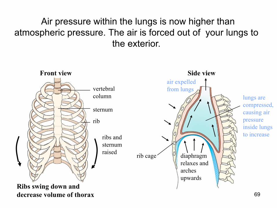
ribs and sternum raised
rib
sternum
vertebral column
Front view
Ribs swing down and decrease volume of thorax
Air pressure within the lungs is now higher than atmospheric pressure. The air is forced out of your lungs to
the exterior.
lungs are compressed, causing air pressure inside lungs to increase
Side view
rib cage diaphragm relaxes and arches upwards
air expelled from lungs
70
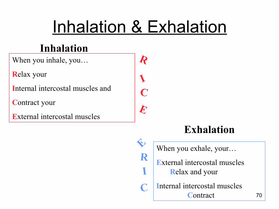
Inhalation & Exhalation
When you inhale, you…
Relax your
Internal intercostal muscles and
Contract your
External intercostal muscles
R
ICE
Inhalation
ERIC
ExhalationWhen you exhale, your…
External intercostal muscles Relax and your
Internal intercostal muscles Contract

71
Breathing MechanismFill in the blanks.
72

73
Lesson 5Learning Objective
1. Tabulate the composition of inspired and expired air.2. Understand the differences in composition.3. Describe the effects of respiratory diseases on the process
of gas exchange – bronchitis, emphysema, lung cancer.
Outline Composition of inspired and expired air. Respiratory diseases
74
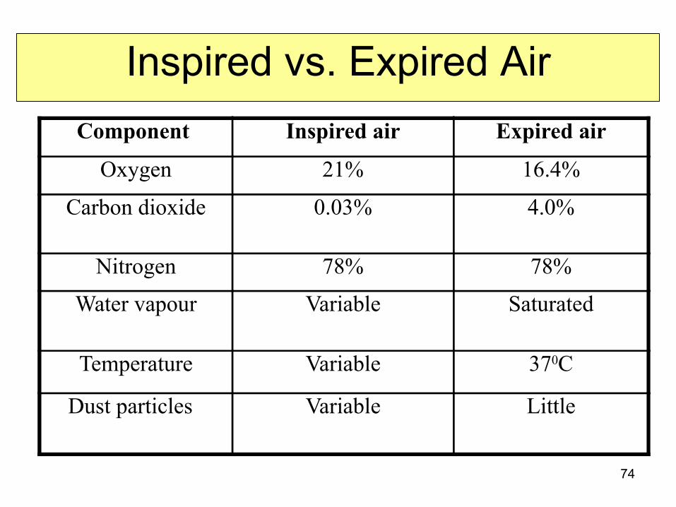
Inspired vs. Expired AirComponent Inspired air Expired air
Oxygen 21% 16.4%
Carbon dioxide 0.03% 4.0%
Nitrogen 78% 78%
Water vapour Variable Saturated
Temperature Variable 370C
Dust particles Variable Little
75
Bronchitis
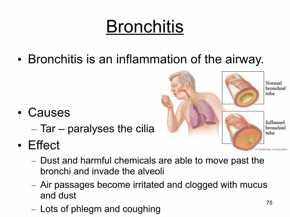
• Bronchitis is an inflammation of the airway.
• Causes– Tar – paralyses the cilia
• Effect– Dust and harmful chemicals are able to move past the
bronchi and invade the alveoli– Air passages become irritated and clogged with mucus
and dust– Lots of phlegm and coughing
76
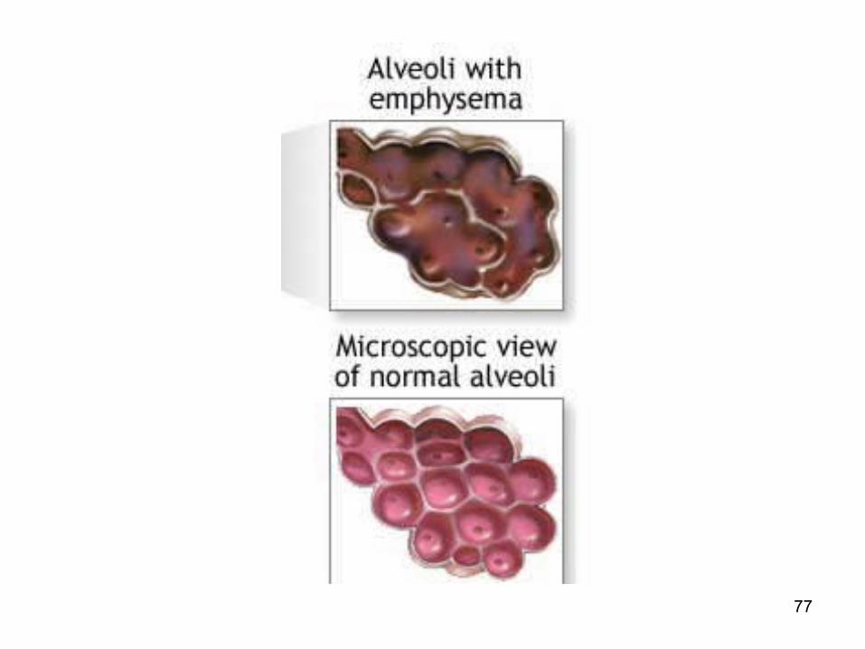
Emphysema
• Causes– Violent coughing causing alveolar walls to
become stretched and lose their elasticity.– Alveolar walls become weak, causing them to
break down.• Effects
– Decrease surface area for gaseous exchange– Lungs become inflated with air– Difficulty in breathing, wheezing
77
78
Asthma
• Air obstruction characterised by narrowing of air passages.
• Causes– Environmental or genetic
• Allergic reactions in the respiratory tract due to irritants
• Effect– Constriction of the respiratory tract– Suffocation and death

79
80
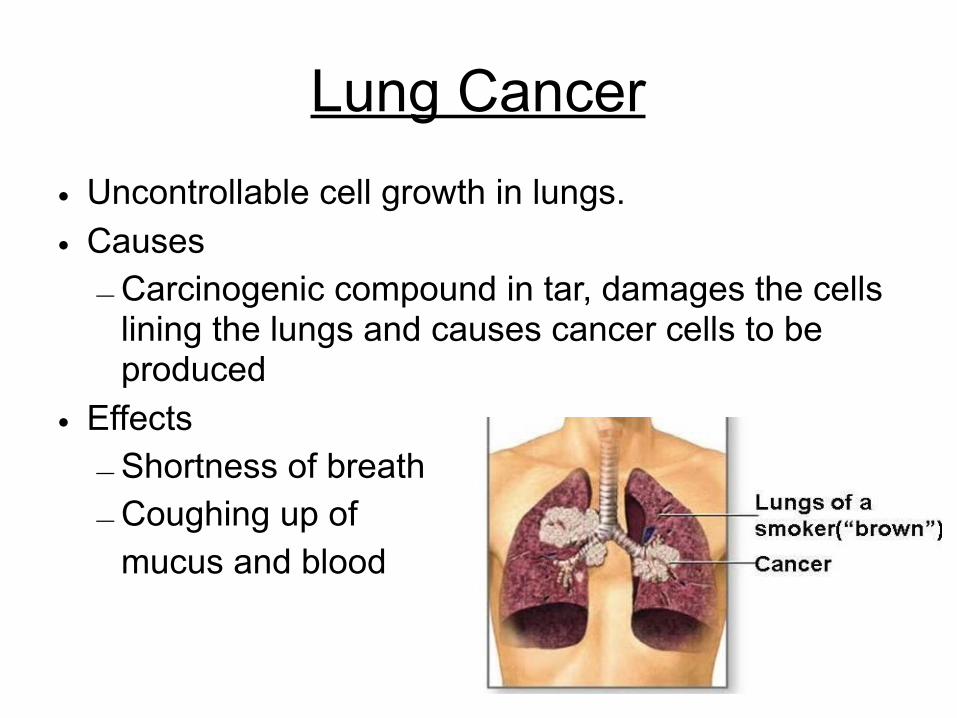
Lung Cancer• Uncontrollable cell growth in lungs.• Causes
– Carcinogenic compound in tar, damages the cells lining the lungs and causes cancer cells to be produced
• Effects– Shortness of breath– Coughing up of
mucus and blood
81
Smoking
• Smoking is the leading cause of lung disease.
• Contents of tobacco smoke– Nicotine– Tar– Carbon monoxide
82
Nicotine• Addictive drug that stimulates release of
adrenaline– Increases blood pressure and heart rate– Increases chance of blood clot and plaque
deposit on the walls of coronary arteries– Affects gaseous exchange between alveoli
and blood capillaries
83
Tar
• Tar is absorbed by cells in the lungs, especially cells lining the bronchi and bronchioles.
• Normally, these cells form a thin, protective layer. But the tar makes them divide and build up into a thicker layer.
• This may lead into cancer.

84
• Tar is also an irritant. – It makes the linings of the respiratory passages
inflamed, causing chronic bronchitis.– It paralyses the cilia and cause extra mucus to be
made by goblet cells.–This mucus trickles down into the lungs–Bacteria breed in the mucus, causing infection. –Coughing moves the mucus upwards.–Constant coughing can damage the delicate
alveoli.–This makes it difficult for the person to get enough
oxygen into their blood.–They have emphysema.
85
Carbon Monoxide
• Carbon monoxide is absorbed into the blood.
• It combines rapidly with haemoglobin in the red blood cells.
• This means there is less haemoglobin available to carry oxygen.
86
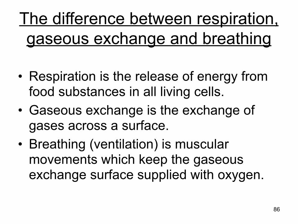
The difference between respiration, gaseous exchange and breathing
• Respiration is the release of energy from food substances in all living cells.
• Gaseous exchange is the exchange of gases across a surface.
• Breathing (ventilation) is muscular movements which keep the gaseous exchange surface supplied with oxygen.
87
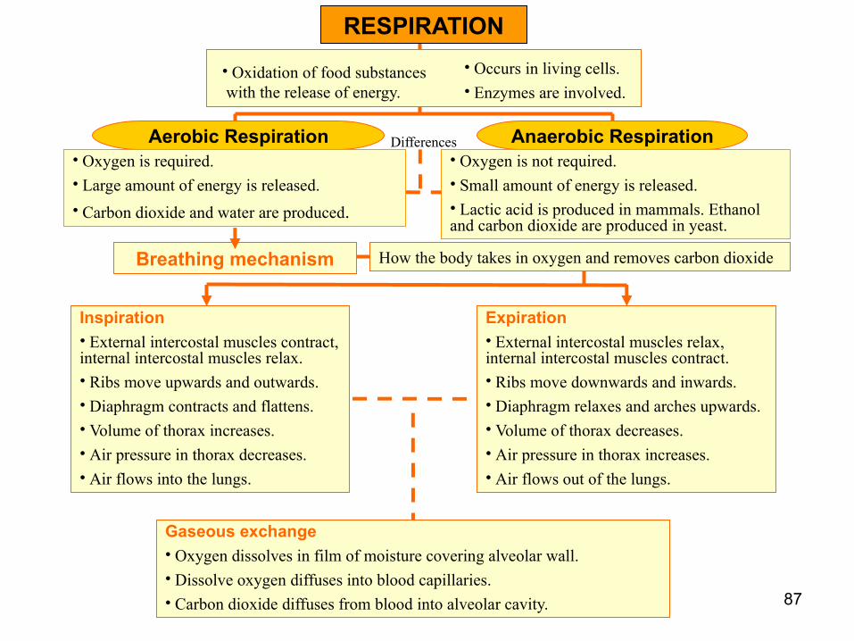
Aerobic Respiration Anaerobic Respiration
How the body takes in oxygen and removes carbon dioxide
RESPIRATION
Breathing mechanism
• Oxygen is required.• Large amount of energy is released.• Carbon dioxide and water are produced.
• Oxygen is not required.• Small amount of energy is released.• Lactic acid is produced in mammals. Ethanol and carbon dioxide are produced in yeast.
Differences
• Occurs in living cells.• Enzymes are involved.
• Oxidation of food substances with the release of energy.
Inspiration• External intercostal muscles contract, internal intercostal muscles relax.• Ribs move upwards and outwards.• Diaphragm contracts and flattens.• Volume of thorax increases.• Air pressure in thorax decreases.• Air flows into the lungs.
Expiration• External intercostal muscles relax, internal intercostal muscles contract.• Ribs move downwards and inwards.• Diaphragm relaxes and arches upwards.• Volume of thorax decreases.• Air pressure in thorax increases.• Air flows out of the lungs.
Gaseous exchange• Oxygen dissolves in film of moisture covering alveolar wall.• Dissolve oxygen diffuses into blood capillaries.• Carbon dioxide diffuses from blood into alveolar cavity.