resolution of inflammation in murine autoimmune arthritis ... · blockbuster drugs that are widely...
TRANSCRIPT

of June 10, 2018.This information is current as Production
4-Mediated Lipoxin A2Prostaglandin ECyclooxygenase-2 Inhibition and Restored byAutoimmune Arthritis Is Disrupted by Resolution of Inflammation in Murine
Marion Man-Ying Chan and Andrea Rossi Moore
ol.0903816http://www.jimmunol.org/content/early/2010/04/30/jimmun
published online 30 April 2010J Immunol
average*
4 weeks from acceptance to publicationFast Publication! •
Every submission reviewed by practicing scientistsNo Triage! •
from submission to initial decisionRapid Reviews! 30 days* •
Submit online. ?The JIWhy
Subscriptionhttp://jimmunol.org/subscription
is online at: The Journal of ImmunologyInformation about subscribing to
Permissionshttp://www.aai.org/About/Publications/JI/copyright.htmlSubmit copyright permission requests at:
Email Alertshttp://jimmunol.org/alertsReceive free email-alerts when new articles cite this article. Sign up at:
Print ISSN: 0022-1767 Online ISSN: 1550-6606. All rights reserved.1451 Rockville Pike, Suite 650, Rockville, MD 20852The American Association of Immunologists, Inc.,
is published twice each month byThe Journal of Immunology
by guest on June 10, 2018http://w
ww
.jimm
unol.org/D
ownloaded from
by guest on June 10, 2018
http://ww
w.jim
munol.org/
Dow
nloaded from

The Journal of Immunology
Resolution of Inflammation in Murine Autoimmune ArthritisIs Disrupted by Cyclooxygenase-2 Inhibition and Restored byProstaglandin E2-Mediated Lipoxin A4 Production
Marion Man-Ying Chan and Andrea Rossi Moore
Acute inflammation follows defined phases of induction, inflammation and resolution, and resolution occurs by an active process that
requires cyclooxygenase-2 (COX-2) activity. This study aims to address whether this paradigm extends to recognized model of
chronic inflammation. We demonstrated that murine collagen-induced arthritis follows a similar sequential course. Interestingly,
COX-2 and its metabolite, the presumably proinflammatory PGE2, are present in the joints during resolution, and blocking COX-2
activity and PGE2 production within this period perpetuated, instead of attenuated, inflammation. Repletion with PGE2 analogs
restored homeostasis, and this function is mediated by the proresolving lipoxygenase metabolite, lipoxin A4, a potent stop signal.
Thus, the study provided in vivo evidence for a natural, endogenous link between the cyclooxygenase–lipoxygenase pathways and
showed that PGE2 serves as a feedback inhibitor essential for limiting chronic inflammation in autoimmune arthritis. These
findings may explain the enigma regarding why COX-2 inhibitors are palliative rather than curative in humans, because blocking
resolution may mitigate the benefit of preventing induction. The Journal of Immunology, 2010, 184: 000–000.
In the field of inflammation, there have been two seminaldiscoveries that precipitated a paradigm shift in the past de-cade. One is the discovery that, contrary to early belief, in-
flammatory response does not dissipate but requires an activeprocess that is mediated by products of cyclooxygenase (COX) forsubsiding. For example, Gilroy et al. (1) observed that blockingCOX activity, whether with the selective COX-2 inhibitor N-[2-(cyclohexyloxy)-4-nitrophenyl]-methanesulfonamide (NS-398) orthe dual COX-1/COX-2 inhibitor indomethacin, inhibited carra-geenin-induced acute pleurisy in the inflammation phase, but then itsignificantly perpetuated inflammation by interfering with resolu-tion. Similarly, Fukunaga et al. (2) have shown that pharmacologicinhibition or gene disruption of COX-2 blocked resolution of acid-induced lung injury. COX-2 has also been shown to mediate reso-lution of acute inflammation in the brain (3), liver (4), small bowel,and colon (5) as well.Another major impact came from the discovery that a class of
arachidonic acid metabolites facilitates resolution of inflammation.These eicosanoids, named lipoxins, resolvins, and protectins, aregenerated from lipoxygenases or aspirin-acetylated COX and serveas stop signals in the evolution of acute inflammation responses.They act by stimulating the uptake of apoptotic polymorphonuclearleukocytes at sites of inflammation to promote a programmed returnto homeostasis (6–8). Analogs and precursors (v-3-fatty acids) of
lipoxins have been shown to relieve inflammatory-related pathol-ogy responses, especially in asthma and various airway injures (9).Currently, it is known that lipoxins are formed in vivo in the courseof inflammation and animals with a mutation in their syntheticenzyme, ALOX12/15, suffer exacerbated inflammation (10).However, our knowledge on the endogenous pathway that regulatestheir biogenic synthesis is still very limited.Autoimmune diseases are cyclical in nature. It is generally as-
sumed that tissue destruction occurs because the inflammation ischronic (11). The first objective of this study is to determine therelevance of the eicosanoid-directed, proresolving process in anautoimmune condition so as to evaluate whether it can be har-nessed for therapeutic intervention; and then to determine whetherthe mechanism is vulnerable to COX-2 inhibitors for these areblockbuster drugs that are widely prescribed for curbing in-flammation (12). We used the murine collagen-induced arthritis(CIA) model, for it has been generally used for evaluating im-munological and pharmacological treatments, including COX-2inhibitors, for rheumatoid arthritis (13). We discovered that PGE2,generally viewed as a culprit for exacerbating arthritis, functionsas a critical endogenous signal sufficient for mediating resolution.It coordinates resolution of inflammation by mediating biogenesisof lipoxin A4 (LXA4) for curbing inflammation. This link betweenPG and lipoxin may explain why clinical treatments with COXinhibitors relieve symptoms but would not halt progression of thedisease in humans.
Materials and MethodsInduction of disease
Experimental protocol used in this study was approved by the TempleUniversity Institutional Animal Care and Use Committee. Arthritis wasinduced in 6- to 8-wk-old, male DBA/1 mice (The Jackson Laboratory, BarHarbor, ME) with intradermal injection of chicken collagen II (Chondrex,Redmond, WA) in CFA according to the procedure of Terato et al. (14).Each mouse received a 0.05 ml volume 50 mg collagen emulsified in CFAintradermally in the tail. Immunity was boosted with another injection of50 mg collagen II in IFA at day 21. Pathogenesis was assessed in a double-blinded manner by measuring the thickness of the hind footpads twicea week with a constant-tension caliper.
Department of Microbiology and Immunology, School of Medicine, Temple Univer-sity, Philadelphia, PA 19140
Received for publication November 30, 2009. Accepted for publication March 24,2010.
This work was supported by National Institutes of Health Grant R21 AR051761 andfunding from the American Institute for Cancer Research (to M.C.).
Address correspondence and reprint requests to Dr. Marion Chan, Department ofMicrobiology and Immunology, School of Medicine, Temple University, 3400 NorthBroad Street, Philadelphia, PA, 19140. E-mail address: [email protected]
Abbreviations used in this paper: 15d-PGJ2, 15-deoxy-Δ12,14-PGJ2 dmPGE2, 16,16dimethyl-PGE2; CIA, collagen-induced arthritis; COX-2, cyclooxygenase-2; LXA4,lipoxin A4; mPGES-1, microsomal PGES-1; NS-398, N-[2-(cyclohexyloxy)-4-nitro-phenyl]-methanesulfonamide; NSAID, nonsteriodal anti-inflammatory drug.
Copyright� 2010 by The American Association of Immunologists, Inc. 0022-1767/10/$16.00
www.jimmunol.org/cgi/doi/10.4049/jimmunol.0903816
Published April 30, 2010, doi:10.4049/jimmunol.0903816 by guest on June 10, 2018
http://ww
w.jim
munol.org/
Dow
nloaded from

Defining resolution
Ability to resolve inflammation was determined by the percent of footpadsthat has decreased, increased, or remains unchanged in their thickness. Anumerical index for each footpad was deduced from the slope of a secantline. In the vehicle control group, the line was drawn from the peak ofinflammation to the point of sacrifice or total resolution (,10% swollen) inthe resolution phase. For therapeutically treated groups, the line was drawnfrom the beginning of NS-398 administration. A value of “0” represents nochange in thickness. Negative values correspond to decrease in swellingand positive values indicate that swelling increased.
Radiology assessment
Mice were anesthetized with ketamine/xylazine then radiographs weretaken using a Faxitron for small animals (model 43855, Faxitron X-Ray,Lincolnshire, IL).
Histological assessment
Knee jointswerefixed in10%neutral buffered formalin for 24h, decalcified inEDTA, embedded in paraffin, and cut in serial sections for histopathologicalanalysis. The sectionswere then stainedwithH&E, and safraninO to examinethe integrity of the cartilage. Formalin-fixed tissue sections were stained withrabbit anti-murine COX-2 Ab (H-62, Santa Cruz Biotechnology, Santa Cruz,CA), then biotinylated anti-rabbit Ab, followed by streptavidin-HRP andDAB solution (all from Dako Scientific, Carpinteria, CA) to assess COX-2expression. Rabbit normal Ig was used for isotype controls.
PG assays
Lipid was extracted before assaying for the PGs. Randomly selectedfootpads were harvested, snap frozen, and stored in liquid nitrogen untilusage, and then weighed before lipid extraction. The frozen joint tissueswere pulverized using amortar and pestle to obtain a fine powder, which washomogenized (Polytron PRO200 homogenizer; PRO Scientific, Oxford,CT) and then sonicated in methanol with 0.01 M butylhydroxytoluene and0.85% formic acid while surrounded by ice. After centrifugation, an aliquotof the supernatants was collected to perform Bradford assay for proteindetermination, then the homogenates were protein-precipitated by theaddition of acetonitrite (pH 3.5). After centrifugation, the supernatants wereloaded onto a C18 SPE column (3M, St. Paul, MN) to collect the lipidmolecules. The cartridge was preconditioned by washing thoroughly with10%methanol/0.1% formic acid and ethyl acetate. The lipid molecules wereeluted with ethyl acetate, followed by methanol with 0.2% formic acid and0.01 M butylhydroxytoluene, and then evaporated to dryness under nitrogenand reconstituted in ELISA buffer. The ELISA kits used for PGE2 andLXA4 were from Cayman Chemicals, Assay Design, and Neogen, re-spectively. Each sample was assessed in triplicate and at two to three di-lutions to ascertain that the reactions occurred within the standard curveand did not reflect interference from cross-reactive substances. The amountof lipid produced was calculated as pg/mg of tissue. A minimum of threerepeats were performed for each experiment and they were merged bynormalization to the corresponding basal value within each experiment forstatistical comparison.
RT-PCR and PCR
Limbs harvested from euthanized mice were weighed, snap frozen, andstored at 280˚C for assessing gene expression. The joints were crushed inliquid nitrogen and homogenized with a micro ultrasonic cell disrupter forRNA extraction in Trizol reagent (Invitrogen, San Diego, CA). Extractsfrom limbs of randomly selected mice from each group were pooled forreverse transcription and real-time PCR analysis as described in Adapalaand Chan (15). Commercial primers and SYBR Green I PCR master mixwere used (Superarray). The housekeeping gene 18S RNA was reversetranscribed with random hexamers, and its cDNA solution was diluted toattain an amplification efficiency that was comparable to that of the ex-perimental gene, which cycling threshold values were normalized to thoseencoding 18S rRNA by the Ct method. A minimum of three repeats wereperformed for each experiment and, in each, relative units were deduced sothe repeats can be normalized and merged for statistical comparison.
Data analyses
For statistical comparison, the data on gene expression and footpad thicknesswere tested for normality using the Kolmogorov-Smirnov test. Then, thenormally distributed populations were compared using unpaired, one-wayANOVA, followed by Bonferroni test. Otherwise, the nonparametricKruskal-Wallis test was used instead. A p value of 0.05 was chosen as thethreshold for statistical significance throughout.
ResultsKinetic studies revealed a resolution phase in the pathogenesisof chronic inflammatory autoimmune arthritis
The study began with examining whether the pathogenesis of au-toimmune arthritis in the CIAmodel would resemble acute injury inhaving discrete phases. Hind foot thickness showed that, similar toacute inflammation, swelling followed three phases: induction,inflammation, and resolution (Fig. 1A). From days 0–30, arthritishad not developed; the mice were asymptomatic and their footpadswere not swollen. Footpad swelling became increasingly prevalentbeginning at day 30 and continued to do so until around day 45when .95% the hind feet were swollen by 20% or more in thick-ness, and the incidence of arthritis in the group of mice was 98%.Subsequently, the footpad swelling began to progressively subsidewith 40% resolved at day 55 and 90% at days 69–70. The pro-gression from induction through inflammation to resolution is il-lustrated in footpad swelling by a representative footpad (Fig. 1B).IL-17 and TNF-a are two of the several cytokines that drive
the progression of inflammation (16, 17). Therefore, groups ofmice were sacrificed at weekly intervals and the joints were col-lected for reverse transcriptase real-time PCR analysis of thesetranscriptionally regulated cytokines to verify the state of in-flammation by their levels. Their mRNA expression increased by4- to 6-fold within the inflammatory phase (days 25–55), then itsubsided as inflammation resolved at day 70 (Fig. 1C). The stateof inflammation was further documented by histology and radi-ography (Fig. 1D, 1E). The H&E sections show that the synoviumof a knee joint at the peak of inflammation is fully infiltrated withneutrophils, whereas that of a resolved one, which footpadthickness had subsided to 18% from 91%, is cleared of infiltrates.The radiographs show that the metatarsophalangeal joints of thefootpad regained alignment as inflammation resolved.
COX-2 was expressed in the resolution phase
Whereas the gene expression of IL-17 and TNF-a subsided asresolution occurred, COX-2 mRNA remained escalated (Fig. 2A).Kinetics studies demonstrated that COX-2 was upregulated by 2- to3.5-fold during the inflammatory phase between days 35 and 45,declined slightly around day 55, then resurged during resolutionthrough day 70. Comparison by unpaired, one-way ANOVAshowed that the level of expression during resolution (day 70) andat the peak of inflammation, day 35 or 45, were both significantlyhigher than baseline (p , 0.005) but they did not differ betweeneach other (p . 0.05). This bimodal pattern was observed in morethan three experimental repeats and was analogous to the nature ofCOX-2 expression in acute inflammation: Gilroy et al. (3) haveshown that when carrageenin is used to induce acute pleurisy, COX-2 expression in the rat model shows a bimodal pattern and theenzyme is expressed 3.5-fold higher in the resolution phase (48 hafter induction) than in the inflammation phase (2 h after in-duction). Blaho et al. (18) have also found a similar pattern in aninfectious disease model, Lyme disease. Furthermore, Kapoor et al.(19) have shown that COX-2 expression is sustained for repair inthe process of wound healing. The presence of COX-2 was con-firmed at the protein level by immunohistochemical staining of theknee joints (Fig. 2B). The enzyme was detected in the synovium aswell as the infiltrating cells of the resolving knees.
Contradictory duality of COX-2: Resolution was COX-2dependent
Because COX-2 is expressed, it is important to determine thefunction of COX-2 in the resolution phase. In this experiment,collagen-induced micewere divided into groups of positive control,which received vehicle only; negative control, which were age-
2 COX-2, PGE2, AND LXA4 IN RESOLVING ARTHRITIS
by guest on June 10, 2018http://w
ww
.jimm
unol.org/D
ownloaded from

matched normal mice; and the experimental, which received 10 mgNS-398, a COX-2–specific inhibitor, dissolved in saline with10% ethanol. The dose of NS-398 given was 0.5 mg/kg, andit was administered by gavage every other day in two manners: 1)treatment began at the induction phase to imitate prophylactic useof COX inhibitor (Fig. 3A), or 2) after inflammation has estab-lished to mimic patients seeking treatment after symptoms haveappeared (Fig. 3B).The effect of NS-398 in the inflammation and the resolution
phases is opposite. Blocking COX-2 prior to manifestation ofsymptoms (initiating NS-398 feeding at day 14) attenuated footpadswelling during the nominal inflammation phase. This was apparentwhen the prevalence of arthritis was enumerated by percent or by thedegree of swelling in the hind footpads. Among mice in the vehiclecontrol group,.75% of the hind feet had swollen, whereas NS-398feeding reduced the incidence to ∼40%. The thickness of theirfootpads had increased by 40.4% on average, whereas those of theNS-398–fed group had swollen by only 21.4% (Fig. 3A). Corre-spondingly, the mRNA levels of proinflammatory cytokines, TNF-a and IL-17, in the footpads that were harvested between days 40
and 45 were also suppressed by NS-398 (Fig. 3C). Henceforth,considering that both groups of mice are comparable in weight,averaging ∼20 g throughout the experiment, our results demon-strate what would be expected; NS-398 reduces inflammation whengiven in the induction phase.In contrast, blocking COX-2 during the resolution phase per-
petuates inflammation (Fig. 3B). This phenomenon is best illus-trated by the percent of mice that have qualitatively been defined bya zero, a positive, or a negative slope. Among the footpads that havedeveloped arthritis (82%), 87% of those in the vehicle control grouphad experienced resolution, as indicated by the rate of change infootpad thickness (negative slope, downward), and only 14% ofthem remained unchanged or continued to swell. In contrast, in-flammation persisted or progressed (zero or positive slope) in 54%of the footpads in the group that was fed NS-398 after inflammationhas established (therapeutically). When secant lines were drawn todeduce numbers, the slopes of the footpads in the vehicle controlgroup average to 22.35, indicating inflammation was resolvingnaturally, whereas those of NS-398 groups that were fed thera-peutically was +0.38, showing that resolution has failed to occur.
FIGURE 2. COX-2 expression during resolution. In A, the gray, shaded
area, duplicated from Fig. 1A, provides a background to show the progression
from induction to inflammation to resolution. Kinetics of COX-2 expression
(black bold line) was derived by real-time PCR. Three independent experi-
ments were performed. In each, 4–6 limbs from randomly selected mice
within a group were used to derive the data points, totaling 12–15 per point.
The data were tested for normality, and statistical analysis was performed
using unpaired, one-way ANOVA, followed by Bonferroni test. The levels at
the peak of inflammation and during resolution (day 70) were not signifi-
cantly different from each other, p . 0.08, but they were significantly
different from when the joints were normal (day 0) p # 0.006. B, Immu-
nohistochemical staining of joints (representative of eight) is shown in the
inflammation (left) and resolution phases (right) for the presence of COX-2 in
the pannus and synovium. Positive staining for COX-2 appears as brown.
Specificity of binding was verified by isotype control. The hematoxylin
counterstain appears as blue, original magnification 340 and 3600.
FIGURE 1. Three phases of pathogenesis in arthritis. In A, open bars
indicate the percent of footpads that have swollen by 20%. Filled bars in-
dicate the percent of swollen limbs that have decreased by .20% in thick-
ness, n = 50–200. The number of footpads decreases as mice were sacrificed
for cytokine analyses. Mean and SDs are plotted. The degree of swelling for
each footpad was calculated as 100 3 (thickness 2 thickness on day 0)/
(thickness on day 0, range from 2.0–2.3 mm). B, The progression of in-
flammation in a typical arthritic limb is shown. In C, IL-17 and TNF-a
mRNA were normalized against 18S RNA and then the relative level of
expression was deduced. Three independent experimental repeats were
performed. Within each experiment, 4–6 limbs from randomly selected mice
were pooled, each data point is averaged from the three repeats, so total n =
12–18. The data were tested for normality, and unpaired one-way ANOVA,
followed by Bonferroni test was used for statistical comparison. Their levels
at peak of inflammation (day 0) were significantly different from when the
joints were normal (day240) or resolved (day +30), p, 0.05, but the levels
in normal and resolved joints were not different, p . 0.05. D, H&E shows
a highly cellular synovium in the inflamed (70% swollen at day 40) but not in
the resolved (subsided from 91% to 12% swelling at day 70) knee joints
(original magnification 340). E, Sequential x-ray of representative meta-
tarsal joints of a footpad is shown; note the subsiding of soft tissue swelling
and the return of joint integrity and bone density in the fifth phalange.
The Journal of Immunology 3
by guest on June 10, 2018http://w
ww
.jimm
unol.org/D
ownloaded from

The degree of inflammation corresponded to the levels of proin-flammatory cytokines in the harvested limbs. Real-time PCRanalysis (Fig. 3D) confirmed that the levels of TNF-a and IL-17mRNAwere significantly higher in the NS-398–treated groups, andthe differences were statistically significant. Fig. 3E illustrates howNS-398 affected the course of resolution kinetically using a repre-sentative limb from each group. The footpads in the vehicle controlswelled and then subsided; however, resolution was impaired in thegroup that was fed NS-398. Even in mice that were fed NS-398preventively (initiated in the induction phase), the suppression ofinflammation was temporary in 67% of the footpads; the onset ofswelling eventually occurred.The medical consequence of loss of resolution was increased
destruction of the joints (Fig. 4). Radiography showed that thedegree of soft tissue swelling, digital misalignment, ankylosis, and
loss of bone density were more severe in the limbs that had delayedonset (preventive treatment) or perpetuated inflammation (thera-peutic treatment) than in those that were able to resolve (vehiclecontrol). The degree of deterioration was further delineated byhistological staining. The pannus in the knee joints of the NS-398–fed mice were more proliferative and the cartilage and bone weremore severely damaged than those of the vehicle controls.
PGE2 was produced in the resolution phase and COX-2dependent
Among the COX-2 metabolites, PGE2 is usually considered asa culprit that causes inflammation, whereas 15-deoxy-Δ12,14-PGJ2(15d-PGJ2) is a ligand for the immunosuppressive transcriptionfactor peroxisome proliferator-activated receptor-g and an in-hibitor of NF-kB (20). A class switching from PGE2 to 15d-PGJ2
FIGURE 3. NS-398 increased the incidence and
severity of swelling in the resolution phase. A, De-
piction of the relative increase in thickness of footpad
(mm) due to arthritis induced by chicken collagen. The
maximum thickness obtained within days 30–55, the
inflamed phase, was plotted for each foot. The data are
compiled from four independent trials. The black di-
amonds (n = 112) represent footpads of mice that were
induced for arthritis but fed vehicle as control. The
gray triangles (n = 58) represent footpads of experi-
mental mice that were fed NS-398 prior to manifes-
tation of symptoms, and they are congregated at the
bottom. B, The incidence and severity of swelling in
the resolution phase. Each black diamond (n = 38) and
gray triangle (n = 48) represents a footpad. The data
were compiled from three independent experiments.
The open squares, in A and B, both n = 20, represent
footpads of normal mice that were not injected. The
horizontal bars indicate the average value in each
group, and the groups were compared by Kruskal-
Wallis test, and p , 0.05 was considered significant. C
and D, The difference in levels of mRNA in a subgroup
of limbs randomly selected and harvested from the
group in A or the group in B, is shown, respectively.
The levels of mRNA expression were normalized
against 18S RNA and the results shown are the mean
6 SEM from 4–6 mice and representative of three
experimental repeats. The data were tested for nor-
mality and then statistical analysis by ANOVA. The
differences between NS-398–fed groups and the vehi-
cle control group was significant (p , 0.05). E, Ki-
netically plots a representative limb from the vehicle
control group (dashed line–open triangles) and a group
where NS-398 treatment was initiated after in-
flammation has established (solid line–filled triangles).
E is also used to delineate the three phases (lines) and
illustrate treatment regimen. Closed arrow points to
when therapeutic NS-398 treatment started.
4 COX-2, PGE2, AND LXA4 IN RESOLVING ARTHRITIS
by guest on June 10, 2018http://w
ww
.jimm
unol.org/D
ownloaded from

has been observed during transition from inflammation to reso-lution on acute models (19, 21). However, in murine CIA, thelevel of PGE2 is sustained.In three independent experiments, similarly, the amount of PGE2
was elevated in the inflamed and the resolution phases. Comparedwith the normal control, the average increases in the two phaseswere 2.406 0.43-fold and 2.576 0.63-fold, respectively (Fig. 5A).They are statistically different from the normal control but not fromeach other. Intrigued by the synthesis of PGE2 in the phase ofresolution, we proceeded to determine whether COX-2 is synthe-sizing the PG during the resolution phase. In the mice that were fedNS-398, the relative level of PGE2 decreased to 1.46 6 0.13 from2.22 6 0.22, by ∼60% when compared with the vehicle control(Fig. 5B, see legend for actual concentrations in pg/mg tissue).
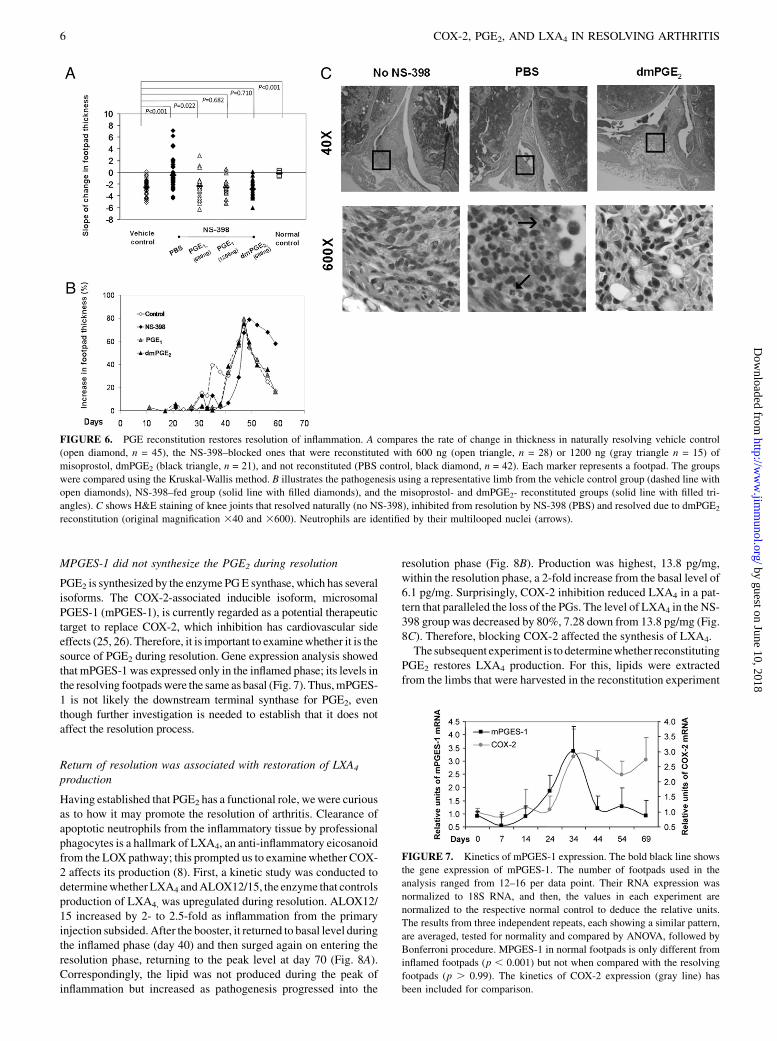
Reconstitution with PGE2 was associated with return ofresolution
To determine whether PGE2 play a functional role in resolvingarthritis, reconstitution studies were performed. Because PGE2 isunstable, we used stable analogs that resist degradation in vivo andwould activate the PGE receptors, EP1-4, 16,16 dimethyl-PGE2
(dmPGE2), and misoprostol (22). The latter is a PGE1 analog usedclinically for preventing nonsteriodal anti-inflammatory drug(NSAID)-induced stomach ulcers in humans (23). Arthritis wasallowed to develop for 45 d and then the mice were divided intogroups: 1) vehicle control that would resolve naturally, 2) NS-398and PBS-treated that would fail to resolve, and 3) NS-398–treatedones reconstituted with PGE analogs, dmPGE2, and misoprostol.Subcutaneous injections were given in 50ml volume along the thighthree times a week (Fig. 6). The analogs were effective in nanogramamounts, which were a significantly lower dose than what had beenused in other studies, for example, daily injection of 60 mg permouse i.p. (24).
In the vehicle control, .89% of the swollen hind footpads re-solved naturally. In the NS-398–fed group in which PBS was ad-ministered, 53% of the footpads remained swollen and theirthickness increased beyond the inflamed phase. Resolution was re-stored by PGE reconstitution. The swollen footpads in these groupsresolved as in the vehicle control. The rate of change in thicknessin the vehicle mice has an average slope of 22.21, whereas thosefed NS-398 was 20.12, suggesting perpetuation of inflammation.The reconstituted groups have slopes of22.49 (600 ngmisoprostol),22.55 (1200 ng misoprostol), and 22.96 (600 ng dmPGE2), in-dicating that resolution was restored. The footpads of normal micedid not show any change in thickness as they were never swollen(Fig. 6A). Statistical comparison by the Kruskal-Wallis methodverified that the reconstituted group and vehicle control group weresimilar, but they were different from the group that received PBSinstead of the PGE analogs. The resolution kinetics in the PGE-reconstituted footpads resembled that observed in footpads thatresolved naturally (Fig. 6B). Histological sections (Fig. 6C) alsorevealed that they were not infiltrated with neutrophils, which wereabundant in the PBS-treated joint of the NS-398–fed mice. Hence-forth, the study supported the hypothesis that PGE2 restored reso-lution in the mice which COX-2 had been inhibited.
FIGURE 5. NS-398 inhibited production of PGE2 in resolution phase.The bars in A show the relative levels of PGE2 in the normal (day 0)
footpads and footpads of mice sacrificed in the inflamed (day 35–40) and
resolution phases (day 65–70). Three independent experimental repeats
were performed and the total numbers of footpads per group were n = 12,
n = 16, and n = 16, respectively. The values for the three experimental
repeats were 36.1, 9.49, and 25.8 pg/mg tissue for normal, 103, 19.89, and
53.96 pg/mg tissue for inflamed, and 108.67, 17.96, and 78.6 pg/mg for
resolved footpads. B shows PGE2 levels in footpads, harvested during
resolution, from mice that were not induced for arthritis (n = 24), fed
vehicle (n = 33), and treated with NS-398 (n = 46). For the individual
experiments, the values for the normal control were 4.6, 24.9, and 5.86 pg/
mg footpad, vehicle control were 9.46, 53.44, and 12.87, and NS-398–
treated group 7.59, 35.86, and 7.52, respectively. In both A and B, relative
units were derived by normalization to their respective normal control, and
then compared by ANOVA, followed by Bonferroni procedure.
FIGURE 4. Joint damages incurred by NS-398. X-rays were taken be-
fore mice were sacrificed between days 65 and 70. Representative footpads
of mice that were fed vehicle and fed NS-398 after inflammation are
shown. Note swelling (halo around ankle), misalignment and loss of space
(circled: distinctive, round ending of the phalanges and metatarsal) and
bone density (circled: loss of opacity) were more severe in the joints of
mice that were treated with NS-398 than the ones from the vehicle. Paraffin
sections of knee joints from a representative mouse that were induced for
arthritis and fed vehicle, fed NS-398 from induction and have delayed onset
of arthritis and fed NS-398 after inflammation has established were stained
with H&E and safranin O. In the H&E sections, the bold black arrow points
to the synovium, which is much more proliferative in those from mice that
received NS-398 before or after their footpad swelled than that from the
vehicle control. In the safranin O sections, the arrow points to the cartilage
(crimson), which is intact in the vehicle control but chipped in those that
became inflamed upon entering the purported resolution period but almost
missing in those that failed to resolve (original magnification 320).
The Journal of Immunology 5
by guest on June 10, 2018http://w
ww
.jimm
unol.org/D
ownloaded from
MPGES-1 did not synthesize the PGE2 during resolution
PGE2 is synthesized by the enzyme PGE synthase, which has severalisoforms. The COX-2-associated inducible isoform, microsomalPGES-1 (mPGES-1), is currently regarded as a potential therapeutictarget to replace COX-2, which inhibition has cardiovascular sideeffects (25, 26). Therefore, it is important to examinewhether it is thesource of PGE2 during resolution. Gene expression analysis showedthat mPGES-1 was expressed only in the inflamed phase; its levels inthe resolving footpadswere the same as basal (Fig. 7). Thus,mPGES-1 is not likely the downstream terminal synthase for PGE2, eventhough further investigation is needed to establish that it does notaffect the resolution process.
Return of resolution was associated with restoration of LXA4
production
Having established that PGE2 has a functional role, wewere curiousas to how it may promote the resolution of arthritis. Clearance ofapoptotic neutrophils from the inflammatory tissue by professionalphagocytes is a hallmark of LXA4, an anti-inflammatory eicosanoidfrom the LOX pathway; this prompted us to examinewhether COX-2 affects its production (8). First, a kinetic study was conducted todeterminewhetherLXA4 andALOX12/15, the enzyme that controlsproduction of LXA4, was upregulated during resolution. ALOX12/15 increased by 2- to 2.5-fold as inflammation from the primaryinjection subsided. After the booster, it returned to basal level duringthe inflamed phase (day 40) and then surged again on entering theresolution phase, returning to the peak level at day 70 (Fig. 8A).Correspondingly, the lipid was not produced during the peak ofinflammation but increased as pathogenesis progressed into the
resolution phase (Fig. 8B). Production was highest, 13.8 pg/mg,within the resolution phase, a 2-fold increase from the basal level of6.1 pg/mg. Surprisingly, COX-2 inhibition reduced LXA4 in a pat-tern that paralleled the loss of the PGs. The level of LXA4 in the NS-398 group was decreased by 80%, 7.28 down from 13.8 pg/mg (Fig.8C). Therefore, blocking COX-2 affected the synthesis of LXA4.The subsequent experiment is to determinewhether reconstituting
PGE2 restores LXA4 production. For this, lipids were extractedfrom the limbs that were harvested in the reconstitution experiment
FIGURE 7. Kinetics of mPGES-1 expression. The bold black line shows
the gene expression of mPGES-1. The number of footpads used in the
analysis ranged from 12–16 per data point. Their RNA expression was
normalized to 18S RNA, and then, the values in each experiment are
normalized to the respective normal control to deduce the relative units.
The results from three independent repeats, each showing a similar pattern,
are averaged, tested for normality and compared by ANOVA, followed by
Bonferroni procedure. MPGES-1 in normal footpads is only different from
inflamed footpads (p , 0.001) but not when compared with the resolving
footpads (p . 0.99). The kinetics of COX-2 expression (gray line) has
been included for comparison.
FIGURE 6. PGE reconstitution restores resolution of inflammation. A compares the rate of change in thickness in naturally resolving vehicle control
(open diamond, n = 45), the NS-398–blocked ones that were reconstituted with 600 ng (open triangle, n = 28) or 1200 ng (gray triangle n = 15) of
misoprostol, dmPGE2 (black triangle, n = 21), and not reconstituted (PBS control, black diamond, n = 42). Each marker represents a footpad. The groups
were compared using the Kruskal-Wallis method. B illustrates the pathogenesis using a representative limb from the vehicle control group (dashed line with
open diamonds), NS-398–fed group (solid line with filled diamonds), and the misoprostol- and dmPGE2- reconstituted groups (solid line with filled tri-
angles). C shows H&E staining of knee joints that resolved naturally (no NS-398), inhibited from resolution by NS-398 (PBS) and resolved due to dmPGE2
reconstitution (original magnification 340 and 3600). Neutrophils are identified by their multilooped nuclei (arrows).
6 COX-2, PGE2, AND LXA4 IN RESOLVING ARTHRITIS
by guest on June 10, 2018http://w
ww
.jimm
unol.org/D
ownloaded from

(Fig. 6). As shown in Fig. 9, when PGE was replenished productionof LXA4 returned (Fig. 9A). In the NS-398 group, the amount of
LXA4 was reduced by 50%, from 3.5 pg/mg in the vehicle controlgroup to 1.5 pg/mg. However, with administration of 600 and 1200ng of misoprostol, in a dose-dependent manner, the levels of LXA4
returned to 2 and 2.7 pg/mg tissue, a reconstitution by 25% and50%, respectively. Reconstitution with 600 ng of dmPGE2 alsorestored LXA4 production to 2.9 pg/mg tissue. Bonnans et al. (27)have shown that PGE2 induces the expression of ALX, the recep-tor of LXA4. DmPGE2 also reconstituted the expression of ALX,thus supporting the argument that PGE2 regulates the operation ofa lipoxin-mediated resolution mechanism (Fig. 9B).
DiscussionChronic inflammation has long been known as the cause of manytissue destructive diseases, such as arthritis, asthma, colitis, andperiodontitis. In the past decade, investigations have uncovered thatthe impact of inflammation is much more extensive. It is the un-derlying culprit in many prevalent diseases that were previously notconsidered to be inflammatory in nature, such as coronary heartconditions, Alzheimer’s, and cancer. This revelation intensifiedinterests in controlling inflammation for disease prevention andtreatment. The momentum has led to a growing body of evidencewithin acute inflammation models, which shows that turning offthe inflammatory response requires an active process mediated atleast in part by products of the COX and lipoxygenase pathways.However, acute inflammation is self-limiting and without relapse;some of its models have been criticized as lacking the complexityof autoimmune diseases and incongruous with chronic inflam-mation, which are cyclic and laden with flares (28). For example,Seibert et al. (28) doubted whether COX-2 inhibitors will apply torheumatoid arthritis when Gilroy et al. (1) first discovered the anti-inflammatory property of COX-2 in 1995. This study in a murine CIA,a model that, to a large extent, has been regarded as resembling theclinical disease, addresses the challenge. It critically expands the clinicalsignificance of the COX-2/PGE-mediated resolution mechanism byrevealing that it is an active and integral process, and hence, potentiallycan be harnessed for therapeutic intervention of the autoimmune con-ditions (Fig. 10).COX inhibitors are first-line therapies of arthritis and have been
widely adopted for treatment of many chronic inflammatory dis-orders and autoimmune diseases (29). In the murine CIA, we foundthat a COX-2 inhibitor, NS-398, at a dose of 0.5 mg/kg, will in-terfere with resolution when given after the disease is symptomatic,and when given earlier will only delay onset. This may explain whyNSAID and COX inhibitors are palliative rather than curative, andwhy they must be given relatively early to be effective and wouldnot halt the progression of disease in humans (12).There are examples that dual inhibitors of COX-1 and COX-2
induce clinical relapse in patients with osteoarthritis and in-flammatory bowel diseases. Reijman et al. (30) observed thatpatients who received long-term treatment with diclofenac (180 d)
FIGURE 9. Association of LXA4 reconstitution
with restoration of resolution. A shows the levels of
LXA4 and B shows the relative expression of ALX in
the naturally resolving vehicle control (n = 15), normal
nonarthritic mice, and NS-398–treated ones that in-
jected with PBS control (n = 15) or were reconstituted
with PGE1 misoprostol at 600 ng (n = 12) and 1200 ng
(n = 5), and dmPGE2 (n = 6). (See Results for actual
concentrations.) In B, the levels between vehicle con-
trol and the reconstituted sample were not significantly
different (p , 0.08).
FIGURE 8. Loss of LXA4 production with NS-398 treatment. A shows the
kinetics of ALOX12/15 mRNA expression, measured by real-time PCR as
described in Fig. 2. The results were averages derived by combining data from
three independent repeats. The total numbers of footpads usedwere as follows:
day 0 = 11, day 7 = 10, day 14 = 11, day 24 = 13, day 35 = 15, day 45 = 15, day
55 = 11, and day 70 = 14. COX-2 was included for comparison. The level at
day 70 is different from those at day 45 and day 0, p, 0.01, and the level at day
45 is similar to that at day 0, p. 0.5. In B and C, LXA4 were measured as pg/
mg of tissue and then, the values in each experiment are normalized to the
respective normal control to deduce the relative units. The results from three
independent repeats, each showing a similar pattern, are averaged, tested for
normality and compared by ANOVA, followed by Bonferroni procedure. B
shows the levels of the lipid, LXA4. The number of limbs used in the analysis
were normal control = 24, inflamed = 9, and resolved = 29. The levels were
significantly different between normal and resolved (p = 0.001). C shows
LXA4 production was reduced by NS-398, administered after inflammation
had established. The relative levels in footpads of normal (n = 20), vehicle
control (n = 23) and NS-398–treated (n = 42) mice are plotted. The levels were
significantly different between vehicle and NS-398 treated (p = 0.001).
The Journal of Immunology 7
by guest on June 10, 2018http://w
ww
.jimm
unol.org/D
ownloaded from

had a 2.4-fold and a 3.2-fold higher risk of osteoarthritic damagein their hips and knee, respectively, than subjects who received theNSAID for a short term (1–30 d). In a small-scaled retrospectivestudy, Matuk et al. (31) found that 40% of patients on coxib and70% of those on celecoxib had a relapse within 6 wk. In a studythat used 109 subjects, Takeuchi et al. (32) found that 17–28% ofpatients with quiescent Crohn’s disease or ulcerative colitis hada relapse after 4 wk on conventional NSAIDs (naproxen, diclo-fenac, or indomethacin), whereas no patients taking acetamino-phen did. It has also been reported that indomethacin exacerbatespsoriasis and rofecoxib prolongs the time needed to resolve in-flammation caused by tooth extraction (33–35).In several acute models, such as heart inflammation and skin
wound healing, production switches from pro- to anti-inflammatorymetabolites, PGE2 to 15d-PGJ2, as the inflammation evolves intoresolution (19, 21). However, in chronic murine autoimmune ar-thritis, PGE2 production is sustained throughout both phases. Sincethe mid-1980s, there have been reports on the pro- as well as anti-inflammatory effects of PGE2; however, the view that PGE2 pro-motes inflammation has dominated. More recently, the pendulumhas begun to swing toward its anti-inflammatory nature. In cancer,for example, PGE2 was thought to promote tumor growth by acti-vating cell proliferation, migration, apoptosis, and/or angiogenesis.However, it is now considered that COX-2 expression promotescarcinogenesis because PGE spares the transformed cells fromimmune elimination (36). The enzyme that degrades PGE, 15-PGdehydrogenase, is now classified as a tumor suppressor. PGE hasbeen found to suppress many immune functions, for example, itactivates regulatory T lymphocytes (37) and inhibits NF-kB acti-vation to reduce the production of proinflammatory mediators (38).It has also been shown to activate the Th-2 subset of T cells, whilesuppressing the Th-1 subset (39). The PG signals cellular events bybinding to its receptors, EP1, EP2, EP3 and EP4, which activatenonidentical functions. The EP1 receptor activates G protein, Gq,and enhances intracellular Ca2+ levels. The EP3 receptor inhibitsadenylate cyclase via Gi to reduce cAMP levels, whereas, con-trarily, the EP2 and EP4 receptors activate G proteins that stimulatecAMP production via adenylate cyclase activation (40). It has beenreported that PGE2-signaling through EP3 suppresses skin in-flammation in murine contact hypersensitivity (41). In contrast, ithas also been shown that PGE2 can exert immunosuppressive ef-fects by binding to EP2 and EP4 (42). To date, a consensus on whichEP is pro- or anti-inflammatory has not been reached.
As early as the 1980s, fish oil has been shown to modulatearthritis in rodent models through products of COX and lipogenase(43, 44). In 1995, Clair and Serhan (45) showed that the in vivoanti-inflammatory action of the NSAID, aspirin, is actually con-ferred by inducing 15-epi-LXA4 synthesis. 15-epi-LXA4 andLXA4 are trihydroxytetraene-containing eicosanoids which pro-mote resolution of inflammation by enhancing apoptotic removalof dying neutrophils. In this study, we showed that, in chronicinflammation, a sustained presence of PGE2 is essential for me-diating production of LXA4 to maintain homeostasis’s returnin vivo (Fig. 10). This protracted dependency is different fromwhat happens in an acute condition or at single cell level. In vitro,with polymorphonuclear neutrophils extracted from air pouch andbronchial epithelial cells isolated from acid-injured lung, PGE2 isonly transiently produced to provide a transitional signal for in-ducing LXA4 production and mediating evolution of inflammationto resolution (2, 46). Whether the need for LXA4 is particular toautoimmunre arthritis remains to be investigated. During resolu-tion of murine Borrelia burgdorferi-induced arthritis, another lipidmediator 15-HETE is formed instead (47).COX is the rate-limiting enzyme for the synthesis of PGE;
however, downstream PGE2 is synthesized by one of three terminalenzymes, cytosolic PG E synthase, mPGES-1, and mPGES-2. Thenoninducible cytosolic PG E synthase colocalizes with COX-1.MPGES-1 is preferentially associated with COX-2 but mPGES-2has no preference. Since the incidence of Vioxx, many drug de-velopers have switched their strategy from targeting COX-2 toblocking mPGES-1 instead (48). We found that mPGES-1 is re-stricted to the inflamed phase, suggesting that this isozyme is nota likely source of the PG in the resolution phase. Thus, mPGES-1inhibitors may possibly be able to limit the severity of inflammationwithout interfering with resolution. The source/s of the PGE2
molecule remains an enigma currently. Perhaps, mPGES-2 isa candidate for providing the PGE involved in tissue homeostasis.In sum, this study provides a new mechanistic insight regarding
the resolution of inflammation in autoimmune arthritis with specificemphasis on the function of COX-2 and PGE2 and LXA4. It dem-onstrates that preserving the PGE2-mediated signaling mechanismsis critical, because if lost, the benefit of preventing induction will bemitigated. Currently, COX-2 inhibitors are widely used as an anti-inflammatory therapy. This is a salient concern, in addition to thefact that they increase the risk of stroke, especially in view of thefact that most patients seek treatment after arthritis has been es-tablished. Apparently, a more in depth understanding on the in-tricacies of the endogenous lipid mediators from the COX–LOXpathways is urgently warranted.
AcknowledgmentsWe thank Dr. Dunne Fong and Dr. Samuel Spadone for valuable discussion
and statistic analysis, Dr. Robert Christman for help in analyzing the x-ray,
and Dr. Suresh Adapala, Dr. Xinmin Zhang, Rodger Brown, and Kyle Evans
for assistance throughout the project.
DisclosuresThe authors have no financial conflicts of interest.
References1. Gilroy, D. W., P. R. Colville-Nash, D. Willis, J. Chivers, M. J. Paul-Clark, and
D. A. Willoughby. 1999. Inducible cyclooxygenase may have anti-inflammatoryproperties. Nat. Med. 5: 698–701.
2. Fukunaga, K., P. Kohli, C. Bonnans, L. E. Fredenburgh, and B. D. Levy. 2005.Cyclooxygenase 2 plays a pivotal role in the resolution of acute lung injury. J.Immunol. 174: 5033–5039.
3. Blais, V., N. P. Turrin, and S. Rivest. 2005. Cyclooxygenase 2 (COX-2) in-hibition increases the inflammatory response in the brain during systemic im-mune stimuli. J. Neurochem. 95: 1563–1574.
FIGURE 10. Proposed model for crosstalk between the COX and LOX
pathways. PGE2 produced through COX activates the production of lip-
oxins from the LOX pathway to mediate resolution of inflammation in
a feedback manner.
8 COX-2, PGE2, AND LXA4 IN RESOLVING ARTHRITIS
by guest on June 10, 2018http://w
ww
.jimm
unol.org/D
ownloaded from

4. Yin, H., L. Cheng, R. Langenbach, and C. Ju. 2007. Prostaglandin I(2) and E(2)mediate the protective effects of cyclooxygenase-2 in a mouse model of immune-mediated liver injury. Hepatology 45: 159–169.
5. Reuter, B. K., S. Asfaha, A. Buret, K. A. Sharkey, and J. L. Wallace. 1996.Exacerbation of inflammation-associated colonic injury in rat through inhibitionof cyclooxygenase-2. J. Clin. Invest. 98: 2076–2085.
6. Gilroy, D. W., T. Lawrence, M. Perretti, and A. G. Rossi. 2004. Inflammatory res-olution: new opportunities for drug discovery. Nat. Rev. Drug Discov. 3: 401–416.
7. Serhan, C. N., S. D. Brain, C. D. Buckley, D. W. Gilroy, C. Haslett,L. A. O’Neill, M. Perretti, A. G. Rossi, and J. L. Wallace. 2007. Resolution ofinflammation: state of the art, definitions and terms. FASEB J. 21: 325–332.
8. Schwab, J. M., M. Arita, and C. N. Serhan. 2007. Resolvin E1 and protectin Dactivate inflammation-resolution programmes. Nature 447: 869–874.
9. Haworth, O., and B. D. Levy. 2007. Endogenous lipid mediators in the resolutionof airway inflammation. Eur. Respir. J. 30: 980–992.
10. Kronke, G., J. Katzenbeisser, S. Uderhardt, M. M. Zaiss, C. Scholtysek,G. Schabbauer, A. Zarbock, M. I. Koenders, R. Axmann, J. Zwerina, et al. 2009.12/15-lipoxygenase counteracts inflammation and tissue damage in arthritis. J.Immunol. 183: 3383–3389.
11. Lawrence, T., and D. W. Gilroy. 2007. Chronic inflammation: a failure of res-olution? Int. J. Exp. Pathol. 88: 85–94.
12. Melnikova, I. 2005. Future of COX2 inhibitors.Nat. Rev. Drug Discov. 4: 453–454.13. Williams, R. O. 2004. Collagen-induced arthritis as a model for rheumatoid
arthritis. In Methods in Molecular Medicine Vol. 98 Tumor Necrosis FactorMethods and Protocols, A. Corti and P. Ghezzi, eds. Humana Press Inc., Totowa,NJ, p. 207-216.
14. Terato, K., K. A. Hasty, M. A. Cremer, J. M. Stuart, A. S. Townes, andA. H. Kang. 1985. Collagen-induced arthritis in mice. Localization of an ar-thritogenic determinant to a fragment of the type II collagen molecule. J. Exp.Med. 162: 637–646.
15. Adapala, N., and M. M. Chan. 2008. Long-term use of an antiinflammatory,curcumin, suppressed type 1 immunity and exacerbated visceral leishmaniasis ina chronic experimental model. Lab. Invest. 88: 1329–1339.
16. Furuzawa-Carballeda, J., M. I. Vargas-Rojas, and A. R. Cabral. 2007. Autoim-mune inflammation from the Th17 perspective. Autoimmun. Rev. 6: 169–175.
17. Maini, R. N., F. M. Brennan, R. Williams, C. Q. Chu, A. P. Cope, D. Gibbons,M. Elliott, and M. Feldmann. 1993. TNF-alpha in rheumatoid arthritis andprospects of anti-TNF therapy. Clin. Exp. Rheumatol. 11(Suppl 8): S173–S175.
18. Blaho, V. A., W. J. Mitchell, and C. R. Brown. 2008. Arthritis develops but failsto resolve during inhibition of cyclooxygenase 2 in a murine model of Lymedisease. Arthritis Rheum. 58: 1485–1495.
19. Kapoor, M., F. Kojima, L. Yang, and L. J. Crofford. 2007. Sequential induction ofpro- and anti-inflammatory prostaglandins and peroxisome proliferators-activatedreceptor-gamma during normal wound healing: a time course study. Prosta-glandins Leukot. Essent. Fatty Acids 76: 103–112.
20. Rajakariar, R., M. Hilliard, T. Lawrence, S. Trivedi, P. Colville-Nash,G. Bellingan, D. Fitzgerald, M. M. Yaqoob, and D. W. Gilroy. 2007. Hemato-poietic prostaglandin D2 synthase controls the onset and resolution of acuteinflammation through PGD2 and 15-deoxyDelta12 14 PGJ2. Proc. Natl. Acad.Sci. USA 104: 20979–20984.
21. Schuligoi, R., M. Grill, A. Heinemann, B. A. Peskar, and R. Amann. 2005.Sequential induction of prostaglandin E and D synthases in inflammation. Bio-chem. Biophys. Res. Commun. 335: 684–689.
22. Kojima, F., S. Kato, and S. Kawai. 2005. Prostaglandin E synthase in thepathophysiology of arthritis. Fundam. Clin. Pharmacol. 19: 255–261.
23. Roth, S., N. Agrawal, M. Mahowald, H. Montoya, D. Robbins, S. Miller,E. Nutting, E. Woods, M. Crager, C. Nissen, and E. Swabb. 1989. Misoprostolheals gastroduodenal injury in patients with rheumatoid arthritis receiving as-pirin. Arch. Intern. Med. 149: 775–779.
24. Sheibanie, A. F., T. Khayrullina, F. F. Safadi, and D. Ganea. 2007. ProstaglandinE2 exacerbates collagen-induced arthritis in mice through the inflammatory in-terleukin-23/interleukin-17 axis. Arthritis Rheum. 56: 2608–2619.
25. Kojima, F., M. Kapoor, L. Yang, E. L. Fleishaker, M. R. Ward, S. U. Monrad,P. C. Kottangada, C. Q. Pace, J. A. Clark, J. G. Woodward, and L. J. Crofford.2008. Defective generation of a humoral immune response is associated witha reduced incidence and severity of collagen-induced arthritis in microsomalprostaglandin E synthase-1 null mice. J. Immunol. 180: 8361–8368.
26. Trebino, C. E., J. L. Stock, C. P. Gibbons, B. M. Naiman, T. S. Wachtmann,J. P. Umland, K. Pandher, J. M. Lapointe, S. Saha, M. L. Roach, et al. 2003.Impaired inflammatory and pain responses in mice lacking an inducible pros-taglandin E synthase. Proc. Natl. Acad. Sci. USA 100: 9044–9049.
27. Bonnans, C., K. Fukunaga, M. A. Levy, and B. D. Levy. 2006. Lipoxin A(4)regulates bronchial epithelial cell responses to acid injury. Am. J. Pathol. 168:1064–1072.
28. Seibert, K., J. Lefkowith, C. Tripp, P. Isakson, and P. Needleman. 1999. COX-2inhibitors—is there cause for concern? Nat. Med. 5: 621–622.
29. Taylor, P. C. 2006. Rheumatoid Arthritis in Practice. Royal Society of MedicinePress, London.
30. Reijman, M., S. M. Bierma-Zeinstra, H. A. Pols, B. W. Koes, B. H. Stricker, andJ. M. Hazes. 2005. Is there an association between the use of different types ofnonsteroidal antiinflammatory drugs and radiologic progression of osteoarthritis?The Rotterdam Study. Arthritis Rheum. 52: 3137–3142.
31. Matuk, R., J. Crawford, M. T. Abreu, S. R. Targan, E. A. Vasiliauskas, andK. A. Papadakis. 2004. The spectrum of gastrointestinal toxicity and effect ondisease activity of selective cyclooxygenase-2 inhibitors in patients with in-flammatory bowel disease. Inflamm. Bowel Dis. 10: 352–356.
32. Takeuchi, K., S. Smale, P. Premchand, L. Maiden, R. Sherwood, B. Thjodleifsson,E. Bjornsson, and I. Bjarnason. 2006. Prevalence and mechanism of nonsteroidalanti-inflammatory drug-induced clinical relapse in patients with inflammatorybowel disease. Clin. Gastroenterol. Hepatol. 4: 196–202.
33. Cohen, A. D., D. Y. Bonneh, H. Reuveni, D. A. Vardy, L. Naggan, and S. Halevy.2005. Drug exposure and psoriasis vulgaris: case-control and case-crossoverstudies. Acta Derm. Venereol. 85: 299–303.
34. Katayama, H., and A. Kawada. 1981. Exacerbation of psoriasis induced by in-domethacin. J. Dermatol. 8: 323–327.
35. Wang, X. M., T. X. Wu, Y. S. Lee, and R. A. Dionne. 2006. Rofecoxib regulatesthe expression of genes related to the matrix metalloproteinase pathway in hu-mans: implication for the adverse effects of cyclooxygenase-2 inhibitors. Clin.Pharmacol. Ther. 79: 303–315.
36. Ben-Baruch, A. 2006. Inflammation-associated immune suppression in cancer:the roles played by cytokines, chemokines and additional mediators. Semin.Cancer Biol. 16: 38–52.
37. Baratelli, F., Y. Lin, L. Zhu, S. C. Yang, N. Heuze-Vourc’h, G. Zeng,K. Reckamp, M. Dohadwala, S. Sharma, and S. M. Dubinett. 2005. Prosta-glandin E2 induces FOXP3 gene expression and T regulatory cell function inhuman CD4+ T cells. J. Immunol. 175: 1483–1490.
38. Gomez, P. F., M. H. Pillinger, M. Attur, N. Marjanovic, M. Dave, J. Park,C. O. Bingham, 3rd, H. Al-Mussawir, and S. B. Abramson. 2005. Resolution ofinflammation: prostaglandin E2 dissociates nuclear trafficking of individual NF-kappaB subunits (p65, p50) in stimulated rheumatoid synovial fibroblasts. J.Immunol. 175: 6924–6930.
39. Shibata, Y., H. Ohata, M. Yamashita, S. Tsuji, J. F. Bradfield, A. Nishiyama,R. A. Henriksen, and Q. N. Myrvik. 2007. Immunologic response enhancesatherosclerosis-type 1 helper T cell (Th1)-to-type 2 helper T cell (Th2) shift andcalcified atherosclerosis in Bacillus Calmette-Guerin (BCG)-treated apolipo-protein E-knockout (apo E-/-) mice. Transl. Res. 149: 62–69.
40. Sugimoto, Y., and S. Narumiya. 2007. Prostaglandin E receptors. J. Biol. Chem.282: 11613–11617.
41. Honda, T., T. Matsuoka, M. Ueta, K. Kabashima, Y. Miyachi, and S. Narumiya.2009. Prostaglandin E(2)-EP(3) signaling suppresses skin inflammation in mu-rine contact hypersensitivity. J. Allergy Clin. Immunol. 124: 809–818, e2.
42. Ogawa, M., J. Suzuki, H. Kosuge, K. Takayama, R. Nagai, and M. Isobe. 2009.The mechanism of anti-inflammatory effects of prostaglandin E2 receptor 4activation in murine cardiac transplantation. Transplantation 87: 1645–1653.
43. Prickett, J. D., D. E. Trentham, and D. R. Robinson. 1984. Dietary fish oilaugments the induction of arthritis in rats immunized with type II collagen. J.Immunol. 132: 725–729.
44. Leslie, C. A., W. A. Gonnerman, M. D. Ullman, K. C. Hayes, C. Franzblau, andE. S. Cathcart. 1985. Dietary fish oil modulates macrophage fatty acids anddecreases arthritis susceptibility in mice. J. Exp. Med. 162: 1336–1349.
45. Claria, J., and C. N. Serhan. 1995. Aspirin triggers previously undescribedbioactive eicosanoids by human endothelial cell-leukocyte interactions. Proc.Natl. Acad. Sci. USA 92: 9475–9479.
46. Levy, B. D., C. B. Clish, B. Schmidt, K. Gronert, and C. N. Serhan. 2001. Lipidmediator class switching during acute inflammation: signals in resolution. Nat.Immunol. 2: 612–619.
47. Blaho, V. A., M. W. Buczynski, C. R. Brown, and E. A. Dennis. 2009. Lipidomicanalysis of dynamic eicosanoid responses during the induction and resolution ofLyme arthritis. J. Biol. Chem. 284: 21599–21612.
48. Couzin, J. 2004. Drug safety. Withdrawal of Vioxx casts a shadow over COX-2inhibitors. Science 306: 384–385.
The Journal of Immunology 9
by guest on June 10, 2018http://w
ww
.jimm
unol.org/D
ownloaded from