resistance to ketolide antibiotics by coordinated...
TRANSCRIPT

Resistance to ketolide antibiotics by coordinatedexpression of rRNA methyltransferases in a bacterialproducer of natural ketolidesMashal M. Almutairia,1, Sung Ryeol Parkb, Simon Rosec, Douglas A. Hansenb,d, Nora Vázquez-Laslopa,Stephen Douthwaitec, David H. Shermanb,d,e,f, and Alexander S. Mankina,2
aCenter for Pharmaceutical Biotechnology, University of Illinois, Chicago, IL 60607; bLife Sciences Institute, University of Michigan, Ann Arbor, MI 48109;cDepartment of Biochemistry and Molecular Biology, University of Southern Denmark, DK-5230 Odense, Denmark; dDepartment of Medicinal Chemistry,University of Michigan, Ann Arbor, MI 48109; eDepartment of Microbiology and Immunology, University of Michigan, Ann Arbor, MI 48109; andfDepartment of Chemistry, University of Michigan, Ann Arbor, MI 48109
Edited by Chaitan Khosla, Stanford University, Stanford, CA, and accepted by the Editorial Board September 4, 2015 (received for review June 22, 2015)
Ketolides are promising new antimicrobials effective against a broadrange of Gram-positive pathogens, in part because of the low propen-sity of these drugs to trigger the expression of resistance genes. Anatural ketolide pikromycin and a related compound methymycin areproduced by Streptomyces venezuelae strain ATCC 15439. The pro-ducer avoids the inhibitory effects of its own antibiotics by expressingtwo paralogous rRNA methylase genes pikR1 and pikR2 with seem-ingly redundant functions. We show here that the PikR1 and PikR2enzymes mono- and dimethylate, respectively, the N6 amino groupin 23S rRNA nucleotide A2058. PikR1 monomethylase is constitu-tively expressed; it confers low resistance at low fitness cost and isrequired for ketolide-induced activation of pikR2 to attain high-levelresistance. The regulatory mechanism controlling pikR2 expressionhas been evolutionary optimized for preferential activation by ketolideantibiotics. The resistancegenes and the inductionmechanism remain fullyfunctional when transferred to heterologous bacterial hosts. The antici-pated wide use of ketolide antibiotics could promote horizontal transferof these highly efficient resistance genes to pathogens. Taken together,these findings emphasized the need for surveillance of pikR1/pikR2-basedbacterial resistance and the preemptive development of drugs that canremain effective against the ketolide-specific resistance mechanism.
ribosome | antibiotics | ketolides | resistance | macrolides
The prototypes of most of the clinically useful antibiotics, in-cluding the diverse group of protein synthesis inhibitors, have
been discovered among the secondary metabolites of bacterialspecies. Antibiotic-producing bacteria have developed an array ofresistance genes to avoid committing suicide (1). The wide med-ical use of antibiotics has created a strong selective pressure forsuch resistance genes to transfer and integrate into the genomesof bacterial pathogens, curbing the beneficial effects of the drugsand shortening their clinical lifespan. Consequently, antibioticproducers are not only our allies in providing useful drugs but also,play an adversary role by facilitating the spread of resistance.Macrolides are among the most medically successful antibiotics
originating from the secondary metabolites of actinomycetes. Theyinhibit translation by binding in the nascent peptide exit tunnel(NPET) close to the peptidyl transferase center (PTC) of the largeribosomal subunit (2–4). The most common mechanism of mac-rolide resistance involves dimethylation of an rRNA nucleotide inthe drug binding site (A2058 in the Escherichia coli 23S rRNA) byErm methyltransferases (5). In the absence of antibiotic, A2058dimethylation is deleterious for the cell, because modification of aresidue in a functional site of the NPET distorts production of asubset of proteins (6). Therefore, to reduce the fitness cost asso-ciated with resistance, expression of the erm genes is often in-ducible and activated only when antibiotic is present. Suchinduction operates through antibiotic-controlled ribosome stallingat an upstream leader ORF (5, 7).Macrolide antibiotics are built on a 14- to 16-atom macrolactone
ring decorated with various side chains. The prototype 14-atom ring
macrolide erythromycin (ERY) and its second generation derivativescarry cladinose at the C3-hydroxyl position of the ring and a desos-amine sugar linked to the C5-hydroxyl group (Fig. 1B). In the newestgeneration of drugs, the ketolides, the C3-cladinose is replaced with aketo function. Ketolides are viewed as one of the most promisingclasses of antibiotics presently under development and offer broadmedical application (8). The first medically useful ketolide teli-thromycin (TEL) and newer ketolides currently in clinical trials showdramatically improved antibacterial activity compared with earliergenerations of macrolides, and to a large extent, because of theirreduced propensity to activate inducible resistance genes (9, 10).All of the clinically relevant ketolides are synthetic or semi-
synthetic derivatives of natural macrolides. The only naturally oc-curring 14-atom macrolactone ring ketolide that is presently knownis pikromycin (PKM) (11) produced by Streptomyces venezuelae(strain ATCC 15439) (Fig. 1). The biosynthesis pathway of PKM isunique because of a modular polyketide synthesis skipping mech-anism that can divert the pathway toward production of a secondmacrolide molecule, methymycin (MTM), which has a smallermacrolactone ring of 12 atoms (Fig. 1B) (12, 13). Although bothPKM andMTM possess antibacterial activity, their modes of bindingto the ribosome and their mechanisms of action remain unclear.The modular polyketide synthesis genes pikAI-pikAV in the
biosynthetic cluster of S. venezuelae ATCC 15439 are preceded by
Significance
Studies of antibiotic resistance are usually initiated in earnestonly after resistance has become established in clinical patho-gens. Here, we forewarn of a resistance mechanism to the novelantibiotics ketolides, which are only coming into broad medicalpractice. We show that the balanced activities and coordinatedexpression of two genes, pikR1 and pikR2, provide efficientprotection to Streptomyces venezuelae, a bacterial producer ofnatural ketolides. Expression of the more potent gene, pikR2, issupported by pikR1 and specifically induced by ketolides. Theresistance mechanism remains fully functional when pikR1 andpikR2 are transferred to other bacterial species and affordsprotection against clinical ketolides. These findings emphasizethe need for the preemptive development of antibiotics thatcan overcome this resistance mechanism.
Author contributions: A.S.M. designed research; M.M.A. and S.R. performed research; S.R.P.and D.A.H. contributed new reagents/analytic tools; M.M.A., N.V.-L., S.D., D.H.S., and A.S.M.analyzed data; and M.M.A., S.D., D.H.S., and A.S.M. wrote the paper.
The authors declare no conflict of interest.
This article is a PNAS Direct Submission. C.K. is a guest editor invited by the EditorialBoard.1Present address: Pharmacognosy Department, College of Pharmacy, King Saud Univer-sity, Riyadh 11451, Saudi Arabia.
2To whom correspondence should be addressed. Email: [email protected].
This article contains supporting information online at www.pnas.org/lookup/suppl/doi:10.1073/pnas.1512090112/-/DCSupplemental.
12956–12961 | PNAS | October 20, 2015 | vol. 112 | no. 42 www.pnas.org/cgi/doi/10.1073/pnas.1512090112

two putative resistance genes, pikR1 and pikR2, arranged tail to tail(Fig. 1A). The protein products PikR1 and PikR2 are 42% iden-tical (Fig. S1) and show similarity to Erm-type rRNA methyl-transferases (e.g., 30% and 28% identities with ErmE for PikR1and PikR2, respectively) (14, 15). Given the significant fitness costassociated with erm-based resistance (6), the duplication of PikRenzymatic function in S. venezuelae is puzzling. Other than beingof general biological interest, the mechanism by which a naturalproducer of ketolides attains resistance is of significant medicalimportance. Not in the least, it can be envisioned that selectivepressure imposed by broad medical use of antibiotics could pro-mote the transfer of natural resistance genes to pathogens. Un-derstanding the function and regulation of pikR1/pikR2 and similargenes would facilitate their surveillance and possibly, help curbtheir dissemination. Study of pikR1/pikR2-like genes might, in turn,stimulate preemptive development of drugs that retain antimicro-bial activity against strains harboring these resistance mechanisms.In this paper, we show that the pikR1 and pikR2 genes render
the ketolide-producing S. venezuelae cells resistant to PKM andMTM as well as clinical ketolide antibiotics. Having determinedthe target, the mode of action, and the regulation of these genes,we reveal how S. venezuelae achieves ketolide resistance at a low
fitness cost by balancing the activities of PikR1 and PikR2 andtiming their expression. We present evidence that the expressionof pikR2 gene has been optimized through evolution to respondto ketolide antibiotics. Finally, we show that transfer of pikR2 toother bacteria renders them resistant to ketolides, and this effectis accentuated in combination with pikR1. These findings illu-minate resistance mechanisms that could potentially be acquiredby clinical pathogens on launching new ketolide antibiotics.
ResultspikR1 and pikR2 Confer Resistance to MTM and PKM.We first assessedwhether pikR1 and pikR2, which show similarity to macrolide re-sistance erm genes (16), confer resistance to the natural ketolidesMTM and PKM produced by S. venezuelae. The pikR ORFs wereindividually expressed from plasmids pPikR1 and pPikR2 (TableS1) in an E. coli strain hypersusceptible to macrolides. Althoughcells lacking these genes were completely inhibited by 4 μg/mLMTM or 8 μg/mL PKM, expression of pikR1 or pikR2 renderedE. coli resistant to >512 μg/mL either of the compounds. Thesedata indicate that pikR1 and pikR2 confer resistance to naturalketolides and thus, have evolved or were acquired to protectS. venezuelae from its endogenous antibiotics.
PikR1 and PikR2 Modify the Same 23S rRNA Nucleotide. The presenceof two resistance genes of potentially similar function may suggestthat one of them is redundant, prompting us to dissect their indi-vidual functions. The binding sites of MTM and PKM in the bac-terial ribosome have not been biochemically defined previously.However, structural evidence from the Deinococcus radioduranslarge ribosomal subunit suggests that MTM binds in the PTC (17)rather than in the NPET, where all conventional macrolides andketolides bind (2–4). Therefore, the site of action and the nature ofthe modifications introduced by PikR1 and PikR2 could not bepredicted with any certainty. The majority of the investigated Erm-type enzymes confer resistance to macrolides by dimethylating theexocyclic amine of A2058 in the 23S rRNA (E. coli numberingthroughout) (5). A2058 dimethylation (but not monomethylation)stalls the progress of reverse transcriptase (RT) on the RNA tem-plate and is readily detected by primer extension. A strong bandcorresponding to RT pausing at A2058 on rRNA extracted fromE. coli cells expressing PikR2 indicated that it probably dimethylatesthis nucleotide (Fig. 2A and Fig. S2A). In contrast, under the stan-dard primer extension conditions, no RT stop was detected at eitherA2058 or any other site within the PTC and the NPET on rRNAfrom cells with pPikR1 (Fig. 2A). However, when we optimizedprimer extension conditions for detecting N6 monomethylation ofadenosine (Materials andMethods and Fig. 2B), RT pausing at A2058was observed on rRNA from cells expressing PikR1 (Fig. 2B, lane 2),supporting the hypothesis that PikR1 monomethylates A2058.The nature of the reactions catalyzed by PikR1 and PikR2 was
corroborated by MS. A 50-nt-long 23S rRNA fragment encom-passing A2058 was isolated from rRNA extracted from E. coli cellsexpressing PikR1 or PikR2 and subjected to RNaseA digestion andMALDI-TOF analysis. In the PikR1 sample, the peak corre-sponding to the unmodified RNA fragment GGA2058AAGAC (m/z2,675) was almost absent, and a new peak at m/z 2,689 appeared(Fig. 2C), indicating that a methyl group had been added. In thecorresponding 23S rRNA fragment from the PikR2 sample, theunmethylated RNA peak was also absent and replaced by a peak atm/z 2,703 showing the addition of two methyl groups (Fig. 2D).Combined with the results of primer extension analysis, the MSdata showed that PikR1 and PikR2 target the same rRNA nucle-otide but generate two different products: PikR1 monomethylatesA2058, whereas PikR2 dimethylates this nucleotide.
pikR1 Is Constitutively Expressed in the Native Host, Whereas pikR2 IsActivated only When Antibiotics Are Produced.Why does a ketolideproducer that is equipped with the A2058 dimethyltransferasegene pikR2 also need the pikR1 gene, which has a product thatmerely monomethylates the same nucleotide? To address thisquestion, we examined the regulation of expression of pikR1 and
Fig. 1. S. venezuelae pikR resistance genes and ketolide antibiotics. (A) Thestructure of theMTM/PKMbiosynthetic gene cluster in S. venezuelaeATCC 15439.The polyketide synthase (pikA) and desosamine biosynthesis (des) gene operonsalong with the pikC and pikD genes are required for production of the activeMTM and PKM antibiotics. The putative resistance genes pikR1 and pikR2 precedethe MTM/PKM biosynthesis operon. (B) Structures of the natural antibiotics MTMand PKM produced by S. venezuelae ATCC 15439, the semisynthetic clinicalketolide TEL, and the C3-cladinose containing macrolide ERY. The dotted box inthe TEL structure highlights the keto group, which replaces the cladinose sugar.
Almutairi et al. PNAS | October 20, 2015 | vol. 112 | no. 42 | 12957
BIOCH
EMISTR
Y

pikR2 in S. venezuelae. When production of antibiotics wasinactivated by deletion of either the pikAI-pikAIV genes (ΔpikA)or the desI gene (ΔdesI) in the biosynthetic operon (leaving thepikR1 and pikR2 resistance genes intact) (Table S2), A2058 wasfound to be fully monomethylated (Fig. 3 and Fig. S2B), butthere was essentially no dimethylation of this residue (Fig. 3 A,D, and E). This result indicated that pikR1 is constitutivelyexpressed, whereas pikR2 remains inactive when no antibiotic isproduced. In contrast, in the antibiotic-producing WT S. venezuelaeATCC 15439, a significant fraction (∼30%) of A2058 was shown byprimer extension to be converted to m2
6A (Fig. 3A and Fig. S3), anobservation confirmed by MS (Fig. 3C). Thus, pikR2 is activatedonly when MTM and/or PKM are present in the cell. Becausedimethylation of A2058 is known to reduce cell fitness (6), theinducible nature of pikR2 is consistent with an evolutionary adap-tation by the antibiotic-producing host to achieve secure protectionagainst endogenously produced ketolides while maintaining a lowfitness cost for this service.
Expression of pikR2 Is Activated in the Natural Host by ClinicallyRelevant Ketolides. Induction of pikR2 by ketolides of its nativehost suggested that the gene could also be activated by medicallyrelevant ketolides. We tested whether preincubation with theclinical ketolide TEL could induce pikR2 expression in S. venezuelae.Derivative strains containing only pikR1 (DHS328) or only pikR2(DHS330) or lacking both genes (DHS332; designated here asR1, R2, or Δ, respectively) were prepared from the ΔpikA strain(DHS2001; designated here as WT*) (18). Without preincubation,the WT* and R1 cells showed an intermediate level of resistance toTEL [minimal inhibitory concentration (MIC) = 8 μg/mL], whereasR2 and Δ were highly sensitive (MIC = 0.25 μg/mL). Preincubationof the WT* and R2 strains with TEL concentrations of one-fourthMICs raised their respective resistance levels 8- and 64-fold, re-spectively (Table 1). The higher resistance of the WT* and R2 cells
on exposure to TEL correlated with increased modification of nu-cleotide A2058 (Fig. S4). The microbiological and biochemical datathus show that, although constitutive expression of pikR1 providessome protection from the clinical ketolide TEL, resistance is dra-matically increased by drug-mediated activation of pikR2.
Molecular Mechanism of pikR2 Induction. Macrolide resistance genesare activated by programmed ribosome stalling at the upstreamleader ORFs (5, 19). Many of the resistance genes are inducedexclusively or preferentially by cladinose-containing macrolides,whereas ketolides are poor inducers (10, 20). Indeed, ketolidesowe their high activity against many clinical isolates to their lowpropensity for activation of inducible resistance genes.We were interested in elucidating the molecular mechanism of
ketolide-dependent induction of the pikR2 gene. Examination ofthe pikR2 upstream regions revealed the presence of a 16-codonORF (pikR2L) 157 bp upstream of pikR2 (Fig. 4A). The role ofpikR2L in ketolide-mediated inducibility was investigated using aplasmid construct containing the pikR2L ORF, the intergenic re-gion, and the first five codons of pikR2 fused to the lacZα reportergene (Fig. 4B). An E. coli Ptac promoter drives transcription of thereporter system. Induction of the pikR2-lacZα chimera was testedin E. coli by a disk diffusion assay (21). Natural and semisyntheticclinical ketolide antibiotics but notably, not the cladinose-con-taining macrolide ERY activated expression of the pikR2L-basedreporter system (Fig. 4B). These data showed that ketolide-spe-cific induction of pikR2 occurs at the level of translation and islikely controlled by the upstream leader ORF.The mechanism of translational induction was investigated in a cell-
free transcription–translation system using a primer extension in-hibition assay (toe printing) (22, 23) to test for ketolide- and macro-lide-induced ribosome stalling on the pikR2LmRNA. BothMTMandPKM induced strong translation arrest at the Leu13 codon of pikR2L
Fig. 2. PikR1 and PikR2 RNA methyltransferases target A2058 in the 23SrRNA. (A) Primer extension analysis of m2
6A modification of rRNA extractedfrom WT E. coli cells (W; lane 3) or those constitutively expressing pikR1 (R1;lane 2) and pikR2 (R2; lane 1) genes. Sequencing lanes are marked C, U, A, G.Full gels are shown in Fig. S2A. (B) Primer extension analysis of the same samples asin A but carried out under conditions optimized for detection of m6Amodification(Materials and Methods). The E. coli ΔrlmJ mutant, which lacks the native m6
modification of A2030 (33), was used as a control (Δ; lane 4). (C and D) MALDI-TOFanalysis of the RNaseA-generated 23S rRNA fragment encompassing nucleotideA2058. rRNA samples were prepared from cells expressing (C) PikR1 or (D) PikR2.
Fig. 3. Expression of pikR2 in S. venezuelae is activated during antibioticproduction. (A and B) Primer extension analysis of rRNA extracted from WTS. venezuelae ATCC 15439 carried under conditions specific for detection of(A) m2
6A or (B) m26A andm6Amodification. Full gels are shown in Fig. S2B. RNA
was extracted from WT cells or mutants unable to produce active antibioticsbecause of deletion of the pikAI-pikAIV (ΔpikA) or desI (ΔdesI) genes. Sequencinglanes in A and B are marked C, U, A, G. (C–E) MALDI-TOF analysis of 23S rRNAfragments from theWT or the ΔpikA and Δδ«σI KOmutants of S. venezuelae; thefragments were generated with RNaseA and encompass nucleotide A2058.
12958 | www.pnas.org/cgi/doi/10.1073/pnas.1512090112 Almutairi et al.

(Fig. 4C, lanes 2 and 3). Unexpectedly, the clinical ketolide TEL waseven more potent in arresting the ribosome at the Leu13 codon (Fig.4C, lane 4). In contrast, consistent with the lack of in vivo induction ofthe pikR2L reporter system by ERY (Fig. 4B), this macrolide failed toinduce ribosome stalling (Fig. 4C, lane 5). Taken together, these re-sults suggest that pikR2 has been evolutionarily optimized for specificactivation by natural ketolide antibiotics and that semisynthetic clinicalketolides can serve as even more potent inducers.Computational analysis shows that a stable stem-loop configuration
of mRNA in the pikR2L-pikR2 intergenic region may sequester thepikR2 ribosome binding site (Fig. S5A). Ketolide-induced ribosomestalling at the Leu13 codon of pikR2L would destabilize the proximalmRNA stem and promote an alternative conformation, in which theinitiation region of pikR2 is accessible for translation (Fig. S5B).
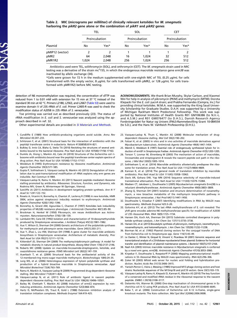
Inducible Expression of pikR2 Alone or Combined with pikR1 Confers aHigh Level of Ketolide Resistance in a Clinically Relevant Host. After es-tablishing that the natural resistance mechanisms conferred by pikR1and pikR2 in the Streptomyces ketolide producer remain operationalon transfer to E. coli, we extended the investigation to a heterologousGram-positive model, because Gram-positive pathogens are the pri-mary clinical targets of macrolides and ketolides. Mycobacterium tu-berculosis is the etiological agent of tuberculosis and considered apossible target for ketolide therapy (24). We constructed strains ofMycobacterium smegmatis, a laboratory model of the pathogenicmycobacteria, which carried either the pikR2 gene controlled by itsregulatory region or the entire pikR1-pikR2 cluster on a plasmid(Fig. 1A). The presence of pikR2 alone elevated the resistance ofM. smegmatis to clinical ketolides by 16- to 32-fold (Table 2). Thecombination of pikR1 and pikR2 conferred a much higher level ofresistance (exceeding 1mg/mL for TEL). Constitutive monomethylationof A2058 by PikR1 most likely facilitates continued ribosome activityand expression of pikR2 on abrupt exposure of the cells to high con-centrations of ketolides. WhenM. smegmatis cells with pikR2 or pikR1-pikR2 were preincubated with subinhibitory concentrations of TEL,resistance reached even higher levels, exceeding those of the controlcells by several hundredfold (Table 2). Thus, the resistance genesoriginating in the producer of natural ketolides can render heterologousbacteria resistant to high concentrations of clinical ketolide antibiotics.
DiscussionIn this paper, we present evidence that the genes pikR1 and pikR2have been evolutionary optimized to confer resistance to ketolideantibiotics. These genes not only render S. venezuelae ATCC 15439resistant to its endogenously produced ketolides but also, conferappreciable resistance to clinical ketolides. The resistance conferredby pikR1 and pikR2 can be transferred to heterologous Gram-neg-ative and -positive hosts. We further show that the fitness cost ofresistance is economized by the ketolide-inducible expression ofpikR2. Taking these factors into account, acquisition of the pikR1
and pikR2 genes is expected to confer a marked selective advantageon bacteria undergoing repeated exposure to ketolides, which couldpromote the transfer of these genes to pathogens when ketolidesbecome more widely used in a clinical setting.Our studies provide insights into the important question of why
S. venezuelae, the producer of the natural ketolides MTM and PKM,maintains two resistance genes with seemingly overlapping functions.The constitutively expressed PikR1 monomethylates 23S rRNA nucle-otide A2058 located at the site of ketolide action, and this modificationconfers an intermediate level of resistance. Higher levels of resistanceare attained with the inducible PikR2, which adds a second methylgroup to the same nucleotide. The combined action of two differen-tially expressed genes ensures active protein synthesis in the ketolide-producing cells across a broad range of the inhibitors concentrations.The inducibility of pikR2 is significant for the operation of the
resistance mechanism. Although dimethylation of A2058, at leastin Staphylococcus aureus, is known to significantly reduce cell fit-ness (6), monomethylation of the same nucleotide seems to havelittle, if any, effect on S. venezuelae fitness (Fig. S6). Thus, with aminimal toll in fitness cost for the constitutive PikR1 mono-methyltransferase, S. venezuelae easily tolerates low concentrationsof MTM and/or PKM. However, when S. venezuelae augments itsproduction of these drugs, activation of the more costly PikR2enzyme is required, resulting in A2058 dimethylation and higherresistance levels. Producers of other natural antibiotics, which carrymore than one resistance gene, may use a similar strategy forachieving the cost-effective protection from inhibitors (25).In contrast to other inducible erm genes, which respond pri-
marily to macrolides bearing a C3-cladinose, the regulation ofpikR2 has been evolutionary optimized for specific activation byketolides (Fig. 4). Ketolide-specific pikR2 induction is most likelycontrolled by programmed translation arrest within the pikR2Lleader ORF. Cladinose-containing macrolides are prone to pro-moting early peptidyl-tRNA drop off occurring within the first 6–10codons (26). Consistently, the sites of programmed macrolide-dependent translation arrest within the leader ORFs of the resistantgenes are located within the ORF’s first 10 codons, so that thedrug-bound ribosome is able to reach the arrest site without pre-maturely dissociating from the mRNA (5, 19, 23, 27). Ribosomestranslating pikR2L have to travel slightly farther and polymerize 13amino acids before reaching the stalling codon. Because ketolidesare less prone to promoting peptidyl-tRNA drop off (28), it iseasier for ketolide-bound ribosomes than for ERY-bound ribo-somes to polymerize the 13-aa-long nascent PikR2L chain. Indeed,the ERY-bound ribosome does not reach the stalling site (Fig. 4and Fig. S5) and thus, fails to trigger pikR2 expression. Ketolide-induced stalling in the pikR2L ORF occurs within the motif RLR,which represents one of the most problematic sequences fortranslation by ketolide-bound ribosomes (28, 29). We note that ashort ORF is also found in front of the constitutively expressed
Table 1. MIC (micrograms per milliliter) of TEL or chloramphenicol for S. venezuelae ΔpikAstrains containing different pikR resistance genes
Strain†
Antibiotics
TEL Chloramphenicol
Preincubation
Fold change
Preincubation
Fold changeNo Yes‡ No Yes‡
WT* 8 64 8 8 8 1Δ 0.25 0.25 1 8 8 1R1 8 16 2 8 8 1R2 0.25 16 64 8 8 1
†The ΔpikA strain, which contained both pikR1 and pikR2 resistance genes but was unable to produce anti-biotics (DHS2001), was designated WT* control. Formal names of the other strains are DHS332 (Δ), DHGS328(R1), and DHS330 (R2).‡Cells were grown for 8 h in LB supplemented with one-fourth MIC of TEL (0.0625 μg/mL for Δ and R2 strains or2 μg/mL for WT* and R1 strains) before MIC testing.
Almutairi et al. PNAS | October 20, 2015 | vol. 112 | no. 42 | 12959
BIOCH
EMISTR
Y

pikR1 gene (Fig. S7A), although this ORF does not promotemacrolide- or ketolide-dependent stalling (Fig. S7B) and thus,seems to play no role in the expression of pikR1.The properties and regulation of the pikR1 and pikR2 genes are
principally important for tolerating the broad range of inhibitorconcentrations experienced by S. venezuelae. Constitutive mono-methylation of A2058 by PikR1 renders ribosomes moderately re-sistant to ketolides, affording protection to S. venezuelae at the earlystages of antibiotic production. Monomethylation of A2058 is acritical step toward acquisition of a high level of resistance, facili-tating efficient translation of pikR2, despite the presence of a keto-lide inhibitor. Indeed, on abrupt exposure of the S. venezuelae pikR1-null mutant (ΔpikR1) to TEL, the lone pikR2 gene was unable toprovide any significant protection (Table 1), but when both pikRgenes were present and cells were preincubated with low ketolideconcentrations (mimicking the onset of antibiotic production), a highlevel of resistance was achieved. In the absence of pikR1, the re-sistance of S. venezuelae to TEL afforded by pikR2 alone, even onpreincubation with the antibiotic, was no higher than that providedby the constitutively expressed monomethylase (Table 1). Thisintermediate level of resistance may be caused by either dimethy-lation of only a small fraction of ribosomes or mixed mono- anddimethylations, because Erm dimethyltransferases are known to addtwo methyl groups to A2058 in two consecutive reactions (30).The synergy between the pikR1 and pikR2 genes was observed to
an even greater extent inM. smegmatis, which was used to model theexpression of the pikR genes in clinical pathogens. Here, the pikR2gene alone conferred considerable resistance to clinical ketolides.However, it was the simultaneous presence of both pikR1 and pikR2genes that provided the highest resistance (Table 2). The resistanceto TEL afforded by pikR1 and pikR2 in M. smegmatis was signifi-cantly higher than in S. venezuelae. Factors that could contribute tothis effect include the increased dosage of the resistance genes in-troduced into mycobacteria on a plasmid, the slower subunit as-sembly resulting in a higher proportion of methylated rRNA, or thelower affinity of the antibiotic for modified mycobacterial ribosomes.The inducibility of pikR2, which lowers the fitness cost of the two-
gene resistance mechanism, may facilitate not only its acquisition bya new host through horizontal gene transfer but also, its mainte-nance on discontinuation of the antibiotic treatment. Horizontaltransfer of the pikR1/pikR2 gene pair may be further facilitated bytheir close physical proximity in the S. venezuelae chromosome (Fig.1A). Despite the high GC content of pikR1 and pikR2, our resultsstrongly suggest that their transfer into at least some clinical strainswould render the strains highly resistant to ketolide therapy. Thesefindings, thus, provide additional justification for renewed drugdiscovery efforts to identify novel antibiotics capable of overcomingprotection rendered by natural resistance genes.
Materials and MethodsFor isolation of the total RNA from S. venezuelae, cells were grown overnight inSGGP media, then diluted 1:100 in SCM media (31), and grown for 5 d at 30 °Cwith constant shaking. Cells were pelleted from 5-mL cultures, resuspended in1mL Tris-EDTA (TE) buffer (10mMTris·HCl, pH 8.0, 1mMEDTA) containing 5mg/mLlysozyme, incubated for 10 min at room temperature, and then, shaken for10min in theMinibead Breaker (Biospec Products) with 50 μL glass beads (particlesize ≤106 μm; Sigma-Aldrich). Total RNA was isolated using the RNeasy MaxiKit (Qiagen).
The RNeasy Plus Mini Kit was used to prepare total RNA from exponentiallygrowing E. coli following the standard manufacturer’s protocol. Primer extensionanalysis under the standard conditions suitable for detection of adenine N6dimethylation was carried out as previously described (21). In experiments where
Fig. 4. The pikR2 regulatory region controls the inducible expression of thepikR2 resistance gene. (A) The putative leader ORF pikR2L precedes the pikR2resistance gene. The nucleotide sequence of the ORF and the amino acid sequenceof the encoded leader peptide are shown. (B) Antibiotic disk diffusion assay re-veals inducibility of pikR2 in E. coli. In the reporter construct in E. coli cells, thepikR2 regulatory region controls expression of the lacZα reporter. The antibioticdisks contained TEL, MTM, PKM, ERY, or chloramphenicol (CHL). The clear areasaround the disks contain antibiotic concentrations that inhibited cell growth. Bluehalos around the ketolide-containing disks indicate drug-dependent induction ofthe reporter. (C, Upper) Toe-printing analysis shows ketolide-induced ribosomestalling at the Leu13 codon of the pikR2L ORF. The band of the ribosomes stalledby the control antibiotic thiostrepton (THS) at the initiation codon is indicated by
black arrows. The ribosomes arrestedwith the Leu13 codon in their P site are shownby the red arrows. The ribosomes that reached the pikR2L 13th codon but failed toarrest translation were captured at the next Arg14 codon (blue arrows) because ofthe depletion of Ile-tRNA from the translation reaction by the presence of theIle-tRNA synthetase inhibitor,mupirocin. The stop codon or theORF is indicatedwithan asterisk. (C, Lower) Stalling efficiency calculated from the ratios of the intensity ofthe bands representing ketolide-dependent arrest (codon 13) vs. read through (co-don 14). Error bars indicate data spreads in two independent experiments.
12960 | www.pnas.org/cgi/doi/10.1073/pnas.1512090112 Almutairi et al.
detection of N6 monomethylation was required, the concentration of dTTP wasreduced from 1 to 0.01 mM with extension for 15 min at 37 °C instead of thestandard 30min at 42 °C. Primers L2180, L2563, and L2667 (Table S3) were used toexamine domain V of 23S rRNA of E. coli. Primer L2405-R was used to check themodification status of A2058 in 23S rRNA of S. venezuelae.
Toe printing was carried out as described previously (23). The status ofrRNA modification in E. coli and S. venezuelae was analyzed using the ap-proach described in ref. 32.
Other experimental details are provided in SI Materials and Methods.
ACKNOWLEDGMENTS. We thank Brian Murphy, Skylar Carlson, and XiaomeiWei for help in analysis of pikromycin (PKM) and methymycin (MTM); DorotaKlepacki for the E. coli ΔacrA strain; and Prabha Fernandes (Cempra, Inc.) forproviding clinical ketolides. M.M.A. was supported by the King Saud Univer-sity Scholarship for Graduate Studies. D.A.H. was supported by a Universityof Michigan Rackham Merit Predoctoral Fellowship. This work was sup-ported by National Institutes of Health Grants R01 GM106386 (to N.V.-L.and A.S.M.) and R01 GM076477 (to D.H.S.), Danish Research AgencyForskningsrådet for Natur og Univers (FNU)-Rammebevilling Grant 10-084554(to S.D.), and the Hans W. Vahlteich Professorship (D.H.S.).
1. Cundliffe E (1989) How antibiotic-producing organisms avoid suicide. Annu RevMicrobiol 43:207–233.
2. Schlünzen F, et al. (2001) Structural basis for the interaction of antibiotics with thepeptidyl transferase centre in eubacteria. Nature 413(6858):814–821.
3. Bulkley D, Innis CA, Blaha G, Steitz TA (2010) Revisiting the structures of several anti-biotics bound to the bacterial ribosome. Proc Natl Acad Sci USA 107(40):17158–17163.
4. Dunkle JA, Xiong L, Mankin AS, Cate JH (2010) Structures of the Escherichia coli ri-bosome with antibiotics bound near the peptidyl transferase center explain spectra ofdrug action. Proc Natl Acad Sci USA 107(40):17152–17157.
5. Weisblum B (1995) Erythromycin resistance by ribosome modification. AntimicrobAgents Chemother 39(3):577–585.
6. Gupta P, Sothiselvam S, Vázquez-Laslop N, Mankin AS (2013) Deregulation of trans-lation due to post-transcriptional modification of rRNA explains why erm genes areinducible. Nat Commun 4:1984.
7. Vázquez-Laslop N, Ramu H, Mankin AS (2011) Nascent peptide–mediated ribosomestalling promoted by antibiotics. Ribosomes: Structure, Function, and Dynamics, edsRodnina MV, Green R, Wintermeyer W (Springer, Vienna).
8. Sutcliffe JA (2011) Antibiotics in development targeting protein synthesis. Ann N YAcad Sci 1241:122–152.
9. Rosato A, Vicarini H, Bonnefoy A, Chantot JF, Leclercq R (1998) A new ketolide, HMR3004, active against streptococci inducibly resistant to erythromycin. AntimicrobAgents Chemother 42(6):1392–1396.
10. Bonnefoy A, Girard AM, Agouridas C, Chantot JF (1997) Ketolides lack inducibilityproperties of MLS(B) resistance phenotype. J Antimicrob Chemother 40(1):85–90.
11. Brockman H, Henkel W (1950) Pikromycin, ein neues Antibiotikum aus Actino-myceten. Naturwissenschaften 37(6):138–139.
12. Lambalot RH, Cane DE (1992) Isolation and characterization of 10-deoxymethynolideproduced by Streptomyces venezuelae. J Antibiot (Tokyo) 45(12):1981–1982.
13. Xue Y, Wilson D, Sherman DH (2000) Genetic architecture of the polyketide synthasesfor methymycin and pikromycin series macrolides. Gene 245(1):203–211.
14. Xue Y, Zhao L, Liu HW, Sherman DH (1998) A gene cluster for macrolide antibioticbiosynthesis in Streptomyces venezuelae: Architecture of metabolic diversity. ProcNatl Acad Sci USA 95(21):12111–12116.
15. Kittendorf JD, Sherman DH (2009) The methymycin/pikromycin pathway: A model formetabolic diversity in natural product biosynthesis. Bioorg Med Chem 17(6):2137–2146.
16. Roberts MC (2008) Update on macrolide-lincosamide-streptogramin, ketolide, andoxazolidinone resistance genes. FEMS Microbiol Lett 282(2):147–159.
17. Auerbach T, et al. (2009) Structural basis for the antibacterial activity of the12-membered-ring mono-sugar macrolide methymycin. Biotechnologia 1(84):24–35.
18. Jung WS, et al. (2006) Heterologous expression of tylosin polyketide synthase andproduction of a hybrid bioactive macrolide in Streptomyces venezuelae. ApplMicrobiol Biotechnol 72(4):763–769.
19. Ramu H, Mankin A, Vazquez-Laslop N (2009) Programmed drug-dependent ribosomestalling. Mol Microbiol 71(4):811–824.
20. Vázquez-Laslop N, et al. (2011) Role of antibiotic ligand in nascent peptide-dependent ribosome stalling. Proc Natl Acad Sci USA 108(26):10496–10501.
21. Bailey M, Chettiath T, Mankin AS (2008) Induction of erm(C) expression by non-inducing antibiotics. Antimicrob Agents Chemother 52(3):866–874.
22. Hartz D, McPheeters DS, Traut R, Gold L (1988) Extension inhibition analysis oftranslation initiation complexes. Methods Enzymol 164:419–425.
23. Vazquez-Laslop N, Thum C, Mankin AS (2008) Molecular mechanism of drug-dependent ribosome stalling. Mol Cell 30(2):190–202.
24. Falzari K, et al. (2005) In vitro and in vivo activities of macrolide derivatives againstMycobacterium tuberculosis. Antimicrob Agents Chemother 49(4):1447–1454.
25. Memili E, Weisblum B (1997) Essential role of endogenously synthesized tylosin for in-duction of ermSF in Streptomyces fradiae. Antimicrob Agents Chemother 41(5):1203–1205.
26. Tenson T, Lovmar M, Ehrenberg M (2003) The mechanism of action of macrolides,lincosamides and streptogramin B reveals the nascent peptide exit path in the ribo-some. J Mol Biol 330(5):1005–1014.
27. Sothiselvam S, et al. (2014) Macrolide antibiotics allosterically predispose the ribo-some for translation arrest. Proc Natl Acad Sci USA 111(27):9804–9809.
28. Kannan K, et al. (2014) The general mode of translation inhibition by macrolideantibiotics. Proc Natl Acad Sci USA 111(45):15958–15963.
29. Davis AR, Gohara DW, Yap MN (2014) Sequence selectivity of macrolide-inducedtranslational attenuation. Proc Natl Acad Sci USA 111(43):15379–15384.
30. Madsen CT, Jakobsen L, Douthwaite S (2005) Mycobacterium smegmatis Erm(38) is areluctant dimethyltransferase. Antimicrob Agents Chemother 49(9):3803–3809.
31. Zhang Q, Sherman DH (2001) Isolation and structure determination of novamethy-mycin, a new bioactive metabolite of the methymycin biosynthetic pathway inStreptomyces venezuelae. J Nat Prod 64(11):1447–1450.
32. Douthwaite S, Kirpekar F (2007) Identifying modifications in RNA by MALDI massspectrometry. Methods Enzymol 425:3–20.
33. Golovina AY, et al. (2012) The last rRNA methyltransferase of E. coli revealed: TheyhiR gene encodes adenine-N6 methyltransferase specific for modification of A2030of 23S ribosomal RNA. RNA 18(9):1725–1734.
34. Hansen DA, Koch AA, Sherman DH (2015) Substrate controlled divergence in poly-ketide synthase catalysis. J Am Chem Soc 137(11):3735–3738.
35. Hansen DA, et al. (2013) Biocatalytic synthesis of pikromycin, methymycin, neomethymycin,novamethymycin, and ketomethymycin. J Am Chem Soc 135(30):11232–11238.
36. Bierman M, et al. (1992) Plasmid cloning vectors for the conjugal transfer of DNAfrom Escherichia coli to Streptomyces spp. Gene 116(1):43–49.
37. Le Dantec C, Winter N, Gicquel B, Vincent V, Picardeau M (2001) Genomic sequence andtranscriptional analysis of a 23-kilobasemycobacterial linear plasmid: Evidence for horizontaltransfer and identification of plasmid maintenance systems. J Bacteriol 183(7):2157–2164.
38. Nash KA (2003) Intrinsic macrolide resistance in Mycobacterium smegmatis is conferredby a novel erm gene, erm(38). Antimicrob Agents Chemother 47(10):3053–3060.
39. Kirpekar F, Douthwaite S, Roepstorff P (2000) Mapping posttranscriptional modifi-cations in 5S ribosomal RNA by MALDI mass spectrometry. RNA 6(2):296–306.
40. Zuker M (2003) Mfold web server for nucleic acid folding and hybridization pre-diction. Nucleic Acids Res 31(13):3406–3415.
41. Yanisch-Perron C, Vieira J, Messing J (1985) ImprovedM13 phage cloning vectors and hoststrains: Nucleotide sequences of the M13mp18 and pUC19 vectors. Gene 33(1):103–119.
42. Vázquez-Laslop N, Ramu H, Klepacki D, Kannan K, Mankin AS (2010) The key functionof a conserved and modified rRNA residue in the ribosomal response to the nascentpeptide. EMBO J 29(18):3108–3117.
43. Datsenko KA, Wanner BL (2000) One-step inactivation of chromosomal genes in Es-cherichia coli K-12 using PCR products. Proc Natl Acad Sci USA 97(12):6640–6645.
44. Baba T, et al. (2006) Construction of Escherichia coli K-12 in-frame, single-geneknockout mutants: The Keio collection. Mol Syst Biol 2(2006):2006.0008.
Table 2. MIC (micrograms per milliliter) of clinically relevant ketolides for M. smegmatisharboring the pikR2 gene alone or the combination of pikR1 and pikR2 genes
Plasmid
TEL SOL CET
Preincubation Preincubation Preincubation
No Yes* No Yes* No Yes*
pMIP12 (vector) 2 2 1 1 1 2pMR2 64 2,048 16 1,024 32 512pMR1R2 1,024 2,048 256 1,024 256 512
Antibiotics used were TEL, solithromycin (SOL), and cethromycin (CET). The M. smegmatis strain used in MICtesting was a derivative of the strain mc2155, in which the endogenous macrolide resistance gene erm38 wasinactivated by allelic exchange (34).*Cells were grown for 72 h in the medium supplemented with one-eighth MIC of TEL (0.25 μg/mL for cellstransformed with the empty vector, 8 μg/mL for cells transformed with pMR2, or 128 μg/mL for cells trans-formed with pMR1R2) before MIC testing.
Almutairi et al. PNAS | October 20, 2015 | vol. 112 | no. 42 | 12961
BIOCH
EMISTR
Y

Supporting InformationAlmutairi et al. 10.1073/pnas.1512090112SI Materials and MethodsAntibiotics, Enzymes, and Chemicals. MTM and PKM were syn-thesized chemically as previously described (35) and repurifiedby preparatory HPLC using a Phenomenex Luna 5u C18 250 ×21.2-mm column (serial 444304–4) monitored at 250 nm at a flowrate of 9 mL/min with an isocratic mobile phase of H2O:MeCN(45:55) and a 0.1% NEt3 modifier. TEL, cethromycin, and soli-thromycin were from Cempra, Inc.; and ERY, clindamyicn, chlor-amphenicol, and thiostrepton were from Sigma Aldrich. Enzymesused for DNA cloning were from Fermentas. [γ32P]-ATP (specificactivity of 6,000 Ci/mmol) was from MP Biomedicals. Other re-agents and chemicals were purchased from either Fisher Scientificor Sigma Aldrich. All oligonucleotides used in the study were syn-thesized by Integrated DNA Technologies and are shown in Table S3.
Strains and Plasmids. To generate Escherichia coli strains consti-tutively expressing PikR1 and PikR2 methyltransferase enzymes,their corresponding genes were PCR-amplified from the Strep-tomyces venezuelae strain ATCC 15439 genomic DNA using theprimers NdeI-pikR1-D2 and AflII-pikR1-R2 or NdeI-pikR2-D2and AflII-pikR2-R2 (Table S3), respectively, which introducedstrong Shine-Dalgarno sequence upstream from the initiator AUGcodons. The PCR products were digested with NdeI and AflII andcloned in the corresponding sites of the plasmid pERMZα (21)(Table S1) behind the Ptac promoter. The resulting plasmidspPikR1 and pPikR2 were used to transform the JM109 ΔacrAstrain, and the transformants were used for MIC testing andRNA preparation.To study the inducibility of pikR1 and pikR2 genes, their regu-
latory regions (including the putative leader ORFs, the intergenicregion, and the first five codons of the resistance genes) were PCR-amplified from the DNA of S. venezuelae ATCC15439 using theprimers ORF-PikR1-D-new and ORF-PikR1-R or ORF2-PikR2-Dand ORF2-PikR2-R, respectively. The PCR products were di-gested with NdeI and AflII and cloned in the corresponding sites ofplasmid pERMZα. The JM109 ΔacrB strain was transformed withthe resulting plasmids pRL1 and pRL2, and the transformantswere used for antibiotic disk diffusion experiments.For studying the expression of the individual resistance genes in
S. venezuelae, strains that lack pikR1, pikR2, or both of them wereengineered. To prepare the strain that had only pikR1, the pikR2gene in the S. venezuelae DHS2001 strain (Table S2) was in-activated by in-frame deletion of 271 codons (codons 31–301) toavoid any polar effects. To construct this deletion, the plasmidpSRP112 (Table S1), based on the E. coli–Streptomyces shuttlevector pKC1139 (36), was constructed by amplifying and cloningleft- and right-flanking regions of the pikR2 genes using the ge-nomic DNA of S. venezuelae DHS2001 as a template, primer pairsSR199–SR200 and SR201–SR202, and KOD Xtreme DNA Po-lymerase (Novagen). The plasmid was assembled using Gibsonassembly (New England Biolabs Kit) according to the manufac-turer’s instructions. Briefly, the amplified PCR products offlanking regions of pikR2 and linearized pKC1139 vector withEcoRI and HindIII were incubated at 50 °C for 2 h. After in-cubation, the samples were transformed into E. coli DH5α. Re-striction digestion and sequencing verified the isolated pSRP112plasmid. The plasmid pSRP112 was then introduced into the S.venezuelae DHS2001 by protoplasts-based transformation. Astrain in which a single cross-over between the pSRP112 plasmidand the S. venezuelae DHS2001 chromosome had occurred wasselected by cultivation of antibiotic-resistant transconjugants at 37°C (the nonpermissive temperature for the pSG5-based replicon)
in the presence of apramycin. Cells from one colony weresubjected to second rounds of propagation in the absence ofapramycin to allow for the second cross-over. The desired doublecross-over mutant ΔpikR2 was selected by its apramycin-sensitivephenotype and verified by PCR. The resulting pikR2 deletionmutant of S. venezuelae DHS2001 was designated R1 (DHS328).The ΔpikR1 and ΔpikR1-pikR2 mutants were generated in thesame way as described above using the primer pairs SR216–SR217and SR218–SR219 for ΔpikR1 and SR219–SR220 and SR221–SR222 for ΔpikR1-pikR2. The pikR1 in-frame deletion encom-passed codons 11–327, and the pikR1-pikR2 deletion left intact thefirst 10 codons of pikR1 and pikR2, removing the entire DNAsegment in between them. The resulting pikR1- and pikR1-pikR2deletion mutants of S. venezuelae DHS2001 were designated R2(DHS330) and Δ (DHS332), respectively.To check the resistance conferred by the pikR2 gene in
Mycobacterium smegmatis, pikR2 was PCR-amplified with itsregulatory region (including the pikR2L and the intergenic re-gion) from the DNA of S. venezuelae strain ATCC15439 usingthe primers pMIP12-R2-D2-short and pMIP12-R2-R-short. ThePCR product was gel-purified and used as a template for thesecond PCR using the primers pMIP12-R2-D2 and pMIP12-R2-R. The PCR product was gel-purified and introduced by Gibsonassembly into the pMIP12 plasmid (37) cut with BamHI andSpeI restriction enzymes. The reaction mixture was transformedinto E. coli JM109. The recombinant plasmid designated pMR2(Table S1) was isolated, sequenced, and introduced into theΔerm38 strain of M. smegmatis (strain mc2155 ermKO-4) (38)(Table S2) by electroporation. The transformed cells were platedon agar plates containing 15 μg/mL kanamycin.To clone the pikR2-pikR1 tandem (that contains pikR2 with
its leader ORF and pikR1 with it native promoter), a 3.25-kbDNA fragment from the genomic DNA of S. venezuelae was ini-tially PCR-amplified using the primers pMIP12-R2-D2-short andpMIP12-R1-R-short. The purified PCR product was reamplifiedusing the primers pMIP12-R2-D2 and pMIP12-R1-R to introduceflanking regions suitable for cloning in the pMIP12 vector. Theresulting PCR product was introduced into the BamHI and SpeIcut plasmid pMIP12 by Gibson assembly as described above. Theresulting plasmid, pMR1R2 (Table S1), was eventually introducedinto M. smegmatis strain mc2155 ermKO-4.
Toe-Printing Assay. Toe printing was carried out as describedpreviously (23). The templates containing genes coding for theputative PikR leader peptides under the control of the T7 pro-moter were generated by PCR using primers T7-pikR1-ORF1-fwd, pikR1-ORF1-rev-spacer-NVI (pikR1L), T7-SD-ORF2-pikR2, O2-R2-IL-R, T7, and O2-R2-NV1-R (piKR2L) (TableS3). The reverse PCR primer replaced the penultimate pikR2Arg codon (CGC) with an Ile codon (ATC) to enable macrolide-independent translation arrest at this codon. The templates weretranslated in 5 μL PURExpress cell-free translation reactions(New England Biolabs) for 30 min at 37 °C. All of the reactionscontained 50 μМ mupirocin, an Ile-tRNA synthetase inhibitor.When needed, other antibiotics (MTM, PKM, ERY, TEL, orthiostrepton) were added to the final concentration of 50 μM.After the addition of 5′-[32P]–radiolabeled primer NV1, reversetranscription was carried out for 15 min at 37 °C. Samples werethen processed and analyzed as described in ref. 23.
MS Analysis of rRNA Modification. The status of rRNA modifica-tion in E. coli and S. venezuelae was analyzed using the approach
Almutairi et al. www.pnas.org/cgi/content/short/1512090112 1 of 8

described previously (32). Briefly, total RNA was extracted from cells(see above) and hybridized with the DNA oligonucleotides Ecprotectand SVprotect (Table S3) complementary to the 23S rRNA se-quence C2035-C2084 (E. coli) or C2025-C2083 (S. venezuelae).rRNA regions that were not protected by hybridization were digestedaway with nucleases, and the protected rRNA fragments were iso-lated by gel electrophoresis. The purified material was digested withRNaseT1 or RNaseA and subjected to MALDI-TOF on a BrukerDaltronics Ultraflextreme Spectrometer recording in reflector andpositive ion modes (39). Spectra were analyzed using Flexanalysissoftware (Bruker).
Microbiological Testing. MICs of antibiotics were determined bythe broth microdilution assay. The MIC values were read after anovernight incubation (E. coli and S. venezuelae) or a 3-d in-cubation (M. smegmatis) at 37 °C.Disk diffusion assays for testing the inducibility of the pRL1
and pRL2 reporters (Table S1) were carried out essentially asdescribed previously (21), with the exception that the JM109ΔacrB strain was used as the host. Briefly, cells transformedwith reporter plasmids were grown overnight in the presenceof 100 μg/mL ampicillin and 0.5 mM isopropyl β-D-1-thiogalactopyranoside (IPTG), and then, 1.5 mL cell cultureswere mixed with 8.5 mL soft agar [0.6% (wt/vol) LB agar at 50 °C]and overlaid on agar plates containing 100 μg/mL ampicillin,0.5 mM IPTG, and 80 μg/mL X-Gal. After the soft agar solidified,5 mmWhatmann 3MM Paper Disks containing 32 μg MTM, 32 μgPKM, 32 μg ERY, 32 μg TEL, or 8 μg chloramphenicol were
placed on top of the agar; plates were incubated for 18–24 h at37 °C and then, photographed.
Growth Competition. S. venezuelae Δ and R1 strains were grownseparately overnight at 30 °C with shaking in CRM medium(10 g/L glucose, 103 g/L sucrose, 10.12 g/L MgCl2·6H2O, 15 g/Ltryptic soy broth, 5 g/L yeast extract). The overnight cultureswere diluted to A600 of 0.04 in fresh CRM medium and grown at30 °C with shaking until they reached A600 of 0.5. Equal culturevolumes of Δ and R1 strains were mixed and grown overnightwith shaking at 30 °C. At each cycle, the culture was diluted1,000-fold into fresh CRM medium and grown for 18 h. Theculture was grown for a total of four passages corresponding to∼40 cell generations. The ratio of cells was determined at cycles2–4 by isolating total RNA from the coculture and examining theextent of monomethylation of A2058 in 23S rRNA by primerextension (Fig. S6). Pure cultures of Δ and R1 strains were usedto prepare control RNA that was used for calibration.
Analysis of the Extent of A2058 Modification. The extent of 23SrRNA A2058 mono- or dimethylation was assessed by primerextension as described previously (21) with minor modifications.Specifically, 0.5 pmol 5′-[32P]–labeled primer SvL2309 (Table S3)was annealed to 1 μg total S. venezuelae RNA and extended withAMV Reverse Transcriptase (Roche) in the presence of 1 mMdCTP and dATP, 1 mM ddGTP, and 0.001 mM dTTP (forA2058 monomethylation) or 0.25 mM dTTP (for A2058 dime-thylation). The cDNA products were purified by phenol extrac-tion, precipitated, resolved in a denaturing 12% polyacrylamidegel, and visualized by phosphoimaging.
Fig. S1. Similarity of proteins encoded in the pikR1 and pikR2 genes in S. venezuelae ATCC 15439.
Almutairi et al. www.pnas.org/cgi/content/short/1512090112 2 of 8

Fig. S2. Primer extension analysis of modification of rRNA extracted from (A) E. coli or (B) S. venezuelae. The segments of the full gels represented in Figs. 2and 3 are boxed. Sequencing lanes are marked with the letters C, U, A, G according to the rRNA nucleotides. W, WT.
Fig. S3. Primer extension on 23S rRNAs from (A) S. venezuelae and (B) E. coli. The extension reactions were performed with 1 mM dTTP and 1 mM ddCTP (H) or0.01 mM dTTP and 1 mM ddCTP (L). The dideoxynucleotide completely stops reverse transcription at G2056 in S. venezuelae rRNA and at G2057 in E. coli rRNA.Gel band intensities were measured by scanning on a Typhoon FLA 9500 (GE Healthcare) and used to quantify the stops at A2058. The band at A2058 inextensions with 1 mM dTTP is caused by m6
2 dimethylation, whereas extensions with the lower dTTP concentration detect both monomethylated and di-methylated A2058. The proportion of A2058 nucleotides that were m6
2-dimethylated after ketolide induction was slightly above 30% (WT H lane). Note that,in contrast to adenine m6
2 dimethylation, m6A monomethylation does not completely arrest primer extension, even under low dTTP concentration conditions;therefore, the primer extension technique can be used to reliably quantify the extent of A2058 dimethylation but not monomethylation. G, A, U, and C aredideoxy sequencing lanes. K, E. coli cells with the empty vector; P, primer band. Sequencing lanes are marked G, A, U, C.
Almutairi et al. www.pnas.org/cgi/content/short/1512090112 3 of 8

Fig. S4. Primer extension analysis of mono- and dimethylation of A2058 in 23S rRNA extracted from different S. venezuelae mutants without (−) and with (+)preincubation for 10 h with one-fourth MIC of TEL. All of the strains used in the experiment were derivatives of the ΔpikA strain (WT*) containing only pikR1(R1), only pikR2 (R2), or lacking both of the pikR genes (Δ).
Fig. S5. The models of the secondary structure of pikR2 mRNA in the (A) noninduced and (B) induced states. The nucleotide sequence of the leader ORFpikR2L is italicized. The RLR sequence in the PikR2L leader peptide encompassing the Leu13 stalling codon is boxed, and the codon is underlined. The Shine–Dalgarno region of the pikR2 gene is shown in red, and the initiator codon is blue. The models are based on mfold predictions (40).
Almutairi et al. www.pnas.org/cgi/content/short/1512090112 4 of 8

Fig. S6. Growth competition reveals low fitness cost associated with A2058 monomethylation in S. venezuelae. S. venezuelae strains Δ and R1 deficient inantibiotic production and lacking both resistance genes (Δ) or carrying only pikR1 gene (R1) were mixed at approximately equal amounts and grown for theindicated number of generations with four consecutive passages with 1:1,000 dilutions. After isolation of the total RNA, the fraction of the R1 strain wasassessed by the extent of A2058 monomethylation using primer extension. If RT did not pause because of A2058 modification, it would completely stop atC2055 because of incorporation of ddGTP. Primer extensions carried out on RNA prepared from the pure cultures of the Δ and R1 strains were used forcalibration. The estimated fraction of the R1 strain in the co-growth culture is plotted.
Almutairi et al. www.pnas.org/cgi/content/short/1512090112 5 of 8

Fig. S7. In contrast to the mechanism seen for pikR2, regulation of the pikR1 gene does not occur by stalling at its putative upstream ORF. (A) The sequence ofthe putative upstream ORF (pikR1L) and the encoded protein sequence. (B) Toe-printing analysis does not show any ketolide- or macrolide-specific stops duringin vitro translation of the pikR1L ORF. The black arrows indicate the toe-printing band corresponding to the ribosome stalled by the control antibiotic thi-ostrepton (THS) at the pikR1L initiator codon (boxed). The green arrows show the band representing the ribosomes that were captured at the pikR1L 11thcodon (boxed) because of the depletion of Ile-tRNA from the translation reaction by the presence of the Ile-RS inhibitor, mupirocin. Sequencing lanes aremarked G, A, U, C.
Almutairi et al. www.pnas.org/cgi/content/short/1512090112 6 of 8

Table S1. Plasmids used in the study
Plasmid name Notes Source
pERMZα The reporter plasmid containing the regulatory region of the macrolide-inducible ermC gene, in whichcodons 3–244 of the ermC gene are replaced with 57 codons of lacZ γeνe
21
pPikR1 pERMZα derivative constitutively expressing the pikR1 gene under the control of the Ptac promoter andoptimized Shine–Dalgarno sequence
This study
pPikR2 pERMZα derivative constitutively expressing the pikR1 gene under the control of the Ptac promoter andoptimized Shine–Dalgarno sequence
This study
pRL1 pERMZα-derived reporter plasmid containing the regulatory region of the pikR1 gene, in which codons 6–331of the pikR1 gene are replaced with 57 codons of lacZα; the transcription is driven by the Ptac promoter
This study
pRL2 The reporter plasmid containing the regulatory region of the pikR1 gene, in which codons 6–317 of the pikR2gene are replaced with 57 codons of lacZα; the transcription is driven by the Ptac promoter
This study
pRL2R2 pERMZα-derived plasmid containing the pikR2 gene and 208 bp of the upstream regulatory region including thepikR2L regulatory ORF; the transcription is driven by the Ptac promoter
This study
pKC1139 E. coli–Streptomyces shuttle vector pKC1139 containing a temperature-sensitive replicon 36pSRP112 pKC1139-derived plasmid containing flanking regions of the pikR2 gene; used for inactivation of pikR2 in the
genome of S. venezuelae DHS2001This study
pSRP113 pKC1139-derived plasmid containing flanking regions of the pikR1 gene; used for deletion of pikR1 from thegenome of S. venezuelae DHS2001
This study
pSRP114 pKC1139 containing flanking regions of the pikR1-pikR2 gene cluster; used for deletion of the pikR1-pikR2cluster from the genome of S. venezuelae DHS2001
This study
pMIP12 E. coli–M. smegmatis shuttle vector containing kanamycin resistance gene 37pMR2 pMIP12-derivative containing pikR2 with its regulatory region under the control of the PBlaF* promoter This studypMR1R2 pMIP12-derivative containing pikR2 with its regulatory region under the control of the PBlaF* promoter This study
Table S2. Strains used in the study
Strain Genotype Source
E. coli JM109 F′[traD36, proAB+ lacIq, Δ(lacZ)M15] endA1 recA1 hsdR17(rk−, mk+) mcrA supE44 λ-gyrA96relA1 Δ(lac-proAB) thi-1
41
E. coli JM109 ΔacrA JM109, ΔacrA This studyE. coli JM109 ΔacrB JM109, ΔacrB 42E. coli BW25113 F-, Δ(araD-araB)567, ΔlacZ4787(::rrnB-3), λ-, rph-1, Δ(rhaD-rhaB)568, hsdR514 43E. coli JW3466 BW25113, ΔrlmJ::kan 44S. venezuelae ATCC15439 WT 12WT* (DHS2001) S. venezuelae ATCC15439, ΔpikAI-pikAIV 18DHS8708 S. venezuelae ATCC15439, ΔdesI 34R1 (DHS328) DHS2001, ΔpikR2 This studyR2 (DHS330) DHS2001, ΔpikR1 This studyΔ (DHS332) DHS2001, ΔpikR1-ΔpikR2 This studyermKO-4 M. smegmatis mc2155 Δerm38 38
Almutairi et al. www.pnas.org/cgi/content/short/1512090112 7 of 8

Table S3. Primers used in the study
Name Sequence
NdeI-pikR1-D2 TCGTTCCATATGGCAATGCGCGACTCCAT
AflII-pikR1-R2 CTTAAGCTTAAGCCAGACCAGCGGGAGGCGGA
NdeI-pikR2-D2 GACTCCATATGGCATTTTCCCCGCAGGGCGG
AflII-pikR2-R2 CCCACCTTAAGGGTCGGATCCGGCTCAGCAC
ORF-PikR1-D-new CAGCTGCATATGGGTAACAGCCGATCCC
ORF-PikR1-R TCTAGCTTAAGGTCGCGCATTGCCATGAACGATCCC
ORF2-PikR2-D GACTATCATATGCAGTTCTGCCACTCTCAG
ORF2-PikR2-R TACTAGCTTAAGCGGGGAAAATGCCATGAG
T7-pikR1-ORF1-fwd ATTAATACGACTCACTATAGGGatataaggaggaaaaCatATGGGTAACAGCCGATCC
pikR1-ORF1-rev-spacer-NVI GGTTATAATGAATTTTGCTTATTAACGATAGAATTCTATCACATTATGTCGGGGGTGAAATCAA
T7-SD-ORF2-pikR2 ATTAATACGACTCACTATAGGGatataaggaggaaaaCatATGcagttc
O2-R2-IL-R TTAACGATAGAATTCTATCACGGACGCGCGAGGATCGAGACGCGTGAGGAGGGGCCCGCCGCTAGGAGATGCGCAGCCTCATGTAACGGG
SvL2309 TGTAGTAAAGGTCCCGGGG
Ecprotect GCAGUGUACCCGCGGCAAGACGGAAAGACCCCGUGAACCUUUACUAUAG
Svprotect GAGUAAAGAUGCUCGUUUCGCGCAGCAGGACGGAAAGACCCCGGGACCUUUACUACAG
T7 ATTAATACGACTCACTATAGGG
O2-R2-NV1-R GGTTATAATGAATTTTGCTTATTAACGATAGAATTCTATCACG
NV1 GGTTATAATGAATTTTGCTTATTAAC
L2405-R AGAGTGGTATTTCAACGGCGA
L2563 TCGCGTACCACTTTA
L2667 GGTCCTCTCGTACTAGGAGCAG
L2180 GGGTGGTATTTCAAGGTCGG
SR199 GCTATGACATGATTACGATTCGTCCCGGAGCGCCACACG
SR200 ACCGCATGCACCAGGCCGTCGATCTCG
SR201 GCCTGGTGCATGCGGTACGGAGCTCC
SR202 CGACGGCCAGTGCCAAGCTTGCGGCGGAAATTCGAAGG
SR216 GCTATGACATGATTACGAATTCGGAGTACTGGCCATCCGGC
SR217 CGAGGCGATCGTCGTACGGACGCCGC
SR218 TACGACGATCGCCTCGGTATGGAGTCG
SR219 CGACGGCCAGTGCCAAGCTTGTCTCCGGAAGCCGCGCT
SR220 GCTATGACATGATTACGAATTCCCACGACCCGACGCAG
SR221 CGAGGCGAGGAAAATGCCATGAGTCTGCTCC
SR222 GCATTTTCCTCGCCTCGGTATGGAGTCG
pMIP12-R2-D2 ATGGATTAGAAGGAGAAGTACCGATGGGATTCTGCCACTCTCAGGCCCGTTACA
pMIP12-R2-R TCGCCCGATCCCGTGTTTCGCTATTTCACGCGCTCTCCGCCCGCC
pMIP12-R2-D2-short TTCTGCCACTCTCAGGCCCGTTACA
pMIP12-R2-R-short TCACGCGCTCTCCGCCCGCC
pMIP12-R1-R TCGCCCGATCCCGTGTTTCGCTATTACGAATTCCTCGGACTCACTCTTGGAC
pMIP12-R1-R-short ACGAATTCCTCGGACTCACTCTTGGAC
acrAup CATATGTTCGTGAATTTACAG
acrAdown GCAATCGTAGGATATTGCG
Almutairi et al. www.pnas.org/cgi/content/short/1512090112 8 of 8