resecting dna samples and aging for finfish
TRANSCRIPT

Standard Operating Procedure
EAP008, Version 1.2
Standard Operating Procedures
for Resecting DNA Samples
and Aging for Finfish
December 2018 Publication 18-03-236 [Recertified 2017]

Purpose of this document
The Washington State Department of Ecology develops Standard Operating Procedures (SOPs) to
document agency practices related to sampling, field and laboratory analysis, and other aspects of the
agency’s technical operations.
Publication information
This SOP is available on the Department of Ecology’s website at
https://fortress.wa.gov/ecy/publications/SummaryPages/1803236.html.
Recommended citation:
Sandvik, P. 2018. Standard Operating Procedure EAP008, Version 1.2: Standard Operating Procedures
for Resecting DNA Samples and Aging for Finfish. Publication 18-03-236. Washington State
Department of Ecology, Olympia.
https://fortress.wa.gov/ecy/publications/SummaryPages/1803236.html. [Recertified 2017.]
Ecology’s Activity Tracker Code for this SOP is 16-073.
Contact information
Publications Coordinator
Environmental Assessment Program
P.O. Box 47600, Olympia, WA 98504-7600
Phone: 360-407-6764
Washington State Department of Ecology – ecology.wa.gov
Location of Ecology Office Phone
Headquarters, Lacey 360-407-6000
Northwest Regional Office, Bellevue 425-649-7000
Southwest Regional Office, Lacey 360-407-6300
Central Regional Office, Union Gap 509-575-2490
Eastern Regional Office, Spokane 509-329-3400
Any use of product or firm names in this publication is for descriptive purposes only
and does not imply endorsement by the author or the Department of Ecology.
To request ADA accommodation for disabilities, or printed materials in a format for the visually
impaired, call Ecology at 360-407-6764 or visit https://ecology.wa.gov/accessibility. People with
impaired hearing may call Washington Relay Service at 711. People with speech disability may call TTY at 877-833-6341.

EAP008 – Resecting DNA Samples and Aging for Finfish – V 1.2 – 5/3/2017 – Page 1
Uncontrolled copy when printed
Washington State Department of Ecology
Environmental Assessment Program
Standard Operating Procedures for Resecting DNA Samples and Aging for Finfish
Version 1.2
Author – Patti Sandvik
Date – 08/30/2006
Original Reviewer– Casey Deligeannis
Date – 08/30/2006
Current Author- Patti Sandvik
Date – 04/20/2017
Current Reviewer – Debby Sargeant, Toxics Studies Unit Supervisor
Date – 04/20/2017
QA Approval – William R. Kammin, Ecology Quality Assurance Officer
EAP008
Recertified – 05/03/2017
Signatures on File.
EAP008 – Resecting DNA Samples and Aging for Finfish – V 1.2 – 5/3/2017 – Page 2
Uncontrolled copy when printed
Please note that the Washington State Department of Ecology’s Standard Operating Procedures
(SOPs) are adapted from published methods, or developed by in-house technical and administrative
experts. Their primary purpose is for internal Ecology use, although sampling and administrative
SOPs may have a wider utility. Our SOPs do not supplant official published methods. Distribution of
these SOPs does not constitute an endorsement of a particular procedure or method.
Any reference to specific equipment, manufacturer, or supplies is for descriptive purposes only
and does not constitute an endorsement of a particular product or service by the author or by
the Department of Ecology.
Although Ecology follows the SOP in most instances, there may be instances in which Ecology uses an
alternative methodology, procedure, or process.

EAP008 – Resecting DNA Samples and Aging for Finfish – V 1.2 – 5/3/2017 – Page 3
Uncontrolled copy when printed
SOP Revision History.
Revision Date Rev
number
Summary of changes Sections Reviser(s)
08/30/06 V1.0 SOP Publication
10/19/10 V1.0 Three year review, minor changes
10/21/10 V1.0 QA approval, recertified
03/13/14 V1.0 Three year review, minor Changes
04/21/14 V1.1 QA approval, recertified
04/20/17 V1.2 Three year review, minor changes all Sandvik,
Sargeant
5/3/2017 V1.2 Cover page and footer,
recertificationdocument formatting
all Kammin
12/28/18 V1.2 added cover page and publication
number
Ponzetti
4/30/19 V1.2 minor edits cover
page,
inside
cover,
page 1
Ponzetti

EAP008 – Resecting DNA Samples and Aging for Finfish – V 1.2 – 5/3/2017 – Page 4
Uncontrolled copy when printed
Environmental Assessment Program
Standard Operating Procedure for Resecting DNA Samples and Aging Structures.
1.0 Purpose and Scope
1.1 This document is the Environmental Assessment Program (EAP) Toxics Study Unit
(TSU) Standard Operating Procedure (SOP) for resecting DNA samples and structures
for determining fish age in fish fin. The Washington State Department of Ecology
(Ecology) works cooperatively with the Washington Department of Fish and Wildlife
(WDFW) in the collection of fish DNA sample tissue and age structures. Age structures
include scales, otoliths, operculum and spines from pectoral and/or dorsal fins.
2.0 Applicability
2.1 This procedure is to be followed by Ecology person(s) conducting fish tissue processing
and at the time of processing the fish in part or as a whole.
3.0 Definitions
3.1 Caudal fin – The tail of fishes.
3.2 DNA – Deoxyribonucleic acid. A nucleic acid that carries the genetic information in the
cell and is capable of self-replication and synthesis of RNA. DNA consists of two long
chains of nucleotides twisted into a double helix and joined by hydrogen bonds between
the complementary bases adenine and thymine or cytosine and guanine. The sequence
of nucleotides determines individual hereditary characteristics (Wikipedia, 2017a).
3.3 Dorsal fin – The main fin located on the back of fishes.
3.4 EAP – Environmental Assessment Program.
3.5 Ecology – Washington State Department of Ecology.
3.6 Lab Analysis & Tracking Plan – A table, usually created in Excel®, used to plan and
document lab analyses of samples for single or multiple projects (Attachment 1).
3.7 Operculum – Any one of the bony plates which support the gill covers of fishes.
3.8 Otolith – One of many minute calcareous particles found in the inner ear of vertebrates.
Fish species have three pairs of otoliths. The largest pair is used for aging.
3.9 Pectoral fin – Either of the anterior pair of fins attached to the pectoral (mid) girdle of
fishes.
3.10 Polymerase Chain Reaction (PCR) amplification – This is a molecular biological
technique for amplifying (creating multiple copies of) DNA without using a living

EAP008 – Resecting DNA Samples and Aging for Finfish – V 1.2 – 5/3/2017 – Page 5
Uncontrolled copy when printed
organism. The technique allows a small sample of DNA to be copied multiple times so
it can be used for analysis. As the PCR reaction proceeds, the number of newly
synthesized DNA strands increases exponentially so that after 20 to 30 cycles, the initial
template is replicated several million-fold, thus facilitating its manipulation for further
analysis such as sequencing, genotyping or probe labeling (Wikipedia, 2017b).
3.11 Processing Bench Sheet – A table, usually created in Excel®, used to plan and
document sample processing data for each fish collected (Attachment 2).
3.12 Resecting – Surgical removal of all or part of an organ, tissue or structure.
3.13 SDS – Safety Data Sheets provides both workers and emergency personnel with the
proper procedures for handling or working with a particular substance. SDS’s include
information such as physical data (melting point, boiling point, flash point, etc.),
toxicity, health effects, first aid, reactivity, storage, disposal, protective equipment and
spill/leak procedures.
3.14 USFWS – United States Fish and Wildlife Service
3.15 WDFW – Washington Department of Fish and Wildlife
4.0 Personnel Qualifications/Responsibilities
4.1 Because this procedure requires use of hazardous materials, training is required as per
Ecology Chemical Hygiene Plan and Hazardous Materials Management Plan (Section
1) (Ecology, 2016), which includes a Laboratory Safety Orientation, Job-Specific
Orientation and must know Chemical Safety Procedures and follow the Standard
Operating Procedures (Section 16).
4.2 Prior to processing, read WDFW genetic sampling goals document “Considerations &
Guidelines for Collecting Representative Samples for Genetic Analysis” (Attachment
3). Should also work with experienced individual for hands on training.
5.0 Equipment, Reagents, and Supplies
5.1 DNA sampling supplies are obtained from the WDFW Molecular Genetics Laboratory.
Please see Section 6.0 for more details. These supplies include:
5.1.1 2ml screw-cap cryovials.
5.1.2 Sample boxes.
5.1.3 DNA preservative solution of ethanol 100 proof – NO SUBSTITUTES ACCEPTED.
See the Toxics TCT Sharepoint site (Toxics Documents) for the Safety Data Sheet and
concentration grade.

EAP008 – Resecting DNA Samples and Aging for Finfish – V 1.2 – 5/3/2017 – Page 6
Uncontrolled copy when printed
5.1.4 Labels for vials. Labels are laser printed on acid resistant paper and contain a four-digit
WDFW code (e.g., “CD05”). Never use labels for a sample set different from the
originally assigned set. Unused labels should be destroyed or returned to the Genetics
Lab. New labels are required for each processing season.
5.1.5 WDFW scale cards.
5.1.6 Aging structure envelopes supplied by WDFW or a commercial supplier.
5.1.7 Otolith sample trays.
5.2 Resecting equipment includes:
5.2.1 Small dissecting scissors.
5.2.2 Fine point, flat and rounded forceps.
5.2.3 Knife with 6-8 inch stainless steel blade.
5.2.4 Marking pens, pencils, pencil sharpener, permanent markers.
5.2.5 Talc-free nitrile exam gloves.
5.2.6 Spine resection tools.
5.2.7 Needle nose pliers for spine resection.
5.2.8 Pliers for spine resection.
5.2.9 Wire clippers for spine or large fish opercle resection.
5.3 Cleaning equipment for age structures.
5.3.1 Microwave.
5.3.2 Casserole dish with glass lid.
5.3.3 Toothbrushes..
5.3.4 Forceps.
5.3.5 Paper Towels.
5.3.6 Garbage cans.

EAP008 – Resecting DNA Samples and Aging for Finfish – V 1.2 – 5/3/2017 – Page 7
Uncontrolled copy when printed
5.4 Documentation
5.4.1 Processing Bench Sheet – A table, usually created in Excel®, used to plan and
document sample processing data for each fish collected.
5.4.2 Lab Analysis & Tracking Plan – A table, usually created in Excel®, used to plan and
document lab analyses of samples for single or multiple projects. Procedure
6.0 DNA Sampling.
6.1.1 DNA Sampling Preparation. In an effort to streamline Ecology’s fish processing, DNA
samples are only collected when requested by WDFW, USFWS, or stated in permit
conditions. Individual project managers typically decide if timeframes and staffing
allow for this extra step in processing.
6.1.1.1 BEFORE BEGINNING DNA SAMPLING, CONTACT WDFW BIOLOGIST. Contact
information is listed in section 6.3 of this document.
6.1.1.2 Inform them of the anticipated workload and discuss potential costs involved. Make
arrangements for supplies.
6.1.1.3 READ WDFW genetic sampling goals document “Considerations & Guidelines for
Collecting Representative Samples for Genetic Analysis”” (Attachment 3).
6.1.1.4 Formulate processing plan: timing, location, staff resources and equipment.
6.1.1.5 Prepare and print Processing Bench Sheet to record DNA sample data for fish to be
processed. See Ecology’s SOP #007 Procedures for Resecting Finfish Whole Body,
Body Parts, or Tissue Samples Section 7.0 for record keeping details, and Attachment 2
of this SOP for an example of the Bench Sheet.
6.1.1.6 Prepare and print Lab Analysis & Tracking Plan spreadsheet for samples to be
processed. See Ecology’s SOP #007 Procedures for Resecting Finfish Whole Body,
Body Parts, or Tissue Samples Section 7.0 for record keeping details, and Attachment 1
of this SOP for an example of the Lab Analysis & Tracking Plan.
6.1.1.7 Clean dissecting areas, sampling instruments and hands with mild soap and water
before beginning DNA collection process. Rinse hands and sampling equipment with
DI water between each specimen or as frequently as necessary to avoid sample-to-
sample contamination. It is also important to wipe dry sampling equipment and hands
after rinsing. This prevents sample to sample contamination and avoids tissue
deterioration from water. Because all the DNA analyses involve Polymerase Chain
Reaction (PCR) amplification of the DNA extracted from the tissue samples, sample-to-
sample contamination can be a problem and must be avoided. Nevertheless, it is not
necessary to wear gloves during the dissection process to avoid contamination of the
samples unless part of another more restrictive process. However, gloves are

EAP008 – Resecting DNA Samples and Aging for Finfish – V 1.2 – 5/3/2017 – Page 8
Uncontrolled copy when printed
recommended as this procedure to date has always been part of another sampling
process which requires that gloves be worn. If DNA resection is part of another process,
see Ecology’s SOP #007 Procedures for Resecting Finfish Whole Body, Body Parts, or
Tissue Samples.
6.1.1.8 Prepare sample box of 100 (or as many needed) 2ml screw-cap cryovials by inserting a
preprinted label in each vial and filling each vial with the DNA preservative ethanol
solution using a small dispensing bottle. Gloves and safety glasses should be worn when
handling preservative solution. See the Toxics TCT Sharepoint Site (Toxics
Documents) for SDS and concentration grade. See Section 4.1 for safety requirements).
DO NOT USE INK ON ANY LABELS inside or outside of the vials; the preservative
solution will dissolve the ink. Pencil on “write-in-the-rain” paper identifying the sample
may be used in an emergency.
6.1.2 DNA Sample Resection.
6.1.2.1 Clip approximately 1-2cm² (about the size of your little finger nail) piece of tissue from
the distal end of the caudal, dorsal or pectoral fin (Figure 1). Although a sample
collected from an opercle may also be used, within a given study, it is best to use the
same tissue source for all samples. For fin-clip or opercle samples, the DNA will
actually be extracted primarily from the epithelial cells covering the surface. Therefore,
it is imperative that there is a reasonably intact layer of skin covering the tissue sample
available. Avoid significant abrasion or freezer-burned tissue for sampling. By cutting
further into a fin and taking an inside clip rather than on the tip of the fin, the chances of
skin covering damage may be minimized.
Figure 1. Fin clip for DNA analysis.
6.1.2.2 Place tissue sample from each specimen in a 2 ml screw-cap cryovial filled with DNA
preservative solution, (ethanol), and a label immediately after dissection (Figure 2).
Caps should be securely tightened on the vials, but not over-tightened. It is critical that
the volume ratio of tissue to preservative not exceed 1:4 (20% tissue: 80%
preservative). An excess of preservative is okay.

EAP008 – Resecting DNA Samples and Aging for Finfish – V 1.2 – 5/3/2017 – Page 9
Uncontrolled copy when printed
Figure 2. Placing clipped fin tissue into ethanol solution in sequenced cryovial.
6.1.2.3 Begin loading vials in the storage boxes in the back left corner cell (A1) and proceed
from left to right and back to front forward to the front right corner cell (J10). Thus, for
a collection of 100 fish DNA samples consecutively numbered from 1-100: sample #1
should be placed in cell A1, sample #2 should be in cell A2, … sample #10 should be in
cell A10, sample #11 should be in cell B1, … sample #20 should be in cell B10, sample
#91 should be in cell J1, … sample #100 should be in cell J10. Note that one collection
of up to 100 samples or two collections of up to 50 samples each (or several smaller
collections) can be stored in a single box. Also note that Ecology fish collection projects
have typically collected DNA under one sampling plan for WDFW, therefore, all
collections of DNA samples can and should be combined consecutively rather than
separate boxes per project unless otherwise instructed.
6.1.2.4 Document that each DNA sample was collected at the time of each resection on the lab
bench sheet. Under the field titled “WDFW DNA ID (code),” enter the number of the
vial for each DNA sample matching the correct “Waterbody” and “ECY Field ID.” In
the field titled “DNA Taken,” enter “Y” for yes. Sampling data should be cross-
referenced to the location of the site where the fish was collected and field
identification.
6.1.2.5 Verify documentation of samples to location, that each vial is filled with DNA
preservative solution and that the caps are secured so the solution won’t evaporate
causing sample deterioration.
6.1.2.6 Store all vials containing DNA samples in the storage boxes provided by WDFW at
room temperature in the Hazardous Chemical Storage Room (room # OL-14). Do not
put tape on the individual vials or write on them. If you need to add a label, write it in
pencil on a piece of paper and put it inside the top of the storage box.
6.2 Aging Structure Resection
6.2.1 Preparation for Aging Structure Resection: CONTACT WDFW BIOLOGISTS WHO
DO FISH AGING SEVERAL MONTHS BEFORE FISH COLLECTIONS. Contact
information listed in section 6.3. Discuss your sampling plans, target numbers and

EAP008 – Resecting DNA Samples and Aging for Finfish – V 1.2 – 5/3/2017 – Page 10
Uncontrolled copy when printed
species of fish. Also discuss timeframes for sampling, getting age structures to them,
and when you need the age data returned.
6.2.1.1 Clean dissecting areas and sampling instruments with mild soap and water before you
begin. Rinse hands and sampling equipment between specimens or as frequently as
necessary to avoid sample-to-sample contamination. If aging structures resection is part
of another process, see Ecology’s SOP #007 Procedures for Resecting Finfish Whole
Body, Body Parts, or Tissue Samples.
6.2.1.2 Refer to the “Fish Aging Structure” spreadsheet for the type of age structure to resect
per fish species (Attachment 4). Aging structures indicated by an “X” need collected per
fish sample. Please pay close attention to footnotes regarding “first choice” structures
vs. secondary (back up) structures.
6.2.1.3 Pre-label aging structure collection containers.
6.2.1.3.1 Label scale cards using a pencil with fish identification (ID) numbers, collection date,
species, length (mm), weight (g), collection site water name (write out full name of
water body or site of collection), collection month and year (i.e. 11/05), and card
number, (which is the consecutive number for the season starting with the number 1)
(Figure 3). The information needed to fill out scale cards can be found on the projects
current Processing Bench Sheet. If the sample length and weight data is absent from the
Processing Bench Sheet, the individual fish will need to be measured and weighed prior
to processing. Clip prepared scale cards together in consecutive order and store in zip
lock bag. Label bag with project name and year of collection.
Figure 3. Fish scale card instructions and example.
EAP008 – Resecting DNA Samples and Aging for Finfish – V 1.2 – 5/3/2017 – Page 11
Uncontrolled copy when printed
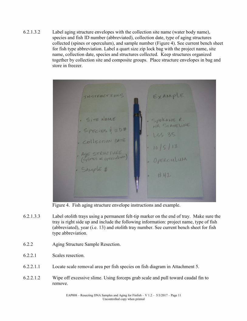
6.2.1.3.2 Label aging structure envelopes with the collection site name (water body name),
species and fish ID number (abbreviated), collection date, type of aging structures
collected (spines or operculum), and sample number (Figure 4). See current bench sheet
for fish type abbreviation. Label a quart size zip lock bag with the project name, site
name, collection date, species and structures collected. Keep structures organized
together by collection site and composite groups. Place structure envelopes in bag and
store in freezer.
Figure 4. Fish aging structure envelope instructions and example.
6.2.1.3.3 Label otolith trays using a permanent felt-tip marker on the end of tray. Make sure the
tray is right side up and include the following information: project name, type of fish
(abbreviated), year (i.e. 13) and otolith tray number. See current bench sheet for fish
type abbreviation.
6.2.2 Aging Structure Sample Resection.
6.2.2.1 Scales resection.
6.2.2.1.1 Locate scale removal area per fish species on fish diagram in Attachment 5.
6.2.2.1.2 Wipe off excessive slime. Using forceps grab scale and pull toward caudal fin to
remove.

EAP008 – Resecting DNA Samples and Aging for Finfish – V 1.2 – 5/3/2017 – Page 12
Uncontrolled copy when printed
6.2.2.1.3 Mount on scale card in the row matching fish ID. Situate scales so the outside (side
exposed to elements) of scale is facing up and the posterior end of scale is toward the
top of card. PRESS DOWN ON THE SCALE to secure the scale to the acetate card.
Scales tend to curl when dried and therefore detach themselves from the card and fall
off. Sometimes adding a bit of moisture (a wet finger) to the scale when pressing them
down will help to secure them.
6.2.2.1.4 Repeat process for a minimum of five to seven scales per fish. If there is room on the
scale card, collect up to ten scales per fish for comparison by the fish biologist.
6.2.2.1.5 Place prepared scale card with other cards in numerical order with the most current
(largest number) on top in a pre-labeled zip seal bag. This exposes the last number done
and is used as a reference starting number for the next scale card to be processed.
6.2.2.1.6 Verify & document that the scales per fish on the scale card match the same fish ID on
the Processing Bench Sheet.
6.2.2.1.7 Record scale card number and scale numbers on Processing Bench Sheet.
6.2.2.2 Opercle resection.
6.2.2.2.1 Bend opercle towards head of fish to break then tear off using your fingers. Larger fish
may need to be cut off using cutters. Collect operculum on both sides of fish.
6.2.2.2.2 Place operculum in pre-labeled envelope matching the fish ID. All envelopes for one
site can be placed in a pre-labeled zip seal bag and placed in freezer to prevent
decomposition until cleaned.
6.2.2.2.3 Verify & document that the operculum per fish in the envelope match the same fish ID
on the Processing Bench Sheet.
6.2.2.3 Spine resection.
6.2.2.3.1 Determine spine resection type per fish species by referring to the “Fish Aging
Structure” spreadsheet for aging structure type (Attachment 4).
6.2.2.3.2 When pulling spines it is important to get the whole spine intact, including the joint
articulation below tissue surface. Using a pair of wire clippers and scalpel, cut around
the base of spine until joint articulation and spine become free from body. This process
should be performed by an experienced individual.
6.2.2.3.3 Place spines in pre-labeled envelope matching the fish ID. All envelopes for one site
can be placed in a pre-labeled zip seal bag and placed in freezer to prevent
decomposition until cleaned.

EAP008 – Resecting DNA Samples and Aging for Finfish – V 1.2 – 5/3/2017 – Page 13
Uncontrolled copy when printed
6.2.2.3.4 Verify & document that the spines per fish in the envelope match the same fish ID on
the Processing Bench Sheet.
6.2.2.4 Otolith resection. There are several techniques used for otolith removal. Ecology
employees have found the following techniques to be very predictable at retrieving
otoliths from a variety of species.
6.2.2.4.1 Using a sharp fillet knife, the first incision should be a vertical cut in line with the
posterior end of gill plate cover. This incision should stop at eye level (Figure 5B).
The second incision should be a parallel cut just above the eyes, starting where first
incision ended (Figure 5C). This incision should create a flap of tissue that can be
folded aside to expose the brain. The largest of typically 3 pairs of otoliths, (the
saggital otoliths), are located just below the brain on each side of the brain cavity.
These otoliths are situated in a protective area that resembles apple seed pits (Figure
5A). A rough landmark for otoliths location is mid-way between the eye and posterior
end of the opercula.
Figure 5. Extraction procedure for walleye saggital otoliths. A. Positioning of otoliths
and location of cuttings. B. Initial cut. C. Second cut to expose brain, otoliths lie
beneath brain in fleshy cavities (Fieler, 2002).
6.2.2.4.2 Remove the brain tissue with forceps carefully so that both otoliths are exposed towards
the back of the brain. They may appear sitting in a divot much like apple seeds sit when
an apple core is sliced in half lengthwise (Figure 6).

EAP008 – Resecting DNA Samples and Aging for Finfish – V 1.2 – 5/3/2017 – Page 14
Uncontrolled copy when printed
Figure 6. Otolith removal from a cod (Miljolare.no, 2006).
6.2.2.4.3 Extract the otoliths using fine-tipped forceps. Be very careful not to force the otolith out
as otoliths tend to break easily. If frozen or unable to see clearly, gently squirt some
water into the brain cavity to thaw and clean. A bowl of water can be useful in thawing
and cleaning the head. Also, if the otolith cannot be found and the “sockets” are clearly
empty or destroyed, rinsing the head in a bowl of water and then “panning” for the
otoliths has brought some retrieval success. Remove any tissue so that otoliths are
clean.
6.2.2.4.4 Place both otoliths from the same fish sample in the same otolith tray cell.
6.2.2.4.5 Record the cell number and otolith tray number on the Processing Benchsheet for each
sample. Validate that each otolith sample recorded on the Processing Benchsheet
matches the correct fish sample ID tag.
6.2.3 Cleaning Operculum and Spines.
6.2.3.1 Cut corners off envelope. Be careful not to cut aging structure. Keep holes small
enough so structures do not come out of envelope while cooking.
6.2.3.2 Fill casserole dish ½ to ¾ full of water.
6.2.3.3 Place 10 – 15 envelopes in casserole dish and cover with glass lid.
6.2.3.4 Place casserole dish with contents in microwave and cook for roughly 4-5 minutes on
high power. May need to adjust cook time up or down depending on ability to remove
tissue from structure.
6.2.3.5 When cooking is complete, pull casserole dish out of microwave. BE CAREFUL,
VERY HOT. Place casserole dish next to sink. Leave envelopes in hot water to soak.
Work on one envelope at a time.

EAP008 – Resecting DNA Samples and Aging for Finfish – V 1.2 – 5/3/2017 – Page 15
Uncontrolled copy when printed
6.2.3.6 Scrub operculum and spines with toothbrush. Make sure to get ALL tissue off structure.
Be careful not to break structure while scrubbing.
6.2.3.7 Once structure is cleaned and rinsed off, place envelope and structure on counter top to
dry. It is important to keep structure with assigned envelope as to keep identification
correct.
6.2.3.8 Repeat Sections 6.2.3.1 through 6.2.3.7 until all structures are cleaned.
6.2.3.9 Place structures back into their original envelope once structures and envelopes are
approximately half dry.
6.2.3.10 Keep samples together in foil box. Label foil box with project name and age structures
(e.g. FFCMP17 AGE STRUCTURES). No need to put structures in freezer once they
are cleaned.
6.2.3.11 Store structures in bottle room (room # OL-17) until ready for delivery to WDFW.
6.3 DNA and Aging Structure Samples Delivery to the lab.
6.3.1 It is important to keep in contact with WDFW to coordinate and communicate project
needs. As collections are completed or at the end of the sampling season, personally
deliver the DNA and aging structure samples to the WDFW Genetic Laboratory and
Ecological Services respectively. Prior to delivery, set up a drop off appointment and
include the following items: otolith trays, scale cards, spine/operculum envelopes, DNA
sample boxes and vials containing tissue, all unused DNA sampling equipment and
appropriate spreadsheets with needed information to process samples.
6.3.2 Deliver the DNA structure samples to WDFW Genetic Laboratory in Olympia, WA at:
WDFW Genetics Laboratory
Natural Resource Building, Rm 665
1111 Washington Street SE
Olympia, WA 98504
Attn. Todd W. Kassler, M.S.
360-902-2722
Or
Sewall F. Young, M.S.
360-902-2773

EAP008 – Resecting DNA Samples and Aging for Finfish – V 1.2 – 5/3/2017 – Page 16
Uncontrolled copy when printed
6.3.3 Deliver the aging structures to Ecological Services in Olympia, WA at:
Ecological Services
Natural Resource Building
1111 Washington Street SE
Olympia, WA 98504
Attn. Lucinda Morrow
360-902-2763
6.3.4 It is best to hand-deliver the samples rather than shipping. This eliminates the
possibility of samples being damaged or lost during shipping. If personal delivery is not
possible, contact the WDFW Lab and set up shipping arrangements prior to sending
samples. WDFW contact information for aging structures is:
7.0 Records Management
7.1 The Lab Analysis & Tracking Plan and Processing Benchsheet are tables, usually
created in Excel®, used to document and coordinate all activities and data for single or
multiple projects per collection and sampling time period and for documenting the
tissue sample preparation for lab analysis. See Ecology’s SOP #007 Procedures for
Resecting Finfish Whole Body, Body Parts, or Tissue Samples Section 7.0 for more
information about these two forms.
7.1.1 Bench sheet - A template of the Processing Benchsheet is located on the Ecology
Intranet at Y:\SHARED Files\ TSU Fish\SOP forms for Fish. The name of the file
should include the project sampling year, the words or abbreviation indicating “Bench
Sheet” and the version number, (e.g. 2017 Bench Sheet.xlsx). Enter all hard copy
documentation into the master electronic Processing Benchsheet. It is important to
ensure that the electronic data is valid for future use. The individual responsible for
entering Processing Benchsheet updates must verify the accuracy of the data and sign
off on each hard copy Processing Benchsheet (e.g. write the word Entered, the date and
individual’s initials). By signing the hard copy, this states data is correct and entered
into the master electronic Processing Benchsheet.
7.1.2 Lab Analysis & Tracking Plan - A template of the lab tracking plan is located on
Ecology Intranet at Y:\SHARED Files\ TSU Fish\SOP forms for Fish. The name of the
file should include the project sampling year, the words or abbreviation indicating “Lab
Tracking Plan” and the version number, (e.g. 2017 fish labplan tracking.xlsx). Enter all
hard copy documentation into the master electronic Lab Analysis & Tracking Plan. As
with the Processing Benchsheet, it is important to ensure that the electronic data is valid
for future use. The individual responsible for entering Lab Analysis & Tracking Plan
updates must verify the accuracy of the data and sign off on each hard copy Lab
Analysis & Tracking Plan (e.g. write the word Entered, the date and individual’s
initials). By signing the hard copy, this states data is correct and entered into the master
electronic Lab Analysis & Tracking Plan.

EAP008 – Resecting DNA Samples and Aging for Finfish – V 1.2 – 5/3/2017 – Page 17
Uncontrolled copy when printed
8.0 Quality Control and Quality Assurance Section
8.1 Verify that all information is filled out on the hard copy bench sheet. There should not
be any blank cells under fields labeled with WDFW DNA ID, DNA Taken, scale card #,
scale #, otolith tray #, otolith cell #, opercle or spine taken. Be sure to document the
process date and crew initials.
8.2 Verify sample number, species, date of collection and any other pertinent information
on the scale card, aging structure envelopes, zip seal bags and otolith tray.
8.3 Check scales on scale card making sure they are attached. If starting to curl and
become loose, rewet the scale with wet finger and press them down again on the card.
8.4 Verify all hard copy and electronic documentation entered on the Processing
Benchsheet and Lab Analysis & Tracking Plan for completeness and accuracy.
9.0 Safety
9.1 Wear gloves and safety glasses when handling the DNA preservative solution.
9.2 Extreme care must be taken when using all knives, not only for you but for others that
may be working in close proximity. Verify that the first aid kit is available in the lab
room and that the contents are complete. Contact the room supervisor and/or the safety
officer if any accident occurs, first aid supplies are inadequate, chemical spills or any
other need or questions. The names and numbers of the room supervisor are posted in
the room. Work with a “buddy” if possible, or at least notify a coworker of your lab
work plans to and put them on your calendar.
9.3 Material Safety Data Sheets for the chemcials used in these procedures are compiled
and stored at the Toxics Coordination Team (TCT) SharePoint website site for review
prior to field activites using this SOP.
10.0 References
10.1 American Fisheries Society (AFS). 1983. Fisheries Techniques. Edited by Nielsen, L.A
and D. L. Johnson. Columbus, Ohio.
10.2 Ecology. 2016. Chemical Hygiene Plan and Hazardous Materials Management Plan.
Olympia, WA.
10.3 Fielder, D.G. 2002. Methodology for immersion marking walleye fry and fingerlings in
oxytetracycline hydrochloride and its detection with fluorescence microscopy. State of
Michigan Department of Natural Resources. Number 2002-1.
http://www.dnr.state.mi.us/PUBLICATIONS/PDFS/ifr/ifrlibra/technical/reports/2002-
1tr.pdf. Accessed April 2017.

EAP008 – Resecting DNA Samples and Aging for Finfish – V 1.2 – 5/3/2017 – Page 18
Uncontrolled copy when printed
10.4 Fisher Scientific. 2016. Safety Data Sheet; Ethyl Alcohol. Thermo Fisher Scientific.
https://www.fishersci.com/us/en/home.html. Accessed April 2017.
10.5 Miljolare.no. 2006. Undersok og del. Disseksjon av torskehode.
http://miljolare.no/fagstoff/vann/artikler/dyr/marint/torskehode.php. Accessed April
2017. (Norwegian to English translation of titles: Dissect and Examine. Dissection of a
cod head).
10.6 Morrow, L. 2006. WDFW biologists. Personal communication with P. Sandvik, Dept.
of Ecology.
10.7 Sneva, J. and L. Morrow. 2003. WDFW biologists. Personal communication with K
Seiders, Dept. of Ecology.
10.8 Sneva, J. 2005. WDFW biologist. Personal communication with B. Era-Miller, Dept. of
Ecology.
10.9 University of Washington (UW). 2017. Ichthyology. Glossary of Fish Characters;
Diagram. http://www.burkemuseum.org/research-and-collections/ichthyology.
Accessed April 2017.
10.10 WDFW Molecular Genetics Laboratory. 2015. Considerations & Guidelines for
Collecting Representative Samples for Genetic Analysis. Genetic Sampling Goals
Olympia, WA.
10.11 Wikipedia. 2017a. The free Encyclopedia. http://en.wikipedia.org/wiki/DNA. Accessed
April 2017.
10.12 Wikipedia. 2017b. The free Encyclopedia. http://en.wikipedia.org/wiki/PCR. Accessed
April 2017.
EAP008 – Resecting DNA Samples and Aging for Finfish – V 1.2 – 5/3/2017 – Page 19
Uncontrolled copy when printed
Attachment 1. Lab Analysis & Tracking Plan, (example only)
Note: The Lab Analysis & Tracking Plan may look different due to different fields and requirements of the project(s) involved, but
fields will be available for documentation and cross reference of each sample’s collection and processing information.
Lab Analysis & Tracking Plan for FFCMP: 2016
FFCMP Work Order # 1701015 jar size -> 2 oz 4 oz 4 oz 4 oz 4 oz 4 oz
SIC DST03 min amount needed per analysis --> 5 g 40 g 40 g min amount needed per jar --> 5 g 80 g 60 g 60 g 80 g 80 g
updated: 1/17/17 NM cost/sample --> 50$ 379$ 242$ 675$ 531$
FF
CM
P (
f)
La
b D
up
ME
L
MS
/MS
D M
EL
La
b D
up
CL
Site 1 Site 2 Species
suffix for
LAR
Field ID
collect
date
# fish
in
comp
Hg
CP,
PCBa (PEST2P
CB)
PBDE
, lipid
PCB
congn
r
PCDD
/F
FFCMP LAR
Field ID
FFCMP
MEL
Lab # 1701015-
nn
FFCMP fish IDsprocess
date
aliquot
per fish
(g)
skin:
off or
on
s
p
a
c
e
Hg
PE
ST
2P
CB
,
PB
DE
, lip
id
ME
L Q
C
PC
B c
on
ge
ne
r,
PC
DD
/F
Arc
hiv
e 1
Arc
hiv
e 2
f Cowlitz R: Castle RockCR LSS LSS-1 8/29/16 5 1 1 CR-LSS-1 01 6,8,15,24,30 12/28/16 300 ON 80 80 80
f Cowlitz R: Castle RockCR LSS LSS-2 8/29/16 5 1 1 CR-LSS-2 02 9,11,13,19,23 12/28/16 300 ON 80 80 80
f Cowlitz R: Castle RockCR LSS LSS-3 8/29/16 5 1 1 CR-LSS-3 03 2,18,22,27,33 12/28/16 300 ON 80 80 80
f Cowlitz R: Castle RockCR MWF MWF-1 8/29/16 5 1 1 1 1 1 CR-MWF-1 04 6,9,10,13,17 1/6/17 79 ON 5 80 60 80 80
f Cowlitz R: Castle RockCR MWF MWF-2 8/29/16 5 1 1 1 1 1 CR-MWF-2 05 8,15,18,22,23 1/6/17 89 ON 6 80 60 80 80
f Cowlitz R: Castle RockCR MWF MWF-3 8/29/16 5 1 1 1 1 1 CR-MWF-3 06 5,14,26,27,28 1/6/17 64 ON 5 80 60 60 60
f a a a Cowlitz R: Castle RockCR NPM NPM-A 8/29/16 5 1 1 1 1 1 CR-NPM-A 07 1,2,3,4,5 1/4/17 90 ON 5 80 60 60 80 80
f p p p Cowlitz R: Castle RockOL CTT CTT-1 8/30/16 5 1 1 1 1 1 OL-CTT-1 08 7,13,14,17,18 1/11/17 83 ON 5 80 60 60 80 80
f a a a Cowlitz R: Castle RockOL CTT CTT-2 8/30/16 5 1 1 1 1 1 OL-CTT-2 09 5,6,9,10,16 1/11/17 94 ON 5 80 60 60 80 80
f Cowlitz R: Castle RockOL CTT CTT-3 8/30/16 5 1 1 1 1 1 OL-CTT-3 10 3,8,11,12,15 1/10/17 51 ON 5 80 60 62 44
f Cowlitz R: Castle RockOL LSS LSS-1 8/30/16 5 1 1 OL-LSS-1 11 16,18,21,29,35 12/29/16 300 ON 80 80 80
f Cowlitz R: Castle RockOL LSS LSS-2 8/30/16 5 1 1 OL-LSS-2 12 6,7,14,27,40 12/29/16 300 ON 80 80 80
f Cowlitz R: Castle RockOL LSS LSS-3 8/30/16 5 1 1 OL-LSS-3 13 8,10,17,32,37 12/29/16 300 ON 80 80 80
f Cowlitz R: Olequa OL MWF MWF-1 8/30/16 5 1 1 1 1 1 OL-MWF-1 14 13,15,25,32,37 12/28/16 91 ON 5 80 80 80 80
f p p p Cowlitz R: Olequa OL MWF MWF-2 8/30/16 5 1 1 1 1 1 OL-MWF-2 15 23,28,29,33,34 12/28/16 80 ON 5 80 60 60 80 75
f Cowlitz R: Olequa OL MWF MWF-3 8/30/16 5 1 1 1 1 1 OL-MWF-3 16 14,20,24,30,40 12/28/16 77 ON 5 80 60 80 80
f Cowlitz R: Olequa OL MWF MWF-L1 8/30/16 3 1 1 1 1 1 OL-MWF-L1 17 3,4,17 1/5/17 103 ON 5 81 80 80 32
f Cowlitz R: Olequa OL MWF MWF-L2 8/30/16 3 1 1 1 1 1 OL-MWF-L2 18 6,10,19 1/5/17 108 ON 5 80 80 80 44
f Cowlitz R: Olequa OL MWF MWF-L3 8/30/16 3 1 1 1 1 1 OL-MWF-L3 19 7,9,16 1/5/17 115 ON 6 80 80 80 75
f Cowlitz R: Olequa OL NPM NPM-1 8/30/16 3 1 1 1 1 1 OL-NPM-1 20 7,10,14 1/5/17 105 ON 5 80 60 80 80
f p p p Cowlitz R: Olequa OL NPM NPM-2 8/30/16 3 1 1 1 1 1 OL-NPM-2 21 8,9,11 1/5/17 102 ON 5 80 60 60 80 19
f Cowlitz R: Olequa OL NPM NPM-3 8/30/16 3 1 1 1 1 1 OL-NPM-3 22 6,12,13 1/5/17 115 ON 5 80 60 80 80
f Mayfield L ML LSS LSS-1 8/31/16 5 1 1 ML-LSS-1 23 1,11,27,30,32 12/27/16 300 ON 80 80 80
f Mayfield L ML LSS LSS-2 8/31/16 5 1 1 ML-LSS-2 24 6,23,31,33,34 12/27/16 300 ON 80 80 80
f Mayfield L ML LSS LSS-3 8/31/16 5 1 1 ML-LSS-3 25 3,12,16,37,40 12/27/16 300 ON 80 80 80
f Mayfield L ML NPM NPM-1 8/31/16 5 1 1 1 1 1 ML-NPM-1 26 12,16,18,30,32 1/4/17 83 ON 5 80 60 80 80
f Mayfield L ML NPM NPM-2 8/31/16 5 1 1 1 1 1 ML-NPM-2 27 4,11,15,20,31 1/4/17 84 ON 5 80 60 80 80
f Mayfield L ML NPM NPM-3 8/31/16 5 1 1 1 1 1 ML-NPM-3 28 13,17,22,26,29 1/4/17 88 ON 5 80 60 80 80
HIDE THESE FIELDS WHEN PRINTING
60 g
Use these fields for sample jar LABEL. Record amount of
tissue in each sample jar.

EAP008 – Resecting DNA Samples and Aging for Finfish – V 1.2 – 5/3/2017 – Page 20
Uncontrolled copy when printed
Attachment 2. Processing Bench Sheet, (example only)
Note: The Bench Sheet used during lab processing may look different due to different fields and requirements of the processes
involved, but fields will be available for documentation and cross reference of each sample’s information.
Field Data for Fish Tissue Samples: FFCMP 2016updated: 12/21/16 PS
Notes: 1. ND = Not Determined; U = checked but could not determine.
Site 1Site
2Species
ECY
Field ID
Total
Length
(mm)
Field
Weight
(gm)
Collect
Date
Collect
Method
Process
date
Process
ing crew
FFCMP fillet
weight (gm)
FFCMP
L, R, or
B fillet
FFCMP
skin
status
On/Off
sex
M/F 1fish
age
scale
card #scale #
otolith
tray #
otolith
cell #
Pull
Opercula
Spines
Y/N
CommentFFCMP LAR
Field ID
FFCMP
MEL ID:
1701015-nn
Castle Rock (CR) CR NPM 1 344 365 8/29/2016 E 1/4/17 PS 139 B ON F Y CR-NPM-A 07
Castle Rock (CR) CR NPM 2 367 454 8/29/2016 E 1/4/17 PS 166 B ON F Y CR-NPM-A 07
Castle Rock (CR) CR NPM 3 330 286 8/29/2016 E 1/4/17 PS 113 B ON F Y CR-NPM-A 07
Castle Rock (CR) CR NPM 4 311 258 8/29/2016 E 1/4/17 PS 105 B ON M Y CR-NPM-A 07
Castle Rock (CR) CR NPM 5 327 292 8/29/2016 E 1/4/17 PS 114 B ON F Y CR-NPM-A 07
Olequa (OL) OL CTT 7 350 498 8/30/2016 E 1/10/17 PS, NM, JM 103 L ON M 4 NNO ADIPOSE
FIN PRESENT OL-CTT-1 08
Olequa (OL) OL CTT 13 316 355 8/30/2016 E 1/10/17 PS, NM, JM 105 B ON F 4 NNO ADIPOSE
FIN PRESENT OL-CTT-1 08
Olequa (OL) OL CTT 14 318 362 8/30/2016 E 1/10/17 PS, NM, JM 103 B ON F 4 NNO ADIPOSE
FIN PRESENT OL-CTT-1 08
Olequa (OL) OL CTT 17 300 262 8/30/2016 E 1/10/17 PS, NM, JM 100 ON F 4 NADIPOSE FIN
PRESENT OL-CTT-1 08
Olequa (OL) OL CTT 18 308 302 8/30/2016 E 1/10/17 PS, NM, JM 100 B ON F 4 NADIPOSE FIN
PRESENT OL-CTT-1 08
Olequa (OL) OL CTT 5 340 487 8/30/2016 E 1/10/17 PS, JM 101 L ON M 5 5 NNO ADIPOSE
FIN PRESENT OL-CTT-2 09
Olequa (OL) OL CTT 6 332 435 8/30/2016 E 1/10/17 PS, JM 126 B ON F 5 6 NNO ADIPOSE
FIN PRESENT OL-CTT-2 09
Olequa (OL) OL CTT 9 337 410 8/30/2016 E 1/10/17 PS, JM 137 B ON F 5 9 NNO ADIPOSE
FIN PRESENT OL-CTT-2 09
Olequa (OL) OL CTT 10 326 422 8/30/2016 E 1/10/17 PS, JM 100 L ON F 5 10 NNO ADIPOSE
FIN PRESENT OL-CTT-2 09
Olequa (OL) OL CTT 16 333 452 8/30/2016 E 1/10/17 PS, JM 104 B ON M 5 16 NNO ADIPOSE
FIN PRESENT OL-CTT-2 09
Olequa (OL) OL LSS 6 416 720 8/30/2016 E 12/29/16 KS, NM whole whole ON Y OL-LSS-2 12
Olequa (OL) OL LSS 7 485 1122 8/30/2016 E 12/29/16 KS, NM whole whole ON Y OL-LSS-2 12
Olequa (OL) OL LSS 14 463 1021 8/30/2016 E 12/29/16 KS, NM whole whole ON Y OL-LSS-2 12
Olequa (OL) OL LSS 27 486 1171 8/30/2016 E 12/29/16 KS, NM whole whole ON Y OL-LSS-2 12
Olequa (OL) OL LSS 40 485 1057 8/30/2016 E 12/29/16 KS, NM whole whole ON Y OL-LSS-2 12
Age Structures

EAP008 – Resecting DNA Samples and Aging for Finfish – V 1.2 – 5/3/2017 – Page 21
Uncontrolled copy when printed
Attachment 3. WDFW Genetic Sampling Goals [genetic sampling goals.doc] rev 14 Oct 2015
Considerations & Guidelines for Collecting Representative Samples for Genetic Analysis
The two most important goals of field sampling for genetic analysis are: 1) to obtain tissue samples
with little or no biochemical degradation, so that the genetic markers to be screened can be
successfully analyzed and 2) to obtain samples from individuals that are representative of the target
population in terms of age, sex, location, and timing.
If the samples are so degraded because of post-mortem tissue decomposition that they cannot be
successfully analyzed to provide reliable genetic data, the time and money spent collecting and
analyzing the samples will be wasted. At the same time, if the collection of samples that is obtained is
not representative of the population as a whole, the genetic data collected may not provide an accurate
profile of the target population and may, therefore, be misleading or useless.
Tissue Quality
Tissue samples obtained from live fish (or fish that have just been sacrificed) are of the highest quality,
while those from relatively fresh carcasses are also suitable for genetic analysis. However, tissue
samples from animals that have been dead for a considerable period of time and have experienced
significant decomposition invariably yield little or no useful data. Poor quality carcasses should NOT
be genetically sampled.
Tissue Preservation - DNA Analysis
Whatever the state of the animal to be sampled is, it is imperative to be sure to preserve the tissue
samples as soon as possible after they are taken. In the case of tissue samples for DNA analysis, we
use a special ethanol preservative solution. This preservative, which is a poison and is flammable,
should be obtained from the WDFW Genetics Lab. The solution preserves the DNA by dehydrating
the tissue. Thus, it is critical that the volume ratio of tissue to preservative not exceed 4 parts of
preservative for every 1 part of tissue. One approach to achieve this is to fill the sampling vial 4/5
full of preservative solution and then add small pieces of tissue until the solution reaches the top of the
vial. Several small pieces of tissue are preferable to a single large piece because the preservative
solution can penetrate the small pieces more quickly, thereby achieving better preservation. Note, an
excess of preservative solution is okay; not using enough preservative is a problem. Once in this
preservative, tissue samples can be stored at room temperature. Be aware that the DNA preservative
solution will dissolve ink; therefore labels in, on, and around the DNA sample vials should either be
printed on a laser printer or written in pencil!
Sampling instruments, dissecting areas, and your hands should be kept clean (rinsed in fresh water
between specimens or as frequently as necessary) to avoid sample-to-sample contamination and blotted
dry after rinsing to minimize dilution of the DNA preservative solution. Because all our DNA
analyses involve PCR amplification of the DNA extracted from the tissue samples, sample-to-sample
contamination can be a problem and must be avoided. Nevertheless, it is not necessary to wear gloves

EAP008 – Resecting DNA Samples and Aging for Finfish – V 1.2 – 5/3/2017 – Page 22
Uncontrolled copy when printed
during the dissection process to avoid contamination of the samples -- just keep your hands, the
sampling instruments, and the work area clean.
Representative Sampling
Obtaining samples that are representative of the target population is a very important, but sometimes
difficult goal to achieve.
Adult Fish - from a single population
The goal here should be to obtain a collection of samples (usually 100 fish to provide adequate
statistical power) that is representative of the target population (e.g., with regard to sex, age, location,
and return/spawn timing). Sampling adults on the spawning grounds at, or immediately after,
spawning is the surest way to obtain a collection that is not contaminated with individuals from other
populations.
Because most populations have an approximate 50:50 sex ratio, collections should consist of about
50% males and 50% females. Similarly, if a population consists of 3, 4, and 5 year old spawners, the
collection should have all of these age classes represented in approximately the same proportions as
they occur in the population at large. In the same way, if a population spawns throughout a 10 mile
stretch of river over approximately 1 month, the collection should ideally include approximately
proportional samples over the same geographic range and time frame. Because of cost and logistical
constraints, this might actually translate into obtaining samples on 3 different days at four locations
distributed throughout the 10 miles of spawning habitat. A collection of samples as described above
provides the best opportunity to obtain an accurate genetic profile of the target population.
Representative (proportional) sampling of spawning adults from a hatchery (or natural) population
over time is schematically shown in Figures 1A & B below.
Smolts, Juveniles, and Fry - from a single population
Although the goal here is also to obtain a collection of samples that is representative of the target
population, the approach and concerns are somewhat different than those described above for adult
sampling. Here there are four, sometimes conflicting sampling concerns. One is to obtain a sample
that is representative of the entire population. A second is to be sure to avoid sampling a mixture of
fish from two or more populations. A third is to avoid sampling sibs (offspring from a single mating),
and half-sibs. A fourth, for DNA-based investigations, is often to sample non-lethally. As a result, the
actual sampling design is usually an attempt to balance these concerns. Sampling small, newly
emerged fry from locations where adult spawning occurred maximizes the likelihood of obtaining a
collection from the target population (and only that population) but this approach maximizes the
likelihood that the collection will contain sibs or half-sibs and that the tissue sampling will be lethal or
at least detrimental to survival (because of the small size of the fish). Sampling larger juveniles, which
have presumably moved around substantially after hatching and emergence from the gravel reduces the
likelihood of sampling sibs and, for DNA studies, increases the likelihood that the sampling will not
affect subsequent survival (because the fish are larger), but increases the likelihood of obtaining a
mixed collection of fish from multiple populations.

EAP008 – Resecting DNA Samples and Aging for Finfish – V 1.2 – 5/3/2017 – Page 23
Uncontrolled copy when printed
Sampling fry or pre-smolts from a hatchery should be designed to include representative samples of
progeny from all egg takes/parents. Taking proportional subsamples from each of the rearing vessels
used for the population is one aspect of this. Sampling outmigrating smolts to characterize a single
population should be stratified over space and time to achieve a representative collection (see Figure
1A & B below).
Sampling Mixtures (e.g., fishery sampling)
Again, the ultimate goal is to obtain a collection of samples (mixture sample sizes of several hundred
are often needed to provide the desired accuracy and precision) that is representative of the mixture
you are trying to characterize (whether this be a mixed-stock fishery sample of adult salmon or a
sample of a mixture of outmigrating smolts from multiple populations). The goal is to obtain
representative numbers of the various components of the mixture. For a fishery, some of the key
factors may be: location and date of catch, type of gear or proportions of gear types, and size and sex
of fish. For a collection of smolts, the key objectives may be to sample proportionally over the entire
period of outmigration, to sample all size classes, and to sample during both day and night.
DNA
When obtaining a non-lethal sample from a small fish, a small sample should be taken. The minimum
amount of tissue that is needed for DNA analysis is approximately the size of this circle: (e.g., a
piece of tissue with the same approximate surface area as a 1.5mm diameter disc). The recommended
sources of such a tissue sample are any of the following:
1) A distal portion of the dorsal lobe of the caudal fin
2) A distal portion of one of the pelvic fins
3) Smaller distal portions of both pelvic fins
4) One entire pelvic fin
By sampling only the distal portion of a fin, we expect that the fish will successfully regenerate the
entire fin over time. In contrast, removing an entire fin often results in little or no fin regeneration,
presumably leaving the fish at a selective disadvantage.
Larger samples are preferred whenever possible (e.g., a piece of tissue approximately the size of this
circle: ); or even as much as approximately 1-2 cm2 area), because this will provide more
material (DNA). The “extra” tissue provides a reserve that can be used to overcome some types of
analytical problems in the lab by repeated analysis and/or it also provides material that can be used for
subsequent analyses (for example to examine additional loci at a future date) or to share with other
laboratories/agencies.

EAP008 – Resecting DNA Samples and Aging for Finfish – V 1.2 – 5/3/2017 – Page 24
Uncontrolled copy when printed
Live fish should be handled appropriately before, during, and after sampling. This will probably
involve: a) anesthetization prior to handling for tissue sampling (and taking of measurements or other
biological samples such as scales), b) careful handling during sampling to avoid injury and
scale/mucous loss, and c) holding fish in a recovery vessel after sampling (until the anesthetic has
worn off) before releasing them in a way that minimizes immediate mortality due to predation of other
effects.
If you have questions or need additional information, please contact Todd Kassler (360-902-2722;
<[email protected]>) or the Genetics Laboratory at 360-902-2775).
Figure 1. Schematic diagrams of proportional sampling designs.
0
10
20
30
40
50
60
70
80
90
100
0
200
400
600
800
1000
1200
1 2 3 4 5 6 7 8 9 10 11 12 13 14 15 16 17 18 19 20
Nu
mb
er
of
ge
ne
tic
sa
mp
les
ta
ke
n
To
tal n
um
be
rs o
f a
du
lts
/sm
olt
s
Time ( in arbitrary units)
A. Minimally Proportional Genetic Sampling Design
genetic sample (N = 100)
total adult return/smolt outmigration (N = 5,460)

EAP008 – Resecting DNA Samples and Aging for Finfish – V 1.2 – 5/3/2017 – Page 25
Uncontrolled copy when printed
0
5
10
15
20
25
30
0
200
400
600
800
1000
1200
1 2 3 4 5 6 7 8 9 10 11 12 13 14 15 16 17 18 19 20
Nu
mb
er
of
gen
eti
c s
am
ple
s t
aken
To
tal n
um
bers
of
ad
ult
s/s
mo
lts
Time ( in arbitrary units)
B. Optimized Proportional Genetic Sampling
genetic sample (N = 100)
total adult retuen/smolt outmigration (N = 5,460)

EAP008 – Resecting DNA Samples and Aging for Finfish – V 1.2 – 5/3/2017 – Page 26
Uncontrolled copy when printed
Attachment 4. Fish Aging Structure Spreadsheet
Fish Aging Structures: updated: 11/28/12 per WDFW Molecular Genetics Laboratory
Spines Otoliths Opercula Scales Cleithra
Ecology
Species Code Common name Scientific name Family name
X X BC 1
Black crappie Pomoxis nigromaculatus Centrarchidae
X X BG 1
Bluegill Lepomis macrochirus Centrarchidae
X X BLS 2
Bridgelip sucker Catostomus columbianus Catostomidae
X X BKT 3
Brook trout Salvelinus fontinalis Salmonidae
X BBH 4
Brown bullhead Ameiurus nebulosus Ictaluridae
X X BNT 3
Brown trout Salmo trutta Salmonidae
X X BLT 3
Bull trout Salvelinus confluentus Salmonidae
X BUR Burbot Lota lota Gadidae
X CC 4
Channel catfish Ictalurus punctatus Ictaluridae
X X CLM 2
Chiselmouth Arocheilus alutaceaus Cyprinidae
X X CCP 5
Common carp Cyprinus carpio Cyprinidae
X X GC 5
Grass carp Ctenopharyngodon idella Cyprinidae
X X CTT 3
Cutthroat trout Oncorhynchus clarki Salmonidae
X ENS English Sole
GST 6
Green sturgeon Acipenser medirostrus Acipenseridae
X X GS 1
Green sunfish Lepomis cyanellus Centrarchidae
X X KOK 3
Kokanee salmon Oncorhynchus nerka Salmonidae
X X LT 9
Lake trout Salvelinus namaycush Salmonidae
X X LWF 9
Lake whitefish Coregonus clupeaformis Salmonidae
X X LMB 1
Largemouth bass Micropterus salmoides Centrarchidae
X X LSS 2
Largescale sucker Catostomus macrochelius Catostomidae
X X LNS 2
Longnose sucker Catostomus catostomus Catostomidae
X X MS 2
Mountain sucker Catostomus platyrhynchus Catostomidae
X X MWF 7
Mountain whitefish Prosopium williamsoni Salmonidae
X NOP Nothern pike Esox lucius Esocidae
X X NPM 2
Northern pikeminnow Ptychocheilus oregonensis Cyprinidae
X X PEA 2
Peamouth Mylocheilus caurinus Cyprinidae
X X PMP 1
Pumpkinseed Lepomis gibbosus Centrarchidae
X X RBT 3
Rainbow trout Oncorhynchus mykiss Salmonidae
X X RKB 1
Rock bass Ambloplites rupestris Centrarchidae
X RKS Rock Sole
X COT Sculpins Cottus sp. Cottidae
X X SMB 1
Smallmouth bass Micropterus dolomieu Centrarchidae
X STF Starry flounder Platicthys stellatus Pleuronectidae
X X WAL 9
Walleye Stizostedion vitreum Percidae
X X WM 1
Warmouth Lepomis gulosis Centrarchidae
X X WC 1
White crappie Pomoxis annularis Centrarchidae
WST 6
White sturgeon Acipenser transmontanus Acipenseridae
X YBH 4
Yellow bullhead Ameiurus natalis Ictaluridae
X X YP 8
Yellow perch Perca flavescens Percidae
Notes:
1 - Otoliths first choice structure if >300mm. Fish <300mm OK to pull scales only.
2 - Opercula first choice structure in all size classes, scales back-up structure if opercula damaged.
3 - Scales first choice structure in all size classes, otoliths back-up structure if unable to pull scales (note: if KOK are spawning stage, pull otoliths).
4 - Pectoral spines (including articulating joint).
5 - Dorsal spine first choice structure, opercula back-up structure if dorsal spine is damaged.
6 - Pull pectoral fin ray. (Mike Wall ages sturgeon for WDFW # 906-6727).
7 - Otoliths first choice structure if >350mm. Fish <350mm OK to pull scales only.
8 - Otoliths first choice structure if >200mm. Fish <200mm OK to pull scales only.
9 - Otoliths first choice structure in all size classes, scales back-up sructure if otoliths not retrieved.
For trout and salmon, add comment to bench sheet for presence or absence of adipose fin.
Aging Questions??? Call Lucinda Morrow @ WDFW #902-2763, or marine lab #902-2859.

EAP008 – Resecting DNA Samples and Aging for Finfish – V 1.2 – 5/3/2017 – Page 27
Uncontrolled copy when printed
Attachment 5. General Areas of Scale Removal
Scales taken for aging samples should be around the same size per fish for a more consistent analysis. Use undamaged scales only. When measuring Walleye
scales in addition to aging, take scale samples above the lateral line, otherwise the preferred scales for aging Walleye are located below the lateral line near the
pectoral fins. (AFS, 1983; Morrow, 2006; Sneva et. al., 2003; Sneva, 2005; and UW, 2017).
Whitefish
7 Rows above lateral line
3 rows above lateral line
1-2 rows above lateral line
Bass, Bluegill, Crappie, Herring,
Perch, Sunfish, Walleye,
Pumpkinseed, Warmouth
Flounders, Haddock,
Kokanee, Sockeye,
Atlantic Salmon
Coho Salmon, Trout, Peamouth
Dace
Chiselmouth, Suckers, Northern
Pikeminnow