research open access particle size dependent deposition
TRANSCRIPT

Braakhuis et al. Particle and Fibre Toxicology 2014, 11:49http://www.particleandfibretoxicology.com/content/11/1/49
RESEARCH Open Access
Particle size dependent deposition andpulmonary inflammation after short-terminhalation of silver nanoparticlesHedwig M Braakhuis1,2*, Ilse Gosens2, Petra Krystek3, John AF Boere2, Flemming R Cassee2,4, Paul HB Fokkens2,Jan Andries Post5, Henk van Loveren1,2 and Margriet VDZ Park2
Abstract
Background: Although silver nanoparticles are currently used in more than 400 consumer products, it is not clearto what extent they induce adverse effects after inhalation during production and use. In this study, we determinedthe lung burden, tissue distribution, and the induction and recovery of adverse effects after short-term inhalationexposure to 15 nm and 410 nm silver nanoparticles.
Methods: Rats were nose-only exposed to clean air, 15 nm silver nanoparticles (179 μg/m3) or 410 nm silver particles(167 μg/m3) 6 hours per day, for four consecutive days. Tissue distribution and the induction of pulmonary toxicitywere determined at 24 hours and 7 days after exposure and compared with the internal alveolar dose. Presence ofsilver nanoparticles in lung cells was visualized by transmission electron microscopy (TEM).
Results: Exposure to 15 nm silver nanoparticles induced moderate pulmonary toxicity compared to the controls,indicated by a 175-fold increased influx of neutrophils in the lungs, a doubling of cellular damage markers in the lungs,a 5-fold increase in pro-inflammatory cytokines, and a 1.5-fold increase in total glutathione at 24 hours after exposure.All the observed effects disappeared at 7 days after exposure. No effects were observed after exposure to 410 nm silverparticles. The internal alveolar mass dose of the 15 nm nanoparticles was 3.5 times higher compared to the 410 nmparticles, which equals to a 66,000 times higher particle number. TEM analysis revealed 15 nm nanoparticles invesicles and nuclei of lung cells, which were decreased in size to <5 nm at 24 hours after exposure. Thisdemonstrates substantial dissolution of the silver nanoparticles.
Conclusion: The results show a clear size-dependent effect after inhalation of similar mass concentrations of15 nm and 410 nm silver (nano)particles. This can be partially explained by the difference in the internal alveolar dosebetween the 15 nm and 410 nm silver (nano)particles as well as by a difference in the release rate of silver ions.
Keywords: Nanoparticles, Inhalation exposure, Pulmonary toxicity, Cellular uptake, Dissolution
BackgroundNanomaterials are used in a rapidly increasing numberof products including consumer products [1]. However,there are concerns these nanomaterials might introducehealth risks upon occupational and consumer exposure.Although many nanomaterials are produced, handledand present in fluids, aerosolization may occur during
* Correspondence: [email protected] of Toxicogenomics, Maastricht University, PO Box 616,Maastricht 6200, MD, the Netherlands2National Institute for Public Health and the Environment (RIVM), PO Box 1,Bilthoven 3720, BA, the NetherlandsFull list of author information is available at the end of the article
© 2014 Braakhuis et al.; licensee BioMed CentCommons Attribution License (http://creativecreproduction in any medium, provided the orDedication waiver (http://creativecommons.orunless otherwise stated.
energetic processes, such as vortexing, weighing, sonic-ation, mixing and blending [2]. In addition, nanomater-ials may be released from their matrix during its use.Therefore, inhalation is considered a relevant route ofexposure [2,3]. Currently, silver nanoparticles are themost common nanoparticles mentioned in product de-scriptions. According to the Nanotechnology ConsumerProducts Inventory, silver nanoparticles are currentlyclaimed to be used in more than 400 consumer products[1]. Silver nanoparticles have antimicrobial activity andare used in food packaging material, food supplements,odour-preventing textiles, cosmetics, kitchen utensils,
ral Ltd. This is an Open Access article distributed under the terms of the Creativeommons.org/licenses/by/4.0), which permits unrestricted use, distribution, andiginal work is properly credited. The Creative Commons Public Domaing/publicdomain/zero/1.0/) applies to the data made available in this article,

Braakhuis et al. Particle and Fibre Toxicology 2014, 11:49 Page 2 of 16http://www.particleandfibretoxicology.com/content/11/1/49
toys, electronics, wound dressings, and room sprays [1,4].Silver (nano)particles and released ions exert antimicrobialproperties by binding to sulphur- and phosphorous-containing biomolecules such as proteins and DNA,thereby potentially also causing damage to mammaliancells [5-9]. Besides worker exposure, the extensive use ofsilver nanoparticles in products might also lead to con-sumer exposure by inhalation of the silver nanoparticlesthat are used in spray applications. Although silver nano-particles are currently claimed to be used in many prod-ucts, it is not clear to what extend they induce adverseeffects after inhalation during production and use.Silver nanoparticles have been studied in several in vivo
inhalation studies showing diverse outcomes. Some stud-ies showed no induction of adverse effects [10,11], whileother studies reported adverse effects varying from a min-imal inflammatory response to the presence of inflamma-tory lesions in the lungs [12-15]. Regarding the tissuedistribution of the silver nanoparticles, some studies re-port a dose-dependent increase in the silver concentrationin the lungs and in the liver [11,13,15]. Two of these stud-ies report a rather high amount of silver detected in thebrain and the olfactory bulb [11,15], causing concerns thatsilver nanoparticles might induce toxicity in the brain.The before mentioned inhalation studies show that sil-
ver nanoparticles can induce pulmonary inflammationand can decrease lung function, depending on the ex-posure time and dosage [10-15]. However, all of thesestudies focused on a single particle size. The previousstudies did not take particle size and surface area intoaccount as explaining variable, whereas these affect theinternal dose and the interaction probability with cells.For particles to induce pulmonary inflammation, theymust deposit in the alveolar region. The deposition of(nano)particles depends mostly on their (agglomerate)size. Nanoparticles with a primary or agglomerate par-ticle size between 10 and 100 nm will deposit more effi-ciently in the alveolar region compared to particles withan agglomerate particle size between 0.1 and 1 μm[16-20]. At a similar mass based exposure dose, particlesof different sizes will have a different deposition patternin the lungs, and the deposited dose in the alveoli ultim-ately determines the extent of the pulmonary toxic ef-fects. The previous studies did not link the depositeddose in the alveoli to the observed effects [10-15].Until now, the formulation in which silver nanoparti-
cles induce toxicity remains unclear. The effects mightbe caused by the silver nanoparticles itself, the releasedsilver ions, or a combination of both. Next to this, it re-mains unclear to what extend the geometric size of silverparticles affect the induction of pulmonary inflamma-tion. Since particle size is the most important particlecharacteristic that determines the deposited dose in thelungs and is of influence on the dissolution rate of silver
nanoparticles, the aim of this study is to investigate theinfluence of particle size on pulmonary toxicity of silvernanoparticles. We hypothesize that small silver nanopar-ticles will induce more prominent pulmonary toxicitycompared to larger silver particles because of the largerdeposited dose in the alveoli and the higher dissolutionrate. In the present study, we tested the effects of a simi-lar mass exposure concentration of 15 nm and 410 nmsilver (nano)particles after short-term nose-only inhal-ation exposure. The total lung burden was measuredand used together with the exposure measurements asan input for the multiple path particle dosimetry(MPPD) model to estimate the alveolar dose. Transmis-sion electron microscopy (TEM) was used to localize sil-ver particles in the lung tissue and tissue distribution wasmeasured to determine any differences in the kinetics ofthe silver particles. To determine the toxicity induced by15 nm and 410 nm silver (nano)particles, body weight,total blood cell counts, inflammatory and cell damagemarkers in the bronchoalveolar lavage fluid (BALF) andhistology of the lungs were analysed. All endpoints weredetermined at 24 hours and 7 days after the last exposureto investigate the possible recovery of adverse effects.
ResultsNanoparticle characteristicsThe particle characteristics are summarized in Table 1 andshown in Additional file 1: Figure S1. The silver nanoparti-cles had a count median diameter of 15 nm (Figure 1) andthe exposed dosage was 179 μg/m3 air. For the larger silverparticles, the 200 nm primary silver particles agglomeratedto a count median diameter of 410 nm (Figure 1) and theexposed dosage was 167 μg/m3 air. While the mass dosagewas similar for both particle groups, the particle numberdosage was 3.8 × 10⁶ particles per cm3 for the 15 nm nano-particles and 2.0 × 10⁴ particles per cm3 for the 410 nmparticles.
Experimental designSince male rats showed more sensitivity towards induc-tion of pulmonary inflammation compared to femalerats after 90 days inhalation exposure [13,14], we choseto expose male rats. The rats were nose-only exposedfor 6 hours per day, 4 consecutive days to fresh air,15 nm silver nanoparticles or 410 nm silver particles, 12rats per exposure group. Of each exposure group, 6 ratswere sacrificed 24 hours after exposure and the other 6rats were sacrificed 7 days after exposure.
Quantification of silver in tissues by high resolutioninductively coupled plasma mass spectrometry (HR-ICP-MS)The presence of silver was measured with HR-ICP-MSin the lungs, liver, spleen, kidneys, brain, testis and lung-associated lymph nodes. Of these tissues, silver was

Table 1 Nanoparticle characteristics
Agglomerate size [nm] Mass [μg/m3] Particle number [#/cm3] Surface area [nm2/cm3]
15.3 (1.26) 179 (107–252) 3.8 × 10⁶ (2.6 - 5.0) 6.72 × 10⁹ (3.97-9.49)
410 (1.43) 167 (147–187) 2.0 × 10⁴ (0.43 - 3.7) 2.33 × 10⁸ (0.28-2.61)
Particle size is given as count median diameter (CMD) with the geometric standard deviation (GSD). Mass, particle number and surface area shown as mean witha 95% confidence interval.
Braakhuis et al. Particle and Fibre Toxicology 2014, 11:49 Page 3 of 16http://www.particleandfibretoxicology.com/content/11/1/49
detected in the lungs and liver (Figure 2). At 24 hoursafter exposure, the lungs of the rats exposed to 15 nmnanoparticles contained 3.4 μg silver per gram lung,whereas the lungs of the rats exposed to 410 nm parti-cles contained 6.0 μg silver per gram lung. This equals atotal lung deposit (TLD) of 5.5 μg of the 15 nm and8.5 μg of the 410 nm silver particles, respectively. Be-cause the silver nanoparticles are spherical, we could usethese measurements to estimate the number of silvernanoparticles deposited in the lungs. A total lungcapacity (TLC) of 13.7 ml [16] was used that results in2.0 × 107 particles per mm3 of 15 nm nanoparticles and1.6 × 103 particles per mm3 of 410 nm particles in thelungs. In the lung-associated lymph nodes, silver couldbe detected in one animal exposed to 15 nm silver nano-particles. In the other animals, the level of silver in thelymph nodes was below the detection limit. In addition,the level of silver in the other tested tissues (spleen, kid-neys, testis, brain) was below the detection limit of0.01 μg/g tissue. At 7 days after exposure, the silver con-tent in the lungs decreased to 1.3 μg/g tissue (2.1 μgTLD) for the rats exposed to 15 nm silver nanoparticlesand 3.9 μg/g tissue (5.9 μg TLD) for the rats exposed to410 nm silver particles. In the liver, silver was detectedin the rats exposed to 15 nm silver nanoparticles at alevel of 0.06 μg/g tissue (0.53 μg total) at 24 hours afterexposure and 0.01 μg/g tissue (0.095 μg total) at 7 daysafter exposure, indicating removal of silver. No silver
A
Figure 1 Scanning electron microscopy (SEM) pictures of the 15 nm silvpolycarbonate filters with 0.22 μm pores (Pictures are made in back-sca
was detected in the liver of rats exposed to 410 nm silverparticles at either time points.
Estimated deposited dose in lungs using Multiple PathParticle Dosimetry Model (MPPD model)According to the MPPD model, the total deposited frac-tion in the lungs is similar for both particle sizes [16] at0.69 for the 15 nm nanoparticles, and 0.67 for the410 nm particles. However, according to our HR-ICP-MS measurements, the TLD was 5.5 μg for the 15 nmnanoparticles and 8.5 μg for the 410 nm particles at24 hours after exposure. The lower silver content in thelungs of the animals exposed to 15 nm particles mightbe explained by clearance of silver in the time betweenthe end of the exposure and the sacrifice and after eachday of exposure. The deposition in the alveoli differs be-tween the two particle sizes: the deposited fraction inthe alveoli is 0.27 for the 15 nm nanoparticles and 0.048for the 410 nm particles [16]. Based on the HR-ICP-MSresults, we estimated the deposited mass in the alveolarregion was 2.1 μg for the 15 nm nanoparticles and0.6 μg for the 410 nm particles at 24 hours after expos-ure, which equals 7.9 × 10⁶ particles per mm3 for the15 nm nanoparticles and 118 particles per mm3 for the410 nm particles. Based on mass, the deposited dose inthe alveoli was 3.5 times higher in the animals exposedto 15 nm silver nanoparticles compared to the animalsexposed to 410 nm silver particles at 24 hours after
B
er nanoparticles (A) and the 410 nm silver particles (B) captured ontter mode).

Figure 2 Amount of silver detected in the lungs and liver by HR-ICP-MS at 24 hours and 7 days after exposure. In the liver, silver couldbe detected in the animals exposed to 15 nm silver nanoparticles and not in the animals exposed to 410 nm silver particles. For both particlesizes, the amount of silver in the lungs at 7 days after exposure was significantly lower compared to 24 hours after exposure.
Braakhuis et al. Particle and Fibre Toxicology 2014, 11:49 Page 4 of 16http://www.particleandfibretoxicology.com/content/11/1/49
exposure. Based on particle number, the deposited dosein the alveoli was 66,000 times higher for the 15 nmcompared to the 410 nm particle group.
Electron microscopy of the lungAs reported above, the estimated number of silver parti-cles in the alveoli was 7.8 × 10⁶ particles per mm3 forthe 15 nm nanoparticles and 118 particles per mm3 forthe 410 nm particles. The ultrathin TEM coupes are70 nm thick; therefore, the expected number of particlesper mm2 is 8 for the 410 nm particles, which may be toofew to be detected. In contrast, for the 15 nm nanoparti-cles, the expected number is much higher: 5.5 × 10⁵
nanoparticles per mm2. Despite of this relatively highnumber neither 15 nor 410 nm silver particles could bedetected in TEM section of the in epon embedded lungtissue.Dissolution of silver nanoparticles is known to occur
and will clearly result in a decrease in the size of the sil-ver nanoparticles. Silver nanoparticles below 5 nm aredifficult to detect in a biological sample by EM. There-fore, we employed the silver enhancement technique, awell-known approach to detect for instance 1 nm goldparticles, normally not detectable in a biological sampleby TEM. Silver enhancement also intensifies the EM sig-nal of the silver nanoparticles by homogenous depositionof metallic silver on the particles surface, thereby in-creasing the particle size of the silver nanoparticles inthe processed tissue. After silver enhancement on gridscontaining ultrathin lung sections, electron dense structures
were detected by TEM in the lungs of the animals ex-posed to 15 nm silver nanoparticles (Figure 3), whereasthey were almost absent in ultrathin lung sections ofcontrol animals. Two different grids containing ultra-thin lung sections of control animals and three differentgrids containing ultrathin lung sections of animals ex-posed to 15 nm silver nanoparticles were examined forthe presence of silver nanoparticles; at least 50 cell pro-files per ultrathin section were examined. After silverenhancement, in the lung sections of the control ani-mals silver dots were detected in 2% of the examinedcells, whereas in the lung sections from the animalsexposed to 15 nm silver nanoparticles silver dots weredetected in 56% of the examined cells. The silver nano-particles were both present in alveolar macrophagesand lung epithelial cells (Figure 3). The nanoparticleswere located mostly in vesicle-like structures and inthe nucleus (Figure 3). However, in some cells the sil-ver dots seemed randomly spread. The size of thesedeposits after silver enhancement was about 15 –20 nm. This indicates that the original particle size ofthe silver nanoparticles in the lung tissue, without depos-ition of metallic silver, was smaller than 5 nm (P. vande Plas, Aurion, Wageningen, the Netherlands; personalcommunication).
Clinical examinationsExposure to silver nanoparticles of 15 nm and silver par-ticles of 410 nm did not induce any premature mortality.There were no effects on animal attitude, fur, activity

Figure 3 TEM pictures of lung tissue of rats exposed to clean air (A and B) or 15 nm silver nanoparticles (C, D, E and F), afterwardsfollowed by silver enhancement. The pictures show the presence of silver nanoparticles in the nucleus, lamellar bodies and lysosomes of lungcells (indicated by the red arrows) at 24 hours after exposure.
Braakhuis et al. Particle and Fibre Toxicology 2014, 11:49 Page 5 of 16http://www.particleandfibretoxicology.com/content/11/1/49
pattern, food and water consumption and faeces andurine production, and no effects on body weights andorgan weights throughout the study (Table 2).
HematologyIn the blood, the total amount of white blood cells andthe amount of neutrophils and lymphocytes were all
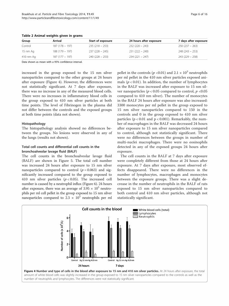
Table 2 Animal weights given in grams
Group Arrival Start of exposure 24 hours after exposure 7 days after exposure
Control 187 (178 – 197) 235 (218 – 253) 232 (220 – 243) 250 (237 – 263)
15 nm Ag 188 (179 – 197) 237 (228 – 245) 231 (222 – 240) 248 (243 – 253)
410 nm Ag 187 (177 – 197) 240 (228 – 253) 234 (221 – 247) 243 (229 – 258)
Data shown as mean with a 95% confidence interval.
Braakhuis et al. Particle and Fibre Toxicology 2014, 11:49 Page 6 of 16http://www.particleandfibretoxicology.com/content/11/1/49
increased in the group exposed to the 15 nm silvernanoparticles compared to the other groups at 24 hoursafter exposure (Figure 4). However, the differences werenot statistically significant. At 7 days after exposure,there was no increase in any of the measured blood cells.There were no increases in inflammatory blood cells inthe group exposed to 410 nm silver particles at bothtime points. The level of Fibrinogen in the plasma didnot differ between the controls and the exposed groupsat both time points (data not shown).
HistopathologyThe histopathology analysis showed no differences be-tween the groups. No lesions were observed in any ofthe lungs (results not shown).
Total cell counts and differential cell counts in thebronchoalveolar lavage fluid (BALF)The cell counts in the bronchoalveolar lavage fluid(BALF) are shown in Figure 5. The total cell numberwas increased 24 hours after exposure to 15 nm silvernanoparticles compared to control (p = 0.063) and sig-nificantly increased compared to the group exposed to410 nm silver particles (p < 0.05). The increased cellnumber is caused by a neutrophil influx (Figure 6). 24 hoursafter exposure, there was an average of 3.95 × 10⁵ neutro-phils per ml cell pellet in the group exposed to 15 nm silvernanoparticles compared to 2.3 × 10³ neutrophils per ml
Figure 4 Number and type of cells in the blood after exposure to 15amount of white blood cells was slightly increased in the group exposed tnumber of neutrophils and lymphocytes. The differences were not statistic
pellet in the controls (p <0.01) and 2.1 × 10³ neutrophilsper ml pellet in the 410 nm silver particles exposed ani-mals (p < 0.01). In addition, the number of lymphocytesin the BALF was increased after exposure to 15 nm sil-ver nanoparticles (p < 0.05 compared to control, p <0.05compared to 410 nm silver). The number of monocytesin the BALF 24 hours after exposure was also increased:3300 monocytes per ml pellet in the group exposed to15 nm silver nanoparticles compared to 150 in thecontrols and 0 in the group exposed to 410 nm silverparticles (p < 0.01 and p < 0.001). Remarkably, the num-ber of macrophages in the BALF was decreased 24 hoursafter exposure to 15 nm silver nanoparticles comparedto control, although not statistically significant. Therewere no differences between the groups in number ofmulti-nuclei macrophages. There were no eosinophilsdetected in any of the exposed groups 24 hours afterexposure.The cell counts in the BALF at 7 days after exposure
were completely different from those at 24 hours afterexposure. At 7 days after exposure, most observed ef-fects disappeared. There were no differences in thenumber of lymphocytes, macrophages and monocytesbetween the exposure groups. There was a slight de-crease in the number of neutrophils in the BALF of ratsexposed to 15 nm silver nanoparticles compared toboth control and 410 nm silver particles, although notstatistically significant.
nm and 410 nm silver particles. At 24 hours after exposure, the totalo 15 nm silver nanoparticles compared to the controls as well as theally significant.
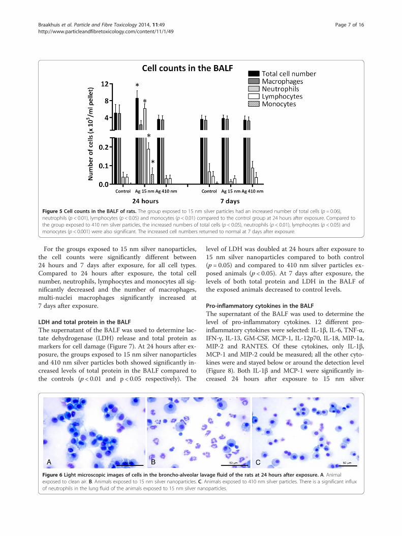
Figure 5 Cell counts in the BALF of rats. The group exposed to 15 nm silver particles had an increased number of total cells (p = 0.06),neutrophils (p < 0.01), lymphocytes (p < 0.05) and monocytes (p < 0.01) compared to the control group at 24 hours after exposure. Compared tothe group exposed to 410 nm silver particles, the increased numbers of total cells (p < 0.05), neutrophils (p < 0.01), lymphocytes (p < 0.05) andmonocytes (p < 0.001) were also significant. The increased cell numbers returned to normal at 7 days after exposure.
Braakhuis et al. Particle and Fibre Toxicology 2014, 11:49 Page 7 of 16http://www.particleandfibretoxicology.com/content/11/1/49
For the groups exposed to 15 nm silver nanoparticles,the cell counts were significantly different between24 hours and 7 days after exposure, for all cell types.Compared to 24 hours after exposure, the total cellnumber, neutrophils, lymphocytes and monocytes all sig-nificantly decreased and the number of macrophages,multi-nuclei macrophages significantly increased at7 days after exposure.
LDH and total protein in the BALFThe supernatant of the BALF was used to determine lac-tate dehydrogenase (LDH) release and total protein asmarkers for cell damage (Figure 7). At 24 hours after ex-posure, the groups exposed to 15 nm silver nanoparticlesand 410 nm silver particles both showed significantly in-creased levels of total protein in the BALF compared tothe controls (p < 0.01 and p < 0.05 respectively). The
Figure 6 Light microscopic images of cells in the broncho-alveolar lavexposed to clean air. B. Animals exposed to 15 nm silver nanoparticles. C. Aof neutrophils in the lung fluid of the animals exposed to 15 nm silver nan
level of LDH was doubled at 24 hours after exposure to15 nm silver nanoparticles compared to both control(p = 0.05) and compared to 410 nm silver particles ex-posed animals (p < 0.05). At 7 days after exposure, thelevels of both total protein and LDH in the BALF ofthe exposed animals decreased to control levels.
Pro-inflammatory cytokines in the BALFThe supernatant of the BALF was used to determine thelevel of pro-inflammatory cytokines. 12 different pro-inflammatory cytokines were selected: IL-1β, IL-6, TNF-α,IFN-γ, IL-13, GM-CSF, MCP-1, IL-12p70, IL-18, MIP-1a,MIP-2 and RANTES. Of these cytokines, only IL-1β,MCP-1 and MIP-2 could be measured; all the other cyto-kines were and stayed below or around the detection level(Figure 8). Both IL-1β and MCP-1 were significantly in-creased 24 hours after exposure to 15 nm silver
age fluid of the rats at 24 hours after exposure. A. Animalnimals exposed to 410 nm silver particles. There is a significant influxoparticles.
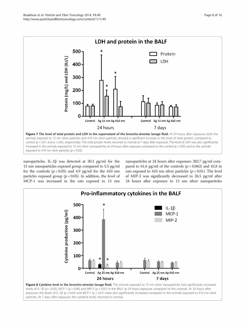
Figure 7 The level of total protein and LDH in the supernatant of the broncho-alveolar lavage fluid. At 24 hours after exposure, both theanimals exposed to 15 nm silver particles and 410 nm silver particles showed a significant increase in the level of total protein compared tocontrol (p < 0.01 and p < 0.05, respectively). The total protein levels returned to normal at 7 days after exposure. The level of LDH was also significantlyincreased in the animals exposed to 15 nm silver nanoparticles at 24 hours after exposure compared to the control (p < 0.05) and to the animalsexposed to 410 nm silver particles (p < 0.05).
Braakhuis et al. Particle and Fibre Toxicology 2014, 11:49 Page 8 of 16http://www.particleandfibretoxicology.com/content/11/1/49
nanoparticles. IL-1β was detected at 30.2 pg/ml for the15 nm nanoparticles exposed group compared to 5.5 pg/mlfor the controls (p < 0.05) and 4.9 pg/ml for the 410 nmparticles exposed group (p < 0.05). In addition, the level ofMCP-1 was increased in the rats exposed to 15 nm
Figure 8 Cytokine level in the broncho-alveolar lavage fluid. The animlevels of IL-1β (p < 0.05), MCP-1 (p = 0.06) and MIP-2 (p < 0.01) in the BALF aexposure, the levels of IL-1β (p < 0.05) and MCP-1 (p < 0.01) were also signiparticles. At 7 days after exposure, the cytokine levels returned to normal.
nanoparticles at 24 hours after exposure: 382.7 pg/ml com-pared to 61.6 pg/ml of the controls (p = 0.063) and 42.8 inrats exposed to 410 nm silver particles (p < 0.01). The levelof MIP-2 was significantly decreased to 26.5 pg/ml after24 hours after exposure to 15 nm silver nanoparticles
als exposed to 15 nm silver nanoparticles had significantly increasedt 24 hours exposure compared to the controls. At 24 hours afterficantly increased compared to the animals exposed to 410 nm silver

Braakhuis et al. Particle and Fibre Toxicology 2014, 11:49 Page 9 of 16http://www.particleandfibretoxicology.com/content/11/1/49
compared to the level of 60.2 pg/ml in the controls (p <0.01). The changes in IL-1β, MCP-1 and MIP-2 levels at24 hours after exposure to 15 nm silver nanoparticles re-covered at 7 days after exposure, when no significant differ-ences were observed in any of the cytokine levels measuredbetween the different exposure groups. There were no sig-nificant differences between the group exposed to 410 nmsilver particles and the controls at both 24 hours and 7 daysafter exposure.
Oxidative stress in the lungsThe levels of total glutathione, oxidized (GSSG) and re-duced (GSH) glutathione were measured in the hom-ogenate of the rinsed right lung of all the rats (Figure 9).The level of total glutathione was significantly increasedat 24 hours after exposure to 15 nm silver nanoparticlescompared to the controls (p < 0.01) and compared to thegroup exposed to 410 nm silver particles (p < 0.05). Thelevel of reduced glutathione was also significantly in-creased at 24 hours after exposure to 15 nm silver nano-particles compared to the controls (p < 0.01) and the410 nm exposed group (p < 0.05), whereas the level ofoxidized glutathione was not. The ratio of GSH/GSSGwas not significantly different between any of the expos-ure groups. 7 days after exposure, the level of total gluta-thione and reduced glutathione in the group exposed to15 nm silver nanoparticles returned to control levels.
DiscussionIn this short-term inhalation study, silver nanoparticlesof two different sizes were tested to determine the differ-ences in the lung deposition, tissue distribution and ad-verse effects. Exposure to 15 nm silver nanoparticlesinduced a moderate inflammatory response at 24 hoursafter the end of the exposure period. All the observed ef-fects disappeared at 7 days after exposure. The most
Figure 9 Glutathione production in the lungs of rats. At 24 hours afternanoparticles exposed group was significantly elevated compared to the con
pronounced observed effect was the influx of neutro-phils at 24 hours after exposure to 15 nm silver nano-particles, which was about 175 times larger compared tothe controls. In addition, we found release of pro-inflammatory cytokines, an increase in total glutathioneand the presence of silver nanoparticles inside lung cells.In contrast, the 410 nm particles induced none of theseeffects in the lung, clearly demonstrating a particle sizedependent difference in the induction of pulmonaryinflammation.There are two main reasons to explain the noted dif-
ferences in effects by the two silver particles. First, thedifferences in effects may be attributed to the differencesin lung deposition of the smaller versus the larger silverparticles. The total lung deposit as measured by HR-ICP-MS was 5.5 μg for the 15 nm silver nanoparticlesand 8.5 μg for the 410 nm silver particles at 24 hoursafter the end of the exposure period. According to theMPPD model, the fraction that reaches the alveoli isabout 5.5 times higher for the 15 nm nanoparticles com-pared to the 410 nm particles. Based on the HR-ICP-MSresults we estimated the amount of 15 nm silver nano-particles present in the alveoli was 7.9 × 10⁶ particlesper mm3 (2.1 μg mass or 5.8 × 10¹² nm2/cm3 surfacearea) and the number of 410 nm silver particles was 118particles per mm3 (0.6 μg mass or 6.2 × 10¹⁰ nm2/cm3
surface area). Therefore, the number of bioavailable15 nm nanoparticles in the alveoli is about 66,000 timeslarger compared to the 410 nm particles, resulting in ahigher interaction probability with cells.Comparing same mass deposits of microparticles and
nanoparticles, the latter contains a much higher numberof particles that need to be cleared. In addition, thesmaller silver nanoparticles deposit deeper in the lungswhere they are less efficiently cleared compared to the lar-ger particles that deposit more in the conducting airways
exposure, the total glutathione production in the 15 nm silvertrols (p < 0.01) and the 410 nm silver particles exposed group (p < 0.05).

Braakhuis et al. Particle and Fibre Toxicology 2014, 11:49 Page 10 of 16http://www.particleandfibretoxicology.com/content/11/1/49
where they are more easily cleared [16-18,21-24]. More-over, the phagocytosis of nanoparticles by macrophages isless efficient [25-27] and slower compared to microparti-cles [28-30]. Our results, however, show a clearance fromthe lungs of 62% of the 15 nm nanoparticles compared to31% of the 410 nm particles at 7 days after exposure. Thehigher clearance rate of the smaller particles might be ex-plained by translocation from the lungs of the smaller sil-ver nanoparticles or the released ions to the blood orinterstitium. This is further supported by the detection ofsilver in the liver of animals at 24 hours after exposure to15 nm silver nanoparticles, indicating systemic availabilityof silver particles or ions. This was not detected after ex-posure to the 410 nm silver particles.Previous inhalation studies with nanosilver showed a
dose-dependent increase in silver concentration in theliver [11,13,15] and even a low translocation to the brainand olfactory bulb [11,15]. In one study, the amount ofsilver in the brain and olfactory bulb was 2.2 ng/g and6.97 ng/g, respectively, after exposure to the highestdose tested of 61 μg/m3 [11]. In the other study, theamount of silver in the brain and olfactory bulb was18.63 ng/g and 30.48 ng/g at the highest dose tested of515 μg/m3 [15]. In both studies, the tissue distributionof silver was detected by atomic absorption spectropho-tometer. The reported level of silver is too low to detectwith the HR-ICP-MS, which has a detection limit of0.01 μg/g, possibly explaining why we did not detect anysilver in the brain or olfactory bulb. It should be notedthat both detection methods do not distinguish betweensilver particles and silver ions.The second reason for the observed differences in ef-
fects between the 15 nm and 410 nm silver particles isthe difference in particle dissolution. It is known thatsmaller particles dissolve faster compared to larger parti-cles because of the high surface to volume ratio [31].Several in vitro studies determined the dissolution rateof silver nanoparticles. In the study of Ma et al. [31], thedissolution of silver nanoparticles ranged from 1% forthe larger particles of 80 nm to 60% for the smallest par-ticles of 5 nm at pH 8 during three and two months in-cubation, respectively. However, there was no constantrelease of silver ions per unit of surface area, indicatingthat surface area alone did not explain the dissolution ofsilver nanoparticles [31]. According to this and otherstudies [31-35], the dissolution of silver nanoparticlesdepends on their particle size, the pH of the solution,the ions present in the solution (e.g. sulphate, chlorideand phosphate causing precipitations with silver ions orcatalysing dissolution), aggregation state and the incuba-tion time. Giving these in vitro results, the dissolutionrate of the 15 nm silver nanoparticles will probably behigher compared to the 410 nm silver particles resultingin an increased ion release. None of these studies report
complete dissolution of silver nanoparticles, the effectsobserved after exposure to silver nanoparticles can beinduced by the released ions, the silver nanoparticles it-self or a combination of both.In a study of Pratsinis et al. [36], the toxicity of silver
nanoparticles in the presence of their released ions, andthe released ions alone was tested in vitro. For small sil-ver nanoparticles with a size below 10 nm, the releasedsilver ions from the surface dominated the cytotoxicitywhereas for larger silver nanoparticles the interactionswith silver particles dominated the toxicity [36]. Accord-ing to Beer et al. [37], at silver ion fractions ≤2.6% thesilver nanoparticles contribute to the observed toxicitywhile at silver ion fractions ≥5.5% the silver nanoparti-cles do not add additional toxicity in vitro [37]. In a re-cent published study [38], oropharyngeal aspiration inmice of 20 nm citrate-coated silver nanoparticles causedpulmonary inflammation indicated by neutrophil influxand the release of pro-inflammatory cytokines, whereas110 nm citrate-coated silver particles caused a smallerneutrophil influx, only at the highest dose tested, and norelease of pro-inflammatory cytokines. At 21 days afterexposure, the silver content in the lungs did not differbetween 20 nm and 110 nm silver particles. In addition,for both particle sizes, there was no silver detected inany of the other selected organs and tissues. Solubilitytest showed that about 4% of the 20 nm nanoparticlesand 2% of the 110 nm particles dissolved in 24 hours.Therefore, the authors concluded the larger pulmonaryinflammation caused by the 20 nm silver nanoparticlesis due to the higher bioavailability of silver ions. How-ever, the effects of 20 nm silver nanoparticles werestronger compared to the effects of silver nitrate indicat-ing the particles itself contributed to the observed ef-fects. In contrast to these results, the 110 nm silverparticles caused mild sub-chronic effects in the lungs at21 days after exposure, which might be caused by aslower and more persistent silver ions release [38]. Simi-lar to these studies, it is likely that the release of silverions contributed to the observed effects after exposureto 15 nm silver nanoparticles in the present study. Tofurther elucidate the mechanism of the silver nanoparti-cle toxicity, the dissolution behaviour of silver nanoparti-cles inside cells should be investigated in detail.In our study, we could detect by TEM silver nanopar-
ticles in the lung tissue of rats at 24 hours after exposureto 15 nm silver nanoparticles. At 24 hours after exposureto 410 nm silver particles, we could not detect silver par-ticles in the lungs because the number of particles wastoo low. The detected silver nanoparticles had a particlesize of 15–20 nm after silver enhancement, indicatingtheir size in the lung tissue was smaller than 5 nm be-fore enhancement. In combination with the literature onthe dissolution of silver nanoparticles, these EM results

Braakhuis et al. Particle and Fibre Toxicology 2014, 11:49 Page 11 of 16http://www.particleandfibretoxicology.com/content/11/1/49
are an indication that the silver nanoparticles probablypartially dissolved after deposition resulting in a decreasein particle size and the release of silver ions. The dissol-ution might occur extra-cellular in the lung lining fluidand intra-cellular in vesicles in epithelial cells or macro-phages. Besides the release of silver ions, we believe thesilver nanoparticles itself may also have contributed tothe observed effects because the TEM images show thepresence of silver nanoparticles inside lung cells. The15 nm silver nanoparticles in our study dissolved overtime resulting on the one hand in the release of toxic sil-ver ions and on the other hand in a decrease in particlesize to <5 nm which gives the particles the opportunityto pass the cellular membranes and the nuclear pores.Previous studies report that particles smaller than 5 nmpossibly can pass the nuclear pores [39], while particlesof 20 nm do not reach the nucleus and could be de-tected in vesicles and lamellar bodies in vitro [40]. Onceinside the cells and eventually inside the nucleus, silvernanoparticles are a continued source of ion release, lead-ing to more damage compared to silver ions releasedfrom silver nitrate [41]. The intracellular ion release hasbeen reported previously as the Trojan-horse type mech-anism [42,43]; nanoparticles can more easily enter thecells compared to the ions itself. The higher dissolutionrate, in combination with the higher alveolar depositionof the 15 nm nanoparticles, might explain why the15 nm nanoparticles caused toxicity and the 410 nmparticles did not. Based on the results of the presentstudy, it is not possible to quantify how much of the ob-served effects can be attributed to the difference in al-veolar deposition and how much of the effects can beattributed to cellular uptake and ion release.Findings of particle size related pulmonary inflamma-
tion are not limited to silver nanoparticles. Other inhal-ation studies with titanium dioxide, carbonaceous, nickeloxide, zinc oxide, ferric oxide, aluminium oxyhydroxide,quartz and cerium oxide particles also focused on the in-fluence of particle size on the induction of pulmonaryinflammation [27,44-52]. Indeed, several studies reportthat smaller nanoparticles caused a greater inflammatoryresponse compared to larger particles when the samemass dosage was administered [27,44-48]. However, al-though particle size will affect the site and amount ofdeposited material, it seems that the local dose alonecannot explain the differences in toxicity. Solubility willat least be one of the other variables in an equation thatwould allow predicting adverse effects of nanoparticlesafter inhalation.
ConclusionThe results of the present short-term inhalation study ofuncoated silver particles indicate that particle size is animportant characteristic that determines the induction
of pulmonary inflammation. Exposure to 15 nm silvernanoparticles induced moderate pulmonary inflamma-tion at 24 hours after exposure, whereas 410 nm silverparticles did not. The lung deposition of the 410 nm sil-ver particles was mainly in the upper airways; the num-ber of particles deposited in the alveoli was calculated tobe 66,000 times higher for the 15 nm silver nanoparti-cles. TEM analysis showed the presence of silver nano-particles in lung cells that were decreased in particlesize, demonstrating the in vivo dissolution of silvernanoparticles. Altogether, these findings strongly suggestthat size-related silver nanoparticle induced pulmonaryinflammation is a consequence of both size related lungdeposition and dissolution rate.
MethodsTest material and characterizationThe test atmosphere was produced by mixing silvernanoparticles with HEPA filtered and conditioned (50%RH, 21°C) compressed air. Depending on the target par-ticle size, one of two types of particle generators wasused. The silver nanoparticles were produced by a PalasGFG 1000 (Palas GmbH, Karlsruhe, Germany) sparkgenerator fitted with silver tipped copper electrodes. Togenerate the 15 nm nanoparticles the output of the gen-erator was immediately diluted in conditioned air. Oxy-gen was added to the dilution air to compensate for theargon flow from the Palas, a final concentration of 20%oxygen in the airflow was maintained. The particle num-ber concentration was controlled by setting the Palasspark frequency. The final condition of the aerosol (55%RH, 21°C) was set by adjusting the relative humidity ofthe mixing air (Figure 10). 200 nm silver particles werepurchased by NanoComposix, inc. San Diego, USA. The200 nm silver particles were polyvinylpyrrolidone (PVP)-coated and supplied in solution of 1 mg/ml in MilliQwater. The silver particle solution was forced through asmall nozzle of a Schlick spray nozzle where it wasbroken up in small droplets (nebulized) by compressedair. The flow of the solution was controlled using a20 ml Terumo syringe and a syringe pump (TSE model540200, Bad homburg, Germany). The nebulized flowwas mixed with compressed air in a heated mixing tube.After mixing and drying, the aerosol passed through atube oven (700°C) to fuse the aggregates into a singleparticle. The heat of the oven removed the PVP-coatingof the silver particles. After the oven, the aerosol dilutedfurther with conditioned air and was allowed to cool.The mass concentration was controlled by adjusting thesyringe pump motor speed and thus the flow of the solu-tion. The final condition of the aerosol (55% RH, 21°C)was set by adjusting the relative humidity of the finalmixing air (Figure 11).

Figure 10 Palas spark generator setup for the nose-only exposure to 15 nm silver nanoparticles. The silver nanoparticles are generated bythe Palas spark generator and subsequently mixed with conditioned air and oxygen. Particle size, number and mass are continuously monitored.
Braakhuis et al. Particle and Fibre Toxicology 2014, 11:49 Page 12 of 16http://www.particleandfibretoxicology.com/content/11/1/49
Several measurements of the test atmosphere wereperformed during the exposure. In the main flow up-stream of the nose only unit particle mass concentration,number concentration, and particle size distributionwere measured. The particle mass concentration was de-termined by time aggregated gravimetric with TeflonR2PJ047 filter (Pall corp., Ann Arbor MI, USA) and bytapered element oscillating microbalance (TEOM)series1400 (Rupprecht & Patashnick, New York, USA).The particle number concentration was measured overtime by a condensation particle counter (CPC) 3022(TSI inc., St Paul MN, USA). Particle size distributionwas monitored over time by an OPS 3330 (TSI inc., StPaul MN, USA), a scanning mobility particle sizer(SMPS) 3080 with 3085 Nano DMA (TSI inc., St Paul MN,USA) and a MOI Model No. 110 (MSP corp, MinneapolisMN, USA). Temperature and relative humidity were deter-mined by a Vaisala M170 (Vaisala Oyj, Helsinki, Finland).The particle size was confirmed by scanning electron mi-croscopy (SEM) using a Nova Nanolab 600 Dualbeam(FEI, Eindhoven, the Netherlands) (Figure 1). In addition,the gravimetric mass concentration was determined. A
Sartorius MC-5 microbalance (Sartorius, Goettingen,Germany) was used in controlled relative humidity (40 –45%) and temperature (21 – 23°C) conditions to do themass measurements, the filters were weighed before andafter each exposure. Laboratory and field blanks were usedfor quality assurance. The filter volume flow was mea-sured with dry gas meters (Gallus 2000 G1.6, ActarisMeterfabriek B.V., Dordrecht, the Netherlands).
AnimalsThe Animal Ethical Committee of the National Institutefor Public Health and the Environment (Bilthoven, theNetherlands) approved the experimental design of theanimal study at the 6th of July 2012. Healthy maleFischer rats (F344/DuCrl) were supplied by Charles River(Sulzfeld, Germany). The animals were kept under specificpathogen free (SPF) conditions. After an acclimatizationperiod of two weeks, the animals were 10 weeks old at thestart of the experiment. The rats were randomly allocatedto the control and the test groups. During exposure, theanimals were restrained in nose-only tubes (CH technolo-gies, Westwood NJ, USA) fixed to the inhalation system.

Figure 11 Spray nozzle setup for the nose-only exposure to 410 nm silver particles. The silver particles are in solution and sprayed into aheated mixing chamber. Subsequently, the particles are dried in an oven and mixed with compressed air. Particle size, number and mass arecontinuously monitored.
Braakhuis et al. Particle and Fibre Toxicology 2014, 11:49 Page 13 of 16http://www.particleandfibretoxicology.com/content/11/1/49
When not exposed, animals were housed up to 3 animalsin macrolon III cages with filtertops to prevent dust enter-ing the cages (bedding material: Lignocel S8-15, AltrominSpezialfutter GmbH & Co. KG). The animal room wasair-conditioned with a 12 hours light- 12 hours dark cycle,the temperature ranging from 20 to 24°C with relative hu-midity ranging from 30 – 70%. Except during exposure,certified feed CRM (SDS Diets, UK) and water were avail-able ad libitum. Before start of the exposure period, allanimals were acclimatized to the nose only tubes on 3consecutive days for 1 hour per day. At 24 hours and7 days after the last exposure, rats were weighed, anesthe-tized by a single intraperitoneal injection of ketamine(75 mg/kg) and xylazine (10 mg/kg) and subsequently ex-sanguinated via the abdominal aorta.
Experimental designThe rats were nose-only exposed for 6 hours per day, 4consecutive days to fresh air, 15 nm silver nanoparticlesor 410 nm silver nanoparticles, 12 rats per exposuregroup. Of each exposure group, 6 rats were sacrificed24 hours after exposure and the other 6 rats were sacri-ficed 7 days after exposure. During nose-only inhalation,rats have a breathing pattern that results in a more real-istic internal exposure than a single high dose in intra-tracheal instillation and oropharyngeal aspiration orpossible additional oral exposure in whole-body expos-ure chambers.
Quantification of silver in tissues by high resolutioninductively coupled plasma mass spectrometry (HR-ICP-MS)The presence of silver in the lungs, liver, spleen, kidneys,brain, testis and lung associated lymph nodes was deter-mined by high resolution inductively coupled plasmamass spectrometer (HR-ICP-MS) [53,54]. Samples of ho-mogenized tissue (max. 0.5 g) were completely dissolvedon a block heater (Stuart SBH200D, supplied by Omni-labo, Breda, The Netherlands) for 24 hours at 120°Cafter the direct addition of aqua regia (0.5 mL 60% nitricacid, ultrapure, and 1.5 mL 30% hydrochloric acid, ultra-pure) and afterwards diluted. The silver concentration inthe digests was determined using high resolution induct-ively coupled plasma mass spectroscopy (HR-ICPMS;Element XR, Thermo Fisher Scientific, Bremen, Germany).Both silver isotopes (107Ag and 109Ag) were measured inthe low-resolution mode. External calibration curves of theinterference-free silver isotopes were used for quantifica-tion. On-line addition and correction with an internalstandard (rhodium with the measured isotope 103Rh in thelow-resolution mode) was applied too. The results of 107Agwere reported and the results of 109Ag were used for con-trol. Sample pre-treatment and analysis were carried out ina cleanroom facility class 100 000.
Estimated deposited dose in lungs using Multi PathParticle Dosimetry model (MPPD model)To estimate the fractions of deposited dose in the head,tracheobronchial, and alveolar region of the rats, the

Braakhuis et al. Particle and Fibre Toxicology 2014, 11:49 Page 14 of 16http://www.particleandfibretoxicology.com/content/11/1/49
multiple path particle dosimetry model (MPPD model)was used [16]. We used the default parameters of themodel for rats, i.e., a forced respiratory capacity of 4 ml,head volume of 0.42 ml, nasal breathing, tidal volume of2.1 ml, and a breathing frequency of 102/min. The in-spiratory fraction was 0.5, and no pause was entered.Calculations were done using the count median diam-eter (CMD), geometric SD, the mass concentration, anda density of 10.49 g/cm3.
Electron microscopy of the lungOf two animals of each group, the left lung was cut outwith the bronchus attached and fixed by pressure (2 kPafor 1 hour) with half-strength Karnovsky fixative (0.08 MSodium-cacodylate buffer, 2.5% glutaraldehyde, 0.025 mMCaCl2, 0.05 mM MgCl2) for electron microscopy. Aftertwo days, the fixative was replaced with 2% para-formaldehyde in sodium-cacodylate buffer for storage. Ofeach lung, three slices of about 1 – 1.5 mm were cut outat the top, middle and bottom of the lung for further ana-lysis. The tissue slices were post-fixed by osmium potas-sium ferro-cyanide solution (0.1 M sodium-cacodylatebuffer, 1% osmium tetra-oxide, 1.5% potassium ferro-cyanide). Next, the tissue slices were dehydrated in acetonseries, cut in smaller pieces and subsequently embeddedin epon. The epon was polymerized at 60°C. The eponblocks were trimmed prior to sectioning. During section-ing, ultrathin sections of 70 nm were sectioned and puton 3 mm hexagonal copper grids coated with a formvar-film. The sections were analysed by transmission electronmicroscopy (TEM) (Tecnai 10, 100 kV, FEI, Eindhoven,the Netherlands).
Silver enhancementA silver enhancement procedure was used to increase theparticle size of the silver nanoparticles in the lung tissue tomake it possible to detect the silver nanoparticles with elec-tron microscopy. The silver enhancement reagents werepurchased at Aurion and performed on grid according tomanufacturer’s instructions (Wageningen, the Netherlands).
Clinical examinationsThe animals were examined three times on exposuredays (before, during and after exposure) and once dailyduring the periods before and after exposure. The clin-ical examination included examination of attitude, ani-mal fur, activity level, food and water intake, and faecesand urine production. The body weight of the animalswas determined upon arrival, at the start of the exposureperiod and at the day of sacrifice.
Hematology analysisBlood samples were collected in EDTA-containing tubesand in citrate vials for analysis of cell types and
inflammatory markers in the blood. The level of Fibrino-gen in the citrate plasma was measured using a ratFribrinogen ELISA kit (GenWay Biotech, Inc., San Diego,USA) according to manufacturer’s instructions.
HistopathologyThe lungs fixed with half-strength Karnovsky fixativewere also used for histopathology. Of these lungs a thinslice was made over the length of the tissue, embeddedin paraffin and stained by haematoxylin-eosin for histo-pathology analysis.
Bronchoalveolar lavage fluid (BALF) analysisThe left lung of the rats was bound just below the bifur-cation of the trachea and the right lung was cannulatedvia the trachea. Bronchoalvelar lavage was performed insitu by infusing the right lung three times with 27 ml/kgphysiological saline. The retrieved bronchoalveolar lav-age fluid (BALF) was kept on ice and centrifuged for10 minutes at 400 g. The pellet was resuspended inphysiological salt for analysis of the total cell numberand cell differentials. Cytospin preparations were stainedand evaluated microscopically for macrophages, poly-nuclear macrophages, polymorph nuclear neutrophils,lymphocytes, monocytes, eosinophils, and atypical cells.At least 400 cells were counted on each slide. The cell-free supernatant was collected to assess cell damage bymeasurement of total protein content and the release oflactate dehydrogenase (LDH) by Beckman Coulter auto-analyser Synchron LX20 (Beckman Coulter, Inc.) and toassess the induction of pro-inflammatory cytokines. Theremainder of the supernatant was stored at −80°C.
Measurement of pro-inflammatory cytokinesThe presence of pro-inflammatory cytokines IL-1beta,IL-6, TNF-alfa, IFN-gamma, IL-13, GM-CSF, MCP-1, IL-12p70, IL-18, MIP-1a, MIP-2 and RANTES in the super-natant of the BALF was measured by a Bio-Plex Proassay for rat cytokines according to manufacturer’s in-structions (Bio-Rad Laboratories, Inc.).
Measurement of oxidative stressThe induction of oxidative stress was determined bymeasuring the amount of reduced, oxidized and totalglutathione in the lungs. The rinsed right lung was ho-mogenized on ice in 4 ml of Phosphate/EDTA buffer(pH 7.5). Subsequently, the homogenate was centrifugedat 600 g for 10 minutes. To remove the proteins fromthe samples, the supernatant was mixed 1:1 with meta-phosphorid acid, incubated for 15 minutes on ice, andcentrifuged at 4000 rpm for 10 minutes. The amount ofreduced, oxidized and total glutathione was measured byBeckmann Coulter Autoanalyser in the supernatant.

Braakhuis et al. Particle and Fibre Toxicology 2014, 11:49 Page 15 of 16http://www.particleandfibretoxicology.com/content/11/1/49
Statistical analysisThe raw data of the BALF was corrected for retrievedfluid. To compare the different exposure groups, thedata was analysed by the Kruskal Wallis nonparametrictest (Graphpad Prism). Statistical significance is indi-cated with a * (p = < 0.05). In all graphs, error bars rep-resent the standard deviation of the mean.
Additional file
Additional file 1: Figure S1. SMPS and OPS particle size distribution(left) and CPC particle number concentration (right) of 15 nm and 410nm silver particles.
Competing interestsThe authors declare that they have no competing interests.
Authors’ contributionsHB designed the study. HB, MP, AJFB, PHBF carried out the study. HBcollected, analysed, interpreted data and drafted the manuscript. PKperformed HR-ICPMS analysis and PHBF performed particle generation, bothgenerated and interpreted data. AJFB, IG, MP and JAP contributed to thestudy design. JAP provided expert input on the electron microscopy data.MP, FRC, JAP and HvL have been involved in revising the manuscript criticallyfor important intellectual content. All authors read and approved the finalmanuscript.
AcknowledgementsThis work was supported by the project ‘Integrated Risk Assessment ofNanomaterials’ from the National Institute for Public Health and theEnvironment and by the NanoNextNL program ‘Risk Analysis and TechnologyAssessment: Human Health Risks’. We would like to thank Liset J.J. de laFoneteyne for the BALF and haematology cells counts, Piet K. Beekhof for theanalysis of the total protein, LDH and glutathione, and Henny W. Verharen,Hans J.C. Strootman, Ron F. Vlug, Christine M.R. Soputan, Jan Bos, JolandaRigters for their excellent technical assistance. In addition, we thank Karin E. M.Vocking, Elly G. van Donselaar for their assistance with the TEM imaging, Josevan den Dungen for assistance with sample pretreatments and D. A. Matthijsde Winter for the assistance with the SEM imaging.
Author details1Department of Toxicogenomics, Maastricht University, PO Box 616,Maastricht 6200, MD, the Netherlands. 2National Institute for Public Healthand the Environment (RIVM), PO Box 1, Bilthoven 3720, BA, the Netherlands.3Philips Innovation Services, High Tech Campus 11, Eindhoven 5656, AE, theNetherlands. 4Institute of Risk Assessment Sciences, Utrecht University, POBox 80.163, Utrecht 3508, TD, the Netherlands. 5Cellbiology, BiologyDepartment, Faculty of Science, University of Utrecht, PO box 80.056, Utrecht3508, TB, the Netherlands.
Received: 4 March 2014 Accepted: 29 August 2014
References1. Nanotechnologies PoE: Consumer Products Inventory. In Retrieved [June,
2014], from http://www.nanotechproject.org/cpi.2. Maynard AD, Kuempel ED: Airborne nanostructured particles and
occupational health. J Nanoparticle Res 2005, 7:587–614.3. Christensen FM, Johnston HJ, Stone V, Aitken RJ, Hankin S, Peters S,
Aschberger K: Nano-silver - feasibility and challenges for human healthrisk assessment based on open literature. Nanotoxicology 2010, 4:284–295.
4. Wijnhoven SWP, Peijnenburg WJGM, Herberts CA, Hagens WI, Oomen AG,Heugens EHW, Roszek B, Bisschops J, Gosens I, Meent D, Dekkers S, JongWH, Zijverden M, Sips AJAM, Geertsma RE: Nano-silver - a review ofavailable data and knowledge gaps in human and environmental riskassessment. Nanotoxicology 2009, 3:109–138.
5. Sotiriou GA, Pratsinis SE: Engineering nanosilver as an antibacterial,biosensor and bioimaging material. Curr Opin Chem Eng 2011, 1:3–10.
6. Sotiriou GA, Pratsinis SE: Antibacterial activity of nanosilver ions andparticles. Environ Sci Technol 2010, 44:5649–5654.
7. Lok CN, Ho CM, Chen R, He QY, Yu WY, Sun H, Tam PK, Chiu JF, Che CM:Silver nanoparticles: partial oxidation and antibacterial activities. J BiolInorg Chem 2007, 12:527–534.
8. Morones JR, Elechiguerra JL, Camacho A, Holt K, Kouri JB, Ramirez JT,Yacaman MJ: The bactericidal effect of silver nanoparticles.Nanotechnology 2005, 16:2346–2353.
9. Xiu ZM, Zhang QB, Puppala HL, Colvin VL, Alvarez PJ: Negligible particle-specific antibacterial activity of silver nanoparticles. Nano Lett 2012,12:4271–4275.
10. Sung JH, Ji JH, Song KS, Lee JH, Choi KH, Lee SH, Yu IJ: Acute inhalationtoxicity of silver nanoparticles. Toxicol Ind Health 2011, 27:149–154.
11. Ji JH, Jung JH, Kim SS, Yoon JU, Park JD, Choi BS, Chung YH, Kwon IH,Jeong J, Han BS, Shin JH, Sung JH, Song KS, Yu IJ: Twenty-eight-dayinhalation toxicity study of silver nanoparticles in Sprague–Dawley rats.Inhal Toxicol 2007, 19:857–871.
12. Stebounova LV, Adamcakova-Dodd A, Kim JS, Park H, O’Shaughnessy PT,Grassian VH, Thorne PS: Nanosilver induces minimal lung toxicity orinflammation in a subacute murine inhalation model. Part Fibre Toxicol2011, 8:5.
13. Song KS, Sung JH, Ji JH, Lee JH, Lee JS, Ryu HR, Lee JK, Chung YH, Park HM,Shin BS, Chang HK, Kelman B, Yu IJ: Recovery from silver-nanoparticle-exposure-induced lung inflammation and lung function changes inSprague Dawley rats. Nanotoxicology 2012, 7:169–180.
14. Sung JH, Ji JH, Yoon JU, Kim DS, Song MY, Jeong J, Han BS, Han JH, ChungYH, Kim J, Kim TS, Chang HK, Lee EJ, Lee JH, Yu IJ: Lung function changesin Sprague–Dawley rats after prolonged inhalation exposure to silvernanoparticles. Inhal Toxicol 2008, 20:567–574.
15. Sung JH, Ji JH, Park JD, Yoon JU, Kim DS, Jeon KS, Song MY, Jeong J, HanBS, Han JH, Chung YH, Chang HK, Lee JH, Cho MH, Kelman BJ, Yu IJ:Subchronic inhalation toxicity of silver nanoparticles. Toxicol Sci 2009,108:452–461.
16. Asgharian B, Price O, Miller F, Subramaniam R, Cassee FR, Freijer J, van BreeL, Winter-Sorkina R: Multiple-Path Particle Dosimetry Model (MPPD v2.11): A Model for Human and Rat Airway Particle Dosimetry. In HamnerInstitutes for Health Sciences Applied Research Associates (ARA), NationalInstitute for Public Health and the Environment (RIVM), and Ministry ofHousing, Spatial Planning and the Environment, Editor. Raleigh, NorthCarolina, USA: Applied Research Associates (ARA); 2009.
17. ICRP: Human respiratory tract model for radiological protection.International Commission on Radiological Protection. ICRP Publication 661994, 24:1–3.
18. Carvalho TC, Peters JI, Williams RO 3rd: Influence of particle size on regionallung deposition–what evidence is there? Int J Pharm 2011, 406:1–10.
19. Cassee FR, Muijser H, Duistermaat E, Freijer JJ, Geerse KB, Marijnissen JC, ArtsJH: Particle size-dependent total mass deposition in lungs determinesinhalation toxicity of cadmium chloride aerosols in rats. Application of amultiple path dosimetry model. Arch Toxicol 2002, 76:277–286.
20. Braakhuis HM, Park MV, Gosens I, De Jong WH, Cassee FR: Physicochemicalcharacteristics of nanomaterials that affect pulmonary inflammation. PartFibre Toxicol 2014, 11:18.
21. Geiser M, Kreyling WG: Deposition and biokinetics of inhalednanoparticles. Part Fibre Toxicol 2010, 7:2.
22. Oberdorster G: Dosimetric principles for extrapolating results of ratinhalation studies to humans, using an inhaled Ni compound as anexample. Health Phys 1989, 57(Suppl 1):213–220.
23. Oberdorster G, Ferin J, Lehnert BE: Correlation between particle size,in vivo particle persistence, and lung injury. Environ Health Perspect 1994,102(Suppl 5):173–179.
24. Geraets L, Oomen AG, Schroeter JD, Coleman VA, Cassee FR: Tissuedistribution of inhaled micro- and nano-sized cerium oxide particles inrats: results from a 28-day exposure study. Toxicol Sci 2012, 127:463–473.
25. Phalen RF, Mendez LB, Oldham MJ: New developments in aerosoldosimetry. Inhal Toxicol 2010, 22(Suppl 2):6–14.
26. Oberdorster G, Pott F: Extrapolation from rat studies with environmentaltobacco smoke (ETS) to humans: comparison of particle mass depositionand of clearance behavior of ETS compounds. Toxicol Lett 1987,35:107–112.

Braakhuis et al. Particle and Fibre Toxicology 2014, 11:49 Page 16 of 16http://www.particleandfibretoxicology.com/content/11/1/49
27. Oberdorster G, Finkelstein JN, Johnston C, Gelein R, Cox C, Baggs R, ElderAC: Acute pulmonary effects of ultrafine particles in rats and mice. ResRep Health Eff Inst 2000, 96:5–74. disc 75–86.
28. Bakand S, Hayes A, Dechsakulthorn F: Nanoparticles: a review of particletoxicology following inhalation exposure. Inhal Toxicol 2012, 24:125–135.
29. Muhlfeld C, Gehr P, Rothen-Rutishauser B: Translocation and cellularentering mechanisms of nanoparticles in the respiratory tract. Swiss MedWkly 2008, 138:387–391.
30. Geiser M, Casaulta M, Kupferschmid B, Schulz H, Semmler-Behnke M,Kreyling W: The role of macrophages in the clearance of inhaled ultrafinetitanium dioxide particles. Am J Respir Cell Mol Biol 2008, 38:371–376.
31. Ma R, Levard C, Marinakos SM, Cheng Y, Liu J, Michel FM, Brown GE, LowryGV: Size-controlled dissolution of organic-coated silver nanoparticles.Environ Sci Technol 2012, 46:752–759.
32. Leo BF, Chen S, Kyo Y, Herpoldt KL, Terrill NJ, Dunlop IE, McPhail DS, ShafferMS, Schwander S, Gow A, Zhang J, Chung KF, Tetley TD, Porter AE, RyanMP: The stability of silver nanoparticles in a model of pulmonarysurfactant. Environ Sci Technol 2013, 47:11232–11240.
33. Stebounova LV, Guio E, Grassian VH: Silver nanoparticles in simulatedbiological media: a study of aggregation, sedimentation, and dissolution.J Nanopar Res 2011, 13:12.
34. Kent RD, Vikesland PJ: Controlled evaluation of silver nanoparticledissolution using atomic force microscopy. Environ Sci Technol 2012,46:6977–6984.
35. Zook JM, Long SE, Cleveland D, Geronimo CL, MacCuspie RI: Measuringsilver nanoparticle dissolution in complex biological and environmentalmatrices using UV-visible absorbance. Anal Bioanal Chem 2011,401:1993–2002.
36. Pratsinis A, Hervella P, Leroux JC, Pratsinis SE, Sotiriou GA: Toxicity of silvernanoparticles in macrophages. Small 2013, 9:2576–2584.
37. Beer C, Foldbjerg R, Hayashi Y, Sutherland DS, Autrup H: Toxicity of silvernanoparticles - nanoparticle or silver ion? Toxicol Lett 2012, 208:286–292.
38. Wang X, Ji Z, Chang CH, Zhang H, Wang M, Liao YP, Lin S, Meng H, Li R,Sun B, Winkle LV, Pinkerton KE, Zink JI, Xia T, Nel AE: Use of coated silvernanoparticles to understand the relationship of particle dissolution andbioavailability to cell and lung toxicological potential. Small 2014,10:385–398.
39. Singh S, Kumar A, Karakoti A, Seal S, Self WT: Unveiling the mechanism ofuptake and sub-cellular distribution of cerium oxide nanoparticles. MolBiosyst 2010, 6:1813–1820.
40. Herzog F, Clift MJ, Piccapietra F, Behra R, Schmid O, Petri-Fink A,Rothen-Rutishauser B: Exposure of silver-nanoparticles and silver-ions tolung cells in vitro at the air-liquid interface. Part Fibre Toxicol 2013, 10:11.
41. Lubick N: Nanosilver toxicity: ions, nanoparticles–or both? Environ SciTechnol 2008, 42:8617.
42. Park EJ, Yi J, Kim Y, Choi K, Park K: Silver nanoparticles induce cytotoxicityby a Trojan-horse type mechanism. Toxicol In Vitro 2010, 24:872–878.
43. Limbach LK, Wick P, Manser P, Grass RN, Bruinink A, Stark WJ: Exposure ofengineered nanoparticles to human lung epithelial cells: influence ofchemical composition and catalytic activity on oxidative stress. EnvironSci Technol 2007, 41:4158–4163.
44. Stoeger T, Reinhard C, Takenaka S, Schroeppel A, Karg E, Ritter B, Heyder J,Schulz H: Instillation of six different ultrafine carbon particles indicates asurface area threshold dose for acute lung inflammation in mice. EnvironHealth Perspect 2006, 114:328–333.
45. Horie M, Fukui H, Endoh S, Maru J, Miyauchi A, Shichiri M, Fujita K, Niki E,Hagihara Y, Yoshida Y, Morimoto Y, Iwahashi H: Comparison of acuteoxidative stress on rat lung induced by nano and fine-scale, soluble andinsoluble metal oxide particles: NiO and TiO2. Inhal Toxicol 2012,24:391–400.
46. Duffin R, Tran L, Brown D, Stone V, Donaldson K: Proinflammogenic effectsof low-toxicity and metal nanoparticles in vivo and in vitro: highlightingthe role of particle surface area and surface reactivity. Inhal Toxicol 2007,19:849–856.
47. Kobayashi N, Naya M, Endoh S, Maru J, Yamamoto K, Nakanishi J:Comparative pulmonary toxicity study of nano-TiO(2) particles ofdifferent sizes and agglomerations in rats: different short- and long-termpost-instillation results. Toxicology 2009, 264:110–118.
48. Ho M, Wu KY, Chein HM, Chen LC, Cheng TJ: Pulmonary toxicity of inhalednanoscale and fine zinc oxide particles: mass and surface area as anexposure metric. Inhal Toxicol 2011, 23:947–956.
49. Zhu MT, Feng WY, Wang B, Wang TC, Gu YQ, Wang M, Wang Y, Ouyang H,Zhao YL, Chai ZF: Comparative study of pulmonary responses tonano- and submicron-sized ferric oxide in rats. Toxicology 2008,247:102–111.
50. Pauluhn J: Pulmonary toxicity and fate of agglomerated 10 and 40 nmaluminum oxyhydroxides following 4-week inhalation exposure of rats:toxic effects are determined by agglomerated, not primary particle size.Toxicol Sci 2009, 109:152–167.
51. Roursgaard M, Poulsen SS, Poulsen LK, Hammer M, Jensen KA, UtsunomiyaS, Ewing RC, Balic-Zunic T, Nielsen GD, Larsen ST: Time-responserelationship of nano and micro particle induced lung inflammation.Quartz as reference compound. Hum Exp Toxicol 2010, 29:915–933.
52. Gosens I, Mathijssen LE, Bokkers BG, Muijser H, Cassee FR: Comparativehazard identification of nano- and micro-sized cerium oxide particlesbased on 28-day inhalation studies in rats. Nanotoxicology 2013,8:643–653.
53. Krystek P, Braakhuis HM, Park MVDZ, Jong WH: Inductively CoupledPlasma-Mass Spectrometry in Biodistribution Studies of (Engineered)Nanoparticles. In Encyclopedia of Analytical Chemistry. John Wiley & Sons,Ltd; 2013.
54. Krystek P: A review on approaches to biodistribution studies about goldand silver engineered nanoparticles by inductively couples plasma massspectrometry. Microchem J 2012, 105:39–43.
doi:10.1186/s12989-014-0049-1Cite this article as: Braakhuis et al.: Particle size dependent depositionand pulmonary inflammation after short-term inhalation of silvernanoparticles. Particle and Fibre Toxicology 2014 11:49.
Submit your next manuscript to BioMed Centraland take full advantage of:
• Convenient online submission
• Thorough peer review
• No space constraints or color figure charges
• Immediate publication on acceptance
• Inclusion in PubMed, CAS, Scopus and Google Scholar
• Research which is freely available for redistribution
Submit your manuscript at www.biomedcentral.com/submit