research open access p7170, a novel inhibitor of … sangana, tausif ahmed, anagha damre, vijaykumar...
TRANSCRIPT

Venkatesha et al. Molecular Cancer 2014, 13:259http://www.molecular-cancer.com/content/13/1/259
RESEARCH Open Access
P7170, a novel inhibitor of mTORC1/mTORC2 andActivin receptor-like Kinase 1 (ALK1) inhibits thegrowth of non small cell lung cancerVenkatasubbaiah A Venkatesha, Asavari Joshi, Magesh Venkataraman, Vinay Sonawane, Dimple Bhatia,Prashant Tannu, Julie Bose, Sarika Choudhari, Ankita Srivastava, Prashant Kumar Pandey, Vaibhavi J Lad,Ramachandra Sangana, Tausif Ahmed, Anagha Damre, Vijaykumar Deore, Bichismita Sahu, Sanjay Kumar,Somesh Sharma and Veena R Agarwal*
Abstract
Background: Lung cancer is the major cause of cancer-related deaths and many cases of Non Small Cell LungCancer (NSCLC), a common type of lung cancer, have frequent genetic/oncogenic activation of EGFR, KRAS, PIK3CA,BRAF, and others that drive tumor growth. Some patients though initially respond, but later develop resistanceto erlotinib/gefitinib with no option except for cytotoxic therapy. Therefore, development of novel targetedtherapeutics is imperative to provide improved survival benefit for NSCLC patients. The mTOR cell survival pathwayis activated in naïve, or in response to targeted therapies in NSCLC.
Methods: We have discovered P7170, a small molecule inhibitor of mTORC1/mTORC2/ALK1 and investigated itsantitumor efficacy using various in vitro and in vivo models of human NSCLC.
Results: P7170 inhibited the phosphorylation of AKT, S6 and 4EBP1 (substrates for mTORC2 and mTORC1) levels by80-100% and growth of NSCLC cells. P7170 inhibited anchorage-independent colony formation of NSCLC patienttumor–derived cells subsistent of disease sub-types. The compound also induced apoptosis in NSCLC cell lines. P7170at a well-tolerated daily dose of 20 mg/kg significantly inhibited the growth of NSCLC xenografts independent ofdifferent mutations (EGFR, KRAS, or PIK3CA) or sensitivity to erlotinib. Pharmacokinetic-pharmacodynamic (PK-PD)analysis showed sub-micro molar tumor concentrations along with mTORC1/C2 inhibition.
Conclusions: Our results provide evidence of antitumor activity of P7170 in the erlotinib –sensitive and –insensitivemodels of NSCLC.
Keywords: NSCLC, mTORC1, mTORC2, STAT3, PI3K, EGFR-TKI, Tumor xenograft, Targeted therapeutics, PK-PD
BackgroundLung cancer remains the leading cause of cancer relateddeaths worldwide. Eighty-five percent of the lung cancercases are presented as Non-Small Cell Lung Cancers(NSCLC) in contrast to fifteen percent of Small CellLung Cancers (SCLC), and typically seventy-five percentof NSCLC new cases are being diagnosed at lateadvanced disease stage (ACS, Surveillance Research,2013; [1]). Current research suggests that the importanceof several molecular signaling pathways in Non-Small
* Correspondence: [email protected] Life Sciences Ltd. # 1 Nirlon Complex, Off: Western Express Highway,Goregaon (East), Mumbai, Maharashtra 400063, India
© 2014 Venkatesha et al.; licensee BioMed CenCommons Attribution License (http://creativecreproduction in any medium, provided the orDedication waiver (http://creativecommons.orunless otherwise stated.
Cell Lung Cancer (NSCLC) cells that promote tumorgrowth. These include, but are not limited to activatingmutations or amplification of EGFR, KRAS, PIK3CA,BRAF, and EML4-ALK gene rearrangements and loss ofPTEN [2-4].The frequencies of activating mutations of EGFR and/
or KRAS in NSCLC varied in different studies (8–60%)depending on the patient selection biases [5-7]. Recently,in a large and unselected cohort prospective screeningof newly diagnosed 552 NSCLC patients, the EGFRmutation rate was found to be only 4.9% [8]. Despite animproved PFS (progression free survival) with EGFR-TKI (tyrosine kinase inhibitor) that effectively targets
tral. This is an Open Access article distributed under the terms of the Creativeommons.org/licenses/by/4.0), which permits unrestricted use, distribution, andiginal work is properly credited. The Creative Commons Public Domaing/publicdomain/zero/1.0/) applies to the data made available in this article,
Venkatesha et al. Molecular Cancer 2014, 13:259 Page 2 of 11http://www.molecular-cancer.com/content/13/1/259
mutant EGFR avidly than wild type, the overall survivalremained controversial [9,10]. These findings suggest apossible role of other molecular pathways in the NSCLCdisease progression. A retrospective study of patientsshowed that KRAS mutation with or without EGFR copynumber alteration could predict chances of NSCLC dis-ease progression [11]. Blocking RAS-RAF-MEK-ERK cellgrowth pathway that channelizes signals from upstreamEGFR, KRAS, and BRAF [12-14] has been shown to beimportant in treating NSCLC. In addition, constitutiveactivation of AKT has emerged as a mechanism of cellsurvival and/or resistance to chemotherapy and radiationin NSCLC [15]. Utilization of ErbB-3 signaling in responseto gefitinib in gefitinib-sensitive cells and IGFIR signalingin gefitinib-resistant cells was shown as a compensatorymechanisms that result in the activation of phosphoino-sitide 3-kinase (PI3K) in EGFR wild type NSCLC cells[16,17]. Also, cooperative up regulation of PI3K andmammalian Target Of Rapamycin (mTOR) pathways inNSCLC patient specimens with or no EGFR mutationssuggested the importance of PI3K-mTOR signaling inNSCLC [18-20]. Additionally, suppression of PI3K-mTORpathway has shown to be effective in inhibiting the growthof KRAS mutant NSCLC tumors in a mouse model [21].Hyper activation of mTOR signaling frequently occurs innearly 70% of patient tumors and because mTOR regulateeukaryotic cellular functions such as cell growth, cellsurvival, metabolism, response to stress, translation, andtranscription through multiple pathways [22], severalmTOR inhibitors are being discovered and evaluated forcancer therapy. It is now understood that both mTORC1and mTORC2 activity is essential for growth of a subset oftumors by activating 4EBP1/ribosomal S6 and AKT re-spectively, hence an inhibitor for the same remain needed.Therefore, we developed an mTOR pathway inhibitorP7170 that showed potent inhibitory activity on mTORC1,mTORC2, and ALK1. [A separate manuscript underrevision in the journal, Molecular Cancer Therapeutics;AACR 2012 conference posters: Agarwal VR et al., CanRes, 2012, 72(8 Supplement): Abstract no 3742 and3759]. In this report we provide evidence for its efficacyin patient tumor-derived lung cancer cells in vitro andin mouse models of erlotinib-sensitive and -insensitiveNSCLC cell line-derived xenografts. The chemical structureof P7170 is included in the patent # WO-2012007926A1.
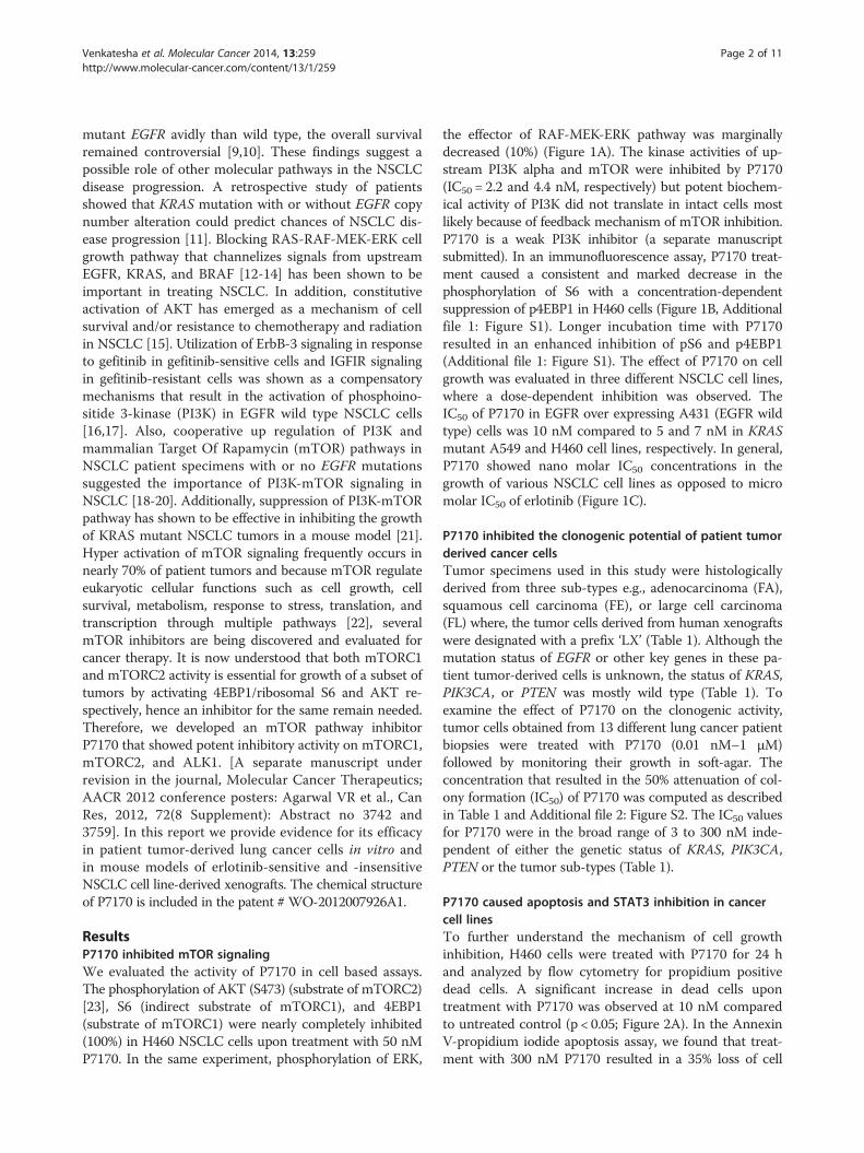
ResultsP7170 inhibited mTOR signalingWe evaluated the activity of P7170 in cell based assays.The phosphorylation of AKT (S473) (substrate of mTORC2)[23], S6 (indirect substrate of mTORC1), and 4EBP1(substrate of mTORC1) were nearly completely inhibited(100%) in H460 NSCLC cells upon treatment with 50 nMP7170. In the same experiment, phosphorylation of ERK,
the effector of RAF-MEK-ERK pathway was marginallydecreased (10%) (Figure 1A). The kinase activities of up-stream PI3K alpha and mTOR were inhibited by P7170(IC50 = 2.2 and 4.4 nM, respectively) but potent biochem-ical activity of PI3K did not translate in intact cells mostlikely because of feedback mechanism of mTOR inhibition.P7170 is a weak PI3K inhibitor (a separate manuscriptsubmitted). In an immunofluorescence assay, P7170 treat-ment caused a consistent and marked decrease in thephosphorylation of S6 with a concentration-dependentsuppression of p4EBP1 in H460 cells (Figure 1B, Additionalfile 1: Figure S1). Longer incubation time with P7170resulted in an enhanced inhibition of pS6 and p4EBP1(Additional file 1: Figure S1). The effect of P7170 on cellgrowth was evaluated in three different NSCLC cell lines,where a dose-dependent inhibition was observed. TheIC50 of P7170 in EGFR over expressing A431 (EGFR wildtype) cells was 10 nM compared to 5 and 7 nM in KRASmutant A549 and H460 cell lines, respectively. In general,P7170 showed nano molar IC50 concentrations in thegrowth of various NSCLC cell lines as opposed to micromolar IC50 of erlotinib (Figure 1C).
P7170 inhibited the clonogenic potential of patient tumorderived cancer cellsTumor specimens used in this study were histologicallyderived from three sub-types e.g., adenocarcinoma (FA),squamous cell carcinoma (FE), or large cell carcinoma(FL) where, the tumor cells derived from human xenograftswere designated with a prefix ‘LX’ (Table 1). Although themutation status of EGFR or other key genes in these pa-tient tumor-derived cells is unknown, the status of KRAS,PIK3CA, or PTEN was mostly wild type (Table 1). Toexamine the effect of P7170 on the clonogenic activity,tumor cells obtained from 13 different lung cancer patientbiopsies were treated with P7170 (0.01 nM–1 μM)followed by monitoring their growth in soft-agar. Theconcentration that resulted in the 50% attenuation of col-ony formation (IC50) of P7170 was computed as describedin Table 1 and Additional file 2: Figure S2. The IC50 valuesfor P7170 were in the broad range of 3 to 300 nM inde-pendent of either the genetic status of KRAS, PIK3CA,PTEN or the tumor sub-types (Table 1).
P7170 caused apoptosis and STAT3 inhibition in cancercell linesTo further understand the mechanism of cell growthinhibition, H460 cells were treated with P7170 for 24 hand analyzed by flow cytometry for propidium positivedead cells. A significant increase in dead cells upontreatment with P7170 was observed at 10 nM comparedto untreated control (p < 0.05; Figure 2A). In the AnnexinV-propidium iodide apoptosis assay, we found that treat-ment with 300 nM P7170 resulted in a 35% loss of cell
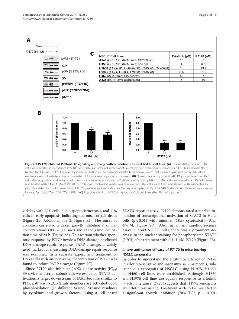
Figure 1 P7170 inhibited PI3K/mTOR signaling and the growth of erlotinib-resistant NSCLC cell lines. (A) Exponentially growing H460cells were seeded in petridishes (2 × 10⁶ cells/dish) and after cell attachment overnight, cells were serum starved for 14-16 h. Cells were thentreated for 1 h with P7170 followed by 0.5 h incubation in the presence of 20% fetal bovine serum. Cells were trypsinized and lysed beforeelectrophoresis of cellular extracts for western blot analyses of proteins of interest. (B) Quantification of pS6 and p4EBP1 protein levels in H460cells after acquisition and analyses of immunofluorescence signals in the Cellomics Array scan platform. H460 cells were seeded in 96-well platesand treated with 0.1 or 1 μM of P7170 for 16 h; drug-containing media was removed and the cells were fixed and stained with antibodies tophosphorylated form of human S6 and 4EBP1 proteins and secondary antibodies conjugated to DyLight 549. Statistical significance values are asfollows: *p < 0.05, **p < 0.01, ***p < 0.001. (C) IC50 of erlotinib or P7170 in various NSCLC cell lines after 48 h of treatment.
Venkatesha et al. Molecular Cancer 2014, 13:259 Page 3 of 11http://www.molecular-cancer.com/content/13/1/259
viability with 22% cells in late apoptosis/necrosis, and 11%cells in early apoptosis indicating the onset of cell death(Figure 2B; Additional file 3: Figure S3). The onset ofapoptosis correlated with cell growth inhibition at similarconcentrations (100 – 300 nM) and at the same incuba-tion time of 24 h (Figure 2A). To ascertain whether apop-totic response by P7170 involves DNA damage or elicitedDNA damage-repair response, PARP cleavage, a widelyused marker for measuring DNA-damage repair responsewas examined. In a separate experiment, treatment ofH460 cells with an increasing concentration of P7170 wasfound to induce PARP cleavage (Figure 2C).Since P7170 also inhibited JAK2 kinase activity (IC50:
39 nM, manuscript submitted), we evaluated STAT3 ac-tivation, a target downstream of JAK2 because similar toPI3K pathway, STAT-family members are activated uponphosphorylation (at different Serine/Tyrosine residues)by cytokines and growth factors. Using a cell based
STAT3-reporter assay, P7170 demonstrated a marked in-hibition of transcriptional activation of STAT3 in HeLacells (p < 0.01) with minimal (18%) cytotoxicity (IC50:4.7nM, Figure 2D). Also, in an immunofluorescenceassay in A549 NSCLC cells, there was a prominent de-crease in the nuclear staining for phosphorylated STAT3(Y705) after treatment with 0.1–1 μM P7170 (Figure 2E).
In vivo anti-tumor efficacy of P7170 in mice bearingNSCLC xenograftsIn order to understand the antitumor efficacy of P7170in erlotinib-sensitive and insensitive in vivo models, sub-cutaneous xenografts of NSCLC, using H1975, H1650,or H460 cell lines were established. Although H1650and H1975 cell lines are equally responsive to erlotinibin vitro, literature [24,25] suggests that H1975 xenograftsare erlotinib-resistant. Treatment with P7170 resulted ina significant growth inhibition (70% TGI, p < 0.001,
Table 1 P7170 inhibits the growth of NSCLC patient-derived cancer cells
Tumor designation Model # Histology Kras PIK3CA PTEN IC50 (nM)
LXFA 1012 adenocarcinoma wt wt wt 45
LXFA 1584 adenocarcinoma NA NA NA 2
LXFA 586 adenocarcinoma wt wt wt 9
LXFA 629 adenocarcinoma wt wt wt 5.5
LXFA 677 adenocarcinoma wt wt wt 300
LXFA 737 adenocarcinoma wt wt wt 300
LXFA 749 adenocarcinoma wt wt wt 17
LXFE 1422 Squamous cell carcinoma wt wt wt 8.5
LXFE 470 Squamous cell carcinoma wt E545K heterozygous wt 50
LXFE 646 Squamous cell carcinoma wt wt wt 100
LXFL 1674 Large cell G12C homozygous wt wt 30
LXFL 430 Large cell wt wt wt 3
LXFL 529 Large cell wt wt wt 50
Various patient tumor xenograft-derived NSCLC cells treated with P7170 at log concentrations (range: 0.01 nM – 1 μM) followed by growth in soft-agar to evaluatecolony formation. Description of patient’s tumor histology sub-type, mutation status, and IC50 of P7170 in these tumor xenograft-derived cells by soft-agar colonyformation is shown.
Venkatesha et al. Molecular Cancer 2014, 13:259 Page 4 of 11http://www.molecular-cancer.com/content/13/1/259
Table 2) of xenograft derived from H1650 erlotinib-sensitive cells [EGFR del E748-A750 (activating mutation);KRAS mutant; PIK3CA wild type] at 5 mg/kg, a dosemuch lower than its MTD. However, the same dose ofP7170 was not very effective in mice bearing erlotinib-insensitive H1975 [EGFR T790M, L858R; KRAS wild type;PIK3CA wild type] xenografts, but treatment with a15mg/kg dose resulted in a significant tumor growthinhibition (92% TGI, p < 0.001, Table 2). Interestingly,P7170 was efficacious (88% TGI at 20 mg/kg; 66% TGI at10 mg/kg with p < 0.001, Table 2 and Figure 3A) inanother model of erlotinib -insensitive H460 (EGFR wildtype; KRAS mutant; PIK3CA mutant) xenografted ani-mals. P7170 administration was well tolerated at doses upto 20 mg in different xenograft models (Additional file 4:Figure S4). These results provide evidence for the efficacyof P7170 in erlotinib-sensitive, and –insensitive NSCLCirrespective of known genetic mutations in these cells.
Pharmacokinetic-pharmacodynamic correlation of P7170 inhuman H460 NSCLC xenograft modelH460 xenograft model was chosen for this study. Tumorbearing mice were treated with different doses of P7170.Administration of P7170 at a 20 mg/kg dose resulted in88% tumor growth inhibition (Figure 3A) confirming theresults of a previous experiment (Table 2). Tumor growthinhibition with 5 and 10 mg/kg doses was significant com-pared to the vehicle (~53 and 66% respectively, p < 0.05)but did not differ much between the doses. In the efficacystudy (Figure 3A); plasma and tumor samples collected at1, 4, 8, or 24 h post P7170-last dose were analyzed todetermine the concentrations of P7170 by LC-MS. The re-sults of this experiment indicate a dose dependent increase
in P7170 concentrations in the tumors based on area underthe curve (AUC) (*p < 0.05 between two doses for plasmaor tumor P7170 concentrations independently; §p < 0.05 isthe significance when concentrations of P7170 in theplasma and tumor were compared; Figure 3B). Also, P7170concentrations were 2.7-fold higher in tumor versus plasmain 20 mg/kg group.In a separate PK-PD study, H460 xenograft-bearing mice
were treated with P7170 (1, 5, 10, or 20 mg/kg/day) con-secutively for 3 days. Plasma and tumor samples were col-lected at 1, 4, 8, and 24 h after the administration of thelast dose for PK-PD-analyses. Based on the time to achieveCmax, plasma and tumor concentrations of P7170 werepooled from two time points (1 and 4 h) in each group(Figure 3B). Tumor to plasma concentrations revealeda preferential accumulation of P7170 in the tumorthan plasma in a dose-dependent manner (3 – 11-fold)(Figure 3B). To ascertain the pharmacodynamic effectof P7170 in tumor xenografts, western blotting of keyproteins in the PI3K-Akt-mTOR pathway in tissueextracts was performed, and the representative proteinblots derived from the samples collected during 1-8 hpost P7170-last-dose are shown (Figure 3C). AKT phos-phorylation (S473) the direct substrate of mTORC2was found to be strongly inhibited in response to 10 or20 mg/kg treatment. Also, the phosphorylation of ribo-somal S6 (S235/236) and 4EBP1 (T37/46), targets down-stream of mTOR were inhibited by 5-20 mg/kg doses(Figure 3C). For the PK-PD-analyses, the normalized indi-vidual protein band intensities (pAKT, pS6, or p4EBP1)were plotted against P7170 concentrations in respectivetumor samples. PK-PD analyses were carried out based onInhibitory Emax models (Additional file 5: Table S1) using
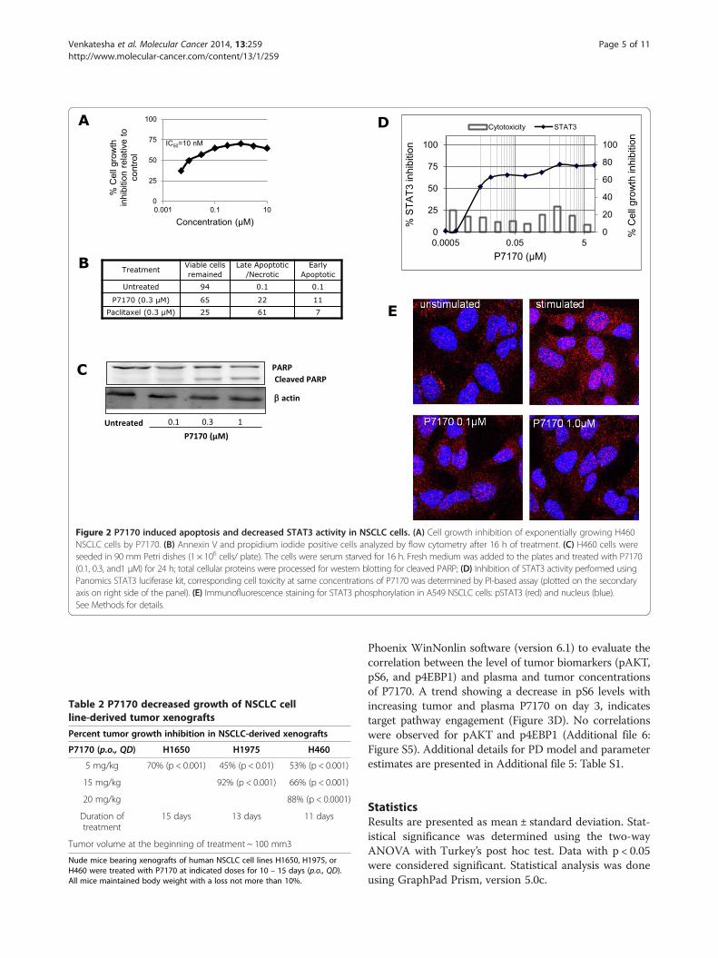
Table 2 P7170 decreased growth of NSCLC cellline-derived tumor xenografts
Percent tumor growth inhibition in NSCLC-derived xenografts
P7170 (p.o., QD) H1650 H1975 H460
5 mg/kg 70% (p < 0.001) 45% (p < 0.01) 53% (p < 0.001)
15 mg/kg 92% (p < 0.001) 66% (p < 0.001)
20 mg/kg 88% (p < 0.0001)
Duration oftreatment
15 days 13 days 11 days
Tumor volume at the beginning of treatment ~ 100 mm3
Nude mice bearing xenografts of human NSCLC cell lines H1650, H1975, orH460 were treated with P7170 at indicated doses for 10 – 15 days (p.o., QD).All mice maintained body weight with a loss not more than 10%.
Figure 2 P7170 induced apoptosis and decreased STAT3 activity in NSCLC cells. (A) Cell growth inhibition of exponentially growing H460NSCLC cells by P7170. (B) Annexin V and propidium iodide positive cells analyzed by flow cytometry after 16 h of treatment. (C) H460 cells wereseeded in 90 mm Petri dishes (1 × 10⁶ cells/ plate). The cells were serum starved for 16 h. Fresh medium was added to the plates and treated with P7170(0.1, 0.3, and1 μM) for 24 h; total cellular proteins were processed for western blotting for cleaved PARP; (D) Inhibition of STAT3 activity performed usingPanomics STAT3 luciferase kit, corresponding cell toxicity at same concentrations of P7170 was determined by PI-based assay (plotted on the secondaryaxis on right side of the panel). (E) Immunofluorescence staining for STAT3 phosphorylation in A549 NSCLC cells: pSTAT3 (red) and nucleus (blue).See Methods for details.
Venkatesha et al. Molecular Cancer 2014, 13:259 Page 5 of 11http://www.molecular-cancer.com/content/13/1/259
Phoenix WinNonlin software (version 6.1) to evaluate thecorrelation between the level of tumor biomarkers (pAKT,pS6, and p4EBP1) and plasma and tumor concentrationsof P7170. A trend showing a decrease in pS6 levels withincreasing tumor and plasma P7170 on day 3, indicatestarget pathway engagement (Figure 3D). No correlationswere observed for pAKT and p4EBP1 (Additional file 6:Figure S5). Additional details for PD model and parameterestimates are presented in Additional file 5: Table S1.
StatisticsResults are presented as mean ± standard deviation. Stat-istical significance was determined using the two-wayANOVA with Turkey’s post hoc test. Data with p < 0.05were considered significant. Statistical analysis was doneusing GraphPad Prism, version 5.0c.
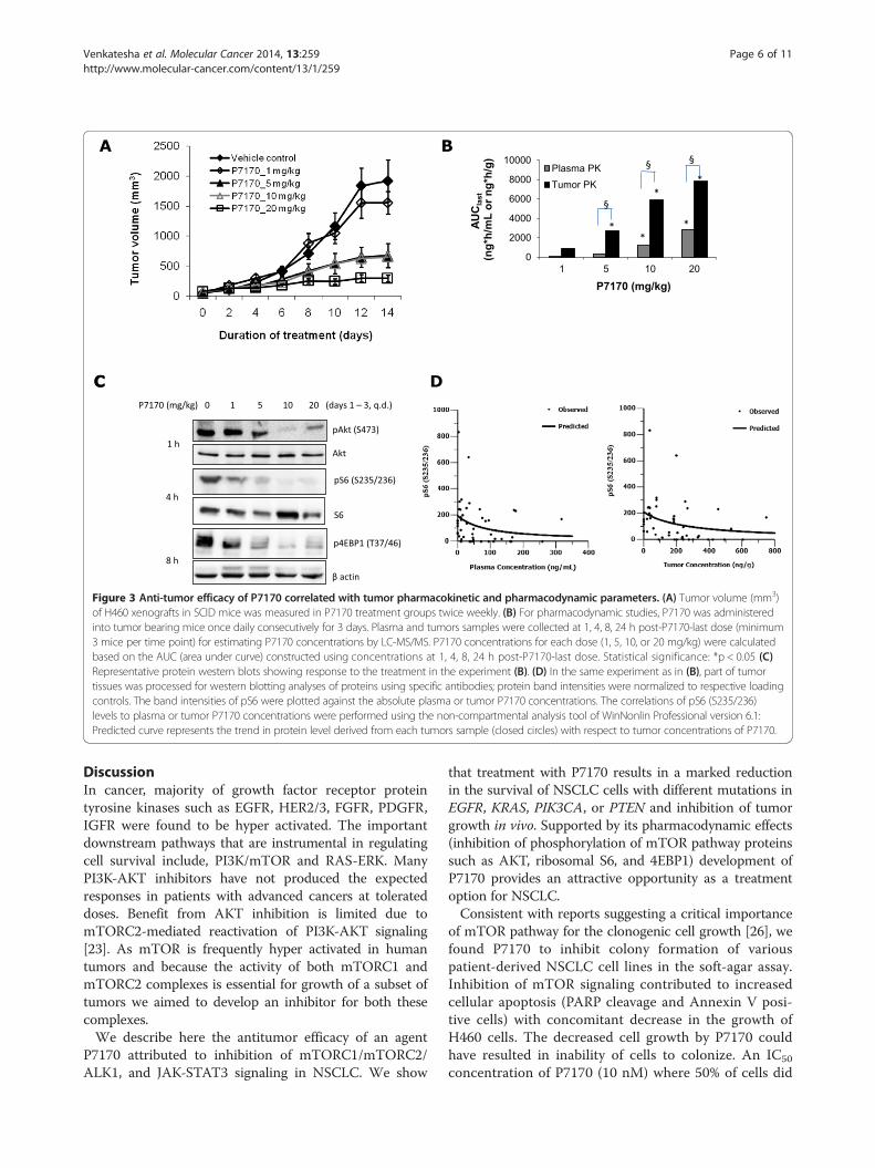
Figure 3 Anti-tumor efficacy of P7170 correlated with tumor pharmacokinetic and pharmacodynamic parameters. (A) Tumor volume (mm3)of H460 xenografts in SCID mice was measured in P7170 treatment groups twice weekly. (B) For pharmacodynamic studies, P7170 was administeredinto tumor bearing mice once daily consecutively for 3 days. Plasma and tumors samples were collected at 1, 4, 8, 24 h post-P7170-last dose (minimum3 mice per time point) for estimating P7170 concentrations by LC-MS/MS. P7170 concentrations for each dose (1, 5, 10, or 20 mg/kg) were calculatedbased on the AUC (area under curve) constructed using concentrations at 1, 4, 8, 24 h post-P7170-last dose. Statistical significance: *p < 0.05 (C)Representative protein western blots showing response to the treatment in the experiment (B). (D) In the same experiment as in (B), part of tumortissues was processed for western blotting analyses of proteins using specific antibodies; protein band intensities were normalized to respective loadingcontrols. The band intensities of pS6 were plotted against the absolute plasma or tumor P7170 concentrations. The correlations of pS6 (S235/236)levels to plasma or tumor P7170 concentrations were performed using the non-compartmental analysis tool of WinNonlin Professional version 6.1:Predicted curve represents the trend in protein level derived from each tumors sample (closed circles) with respect to tumor concentrations of P7170.
Venkatesha et al. Molecular Cancer 2014, 13:259 Page 6 of 11http://www.molecular-cancer.com/content/13/1/259
DiscussionIn cancer, majority of growth factor receptor proteintyrosine kinases such as EGFR, HER2/3, FGFR, PDGFR,IGFR were found to be hyper activated. The importantdownstream pathways that are instrumental in regulatingcell survival include, PI3K/mTOR and RAS-ERK. ManyPI3K-AKT inhibitors have not produced the expectedresponses in patients with advanced cancers at tolerateddoses. Benefit from AKT inhibition is limited due tomTORC2-mediated reactivation of PI3K-AKT signaling[23]. As mTOR is frequently hyper activated in humantumors and because the activity of both mTORC1 andmTORC2 complexes is essential for growth of a subset oftumors we aimed to develop an inhibitor for both thesecomplexes.We describe here the antitumor efficacy of an agent
P7170 attributed to inhibition of mTORC1/mTORC2/ALK1, and JAK-STAT3 signaling in NSCLC. We show
that treatment with P7170 results in a marked reductionin the survival of NSCLC cells with different mutations inEGFR, KRAS, PIK3CA, or PTEN and inhibition of tumorgrowth in vivo. Supported by its pharmacodynamic effects(inhibition of phosphorylation of mTOR pathway proteinssuch as AKT, ribosomal S6, and 4EBP1) development ofP7170 provides an attractive opportunity as a treatmentoption for NSCLC.Consistent with reports suggesting a critical importance
of mTOR pathway for the clonogenic cell growth [26], wefound P7170 to inhibit colony formation of variouspatient-derived NSCLC cell lines in the soft-agar assay.Inhibition of mTOR signaling contributed to increasedcellular apoptosis (PARP cleavage and Annexin V posi-tive cells) with concomitant decrease in the growth ofH460 cells. The decreased cell growth by P7170 couldhave resulted in inability of cells to colonize. An IC50
concentration of P7170 (10 nM) where 50% of cells did
Venkatesha et al. Molecular Cancer 2014, 13:259 Page 7 of 11http://www.molecular-cancer.com/content/13/1/259
not undergo apoptosis indicated other mechanismsmay be involved mediating cell death. To delineate this,we found studies reporting a persistent activation ofSTAT3 (pSTAT3) in nearly 50% of lung adenocarcin-omas [27]. It is known that JAK2 directly regulateSTAT3 activation [28], and since P7170 is known tohave activity against the JAK2 kinase, it was hypothe-sized that P7170 may inhibit STAT3 activation. As ex-pected, P7170 inhibited STAT3 activity in a luciferaseexpression system in HeLa cells. In an immunofluores-cence assay, P7170 has also inhibited STAT3 phosphor-ylation (Y705) in situ in A549 NSCLC cells. Inunderstanding the functional effect of these results, astudy showed that inhibition of STAT3 was responsiblefor NSCLC tumor growth suppression [29] suggestingthat inhibition of JAK2-STAT3 could be a mechanismfor cellular apoptosis in P7170 treated cells.We have demonstrated the in vivo antitumor efficacy of
P7170 in xenograft models of different erlotinib-sensitive,and –resistant NSCLC cell lines at tolerated doses, andparticularly, in xenografts derived from EGFR T790M,L858R erlotinib-resistant cells. The results of the presentPK/PD study show mTORC1 and mTORC2 targetengagement with a negative correlation between tumorvolume and plasma/tumor P7170 concentrations. Withthe patient treatment in perspective, second generationEGFR TKI or combination with mTOR inhibitor forNSCLC patients with EGFR T790M mutation [30] couldpotentially become options. Although therapeutic optionsfor NSCLC patients with KRAS mutation is lacking, newapproaches in the clinical testing such as dual MEK andmTOR inhibition or cytotoxic drugs [31] are beingevaluated. Therefore, targeting of mTOR cell survival/growth pathways in subjects with erlotinib –sensitive and–insensitive NSCLC might be beneficial. In summary,P7170 showed a target engagement, resulted in tumor cellkilling leading to tumor growth inhibition in vivo.
ConclusionsTaken together, our results demonstrate that P7170 hasNSCLC cell killing activity via inhibition of mTORC1/mTORC2 and JAK2-STAT3 signaling. This activity resultedin tumor growth inhibition as a single agent in thein vivo xenograft models. Therefore, we propose thatP7170 could produce benefit in patients with erlotinib-sensitive, and –insensitive NSCLC.
MethodsCell lines and cultureA549, H358, H1975, H1650, and H460 cell lines werepurchased from ATCC and maintained in RPMI1640,supplemented with 10% heat inactivated FBS (Gibco).STAT3 Reporter HeLa Stable Cell Line (stably trans-fected with Luciferase gene under the control of STAT3
promoter) was purchased from Panomics, USA and wasmaintained in DMEM supplemented with 10% FBS underhygromycin B (Sigma H3274) selection condition. Experi-mental culture conditions for patient tumor derived cellsare given below in methods.
Cell growth inhibition assayPropidium iodide assayExponentially growing cells were plated in 96-well plate24 h before treatment with P7170. After 48 h of incuba-tion plates were washed with 1× PBS followed by additionof PI-containing PBS (7 μg/ml) and storage at -80°C. Afterthawing the plate PI fluorescence was measured inPOLAR STAR OPTIMA (BMG Lab technologies). CellGrowth Inhibition was normalized to the controls.
Soft-agar colony formation of patient tumor-derivedxenograft cellsP7170 was evaluated at log concentrations from 0.01 nMto 1 μM in a 14-day soft agar colony formation assay. ThePatient tumor explants are passaged as subcutaneousxenograft in NMRI nu/nu mice. For the clonogenic assay,cells were isolated from tumor xenografts (also referredto as patient derived tumor xenografts, PDX) of 13 NSCLCpatient specimens established in mice under sterile condi-tions followed by mechanical disaggregation and subse-quent incubation with an enzyme cocktail [consisting ofcollagenase type IV (41 U /ml), DNase I (125 U /ml), hyal-uronidase type III (100 U /ml) and dispase II (1.0 U /ml) inRPMI 1640 medium] at 37°C for 45 minutes. Cells wereallowed to pass through sieves of 200 μm and 50 μm sterilenylon mesh and washed twice with sterile PBS buffer. Thepercentage of viable cells is determined in NeubauerHemocytometer using trypan blue exclusion. The bottomlayer consisted of 0.2 ml/well Iscove’s Modified Dulbecco’sMedium (Invitrogen), supplemented with 20% (v/v) fetalcalf serum (Sigma), 0.01% (w/v) gentamicin (Invitrogen)and 0.75% (w/v) agar (BD Biosciences). Tumor cells wereadded to 0.2 ml of the same culture medium supplementedwith 0.4% (w/v) agar and plated in 24-multiwell dishesonto the bottom layer. P7170 at desired concentration in0.2 ml culture medium was overlaid on the bottom layer.Every dish included six untreated control wells and drugtreated groups in triplicate at 6 concentrations. Cultureswere incubated at 37°C and 7.5% CO2 in the humidifiedatmosphere for up to 20 days and monitored closely forcolony growth using an inverted microscope. Within thisperiod, in vitro tumor growth leads to the formation ofcolonies with diameter > 50 μm. At the time of maximumcolony formation, counts were performed with an auto-mated image analysis system (OMNICON 3600, BiosysGmbH). 24 h prior to evaluation, vital colonies are stainedwith a sterile aqueous solution of 2-(4-iodophenyl)-3-(4-nitrophenyl)-5-phenyltetrazolium chloride (1 mg/ml, 100

Venkatesha et al. Molecular Cancer 2014, 13:259 Page 8 of 11http://www.molecular-cancer.com/content/13/1/259
μl/well). IC50 values were calculated by two point curve fit[(conc. Of inhibitor) versus response (%T/C)] using Oncotestin house database system) or nonlinear regression [log (conc.of inhibitor)] versus response (% T/C) using Graph pad Prism5 for windows, version 5.01, Graph pad software Inc. CA). Forcalculation of mean IC50 values the geometric mean is used.Results are presented as mean graph plots or heat maps(individual IC50 values relative to the geometric mean IC50
value) over all cell lines as tested.
ImmunoblottingWhole cell extracts from treated cell lines or xenograftswere prepared using cell lysis buffer [a mixture of proteaseinhibitor cocktail (Sigma, Cat #P8340) and phosphataseinhibitors (40 mM Beta-glycerol phosphate (Sigma, Cat#G9422), 4 mM DTT (MP Biomedicals, Cat #194821),0.4 mM NaF (Sigma Cat #S1504), 0.4 mM Sodium-orthovanadate (Sigma, Cat #S6508)]. Total protein con-centration from the soluble fraction was determined byBradford’s method. Equal amount of total protein wasresolved on SDS-PAGE gels; protein bands were electro-transferred onto PVDF membrane. Antibodies for im-munoblotting are from Cell Signaling Technology [pAKT(S473) (#9271); AKT (#9272); pS6 (S235/236) (#2211); S6(#2217); p4EBp1 (T37/46) (#9459); pERK (T202/Y204)(#9101); ERK (#4695)]; and from Sigma (Actin #A2228).SuperSignal West Femto (Thermo Scientific Cat #34096)was used for chemiluminescence detection and the signalswere captured in the ChemiDoc XRS image system (Bio-Rad). The protein western band intensities were calculatedusing NIH Image-J software.
ImmunofluorescencepS6 and p4EBP1 stainingH460 cells were seeded in 96-well Black/clear plates(Nunc) followed by treatment on the next day withP7170 for 1 h. Cells were fixed with 3.7% PFA in 1× PBSat RT for 20 min, cell membranes permeabilized with0.1% Triton X-100 for 90 sec, followed by blocking with5% BSA (Sigma-Aldrich Cat #A7030) (w/v) in 1 × PBSfor 2 h before immunostaining. Primary antibodies usedwere pS6 and p4EBP1 (Cell Signaling) at dilution (1:500)at RT for 1 h. Secondary Goat Anti-Rabbit antibody IgG(H + L) DyLight 549 Thermo Scientific Cat #35557)probed at 1:1000 dilution for 1 h. Nuclei were stained withHoechst 33342 (AnaSpec Inc. Cat #83218). Plates werescanned on Cellomics Array Scan VTI HCS Reader.
pSTAT3 stainingExponentially growing A549 cells were seeded on cover-slips in a 24 well plate (Nunc) in complete growth mediafor 14-16 h followed by culture in no serum media foranother 16-20 h. Serum starved cells were treated withP7170 (0.1-1 μM) for 1 h followed by stimulation with
30 ng/ml rhIL-6 (R&D systems Cat. No. 206-IL) for20 min. Cells were washed with 1× PBS, fixed andpermeabilized as described earlier. Primary antibodiesused were pSTAT3(Y705) (Cell Signaling Cat #9131) incu-bated (1:100 dilution) at RT for 1 h. Secondary antibodyand nuclear staining performed as earlier. Cell imageswere captured by confocal microscopy (Leica, LSM700)with ZEN2009 software.
Annexin V – FITC staining and flow cytometryExponentially growing H460 cells were seeded in 6 wellplate, treated 16 h later with P7170 at 0.3 μM for 24 h.Cells were harvested by trypsinization and processed forFITC Annexin V staining as per manufacture’s protocol(BD Pharmingen Cat #556420). The samples were acquiredby FACSCalibur flow cytometer and the signal inten-sities were analyzed using CellQuest Pro software (BectonDickinson).
STAT3 luciferase assaySTAT3 Reporter HeLa cells were seeded (2 × 10⁴ cells/well)in a 96- well white plate (Nunclon Cat #167008). After16 h, cells were treated for 1 h with P7170 followed bystimulation with 100 ng/ml Oncostatin M (CalbiochemCat #496260) for 7 h. Cells were lysed in a buffer (a mix-ture of 125 mM Tris phosphate buffer, 10 mM EDTA,10 mM DTT, 50% glycerol, 5% Triton ×100) before theaddition of luciferin (Promega Cat #E160E), ATP (SigmaCat #A2383), and coenzyme A (Sigma Cat #C3019). Theluminescence generated was measured using POLARstarOPTIMA (BMG Lab technologies). Cytotoxicity due todrug treatment (total 8 h) was determined using propidiumiodide.
In vivo xenograft models of NSCLC cell lines and treatmentNude mice (male, ~6 wks.) from Harlan Labs were housedin animal isolators (Harlan Inc.) with 12 h light dark cycle,55-75% relative humidity at 22-25°C of room temperature,given access to autoclaved rodent diet (National Centrefor Laboratory Animal Sciences, Hyderabad, India) andwater ad libitum. Animals were acclimatized for a weekbefore implanting subcutaneously with 5×10⁶ NSCLCcells (0.2 ml/site with BD matrigel) in the right flank.When the mean xenograft size reached ~100 mm3, micewere randomized into the study groups (n = 9/group).P7170 was suspended in 0.25% CMC +0.1% Tween-80for administering via oral (p.o.) route. Animal bodyweight and physical signs were monitored throughoutthe experiment every day. The tumor size was measuredwith calipers 2-3 times/wk. and the tumor volumeswere calculated using the following formula: (length ×width)2 × 0.5.

Venkatesha et al. Molecular Cancer 2014, 13:259 Page 9 of 11http://www.molecular-cancer.com/content/13/1/259
Pharmacokinetic (PK) and Pharmacodynamic (PD) studyPK analysesPlasma samples or tumor homogenates were processedto extract P7170 followed by determining P7170 concen-trations using LC-MS/MS.
Tumor homogenate preparationTumor samples were diluted 4 times of its weight withhomogenization solvent methanol: saline (75:25 v/v) andthen homogenized to obtain tissue extracts until a uni-form homogenate was obtained.
Sample preparationAn aliquot (100 μL) of plasma or tumor homogenatewas spiked individually with 10 μL of internal standard(P6569, a compound structurally similar to P7170,1.0 μg/mL). The samples were vortexed for 10 secondsfollowed by addition of 1 mL ethyl acetate and furthervortexed for 5 min. The samples were then centrifugedat 10000 rpm for 5 minutes at 4°C. Supernatants (800 μL)were transferred to relabeled glass tubes and its solventportion was allowed to evaporate under nitrogen. The driedresidues were reconstituted using 200 μL acetonitrile: water(90:10 v/v) by vortex and centrifuged. The supernatantswere analyzed for P7170 by a fit-for-purpose LC-MS/MSmethod. Pharmacokinetic analyses were carried out at eachdose level using the non-compartmental analysis tool ofPhoenix WinNonlin software (Version 6.1).
LC-MS/MS analysisThe chromatographic LC-MS/MS system consisted aShimadzu LC pump with an API 4000 triple mass spec-trometer (Applied Biosystems/MDS Sciex, Foster City,CA) fitted with a TurboIon- Spray interface. P7170 andP6569 (internal standard) were separated on a ThermoBDS Hypersil C18 column of 100 × 4.6 mm I.D. andparticle size of 5 μ. The mobile phase composed of twosolvents: Solvent A, 5 mM Ammonium formate in 0.1%formic acid and the Solvent B, Acetonitrile in a ratio of20:80% v/v, respectively. The isocratic HPLC conditionwas used for analysis. The samples were introduced intothe HPLC column with a SIL-20 AC XR autosampler(Shimadzu) and an integrated HPLC pumping system(Shimadzu LC-20 AD XR) with 5 μL injection volume ata flow rate of 0.8 mL/min for a 3 min run time. The ana-lytes were then eluted at RT 1.45 and 1.50 min forP7170 and P6569, respectively followed by MS detection.Electro-spray ionization in positive ion mode (ESI+) wasused for ionization and multiple reaction monitor(MRM) mode was chosen for detection. The precursor-product ions pairs were m/z 528.0→ 461.4 (for P7170),and m/z 504.3→ 437.2 (for internal standard, P6569).The optimized acquisition parameters were: Temperatureset at 400°C; Curtain gas (CUR), 30 psi (99.99% Nitrogen);
Nebulizer Gas (Gas 1), 60 psi (99.99% Nitrogen); HeaterGas (Gas 2), 50 psi (99.99% Nitrogen); Collision-ActivatedDissociation (CAD) Gas: 10 v; Ion Spray Voltage (IS),5500 v.The unknown plasma and tumor concentrations of
P7170 (unbound and plasma protein bound) were calcu-lated from the calibration standards at 0 to 15000 ng/mLby spiking 10 × standards in blank plasma and tumorhomogenates collected from untreated nude mice usinglinear regression according to the following equation:
y ¼ axþ b
Where,y = Peak area ratio of analyte: internal standardb = Intercept of the corresponding standard curvea = Slope of the standard curvex = Concentration of analyte (ng/mL)Pharmacokinetic analysis was carried out at each dose
level using the non-compartmental analysis tool of Win-Nonlin Professional version 6.1. Pharmacokinetic param-eters were determined from mean plasma and tumorconcentrations thus obtained at each time point by non-compartmental analysis using Phoenix WinNonlin Pro-fessional version 6.1. Concentrations below limit ofquantification (LLOQ = 0.5 ng/mL) were considered aszero for PK analysis. Nominal time points were usedfor PK analysis.
PK-PD analysesPD analysis was performed to derive a correlation betweentumor marker levels (pS6, pAKT and p4EBP1) and thecorresponding plasma and tumor P7170 concentrationsafter dosing the tumor-bearing mice with P7170 oncedaily for 3 days. Plasma and tumors samples were col-lected at 1, 4, 8, 24 h post-P7170-last dose for estimatingP7170 concentrations by LC-MS/MS. P7170 tumor con-centrations for each dose (1, 5, 10, or 20 mg/kg) were cal-culated based on the AUC (area under curve) constructedusing concentrations at 1, 4, 8, 24 h post-P7170-last dose.The normalized phosphoprotein western band intensitiesof AKT (S473), S6 (S235/236), and 4EBP1 (T37/46) asdetermined in tumor xenografts (in 1, 4, 8, 24 h postP7170-last dose) of different experimental arms were plot-ted against corresponding tumor xenograft P7170 concen-tration using the non-compartmental analysis tool usingPhoenix WinNonlin Professional version 6.1. The analyseswere performed using various inhibitory Emax modelsavailable in Phoenix WinNonlin software (version 6.1).The model with the lowest AIC (Akaike InformationCriteria) value is selected as the best fit model. If twomodels had similar AIC values, then selection wasmade qualitatively by visual inspection of the model fitand % CV of the parameter estimates.
Venkatesha et al. Molecular Cancer 2014, 13:259 Page 10 of 11http://www.molecular-cancer.com/content/13/1/259
Additional files
Additional file 1: Figure S1. P7170 inhibited PI3K-mTOR signaling.pS6 (S235/236) and p4EBP1 (T37/46) protein levels were determined byimmunofluorescence staining in H460 cells. H460 cells were seeded in 96-wellplates (black and transparent bottom) before treatment with 0.1 or 1 μM ofP7170 for 1 h; drug containing media was removed and the cells were fixedand stained with antibodies to phosphorylated form of human S6 and 4EBP1proteins and secondary antibodies conjugated to DyLight 549, and signalsacquired and analyzed in Cellomics high content array scan reader.
Additional file 2: Figure S2. P7170 inhibits the colony formation oftumor cells isolated from Non Small Cell Lung Cancer patients. Doseresponse curves for various patient tumor xenograft-derived NSCLC cellstreated with P7170 in a soft-agar colony formation assay.
Additional file 3: Figure S3. Cellular apoptotic analysis after P7170treatment. In the flow cytometry analysis gating was set using untreatedcells (A); Increased cellular apoptosis and necrosis after P7170 treatment(B) or Paclitaxel treatment (C).
Additional file 4: Figure S4. Body weight changes of nude mice bearingNSCLC cell line-derived xenografts treated with P7170. (A) Percent bodyweight changes in the H1650 NSCLC cell-derived xenograft treated withP7170 (see Table 2); (B) Percent body weight changes in the H1975 NSCLCcell-derived xenograft treated with P7170 (see Table 2); Percent body weightchanges in the H460 NSCLC cell-derived xenograft treated with P7170(see Figure 3A).
Additional file 5: Table S1. Summary of PK/PD study. Correlation analysisof tumor volume to P7170 concentration. E0 represents the level of biomarkerin plasma and tumor at baseline i.e. when the concentration of drug inplasma and tumor is 0 (zero). IC50 represents the concentration of drug inplasma and tumor required to produce 50% of the maximal inhibition.
Additional file 6: Figure S5. Pharmacodynamic correlation of pAKT (S473)and p4EBP1 (T37/46) with tumor P7170 concentrations. Based on the studydescribed in Figure 3, pharmacodynamic correlations of tumor pAKT (S473)and p4EBP1 (T37/46) levels to the plasma and tumor concentrations ofP7170 were performed. The correlation plots calculated using the model(Additional file 5: Table S1): Correlation of tumor pAKT levels with P7170concentrations in plasma (A) and tumor (B); and correlation of tumorp4EBP1 levels with P7170 concentrations in plasma (C) and tumor (D).
AbbreviationsNSCLC: Non small cell lung cancer; mTOR: Mammalian target of Rapamycin;mTORC1: Mammalian target of Rapamycin complex 1; mTORC2: Mammaliantarget of Rapamycin Complex 2; EGFR: Epidermal growth factor receptor;TKI: Tyrosine kinase inhibitor; PI3K: Phosphoinositide 3-kinase; KRAS: Kirstenrat sarcoma virus; PK: Pharmacokinetics; PD: Pharmacodynamics; JAK2: Januskinase 2; STAT3: Signal transducer and activator of transcription 3; TGI: TumorGrowth Inhibition.
Competing interestsThe authors declare that they have no competing interests.
Authors’ contributionsAJ carried out protein western blotting, cell growth inhibition, STAT3 reporter,immunofluorescence assays, and pharmacodynamic studies. MV performedmouse tumor xenograft and pharmacodynamic studies. VS and PP carried outexperiments in mouse tumor xenograft models. JB set up JAK2 kinase assay. DBparticipated in general experimental planning. SC carried out kinase assays, flowcytometry, and protein western blotting experiments. AS carried outimmunofluorescence experiments. VL performed cell growth inhibition assaysin NSCLC cell lines. PT and AD performed pharmacokinetic experiments andanalysis. RS and TA carried out PK-PD correlations. VD, BS, and SK involved inthe design and synthesis of P7170. SS contributed in the study concept. VVdrafted the work and prepared the manuscript to be published. VA conceivedthe studies and design of most of the studies. All authors read and approvedthe final manuscript.
AcknowledgementColony forming assays were performed at Oncotest, Germany.
Received: 9 June 2014 Accepted: 24 November 2014Published: 2 December 2014
References1. Morace C, Spadaro A, Cucunato M, Tortorella V, Consolo P, Luigiano C,
Stabile G, Bonfiglio C, Bellerone R, Fortiguerra A, Alibrandi A, Crino S,Carducci A, Resta ML, Ferrau O, Freni MA: High serum resistin in chronicviral hepatitis is not a marker of metabolic disorder.Hepatogastroenterology 2010, 57:1215–1219.
2. Pao W, Girard N: New driver mutations in non-small-cell lung cancer.Lancet Oncol 2011, 12:175–180.
3. Stella GM, Luisetti M, Inghilleri S, Cemmi F, Scabini R, Zorzetto M, Pozzi E:Targeting EGFR in non-small-cell lung cancer: lessons, experiences,strategies. Respir Med 2012, 106:173–183.
4. Johnson JL, Pillai S, Chellappan SP: Genetic and biochemical alterations innon-small cell lung cancer. Biochem Res Int 2012, 2012:940405.
5. van Zandwijk N, Mathy A, Boerrigter L, Ruijter H, Tielen I, de Jong D, Baas P,Burgers S, Nederlof P: EGFR and KRAS mutations as criteria for treatmentwith tyrosine kinase inhibitors: retro- and prospective observations innon-small-cell lung cancer. Ann Oncol 2007, 18:99–103.
6. Mok TS, Wu YL, Thongprasert S, Yang CH, Chu DT, Saijo N, SunpaweravongP, Han B, Margono B, Ichinose Y, Nishiwaki Y, Ohe Y, Yang JJ,Chewaskulyong B, Jiang H, Duffield EL, Watkins CL, Armour AA, Fukuoka M:Gefitinib or carboplatin-paclitaxel in pulmonary adenocarcinoma. N EnglJ Med 2009, 361:947–957.
7. Tapia C, Savic S, Bihl M, Rufle A, Zlobec I, Terracciano L, Bubendorf L: [EGFRmutation analysis in non-small-cell lung cancer : experience from routinediagnostics]. Pathologe 2009, 30:384–392.
8. Boch C, Kollmeier J, Roth A, Stephan-Falkenau S, Misch D, Gruning W, BauerTT, Mairinger T: The frequency of EGFR and KRAS mutations in non-smallcell lung cancer (NSCLC): routine screening data for central Europe froma cohort study. BMJ Open 2013, 3:1–6.
9. Douillard JY, Shepherd FA, Hirsh V, Mok T, Socinski MA, Gervais R, Liao ML,Bischoff H, Reck M, Sellers MV, Watkins CL, Speake G, Armour AA, Kim ES:Molecular predictors of outcome with gefitinib and docetaxel inpreviously treated non-small-cell lung cancer: data from the randomizedphase III INTEREST trial. J Clin Oncol 2010, 28:744–752.
10. Zhou C, Wu YL, Chen G, Feng J, Liu XQ, Wang C, Zhang S, Wang J, Zhou S,Ren S, Lu S, Zhang L, Hu C, Hu C, Luo Y, Chen L, Ye M, Huang J, Zhi X,Zhang Y, Xiu Q, Ma J, Zhang L, You C: Erlotinib versus chemotherapy asfirst-line treatment for patients with advanced EGFR mutation-positivenon-small-cell lung cancer (OPTIMAL, CTONG-0802): a multicentre,open-label, randomised, phase 3 study. Lancet Oncol 2011, 12:735–742.
11. Massarelli E, Varella-Garcia M, Tang X, Xavier AC, Ozburn NC, Liu DD,Bekele BN, Herbst RS, Wistuba II: KRAS mutation is an important predictorof resistance to therapy with epidermal growth factor receptor tyrosinekinase inhibitors in non-small-cell lung cancer. Clin Cancer Res 2007,13:2890–2896.
12. Riely GJ, Marks J, Pao W: KRAS mutations in non-small cell lung cancer.Proc Am Thorac Soc 2009, 6:201–205.
13. Brognard J, Dennis PA: Variable apoptotic response of NSCLC cells toinhibition of the MEK/ERK pathway by small molecules or dominantnegative mutants. Cell Death Differ 2002, 9:893–904.
14. Wan PT, Garnett MJ, Roe SM, Lee S, Niculescu-Duvaz D, Good VM, Jones CM,Marshall CJ, Springer CJ, Barford D, Marais R, Cancer Genome P: Mechanismof activation of the RAF-ERK signaling pathway by oncogenic mutations ofB-RAF. Cell 2004, 116:855–867.
15. Brognard J, Clark AS, Ni Y, Dennis PA: Akt/protein kinase B is constitutivelyactive in non-small cell lung cancer cells and promotes cellular survival andresistance to chemotherapy and radiation. Cancer Res 2001, 61:3986–3997.
16. Engelman JA, Janne PA, Mermel C, Pearlberg J, Mukohara T, Fleet C,Cichowski K, Johnson BE, Cantley LC: ErbB-3 mediates phosphoinositide3-kinase activity in gefitinib-sensitive non-small cell lung cancer celllines. Proc Natl Acad Sci U S A 2005, 102:3788–3793.
17. Guix M, Faber AC, Wang SE, Olivares MG, Song Y, Qu S, Rinehart C, Seidel B,Yee D, Arteaga CL, Engelman JA: Acquired resistance to EGFR tyrosine kinaseinhibitors in cancer cells is mediated by loss of IGF-binding proteins. J ClinInvest 2008, 118:2609–2619.
18. Yamamoto H, Shigematsu H, Nomura M, Lockwood WW, Sato M, Okumura N,Soh J, Suzuki M, Wistuba II, Fong KM, Lee H, Toyooka S, Date H, Lam WL,

Venkatesha et al. Molecular Cancer 2014, 13:259 Page 11 of 11http://www.molecular-cancer.com/content/13/1/259
Minna JD, Gazdar AF: PIK3CA mutations and copy number gains in humanlung cancers. Cancer Res 2008, 68:6913–6921.
19. Trigka EA, Levidou G, Saetta AA, Chatziandreou I, Tomos P, Thalassinos N,Anastasiou N, Spartalis E, Kavantzas N, Patsouris E, Korkolopoulou P: Adetailed immunohistochemical analysis of the PI3K/AKT/mTOR pathwayin lung cancer: correlation with PIK3CA, AKT1, K-RAS or PTEN mutationalstatus and clinicopathological features. Oncol Rep 2013, 30:623–636.
20. Han SW, Kim TY, Jeon YK, Hwang PG, Im SA, Lee KH, Kim JH, Kim DW, Heo DS,Kim NK, Chung DH, Bang YJ: Optimization of patient selection for gefitinibin non-small cell lung cancer by combined analysis of epidermal growthfactor receptor mutation, K-ras mutation, and Akt phosphorylation.Clin Cancer Res 2006, 12:2538–2544.
21. Engelman JA, Chen L, Tan X, Crosby K, Guimaraes AR, Upadhyay R, Maira M,McNamara K, Perera SA, Song Y, Chirieac LR, Kaur R, Lightbown A, SimendingerJ, Li T, Padera RF, Garcia-Echeverria C, Weissleder R, Mahmood U, Cantley LC,Wong KK: Effective use of PI3K and MEK inhibitors to treat mutantKras G12D and PIK3CA H1047R murine lung cancers. Nat Med 2008,14:1351–1356.
22. Watanabe R, Wei L, Huang J: mTOR signaling, function, novel inhibitors,and therapeutic targets. J Nucl Med 2011, 52:497–500.
23. Sarbassov DD, Guertin DA, Ali SM, Sabatini DM: Phosphorylation andregulation of Akt/PKB by the rictor-mTOR complex. Science 2005,307:1098–1101.
24. Suzuki T, Fujii A, Ohya J, Nakamura H, Fujita F, Koike M, Fujita M: Antitumoractivity of a dual epidermal growth factor receptor and ErbB2 kinaseinhibitor MP-412 (AV-412) in mouse xenograft models. Cancer Sci 2009,100:1526–1531.
25. Iwai T, Moriya Y, Shirane M, Fujimoto-Ouchi K, Mori K: Continuous inhibitionof epidermal growth factor receptor phosphorylation by erlotinib enhancesantitumor activity of chemotherapy in erlotinib-resistant tumor xenografts.Oncol Rep 2012, 27:923–928.
26. Liu X, Powlas J, Shi Y, Oleksijew AX, Shoemaker AR, De Jong R, Oltersdorf T,Giranda VL, Luo Y: Rapamycin inhibits Akt-mediated oncogenictransformation and tumor growth. Anticancer Res 2004, 24:2697–2704.
27. Gao SP, Mark KG, Leslie K, Pao W, Motoi N, Gerald WL, Travis WD, BornmannW, Veach D, Clarkson B, Bromberg JF: Mutations in the EGFR kinasedomain mediate STAT3 activation via IL-6 production in human lungadenocarcinomas. J Clin Invest 2007, 117:3846–3856.
28. Hedvat M, Huszar D, Herrmann A, Gozgit JM, Schroeder A, Sheehy A,Buettner R, Proia D, Kowolik CM, Xin H, Armstrong B, Bebernitz G, Weng S,Wang L, Ye M, McEachern K, Chen H, Morosini D, Bell K, Alimzhanov M,Ioannidis S, McCoon P, Cao ZA, Yu H, Jove R, Zinda M: The JAK2 inhibitorAZD1480 potently blocks Stat3 signaling and oncogenesis in solidtumors. Cancer Cell 2009, 16:487–497.
29. Weerasinghe P, Garcia GE, Zhu Q, Yuan P, Feng L, Mao L, Jing N: Inhibitionof Stat3 activation and tumor growth suppression of non-small cell lungcancer by G-quartet oligonucleotides. Int J Oncol 2007, 31:129–136.
30. Li D, Shimamura T, Ji H, Chen L, Haringsma HJ, McNamara K, Liang MC,Perera SA, Zaghlul S, Borgman CL, Kubo S, Takahashi M, Sun Y, Chirieac LR,Padera RF, Lindeman NI, Janne PA, Thomas RK, Meyerson ML, Eck MJ,Engelman JA, Shapiro GI, Wong KK: Bronchial and peripheral murine lungcarcinomas induced by T790M-L858R mutant EGFR respond to HKI-272and rapamycin combination therapy. Cancer Cell 2007, 12:81–93.
31. Larsen JE, Cascone T, Gerber DE, Heymach JV, Minna JD: Targetedtherapies for lung cancer: clinical experience and novel agents. Cancer J2011, 17:512–527.
doi:10.1186/1476-4598-13-259Cite this article as: Venkatesha et al.: P7170, a novel inhibitor ofmTORC1/mTORC2 and Activin receptor-like Kinase 1 (ALK1) inhibits thegrowth of non small cell lung cancer. Molecular Cancer 2014 13:259.
Submit your next manuscript to BioMed Centraland take full advantage of:
• Convenient online submission
• Thorough peer review
• No space constraints or color figure charges
• Immediate publication on acceptance
• Inclusion in PubMed, CAS, Scopus and Google Scholar
• Research which is freely available for redistribution
Submit your manuscript at www.biomedcentral.com/submit