research open access molecular characterization of the
TRANSCRIPT

RESEARCH Open Access
Molecular characterization of the PhoPQ-PmrD-PmrAB mediated pathway regulating polymyxinB resistance in Klebsiella pneumoniae CG43Hsin-Yao Cheng1, Yi-Fong Chen2, Hwei-Ling Peng1,2*
Abstract
Background: The cationic peptide antibiotic polymyxin has recently been reevaluated in the treatment of severeinfections caused by gram negative bacteria.
Methods: In this study, the genetic determinants for capsular polysaccharide level and lipopolysaccharidemodification involved in polymyxin B resistance of the opportunistic pathogen Klebsiella pneumoniae werecharacterized. The expressional control of the genes responsible for the resistance was assessed by a LacZ reportersystem. The PmrD connector-mediated regulation for the expression of pmr genes involved in polymyxin Bresistance was also demonstrated by DNA EMSA, two-hybrid analysis and in vitro phosphor-transfer assay.
Results: Deletion of the rcsB, which encoded an activator for the production of capsular polysaccharide, had aminor effect on K. pneumoniae resistance to polymyxin B. On the other hand, deletion of ugd or pmrF generesulted in a drastic reduction of the resistance. The polymyxin B resistance was shown to be regulated by thetwo-component response regulators PhoP and PmrA at low magnesium and high iron, respectively. Similar to thecontrol identified in Salmonella, expression of pmrD in K. pneumoniae was dependent on PhoP, the activated PmrDwould then bind to PmrA to prolong the phosphorylation state of the PmrA, and eventually turn on theexpression of pmr for the resistance to polymyxin B.
Conclusions: The study reports a role of the capsular polysaccharide level and the pmr genes for K. pneumoniaeresistance to polymyxin B. The PmrD connector-mediated pathway in governing the regulation of pmr expressionwas demonstrated. In comparison to the pmr regulation in Salmonella, PhoP in K. pneumoniae plays a majorregulatory role in polymyxin B resistance.
BackgroundKlebsiella pneumoniae, an important nosocomial patho-gen, causes a wide range of infections including pneu-monia, bacteremia, urinary tract infection, andsometimes even life-threatening septic shock [1]. Theemergence of multi-drug resistant K. pneumoniae hasreduced the efficacy of antibiotic treatments andprompted the reevaluation of previously but not cur-rently applied antibiotics [2,3] or a combined therapy[4]. Polymyxins, originally isolated from Bacillus poly-myxa, have emerged as promising candidates forthe treatment of infections [5]. As a member of
antimicrobial peptides (APs), the bactericidal agentexerts its effects by interacting with the lipopolysacchar-ide (LPS) of gram-negative bacteria. The polycationicpeptide ring on polymyxin competes for and substitutesthe calcium and magnesium bridges that stabilize LPS,thus disrupting the integrity of the outer membraneleading to cell death [5,6].The Klebsiella capsular polysaccharide (CPS), which
enabled the organism to escape from complement-mediated serum killing and phagocytosis [7,8], has beenshown to physically hinder the binding of C3 comple-ment [9] or polymyxin B [10]. The assembly and trans-port of Klebsiella CPS followed the E. coli Wzy-dependent pathway [11], in which mutations at wzaencoding the translocon protein forming the complexresponsible for CPS polymer translocation and export
* Correspondence: [email protected] of Biological Science and Technology, National Chiao-TungUniversity, Hsin Chu, Taiwan, China
Cheng et al. Journal of Biomedical Science 2010, 17:60http://www.jbiomedsci.com/content/17/1/60
© 2010 Cheng et al; licensee BioMed Central Ltd. This is an Open Access article distributed under the terms of the Creative CommonsAttribution License (http://creativecommons.org/licenses/by/2.0), which permits unrestricted use, distribution, and reproduction inany medium, provided the original work is properly cited.

resulted in an inability to assemble a capsular layer onthe cell surface [12]. The CPS biosynthesis in K. pneu-moniae was transcriptionally regulated by the two-com-ponent system (2CS) RcsBCD [13] where the deletion ofthe response regulator encoding gene rcsB in K. pneu-moniae caused a loss of mucoid phenotype and reduc-tion in CPS production [14].In Escherichia coli and Salmonella enterica serovar
Typhimurium, polymyxin B resistance is achievedmainly through the expression of LPS modificationenzymes, including PmrC, an aminotransferase for thedecoration of the LPS with phosphoethanolamine [15]and the pmrHFIJKLM operon [16,17] (also called pbgPor arn operon [18,19]) encoding enzymes. Mutations atpmrF, which encoded a transferase for the addition of4-aminoarabinose on bactoprenol phosphate, renderedS. enterica and Yersinia pseudotuberculosis more suscep-tible to polymyxin B [16,20]. The S. enterica ugd geneencodes an enzyme responsible for the supply of theamino sugar precursor L-aminoarabinose for LPS modi-fications and hence the Ugd activity is essential for theresistance to polymyxin B [21]. On the other hand, theE. coli ugd mutant with an impaired capsule alsobecame highly susceptible to polymyxin B [22].The 2CS PmrA/PmrB, consisting of the response reg-
ulator PmrA and its cognate sensor kinase PmrB, hasbeen identified as a major regulatory system in poly-myxin B resistance [23,24]. The resistance in S. entericaor E. coli has been shown to be inducible by the extra-cellular iron [25]. In addition to acidic pH [26], the roleof ferric ions as a triggering signal for the expression ofPmrA/PmrB has been demonstrated [23]. The 2CSPhoP/PhoQ which regulates the magnesium regulon[27] could also activate polymyxin B resistance underlow magnesium in S. enterica, in which the PhoP/PhoPQ-dependent control is connected by the smallbasic protein PmrD. The expression of pmrD could beactivated by PhoP while repressed by PmrA forming afeedback loop [28,29]. The activated PmrD could thenbind to the phosphorylated PmrA leading to a persistentexpression of the PmrA-activated genes [30].The PmrD encoding gene was also identified in E.
coli and K. pneumoniae. However, pmrD deletion in E.coli had no effect on the bacterial susceptibility topolymyxin B [25]. Recently, the PhoP-dependentexpression of pmrD has also been demonstrated in K.pneumoniae. According to the predicted semi-con-served PhoP box in the pmrD upstream region, adirect binding of PhoP to the pmrD promoter for theregulation was speculated [31].In this study, specific deletions of genetic loci involved
in CPS biosynthesis and LPS modifications were intro-duced into K. pneumoniae CG43, a highly virulent clini-cal isolate of K2 serotype [32]. Involvement of the
genetic determinants in polymyxin B resistance wasinvestigated.
MethodsPlasmids, bacterial strains, and growth conditionsBacterial strains and plasmids used in this study arelisted in Table 1, and the primers used are listed inTable 2. E. coli, K. pneumoniae CG43 [32,33] and itsderivatives were propagated at 37°C in Luria-Bertani(LB) broth or M9 minimal medium. Bacterial growthwas assessed by OD600. The antibiotics used includeampicillin (100 μg/ml), chloramphenicol (35 μg/ml),kanamycin (25 μg/ml), tetracycline (12.5 μg/ml) andstreptomycin (500 μg/ml). Polymyxin B sulfate salt(Sigma-Aldrich) was prepared as 1 unit/μl stock solutionin PBS and stored at 4°C before use.
Construction of specific gene-deletion mutantsSpecific gene deletion was individually introduced intothe chromosome of K. pneumoniae CG43S3 by allelicexchange strategy [14]. In brief, two approximately1000-bp DNA fragments flanking both sides of thedeleted region were cloned into the suicide vectorpKAS46 [34]. The resulting plasmid was then mobilizedfrom E. coli S17-1 lpir [34] to K. pneumoniae CG43S3,K. pneumoniae CG43S3ΔlacZ [35], or K. pneumoniaeCG43S3ΔrcsB [14], by conjugation. The transconjugantswere selected with ampicillin and kanamycin on M9agar plates. Colonies were grown overnight in LB brothat 37°C and then spread onto an LB agar plate contain-ing 500 μg/ml of streptomycin. The streptomycin-resis-tant and kanamycin-sensitive colonies were selected,and the deletion was verified by PCR and Southernanalysis using gene-specific probe. The resultingK. pneumoniae mutants are listed Table 1.To obtain the complementation plasmids, DNA frag-
ments containing the coding sequence of pmrA, phoP,pmrF, or pmrD were PCR-amplified with primer setspmrAp03/pmrA06, phoP01/phoP02, ppmrF01/ppmrF02or pmrDp01/pmrDe02 (Table 2) and cloned into theshuttle vector pRK415 [36] to generate pRK415-PmrA,pRK415-PhoP, pRK415-PmrF and pRK415-PmrD(Table 1), respectively.
Extraction and quantification of CPSBacterial CPS was extracted using the method described[37]. Briefly, 500 μl of overnight culture was mixed with100 μl of 1% Zwittergent 3-14 (Sigma-Aldrich) in100 mM citric acid (pH 2.0) and incubated at 50°C for20 min. After centrifugation, 250 μl of the supernatantwas used to precipitate CPS with 1 ml of absolute etha-nol. The pellet was dissolved in 200 μl distilled water,and then 1,200 μl of 12.5 mM borax in H2SO4 wasadded. The mixture was vigorously mixed, boiled for
Cheng et al. Journal of Biomedical Science 2010, 17:60http://www.jbiomedsci.com/content/17/1/60
Page 2 of 16

Table 1 Bacterial strains and plasmids used in this study
Strain or plasmid Description Reference orsource
Strains
K. pneumponiae
CG43S3 CG43 Smr [14]
ΔpmrF CG43S3ΔpmrF Smr This study
ΔphoP CG43S3ΔphoP Smr This study
ΔpmrD CG43S3ΔpmrD Smr This study
ΔpmrA CG43S3ΔpmrA Smr This study
Δugd CG43S3Δugd Smr This study
Δwza CG43S3Δwza Smr This study
ΔlacZ CG43S3ΔlacZ Smr [35]
ΔlacZΔphoP CG43S3ΔlacZΔphoP Smr This study
ΔlacZΔpmrD CG43S3ΔlacZΔpmrD Smr This study
ΔlacZΔpmrA CG43S3ΔlacZΔpmrA Smr This study
ΔpmrAΔphoP CG43S3ΔpmrAΔphoP Smr This study
ΔrcsB (B2202) CG43S3ΔrcsB Smr [14]
ΔpmrAΔrcsB CG43S3ΔpmrAΔrcsB Smr This study
ΔpmrDΔrcsB CG43S3ΔpmrDΔrcsB Smr This study
ΔphoPΔrcsB CG43S3ΔphoPΔrcsB Smr This study
E. coli
S17-1lpir hsdR recA pro RP4-2 (Tc::Mu; Km::Tn7)(lpir) [34]
XL1-Blue MRF’Kan
Δ(mcrA)183 Δ(mcrCB-hsdSMR-mrr)173 endA1 supE44 thi-1 recA1 gyrA96 relA1 lac [F’ proAB lacIqZ ΔM15 Tn5(Kanr)]
Stratagene
BL21(DE3) F- ompT hsdSB(rB-mB
-) gal dcm trxB15::kan (DE3) Novagen
Plasmids
yT&A T/A-type PCR cloning vector, Apr Yeastern
pET30b His-tagged protein expression vector, Kmr Novagen
pBT Bait plasmid, p15A origin of replication, lac-UV5 promoter, l-cI open reading frame, Cmr Stratagene
pTRG Target plasmid, ColE1 origin of replication, lac-UV5 promoter, RNAPaopen reading frame, Tcr, Stratagene
pBT-LGF2 Control plasmid containing a fragment encoding the yeast transcriptional activator Gal4 fused with l-cI,Cmr
Stratagene
pTRG-GAL11P Control plasmid containing a fragment encoding a mutant form of Gal11 protein, called Gal11P, fusedwith RNAPa, Tcr
Stratagene
pKAS46 Suicide vector, rpsL, Apr, Kmr [34]
pRK415 Shuttle vector, mob+, Tcr [36]
placZ15 promoter selection vector, lacZ+, Cmr [35]
pRK415-PmrF 1.3-kb fragment containing a pmrF allele cloned into pRK415, Tcr This study
pRK415-RcsB 1.2-kb fragment containing the entire rcsB locus cloned into pRK415, Tcr [39]
pRK415-PmrA 1.1-kb fragment containing a pmrA allele cloned into pRK415, Tcr This study
pRK415-PhoP 900-bp fragment containing a phoP allele cloned into pRK415, Tcr This study
pRK415-PmrD 550-bp fragment containing a pmrD allele cloned into pRK415, Tcr This study
placZ15-PpmrH
500-bp fragment containing the upstream region of the K. pneumoniae pbgP genes cloned intoplacZ15, Cmr
This study
placZ15-PpmrD
350-bp fragment containing the upstream region of the K. pneumoniae pmrD genes cloned intoplacZ15, Cmr
This study
pET30b-PhoP 711-bp fragment encoding full-length PhoP cloned into pET30b, Kmr This study
pET30b-PhoPN 447-bp fragment encoding residues 1-149 of PhoP cloned into pET30b, Kmr This study
pET30b-PmrBC 828-bp fragment encoding residues 90-365 of PmrB cloned into pET30b, Kmr This study
pET-PmrA 669-bp fragment encoding full-length PmrA cloned into pET29b, Kmr This study
pET-PmrD 243-bp fragment encoding full-length PmrD cloned into pET29b, Kmr This study
pBT-PmrA 669-bp fragment encoding full-length RcsB cloned into pBT, Cmr This study
pTRG-PmrD 243-bp fragment encoding full-length RcsA cloned into pTRG, Tcr This study
Cheng et al. Journal of Biomedical Science 2010, 17:60http://www.jbiomedsci.com/content/17/1/60
Page 3 of 16
5 min, cooled, and then 20 μl 0.15% 3-hydroxydiphenol(Sigma-Aldrich) was added. OD520 was measured and theuronic acid content was determined from a standard curveof glucuronic acid and expressed as μg per 109 CFU.
Polymyxin B resistance assayPolymyxin B resistance assay was performed essentiallyas described [10] with some modifications. In brief, theovernight-grown K. pneumoniae strains were washedtwice with saline (0.85% NaCl solution, w/v) and subcul-tured in LB broth alone or supplemented with 1 mMFeCl3 or with 10 mM MgCl2 at 37°C. The log-phased(OD600 of 0.7) bacterial culture was then washed twiceand a suspension containing ca. 2.5 × 10⁴ CFU/ml in LBwas prepared. Then, 100 μl of the suspension wasplaced in each well of a 96-well micro-titer plate and100 μl PBS or PBS-diluted polymyxin B was added toeach well to final concentrations of 0, 1, 2, or 4 units/mlof polymyxin B. The plate was incubated at 37°C for 1 hwith shaking. Subsequently, 100 μl of the suspensionwas directly plated on LB agar plates and incubated at37°C overnight to determine the number of viable bac-teria. The survival rates were expressed as colony countsdivided by the number of the same culture treated withPBS and multiplied by 100. The assays were performedthrice, and the results were shown as the average ±
standard deviation from triplicate samples. The survivalrates at 1 and 2 units/ml (Figure 1C) and at 2 units(Figure 2A and Figure 3AB) of polymyxin B wereshown.
Cell line, cell culture and phagocytosis assayThe mouse macrophage cell line RAW264.7 was culti-vated in Dulbecco’s Modified Eagle Medium (DMEM)(Gibco) supplemented with 10% fetal bovine serum(Gibco), 100 units/ml of penicillin and 100 μg/ml ofstreptomycin (Gibco) at 37°C under 5% CO2. The eva-luation of bacterial phagocytosis was carried out asdescribed with some modifications [9]. In brief, cellswere washed, resuspended in DMEM containing 10%FBS, and approximately 106 cells per well were seededin a 24 well tissue culture plate and incubated at 37°Cfor 16 h. Then 100 μl of the bacterial suspension(approximately 3 × 10⁸ CFU/ml in PBS) was used toinfect each well to obtain a ratio of ca. 30 bacteria permacrophage. After incubation for 2 h, the cells werewashed thrice, then 1 ml of DMEM containing 100 μg/ml of gentamycin was added and incubated for another2 h to kill the extracellular bacteria. Cells were washedthrice, 1 ml of 0.1% Triton X-100 was added and incu-bated at room temperature for 10 min with gentle shak-ing to disrupt the cell membrane. The cell lysate was
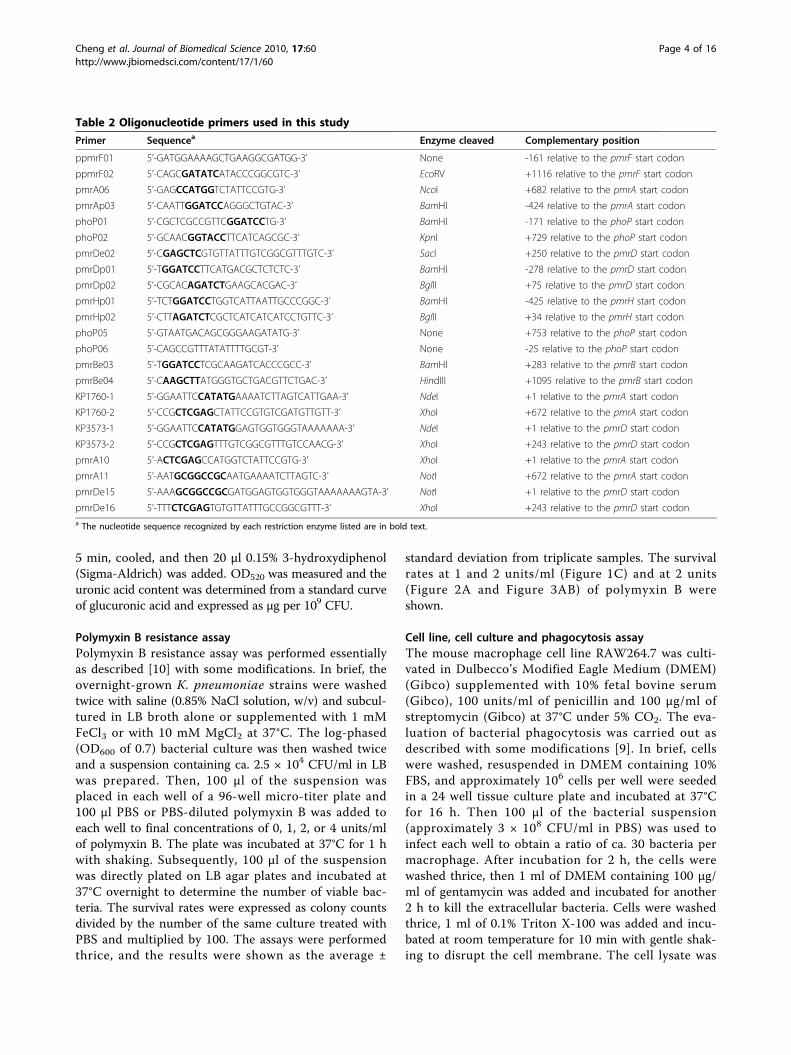
Table 2 Oligonucleotide primers used in this study
Primer Sequencea Enzyme cleaved Complementary position
ppmrF01 5’-GATGGAAAAGCTGAAGGCGATGG-3’ None -161 relative to the pmrF start codon
ppmrF02 5’-CAGCGATATCATACCCGGCGTC-3’ EcoRV +1116 relative to the pmrF start codon
pmrA06 5’-GAGCCATGGTCTATTCCGTG-3’ NcoI +682 relative to the pmrA start codon
pmrAp03 5’-CAATTGGATCCAGGGCTGTAC-3’ BamHI -424 relative to the pmrA start codon
phoP01 5’-CGCTCGCCGTTCGGATCCTG-3’ BamHI -171 relative to the phoP start codon
phoP02 5’-GCAACGGTACCTTCATCAGCGC-3’ KpnI +729 relative to the phoP start codon
pmrDe02 5’-CGAGCTCGTGTTATTTGTCGGCGTTTGTC-3’ SacI +250 relative to the pmrD start codon
pmrDp01 5’-TGGATCCTTCATGACGCTCTCTC-3’ BamHI -278 relative to the pmrD start codon
pmrDp02 5’-CGCACAGATCTGAAGCACGAC-3’ BglII +75 relative to the pmrD start codon
pmrHp01 5’-TCTGGATCCTGGTCATTAATTGCCCGGC-3’ BamHI -425 relative to the pmrH start codon
pmrHp02 5’-CTTAGATCTCGCTCATCATCATCCTGTTC-3’ BglII +34 relative to the pmrH start codon
phoP05 5’-GTAATGACAGCGGGAAGATATG-3’ None +753 relative to the phoP start codon
phoP06 5’-CAGCCGTTTATATTTTGCGT-3’ None -25 relative to the phoP start codon
pmrBe03 5’-TGGATCCTCGCAAGATCACCCGCC-3’ BamHI +283 relative to the pmrB start codon
pmrBe04 5’-CAAGCTTATGGGTGCTGACGTTCTGAC-3’ HindIII +1095 relative to the pmrB start codon
KP1760-1 5’-GGAATTCCATATGAAAATCTTAGTCATTGAA-3’ NdeI +1 relative to the pmrA start codon
KP1760-2 5’-CCGCTCGAGCTATTCCGTGTCGATGTTGTT-3’ XhoI +672 relative to the pmrA start codon
KP3573-1 5’-GGAATTCCATATGGAGTGGTGGGTAAAAAAA-3’ NdeI +1 relative to the pmrD start codon
KP3573-2 5’-CCGCTCGAGTTTGTCGGCGTTTGTCCAACG-3’ XhoI +243 relative to the pmrD start codon
pmrA10 5’-ACTCGAGCCATGGTCTATTCCGTG-3’ XhoI +1 relative to the pmrA start codon
pmrA11 5’-AATGCGGCCGCAATGAAAATCTTAGTC-3’ NotI +672 relative to the pmrA start codon
pmrDe15 5’-AAAGCGGCCGCGATGGAGTGGTGGGTAAAAAAAGTA-3’ NotI +1 relative to the pmrD start codon
pmrDe16 5’-TTTCTCGAGTGTGTTATTTGCCGGCGTTT-3’ XhoI +243 relative to the pmrD start codona The nucleotide sequence recognized by each restriction enzyme listed are in bold text.
Cheng et al. Journal of Biomedical Science 2010, 17:60http://www.jbiomedsci.com/content/17/1/60
Page 4 of 16

Figure 1 Deletion effects of ugd, wza and rcsB genes on Klebsiella CPS production and resistance to polymyxin B. (A) Comparison ofcolony morphology. The K. pneumoniae strains were streaked on an LB agar plate, incubated at 37°C overnight and photographed. (B)Sedimentation test. The strains were cultured overnight in LB broth at 37°C and subjected to centrifugation at 4,000 ×g for 5 min. Quantificationof K2 CPS amounts of each strain is shown below the figure. Values are shown as averages ± standard deviations from triplicate samples. (C)Polymyxin resistance assay. The log-phased cultures of K. pneumoniae CG43S3, Δugd, Δwza or ΔrcsB mutants were challenged with 1 or 2 units/ml of polymyxin B. (D) Polymyxin resistance assay. The log-phased culture of K. pneumoniae strains were challenged with 2 or 4 units/ml ofpolymyxin B. The survival rates are shown as the average ± standard deviations from triplicate samples. *, P < 0.01 compared to the parentalstrain CG43S3. **, P < 0.01 compared to each strain carrying pRK415.
Cheng et al. Journal of Biomedical Science 2010, 17:60http://www.jbiomedsci.com/content/17/1/60
Page 5 of 16

diluted serially with PBS, plated onto LB agar plates andincubated overnight for determining viable bacteriacount. The relative survival rates after phagocytosis wereexpressed as the colony counts of viable bacteria dividedby those of the original inoculums and multiplied by100. Three independent trials were performed, and thedata shown were the average ± standard deviation fromfive replicas.
Construction of reporter fusion plasmid andmeasurement of promoter activityThe approximately 350 or 500-bp DNA fragments con-taining the upstream region of the K. pneumoniae pmrDor pmrHFIJKLM gene cluster were PCR-amplified withprimers pmrDp01/pmrDp02 or pmrHp01/pmrHp02(Table 2), respectively and cloned in front of a promo-ter-less lacZ gene of the promoter selection plasmidplacZ15 [35]. The resulting plasmids, placZ15-PpmrD
and placZ15-PpmrH were mobilized from E. coli S17-1lpir to K. pneumoniae strains by conjugation. b-galacto-sidase activity was determined as previously described[35]. In brief, overnight cultures were washed twice withsaline and subcultured in LB alone or supplementedwith 10 mM MgCl2, 0.1 mM FeCl3, or 0.1 mM FeCl3plus 0.3 mM ferric iron scavenger deferoxamine (Sigma-Aldrich) to mid-log phase (OD600 of 0.7). Then 100 μlof the culture was mixed with 900 μl of Z buffer (60mM Na2HPO4, 40 mM NaH2PO4, 10 mM KCl, 1 mMMgSO4, 50 mM b-mercaptoethanol), 17 μl of 0.1% SDS,and 35 μl of chloroform and the mixture was shakenvigorously. After incubation at 30°C for 10 min, 200 μlof 4 mg/ml ONPG (o-nitrophenyl-b-D-galactopyrano-side) (Sigma-Aldrich) was added. Upon the appearanceof yellow color, the reaction was stopped by adding 500μl 1 M Na2CO3. OD420 was recorded and the b-galacto-sidase activity was expressed as Miller units [38]. Each
Figure 2 Involvement of K. pneumoniae pmrF gene in polymyxin B resistance and intramacrophage survival. (A) The log-phased culturesof K. pneumoniae CG43S3, the ΔpmrF mutant or ΔpmrF carrying pRK415-PmrF were grown in LB or LB supplemented with 1 mM Fe3+ and thenchallenged with 2 units/ml of polymyxin B. The survival rates are shown as the average ± standard deviations from triplicate samples. (B) Thesurvival rates of K. pneumoniae CG43S3ΔrcsB, the isogenic ΔpmrFΔrcsB mutant, and ΔpmrFΔrcsB mutant strain carrying the complementationplasmid pRK415-PmrF within the mouse macrophage RAW264.7 were determined. The results shown are relative survival rates which werecalculated from the viable colony counts of intracellular bacteria divided by individual original inoculums. Values are shown as the average offive replicas. Error bars, standard deviations. *, P < 0.01 compared to each parental strain; **, P < 0.01 compared to each mutant strain carryingpRK415-PmrF.
Cheng et al. Journal of Biomedical Science 2010, 17:60http://www.jbiomedsci.com/content/17/1/60
Page 6 of 16

sample was assayed in triplicate, and at least three inde-pendent experiments were carried out. The data shownwere calculated from one representative experiment andshown as the means and standard deviation from tripli-cate samples.
Cloning, expression and purification of recombinantproteinsThe DNA fragment of PhoP coding region was PCRamplified from the genomic DNA of K. pneumoniae
CG43S3 with primers phoP05/phoP06 (Table 2). Theamplified PCR products were cloned into the PCR clon-ing vector yT&A (Yeastern Biotech, Taiwan). TheEcoRI/BamHI and SalI fragments from the resultingplasmid were then cloned individually into pET30b(Novagen, Madison, Wis) to generate pET30b-PhoP andpET30b-PhoPN to allow the in-frame fusion to theN-terminal His codons. Plasmid pET30b-PmrBC wasconstructed by cloning DNA fragments PCR-amplifiedwith pmrBe03/pmrBe04 (Table 2) into a BamHI/HindIII
Figure 3 Effects of K. pneumoniae pmrA, pmrD and phoP deletion and complementation in polymyxin B resistance andintramacrophage survival. (A) The log-phased cultures of K. pneumoniae CG43S3, the ΔpmrA, ΔpmrD or ΔphoP mutants were grown in LB, LBsupplemented with 10 mM Mg2+ or LB supplemented with 1 mM Fe3+ and then challenged with 2 units/ml of polymyxin B. The survival ratesare shown as the average ± standard deviations from triplicate samples. (B) The log-phased cultures of K. pneumoniae CG43S3 carrying pRK415,the ΔpmrAΔphoP mutant strains carrying pRK415, pRK415-PhoP or pRK415-PmrA were grown in LB and challenged with 2 units/ml of polymyxinB. The survival rates are shown as the average ± standard deviations from triplicate samples. (C) The survival rates of K. pneumoniaeCG43S3ΔrcsB, the isogenic ΔpmrAΔrcsB, ΔphoPΔrcsB and ΔpmrDΔrcsB mutants, and each mutant strain carrying the complementation plasmidspRK415-PmrA, pRK415-PhoP or pRK415-PmrD within the mouse macrophage RAW264.7 were determined. The results shown are relative survivalrates which were calculated from the viable colony counts of intracellular bacteria divided by individual original inoculums. Values are shown asthe average of five replicas. Error bars, standard deviations. *, P < 0.01 compared to each parental strain; **, P < 0.01 compared to each mutantstrain carrying the complementation plasmid.
Cheng et al. Journal of Biomedical Science 2010, 17:60http://www.jbiomedsci.com/content/17/1/60
Page 7 of 16

site on pET30b. Plasmids pET-PmrA and pET-PmrD(courtesy of Dr. Chinpan Chen, Academia Sinica, Taipei,Taiwan) were constructed by cloning DNA fragmentsPCR-amplified with KP1760-1/KP1760-2 and KP3573-1/KP3573-2 (Table 2) into an NdeI/XhoI site, respectivelyinto pET29b. The resulting plasmids were transformedinto E. coli BL21(DE3) (Invitrogen, USA), and therecombinant proteins were over-expressed by inductionwith 0.5 mM isopropyl 1-thio-b-D-galactopyranoside(IPTG) for 3 h at 37°C. The proteins were then purifiedfrom total cell lysate by affinity chromatography usingHis-Bind resin (Novagen, Madison, Wis). After purifica-tion, the eluent was dialyzed against 1× protein storagebuffer (10 mM Tris-HCl pH 7.5, 138 mM NaCl,2.7 mM KCl, and 10% glycerol) at 4°C overnight, fol-lowed by condensation with PEG20000, and the puritywas determined by SDS-PAGE analysis.
DNA electrophoretic mobility shift assay (EMSA)EMSA was performed as previously described [14]. Inbrief, the DNA fragment encompassing the putativepmrD promoter region was obtained by PCR amplifica-tion and then end-labeled with [g-32P]ATP by T4 poly-nucleotide kinase. The purified His-PhoP or His-PhoPN149 protein was mixed with the DNA probe in a50-μl reaction mixture containing 20 mM Tris-HCl pH8.0, 50 mM KCl, 1 mM MgCl2, 1 mM dithiothreitol,and 7.5 mM acetyl phosphate. The mixture was incu-bated at room temperature for 30 min, mixed with 0.1volume of DNA loading dye, and then loaded onto a 5%nondenaturing polyacrylamide gel containing 5% gly-cerol in 0.5× TBE buffer (45 mM Tris-HCl pH 8.0, 45mM boric acid, 1.0 mM EDTA). After electrophoresis ata constant current of 20 mA at 4°C, the result wasdetected by autoradiography.
Bacterial two-hybrid assayThe bacterial two-hybrid assay was performed asdescribed previously [20,30]. The DNA fragmentsencoding full-length PmrA and PmrD were PCR-amplified with primer pairs pmrA10/pmrA11 andpmrDe15/pmrDe16 (Table 2) respectively, and clonedinto the 3′ end of genes encoding the a subunit ofRNA polymerase (RNAPa) domain on pBT and l-cIrepressor protein domain on pTRG. The resultingRNAPa-PmrA and l-cI-PmrD encoding plasmids,pBT-PmrA and pTRG-PmrD, were confirmed by DNAsequencing. The positive control plasmids used werepTRG-Gal11P and pBT-LGF2 (Stratagene). The pBTand pTRG derived plasmids were co-transformed intoE. coli XL1-Blue MRF’ Kan cells and selected on LBagar plates supplemented with 12.5 μg/ml tetracycline,
25 μg/ml chloramphenicol, and 50 μg/ml kanamycin.To investigate the protein-protein interaction in vivo,cells were grown until the OD600 reached 0.3 and thendiluted serially (10-1, 10-2, 10-3, and 10-4 order). Two-microliters of the bacterial culture were spotted ontoLB agar plates supplemented with 350 μg/ml carbeni-cillin, 25 μg/ml chloramphenicol, 50 μg/ml kanamycin,12.5 μg/ml tetracycline, 50 μg/ml X-gal (5-bromo-4-chloro-3-indolyl-b-D-galactopyranoside), and 20 μMIPTG. Growth of the bacterial cells was observed afterincubation at 30°C for 36 h.
In vitro phosphotransfer assayThe in vitro phosphotransfer assay was performed essen-tially as described [30]. The phospho-PmrBC276 proteinwas obtained by pre-incubation of His-PmrBC276 protein(5 μM) with 40 μCi of [g-32P]ATP in 80 μl of 1× phos-phorylation buffer (10 mM Tris-HCl, pH 7.5; 138 mMNaCl; 2.7 mM KCl; 1 mM MgCl2; 1 mM DTT) for 1 h atroom temperature. The reaction mixture was then chilledon ice, and 5 μl of the mixture was removed and mixedwith 2.5 μl of 5× SDS sample buffer as a reference sam-ple. The phospho-PmrBC276 protein mixture (30 μl) wasthen mixed with equal volumes of 1× phosphorylationbuffer containing either PmrA (10 μM) or PmrA withPmrD (each at 10 μM) to initiate the phosphotransferreaction. A 10-μl aliquot was removed at specific timepoints, mixed with 2.5 μl of 5× SDS sample buffer to stopthe reaction, and the samples were kept on ice until theperformance of SDS-PAGE. After electrophoresis at 4°C,the signal was detected by autoradiography.
Kinase/phosphatase and autokinase assayThe assays were performed essentially as described [30].The recombinant protein His-PmrBC276 (2.5 μM) wasincubated with His-PmrA (5 μM) alone or with His-PmrD (5 μM) for kinase/phosphatase assay or incubatedwith His-PmrD (5 μM) alone for autokinase assay. Thereactions were carried out in 30 μl of 1× phosphoryla-tion buffer with 3.75 μCi [g-32P]ATP at room tempera-ture and started with the addition of His-PmrBC276. Analiquot of 10-μl was removed at specific time points,mixed with 5× SDS sample buffer to stop the reaction,and the samples were kept on ice until the performanceof SDS-PAGE. After electrophoresis at 4°C, the signalwas detected by autoradiography.
Statistical analysisStudent’s t test was used to determine the significanceof the differences between the CPS amounts and thelevels of b-galactosidase activity. P values less than 0.01were considered statistically significant.
Cheng et al. Journal of Biomedical Science 2010, 17:60http://www.jbiomedsci.com/content/17/1/60
Page 8 of 16

ResultsReduced production of capsular polysaccharide hadminor effect on polymyxin B resistance in K. pneumoniaeK. pneumoniae CG43 is a highly encapsulated virulentstrain [32]. In order to verify the role of CPS in poly-myxin B resistance, the Δugd and Δwza mutants weregenerated by allelic exchange strategy, and their phe-notype as well as the amount of CPS produced werecompared with the parental strain CG43S3 and ΔrcsBmutant [14]. As shown in Figure 1A, the Δugd andΔwza mutants formed apparently smaller colonies onLB agar plate compared with the glistering colony ofthe parental strain CG43S3. Although the colony mor-phology of the ΔrcsB mutant was indistinguishablefrom CG43S3, the CPS-deficient phenotype was evi-dent as assessed using sedimentation assay and theamount of K2 CPS produced (Figure 1B). Deletion ofrcsB resulted in an approximately 50% reduction of theCPS, while the Δwza mutant produced less than 20%of that of its parental strain CG43S3. The CPS bio-synthesis in Δugd mutant was almost abolished, indi-cating an indispensible role of Ugd in CPSbiosynthesis. To investigate how the CPS level wasassociated with polymyxin B resistance, the survivalrates of the strains challenged with polymyxin B werecompared. The Δugd mutant producing the lowestamount of CPS was extremely sensitive to the treat-ment of polymyxin B (Figure 1C). Although the Δugdmutant was CPS-deficient, the impaired polymyxinresistance may have been largely attributed to thedefect in LPS biosynthesis since the survival rates ofΔwza and ΔrcsB mutants appeared to be comparablewith the parental strain CG43S3. This argues againstthe notion that the level of polymyxin B resistance ispositively correlated to the amount of CPS [10]. Never-theless, the possibility that a higher amount of CPSwas required for the resistance could not be ruled out.As shown in Figure 1D, the introduction of pRK415-RcsB [39] resulted in a significantly higher resistanceto polymyxin B in both ΔrcsB mutant and its parentalstrain. This indicated a protective effect of largeamounts of CPS in polymyxin resistance.
PmrF is involved in polymyxin B resistance and survivalwithin macrophageTo investigate if the K. pneumoniae pmr homologuesplayed a role in polymyxin B resistance, a pmrF deletionmutant strain and a plasmid pRK415-PmrF were gener-ated. As shown in Figure 2A, when the strains weregrown in LB medium, a low magnesium condition [40],differences in the survival rates were not apparent.When the strains were grown in LB supplemented with1 mM FeCl3, an apparent deleting effect of pmrF in
polymyxin B resistance was observed, and the survivalrate could be restored by the introduction of pRK415-PmrF. The results indicated a role of PmrF in the poly-myxin B resistance in high iron condition.In addition to the mucosa surfaces, antimicrobial pep-
tides and proteins play important roles in the microbici-dal activity of phagosome [41]. To investigate the effectof pmrF deletion in the bacterial survival within phago-some, phagocytosis assay was carried out. Since K. pneu-moniae CG43S3 was highly resistant to engulfment byphagocytes in our initial experiments, the ΔrcsB mutantwhich produced less CPS was used as the parental strainto generate ΔpmrFΔrcsB mutant. As shown in Figure2B, deletion of pmrF resulted in an approximately four-fold reduction in the recovery rate, which was restoredafter the introduction of pRK415-PmrF. This indicatedan important role of pmrF not only in polymyxin Bresistance but also in bacterial survival withinmacrophage.
Deletion effect of pmrA, pmrD or phoP on polymyxin Bresistance in K. pneumoniaeTo investigate how PmrA, PhoP and PmrD wereinvolved in the regulation of polymyxin B resistance inK. pneumoniae, ΔpmrA, ΔphoP and ΔpmrD mutantstrains were generated. Deletion of either one of thesegenes resulted in a dramatic reduction of resistance topolymyxin B when the strains were grown in LB med-ium (Figure 3A). The deleting effects were no longerobserved when the strains grown in LB supplementedwith 10 mM magnesium, implying an involvement ofthe PhoP-dependent regulation in LB, a low magnesiumenvironment. Under high-iron conditions, the deletionof pmrA caused the greatest reduction in the survivalrate. Introduction of pRK415-PmrA or pRK415-PhoPinto the ΔpmrAΔphoP double mutant strain not onlyrestored but also enhanced the bacterial resistance topolymyxin B (Figure 3B), which is likely due to an over-expression level of phoP or pmrA by the multicopy plas-mid. Finally, whether the deletion of pmrA, phoP orpmrD affected the survival rate in phagosomes was alsoinvestigated. Interestingly, deletion of phoP resulted inmost apparent effect while the pmrA deletion had lesseffect on the bacterial survival in macrophages. This wasprobably due to low iron concentration in the phago-somes [40]. The introduction of pRK415-PhoP orpRK415-PmrD could restore the recovery rates ofΔphoPΔrcsB and ΔpmrDΔrcsB, although not tothe extent displayed by the parental strain. Takentogether, our results indicate the presence of two inde-pendent pathways in the regulation of polymyxin Bresistance and the bacterial survival within macrophagephagosomes.
Cheng et al. Journal of Biomedical Science 2010, 17:60http://www.jbiomedsci.com/content/17/1/60
Page 9 of 16

Effect of pmrA, phoP or pmrD deletion on PpmrH::lacZ orPpmrD::lacZ activityAs the functional role of the structural gene pmrF and theregulator genes phoP, pmrD and pmrA was verified, itwould be of importance to investigate the regulatory net-work govern by PhoPQ-PmrD-PmrAB on the expressionof pmr genes. Sequence analysis has revealed PhoP andPmrA box consensus in the upstream region of pmrH andPhoP box consensus in the upstream region of pmrD (Fig-ure 4A). To investigate the interplay of PhoP, PmrA, andPmrD on the expression of pmr and pmrD genes, thereporter plasmids placZ15-PpmrH and placZ15-PpmrDwere constructed and mobilized into K. pneumoniaeCG43S3ΔlacZ and its derived ΔpmrAΔlacZ, ΔpmrDΔlacZor ΔphoPΔlacZ isogenic strains, respectively. The b-galac-tosidase activities of K. pneumoniae transformants underdifferent environmental conditions were determined. Inthe wild-type strain CG43S3ΔlacZ, the PpmrH::lacZ activitywas repressed in the presence of high magnesium butenhanced in high ferric ion (Figure 4B). Such iron-induci-ble activity was abolished after the addition of iron scaven-ger deferoxamine. As shown in Figure 4B, deleting effectof pmrA or phoP on the activity of PpmrH::lacZ could beobserved in LB or LB supplemented with ferric iron. Thenegative effect of pmrD deletion was also apparent at highiron condition but was abolished after the addition ofdeferoxamine. The results clearly demonstrate the involve-ment of PmrA, PhoP and PmrD in the regulation of theexpression of pmr genes, particularly in the presence ofhigh ferric irons. As shown in Figure 4C, the PpmrD::lacZactivity was significantly reduced in high-magnesium con-ditions or upon the deletion of phoP. Interestingly, thedeletion of pmrA or high ferric irons had little effect onthe activity of PpmrD::lacZ. The results suggest that theexpression of K. pneumoniae pmrD is regulated in aPhoP-dependent but PmrA-independent manner.
Analysis of EMSA indicates a direct binding of therecombinant PhoP to pmrD
The binding of PhoP or PmrA to PpmrH has been deter-mined recently [31]. In order to determine whetherPhoP binds directly to PpmrD, EMSA was performed. Asshown in Figure 5A, binding of the recombinant His-PhoP protein to PpmrD was evident by the formation ofa protein/DNA complex with a slower mobility. Thebinding specificity was also examined by the addition ofspecific DNA competitor or non-specific DNA competi-tor. As shown in Figure 5B, the formation of protein/DNA complex diminished when His-PhoPN149, in whichthe carboxyl-terminal helix-turn-helix domain has beentruncated, was used instead of His-PhoP. The resultsstrongly suggest the PhoP binds via its C-terminaldomain to the promoter of pmrD for the activation ofthe pmrD expression in K. pneumoniae.
Two-hybrid analysis of the in vivo interaction betweenKlebsiella PmrD and PmrAThe interaction between Klebsiella PmrD and PmrA hasbeen shown as a prerequisite for the connector-mediated pathway [31]. To demonstrate in vivo interac-tion, a bacterial two-hybrid assay was performed. Theplasmid pBT-PmrA carrying the RNAPa-PmrA codingregion and the plasmid pTRG-PmrD carrying the l-cI-PmrD coding sequence were generated. In vivo interac-tion between the two reporter strains allowed the bind-ing of l-cI to the operator region as well as therecruitment of a-RNAP for the expression of the ampRand lacZ reporter genes. The bacteria harboring thepositive control plasmids pTRG-Gal11P/pBT-LGF2showed a more vigorous growth on the indicator plate,as reflected by the apparent colony formation when theculture was diluted serially (Figure 6A). In contrast, thestrain carrying the negative control vectors pBT/pTRGrevealed impaired colony formation. As shown in Figure6A, a similar growth pattern of the E. coli cells harbor-ing pBT-PmrA/pTRG-PmrD to that of the positive con-trol cells was observed indicating an interaction betweenthe PmrD and PmrA.
The PmrD binds to PmrA to prevent dephosphorylationIn S. enterica, the phosphorylation of PmrA by the cog-nate sensor protein PmrB has been demonstrated toenhance its affinity in binding to its target promoter.The subsequent dephosphorylation of PmrA by PmrBhelped to relieve from over-activation of this system (1).In Salmonella, PmrD has been shown to be able to pro-tect PmrA from both intrinsic and PmrB-mediateddephosphorylation (22). To verify if Klebsiella PmrDalso participates in the phosphorylation, in vitro phos-photransfer assay was carried out with the recombinantproteins His-PmrA, His-PmrD and His-PmrBC276. Asshown in Figure 6B, the His-PmrA was rapidly phos-phorylated upon addition of the autophosphorylatedHis-PmrBC276 and then gradually dephosphorylated.Addition of His-PmrD apparently prolong the phosphor-ylation state of the His-PmrA, which could be main-tained for at least 60 min (Figure 6B). Thephosphorylated His-PmrA appeared to be very stable inthe presence of the His-PmrD since the phosphorylationsignal was still detectable 4 h later (data not shown). Asshown in Figure 6C, the specificity of the interactionbetween His-PmrD and His-PmrA was also demon-strated since the phosphorylation state of His-PmrAcould not be detected when incubated with the smallcationic proteins RNase A or cytochrome C [30]. Similarlevels of phospho-PmrBC276 were observed in the pre-sence or absence of PmrD (Figure 6D), suggesting theHis-PmrD had no effect on the phorphorylation state ofHis-PmrBC276.
Cheng et al. Journal of Biomedical Science 2010, 17:60http://www.jbiomedsci.com/content/17/1/60
Page 10 of 16

Figure 4 Schematic representation of pmrH and pmrD loci and determination of K. pneumoniae PpmrH::lacZ and PpmrD::lacZ activity.(A) Diagrammatic representation of the pmrH and pmrD loci. The large arrows represent the open reading frames. The relative positions of theprimer sets used in PCR-amplification of the DNA fragments encompassing the PpmrH and PpmrD regions are indicated, and the numbers denotethe relative positions to the translational start site. The name and approximate size of the DNA probes used in electro-mobility shift assay (EMSA)are shown on the left. The dashed boxes indicate the predicted PhoP and PmrA binding sequences and the alignment result is shown below.The identical nucleotide sequences are underlined. HP, hypothetical protein. (B) The b-galactosidase activities of log-phased cultures of K.pneumoniae strains carrying placZ15-PpmrH grown in LB, LB containing 10 mM MgCl2, LB containing 0.1 mM FeCl3 or 0.1 mM FeCl3 plusing 0.3mM deferoxamine were determined and expressed as Miller units. (C) The b-galactosidase activities of log-phased cultures of K. pneumoniaestrains carrying placZ15-PpmrD grown in LB, LB containing 10 mM MgCl2 or LB containing 0.1 mM FeCl3 were determined and expressed asMiller units. The data shown were the average ± standard deviations from triplicate samples. *, P < 0.01 compared to the same strain grown inLB medium. **, P < 0.01 compared to the parental strain grown in LB medium. #, P < 0.01 compared to the parental strain grown in LB mediumsupplemented with ferric ions.
Cheng et al. Journal of Biomedical Science 2010, 17:60http://www.jbiomedsci.com/content/17/1/60
Page 11 of 16

Figure 5 Binding of His-PhoP and His-PhoPN149 to PpmrD. (A) Specific binding of recombinant His-PhoP protein to the putative pmrDpromoter. EMSA was performed by using the 32P-labeled DNA probe of PpmrD incubated with increasing amounts of the His-PhoP (lanes 2 to 5),with 40 pmole of His-PhoP plus increasing amounts of the unlabeled PpmrD DNA (specific competitor, lane 6 to 9), or with excess amounts ofnon-specific competitor DNA (lane 10 and 11). The amounts of recombinant proteins and DNA probes used are indicated in the figure. (B)EMSA was performed with 0, 4 or 40 pmole of His-PhoP (lanes 1 to 3), His-PhoPN149 (lanes 4 to 6) or 100 pmole of BSA (lane 7). The arrowsindicate the PhoP/PpmrD complex and free probe of PpmrD.
Cheng et al. Journal of Biomedical Science 2010, 17:60http://www.jbiomedsci.com/content/17/1/60
Page 12 of 16

Figure 6 Klebsiella PmrD interacts with PmrA to prevent dephosphorylation. (A) Bacterial two-hybrid analysis of PmrD/PmrA interaction invivo. The E. coli XL1-Blue cells co-transformed with various combinations of pTRG and pBT-derived plasmids were diluted serially and spottedonto the indicator plate. The bacterial growth after 36 h was investigated and photographed. Combinations of plasmids carried by each strainwere indicated above the figure. (B) Klebsiella PmrD prevents the dephosphorylation of PmrA by its cognate sensor protein. The phosphorylationstate of the recombinant His-PmrA protein was monitored upon the addition of the sensor protein His-PmrBC276 in the presence (PmrD) orabsence (-) of purified His-PmrD protein at specific time points as indicated. The arrows indicate phospho-PmrA (P-PmrA) and phospho-PmrBC276(P-PmrBC276). (C) Kinase/phosphatase assay was carried out using the recombinant His-PmrA (final concentration 5 μM) and His-PmrBC276 (finalconcentration 2.5 μM) in the presence (PmrD) or absence (-) of the recombintant His-PmrD protein (final concentration 5 μM). The small cationicproteins RNase A and cytochrome C were introduced individually as a negative control at a final concentration of 5 μM. (D) Autokinase assay ofthe recombinant His-PmrBC276 (final concentration 2.5 μM) was performed in the presence (PmrD) or absence (-) of the recombintant His-PmrDprotein (final concentration 5 μM).
Cheng et al. Journal of Biomedical Science 2010, 17:60http://www.jbiomedsci.com/content/17/1/60
Page 13 of 16

DiscussionAlthough the amount of CPS produced by ΔrcsB mutantwas more than twice of that produced by Δwza mutant,no apparent difference between the wild type strainCG43S3, Δwza mutant, and ΔrcsB mutant in polymyxinB resistance could be observed. This is different fromthe previous finding that K. pneumoniae CPS was animportant physical barrier for the APs [10]. This discre-pancy may be attributed to some of the K. pneumoniaestrains used for comparison in the previous study pro-duced extremely low level of the CPS. Nevertheless, ahigher amount of CPS was protective for the bacterialresistance to polymyxin B.On the other hand, the deletion of ugd resulted in the
loss of resistance to polymyxin B. Sequence analysis ofthe available K. pneumoniae genome NTHU-K2044 [42],MGH78578 (http://genome.wustl.edu/) and 342 [43]revealed no PmrA [17] or PhoP box [27] in theupstream region of the manC-manB-ugd genes [44].This implies the involvement of a regulatory mechanismdifferent from that for S. enterica ugd, which was posi-tively regulated by the three 2CS regulators PhoP, PmrAand RcsB [45].Consistent with the reported findings [31], deletion of
Klebsiella pmrF which encodes one of the enzymesrequired for synthesis and incorporation of aminoarabi-nose in LPS resulted in decreased resistance to poly-myxin B and survival within macrophages. The pmrexpression has been shown to be directly regulated byPhoP under low magnesium or by PmrA in high ferricions, or by the connector-mediated pathway reportedfor Salmonella,[31]. Similar to the observations inE. coli, S. enterica [25], Yersinia pestis [46], and Pseudo-monas aeruginosa [47], a positive regulatory role ofPmrA and PhoP in polymyxin B resistance in K. pneu-moniae was also demonstrated.The deletion of phoP resulted in more drastic effect
on the bacterial survival in macrophage than the pmrAdeletion, implying a different level of control betweenPhoP and PmrA in K. pneumoniae resistance to phago-cytosis. During phagocytosis, phagosomal maturationand phagolysosomes formation are accompanied by pro-gressive acidification and acquisition of various hydro-lases, reactive oxygen, nitrogen species, and APs [48].Low pH and low-magnesium have been shown to beable to stimulate expression of the PhoP-activated genes[40,49]. Apart from its microbicidal activity, the APsinside phagosomes has even been reported as an indu-cing signal for the activation of the PhoP/PhoQ system[50]. The deletion of pmrF or phoP caused a significantreduction in intramacrophage survival of the bacterial,implying a role of the AP resistance regulation in thebacterial pathogenesis.
Until now, PmrD was only found in E. coli, Shigellaflexneri, S. enterica and K. pneumoniae. Although PmrDin Klebsiella appeared to act in a way similar to thePmrD in S. enterica, they share only about 40%sequence identity. The expression of K. pneumoniaepmrD was shown to be PhoP-dependent and the regula-tion was achieved through a direct binding of PhoP tothe putative pmrD promoter. In addition, the binding ofPmrD was shown to efficiently protect the PmrA fromdephosphorylation. The in vivo interaction betweenPmrD and PmrA demonstrated using 2-hybrid analysisfurther supported the presence of the connector-mediated pathway in K. pneumoniae.In summary, involvement of Klebsiella pmr in poly-
myxin B resistance and the regulation for the expressionof pmr genes were analyzed. The regulatory network forthe expression of the pmr genes is comprised of 2CSresponse regulators PhoP and PmrA, and the connectorprotein PmrD. The demonstration of PmrD in prolong-ing the phosphorylation state of phosphor-PmrA furtherconfirmed the presence of a connector-mediated path-way in K. pneumoniae. The complexity in the control ofpmr genes expression may provide ecological niches forK. pneumoniae in response to a variety of environmentalclues; for example, in the process of infection.
AcknowledgementsWe thank Dr. Chinpan Chen (Academia Sinica, Taipei, Taiwan) for providingthe plasmids pET-PmrD and pET-PmrA. The work is supported by the grantsfrom the National Research Program for Genome Medicine (NSC96-3112-B009-001) and National Science Council (97-2320-B-009-001-MY3).
Author details1Department of Biological Science and Technology, National Chiao-TungUniversity, Hsin Chu, Taiwan, China. 2Institute of Molecular Medicine andBioengineering, National Chiao-Tung University, Hsin Chu, Taiwan, China.
Authors’ contributionsHYC conceived the study, designed and performed the experiments,interpreted the data, drafted and revised the manuscript. YFC helped withthe polymyxin B resistance assay. HLP coordinated the study and revised themanuscript for scientific content as the corresponding author. All authorshave read and approved the final manuscript.
Competing interestsThe authors declare that they have no competing interests.
Received: 16 April 2010 Accepted: 24 July 2010 Published: 24 July 2010
References1. Podschun R, Ullmann U: Klebsiella spp. as nosocomial pathogens:
epidemiology, taxonomy, typing methods, and pathogenicity factors.Clin Microbiol Rev 1998, 11:589-603.
2. Falagas ME, Kopterides P: Old antibiotics for infections in critically illpatients. Current opinion in critical care 2007, 13:592-597.
3. Falagas ME, Bliziotis IA: Pandrug-resistant Gram-negative bacteria: thedawn of the post-antibiotic era? Int J Antimicrob Agents 2007, 29:630-636.
4. Kasiakou SK, Michalopoulos A, Soteriades ES, Samonis G, Sermaides GJ,Falagas ME: Combination therapy with intravenous colistin formanagement of infections due to multidrug-resistant Gram-negativebacteria in patients without cystic fibrosis. Antimicrob Agents Chemother2005, 49:3136-3146.
Cheng et al. Journal of Biomedical Science 2010, 17:60http://www.jbiomedsci.com/content/17/1/60
Page 14 of 16
5. Zavascki AP, Goldani LZ, Li J, Nation RL: Polymyxin B for the treatment ofmultidrug-resistant pathogens: a critical review. The Journal ofantimicrobial chemotherapy 2007, 60:1206-1215.
6. Hancock RE: Peptide antibiotics. Lancet 1997, 349:418-422.7. Kabha K, Nissimov L, Athamna A, Keisari Y, Parolis H, Parolis LA, Grue RM,
Schlepper-Schafer J, Ezekowitz AR, Ohman DE, et al: Relationships amongcapsular structure, phagocytosis, and mouse virulence in Klebsiellapneumoniae. Infect Immun 1995, 63:847-852.
8. Domenico P, Salo RJ, Cross AS, Cunha BA: Polysaccharide capsule-mediated resistance to opsonophagocytosis in Klebsiella pneumoniae.Infect Immun 1994, 62:4495-4499.
9. Cortes G, Borrell N, de Astorza B, Gomez C, Sauleda J, Alberti S: Molecularanalysis of the contribution of the capsular polysaccharide and thelipopolysaccharide O side chain to the virulence of Klebsiellapneumoniae in a murine model of pneumonia. Infect Immun 2002,70:2583-2590.
10. Campos MA, Vargas MA, Regueiro V, Llompart CM, Alberti S,Bengoechea JA: Capsule polysaccharide mediates bacterial resistance toantimicrobial peptides. Infect Immun 2004, 72:7107-7114.
11. Whitfield C, Paiment A: Biosynthesis and assembly of Group 1 capsularpolysaccharides in Escherichia coli and related extracellularpolysaccharides in other bacteria. Carbohydr Res 2003, 338:2491-2502.
12. Drummelsmith J, Whitfield C: Translocation of group 1 capsularpolysaccharide to the surface of Escherichia coli requires a multimericcomplex in the outer membrane. EMBO J 2000, 19:57-66.
13. Majdalani N, Gottesman S: The Rcs phosphorelay: a complex signaltransduction system. Annu Rev Microbiol 2005, 59:379-405.
14. Lai YC, Peng HL, Chang HY: RmpA2, an activator of capsule biosynthesisin Klebsiella pneumoniae CG43, regulates K2 cps gene expression at thetranscriptional level. J Bacteriol 2003, 185:788-800.
15. Kim SH, Jia W, Parreira VR, Bishop RE, Gyles CL: Phosphoethanolaminesubstitution in the lipid A of Escherichia coli O157: H7 and itsassociation with PmrC. Microbiology (Reading, England) 2006,152:657-666.
16. Gunn JS, Ryan SS, Van Velkinburgh JC, Ernst RK, Miller SI: Genetic andfunctional analysis of a PmrA-PmrB-regulated locus necessary forlipopolysaccharide modification, antimicrobial peptide resistance, andoral virulence of Salmonella enterica serovar typhimurium. Infect Immun2000, 68:6139-6146.
17. Wosten MM, Groisman EA: Molecular characterization of the PmrAregulon. The Journal of biological chemistry 1999, 274:27185-27190.
18. Yan A, Guan Z, Raetz CR: An undecaprenyl phosphate-aminoarabinoseflippase required for polymyxin resistance in Escherichia coli. The Journalof biological chemistry 2007, 282:36077-36089.
19. Breazeale SD, Ribeiro AA, McClerren AL, Raetz CR: A formyltransferaserequired for polymyxin resistance in Escherichia coli and themodification of lipid A with 4-Amino-4-deoxy-L-arabinose. Identificationand function oF UDP-4-deoxy-4-formamido-L-arabinose. The Journal ofbiological chemistry 2005, 280:14154-14167.
20. Marceau M, Sebbane F, Ewann F, Collyn F, Lindner B, Campos MA,Bengoechea JA, Simonet M: The pmrF polymyxin-resistance operon ofYersinia pseudotuberculosis is upregulated by the PhoP-PhoQ two-component system but not by PmrA-PmrB, and is not required forvirulence. Microbiology (Reading, England) 2004, 150:3947-3957.
21. Tamayo R, Ryan SS, McCoy AJ, Gunn JS: Identification and geneticcharacterization of PmrA-regulated genes and genes involved inpolymyxin B resistance in Salmonella enterica serovar typhimurium.Infect Immun 2002, 70:6770-6778.
22. Lacour S, Bechet E, Cozzone AJ, Mijakovic I, Grangeasse C: Tyrosinephosphorylation of the UDP-glucose dehydrogenase of Escherichia coliis at the crossroads of colanic acid synthesis and polymyxin resistance.PLoS One 2008, 3:e3053.
23. Wosten MM, Kox LF, Chamnongpol S, Soncini FC, Groisman EA: A signaltransduction system that responds to extracellular iron. Cell 2000,103:113-125.
24. Gunn JS: The Salmonella PmrAB regulon: lipopolysaccharidemodifications, antimicrobial peptide resistance and more. TrendsMicrobiol 2008, 16:284-290.
25. Winfield MD, Groisman EA: Phenotypic differences between Salmonellaand Escherichia coli resulting from the disparate regulation ofhomologous genes. Proc Natl Acad Sci USA 2004, 101:17162-17167.
26. Perez JC, Groisman EA: Acid pH activation of the PmrA/PmrB two-component regulatory system of Salmonella enterica. Molecularmicrobiology 2007, 63:283-293.
27. Kato A, Tanabe H, Utsumi R: Molecular characterization of the PhoP-PhoQtwo-component system in Escherichia coli K-12: identification ofextracellular Mg2+-responsive promoters. J Bacteriol 1999, 181:5516-5520.
28. Kox LF, Wosten MM, Groisman EA: A small protein that mediates theactivation of a two-component system by another two-componentsystem. Embo J 2000, 19:1861-1872.
29. Kato A, Latifi T, Groisman EA: Closing the loop: the PmrA/PmrB two-component system negatively controls expression of itsposttranscriptional activator PmrD. Proc Natl Acad Sci USA 2003,100:4706-4711.
30. Kato A, Groisman EA: Connecting two-component regulatory systems bya protein that protects a response regulator from dephosphorylation byits cognate sensor. Genes Dev 2004, 18:2302-2313.
31. Mitrophanov AY, Jewett MW, Hadley TJ, Groisman EA: Evolution anddynamics of regulatory architectures controlling polymyxin B resistancein enteric bacteria. PLoS genetics 2008, 4:e1000233.
32. Chang HY, Lee JH, Deng WL, Fu TF, Peng HL: Virulence and outermembrane properties of a galU mutant of Klebsiella pneumoniae CG43.Microb Pathog 1996, 20:255-261.
33. Peng HL, Wang PY, Wu JL, Chiu CT, Chang HY: Molecular epidemiology ofKlebsiella pneumoniae. Zhonghua Min Guo Wei Sheng Wu Ji Mian Yi XueZa Zhi 1991, 24:264-271.
34. Skorupski K, Taylor RK: Positive selection vectors for allelic exchange. Gene1996, 169:47-52.
35. Lin CT, Huang TY, Liang WC, Peng HL: Homologous response regulatorsKvgA, KvhA and KvhR regulate the synthesis of capsular polysaccharidein Klebsiella pneumoniae CG43 in a coordinated manner. J Biochem(Tokyo) 2006, 140:429-438.
36. Keen NT, Tamaki S, Kobayashi D, Trollinger D: Improved broad-host-rangeplasmids for DNA cloning in gram-negative bacteria. Gene 1988,70:191-197.
37. Domenico P, Schwartz S, Cunha BA: Reduction of capsular polysaccharideproduction in Klebsiella pneumoniae by sodium salicylate. Infect Immun1989, 57:3778-3782.
38. Miller JH: Experiments in Molecular Genetics Cold Spring Harbor LaboratoryPress, Cold Spring Harbor, N.Y. 1972.
39. Cheng HY, Chen YS, Wu CY, Chang HY, Lai YC, Peng HL: RmpA regulationof capsular polysaccharide biosynthesis in Klebsiella pneumoniae CG43.J Bacteriol 192:3144-3158.
40. Groisman EA: The ins and outs of virulence gene expression: Mg2+ as aregulatory signal. Bioessays 1998, 20:96-101.
41. Benincasa M, Mattiuzzo M, Herasimenka Y, Cescutti P, Rizzo R, Gennaro R:Activity of antimicrobial peptides in the presence of polysaccharidesproduced by pulmonary pathogens. J Pept Sci 2009, 15:595-600.
42. Wu KM, Li LH, Yan JJ, Tsao N, Liao TL, Tsai HC, Fung CP, Chen HJ, Liu YM,Wang JT, Fang CT, Chang SC, Shu HY, Liu TT, Chen YT, Shiau YR,Lauderdale TL, Su IJ, Kirby R, Tsai SF: Genome Sequencing andComparative Analysis of Klebsiella pneumoniae NTUH-K2044, a StrainCausing Liver Abscess and Meningitis. Journal of bacteriology 2009.
43. Fouts DE, Tyler HL, DeBoy RT, Daugherty S, Ren Q, Badger JH, Durkin AS,Huot H, Shrivastava S, Kothari S, Dodson RJ, Mohamoud Y, Khouri H,Roesch LF, Krogfelt KA, Struve C, Triplett EW, Methé BA: Complete genomesequence of the N2-fixing broad host range endophyte Klebsiellapneumoniae 342 and virulence predictions verified in mice. PLoS genetics2008, 4:e1000141.
44. Chuang YP, Fang CT, Lai SY, Chang SC, Wang JT: Genetic determinants ofcapsular serotype K1 of Klebsiella pneumoniae causing primarypyogenic liver abscess. J Infect Dis 2006, 193:645-654.
45. Mouslim C, Groisman EA: Control of the Salmonella ugd gene by threetwo-component regulatory systems. Molecular microbiology 2003,47:335-344.
46. Winfield MD, Latifi T, Groisman EA: Transcriptional regulation of the 4-amino-4-deoxy-L-arabinose biosynthetic genes in Yersinia pestis. J BiolChem 2005, 280:14765-14772.
47. Moskowitz SM, Ernst RK, Miller SI: PmrAB, a two-component regulatorysystem of Pseudomonas aeruginosa that modulates resistance tocationic antimicrobial peptides and addition of aminoarabinose to lipidA. J Bacteriol 2004, 186:575-579.
Cheng et al. Journal of Biomedical Science 2010, 17:60http://www.jbiomedsci.com/content/17/1/60
Page 15 of 16

48. Flannagan RS, Cosio G, Grinstein S: Antimicrobial mechanisms ofphagocytes and bacterial evasion strategies. Nat Rev Microbiol 2009,7:355-366.
49. Groisman EA: The pleiotropic two-component regulatory system PhoP-PhoQ. J Bacteriol 2001, 183:1835-1842.
50. Otto M: Bacterial sensing of antimicrobial peptides. Contrib Microbiol2009, 16:136-149.
doi:10.1186/1423-0127-17-60Cite this article as: Cheng et al.: Molecular characterization of thePhoPQ-PmrD-PmrAB mediated pathway regulating polymyxinB resistance in Klebsiella pneumoniae CG43. Journal of Biomedical Science2010 17:60.
Submit your next manuscript to BioMed Centraland take full advantage of:
• Convenient online submission
• Thorough peer review
• No space constraints or color figure charges
• Immediate publication on acceptance
• Inclusion in PubMed, CAS, Scopus and Google Scholar
• Research which is freely available for redistribution
Submit your manuscript at www.biomedcentral.com/submit
Cheng et al. Journal of Biomedical Science 2010, 17:60http://www.jbiomedsci.com/content/17/1/60
Page 16 of 16