research open access identification of rare dna sequence
TRANSCRIPT

Matsunami et al. Molecular Autism 2014, 5:5http://www.molecularautism.com/content/5/1/5
RESEARCH Open Access
Identification of rare DNA sequence variants inhigh-risk autism families and their prevalence in alarge case/control populationNori Matsunami1†, Charles H Hensel2*†, Lisa Baird1, Jeff Stevens1, Brith Otterud1, Tami Leppert1, Tena Varvil1,Dexter Hadley3, Joseph T Glessner3, Renata Pellegrino3, Cecilia Kim3, Kelly Thomas3, Fengxiang Wang3,Frederick G Otieno3, Karen Ho2, Gerald B Christensen4, Dongying Li5, Rytis Prekeris5, Christophe G Lambert4,Hakon Hakonarson3,6 and Mark F Leppert1
Abstract
Background: Genetics clearly plays a major role in the etiology of autism spectrum disorders (ASDs), but studies todate are only beginning to characterize the causal genetic variants responsible. Until recently, studies using multipleextended multi-generation families to identify ASD risk genes had not been undertaken.
Methods: We identified haplotypes shared among individuals with ASDs in large multiplex families, followed bytargeted DNA capture and sequencing to identify potential causal variants. We also assayed the prevalence of theidentified variants in a large ASD case/control population.
Results: We identified 584 non-conservative missense, nonsense, frameshift and splice site variants that mightpredispose to autism in our high-risk families. Eleven of these variants were observed to have odds ratios greaterthan 1.5 in a set of 1,541 unrelated children with autism and 5,785 controls. Three variants, in the RAB11FIP5,ABP1, and JMJD7-PLA2G4B genes, each were observed in a single case and not in any controls. These variantsalso were not seen in public sequence databases, suggesting that they may be rare causal ASD variants.Twenty-eight additional rare variants were observed only in high-risk ASD families. Collectively, these 39 variantsidentify 36 genes as ASD risk genes. Segregation of sequence variants and of copy number variants previouslydetected in these families reveals a complex pattern, with only a RAB11FIP5 variant segregating to all affectedindividuals in one two-generation pedigree. Some affected individuals were found to have multiple potential riskalleles, including sequence variants and copy number variants (CNVs), suggesting that the high incidence of autismin these families could be best explained by variants at multiple loci.
Conclusions: Our study is the first to use haplotype sharing to identify familial ASD risk loci. In total, we identified39 variants in 36 genes that may confer a genetic risk of developing autism. The observation of 11 of these variantsin unrelated ASD cases further supports their role as ASD risk variants.
Keywords: Familial autism, Haplotype sharing, DNA sequence variants, Case/control study
* Correspondence: [email protected]†Equal contributors2Lineagen, Inc, Salt Lake City, UT, USAFull list of author information is available at the end of the article
© 2014 Matsunami et al.; licensee BioMed Central Ltd. This is an Open Access article distributed under the terms of theCreative Commons Attribution License (http://creativecommons.org/licenses/by/2.0), which permits unrestricted use,distribution, and reproduction in any medium, provided the original work is properly cited. The Creative Commons PublicDomain Dedication waiver (http://creativecommons.org/publicdomain/zero/1.0/) applies to the data made available in thisarticle, unless otherwise stated.
Matsunami et al. Molecular Autism 2014, 5:5 Page 2 of 18http://www.molecularautism.com/content/5/1/5
BackgroundTwin and family studies clearly demonstrate that genet-ics plays a major role in the etiology of ASDs [1-7].However, family-based approaches using sib-ships or nu-clear families have not resulted in the identification ofgenes or variants that explain a significant portion of theaffected population. Similarly, genetic linkage studieshave identified a number of chromosomal regions thatare thought to contain genes that predispose to ASDs,but identification of the relevant gene(s) in these regionshas proven difficult.More recent analyses have revealed that many of the
point mutations thought to predispose children to ASDswere not present in either parent, and thus occurredspontaneously in the affected individuals [8-12]. Such denovo variants likely account for as much as 20 to 30% ofthe genetic variation which results in ASDs. Additionalcases are likely due to recessive inheritance of non-functional or hypo-functional alleles, but autosomal re-cessive inheritance is thought to explain only about 1%of autism cases [13,14].In addition to single nucleotide variants and small
insertions/deletions that can be identified by DNA se-quencing, larger deletions or duplications (copy numbervariants, CNVs) have been shown to play a role in theetiology of ASDs [15-27]. Despite the observed inherit-ance of many ASD predisposition CNVs from an un-affected parent, the lack of extended, multi-generationpedigrees has precluded a comprehensive analysis of segre-gation of putative ASD predisposition CNVs and single nu-cleotide polymorphisms (SNPs) and the characterization ofother genetic factors necessary for their expression.The large families available in Utah coupled with the
willingness of family members to participate in geneticstudies have resulted in the identification of a largenumber of disease predisposition genes for both Men-delian and complex diseases. The pedigrees used in thisstudy were part of a 70-family linkage study publishedpreviously [28] and two smaller studies that evaluated asingle extended pedigree in this collection of families[29,30]. In this work, we analyzed members of 26extended multi-generational ASD families and four two-generation multiplex ASD families by performing haplo-type sharing analysis to identify chromosomal regionsthat might harbor ASD predisposition genes. We thenused DNA capture and sequencing of all genes in sharedregions and of additional candidate autism risk genes toidentify SNPs that might predispose to ASD in thesefamilies. These SNPs were analyzed in a large case/con-trol study and for segregation in these families. We alsoevaluated the segregation of CNVs, reported previously[27], in these families. Consistent with earlier studies, nosingle locus could account for more than a subset of theaffected individuals in any extended pedigree. In particular,
multiple potential risk alleles, including in some casesCNVs and SNPs, were identified in an extended pedigree,suggesting that no single variant is the genetic predispos-ition locus for all affected family members. The data pre-sented here identify several genes that may harbor ASDpredisposition mutations, and add to the growing list ofgenes that are targets for clinical DNA sequencing to aidin the understanding of an ASD genetic diagnosis. Thesedata further suggest that in some individuals multiple gen-etic variants may be necessary to elicit the observed clinicalcharacteristics and that for a complete understanding of ASDgenetics, both sequence variants and CNVs must be analyzed.
MethodsDNA samplesA total of 386 DNA samples from 26 extended multi-generation and four two-generation Utah multiplex ASDpedigrees were used in this study. Families were ascer-tained and recruited using the Utah Population Database(UPDB) as described [28]. Affection status was deter-mined using the Autism Diagnostic Interview-Revised(ADI-R) and the Autism Diagnostic Observation Schedule(ADOS), for both the familial ASD cases and the unre-lated ASD cases, as described previously [27]. The averagenumber of affected individuals in each pedigree is 7.9. Thepedigrees described here are a subset of those describedpreviously [28]. Pedigree details are shown in Additionalfile 1: Table S1. The 55 samples used for our previousCNV discovery were included in these families. A total of9,000 DNA samples previously described in a case/controlstudy [27], including 3,000 individuals with ASD and6,000 controls, were used to evaluate these variants ina broader population. All samples collected for thework described here were collected under methods ap-proved by the University of Utah Institutional ReviewBoard (IRB) (University of Utah IRB#: 6042–96) or TheChildren’s Hospital of Philadelphia IRB (CHOP IRB#:IRB 06–004886). Patients and their families were re-cruited through the University of Utah Department ofPsychiatry or The Children’s Hospital of Philadelphiaclinic or CHOP outreach clinics. Written informedconsent was obtained from the participants or theirparents using IRB approved consent forms prior to enroll-ment in the project. There was no discrimination againstindividuals or families who chose not to participate in thestudy. All data were analyzed anonymously and all clinicalinvestigations were conducted according to the principlesexpressed in the Declaration of Helsinki.
SNP microarray genotypingAffymetrix 250 K NspI SNP chip genotyping was carriedout on all 386 DNA samples using the manufacturer’srecommended procedure. Genotypes were called by Affy-metrix Genotyping Console software using the BRLMM
Matsunami et al. Molecular Autism 2014, 5:5 Page 3 of 18http://www.molecularautism.com/content/5/1/5
[31] genotype calling algorithm. Only SNPs with call ratesgreater than or equal to 99% were used for further ana-lyses. SNPs demonstrating Mendelian errors also wereidentified using PedCheck [32] and were excluded.
Shared haplotype analysisWe performed shared haplotype analysis on each pedi-gree to identify genomic regions that have significantsharing among the affected individuals in that pedigree.The HapShare algorithm [33] was used to performhaplotype phasing based on Mendelian inheritance andto identify shared genomic segments. The comparisonsincluded N out of N affected individuals, (N-1) out of N,(N-2) out of N, (N-3) out of N, and so on (See Figureone in reference [33]). In two-generation pedigrees, insome cases co-segregation of haplotypes was observed inall affected individuals analyzed, but the shared regionswere large, including up to half of a chromosome. Con-sequently, shared regions from nuclear families were notselected for sequencing unless they overlapped regionsobserved in additional families.
Custom targeted exome DNA sequencingNimbleGen (Roche NimbleGen, Inc., Madison, WI, USA)custom sequence capture arrays were designed to capture2,000 base pairs upstream of the transcription start siteand all exons and exon-intron boundaries of genes withinthe shared genomic segments. An additional 23 genes fromoutside of our haplotype sharing regions were selectedfrom the literature based on their potential roles in autismor neuronal functions (see Additional file 1: Table S2). Atotal of approximately 1,800 genes were captured. Captureand Illumina DNA sequencing were performed by the Van-derbilt University Microarray Shared Resource facility onDNA from 26 affected individuals from 11 families thatshowed sharing of genomic segments. Short reads werealigned to the National Center for Biotechnology Informa-tion (NCBI) reference human genome build 36 (GRCh36/hg18) and variants were called using the software align-ment and variant calling methods described in Table 1[34-36]. Potential variants detected by at least two of themethods were selected for further analysis.
Table 1 Sequence alignment and variant detectionmethods
Alignment andassembly
Sequence variantdetection
Method 1 Bowtie Maq
Method 2 MOSAIK GigaBayes
Method 3 CLC Bio GenomicsWorkbench (CLC Bio Inc.)
CLC Bio Genomics Workbench
(CLC Bio Inc., Cambridge, MA, USA).
Variant annotationIn silico functional analysis was carried out initially usingcSNP classifier, a preliminary program later incorporatedinto VAAST [37], to classify variants as synonymous, con-servative missense, non-conservative missense, nonsense,frameshift, or splice site changes. Later, variants were re-annotated using the ANNOVAR program [38]. TheKnownGene and RefSeq gene tracks from the UCSC gen-ome browser were used to annotate functional variants,and the LiftOver tool was used to convert human genomebuild 36 (GRCh36/hg18) coordinates to human genomebuild 37 (GRCh37/hg19) coordinates [39,40].
Custom microarray design and array processingDesign of the custom iSelect InfiniumTM II BeadChiparray (Illumina Inc., San Diego, CA, USA) includingprobes for 2,799 putative functional candidate SNPs and7,134 CNV probes was described previously [27]. Thecustom iSelect array was processed on 3,000 case and6,000 control samples at the Center for Applied Genom-ics at The Children’s Hospital of Philadelphia (CHOP)[27]. The same array was also used to analyze DNA from196 Utah discovery cohort family members at theUniversity of Utah Genomics Core facility for variantvalidation and analysis of SNP segregation in families.
Array data quality controlSample QCSubjects were withheld from SNP analysis if any of thefollowing were true: 1) subsequent to genotyping, theDNA sample was of apparent poor quality, evidenced byvery low call rates (N = 134); 2) the subject was identi-fied as a trisomy-21 (N = 51); 3) the subject was outsideof the central cluster of Caucasian subjects identified byprincipal component analysis (PCA) (N = 903) [27].Relatedness estimation further indicated that some of
the case subjects and controls were part of families withmultiple relatives represented in the data. Re-evaluationof family structure in the sample cohorts used subse-quently identified additional relationships. Subsequentassociation tests were therefore conducted using onlyone member of each known family in order to reducethe possibility of statistical confounding due to related-ness. For these tests, the subject selected from each fam-ily was the individual located nearest to the mediancentroid of the first two principal components. Thenumber of subjects removed due to relatedness was 688.This resulted in a final sample set for association testingcomprising 7,326 subjects, of which 1,541 were casesand 5,785 were controls.PCA was used to avoid artifacts due to population
stratification. Principal components were calculated inGolden Helix SNP and Variation Suite (SVS) usingdefault settings. All subjects were included in the

Matsunami et al. Molecular Autism 2014, 5:5 Page 4 of 18http://www.molecularautism.com/content/5/1/5
calculation except those that failed sample quality con-trol (QC). Prior to calculating principal components, theSNPs were filtered according to the following criteria:autosomes only, call rate > 0.95, minor allele frequency(MAF) > 0.05, linkage disequilibrium R2 < 25% for allpairs of SNPs within a moving window of 50 SNPs. Twothousand and eight SNPs, including those used for CNVanalysis, were used for the principal component calcula-tions. No genotype data were available for referencepopulations, as would typically be preferred for makinginferences about population stratification. However, aself-reported ethnicity variable was available for mostsubjects. A plot of the first two principal componentsshows a primary central cluster of subjects, with outliergroups extending along two axes. These roughly corres-pond to Asian and African-American ancestry as self-reported in the phenotype data. A simple outlier detectionalgorithm was applied to stratify the subjects into twogroups representing the most probable Caucasians andnon-Caucasians. This was done by first calculating theCartesian distance of each subject from the median cen-troid of the first two principal component vectors. Afterdetermining the third quartile (Q3) and inter-quartilerange (IQR) of the distances, any subject with a distanceexceeding:
Q3þ 1:5� IQR
was determined to be outside of the main cluster, andtherefore non-Caucasian. Six hundred and eighty-twosubjects were placed in the non-Caucasian category. Agraphical representation of the results of this PCA ana-lysis were reported previously [27].
SNP QCPrior to association testing, SNPs were evaluated for callrate, Hardy-Weinberg equilibrium (HWE) and allele fre-quency. All SNPs with call rates lower than 99% were re-moved from further analysis. No SNPs had significantHardy-Weinberg disequilibrium.
Laboratory confirmation of SNPs and CNVsFor molecular validation of SNPs, PCR products werefirst screened by LightScanner High Resolution Meltcurve analysis (BioFire Diagnostics Inc., Salt Lake City,UT, USA) for the presence of sequence variants. PCRprimer sequences are shown in Additional file 1: TableS3. Any samples that gave abnormal melt profiles weresequenced using the Sanger method at the UniversityUtah Sequencing Core facility to confirm the presenceof a sequence variant. For CNVs, pre- or custom-designed TaqMan copy number assays (Applied Biosys-tems Inc., Foster City, CA, USA) were used as describedpreviously [27].
Protein binding assaysAll glutathione S-transferase (GST)-tagged proteins wereexpressed and purified as described previously [41]. Totest Rab11FIP5 binding to various Rab GTPases, purifiedrecombinant FIP5(490–653) or FIP5(490–653)-P652Lwere incubated with glutathione beads coated with GST,GST-Rab11a, GST-Rab4a or GST-Rab3a in the presenceof 1 μm GMP-PNP. Beads were then washed with PBSand eluted with 1% sodium dodecyl sulphate (SDS).Eluates were then analyzed for the presence of FIP5(490–653) by immunoblotting with anti-Rab11FIP5 anti-bodies. A similar assay also was used to test the abilityof Rab11FIP5 (wild-type or P652L mutant) to dimerize.
Flow cytometry analysis of transferrin recyclingTo test the effect of the Rab11FIP5-P652L mutant onendocytic recycling, the transferrin recycling assay wasused as described previously [42]. Briefly, HeLa cells ex-pressing either wild-type FIP5-GFP or FIP5-GFP-P652Lwere incubated with transferrin conjugated to Alexa488.Cells were then washed and incubated with serum-supplemented media for varying amounts of time. Thecell-associated (not recycled) Tf-Alexa488 was analyzedby flow cytometry.
ResultsTo identify genes that predispose to ASDs in multiplexASD families, we took a haplotype sharing/custom DNAcapture and sequencing approach. We utilized the work-flow outlined in Figure 1, first to identify chromosomalregions with excessive sharing among affected individ-uals in multiplex ASD families. We then used sequencecapture to identify potential functional sequence variantsin the genes lying in the shared regions, as well as inadditional ASD candidate genes. Finally, we evaluatedthe segregation of those variants in our ASD familiesand determined their prevalence in a large set of ASDcases and a large set of controls. The details of thisprocess are described below.
Affymetrix 250 K SNP genotyping and haplotype sharingSNP genotyping was carried out on 386 DNA samplesfrom 26 extended multi-generation and four two-generation Utah multiplex ASD pedigrees. SNPs with nomap location were not included in the analysis. Theaverage call rate was 99.1% for the entire dataset.We used the HapShare method [33] to identify gen-
omic regions that have significant sharing among the af-fected individuals in each of the 30 pedigrees we studied.Paternal and maternal haplotypes were determinedbased on Mendelian inheritance using only informativemarkers. These haplotypes then were compared amongaffected individuals within each extended or nuclearfamily. We selected 18 regions of haplotype sharing
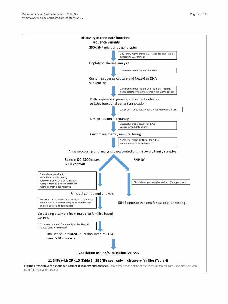
Figure 1 Workflow for sequence variant discovery and analysis. Only ethnicity and gender matched, unrelated, cases and controls wereused for association testing.
Matsunami et al. Molecular Autism 2014, 5:5 Page 5 of 18http://www.molecularautism.com/content/5/1/5

Matsunami et al. Molecular Autism 2014, 5:5 Page 6 of 18http://www.molecularautism.com/content/5/1/5
based on sharing in extended pedigrees for further ana-lysis. The degree of sharing that we observed among af-fected individuals and the coordinates of the regionsselected for DNA capture and sequencing are shown inTable 2. Two additional regions were selected for DNAcapture and sequencing based on a published linkageanalysis using an overlapping set of families [28].
Sequence capture, sequence analysis and variantidentificationWe performed capture and DNA sequencing usingDNA from 26 affected individuals from 11 families thatshowed the best sharing of genomic segments. Thesesamples included individuals from two-generation pe-digrees that had shared haplotypes overlapping regionsidentified in the extended pedigrees. Eight to ninemillion 36 base short reads were obtained from eachsample. The short reads alignment against the NCBIreference human genome build 36 revealed coverage of86 to 97% of the designed capture area, with the averageread depth over the designed capture area of 30 to 47X.Because 1) the capture library was constructed in adirectional manner, 2) all capture probes represented thesame DNA strand, and 3) the library was sequenced onlyfrom one direction, some portions of exons lacked thesequence depth of coverage that we desired. Conse-quently, there could be additional variants (false negativeresults) that we did not detect in some of the genes. For
Table 2 Chromosomal regions selected for sequencing based
Eighteen shared haplotype regions Chromosome Location
2p14-p12 2 65612029-7
2q23-q31 2 153638312-1
2q37 2 231435643-2
3q13 3 111604019-1
3q26-q27 3 174594938-1
4q28-q31 4 137362554-1
7p21 7 7381742-11
7p14 7 36090817-4
7q21-q31 7 90511244-10
7q35-36 7 142750349-1
12q21 12 76119990-7
12q21 12 79689788-8
14q11-q21 14 22912579-4
14q32 14 92331535-10
15q12-q21 15 24339787-4
16q22-23 16 73415053-7
20p11-q13 20 25253250-4
20q13 20 49062886-5
Where multiple numbers are given, multiple families shared overlapping haplotypeshare the same haplotype.
example no variants were identified on haplotypes thatsegregate to all affected individuals in pedigree 10 onchromosomes 2 and 14 (Additional file 2: Figure S1, Aand B, Additional file 3: Figure S2). Nonetheless, variantcalling using the three methods shown in Table 1 identifiedover one million sequence variants called by at least two ofthe three methods. Analysis using cSNP classifier resultedin the detection of 2,825 putative functional candidateSNPs, including 210 nonsense variants, 1,614 non-conservative missense variants, 35 frameshift variants and966 splice site variants.We chose to design a custom microarray to evaluate
the variants that we identified by sequencing in order to1) interrogate the entire set of candidate functional SNPsin the discovery families for validation, and 2) to per-form a large scale case/control study to determine if anyof the variants identified predisposition genes importantto the broad population of children with ASD (Figure 1).Following array design and manufacture, probes for2,413 variants were created successfully. Custom microarrayexperiments on Utah discovery and CHOP case/controlsamples revealed 584 out of 2,413 putative variants tobe polymorphic. The complete list of polymorphic vari-ants is shown in Additional file 1: Table S4. Theremaining array probes did not detect a non-referencesequence allele. These 1,829 variants thus were inter-preted to be false positives due to the variant callingand alignment process of single end sequence data.
on haplotype sharing
(hg18) Location (hg19) Affecteds sharing haplotype
6349401 65758525-76495893 6 of 6
74296304 153930066-174588058 6 of 6
38617145 231727399-238952406 5 of 7
12685490 110121329-111202800 4 of 7
85701563 173112244-184218869 4 of 7, 4 of 4
41629142 137143104-141409692 6 of 6
861952 7415217-11895427 4 of 4, 4 of 6
1521542 36124292-41555017 4 of 7
7823133 90673308-108035897 5 of 8a
51152511 143040227-151521578 4 of 6
7788028 77595859-79263897 5 of 7
7939487 81165657-89415356 5 of 8
5661808 23842739-46592058 3 of 4, 6 of 6
3509782 93261782-104440029 4 of 4
3759484 26788694-45972192 3 of 4, 4 of 6, 5 of 8
7780513 74857552-79223012 4 of 7, 5 of 6, 3 of 4
1225971 25305250-41792557 4 of 7
7757418 49629479-58324023 5 of 6, 5 of 6
s. aIndicates a family where a ninth affected individual was later shown not to

Matsunami et al. Molecular Autism 2014, 5:5 Page 7 of 18http://www.molecularautism.com/content/5/1/5
All autosomal SNP variants were tested for associationwith autism in our case/control study using an allelicassociation test. Statistical significance of each wasassessed using both Fisher’s exact test and a chi-squaredtest. The allelic association test detects any significantresult regardless of the direction of the effect. ElevenSNPs (see clustering in Additional file 4: Figure S3) wereeither unique to cases or had odds ratios (minor allele)greater than 1.5 (Table 3). We prioritized the variantsobserved in our case/control study for additional workbased on an odds ratio cutoff of 1.5. We also includedvariants unique to cases. We chose this approach ratherthan using P values since these variants were too rare toselect based on P values, and for relatively rare diseasesodds ratios are approximately equivalent to relative riskvalues. In addition, 28 SNPs were detected only in theUtah discovery cohort and not in the CHOP cases orcontrols (Table 4). We consider these 28 SNPs to bepotential ASD risk alleles because a) they are rare ornon-existent in the general population and thus couldrepresent ‘private mutations’, b) they may affect proteinfunction, and c) they segregate to one or more childrenwith autism in high-risk autism pedigrees. Thus, wecharacterize these 39 SNPs, found in 36 different genes,as potential autism risk variants. Each of these 39 vari-ants was localized to our targeted regions (Table 2), and30 of the 39 variants were predicted to be damaging byat least one program embedded in ANNOVAR [38],including SIFT, Polyphen2, LRT and MutationTaster.Details of the analysis of these variants are shown inAdditional file 1: Table S5. All 39 SNPs were furtherconfirmed by Sanger DNA sequencing of PCR amplicons(see Additional file 5: Figure S4, Additional file 6: Figure S5,
Table 3 Sequence variants identified in families and observedVariant(Ref/Obs)
Gene Coordinate(hg19)
Fisher’sexact P
Chi-squared P Odds rat(Minorallele)
G/T RAB11FIP5 chr2:73302656 2.10E-01 5.27E-02 infinite
G/C ABP1 chr7:150554592 2.10E-01 5.27E-02 infinite
T/A JMJD7-PLA2G4B
chr15:42133295 2.10E-01 5.27E-02 infinite
C/T C7orf10 chr7:40498796 4.02E-02 3.13E-02 1.62
C/T AKAP9 chr7:91724455 6.62E-02 4.44E-02 3.76
C/T HEPACAM2 chr7:92825188 5.84E-02 3.88E-02 1.83
G/T ALX1 chr12:85674230 2.22E-02 1.49E-02 1.75
G/A AP1G2 chr14:24035159 1.66E-01 1.35E-01 1.67
G/C CLMN chr14:95679692 2.29E-01 2.23E-01 1.67
G/A MOK chr14:102749873 1.97E-01 1.55E-01 3.76
G/A OIP5 chr15:41611874 3.77E-01 2.53E-01 2.25
Het. Refers to individuals heterozygous for the indicated variant.
for sequence chromatograms). The transcripts used forvariant annotation are found in Additional file 1: Table S5.
Segregation of variants in high-risk pedigreesTo determine the potential significance of variants thatwe identified, we evaluated the segregation pattern ofthese variants in the relevant pedigrees. We identifiedpotentially detrimental sequence variants in 10 of the 11pedigrees from which individuals were selected for DNAcapture and sequencing. Several of the pedigrees segre-gated more than one variant, indicating the complexityof the underlying genetics in high-risk ASD pedigrees.Moreover, many of these pedigrees also have CNVs thatwere identified in our previous work [27]. Adding tothe genetic complexity, many of these CNVs also segre-gate to affected individuals. Five families that demonstratethese complex inheritance patterns are shown here(Figures 2, 3, 4, 5 and 6). Five additional pedigrees withmultiple variants are shown in Additional file 7: Figure S6,Additional file 8: Figure S7, Additional file 9: Figure S8,Additional file 10: Figure S9, Additional file 3: Figure S9.Pedigree 1 (Figure 2) shows a two-generation family co-
segregating a missense variant in RAB11FIP5 (Table 4).This variant is present in the mother and segregates to allthree male affected children in the family, and not to theunaffected female child. RAB11FIP5 has previously beenimplicated as an ASD risk gene based on its disruption bya translocation observed in a 10 year-old male child with adiagnosis of pervasive developmental disorder not other-wise specified (PDD-NOS) [43]. The variant detected inpedigree 1 results in a P652L substitution. Proline is con-served at this residue in all of the mammalian RAB11FIP5genes sequenced to date, suggesting that it is important
in the case/control studyio Odds ratio
95% lowerconfidencebound
Odds ratio95% upperconfidencebound
Het.cases
Het.controls
W-Tcases
W-Tcontrols
Variant
N/A N/A 1 0 1,540 5,785 P652H
N/A N/A 1 0 1,540 5,785 R345P
N/A N/A 1 0 1,540 5,785 splice site
1.04 2.53 28 65 1,513 5,720 R288W,splice site
0.94 15.03 4 4 1,537 5,781 R3233C
1.02 3.27 17 35 1,524 5,750 G398R
1.11 2.77 27 58 1,514 5,727 R64L
0.85 3.30 12 27 1,529 5,757 R99C
0.73 3.84 8 18 1,533 5,767 P158A
0.53 26.67 2 2 1,539 5,783 Q22X
0.54 9.44 3 5 1,538 5,780 S165F
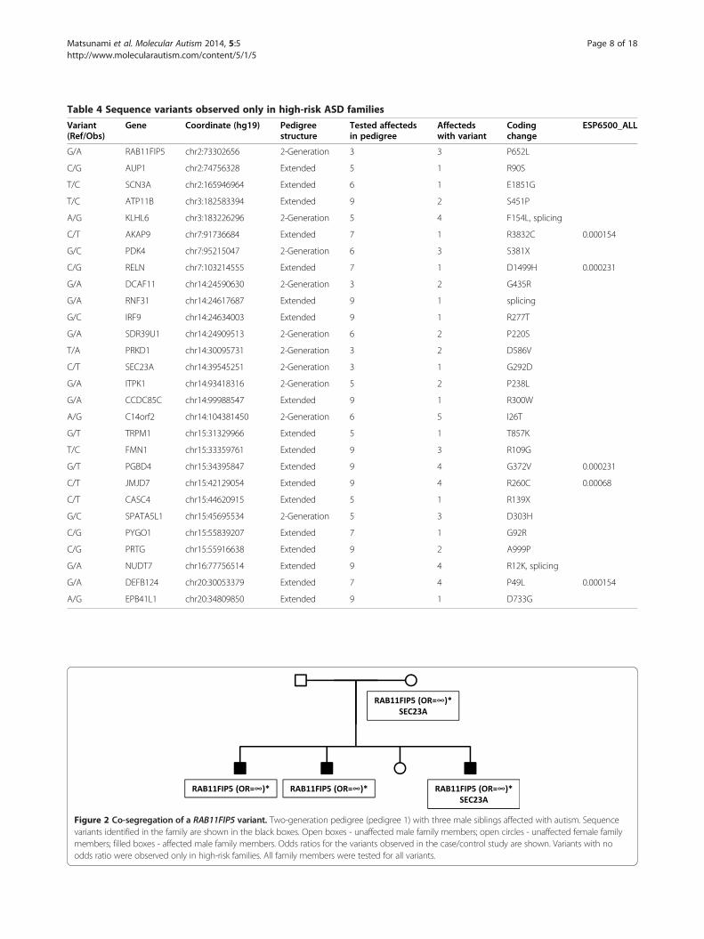
Figure 2 Co-segregation of a RAB11FIP5 variant. Two-generation pedigree (pedigree 1) with three male siblings affected with autism. Sequencevariants identified in the family are shown in the black boxes. Open boxes - unaffected male family members; open circles - unaffected female familymembers; filled boxes - affected male family members. Odds ratios for the variants observed in the case/control study are shown. Variants with noodds ratio were observed only in high-risk families. All family members were tested for all variants.
Table 4 Sequence variants observed only in high-risk ASD families
Variant(Ref/Obs)
Gene Coordinate (hg19) Pedigreestructure
Tested affectedsin pedigree
Affectedswith variant
Codingchange
ESP6500_ALL
G/A RAB11FIP5 chr2:73302656 2-Generation 3 3 P652L
C/G AUP1 chr2:74756328 Extended 5 1 R90S
T/C SCN3A chr2:165946964 Extended 6 1 E1851G
T/C ATP11B chr3:182583394 Extended 9 2 S451P
A/G KLHL6 chr3:183226296 2-Generation 5 4 F154L, splicing
C/T AKAP9 chr7:91736684 Extended 7 1 R3832C 0.000154
G/C PDK4 chr7:95215047 2-Generation 6 3 S381X
C/G RELN chr7:103214555 Extended 7 1 D1499H 0.000231
G/A DCAF11 chr14:24590630 2-Generation 3 2 G435R
G/A RNF31 chr14:24617687 Extended 9 1 splicing
G/C IRF9 chr14:24634003 Extended 9 1 R277T
G/A SDR39U1 chr14:24909513 2-Generation 6 2 P220S
T/A PRKD1 chr14:30095731 2-Generation 3 2 D586V
C/T SEC23A chr14:39545251 2-Generation 3 1 G292D
G/A ITPK1 chr14:93418316 2-Generation 5 2 P238L
G/A CCDC85C chr14:99988547 Extended 9 1 R300W
A/G C14orf2 chr14:104381450 2-Generation 6 5 I26T
G/T TRPM1 chr15:31329966 Extended 5 1 T857K
T/C FMN1 chr15:33359761 Extended 9 3 R109G
G/T PGBD4 chr15:34395847 Extended 9 4 G372V 0.000231
C/T JMJD7 chr15:42129054 Extended 9 4 R260C 0.00068
C/T CASC4 chr15:44620915 Extended 5 1 R139X
G/C SPATA5L1 chr15:45695534 2-Generation 5 3 D303H
C/G PYGO1 chr15:55839207 Extended 7 1 G92R
C/G PRTG chr15:55916638 Extended 9 2 A999P
G/A NUDT7 chr16:77756514 Extended 9 4 R12K, splicing
G/A DEFB124 chr20:30053379 Extended 7 4 P49L 0.000154
A/G EPB41L1 chr20:34809850 Extended 9 1 D733G
Matsunami et al. Molecular Autism 2014, 5:5 Page 8 of 18http://www.molecularautism.com/content/5/1/5
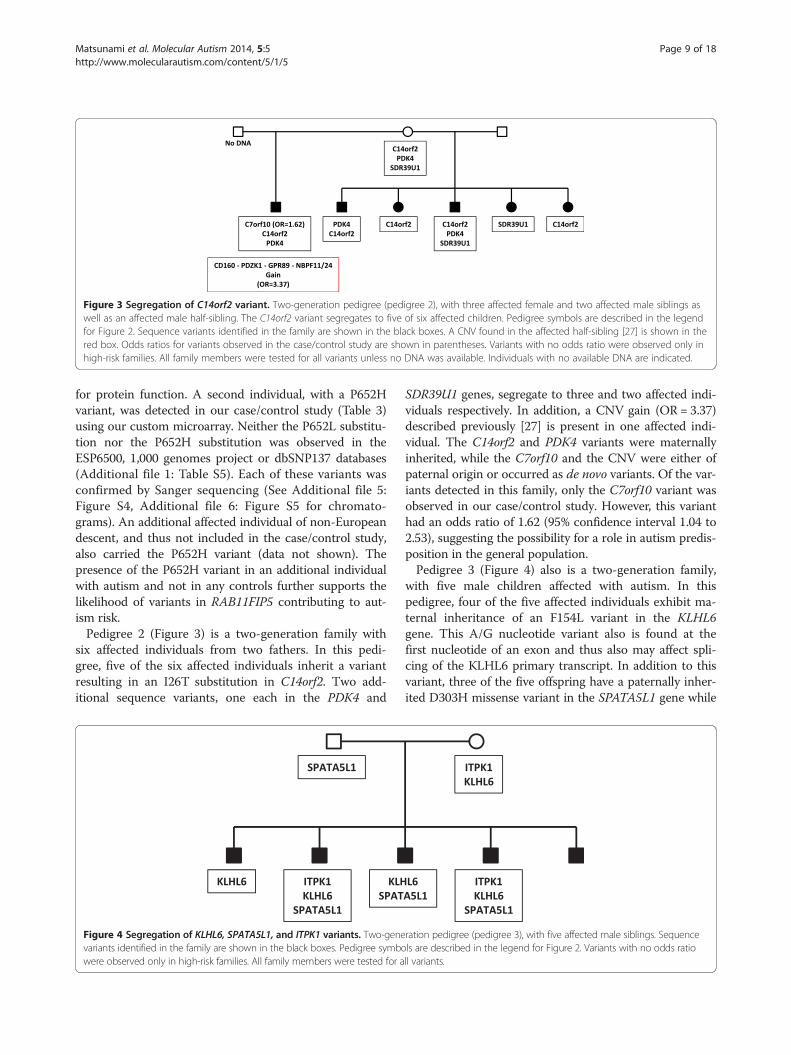
Figure 3 Segregation of C14orf2 variant. Two-generation pedigree (pedigree 2), with three affected female and two affected male siblings aswell as an affected male half-sibling. The C14orf2 variant segregates to five of six affected children. Pedigree symbols are described in the legendfor Figure 2. Sequence variants identified in the family are shown in the black boxes. A CNV found in the affected half-sibling [27] is shown in thered box. Odds ratios for variants observed in the case/control study are shown in parentheses. Variants with no odds ratio were observed only inhigh-risk families. All family members were tested for all variants unless no DNA was available. Individuals with no available DNA are indicated.
Matsunami et al. Molecular Autism 2014, 5:5 Page 9 of 18http://www.molecularautism.com/content/5/1/5
for protein function. A second individual, with a P652Hvariant, was detected in our case/control study (Table 3)using our custom microarray. Neither the P652L substitu-tion nor the P652H substitution was observed in theESP6500, 1,000 genomes project or dbSNP137 databases(Additional file 1: Table S5). Each of these variants wasconfirmed by Sanger sequencing (See Additional file 5:Figure S4, Additional file 6: Figure S5 for chromato-grams). An additional affected individual of non-Europeandescent, and thus not included in the case/control study,also carried the P652H variant (data not shown). Thepresence of the P652H variant in an additional individualwith autism and not in any controls further supports thelikelihood of variants in RAB11FIP5 contributing to aut-ism risk.Pedigree 2 (Figure 3) is a two-generation family with
six affected individuals from two fathers. In this pedi-gree, five of the six affected individuals inherit a variantresulting in an I26T substitution in C14orf2. Two add-itional sequence variants, one each in the PDK4 and
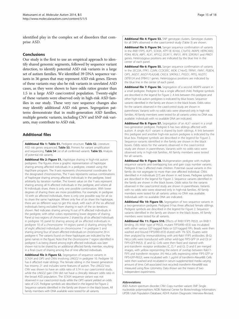
Figure 4 Segregation of KLHL6, SPATA5L1, and ITPK1 variants. Two-genevariants identified in the family are shown in the black boxes. Pedigree symbowere observed only in high-risk families. All family members were tested for a
SDR39U1 genes, segregate to three and two affected indi-viduals respectively. In addition, a CNV gain (OR = 3.37)described previously [27] is present in one affected indi-vidual. The C14orf2 and PDK4 variants were maternallyinherited, while the C7orf10 and the CNV were either ofpaternal origin or occurred as de novo variants. Of the var-iants detected in this family, only the C7orf10 variant wasobserved in our case/control study. However, this varianthad an odds ratio of 1.62 (95% confidence interval 1.04 to2.53), suggesting the possibility for a role in autism predis-position in the general population.Pedigree 3 (Figure 4) also is a two-generation family,
with five male children affected with autism. In thispedigree, four of the five affected individuals exhibit ma-ternal inheritance of an F154L variant in the KLHL6gene. This A/G nucleotide variant also is found at thefirst nucleotide of an exon and thus also may affect spli-cing of the KLHL6 primary transcript. In addition to thisvariant, three of the five offspring have a paternally inher-ited D303H missense variant in the SPATA5L1 gene while
ration pedigree (pedigree 3), with five affected male siblings. Sequencels are described in the legend for Figure 2. Variants with no odds ratioll variants.

Figure 5 Segregation of DEFB124 variant in a multi-generation pedigree. Pedigree 4 has seven children affected with autism. Links betweenthis pedigree and other high-risk autism pedigrees are indicated by blue boxes. Sequence variants identified in the family are shown in the blackboxes. CNVs inherited by two individuals [27] are shown in red boxes. Pedigree symbols are described in the legend for Figure 2. Odds ratios forthe variants observed in the case/control study are shown in parentheses. Variants with no odds ratio were observed only in high-risk families. Allfamily members were tested for all variants unless no DNA was available. Individuals with no available DNA are indicated.
Matsunami et al. Molecular Autism 2014, 5:5 Page 10 of 18http://www.molecularautism.com/content/5/1/5
two of five also have a maternally inherited P238L changein the ITPK1 gene. One affected child does not inherit anyof these variants. Of interest, none of the variants ob-served in this small family were observed in any cases orcontrols in our population study, demonstrating that theyare not common autism predisposition loci.Pedigree 4 (Figure 5) is a six generation family with an
ancestor common to all seven male children that are af-fected with autism. These children all are in the fifth orsixth generations of the pedigree. Linkage analysis wasperformed previously on this family using Affymetrix10 K SNP genotype data [29,30], and three regions ofsignificant linkage were identified. These include 3q13.2-q13.31, 3q26.31-q27.3, and 20q11.21-q13.12. These threeregions also were identified by haplotype sharing in thisstudy (Figure 5, see Additional file 2: Figure S1C forchromosome 20 haplotype sharing). Four of the sevenaffected individuals in this family share a P49L variantthat is the result of an A/G transition in the DEFB124gene on chromosome 20q11.21, consistent with thehaplotype sharing that we observed (Additional file 2:Figure S1c) and with the published linkage result. Thisvariant was not observed in cases or controls in our
population study. One affected individual in this pedi-gree does not share the DEFB124 variant, but insteadhas a chromosome 3q gain CNV, inherited from hisfather, that had an odds ratio of 3.74 in our previousstudy [27]. The elevated odds ratio suggests that thisCNV is an autism risk locus.Two additional affected individuals in pedigree 4 do
not carry any variant that we detected in our families.However, as indicated in Figure 5, each of these two in-dividuals is descended from a marry-in spouse with astrong family history of autism, suggesting the possibilityof additional undetected variants.Finally, one affected individual who carries the DEFB124
variant carries variants in the HEPACAM2 gene (odds ra-tio 1.83 in our population study, Table 3), the AP1G2 gene(odds ratio 1.67, Table 3), the PYGO1 gene and the RELNgene. Neither the RELN variant nor the PYGO1 variantwas observed in the case/control study (Table 4). Interest-ingly, a recent unpublished clinical case identified a het-erozygous deletion involving 15 exons of the RELN genein a patient diagnosed with autism and behavior/conductdisorder (Rena Vanzo, personal communication). Homo-zygous or compound heterozygous mutations in RELN

Figure 6 Segregation of multiple variants including a sequence variant in AKAP9 and a copy number variant in NRXN1 in amulti-generation pedigree. Pedigree 5 has nine children affected with autism. A link between this pedigree and another high-risk autismpedigree is indicated by the blue box. Sequence variants identified in the family are shown in the black boxes. CNVs identified in four individuals[27] are shown in red boxes. Pedigree symbols are described in the legend for Figure 2. Odds ratios for the variants observed in the case/controlstudy are shown in parentheses. Variants with no odds ratio were observed only in high-risk families. All family members were tested for allvariants unless no DNA was available. Individuals with no available DNA are indicated.
Matsunami et al. Molecular Autism 2014, 5:5 Page 11 of 18http://www.molecularautism.com/content/5/1/5
are associated with lissencephaly [44,45], but this RELNdeletion is the first description of an individual with a de-velopmental phenotype that may be due to haploinsuffi-ciency at this locus.Pedigree 5 (Figure 6) is a four-generation family with nine
individuals affected with autism (seven male, two female).Two variants are of particular interest in this family. Thefirst is a CNV including the 5′-flanking region of theNRXN1 gene. This CNV is inherited from a father whomarries into the family in the second generation. This CNVsegregates to three of the four descendants of this individ-ual who are diagnosed with autism. An overlapping NRXN1CNV was shown in our previous work to have an odds ra-tio of 14.96 [27], consistent with other publications suggest-ing a role for NRXN1 variants in autism, as well as otherneurological disorders [46-48]. However, that CNV wasshown to extend into the coding region of NRXN1, whileTaqMan CNV analysis demonstrates that the CNV in
pedigree 5 did not (data not shown). Thus, the significanceof the NRXN1 CNV observed in this family is uncertain.A second variant identified in this family, found on a
haplotype shared by all five affected individuals in twobranches of the family (Additional file 2: Figure S1c), is aC/T transition in the AKAP9 gene that results in anR3233C missense substitution. Note that none of theindividuals in these two branches of the family carry theNRXN1 CNV. The AKAP9 variant was observed in4/1541 cases and 4/5785 controls in our population study(odds ratio of 3.76, 95% confidence interval 0.94 to 15.03)(Table 3). A second missense variant in the AKAP9 genewas observed in a single affected individual in a nuclearfamily (pedigree 6, Additional file 7: Figure S6). This sec-ond AKAP9 variant was not observed in the case/controlstudy (Table 4). The AKAP family of proteins has beensuggested to connect different biological pathways that areinvolved in nervous system development [49].

Matsunami et al. Molecular Autism 2014, 5:5 Page 12 of 18http://www.molecularautism.com/content/5/1/5
Pedigree 5 also segregates other variants that areinherited by multiple children affected with autism. Onebranch of the pedigree segregates a G/C transversion inthe CLMN gene that results in a P158A missense substi-tution. This variant yielded an odds ratio of 1.67 (95%confidence interval 0.73 to 3.84) in our case/controlstudy, suggesting that it is an ASD risk allele. A variantin the ABP1 gene, also the result of a G/C transversionand resulting in an R345P missense substitution, was ob-served in two affected individuals in a single branch ofthe family. This variant was maternally inherited and notseen elsewhere in the pedigree. However, this variantwas observed in 1/1,541 cases and 0/5,785 controls inour population study (Table 3) and was not observed inthe ESP6500, 1,000 Genomes, or dbSNP137 databases(Additional file 1: Table S5), indicating that it may be avery rare ASD risk variant. Finally, a G/T transversion inthe ALX1 gene that results in an R64L missense substi-tution was paternally inherited by a single individual. Thisvariant also was seen in pedigree 7 (Additional file 8:Figure S7) and was observed multiple times in ourpopulation study (27/1,541 cases and 58/5,785 controls)yielding an odds ratio of 1.75 (95% confidence interval1.11 to 2.77) (Table 3). Expression of this gene also maybe increased by a downstream balanced translocation ina family with mental retardation, language delay andmicrocephaly that segregate with the translocation [50].Pedigrees 8 to 10 are shown in Additional file 9:
Figure S8, Additional file 10: Figure S9, Additional file3: Figure S2. One of these pedigrees, pedigree 10, car-ried two haplotypes (chromosomes 2 and 14) segregat-ing to all six affected individuals (Additional file 2:Figure S1a-b). Sequencing of the genes encompassed bythese regions did not identify potential causal variants.This could be due to poor sequence coverage of someportions of the genes. However, sequencing of affectedindividuals in these families did result in the identifica-tion of variants that could be autism risk alleles. One ofthese variants, a G/A transition that result in a Q22Xchange in the MOK gene observed in a single affectedindividual and inherited from her father, is interesting,as it was observed in our population study and yieldedan odds ratio of 3.76 (95% confidence interval 0.53 to26.67) (Table 3). Other variants in pedigrees 8 to 10(Additional file 9: Figure S8, Additional file 10: FigureS9, Additional file 3: Figure S2), including some onlyseen in Utah families and others seen in both familiesand in our population study also were identified. Thesevariants are included in Table 3 and Table 4.
Functional analysis of RAB11FIP5To uncover the functional consequences of the Rab11FIP5-P652L variant we evaluated binding of Rab11FIP5 to Rab11.Rab11 is a small monomeric GTPase that mediates
Rab11FIP5 recruitment to endocytic membranes and isrequired for Rab11FIP5 function [41]. As shown inAdditional file 11: Figure S10A, the P652L substitutiondid not affect Rab11FIP5 binding to Rab11, nor did itaffect its specificity toward the Rab11 GTPase. It waspreviously shown that Rab11FIP5 forms homodimersand that its ability to dimerize is also required for Rab11-FIP5 cellular functions [41]. Thus, we tested the effect ofP652L substitution on Rab11FIP5 ability to dimerize. Asshown in Additional file 11: Figure S10B, the Rab11FIP5-P652L mutant was still able to form dimers. Consistentwith in vitro binding data, FIP5-GFP-P652L endocyticlocalization in HeLa cells was also not affected (Additionalfile 11: Figure S10B-E).It is now well established that Rab11FIP5 functions by
regulating endocytic recycling [51]. To that end, we nexttested whether Rab11FIP5-P652L has an effect on recyc-ling of transferrin receptors in HeLa cells and found thatthe P652L substitution did not alter recycling (Additionalfile 11: Figure S10H). Thus, so far we were unable to findany functional consequences of Rab11FIP5-P652L substi-tution, suggesting that core Rab11FIP5 properties are notaffected. Further studies will be needed to uncover thekinetic or binding defects of Rab11FIP5 variants, espe-cially within the context of neuronal cells.
DiscussionNumerous studies have implicated genetics in the eti-ology of ASDs, and recent results have implicated hun-dreds of genes and chromosomal regions as candidateASD predisposition loci. These include chromosomal re-arrangements as well as sequence variants. None ofthese candidates accounts for more than a small portionof ASD cases, and it is clear, based on the apparent com-plexity of autism genetics, that many genes are still un-discovered. In multiplex ASD pedigrees, it has beensuggested that inherited genetic variants are more likelyto be responsible, while in simplex families de novo vari-ants may be more important [8,16].We used a discovery/validation strategy based on identi-
fying inherited genetic variants in two- to nine-generationASD families, followed by a case/control analysis of thosevariants in DNA samples from unrelated children with aut-ism and children with normal development, to identify pu-tative familial ASD predisposition genes. Using haplotypeanalysis we identified shared genomic segments within thefamilies, and used DNA sequencing and CNV analysis toidentify potential causal mutations on those haplotypes. Wealso followed this with a large case/control study to deter-mine if any of the variants we identified might play a role inthe general population of individuals with ASD.We showed previously that identification of CNVs in a
family-based discovery cohort could identify copy num-ber variants relevant to the general ASD population

Matsunami et al. Molecular Autism 2014, 5:5 Page 13 of 18http://www.molecularautism.com/content/5/1/5
[27]. In this paper, we applied the same strategy to thefamily-based identification of DNA sequence variants,and also follow up on our CNV analysis by providinginformation regarding segregation of some of theseCNVs in our high-risk ASD families.We identified 39 SNPs that are likely to affect protein
function that have segregation patterns and ASD caseallele frequencies suggestive of a role in ASD predispo-sition. Thirty-one of these variants result in non-conservative amino acid substitutions, five are predictedto affect splicing (three of these are predicted to affectboth splicing and protein coding), and three introducepremature termination codons. Two variants wereidentified in the AKAP9 gene and the JMJD7 gene (orthe JMJD7-PLA2G4B fusion gene), and two differentvariants were identified that affect the same amino acidresidue in the RAB11FIP5 gene; so collectively, theseSNPs identify 36 potential ASD risk genes.With the exception of two-generation families, and
consistent with our haplotype sharing results, no se-quence variants or CNVs implicated as ASD predispos-ition loci segregate to all affected individuals in apedigree. This is consistent with previous genetic stud-ies, which to date have been unable to demonstratesegregation of a single ASD risk locus in an extendedfamily (for example see [52]). In our pedigree 5(Figure 6), two independent risk variants, a singlenucleotide variant in AKAP9 and a deletion CNV in ornear NRXN1, segregate to different branches of thefamily. Other risk variants also are found in individualswith ASD in this family, including two sequence variantswith odds ratios greater than 1.5 in our populationstudy. These results suggest that even in extendedfamilies that might be predicted to be segregating asingle risk allele with reduced penetrance, multiple riskalleles in different ASD predisposition loci may benecessary. The results further suggest that use of specificinheritance models when evaluating autism genetics inlarge families should be approached with caution. Further,given our sequencing results and CNVs, the data suggestthat a two-pronged approach, involving both DNA sequen-cing and microarray-based CNV analysis, may be necessaryfor a complete genetic diagnosis of children with ASDs.
Table 5 Biological functions/pathways of genes with variants
Function Gene names
Previously associated with autism TRPM1, RAB11FIP5, AKAP9, SCN
Previously associated with neurologicaldisorder (other than autism)
RELN (autosomal recessive lissCCDC85C (seizures), EPB41L1 (
Neural function ITPK1, CLMN, PRTG
Mitochondrial function PLA2G4B, c7orf10, PDK4, C14or
Inflammatory response/Immune function DEFB124, BPI, RNF31, IRF9
Eleven of the putative autism risk variants that weidentified in our high-risk families are further supportedby data from our case/control study. Three of these vari-ants each were seen in a single ASD case (out of 1,541total cases) and in none of 5,785 controls. Familial vari-ants that we detected in eight additional genes are morecommon in ASD cases than in controls, and each has anodds ratio greater than 1.5. Although these variants arerare (all have frequencies of < 0.01 in our case/controlstudy), their identification in affected individuals in ourASD families and their increased prevalence in unrelatedaffected individuals support their role as potential ASDrisk loci.Several intriguing observations resulted from an exten-
sive literature review of the functions and mechanisticactions of each of these 36 genes and their encoded pro-teins. A number of the genes have been previouslylinked to autism or other neurological disorders or haveknown neurological functions (Table 5) (11 out of 36genes, or 31%). The functions of several other genes be-long to pathways often cited as having relevance toautism. These include genes encoding proteins with im-munological functions (inflammatory response), andgenes encoding proteins important for energy metabol-ism and mitochondrial function. These groups accountfor 19 of the 36 genes on the list (53%). Other geneshave as yet unexplored functions, can only be linked tofunctions based on sequence similarity, or have scatteredroles in many other cellular or organismal processes,such as cell cycle control, angiogenesis, protein degra-dation, or metalloproteinase activity.The incidence/prevalence of autism has been reported
to be as high as 1 in 38 children [68], while the estimatedprevalence in the US is reported to be approximately 1 in88 [69]. Multiple factors, including genetic, epigenetic andenvironmental, are thought to be necessary for autism de-velopment. As with previous work involving familial aut-ism, we found that no single gene variant described herecan account for every case of autism in our pedigrees. Thismay reasonably be expected of a multigenic disorder withhigh prevalence in the population. It also is consistentwith the high likelihood that autism represents a numberof different conditions, at least at the genetic level, all
found in children with ASDs
References
3A [27,43,49,53]
encephaly), ALX1 (facial clefting, micropthalmia),intellectual disability)
[44,45,50,54-56]
[57-59]
f2 [60-63]
[64-67]

Matsunami et al. Molecular Autism 2014, 5:5 Page 14 of 18http://www.molecularautism.com/content/5/1/5
under the same phenotypic umbrella called autism. Sev-eral variants that we identified, however, represent excel-lent candidates for significant risk factors for autism basedon their known functions and the currently understoodmolecular pathways involved in autism. We discuss themost compelling cases below.
RAB11FIP5RAB11FIP5 is a member of a family of scaffolding pro-teins for the RAS GTPase, Rab11. Specifically, RAB11-FIP5 has been characterized as a key player in apicalendosome recycling, plasma membrane recycling andtranscytosis [70,71]. We identified a P652L variant inthree affected siblings in a family of six members, inwhich the mother is an unaffected P652L carrier. Anadditional variant resulting in a P652H substitution alsowas detected in 1/1,541 Caucasian ASD cases and 0/5,785 Caucasian children with normal development(Table 3). These variants modify a conserved prolinewithin the C-terminus of RAB11FIP5.Heterozygous disruption of RAB11FIP5 was observed
previously in a ten year old boy with a balanced trans-location (46, XY, t(2;9)(p13;p24)) that disrupts only theRAB11FIP5 gene [43]. This individual has a clinical diag-nosis of PDD-NOS, an autism spectrum disorder. Thistranslocation led the authors to suggest that haploinsuf-ficiency of RAB11FIP5 contributes to the subject’s ASD.RAB11FIP5 works closely in conjunction with RAB11,and its presence has been detected in both presynapticand post-synaptic densities where Rab11 plays a key rolein determining synaptic strength in long-term depression[72], regulates norepinephrine transporter trafficking [73],carries out synaptic glutamate receptor recycling [74], andregulates dendritic branching in response to BDNF[75,76]. All of these functions have been suggested to besignificant contributors to the etiology of ASDs [77,78]and further support the role of mutations in RAB11FIP5as ASD risk alleles.
AKAP9AKAP9 is a member of a family of over 50 proteins thatserve as scaffolding partners for PKA, its effectors, andphosphorylation targets. AKAP9, also known as Yotiao,is chiefly expressed in the heart and brain, where theencoded protein serves as a scaffold for PKA, proteinphosphatase I, NMDA receptors, the heart potassiumchannel subunit KCNQ1, IP3R1, and specific isoforms ofadenylyl cyclase [79-83]. The subcellular localization andassembly of these multimeric protein scaffolds, mediatedby AKAPs, are thought to be essential for function, sincedisruption of the interaction between the AKAP and itseffectors leads to a loss of activity. In the case of KCNQ1,loss of interaction between AKAP9 and KCNQ1 leads to apotentially fatal heart condition, long-QT syndrome,
which also arises in cases with loss of function mutationsin KCNQ1 itself [84].We identified two variants in the AKAP9 gene. These
variants result in R3233C and R3832C substitutions inthe encoded protein. These two variants were coincidentwith autism and were found in two unrelated extendedASD pedigrees (Figure 6, Additional file 7: Figure S6).The R3233C variant was additionally found in ourcase/control study. A recent meta-study of the genesidentified from the five major autism GWAS studies andautism candidate genes arising from alternative method-ologies, such as large scale CNV studies, placed AKAPS asa central, integral gene family linking many of the path-ways identified by bioinformatics [49]. Given its role in lo-calizing PKA, adenylyl cyclase isoforms and NMDAR inthe postsynaptic scaffold, AKAP9 represents a proteinthat, like its better-characterized counterpart AKAP5,could function in synaptic transmission and plasticity, glu-tamatergic receptor function regulation and recycling, anddendritic spine morphology [85].It is notable that two of the genes (MOK, TRPM1)
containing potential ASD risk alleles were partially orcompletely encompassed by risk CNVs observed in ourprevious study [27]. This suggests that the same genesmay be affected by different genetic mechanisms withthe same or similar phenotypic result. The CNVs con-taining these genes were both copy number losses. TheMOK sequence variant described here was a nonsensechange, while the TRPM1 variant was a missensechange. These results are consistent with the MOK andTRPM1 effects being due to haploinsufficiency at thesetwo loci.Our data demonstrate the complex nature of autism
genetics. Although the heritability for autism is quitehigh, our data show that numerous genetic variants mayconfer risk to ASD even in a single family. This findingis consistent with the results of a whole genome sequen-cing study that used both a recessive model and modelindependent analyses to identify several potential ASDrisk variants in an ASD family with two affected individ-uals [86]. Consistent with the large number of potentialASD risk genes identified to date, none of the genesidentified in this single multiplex ASD family overlappedwith the genes identified in our study. Our study adds tothis complexity by identifying sequence variants in re-gions of haplotype sharing in 30 high-risk ASD familiesof two to six generations. Our data further demonstratethat in very large multi-generation families, the likeli-hood of additional risk variants entering the family fromindividuals who marry into the pedigree is high. Theseresults are consistent with the prevalence of autism inthe population and the high heritability for autism seenin numerous studies. Future studies will be necessary tocharacterize the biological roles that the variants we
Matsunami et al. Molecular Autism 2014, 5:5 Page 15 of 18http://www.molecularautism.com/content/5/1/5
identified play in the complex set of disorders that com-prise ASD.
ConclusionsOur study is the first to use an empirical approach to iden-tify shared genomic segments, followed by sequence variantdetection, to identify potential ASD risk variants in a largeset of autism families. We identified 39 DNA sequence var-iants in 36 genes that may represent ASD risk genes. Elevenof these variants may also be risk variants in unrelated ASDcases, as they were shown to have odds ratios greater than1.5 in a large ASD case/control population. Twenty-eightof these variants were observed only in high-risk ASD fam-ilies in our study. These very rare sequence changes alsomay identify additional ASD risk genes. Segregation pat-terns demonstrate that in multi-generation ASD families,multiple genetic variants, including CNV and SNP risk vari-ants, may contribute to ASD risk.
Additional files
Additional file 1: Table S1. Pedigree structure. Table S2. LiteratureASD risk genes sequenced. Table S3. Primers for variant amplificationand sequencing. Table S4 List of all confirmed variants. Table S5. Analysisof potential risk variants.
Additional file 2: Figure S1. Haplotype sharing in high-risk autismpedigrees. The figures show a graphic representation of haplotypesharing among affected individuals in a pedigree, created using theHapShare program. The X-axis represents chromosomal coordinates forthe designated chromosomes. The Y-axis represents various combinationsof haplotype sharing among affected individuals in the pedigree, listedarbitrarily by iteration number. The lowest value on the Y-axis representsharing among all N affected individuals in the pedigree, and where allN individuals share, there is only one possible combination. With lowerdegrees of sharing there are more possibilities. For example, in pedigree10 with six affected individuals, there is only one possible way for all sixto share the same haplotype. Where only five of six share the haplotype,there are six different ways to get this result, with each of the six affectedindividuals being excluded from sharing in each of the six iterationsshown. Red indicates sharing among N out of N affected individuals inthe pedigree, with other colors representing lower degrees of sharing.Panel a) two regions of chromosome 2 shared by all six affected individualsin pedigree 10; panel b) sharing among all six affected individuals inpedigree 10 of a chromosome 14 region; panel c) sharing among fiveof eight affected individuals on chromosome 7 in pedigree 5 andsharing among four of seven affected individuals on chromosome 20 inpedigree 4. The variants found on these haplotypes are indicated by thegene names in the figure. Note that the chromosome 7 region identified inpedigree 5 as being shared among eight affected individuals was latershown not to be shared by an additional affected family member, resultingin a final count of sharing among five of nine affected individuals.
Additional file 3: Figure S2. Segregation of sequence variants inSCN3A and OIP5 and CNVs involving LINGO2 in pedigree 10. Pedigree 10has 6 affected male siblings. The female sibling in the lowest generationhas trisomy 21 and includes some features of autism. The LINGO2 lossCNV was shown to have an odds ratio of 3.74 in our case/control study,while the LINGO2 gain CNV did not have a clinically relevant odds ratio inthe broad ASD population. The SCN3A sequence variant was notobserved in our case/control study while the OIP5 variant yielded an oddsratio of 2.25. Pedigree symbols are described in the legend for Figure 2.Sequence variants identified in the family are shown in the black boxes. Allfamily members with DNA available were tested for all variants.
Additional file 4: Figure S3. SNP genotype clusters. Genotype clustersfor all SNPs observed in the case/control study (Table 3) are shown.
Additional file 5: Figure S4. Sanger sequence confirmation of variantsin the RAB11FIP5, AUP1, SCN3A, ATP11B, KLHL6, C7orf10, AKAP9, HEPACAM2,PDK4, RELN, ABP1, ALX1, AP1G2, DCAF11, RNF31, IRF9, SDR39U1 and PRKD1genes. Heterozygous positions are indicated by the blue line in thecenter of each panel.
Additional file 6: Figure S5. Sanger sequence confirmation of variantsin the SEC23A, ITPK1, CLMN, CCDC85C, MOK, C14orf2, TRPM1, FMN1, PGBD4,OIP5, JMJD7, JMJD7-PLA2G4B, CASC4, SPATA5L1, PYGO1, PRTG, NUDT7,DEFB124 and EPB41L1 genes. Heterozygous positions are indicated bythe blue line in the center of each panel.
Additional file 7: Figure S6. Segregation of a second AKAP9 variant ina small pedigree. Pedigree 6 has a single affected child. Pedigree symbolsare described in the legend for Figure 2. A link between this pedigree andother high-risk autism pedigrees is indicated by blue boxes. Sequencevariants identified in the family are shown in the black boxes. Odds ratiosfor the variants observed in the case/control study are shown inparentheses. Variants with no odds ratio were observed only in high-riskfamilies. All family members were tested for all variants unless no DNA wasavailable. Individuals with no available DNA are indicated.
Additional file 8: Figure S7. Segregation of an ALX1 variant in a smalltwo-generation pedigree. Pedigree 6 has two siblings affected withautism. A single ALX1 variant is shared by both siblings. A link betweenthis pedigree and another high-risk autism pedigree is indicated by theblue box. Pedigree symbols are described in the legend for Figure 2.Sequence variants identified in the family are shown in the blackboxes. Odds ratios for the variants observed in the case/controlstudy are shown in parentheses. Variants with no odds ratio wereobserved only in high-risk families. All family members were testedfor all variants.
Additional file 9: Figure S8. Multigeneration pedigree with multiplesequence variants and overlapping loss and gain copy number variants.Pedigree 8 has 5 affected male children. Potential causal variants in thisfamily do not segregate to more than one affected individual. CNVsidentified in 4 individuals [27] are shown in red boxes. Pedigree symbolsare described in the legend for Figure 2. Sequence variants identified inthe family are shown in the black boxes. Odds ratios for the variantsobserved in the case/control study are shown in parentheses. Variantswith no odds ratio were observed only in high-risk families. All familymembers were tested for all variants unless no DNA was available.Individuals with no available DNA are indicated.
Additional file 10: Figure S9. Segregation of two sequence variants ina two-generation pedigree. Pedigree 9 has three affected female siblings.Pedigree symbols are described in the legend for Figure 2. Sequencevariants identified in the family are shown in the black boxes. All familymembers were tested for all variants.
Additional file 11: Figure S10. Effects of RAB11FIP5 P652L on RAB11binding. (A) Wild- type of P652L mutant FIP5(490–653) was incubatedwith either various GST-tagged Rabs or GST-tagged FIPs. Beads were thenwashed and bound FIP5(490–653) eluted with 1% SDS. Eluates werethen analyzed by immunoblotting with anti-Rab11FIP5 antibodies. (B-G)HeLa cells were transduced with either wild-type FIP5-GFP (A and D) orFIP5-GFP-P652L (E and G). Cells were then fixed and stained withanti-transferrin receptor antibodies (C, D, F and G). D and E are mergedimages, with yellow representing the extent of overlap between Rab11-FIP5 and transferrin receptor. (H) HeLa cells expressing either FIP5-GFP orFIP5-GFP-P652L were incubated with 1 μg/ml of transferrin-Alexa488. Cellswere then washed and incubated in serum-supplemented media varyingamount of time. Cell-associated (not recycled) transferrin-Alexa488 wasmeasured using flow cytometry. Data shown are the means of twoindependent experiments.
AbbreviationsASD: Autism spectrum disorder; CNV: Copy number variant; SNP: Singlenucleotide polymorphism; NCBI: National Center for Biotechnology Information;UPDB: Utah Population Database; ADI-R: Autism Diagnostic Interview-Revised;

Matsunami et al. Molecular Autism 2014, 5:5 Page 16 of 18http://www.molecularautism.com/content/5/1/5
ADOS: Autism Diagnostic Observation Schedule; CHOP: The Children’s Hospitalof Philadelphia; PCA: Principal component analysis; MAF: Minor allele frequency;HWE: Hardy-Weinberg equilibrium; GST: Glutathione S-transferase; SDS: Sodiumdodecyl sulfate; PDD-NOS: Pervasive developmental disorder not otherwisespecified.
Competing interestsGBC and CGL are paid employees of Golden Helix Inc., which derivescommercial revenue from the SNP & Variation Suite software used for dataanalysis for this publication. NM, CHH and MFL have stock options andpatent applications with Lineagen, Inc. MFL also is an unpaid scientificadvisor for Lineagen, Inc. CHH is an employee of, and KH is a formeremployee of, Lineagen, Inc. The remaining authors declare no competinginterests. Financial support from Lineagen, Inc. does not alter our adherenceto all the Molecular Autism policies on sharing data and materials.
Authors’ contributionsNM designed experiments, performed DNA sequencing and molecularanalysis, analyzed data and contributed to writing of the manuscript.CHH designed experiments, oversaw data analysis, and wrote much of themanuscript. LB performed molecular confirmation assays on high-risk familysequence variants and designed and performed melting curve assays forSNP confirmation. JS and BO performed sequence analysis for identifyingsequence variants in our high-risk autism families. TL performed haplotypesharing analysis on our high-risk ASD families. TV handled all aspects ofautism family DNA sample QC and tracking. DH contributed to experimentaldesign. JTG supervised all data release and performed QC on all datagenerated at CHOP. RP supervised sequencing work at CHOP and oversawdata quality. CK and KT generated and helped analyze most of the array-based SNP data generated at CHOP. FW oversaw all aspects of DNA samplehandling of CHOP samples. FGO performed Sanger sequencing and analysisto confirm variants in CHOP samples. KH evaluated the biological roles ofall genes with variants and contributed to writing of the manuscript.GBC performed data QC and analysis, and contributed to writing of themanuscript. DL and RP performed and evaluated RAB11FIP5 functional assaysand wrote portions of the manuscript. CGL, HH and MFL, oversaw lab andanalytical operations, aided significantly in experimental design and setup,contributed to the writing of the manuscript and approved the final versionof the manuscript. All authors read and approved the final manuscript.
AcknowledgementsAll family subjects were ascertained and DNA collected with support fromR01 MH 06359 from the National Institute of Mental Health andU19HD035476 from the National Institute of Child Health and HumanDevelopment. Utah DNA samples were processed with support from GCRCM01-RR025764 from the National Center for Research Resources. The authorsgratefully acknowledge the resources provided by the Autism GeneticResource Exchange (AGRE) Consortium and the participating AGRE families.
Author details1Department of Human Genetics, University of Utah, Salt Lake City, UT, USA.2Lineagen, Inc, Salt Lake City, UT, USA. 3Center for Applied Genomics, TheChildren’s Hospital of Philadelphia, Philadelphia, PA, USA. 4Golden Helix, Inc,Bozeman, MT, USA. 5Department of Cell and Developmental Biology, Schoolof Medicine, University of Colorado Anschutz Medical Campus, Aurora, CO,USA. 6Department of Pediatrics, University of Pennsylvania School ofMedicine, Philadelphia, PA, USA.
Received: 11 September 2013 Accepted: 24 December 2013Published: 27 January 2014
References1. Rosenberg RE, Law JK, Yenokyan G, McGready J, Kaufmann WE, Law PA:
Characteristics and concordance of autism spectrum disorders among277 twin pairs. Arch Pediatr Adolesc Med 2009, 163:907–914.
2. Hallmayer J, Cleveland S, Torres A, Phillips J, Cohen B, Torigoe T, Miller J,Fedele A, Collins J, Smith K, Lotspeich L, Croen LA, Ozonoff S, Lajonchere C,Grether JK, Risch N: Genetic heritability and shared environmental factorsamong twin pairs with autism. Arch Gen Psychiatry 2011, 68:1095–1102.
3. Lichtenstein P, Carlström E, Råstam M, Gillberg C, Anckarsäter H:The genetics of autism spectrum disorders and related neuropsychiatricdisorders in childhood. Am J Psychiatry 2010, 167:1357–1363.
4. Ronald A, Hoekstra RA: Autism spectrum disorders and autistic traits: adecade of new twin studies. Am J Med Genet B Neuropsychiatr Genet 2011,156B:255–274.
5. International Molecular Genetic Study of Autism Consortium (IMGSAC):A full genome screen for autism with evidence for linkage to a regionon chromosome 7q. Hum Mol Genet 1998, 7:571–578.
6. International Molecular Genetic Study of Autism Consortium (IMGSAC):A genomewide screen for autism: strong evidence for linkage tochromosomes 2q, 7q, and 16p. Am J Hum Genet 2001, 69:570–581.
7. Buxbaum JD, Silverman J, Keddache M, Smith CJ, Hollander E, Ramoz N,Reichert JG: Linkage analysis for autism in a subset families withobsessive-compulsive behaviors: evidence for an autism susceptibilitygene on chromosome 1 and further support for susceptibility genes onchromosome 6 and 19. Mol Psychiatry 2004, 9:144–150.
8. Iosifov I, Ronemus M, Levy D, Wang Z, Hakker I, Rosenbaum J, Yamrom B,Lee Y-h, Narzisi G, Leotta A, Kendall J, Grabowska E, Ma B, Marks S,Rodgers L, Stepansky A, Troge J, Andrews P, Bekritsky M, Pradhan K,Ghiban E, Kramer M, Parla J, Demeter R, Fulton LL, Fulton RS, Magrini VJ, YeK, Darnell J, Darnell RB, et al.: De novo gene disruptions in children on theautistic spectrum. Neuron 2012, 74(2):285–299.
9. Sanders SJ, Murtha MT, Gupta AR, Murdoch JD, Raubeson MJ, Willsey AJ,Ercan-Sencicek AG, DiLullo NM, Parikshak NN, Stein JL, Walker MF, Ober GT,Teran NA, Song Y, El-Fishawy P, Murtha RC, Choi M, Overton JD, BjornsonRD, Carriero NJ, Meyer KA, Bilguvar K, Mane SM, Sestan N, Lifton RP, GünelM, Roeder K, Geschwind DH, Devlin B, State MW: Disruptive de novo pointmutations, revealed by whole-exome sequencing, are strongly associatedwith autism spectrum disorders. Nature 2012, 485(7397):237–241.
10. Neale BM, Kou Y, Liu L, Ma’ayan A, Samocha KE, Sabo A, Lin CF, Stevens C,Wang LS, Makarov V, Polak P, Yoon S, Maguire J, Crawford EL, Campbell NG,Geller ET, Valladares O, Schafer C, Liu H, Zhao T, Cai G, Lihm J, DannenfelserR, Jabado O, Peralta Z, Nagaswamy U, Muzny D, Reid JG, Newsham I, Wu Y,et al: Patterns and rates of exonic de novo mutations in autism spectrumdisorders. Nature 2012, 485(7397):242–245.
11. O’Roak BJ, Deriziotis P, Lee C, Vives L, Schwartz JJ, Girirajan S, Karakoc E,Mackenzie AP, Ng SB, Baker C, Rieder MJ, Nickerson DA, Bernier R, Fisher SE,Shendure J, Eichler EE: Exome sequencing in sporadic autism reveals ahighly interconnected protein network and extreme locusheterogeneity. Nature 2012, 485(7397):246–250.
12. O’Roak BJ, Vives L, Fu W, Egertson JD, Stanaway IB, Phelps IG, Carvill G,Kumar A, Lee C, Ankenman K, Munson J, Hiatt JB, Turner EH, Levy R, O’DayDR, Krumm N, Coe BP, Martin BK, Borenstein E, Nickerson DA, Mefford HC,Doherty D, Akey JM, Bernier R, Eichler EE, Shendure J: Multiplex targetedsequencing identifies recurrently mutated genes in autism spectrumdisorders. Science 2012, 338(6114):1619–1622.
13. Lim ET, Raychaudhuri S, Sanders SJ, Stevens C, Sabo A, MacArthur DG, NealeBM, Kirby A, Ruderfer DM, Fromer M, Lek M, Liu L, Flannick J, Ripke S,Nagaswamy U, Muzny D, Reid JG, Hawes A, Newsham I, Wu Y, Lewis L, DinhH, Gross S, Wang LS, Lin CF, Valladares O, Gabriel SB, De Pristo M, AltshulerDM, Purcell SM, et al: Rare complete knockouts in humans: populationdistribution and significant role in autism spectrum disorders. Neuron2013, 77(2):235–242.
14. Yu TW, Chahrour MH, Coulter ME, Jiralerspong S, Okamura-Ikeda K, AtamanB, Schmitz-Abe K, Harmin DA, Adli M, Malik AN, D’Gama AM, Lim ET, San-ders SJ, Mochida GH, Partlow JN, Sunu CM, Felie JM, Rodriguez J, Nasir RH,Ware J, Joseph RM, Hill RS, Kwan BY, Al-Saffar M, Mukaddes NM, Hashmi A,Balkhy S, Gascon GG, Hisama FM, LeClair E, et al: Using whole-exome se-quencing to identify inherited causes of autism. Neuron 2013,77(2):259–273.
15. Girirajan S, Brkanac Z, Coe BP, Baker C, Vives L, Vu TH, Shafer N, Bernier R,Ferrero GB, Silengo M, Warren ST, Moreno CS, Fichera M, Romano C,Raskind WH, Eichler EE: Relative burden of large CNVs on a range ofneurodevelopmental phenotypes. PLoS Genet 2011, 7:e1002334.
16. Sebat J, Lakshmi B, Malhotra D, Troge J, Lese-Martin C, Walsh T, Yamrom B,Yoon S, Krasnitz A, Kendall J, Leotta A, Pai D, Zhang R, Lee YH, Hicks J,Spence SJ, Lee AT, Puura K, Lehtimäki T, Ledbetter D, Gregersen PK,Bregman J, Sutcliffe JS, Jobanputra V, Chung W, Warburton D, King MC,Skuse D, Geschwind DH, Gilliam TC, et al: Strong association of de novocopy number mutations with autism. Science 2007, 316:445–449.

Matsunami et al. Molecular Autism 2014, 5:5 Page 17 of 18http://www.molecularautism.com/content/5/1/5
17. Marshall CR, Noor A, Vincent JB, Lionel AC, Feuk L, Skaug J, Shago M,Moessner R, Pinto D, Ren Y, Thiruvahindrapduram B, Fiebig A, Schreiber S,Friedman J, Ketelaars CE, Vos YJ, Ficicioglu C, Kirkpatrick S, Nicolson R,Sloman L, Summers A, Gibbons CA, Teebi A, Chitayat D, Weksberg R,Thompson A, Vardy C, Crosbie V, Luscombe S, Baatjes R, et al: Structuralvariation of chromosomes in autism spectrum disorder. Am J Hum Genet2008, 82:477–488.
18. Christian SL, Brune CW, Sudi J, Kumar RA, Liu S, Karamohamed S, Badner JA,Matsui S, Conroy J, McQuaid D, Gergel J, Hatchwell E, Gilliam TC, GershonES, Nowak NJ, Dobyns WB, Cook EH Jr: Novel submicroscopicchromosomal abnormalities detected in autism spectrum disorder.Biol Psychiatry 2008, 63:1111–1117.
19. Glessner JT, Wang K, Cai G, Korvatska O, Kim CE, Wood S, Zhang H, Estes A,Brune CW, Bradfield JP, Imielinski M, Frackelton EC, Reichert J, Crawford EL,Munson J, Sleiman PM, Chiavacci R, Annaiah K, Thomas K, Hou C, GlabersonW, Flory J, Otieno F, Garris M, Soorya L, Klei L, Piven J, Meyer KJ,Anagnostou E, Sakurai T, et al: Autism genome-wide copy numbervariation reveals ubiquitin and neuronal genes. Nature 2009, 459:569–573.
20. Bucan M, Abrahams BS, Wang K, Glessner JT, Herman EI, Sonnenblick LI,Alvarez Retuerto AI, Imielinski M, Hadley D, Bradfield JP, Kim C, Gidaya NB,Lindquist I, Hutman T, Sigman M, Kustanovich V, Lajonchere CM, Singleton A,Kim J, Wassink TH, McMahon WM, Owley T, Sweeney JA, Coon H, NurnbergerJI, Li M, Cantor RM, Minshew NJ, Sutcliffe JS, Cook EH, et al: Genome-wideanalyses of exonic copy number variants in a family-based study point tonovel autism susceptibility genes. PLoS Genet 2009, 5:e1000536.
21. Pinto D, Pagnamenta AT, Klei L, Anney R, Merico D, Regan R, Conroy J,Magalhaes TR, Correia C, Abrahams BS, Almeida J, Bacchelli E, Bader GD, BaileyAJ, Baird G, Battaglia A, Berney T, Bolshakova N, Bölte S, Bolton PF, BourgeronT, Brennan S, Brian J, Bryson SE, Carson AR, Casallo G, Casey J, Chung BH,Cochrane L, Corsello C, et al: Functional impact of global rare copy numbervariation in autism spectrum disorders. Nature 2010, 466:368–372.
22. Szatmari P, Paterson AD, Zwaigenbaum L, Roberts W, Brian J: Mappingautism risk loci using genetic linkage and chromosomal rearrangements.Nat Genet 2007, 39:319–328.
23. Sanders SJ, Ercan-Sencicek AG, Hus V, Luo R, Murtha MT, Moreno-De-LucaD, Chu SH, Moreau MP, Gupta AR, Thomson SA, Mason CE, Bilguvar K,Celestino-Soper PB, Choi M, Crawford EL, Davis L, Wright NR, DhodapkarRM, DiCola M, DiLullo NM, Fernandez TV, Fielding-Singh V, Fishman DO,Frahm S, Garagaloyan R, Goh GS, Kammela S, Klei L, Lowe JK, Lund SC, et al:Multiple recurrent de novo CNVs, including duplications of the 7q11.23Williams syndrome region, are strongly associated with autism. Neuron2011, 70:863–885.
24. Weiss LA, Shen Y, Korn JM, Arking DE, Miller DT, Fossdal R, Saemundsen E,Stefansson H, Ferreira MA, Green T, Platt OS, Ruderfer DM, Walsh CA,Altshuler D, Chakravarti A, Tanzi RE, Stefansson K, Santangelo SL, Gusella JF,Sklar P, Wu BL, Daly MJ: Autism consortium: association betweenmicrodeletion and microduplication at 16p11.2 and autism. N Engl J Med2008, 358:667–675.
25. Morrow EM, Yoo SY, Flavell SW, Kim TK, Lin Y, Hill RS, Mukaddes NM, BalkhyS, Gascon G, Hashmi A, Al-Saad S, Ware J, Joseph RM, Greenblatt R, GleasonD, Ertelt JA, Apse KA, Bodell A, Partlow JN, Barry B, Yao H, Markianos K,Ferland RJ, Greenberg ME, Walsh CA: Identifying autism loci and genes bytracing recent shared ancestry. Science 2008, 321:218–223.
26. Jacquemont ML, Sanlaville D, Redon R, Raoul O, Cormier-Daire V, Lyonnet S,Amiel J, Le Merrer M, Heron D, de Blois MC, Prieur M, Vekemans M, CarterNP, Munnich A, Colleaux L, Philippe A: Array-based comparative genomichybridization identifies high frequency of cryptic chromosomalrearrangements in patients with syndromic autism spectrum disorders.J Med Genet 2006, 43:843–849.
27. Matsunami N, Hadley D, Hensel CH, Christensen GB, Kim C, Frackelton E,Thomas K, da Silva RP, Stevens J, Baird L, Otterud B, Ho K, Varvil T, Leppert T,Lambert CG, Leppert M, Hakonarson H: Identification of rare recurrentcopy number variants in high-risk autism families and their prevalencein a large ASD population. PLoS One 2013, 8(1):e52239.
28. Allen-Brady K, Robison R, Cannon D, Varvil T, Villalobos M, Pingree C,Leppert MF, Miller J, McMahon WM, Coon H: Genome-wide linkage inUtah autism pedigrees. Mol Psychiatry 2010, 15(10):1006–1015.
29. Coon H, Matsunami N, Stevens J, Miller J, Pingree C, Camp NJ, Thomas A,Krasny L, Lainhart J, Leppert MF, McMahon W: Evidence for linkage onchromosome 3q25-27 in a large autism extended pedigree. Hum Hered2005, 60(4):220–226.
30. Allen-Brady K, Miller J, Matsunami N, Stevens J, Block H, Farley M, Krasny L,Pingree C, Lainhart J, Leppert M, McMahon WM, Coon H: A high-densitySNP genome-wide linkage scan in a large autism extended pedigree.Mol Psychiatry 2009, 14(6):590–600.
31. BRLMM: An improved genotype calling method for the GeneChip®human mapping 500K array Set. http://media.affymetrix.com/support/technical/whitepapers/brlmm_whitepaper.pdf.
32. O’Connell JR, Weeks DE: PedCheck: a program for identification ofgenotype incompatibilities in linkage analysis. Am J Hum Genet 1998,63(1):259–266.
33. Arrington CB, Bleyl SB, Matsunami N, Bowles NE, Leppert TI, Demarest BL,Osborne K, Yoder BA, Byrne JL, Schiffman JD, Null DM, DiGeronimo R,Rollins M, Faix R, Comstock J, Camp NJ, Leppert MF, Yost HJ, Brunelli L:A family-based paradigm to identify candidate chromosomal regions forisolated congenital diaphragmatic hernia. Am J Med Genet A 2012,158A(12):3137–3147.
34. Langmead B, Trapnell C, Pop M, Salzberg SL: Ultrafast and memory-efficient alignment of short DNA sequences to the human genome.Genome Biol 2009, 10(3):R25.
35. Li H, Ruan J, Durbin R: Mapping short DNA sequencing reads and callingvariants using mapping quality scores. Genome Res 2008, 18(11):1851–1858.
36. Hillier LW, Marth GT, Quinlan AR, Dooling D, Fewell G, Barnett D, Fox P,Glasscock JI, Hickenbotham M, Huang W, Magrini VJ, Richt RJ, Sander SN,Stewart DA, Stromberg M, Tsung EF, Wylie T, Schedl T, Wilson RK, Mardis ER:Whole-genome sequencing and variant discovery in C. elegans. NatMethods 2008, 5(2):183–188.
37. Yandell M, Huff C, Hu H, Singleton M, Moore B, Xing J, Jorde LB, Reese MG:A probabilistic disease-gene finder for personal genomes. Genome Res2011, 21(9):1529–1542.
38. Wang K, Li M, Hakonarson H: ANNOVAR: functional annotation of geneticvariants from high-throughput sequencing data. Nucleic Acids Res 2010,38(16):e164.
39. Kent WJ, Sugnet CW, Furey TS, Roskin KM, Pringle TH, Zahler AM, Haussler D:The human genome browser at UCSC. Genome Res 2002, 12(6):996–1006.
40. Meyer LR, Zweig AS, Hinrichs AS, Karolchik D, Kuhn RM, Wong M, Sloan CA,Rosenbloom KR, Roe G, Rhead B, Raney BJ, Pohl A, Malladi VS, Li CH, Lee BT,Learned K, Kirkup V, Hsu F, Heitner S, Harte RA, Haeussler M, Guruvadoo L,Goldman M, Giardine BM, Fujita PA, Dreszer TR, Diekhans M, Cline MS, ClawsonH, Barber GP, et al: The UCSC Genome Browser database: extensions andupdates 2013. Nucleic Acids Res 2013, 41(Database issue):D64–D69.
41. Junutula JR, Schonteich E, Wilson GM, Peden AA, Scheller RH, Prekeris R:Molecular characterization of Rab11 interactions with members of thefamily of Rab11-interacting proteins. J Biol Chem 2004, 279(32):33430–33437.
42. Peden AA, Schonteich E, Chun J, Junutula JR, Scheller RH, Prekeris R:The RCP-Rab11 complex regulates endocytic protein sorting. Mol Biol Cell2004, 15(8):3530–3541.
43. Roohi J, Tegay DH, Pomeroy JC, Burkett S, Stone G, Stanyon R, Hatchwell E:A de novo apparently balanced translocation [46, XY, t(2;9)(p13;p24)]interrupting RAB11FIP5 identifies a potential candidate gene for autismspectrum disorder. Am J Med Genet B Neuropsychiatr Genet 2008,147B(4):411–417.
44. Hong SE, Shugart YY, Huang DT, Shahwan SA, Grant PE, Hourihane JO,Martin ND, Walsh CA: Autosomal recessive lissencephaly with cerebellarhypoplasia is associated with human RELN mutations. Nat Genet 2000,26(1):93–96.
45. Zaki M, Shehab M, El-Aleem AA, Abdel-Salam G, Koeller HB, Ilkin Y, Ross ME,Dobyns WB, Gleeson JG: Identification of a novel recessive RELN mutationusing a homozygous balanced reciprocal translocation. Am J Med GenetA 2007, 143A(9):939–944.
46. Béna F, Bruno DL, Eriksson M, van Ravenswaaij-Arts C, Stark Z, Dijkhuizen T,Gerkes E, Gimelli S, Ganesamoorthy D, Thuresson AC, Labalme A, Till M, Bilan F,Pasquier L, Kitzis A, Dubourgm C, Rossi M, Bottani A, Gagnebin M, Sanlaville D,Gilbert-Dussardier B, Guipponi M, van Haeringen A, Kriek M, Ruivenkamp C,Antonarakis SE, Anderlid BM, Slater HR, Schoumans J: Molecular and clinicalcharacterization of 25 individuals with exonic deletions of NRXN1 andcomprehensive review of the literature. Am J Med Genet B NeuropsychiatrGenet 2013, 162B(4):388–403.
47. Nag A, Bochukova EG, Kremeyer B, Campbell DD, Muller H, Valencia-DuarteAV, Cardona J, Rivas IC, Mesa SC, Cuartas M, Garcia J, Bedoya G, Cornejo W,Herrera LD, Romero R, Fournier E, Reus VI, Lowe TL, Farooqi IS, Mathews CA,McGrath LM, Yu D, Cook E, Wang K, Scharf JM, Pauls DL, Freimer NB, Plagnol V,

Matsunami et al. Molecular Autism 2014, 5:5 Page 18 of 18http://www.molecularautism.com/content/5/1/5
Ruiz-Linares A: Tourette syndrome association international consortiumfor genetics: CNV analysis in Tourette syndrome implicates large genomicrearrangements in COL8A1 and NRXN1. PLoS One 2013, 8(3):e59061.
48. Schaaf CP, Boone PM, Sampath S, Williams C, Bader PI, Mueller JM,Shchelochkov OA, Brown CW, Crawford HP, Phalen JA, Tartaglia NR, Evans P,Campbell WM, Tsai AC, Parsley L, Grayson SW, Scheuerle A, Luzzi CD,Thomas SK, Eng PA, Kang SH, Patel A, Stankiewicz P, Cheung SW:Phenotypic spectrum and genotype-phenotype correlations of NRXN1exon deletions. Eur J Hum Genet 2012, 20(12):1240–1247.
49. Poelmans G, Franke B, Pauls DL, Glennon JC, Buitelaar JK: AKAPs integrategenetic findings for autism spectrum disorders. Transl Psychiatry 2013,3:e270.
50. Liao HM, Fang JS, Chen YJ, Wu KL, Lee KF, Chen CH: Clinical and molecularcharacterization of a transmitted reciprocal translocation t(1;12)(p32.1;q21.3) in a family co-segregating with mental retardation, languagedelay, and microcephaly. BMC Med Genet 2011, 12:70.
51. Schonteich E, Wilson GM, Burden J, Hopkins CR, Anderson K, Goldenring JR,Prekeris R: The Rip11/Rab11-FIP5 and kinesin II complex regulatesendocytic protein recycling. J Cell Sci 2008, 121(Pt 22):3824–3833.
52. Kilpinen H, Ylisaukko-oja T, Rehnström K, Gaál E, Turunen JA, Kempas E, vonWendt L, Varilo T, Peltonen L: Linkage and linkage disequilibrium scan forautism loci in an extended pedigree from Finland. Hum Mol Genet 2009,18(15):2912–2921.
53. Chen CP, Lin SP, Chern SR, Chen YJ, Tsai FJ, Wu PC, Wang W: Array-CGHdetection of a de novo 2.8 Mb deletion in 2q24.2→q24.3 in a girl with autisticfeatures and developmental delay. Eur J Med Genet 2010, 53(4):217–220.
54. Uz E, Alanay Y, Aktas D, Vargel I, Gucer S, Tuncbilek G, von Eggeling F, Yilmaz E,Deren O, Posorski N, Ozdag H, Liehr T, Balci S, Alikasifoglu M, Wollnik B, AkarsuNA: Disruption of ALX1 causes extreme microphthalmia and severe facialclefting: expanding the spectrum of autosomal-recessive ALX-relatedfrontonasal dysplasia. Am J Hum Genet 2010, 86(5):789–796.
55. Mori N, Kuwamura M, Tanaka N, Hirano R, Nabe M, Ibuki M, Yamate J: Ccdc85cencoding a protein at apical junctions of radial glia is disrupted inhemorrhagic hydrocephalus (hhy) mice. Am J Pathol 2012, 180(1):314–327.
56. Hamdan FF, Gauthier J, Araki Y, Lin DT, Yoshizawa Y, Higashi K, Park AR,Spiegelman D, Dobrzeniecka S, Piton A, Tomitori H, Daoud H, Massicotte C,Henrion E, Diallo O, S2D Group, Shekarabi M, Marineau C, Shevell M,Maranda B, Mitchell G, Nadeau A, D’Anjou G, Vanasse M, Srour M, LafrenièreRG, Drapeau P, Lacaille JC, Kim E et al: Excess of de novo deleteriousmutations in genes associated with glutamatergic systems innonsyndromic intellectual disability. Am J Hum Genet 2011, 88(3):306–316.
57. Majerus PW, Wilson DB, Zhang C, Nicholas PJ, Wilson MP: Expression ofinositol 1,3,4-trisphosphate 5/6-kinase (ITPK1) and its role in neural tubedefects. Adv Enzyme Regul 2010, 50(1):365–372.
58. Marzinke MA, Clagett-Dame M: The all-trans retinoic acid (atRA)-regulatedgene Calmin (Clmn) regulates cell cycle exit and neurite outgrowth inmurine neuroblastoma (Neuro2a) cells. Exp Cell Res 2012, 318(1):85–93.
59. Wong YH, Lu AC, Wang YC, Cheng HC, Chang C, Chen PH, Yu JY, Fann MJ:Protogenin defines a transition stage during embryonic neurogenesisand prevents precocious neuronal differentiation. J Neurosci 2010,30(12):4428–4439.
60. Ghosh M, Loper R, Gelb MH, Leslie CC: Identification of the expressedform of human cytosolic phospholipase A2beta (cPLA2beta): cPLA2beta3is a novel variant localized to mitochondria and early endosomes. J BiolChem 2006, 281(24):16615–16624.
61. Sherman EA, Strauss KA, Tortorelli S, Bennett MJ, Knerr I, Morton DH,Puffenberger EG: Genetic mapping of glutaric aciduria, type 3, tochromosome 7 and identification of mutations in c7orf10. Am J HumGenet 2008, 83(5):604–609.
62. Korotchkina LG, Patel MS: Site specificity of four pyruvate dehydrogenasekinase isoenzymes toward the three phosphorylation sites of humanpyruvate dehydrogenase. J Biol Chem 2001, 276(40):37223–37229.
63. Meyer B, Wittig I, Trifilieff E, Karas M, Schägger H: Identification of twoproteins associated with mammalian ATP synthase. Mol Cell Proteomics2007, 6(10):1690–1699.
64. Jarczak J, Kościuczuk EM, Lisowski P, Strzałkowska N, Jóźwik A, Horbańczuk J,Krzyżewski J, Zwierzchowski L, Bagnicka E: Defensins: natural componentof human innate immunity. Hum Immunol 2013, 74(9):1069–1079.
65. Holweg A, Schnare M, Gessner A: The bactericidal/permeability-increasingprotein (BPI) in the innate defense of the lower airways. Biochem SocTrans 2011, 39(4):1045–1050.
66. Tokunaga F, Iwai K: Linear ubiquitination: a novel NF-κB regulatorymechanism for inflammatory and immune responses by the LUBACubiquitin ligase complex. Endocr J 2012, 59(8):641–652.
67. Nguyen H, Hiscott J, Pitha PM: The growing family of interferonregulatory factors. Cytokine Growth Factor Rev 1997, 8(4):293–312.
68. Kim YS, Leventhal BL, Koh YJ, Fombonne E, Laska E, Lim EC, Cheon KA, KimSJ, Kim YK, Lee H, Song DH, Grinker RR: Prevalence of autism spectrumdisorders in a total population sample. Am J Psychiatry 2011,168(9):904–912.
69. Center for Disease Control and Prevention. http://www.cdc.gov/ncbddd/autism/data.html.
70. Prekeris R, Klumperman J, Scheller RH: A Rab11/Rip11 protein complexregulates apical membrane trafficking via recycling endosomes. Mol Cell2000, 6(6):1437–1448.
71. Hales CM, Griner R, Hobdy-Henderson KC, Dorn MC, Hardy D, Kumar R,Navarre J, Chan EK, Lapierre LA, Goldenring JR: Identification andcharacterization of a family of Rab11-interacting proteins. J Biol Chem2001, 276(42):39067–39075.
72. Fernández-Monreal M, Brown TC, Royo M, Esteban JA: The balancebetween receptor recycling and trafficking toward lysosomesdetermines synaptic strength during long-term depression. J Neurosci2012, 32(38):13200–13205.
73. Matthies HJ, Moore JL, Saunders C, Matthies DS, Lapierre LA, Goldenring JR,Blakely RD, Galli A: Rab11 supports amphetamine-stimulated norepinephrinetransporter trafficking. J Neurosci 2010, 30(23):7863–7877.
74. van der Sluijs P, Hoogenraad CC: New insights in endosomal dynamicsand AMPA receptor trafficking. Semin Cell Dev Biol 2011, 22(5):499–505.
75. Park M, Salgado JM, Ostroff L, Helton TD, Robinson CG, Harris KM, EhlersMD: Plasticity-induced growth of dendritic spines by exocytic traffickingfrom recycling endosomes. Neuron 2006, 52(5):817–830.
76. Lazo OM, Gonzalez A, Ascaño M, Kuruvilla R, Couve A, Bronfman FC:BDNF regulates Rab11-mediated recycling endosome dynamics toinduce dendritic branching. J Neurosci 2013, 33(14):6112–6122.
77. Penzes P, Cahill ME, Jones KA, VanLeeuwen JE, Woolfrey KM: Dendriticspine pathology in neuropsychiatric disorders. Nat Neurosci 2011,14(3):285–293.
78. Ebert DH, Greenberg ME: Activity-dependent neuronal signaling andautism spectrum disorder. Nature 2013, 493(7432):327–337.
79. Piggot J, Shirinyan D, Shemmassian S, Vazirian S, Alarcón M: Neural systemsapproaches to the neurogenetics of autism spectrum disorders.Neuroscience 2009, 164(1):247–256.
80. Lin L, Sun W, Kung F, Dell’Acqua ML, Hoffman DA: AKAP79/150 impactsintrinsic excitability of hippocampal neurons through phospho-regulation of A-type K + channel trafficking. J Neurosci 2011,31(4):1323–1332.
81. Westphal RS, Tavalin SJ, Lin JW, Alto NM, Fraser ID, Langeberg LK, Sheng M,Scott JD: Regulation of NMDA receptors by an associated phosphatase-kinase signaling complex. Science 1999, 285(5424):93–96.
82. Marx SO, Kurokawa J, Reiken S, Motoike H, D’Armiento J, Marks AR, Kass RS:Requirement of a macromolecular signaling complex for beta adrenergicreceptor modulation of the KCNQ1-KCNE1 potassium channel. Science2002, 295(5554):496–499.
83. Tu H, Tang TS, Wang Z, Bezprozvanny I: Association of type 1 inositol1,4,5-trisphosphate receptor with AKAP9 (Yotiao) and protein kinase A.J Biol Chem 2004, 279(18):19375–19382.
84. Chen L, Marquardt ML, Tester DJ, Sampson KJ, Ackerman MJ, Kass RS:Mutation of an A-kinase-anchoring protein causes long-QT syndrome.Proc Natl Acad Sci USA 2007, 104(52):20990–20995.
85. Keith DJ, Sanderson JL, Gibson ES, Woolfrey KM, Robertson HR, Olszewski K,Kang R, El-Husseini A, Dell’acqua ML: Palmitoylation of A-kinase anchoringprotein 79/150 regulates dendritic endosomal targeting and synapticplasticity mechanisms. J Neurosci 2012, 32(21):7119–7136.
86. Shi L, Zhang X, Golhar R, Otieno FG, He M, Hou C, Kim C, Keating B, LyonGJ, Wang K, Hakonarson H: Whole-genome sequencing in an autismmultiplex family. Mol Autism 2013, 4(1):8.
doi:10.1186/2040-2392-5-5Cite this article as: Matsunami et al.: Identification of rare DNA sequencevariants in high-risk autism families and their prevalence in a largecase/control population. Molecular Autism 2014 5:5.