research open access excitotoxin-induced caspase-3
TRANSCRIPT
King et al. Acta Neuropathologica Communications 2013, 1:59http://www.actaneurocomms.org/content/1/1/59
RESEARCH Open Access
Excitotoxin-induced caspase-3 activation andmicrotubule disintegration in axons is inhibitedby taxolAnna Elizabeth King1*, Katherine Adriana Southam2, Justin Dittmann1 and James Clement Vickers1
Abstract
Background: Axon degeneration, a key pathological event in many neurodegenerative diseases and injury, can beinduced by somatodendritic excitotoxin exposure. It is currently unclear, however, whether excitotoxin-inducedaxon degeneration is mechanistically similar to Wallerian degeneration, which occurs following axon transection,but does not involve axonal caspase activation.
Results: We have used mouse primary cortical neurons at 9 days in vitro, in a compartmented culture model thatallows separation of the axon from the soma, to examine the pathological cascade of excitotoxin-induced axondegeneration. Excitotoxicity induced by chronic exposure to kainic acid, resulted in axonal fragmentation, whichwas associated with activation of caspase-3 in the axonal compartment. To examine the role of microtubules inthese events, the microtubule-stabilizing agent, taxol, was added to either the axonal or somatodendriticcompartment. Our results demonstrated that microtubule stabilization of axons resulted in a significant reduction inthe number of fragmented axons following excitotoxin exposure. Interestingly, taxol exposure to either thesomatodendritic or axonal compartment resulted in reduced caspase-3 activation in axons, suggesting that caspaseactivation is a downstream event of microtubule destabilization and involves signalling from the cell soma.
Conclusion: These data suggest that excitotoxin-induced axon degeneration shows some mechanistic differencesto Wallerian degeneration, and that microtubule stabilization may assist in protecting nerve cells from excitotoxiceffects.
Keywords: Axon degeneration, Caspase, Excitotoxicity, Taxol, Microtubule
BackgroundAxon pathology is a feature of many neurodegenerativediseases recently reviewed in [1] and following braintrauma [2]. The causes of axon pathology and degener-ation in specific disease and injury conditions is cur-rently unknown and may arise from a variety of insults.However, there is general consensus that axon degener-ation can arise independently of cell death [3,4]. In thisrespect, several studies have shown that somal protec-tion in disease models may fail to protect the axons fromdegeneration and this is a potential cause of lack ofbeneficial clinical outcomes for many therapeutic agentsdespite obvious rescue from neuronal loss [5,6]. Two
* Correspondence: [email protected] Dementia Research and Education Centre, University of Tasmania,Private Bag 23, Hobart, Tasmania 7000, AustraliaFull list of author information is available at the end of the article
© 2013 King et al.; licensee BioMed Central LtCommons Attribution License (http://creativecreproduction in any medium, provided the or
causes of axonal degeneration have been well docu-mented in the literature. The first is axon transection,which results in complete disconnection of the distalaxon from the soma and proximal axon regions, and thesecond is developmental axon pruning, which occursduring establishment of neuronal circuitry [7] and in cellculture is often modelled by growth factor withdrawal indorsal root ganglion (DRG) neurons [8].It has been well established that, following axon tran-
section, the distal axon segment undergoes a cascade ofstereotypical degenerative alterations, termed Walleriandegeneration. Much of what we know about Walleriandegeneration comes from the discovery of the Wldsmouse, which undergoes delayed Wallerian degenerationfollowing peripheral axon transection, reviewed in [9,10].This mouse expresses an abnormal fusion proteinresulting from the splicing of two mRNAS which encode
d. This is an Open Access article distributed under the terms of the Creativeommons.org/licenses/by/2.0), which permits unrestricted use, distribution, andiginal work is properly cited.

King et al. Acta Neuropathologica Communications 2013, 1:59 Page 2 of 9http://www.actaneurocomms.org/content/1/1/59
Nmnat1 (an NAD+ salvage enzyme) and the N-terminalprotein of Ube4b (an E4 ubiquitin ligase) [11]. AlthoughWlds is protective in models of axonal severing [9] anda number of other disease models [12-14], it fails to pro-tect against all forms of axonal degeneration in diseasemodels [15-17] and does not protect against axon de-generation that occurs in developmental axon pruning[18]. Recent studies have highlighted other differencesbetween Wallerian degeneration and developmentalaxon pruning, despite both being characterized morpho-logically by axonal beading and fragmentation. Simonet al. [8] demonstrated differences in proteolytic cas-cades in these types of degeneration, with developmentalpruning being associated with axonal caspase-3 activa-tion, through the mitochondrial activation of caspase-9.Caspase-3 activation is not involved in Wallerian degen-eration [3,8]. Differences have also been demonstrated inthe spatial activation of kinase pathways in these twoforms of axon degeneration [19].Axon degeneration in neurological disease is rarely asso-
ciated with direct axonal severing and it is, thus, pertinentto investigate other potential causes of axon degeneration.Determining the pathological cascade within the axon willaid in developing therapeutic agents that will preserve axonintegrity and function. Our previous studies and those ofothers, have demonstrated that both somatodendritic andaxonal insults can result in a pathological cascade of axondegeneration in vivo and in vitro [20-27]. Of potential rele-vance to a number of neurodegenerative conditions, wehave shown that chronic somatodendritic excitotoxin ex-posure in cultured cortical neurons results in degenerationof the unexposed axon [27]. Aberrant excitatory activity,including excitotoxicity, has been implicated in a numberof neurodegenerative conditions, including amyotrophiclateral sclerosis (ALS), Alzheimer’s disease (AD), multiplesclerosis (MS), stroke, epilepsy and CNS trauma [28-31]many of which are characterized by extensive axonal de-generation [32,33], suggesting that excitotoxic mechanismsmay contribute to axonal pathology in these conditions.Axon degeneration following excitotoxicity in cultured
cortical neurons has been characterized morphologicallyby axonal beading and fragmentation, sharing similaritieswith Wallerian degeneration and axon pruning. Axondegeneration involves a breakdown of the neuronal cyto-skeleton, including microtubule depolymerisation [20]and early loss of neurofilament proteins [21] prior tofrank destruction/dissolution of microtubules [20,21].Microtubules are highly dynamic molecules capable ofdepolymerization at the ends (dynamic instability) andsubject to severing along their length. These propertiesallow rapid growth and disassembly, which is essentialfor their role in cell division, motility, morphology andtransport. The clear organization of MTs throughout thecell, their large surface area, polar configuration, highly
dynamic properties and role in transport makes them anideal candidate for the spatial organization of signaltransduction molecules within different parts of the cell[34]. The role that microtubule stability plays in axondegeneration and, in particular, whether microtubulebreakdown is a downstream process resulting from acti-vation of proteolytic cascades or has a more active rolein the onset of degradation, is currently unclear.Therefore, in this in vitro study using cultured mouse
cortical neurons, we have investigated axon degenerationfollowing excitotoxicity induced by kainic acid to deter-mine mechanistic similarities to other forms of axondegeneration. Utilizing compartmented microfluidicchambers that allow spatial separation and manipulationof the soma and axons, we have investigated the role ofmicrotubule destabilization in subsequent axonal anddendritic pathology in relation to excitotoxicity, inaddition to the potential role of activation of apoptoticcascades within the axon. Our study indicates that kainicacid induced axon degeneration involves the activation ofcaspase-3 within the axon compartment and is inhibitedby stabilization of the axonal microtubule network. Ourdata suggests that excitotoxin induced axon degenerationshares features of both Wallerian degeneration and devel-opmental axon pruning.
MethodsPreparation of microfluidic chambersMicrofluidic chamber (Figure 1) were prepared as de-scribed previously [27]. Briefly, chambers were sterilized inethanol, dried and attached to 22 mm2 glass coverslips(Marienfeld). Poly-L lysine substrate (0.001%, Gibco) wasloaded into both sides of the chamber and left overnight.Substrate was removed and chambers were filled with cellplating media consisting of Neurobasal media (Gibco), 2%B27 supplement (Gibco), 0.5 mM glutamine, 25 μM glu-tamate, 10% fetal calf serum (Gibco) and 1% antibiotic/antimycotic (Gibco). Chambers were equilibrated for aminimum of 4 hours prior to culture.
Primary cell culturePrimary mouse cortical cell culture was performed as wehave described previously [27,35]. Briefly, cortical tissuewas dissected from E15.5 mouse embryos and dissoci-ated using 0.0125% trypsin (5 minutes at 37°C). Tissuewas washed in cell plating media followed by mechanicaltituration using a 1 ml pipette. Cell viability and densitywas assessed using a trypan blue exclusion assay. Cellconcentration was adjusted to 8×10⁶ cells/ml. Media wasremoved from the wells of the microfluidic chambersand 20 μl cells were loaded into one side of themicrofluidic chambers (somatodendritic chamber). Cellswere allowed to adhere to the substrate for 15 minutesprior to filling chambers with cell plating media. The

Figure 1 Timecourse of axonal and dendritic alterationsfollowing somatodendritic exposure to kainic acid. Top panelshow schematic of neuronal arrangement in microfluidic chambers.Β-III tubulin (axonal) and MAP2 (dendrite) immunoreactivityindicated beading of a small number of both distal axons and distaldendrites by 1 hour post exposure to kainic acid. By 6 hoursextensive axonal beading was present and dendritic arbours werereduced in area. By 16 hours axons were fragmented, although β-IIItubulin immunoreactivity was still present within axonal fragmentsand MAP2 immunoreactivity was restricted to neuronal soma and asmall number of short dendrites. Arrows indicate beading; arrowhead indicates shortened dendrites. Scale bar: 20 μm.
King et al. Acta Neuropathologica Communications 2013, 1:59 Page 3 of 9http://www.actaneurocomms.org/content/1/1/59
following day, media was changed to cell maintenancemedia consisting of plating media without the fetal calfserum and glutamate. Cells were grown in 5% CO2 at37°C.
Pharmacological manipulationCells were treated with 100 μM kainic acid or vehicle con-trol at 9 days in vitro (DIV) corresponding to a time ofdense axonal growth in the axon chamber. For taxol treat-ments, 3 μg/ml taxol (in a maximum of 0.1% DMSO) orDMSO vehicle control (0.1%) was added to the axonal orsomatodendritic chamber at the time of treatment withkainic acid.
ImmunohistochemistryCell cultures were fixed in 4% paraformaldehyde prior toimmunolabelling. Primary antibodies including activated
caspase-3 (Millipore, 1:200), β-III tubulin (Promega,1:1000) and MAP2 (Millipore, 1:1000) were diluted in0.3% triton-X (Fluka) and applied for 1 hour at roomtemperature and overnight at 4°C. Isotype and speciesspecific secondary antibodies were applied at 1:1000 for2 hours at room temperature. The nuclear stain DAPIwas applied at for 15 minutes at room temperature.
Live cell imaging and quantitationTaxol toxicityTo determine taxol toxicity, propidium iodide was appliedto live cultures for 5 minutes prior to imaging on a Leica(DM LB2) fluorescent upright microscope fitted with a40× dipping lens. Propidium iodide labelling in the nucleus(indicating loss of cell viability) was quantitated in 100neuronal cells (identified by morphological characteristicsunder light microscopy) in randomly selected fields ofview.
Axon fragmentationLive images of axons were acquired on a Nikon TiEmotorized inverted microscope, with chambers maintainedat 37°C. For quantitation of axonal degeneration, imageswere acquired of the axonal compartment prior to celltreatments and from identical areas at 16 hours after treat-ments. A 3X3 square grid was placed over images (totalarea 0.2 mm2) and the percentage of fragmented or intactaxons was quantitated by an observer blinded to condi-tions. Numbers of fragmented axons were normalized topre-treatment counts for individual images giving a reliablequantitation of change in axonal fragmentation. Three im-ages for each coverslip were taken for quantitation and aminimum of n = total of 5 coverslips from 3 separate cul-ture days were used for all analysis except the dose re-sponse data where n = total of 3 coverslips across 3separate cultures.
Dendritic arbour sizeTo determine dendritic arbour length, treated andfixed chambers (n = total of 4 coverslips across 3separate cultures) were immunolabelled with MAP2and labelled with the nuclear stain DAPI. Six imageswere acquired from randomly selected fields of thesomatodendritic compartment on a Leica (DM LB2)fluorescent upright microscope with a 40× objective.Acquisition parameters were constant between datasets. Dendritic arbourization was quantitated inImageJ using the NeuriteTracer plugin using standardprotocols, with thresholds constant between data setsfor individual experiments.
Caspase quantitationCaspase quantitation was performed on n = 5 coverslipsfrom 3 separate cultures. Treated and fixed cultures

King et al. Acta Neuropathologica Communications 2013, 1:59 Page 4 of 9http://www.actaneurocomms.org/content/1/1/59
were immunolabelled with an anti-activated caspase-3antibody and 6 images were acquired from randomly se-lected fields of view of the axon compartment on a Leica(DM LB2) fluorescent upright microscope with a 40×objective. Positive labelling was selected form back-ground using the threshold function of ImageJ withvalues constant between data sets. Area of positive im-munoreactivity, as a proportion of total area was quanti-tated using ImageJ.
Statistical analysisAll statistical analysis and graphs were prepared ingraphpad Prism (6.0) using one-way Anova with BonferroniPost-hoc tests. P < 0.05 was considered significant.
ResultsSomatodendritic kainic acid exposure induced axonal anddendritic cytoskeletal alterationsWe have previously described morphological changes toaxons following somatodendritic glutamate exposure [27].To determine the sequence of cytoskeletal changes in theaxons of mouse cortical neurons exposed to kainic acid inthe somatodendritic compartment, treated (100 μM kainicacid) and untreated cultures were fixed over a timecourseof 1, 6 and 16 hrs and labelled with antibodies againstneuron-specific β-III tubulin or the dendrite specificmicrotubule associated protein MAP2. Loss of MAP2 im-munoreactivity in dendrites has been shown previously tobe an early feature of excitotoxicity and other insults [35].In untreated cultures, β-III tubulin was present throughoutthe axons with occasional blebbing in the axon (Figure 1).At 1 hour post exposure to kainic acid, there was a distinctbeading of the distal terminals of many axons in the axonchambers (Figure 1). MAP2 immunoreactivity demon-strated that beading was also present in distal den-dritic arbours (Figure 1). At 6 hours following kainicacid exposure, β-III tubulin immunoreactivity demon-strated increased beading throughout the axon. MAP2 immunolabelling demonstrated loss of immunoreac-tivity in dendritic arbours at this stage relative to untreatedcultures (Figure 1). By 16 hours, extensive axon fragmenta-tion had occurred and β-III tubulin was highly beaded andfragmented, but still present in axonal fragments (Figure 1).MAP2 immunoreactivity in dendritic arbours following16 hours kainic acid exposure was restricted to the somaand short lengths of dendrite (Figure 1).
Kainic acid induced axon fragmentation involveddestabilization of axonal microtubules, which wasrescued by axonal taxol exposureOur data showed that axon fragmentation and microtubulealterations are an early feature of excitotoxin induced axondegeneration. We next determined if preventing micro-tubule destabilization with the drug taxol, would rescue the
axon from fragmentation. Initial investigations demon-strated that taxol concentrations of 1-3 μg/ml did not causeneuronal death over a period of 24 hours. However concen-tration of 10 μg/ml taxol caused some loss of nuclear mem-brane integrity, demonstrated by uptake of propidiumiodide, indicating cell death (data not shown). For subse-quent experiments, 3 μg/ml taxol or vehicle control wasadded to the somatodendritic or axonal compartment atthe time of addition of kainic acid or vehicle to thesomatodendritic compartment. Axon degeneration (frag-mentation) was quantitated at 16-18 hrs post kainic acidtreatment. Axonal or somatodendritic application of taxolalone did not cause a significant increase in axonal frag-mentation in these cultures (Figure 2a). Kainic acid induceda significant (p < 0.05) increase in axon fragmentation(41.3 ± 8.6%, mean ± SEM) relative to vehicle-treated cul-tures (4.4 ± 2.9%, mean ± SEM, Figure 2b). Application oftaxol to the somatodendric compartment did not signifi-cantly alter the percentage of fragmented axons (25.8 ±5.2%, mean ± SEM, Figure 2b) induced by kainic acid, al-though there was a trend towards reduction. However,axonal exposure to taxol resulted in a significant protectionof the axon with reduced axonal fragmentation (12.2 ±2.3%, mean ± SEM) relative to kainic acid treated axonstreated with vehicle alone (Figure 2b). These data indicatethat taxol protects the axon directly by preventing micro-tubule destabilization within this compartment and thatsomatodendritic microtubule stabilization does not signifi-cantly prevent axon degeneration. To determine the con-centration range that taxol is able to provide axonalprotection against fragmentation, kainic acid treated cul-tures were exposed to axonal taxol from 10-1000 ng/ml.Axonal fragmentation was significantly reduced with100 ng/ml taxol, while 10 ng/ml showed a non-significanttrend towards reduced fragmentation (Figure 3).
Stabilization of microtubules in the somatodendritic oraxonal compartment does not prevent loss of dendriticMAP2Similar to axonal microtubule destabilization, alterationsto MAP2 are an early feature of excitotoxin-inducedaxon degeneration. We next examined whether taxolprotection in either the axonal or somatodendritic com-partment prevented loss of MAP2 immunoreactivity indendrites following kainic acid exposure. Application oftaxol to either the axonal or somatodendritic compart-ment did not protect the neuron from loss of MAP2 im-munoreactivity following kainic acid exposure (Figure 4).
Kainic acid induced axon degeneration involvedactivation of caspase-3 in the unexposed axonal segmentCytoskeletal degradation can involve activation of pro-teolytic cascades. We investigated if axonal caspase acti-vation was involved in axon degeneration following

Figure 2 Taxol rescue of kainic acid induced axondegeneration. a. Taxol applied to axon or somatodendriticcompartment alone did not induce axonal fragmentation. b. Kainicacid applied to the somatodendritic compartment induced asignificant increase in axonal fragmentation in the axoncompartment. Axonal, but not somatodendritic, taxol significantlyprotected the axons from kainic acid induced axon fragmentation.c. βIII tubulin immunoreactivity in the axon compartmentdemonstrates extensive fragmentation following kainic acidtreatment relative to vehicle treatment (control). Axonal taxoltreatment rescued the axons from fragmentation. d. Phase contrastimage of control (vehicle) treated chamber shows healthy soma andunfragmented axons. Following kainic acid exposure, cell somaappear bright and condensed indicating degeneration, howevertaxol in the axon compartment maintains the integrity of the axons.KA, kainic acid. * Significantly different from control. # Significantlydifferent from kainic acid treatment. P < 0.05. Mean values shown ±SEM. Scale bars: c, 20 μm; d, 100 μm.
Figure 3 Dose response curve of rescue of axonalfragmentation by taxol. Graph shows axonal fragmentation invehicle (control) treated cultures and those exposed to kainic acid(KA) with 10, 100, 500 and 1000 ng/ml taxol. 100 ng/ml taxol wassufficient to significantly protect axons from fragmentation followingkainic acid exposure.
Figure 4 Effect of kainic acid on dendritic arbour. Kainic acidsignificantly reduced the length of MAP2 immunoreactive dendriticarbours and this was not prevented by taxol in either the axonal ordendritic compartment. KA, kainic acid. * Significantly different fromcontrol. P < 0.05. Mean values shown ± SEM.
King et al. Acta Neuropathologica Communications 2013, 1:59 Page 5 of 9http://www.actaneurocomms.org/content/1/1/59
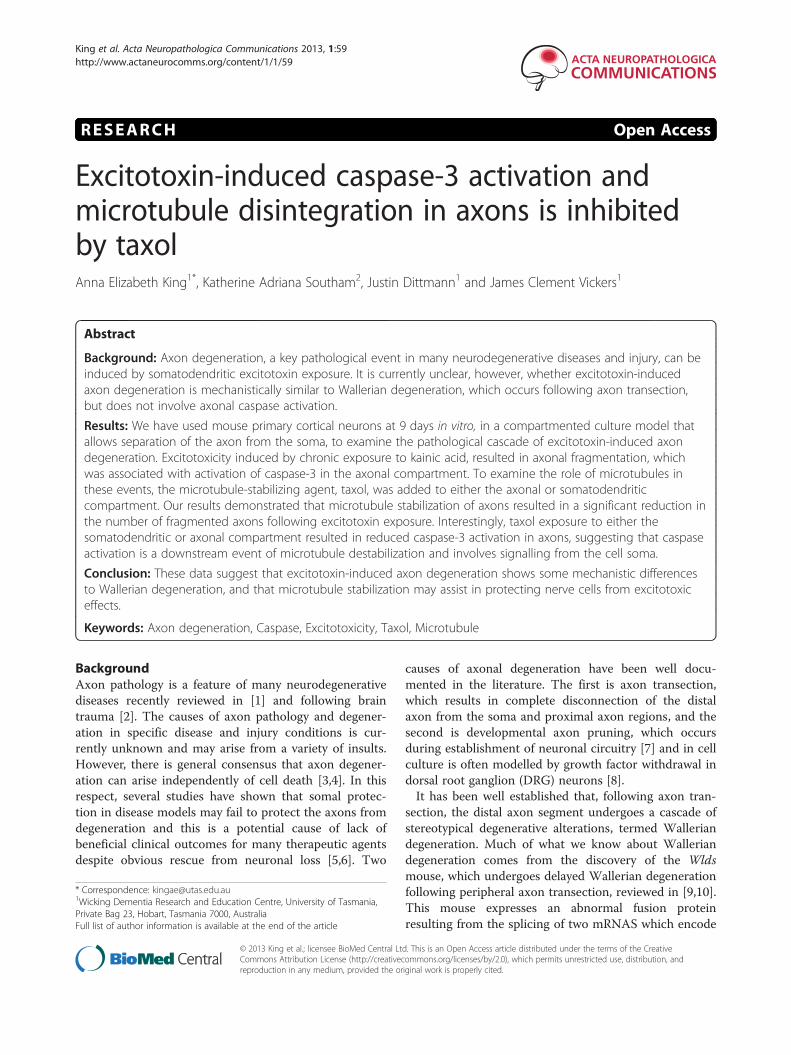
kainic acid exposure. Immunocytochemical analysis withan antibody against activated caspase-3 demonstratedthat exposure to kainic acid resulted in induction of ac-tive caspase-3 in axons at 16–18 hours post treatment(Figure 5a,b). Immunoreactivity was present as regularlyspaced puncta along the length of the axon and waspresent in axons in both the axonal and somatodendritic
chambers. Caspase immunoreactivity was not present inMAP2 immunoreactive dendrites. To determine the sta-ging of activation of caspase-3 following kainic acid ex-posure, cultures were treated and fixed over atimecourse of 1 and 6 hours. Increased numbers ofcaspase immunoreactive axons were not present at1 hour post kainic acid exposure (Figure 5a). At 6 hoursfollowing kainic acid exposure there was a non-significant trend towards increase in caspase activation,
Figure 5 Excitotoxicity induces activation of caspase 3 which isprevented by microtubule stabilization. a. Active caspase-3immunoreactivity was low in control (vehicle) treated chambers butwas present in many axons as discrete puncta following kainic acid.Caspase immunoreactivity was not altered at 1 hour following kainicacid exposure but was significantly increased at 16 hrs. b,c. Kainicacid induced active caspase-3 immunoreactivity in the axoncompartment was significantly attenuated by either somatodendriticor axonal taxol treatment. KA, kainic acid. * Significantly differentfrom control. # Significantly different from kainic acid treatment.P < 0.05. Mean values shown ± SEM. Scale bar: 20 μm.
King et al. Acta Neuropathologica Communications 2013, 1:59 Page 6 of 9http://www.actaneurocomms.org/content/1/1/59
which was variable between cultures, indicating thatcaspase activation occurs at a later stage of the degen-erative process.
Microtubule destabilization following kainic acidexposure is upstream of caspase activationWe next determined if axonal caspase activation occuredprior to, or resulted from, microtubule destabilization.Kainic acid-treated cultures were co-incubated withtaxol in the axonal or somatodendritic compartments(as described above) and fixed after 16–18 hours.Immunolabelling with active caspase-3 demonstratedthat axonal taxol prevented kainic acid induced activa-tion of caspase-3 in the axonal compartment with a sig-nificant reduction in caspase immunofluorescence intaxol-treated cultures (51.3 ± 14.7 × 10⁻³% of total area,mean ± SEM) relative to cultures exposed to kainic acidin the absence of axonal taxol (227.9 ± 24.5 × 10⁻³% oftotal area, mean ± SEM Figure 5b,c). This suggests thatthat caspase 3 activation occurs downstream of micro-tubule destabilization in the axon compartment. Interestingly,
somatodendritic taxol application also significantly re-duced activation of caspase-3 by kainic acid (28.5 ±8.8 ×10⁻³% of total area, mean ± SEM), (Figure 5b,c)suggesting that somatodendritic signalling is involvedin the activation of caspase in the axon.
DiscussionOur previous studies and those of others have demon-strated that excitotoxicity can induce axon degeneration.However, the relationship between excitotoxin-inducedaxon degeneration and other forms of axon degenerationremain unclear. In this study we have investigatedwhether excitotoxiciy induces activation of axonalcaspases as this has been implicated in developmentalaxon pruning, but is not involved in Wallerian degener-ation following axon severing. Our study demonstratedthat excitotoxicity results in activation of caspase-3within the axon, suggesting a mechanistic difference toWallerian-type degeneration. To investigate the relation-ship between cytoskeletal breakdown and caspase activa-tion, we exposed cultures to excitotoxins in the presenceor absence of the microtubule stabilizing agent, taxol,in the axonal or somatodendritic compartment, andexamined the effect on axon fragmentation, axonalcaspase activation and loss of dendritic MAP2. Ourdata demonstrated that application of axonal, but notsomatodendritic, taxol, prevented axonal fragmenta-tion. Furthermore both axonal and somatodendritictaxol prevented the activation of axonal caspase-3, al-though MAP2 loss was not altered. These data implythat caspase activation occurs downstream of micro-tubule destabilization within the axon and involvessomal signalling.
Axonal caspase 3 activation in axon degenerationCaspases are cysteine proteases that are important inmediating apoptosis. However, recently caspases havebeen implicated in other cellular processes that are notassociated with cell death including synaptic [36] andaxon [8,37,38] pruning. Caspase activation in the axonhas been associated with a number of neurodegenerativediseases and condition. For example, axonal caspase hasbeen associated with beta amyloid toxicity [37] and isfound following injury [39]. Furthermore in models ofKrabbes disease, which is caused by deficiency ingalactosylsphingosine and characterized by demyelin-ation, caspase activation in the axon, but not the soma,is present prior to demyelination [40]. These data sug-gest that axonal caspase activation in these conditionsrepresents a type of axon degeneration that is differentfrom classical Wallerian degeneration.Despite these finding the axonal role of caspases re-
main unclear. Caspases have a number of substrates, in-cluding cytoskeletal proteins, which could account for
King et al. Acta Neuropathologica Communications 2013, 1:59 Page 7 of 9http://www.actaneurocomms.org/content/1/1/59
their activation. Both precaspase-3 and biochemically ac-tive caspase-3 have been shown to be present in healthyaxons of cultured neurons [38] although the epitope ofactive caspase-3 may be masked by binding to apoptosisinhibitors as it can not be detected by immunohisto-chemistry. However, active caspase-3 immunoreactivitywas localized to tubulin aggregates following nervegrowth factor withdrawal [38], suggesting releasefrom their inhibitors during degeneration. This isconsistent with our findings of punctate expressionof active caspase-3 following excitotoxicity. Growthfactor deprivation may be involved in axonal degener-ation following excitotoxicity as excitotoxicity has beenshown to disrupt axon transport mechanisms and can re-sult in the disruption and cleavage of components of thedynein- dynactin complex [41]. These molecular motorsare responsible for retrograde transport and signaling,thus specifically implicating reduced growth factor signal-ing from the axon terminal [41].Caspase activation following growth factor withdrawal
is eliminated by knockdown of Bax [37,38], althoughpan caspase inhibitors do not provide complete protec-tion against axon degeneration [38]. In this respect,caspase activation may act in parallel with other axondegeneration pathways involving NAD+ in this model[38]. This is similar to our previous findings in whichcortical neurons treated with glutamate underwent axondegeneration that was partially protected by pan caspaseinhibitors [27]. This suggests that caspase activation isnot the only axon degeneration mechanisms activated inthese pathways and also fits with our current study dem-onstrating that preventing axonal caspase activation byapplication of somatodendritic taxol does not preventaxon fragmentation. Thus, although the role of caspase-3 activation in axon degeneration is unclear, it may notbe critical to degeneration and alterations to micro-tubule proteins may be more important in axonalmaintenance.
The role of microtubule destabilization in excitotoxicininduced axon degenerationMicrotubule fragmentation has previously been implicatedin axonal degeneration mechanisms and was the earliestdetectable change in axons following severing when exam-ining cytoskeletal alterations [20]. Breakdown of the micro-tubule network in Wallerian degeneration involves theubiquitin proteasome system. However, while this is anearly event relative to cytoskeletal breakdown, the role ofmicrotubule dynamics in the initial intracellular biochem-ical cascade that results in the breakdown of the cytosol,remain unclear. An early study demonstrated the protect-ive role of microtubule stabilization with taxol on hippo-campal neurons exposed to kainic acid, [42]. Similarly themicrotubule stabilizing agent davunetide protects against
kainic acid excitotoxicity in hippocampal cells both in vitroand in vivo [43]. However in these studies taxol was glo-bally applied to neurons and protection attributed to pre-vention of calcium influx through calcium permeableAMPA receptors [42]. The role of microtubules in theaxonal compartment was not examined. Interestingly inour study, somatodendritic taxol did not significantly pro-tect the unexposed axon compartment, although a trend toreduced fragmentation was observed. This may reflect dif-ferences in the neurons cultured or suggest that axonaltaxol was also contributing to axonal protection in the pre-vious study.Recently protective effects of axonal taxol have been
reported in neurons exposed to 3 hours glutamate,strengthening our current results and suggesting thatmicrotubule destabilization is an early and upstreamevent in axon degeneration following excitotoxicity [41].Similarly several studies have now demonstrated protect-ive effects of microtubule stabilization or increasedmicrotubule acetylation in other models involving axonaldegeneration such as mutant tau induced axon degener-ation [44-46] and models of ALS [47]. In this respectmicrotubule stabilization protects neurons from activa-tion of cdk5 and subsequent tau hyperphosphorylationin a cell culture model of amyloid toxicity [48] againsuggesting that microtubule destabilization is an up-stream event. Similarly, studies looking at dendritic de-generation following NMDA insult demonstrate thatmicrotubule stabilization is upstream of calpain activa-tion and loss of MAP2, suggesting that loss of MAP2 isnot the cause of microtubule destabilization in this in-stance [49]. However in our study somatodendriticmicrotubule stabilization did not prevent MAP2 loss fol-lowing kainic acid exposure perhaps reflecting differencein the excitotoxic pathways activated or the length ofexposure.Although microtubule stabilization is protective against
degeneration in a number of models, overstabilization ofmicrotubules may also be detrimental as seen in cases ofperipheral neuropathy in cancer patients treated therapeut-ically with taxol [50]. Furthermore mutations in smallheatshock proteins in Charcot Marie tooth disease causethem to bind more strongly to microtubules and alter-ations in microtubule dynamics may be the cause of per-ipheral neuropathy in this disease. Thus precise balance inmicrotubule stability may be necessary to prevent axonaldegeneration mechanisms.
Somal- axonal signaling in axon degenerationThe effect of compartment specific microtubule stabilizationis of interest in the current study. We show that stabilizationof axonal microtubules significantly reduced caspase-3activation within the axon suggesting that microtubuledestabilization is required for caspase activation. However,

King et al. Acta Neuropathologica Communications 2013, 1:59 Page 8 of 9http://www.actaneurocomms.org/content/1/1/59
the effect of somatodendritic taxol on axonal caspase acti-vation suggests that axonal microtubule destabilizationalone is not sufficient for caspase activation, but that sig-nalling from the soma to axon may be involved. The na-ture of this signal is currently unclear, however a recentstudy by Chen and colleagues [19] demonstrated that fol-lowing growth factor withdrawal, spatially distinct kinasepathways are activated that differentially affect axonalbeading and degeneration. In particular GSK3 activationor transcriptional repression in the soma was suffi-cient to block axonal degeneration but not axonalbeading, whereas ErbB and p38 inhibition in the axonprevented both beading and fragmentation [19]. Thesedata suggest that axon and soma may regulate differ-ent aspects of axonal degeneration and that in ourexcitotoxicity model these may be regulated in partby microtubule dynamics. This strengthens the hypothesisthat parallel degeneration pathways are involved inexcitotoxin induced axon degeneration. Despite this, al-though an early event, microtubule stabilization is not suf-ficient to prevent cleavage of components of the dynein-dynactin complex [41].
ConclusionThe current study suggests that excitotoxin induced axondegeneration involves features of both Wallerian degener-ation and growth factor withdrawal and that alteration inmicrotubule dynamics are an upstream event in axon de-generation. Further deciphering the alterations to micro-tubule dynamics in relation to their post translationalmodifications, binding of microtubule associated proteinsand association with downstream signalling pathways willbe important in providing effective therapeutic interven-tion in a number of degenerative diseases and conditionsin the central nervous system.
Competing interestThe authors declare that they have no conflict of interest for this manuscript.
Authors’ contributionsAK designed and performed the experiments, analysed data and drafted themanuscript. KS performed analysis on images from cultures. JD preparedmicrofluidic cultures. JV aided in study design and manuscript preparation.All authors read and approved the final manuscript.
AcknowledgementsThis research was supported by the Motor Neuron Disease Research Instituteof Australia, Alzheimer’s Australia Research, National Health and MedicalResearch Council grant (APP1003931) and the JO and JR Wicking trust.
Author details1Wicking Dementia Research and Education Centre, University of Tasmania,Private Bag 23, Hobart, Tasmania 7000, Australia. 2Menzies Research Institute,University of Tasmania, Hobart, Tasmania 7000, Australia.
Received: 8 July 2013 Accepted: 1 September 2013Published: 9 September 2013
References1. Adalbert R, Coleman MP: Axon pathology in age-related
neurodegenerative disorders. Neuropathol Appl Neurobiol 2012,10(10):1365–2990.
2. Smith DH, Hicks R, Povlishock JT: Therapy development for diffuse axonalinjury. J Neurotrauma 2013, 30(5):307–323.
3. Finn JT, et al: Evidence that Wallerian degeneration and localized axondegeneration induced by local neurotrophin deprivation do not involvecaspases. J Neurosci 2000, 20(4):1333–1341.
4. Osterloh JM, et al: dSarm/Sarm1 is required for activation of an injury-induced axon death pathway. Science 2012, 337(6093):481–484.
5. Ferri A, et al: Inhibiting axon degeneration and synapse loss attenuatesapoptosis and disease progression in a mouse model of motoneurondisease. Curr Biol 2003, 13(8):669–673.
6. Gould TW, et al: Complete dissociation of motor neuron death frommotor dysfunction by Bax deletion in a mouse model of ALS.J Neurosci 2006, 26(34):8774–8786.
7. Luo L, O’Leary DD: Axon retraction and degeneration in developmentand disease. Annu Rev Neurosci 2005, 28:127–156.
8. Simon DJ, et al: A caspase cascade regulating developmental axondegeneration. J Neurosci 2012, 32(49):17540–17553.
9. Lunn ER, et al: Absence of wallerian degeneration does not hinderregeneration in peripheral nerve. Eur J Neurosci 1989, 1(1):27–33.
10. Wang JT, Medress ZA, Barres BA: Axon degeneration: molecularmechanisms of a self-destruction pathway. J Cell Biol 2012, 196(1):7–18.
11. Mack TG, et al: Wallerian degeneration of injured axons and synapses isdelayed by a Ube4b/Nmnat chimeric gene. Nat Neurosci 2001,4(12):1199–1206.
12. Samsam M, et al: The Wlds mutation delays robust loss of motor andsensory axons in a genetic model for myelin-related axonopathy.J Neurosci 2003, 23(7):2833–2839.
13. Howell GR, et al: Axons of retinal ganglion cells are insulted in the opticnerve early in DBA/2J glaucoma. J Cell Biol 2007, 179(7):1523–1537.
14. Mi W, et al: The slow Wallerian degeneration gene, WldS, inhibits axonalspheroid pathology in gracile axonal dystrophy mice. Brain 2005,128(Pt 2):405–416.
15. Vande Velde C, et al: The neuroprotective factor Wlds does not attenuatemutant SOD1-mediated motor neuron disease. Neuromolecular Med 2004,5(3):193–203.
16. Rose FF Jr, et al: The Wallerian degeneration slow (Wld(s)) gene does notattenuate disease in a mouse model of spinal muscular atrophy.Biochem Biophys Res Commun 2008, 375(1):119–123.
17. Kariya S, et al: The neuroprotective factor Wld(s) fails to mitigate distalaxonal and neuromuscular junction (NMJ) defects in mouse models ofspinal muscular atrophy. Neurosci Lett 2009, 449(3):246–251.
18. Parson SH, Mackintosh CL, Ribchester RR: Elimination of motor nerveterminals in neonatal mice expressing a gene for slow walleriandegeneration (C57Bl/Wlds). Eur J Neurosci 1997, 9(8):1586–1592.
19. Chen M, et al: Spatially coordinated kinase signaling regulates local axondegeneration. J 2012, 32(39):13439–13453.
20. Zhai Q, et al: Involvement of the ubiquitin-proteasome system in theearly stages of wallerian degeneration. Neuron 2003, 39(2):217–225.
21. Chung RS, et al: Glutamate induces rapid loss of axonal neurofilamentproteins from cortical neurons in vitro. Exp Neurol 2005, 193(2):481–488.
22. King AE, et al: Excitotoxicity mediated by non-NMDA receptors causesdistal axonopathy in long-term cultured spinal motor neurons.Eur J Neurosci 2007, 26(8):2151–2159.
23. Underhill SM, Goldberg MP: Hypoxic injury of isolated axons isindependent of ionotropic glutamate receptors. Neurobiol Dis 2007,25(2):284–290.
24. Saggu SK, et al: The spatiotemporal pattern of somal and axonalpathology after perikaryal excitotoxic injury to retinal ganglion cells: ahistological and morphometric study. Exp Neurol 2008, 211(1):52–58.
25. Saggu SK, et al: Wallerian-like axonal degeneration in the optic nerveafter excitotoxic retinal insult: an ultrastructural study. BMC Neurosci 2010,11:p. 97.
26. Hou ST, et al: CaMKII phosphorylates collapsin response mediator protein 2and modulates axonal damage during glutamate excitotoxicity.J Neurochem 2009, 111(3):870–881.
27. Hosie KA, et al: Chronic excitotoxin-induced axon degeneration in acompartmented neuronal culture model. ASN Neuro 2012, 4(1).

King et al. Acta Neuropathologica Communications 2013, 1:59 Page 9 of 9http://www.actaneurocomms.org/content/1/1/59
28. Palop JJ, et al: Aberrant excitatory neuronal activity and compensatoryremodeling of inhibitory hippocampal circuits in mouse models ofAlzheimer’s disease. Neuron 2007, 55(5):697–711.
29. Bogaert E, d’Ydewalle C, Van Den Bosch L: Amyotrophic lateral sclerosisand excitotoxicity: from pathological mechanism to therapeutic target.CNS Neurol Disord Drug Targets 2010, 9(3):p. 297–p. 304.
30. Mehta A, et al: Excitotoxicity: bridge to various triggers inneurodegenerative disorders. Eur J Pharmacol 2013, 698(1–3):6–18.
31. Kostic M, Zivkovic N, Stojanovic I: Multiple sclerosis and glutamateexcitotoxicity. Rev Neurosci 2013, 24(1):71–88.
32. Vickers JC, et al: Axonopathy and cytoskeletal disruption in degenerativediseases of the central nervous system. Brain Res Bull 2009,80(4–5):217–223.
33. King AE, Vickers JC: Excitotoxicity and axon degeneration. In Handbook ofneurotoxicity. Edited by Guillemin G. New York: Springer Science; 2013(Accepted).
34. Gundersen GG, Cook TA: Microtubules and signal transduction. Curr OpinCell Biol 1999, 11(1):81–94.
35. King AE, et al: Localization of glutamate receptors in developing corticalneurons in culture and relationship to susceptibility to excitotoxicity.J Comp Neurol 2006, 498(2):277–294.
36. D’Amelio M, et al: Caspase-3 triggers early synaptic dysfunction in amouse model of Alzheimer’s disease. Nat Neurosci 2011, 14(1):69–76.
37. Nikolaev A, et al: APP binds DR6 to trigger axon pruning and neurondeath via distinct caspases. Nature 2009, 457(7232):981–989.
38. Schoenmann Z, et al: Axonal degeneration is regulated by the apoptoticmachinery or a NAD + −sensitive pathway in insects and mammals.J Neurosci 2010, 30(18):6375–6386.
39. Chen XH, et al: Long-term accumulation of amyloid-beta, beta-secretase,presenilin-1, and caspase-3 in damaged axons following brain trauma.Am J Pathol 2004, 165(2):357–371.
40. Smith B, et al: Peripheral neuropathy in the twitcher mouse involves theactivation of axonal caspase 3. ASN Neuro 2011, 3(4).
41. Fujiwara T, Morimoto K: Cooperative effect of p150Glued andmicrotubule stabilization to suppress excitotoxicity-induced axondegeneration. Biochem Biophys Res Commun 2012, 424(1):82–88.
42. Furukawa K, Mattson MP: Taxol stabilizes [Ca2+]i and protectshippocampal neurons against excitotoxicity. Brain Res 1995,689(1):141–146.
43. Zemlyak I, et al: The microtubule interacting drug candidate NAPprotects against kainic acid toxicity in a rat model of epilepsy.J Neurochem 2009, 111(5):1252–1263.
44. Shemesh OA, Spira ME: Rescue of neurons from undergoing hallmark tau-induced Alzheimer’s disease cell pathologies by the antimitotic drugpaclitaxel. Neurobiol Dis 2011, 43(1):163–175.
45. Barten DM, et al: Hyperdynamic microtubules, cognitive deficits, andpathology are improved in tau transgenic mice with low doses of themicrotubule-stabilizing agent BMS-241027. J Neurosci 2012,32(21):7137–7145.
46. Xiong Y, et al: HDAC6 mutations rescue human tau-induced microtubuledefects in Drosophila. Proc Natl Acad Sci USA 2013, 110(12):4604–4609.
47. Taes I, et al: Hdac6 deletion delays disease progression in the SOD1G93Amouse model of ALS. Hum Mol Genet 2013, 30:30.
48. Li G, et al: Stabilization of the cyclin-dependent kinase 5 activator, p35,by paclitaxel decreases beta-amyloid toxicity in cortical neurons.J Neurochem 2003, 84(2):347–362.
49. Hoskison MM, Shuttleworth CW: Microtubule disruption, not calpain-dependent loss of MAP2, contributes to enduring NMDA-induceddendritic dysfunction in acute hippocampal slices. Exp Neurol 2006,202(2):302–312.
50. Quasthoff S, Hartung HP: Chemotherapy-induced peripheral neuropathy.J Neurol 2002, 249(1):9–17.
doi:10.1186/2051-5960-1-59Cite this article as: King et al.: Excitotoxin-induced caspase-3 activationand microtubule disintegration in axons is inhibited by taxol. ActaNeuropathologica Communications 2013 1:59.
Submit your next manuscript to BioMed Centraland take full advantage of:
• Convenient online submission
• Thorough peer review
• No space constraints or color figure charges
• Immediate publication on acceptance
• Inclusion in PubMed, CAS, Scopus and Google Scholar
• Research which is freely available for redistribution
Submit your manuscript at www.biomedcentral.com/submit