research open access effect of jaw opening on the stress
TRANSCRIPT

HEAD & FACE MEDICINE
Li et al. Head & Face Medicine 2014, 10:24http://www.head-face-med.com/content/10/1/24
RESEARCH Open Access
Effect of jaw opening on the stress pattern in anormal human articular disc: finite elementanalysis based on MRI imagesQihong Li1,3†, Shuang Ren2†, Cheng Ge3, Haiyan Sun3, Hong Lu3, Yinzhong Duan1 and Qiguo Rong2*
Abstract
Introduction: Excessive compressive and shear stresses are likely related to condylar resorption and discperforation. Few studies have reported the disc displacement and deformation during jaw opening. The aim of thisstudy was to analyze stress distribution in a normal articular disc during the jaw opening movement.
Methods: Bilateral MRI images were obtained from the temporomandibular joint of a healthy subject for the jawopening displacement from 6 to 24 mm with 1 mm increments. The disc contour for the jaw opening at 6 mmwas defined as the reference state, and was used to establish a two dimensional finite element model of the disc.The contours of the disc at other degrees of jaw opening were used as the displacement loading. Hyperelasticmaterial models were applied to the anterior, intermediate and posterior parts of the disc. Stress and straintrajectories were calculated to characterize the stress/strain patterns in the disc.
Results: Both the maximum and minimum principal stresses were negative in the intermediate zone, therefore, theintermediate zone withstood mainly compressive stress. On the contrary, the maximum and minimum principal stresseswere most positive in the anterior and posterior zones, which meant that the anterior and posterior bands suffered highertensile stresses. The different patterns of stress trajectories between the intermediate zone and the anterior and posteriorbands might be attributed to the effect of fiber orientation. The compression of the intermediate zone and stretching ofthe anterior and posterior bands caused high shear deformation in the transition region, especially at the disc surfaces.
Conclusions: The stress and strain remained at a reasonable level during jaw opening, indicating that the disc experiencesno injury during functional opening movements in a healthy temporomandibular joint.
Keywords: Temporomandibular joint disc, Finite element analysis, Stress trajectory, Jaw opening
IntroductionThe temporomandibular joint (TMJ), a load-bearingorgan in the human body, contains an articular disc lo-cated between the glenoid fossa and the condyle that,during mandibular movements, plays an important roleas a stress absorber during mouth function, resulting instress reduction and redistribution within the joint [1].The group of ‘temporomandibular disorders’ (TMD)
comprises a number of related clinical problems involv-ing pain and dysfunction of the masticatory system, thetemporomandibular joint and its associated structures.
* Correspondence: [email protected]†Equal contributors2Department of Mechanics and Engineering Science, College of Engineering,Peking University, Beijing 100871, ChinaFull list of author information is available at the end of the article
© 2014 Li et al.; licensee BioMed Central Ltd. TCommons Attribution License (http://creativecreproduction in any medium, provided the orDedication waiver (http://creativecommons.orunless otherwise stated.
The main cause of TMD has not yet been established[2], although functional overloading is considered to bea major etiological factor [1]. Indeed, excessive compres-sive and shear stresses are likely to be common sourcesof condylar resorption and disc perforation [3].Stress distribution in the TMJ is hard to measure ex-
perimentally, and is thus poorly understood. However, fi-nite element (FE) analysis is a promising research toolfor evaluating dental biomechanics [4]. It can be used toanalyze stress distribution patterns in the TMJ tissues afterapplication of force or deformation. Two-dimensional (2D)and three-dimensional (3D) FE models have been used tosimulate the in vivo biomechanics of the human TMJ[5-10]. Most previous studies focused on clenching behav-iors, since maximum TMJ loading occurs during forceful
his is an Open Access article distributed under the terms of the Creativeommons.org/licenses/by/4.0), which permits unrestricted use, distribution, andiginal work is properly credited. The Creative Commons Public Domaing/publicdomain/zero/1.0/) applies to the data made available in this article,

Li et al. Head & Face Medicine 2014, 10:24 Page 2 of 9http://www.head-face-med.com/content/10/1/24
clenching or aggressive episodes [11]. However, the TMJ isalso sub-maximally loaded during many other activities,such as drinking, screaming, biting, and masticatory open-ing and closing [12]. The condylar movement during thesevarious mandibular movements, especially jaw opening,produces remarkable ranges of disc mobility.Until now, very few studies have reported dynamic simu-
lation of the disc to explore disc displacement and deform-ation during jaw opening [12,13]. Biomechanical analysis ofmusculoskeletal system dynamics has been widely per-formed by applying rigid-body dynamics [14,15]. The dis-tribution of forces in irregularly-shaped joint structures,however, cannot be analyzed, and deformations of the ar-ticular disc cannot be taken into account. Recently,methods combining 3D imaging and motion-tracking data(both optoelectric and electromagnetic) were introduced tostudy temporomandibular joint (TMJ) kinematics [16].The combination of 3D TMJ anatomies and jaw trackingwith six degrees of freedom permits a subject-specific dy-namic analysis of TMJ loading during opening, closing andchewing. The location of the minimum intra-articularspace was thought to bear the greatest force, although thenature of the articular disc gives it uneven thickness and ir-regular patterns of deformation, meaning that this relation-ship between force transfer and minimum intra-articulardistance is somewhat ambiguous [12].The aim of this study was to analyze the strain/stress pat-
tern in the articular disc during jaw opening [1,6,9,10,17].The contours of the disc at different opening distanceswere used as the displacement loading for FE analysis.Since this study was mainly a methodological validationof the applicability of the displacement loading basedon MRI data, a 2D finite element model of the humanmandible disc was applied.
Materials and methodsMagnetic resonance imaging and model reconstructionBilateral MRI images were obtained from the TMJ of a 14-year-old female volunteer with no history of TMD. TheMRI machine used was a high intensity 1.5-T magnet(Signa, Excite, General Electric, Chalfont St. Giles, UnitedKingdom) and a 3-in dual surface coil. An optimized
Figure 1 Boundary tracing of the TMJ. (a) MRI image of the TMJ at 6 mdemarcated with white lines. (b) Diagrammatic representation of the conto
proton density weighed fast spin echo sequence (repetitiontime 1600 ms/echo time 13.39 ms) was used for scanning.A field of view (FOV) of 100 × 150 mm and a slice thick-ness of 2 mm were used. Informed consent was obtainedfrom the volunteer’s parents to participate in the study. Wepromise to protect life, health, dignity, integrity, right toself-determination, privacy, and confidentiality of personalinformation of research subject. Ethical permission hasbeen offered by Scientific research and clinical applicationof medical technology ethics committee of Afiliated Hos-pital of Academy of Military Medical Sciences with a refer-ence number of KY-2013-03-06. MRI was taken in thesagittal direction, perpendicular to the long axis of the con-dyle. A mouth gag was placed in the mouth before imageacquisition, and the subject was asked to close her man-dible onto the mouth gag. After one cycle of imaging, shewas asked to press a button on the gag to increase jawopening by 1 mm. Contiguous 2 mm-thick sagittal slices ofthe dentition were obtained at each opening distance. Sa-gittal MRI images of the same slice were selected for 19different opening distances at 1 mm increments. The mini-mum opening distance between central incisors is 6 mmby the mouth gag. The range of jaw opening was from 6–24 mm. The contours of the articular fossa, articular discand condyle were traced by a trained dentist (Figure 1).To make sure that slices from the same position, the
volunteer’s head was fixed in the same place. All MR im-ages were taken by the same slice. Besides, the imageswere registered in Matlab (version: 2010a, MathWorks,USA) by alignment of the articular fossa which is con-sidered no movement during jaw opening.
Finite element modeling and analysisThe contour of the disc with the jaw open at 6 mm was de-fined as the reference state, and was used to establish a twodimensional finite element model of the disc. The initialstress state of the FE model was assumed to be zero. Allother disc contours were meshed in the same way as for theFE model. For a given opening distance, the difference in thecoordinates of the corresponding nodes (i.e. the displace-ment of the boundary nodes of the FE model) was usedas the displacement loading, as shown in Figure 2a.
m of jaw opening. The contours of the fossa, disc and condyle areurs of the fossa, disc and condyle at jaw opening of 8, 16 and 24 mm.

Figure 2 Loading conditions and zoning of the disc. (a) Loading conditions of the disc, obtained by the displacement of corresponding nodes onthe boundary of disc in the reference state (6 mm opening; grey outline) and the disc in the current states at varying distances of opening (7–24 mm;white outline). (b) Diagram showing the fifteen zones analyzed in this study, comprising posterior (P), medial-posterior (M-P), medial (M), medial-anterior(M-A) and anterior (A) positions in each of the superior (S), middle (M) and inferior (I) layers. The central line represents the path used to evaluate thestretch of the disc.
Table 1 Material data of the anterior, intermediate andposterior parts of the disc
Strain Stress (MPa) AB/ PB Stress (MPa) IZ
0 0 0,0
0.01 0.0233 0.0311
0.02 0.0506 0.0638
0.03 0.0801 0.0988
0.04 0.1268 0.1524
0.05 0.1999 0.2186
0.06 0.2862 0.3267
0.08 0.6143 0.6628
0.1 0.9284 0.9815
0.13 1.1316 1.351
0.15 1.2979 1.6351
0.17 1.388 1.7575
0.2 1.4688 1.8083
AB: anterior band; PB: posterior band; IZ: intermediate zone.
Li et al. Head & Face Medicine 2014, 10:24 Page 3 of 9http://www.head-face-med.com/content/10/1/24
Altogether, 18 simulations (jaw opening range: 7–24 mm) were performed. The stress analysis was exe-cuted by the FE software, ANSYS 14.0 from ANSYSInc. (Houston, USA).The TMJ disc is a dense fibrocartilaginous structure
[1] and its mechanical behavior has been proven to benonlinear, anisotropic and time-dependent [18-20]. Inaddition, there are differences in the mechanical behav-ior of the anterior, posterior and medial parts of the disc[10]. In recent studies, Perez [6] developed an accurateTMJ model using a fiber-reinforced porohyperelasticmaterial for the articular disc. However, the constants forthis material model were obtained from dogs. In thecurrent study, we also used a hyperelastic model of theTMJ disc, described by the experimental response function.For hyperelastic materials, the stress–strain relationshipderives from a strain energy density function:
Sij ¼ ∂W∂Eij
where Sij is a component of the second Piola-Kirchhoffstress tensor; W is the strain energy density: and Eij is acomponent of the Lagrangian strain tensor.The Cauchy stress components of a volumetrically
constrained material can be shown to be [21]:
σij ¼ −pδij þ dev 2∂W∂I1
bij−2Iα∂W∂I2
b−1ij
� �
where δij is the Kronecker delta; p is the pressure; andbij is the left Cauchy-Green deformation tensor. Thedeviatoric stress is therefore determined solely by the de-formation and the response functions (derivatives ∂W
∂I1, ∂W
∂I2and ∂W
∂J ), which are determined analytically for the hypere-lastic potentials in isotropic and anisotropic hyperelasticity.The parameters of the response functions (shown in
Table 1) were obtained from the experimental data re-ported by Kang [18], where Kang and coworkers per-formed tensile tests of the anterior, intermediate andposterior bands along the medial-lateral axis of 13
human TMJ discs. Considering the fiber directions inthe disc, the mechanical properties of the anterior andposterior bands can be characterized with data obtainedfrom the intermediate zone, since the fibers of the inter-mediate zone run perpendicular to the axis of the tensiletest [10]. Conversely, the elastic constants related to theintermediate zone can be obtained from tests conductedon tissue in the anterior band. Since tests performed onthe posterior band were along the fibers, it was expectedto be stiffer than the intermediate zone; however, itturned out to be softer. This suggests that the test mayhave been conducted too close to the end of the disc, sowe eliminated the posterior band data. Since no com-pression test data can be found in literatures, the discwas assumed to have the same material behavior incompression and tension.To evaluate stress variation in the anterior and poster-
ior bands and the intermediate zone, a path was definedin the articular disc. In addition, the disc was divided

Figure 3 Trajectory of the disc center during jaw opening.
Li et al. Head & Face Medicine 2014, 10:24 Page 4 of 9http://www.head-face-med.com/content/10/1/24
into 15 zones to characterize the stress/strain patternsduring jaw opening (Figure 2b).
ResultsFigure 3 shows the positions of the disc center duringthe jaw opening process. These appeared to be distrib-uted randomly and the movement of the disc center wasnot strongly correlated with the opening distance. Afterfitting, the trajectory of the disc center was ovoid inshape. This result shows that the opening distance doesnot describe the disc position on its own. Although jawopening is a continuous movement, the disc center mightundergo intermittent and rapid slippage or displacement.
Figure 4 Stress trajectories of the disc at jaw opening of 7, 12, 17 and
Indeed, the disc center jumped suddenly when the jawopened from 15 mm to 16 mm, perhaps as a consequenceof friction between the disc and the condyle.Figure 4 shows the stress trajectories of the disc at jaw
opening of 7, 12, 17 and 22 mm, and the correspondingstress distributions are shown in Figure 5. Except at7 mm, where the stress level was relatively low, thestress trajectories had similar patterns in each condition.It can be observed that both the maximum and mini-mum principal stresses were negative in the intermediatezone. Stress trajectories run vertically and horizontally,with the disc bearing mainly the vertical pressure. Themaximum and minimum principal stresses were mostpositive in the anterior and posterior bands. The tensilestresses in the anterior band may be caused by stretchingof the disc by the lateral pterygoid muscle, while that inthe posterior band may be caused by contracting of thebilaminar zone. The transition from compressive stress inthe intermediate zone to tensile stress in the anterior andposterior bands did not occur smoothly, instead being ac-complished at a singular point in the transition area.Figure 6 shows the pattern of stresses on the disc along
the posterior-anterior path at jaw opening of 18 mm. Thefitted curves of the maximum and minimum principalstresses appeared to be funnel-shaped, confirming that theposterior and anterior bands were stretched, whereas theintermediate zone bore compressive stress. The shear
22 mm (MPa).
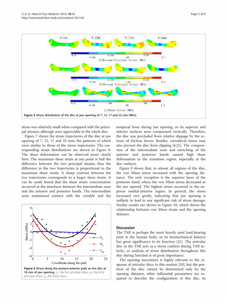
Figure 5 Stress distribution of the disc at jaw opening of 7, 12, 17 and 22 mm (MPa).
Li et al. Head & Face Medicine 2014, 10:24 Page 5 of 9http://www.head-face-med.com/content/10/1/24
stress was relatively small when compared with the princi-pal stresses, although were appreciable in the whole disc.Figure 7 shows the strain trajectories of the disc at jaw
opening of 7, 12, 17 and 22 mm, the patterns of whichwere similar to those of the stress trajectories. The cor-responding strain distributions are shown in Figure 8.The shear deformation can be observed more clearlyhere. The maximum shear strain at any point is half thedifference between the two principal strains; thus thedifference in the two trajectories is proportional to themaximum shear strain. A sharp contrast between thetwo trajectories corresponds to a larger shear strain. Itcan be easily found that the shear strain concentrationoccurred at the interfaces between the intermediate zoneand the anterior and posterior bands. The intermediatezone maintained contact with the condyle and the
Figure 6 Stress along the postero-anterior path on the disc at18 mm of jaw opening. σ1: the first principal stress, σ3: the thirdprincipal stress, τxy: the shear stress.
temporal bone during jaw opening, so its superior andinferior surfaces were compressed vertically. Therefore,the disc was precluded from relative slippage by the ac-tions of friction forces. Besides, retrodiscal tissue mayalso prevent the disc from slipping [6,21]. The compres-sion of the intermediate zone and stretching of theanterior and posterior bands caused high sheardeformation in the transition region, especially at thedisc surfaces.Figure 9 shows that, in almost all regions of the disc,
the von Mises stress increased with the opening dis-tance. The only exception is the superior layer of theposterior band, where the von Mises stress decreased asthe jaw opened. The highest stress occurred in the su-perior medial-anterior region. In general, the stressincreased very gently, indicating that jaw opening isunlikely to lead to any significant risk of stress damage.Similar results are shown in Figure 10, which shows therelationship between von Mises strain and the openingdistance.
DiscussionThe TMJ is perhaps the most heavily used load-bearingjoint in the human body, so its biomechanical balancehas great significance to its function [21]. The articulardisc in the TMJ acts as a stress cushion during TMJ ac-tivity, so analysis of stress distribution throughout thisdisc during function is of great importance.The opening movement is highly relevant to the re-
sponse of articular discs to this motion [10], but the pos-ition of the disc cannot be determined only by theopening distance; other influential parameters are re-quired to describe the configuration of this disc. In
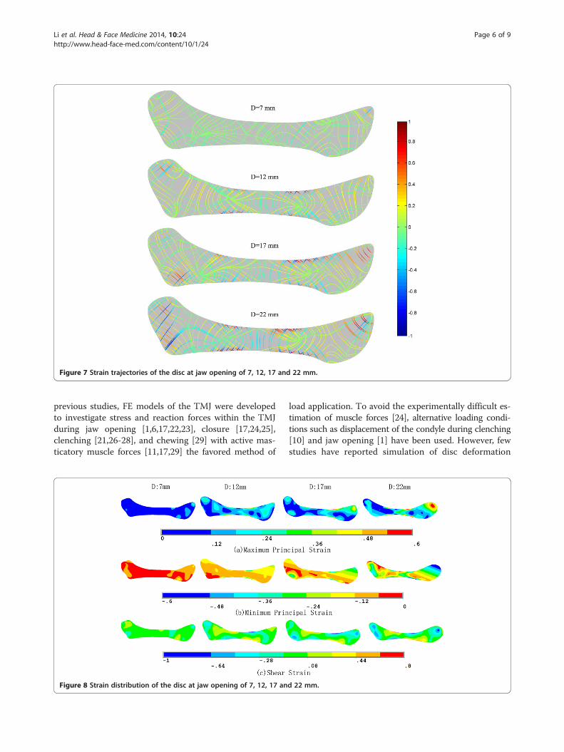
Figure 7 Strain trajectories of the disc at jaw opening of 7, 12, 17 and 22 mm.
Li et al. Head & Face Medicine 2014, 10:24 Page 6 of 9http://www.head-face-med.com/content/10/1/24
previous studies, FE models of the TMJ were developedto investigate stress and reaction forces within the TMJduring jaw opening [1,6,17,22,23], closure [17,24,25],clenching [21,26-28], and chewing [29] with active mas-ticatory muscle forces [11,17,29] the favored method of
Figure 8 Strain distribution of the disc at jaw opening of 7, 12, 17 an
load application. To avoid the experimentally difficult es-timation of muscle forces [24], alternative loading condi-tions such as displacement of the condyle during clenching[10] and jaw opening [1] have been used. However, fewstudies have reported simulation of disc deformation
d 22 mm.

Figure 9 Relationship of von Mises stress and the opening distance. Von Mises stress in the superior (S), middle (M) and inferior (I) disc layers isplotted against the opening distance.
Li et al. Head & Face Medicine 2014, 10:24 Page 7 of 9http://www.head-face-med.com/content/10/1/24
during jaw opening that uses the disc contours as thedisplacement loading. In the present study, we observeddisplacement of the disc boundary by measuring thecontours of the disc in MRI images.The mechanical behavior of the TMJ disc, when investi-
gated experimentally in humans [18], dogs [19] and pigs [20],was found to be nonlinear, anisotropic and time-dependent,and varied between different regions of the disc. However, inmost previous studies [1,5,6,10,23,24], the material propertiesof the disc were considered as entirely homogeneous. Re-cently, Perez [6] developed an accurate TMJ model that useda fiber-reinforced porohyperelastic constitutive model forthe disc, where the constants for material models wereextrapolated from tensile tests in dogs [19].Similarly, an experimental response function was applied
in this study to describe the hyperelastic property of thedisc. Parameters of the response function were obtainedfrom tensile tests of human TMJ discs [15]. The incom-pressibility of the disc was not considered in this study,since the jaw opening here was a quasistatic process,the water seepage in the disc was therefore neglected. Thestress/strain trajectory patterns show that the fiber
Figure 10 Relationship of von Mises strain and the opening distance. Vplotted against the opening distance.
orientation has a significant influence on disc deformation,meaning that it is reasonable to consider the effects of fiberorientation and distribution in the disc. Because of itshyperelasticity, the disc endures high strains and relativelylow stresses. During jaw opening, the intermediate zonebears mainly compressive stress, in agreement with previ-ous studies [6]. Conversely, the anterior and posteriorbands bear mainly tensile stress. Other studies [30,31] havefound the same situation during clenching. These resultsindicate that the function of the disc is companied by com-bined impact of stretching of the ligaments and compres-sion of the condyle and the articular fossa. The von Misesstress and strain increase gradually with the openingdistance, but remain at a reasonable level, indicating thatthe opening movement does no harm to the disc, consist-ent with expectations for the performance of a healthy disc.Likewise, Tanaka [22] also found that the von Mises stressincreased in the disc as jaw opening progressed. The onlyexception in the present study is the superior layer ofthe posterior band. This may be caused by a big clockwiserotation of the disc, and therefore reduce the stress inthat zone.
on Mises strain in the superior (S), middle (M) and inferior (I) disc layers is

Li et al. Head & Face Medicine 2014, 10:24 Page 8 of 9http://www.head-face-med.com/content/10/1/24
Some studies have reported that perforation of thedisc may arise due to high shear stresses [32-34]. Oursimulation results show that the highest shear stress oc-curs at the boundaries of the middle-anterior andmiddle-posterior regions. This may be due to the tran-sition of the loading conditions from compressive inthe intermediate zone to tensile in the anterior andposterior bands.There are some limitations in the present study. Only
oblique sagittal jaw displacement has been consideredusing this 2D FE model, so our results are not as vividas they might have been with a 3D model. Also, somesimplifications were made with respect to the displace-ment loading: displacement of the disc boundary wasobtained from the corresponding node pairs of the twodisc configurations, which may lead to stress concentra-tion on parts of the boundary. However, most simulationresults are highly reasonable.The present work represents an innovative trial of a
more accurate technique for predicting the stress re-sponse during similar motion problems, not only fordiscs but also for other organs, tissues and joints in thehuman body.
ConclusionThe simulations showed that the highest compressive stressoccurred in the intermediate zone, whereas the anteriorand posterior bands experienced mainly tensile stress. Fiberorientation had a significant effect on the stress/strain pat-terns. The stress and strain increased slightly with theopening distance, but were remarkably stable. The highestshear stress was at the interfaces between the medial-anterior and medial-posterior zones. Generally, the stressand strain remained at a reasonable level during jaw open-ing, indicating that the disc experiences no injury duringfunctional opening movements in a healthy joint.
Competing interestsThere are no potential competing interest to disclose.
Authors’ contributionsQR and YD initiated the investigation and designed the study. QL tracedcontours of the temporomandibular joint. SR preformed the finite elementanalysis. QL and SR drafted the manuscript. QR and YD critically reviewedand revised the manuscript. CG, HS, HL participated in MR image acquisitionand analysis. All authors read and approved the final manuscript.
AcknowledgementsThis study was supported by Beijing Natural Science Foundation Grants7133248 and 3122020.
Author details1Department of Orthodontics, School of Stomatology, Fourth Military MedicalUniversity, Xi’an 710032, China. 2Department of Mechanics and EngineeringScience, College of Engineering, Peking University, Beijing 100871, China.3Department of Stomatology, Afiliated Hospital of Academy of MilitaryMedical Sciences, Beijing 100071, China.
Received: 2 April 2014 Accepted: 13 June 2014Published: 19 June 2014
References1. Tanaka E, del Pozo R, Tanaka M, Asai D, Hirose M, Iwabe T, Tanne K:
Three-dimensional finite element analysis of human temporomandibularjoint with and without disc displacement during jaw opening. Med EngPhys 2004, 26:503–511.
2. Greene CS: Etiology of temporomandibular disorders. Semin Orthod 1995,1:222–228.
3. Arnett GW, Milam SB, Gottesman L: Progressive mandibular retrusion-idiopathic condylar resorption. Part II. Am J Orthod Dentofacial Orthop1996, 110:117–127.
4. Hannam AG: Current computational modelling trends incraniomandibular biomechanics and their clinical implications.J Oral Rehabil 2011, 38:217–234.
5. Beek M, Koolstra J, Van Ruijven L, Van Eijden T: Three-dimensional finiteelement analysis of the human temporomandibular joint disc. J Biomech2000, 33:307–316.
6. del Palomar Perez A, Doblare M: An accurate simulation model ofanteriorly displaced TMJ discs with and without reduction. Med Eng Phys2007, 29:216–226.
7. DeVocht JW, Goel VK, Zeitler DL, Lew D: A study of the control of discmovement within the temporomandibular joint using the finite elementtechnique. J Oral Maxil Surg 1996, 54:1431–1437.
8. Chen J, Akyuz U, Xu L, Pidaparti RM: Stress analysis of the humantemporomandibular joint. Med Eng Phys 1998, 20:565–572.
9. Donzelli PS, Gallo LM, Spilker RL, Palla S: Biphasic finite element simulationof the TMJ disc from in vivo kinematic and geometric measurements.J Biomech 2004, 37:1787–1791.
10. del Palomar Perez A, Doblare M: The effect of collagen reinforcement inthe behaviour of the temporomandibular joint disc. J Biomech 2006,39:1075–1085.
11. Hirose M, Tanaka E, Tanaka M, Fujita R, Kuroda Y, Yamano E, van Eijden TM,Tanne K: Three-dimensional finite-element model of the humantemporomandibular joint disc during prolonged clenching. Eur J Oral Sci2006, 114:441–448.
12. Tuijt M, Koolstra JH, Lobbezoo F, Naeije M: Differences in loading of thetemporomandibular joint during opening and closing of the jaw.J Biomech 2010, 43:1048–1054.
13. Koolstra JH, van Eijden TM: Combined finite-element and rigid-bodyanalysis of human jaw joint dynamics. J Biomech 2005, 38:2431–2439.
14. Peck CC, Langenbach GE, Hannam AG: Dynamic simulation of muscle andarticular properties during human wide jaw opening. Arch Oral Biol 2000,45:963–982.
15. McLean SG, Su A, van den Bogert AJ: Development and validation of a3-D model to predict knee joint loading during dynamic movement.J Biomech Eng 2003, 125:864–874.
16. Palla S, Gallo LM, Gossi D: Dynamic stereometry of thetemporomandibular joint. Orthod Craniofac Res 2003, 6(Suppl 1):37–47.
17. Cheng HY, Peng PW, Lin YJ, Chang ST, Pan YN, Lee SC, Ou KL, Hsu WC:Stress analysis during jaw movement based on vivo computedtomography images from patients with temporomandibular disorders.Int J Oral Maxillofac Surg 2013, 42:386–392.
18. Kang H, Bao GJ, Qi SN: Biomechanical responses of humantemporomandibular joint disc under tension and compression. Int J OralMaxillofac Surg 2006, 35:817–821.
19. Shengyi T, Xu Y: Biomechanical properties and collagen fiber orientationof TMJ discs in dogs: Part 1. Gross anatomy and collagen fiberorientation of the discs. J Craniomandib Disord 1991, 5:28–34.
20. Kuboki T, Shinoda M, Orsini MG, Yamashita A: Viscoelastic properties of thepig temporomandibular joint articular soft tissues of the condyle anddisc. J Dent Res 1997, 76:1760–1769.
21. del Palomar AP, Doblare M: 3D finite element simulation of the openingmovement of the mandible in healthy and pathologic situations.J Biomech Eng 2006, 128:242–249.
22. Tanaka E, Rodrigo DP, Tanaka M, Kawaguchi A, Shibazaki T, Tanne K: Stressanalysis in the TMJ during jaw opening by use of a three-dimensionalfinite element model based on magnetic resonance images. Int J OralMaxillofac Surg 2001, 30:421–430.
23. Osborn J: The disc of the human temporomandibular joint: design,function and failure. J Oral Rehabil 1985, 12:279–293.
24. Chen J, Xu L: A finite element analysis of the human temporomandibularjoint. J Biomech Eng 1994, 116:401–407.

Li et al. Head & Face Medicine 2014, 10:24 Page 9 of 9http://www.head-face-med.com/content/10/1/24
25. Savoldelli C, Bouchard PO, Loudad R, Baque P, Tillier Y: Stress distributionin the temporo-mandibular joint discs during jaw closing: ahigh-resolution three-dimensional finite-element model analysis. Surg RadiolAnat 2012, 34:405–413.
26. Mori H, Horiuchi S, Nishimura S, Nikawa H, Murayama T, Ueda K, Ogawa D,Kuroda S, Kawano F, Naito H, Tanaka M, Koolstra JH, Tanaka E: Three-dimensionalfinite element analysis of cartilaginous tissues in human temporomandibularjoint during prolonged clenching. Arch Oral Biol 2010, 55:879–886.
27. Tanaka E, Tanne K, Sakuda M: A three-dimensional finite element modelof the mandible including the TMJ and its application to stress analysisin the TMJ during clenching. Med Eng Phys 1994, 16:316–322.
28. Abe S, Kawano F, Kohge K, Kawaoka T, Ueda K, Hattori-Hara E, Mori H,Kuroda S, Tanaka E: Stress analysis in human temporomandibular jointaffected by anterior disc displacement during prolonged clenching.J Oral Rehabil 2013, 40:239–246.
29. Jaisson M, Lestriez P, Taiar R, Debray K: Finite element modelling of thearticular disc behaviour of the temporo-mandibular joint under dynamicloads. Acta Bioeng Biomech 2011, 13:85–91.
30. del Palomar Pérez A, Doblaré M: On the numerical simulation of themechanical behaviour of articular cartilage. Int J Numer Meth Eng 2006,67:1244–1271.
31. Nagahara K, Murata S, Nakamura S, Tsuchiya T: Displacement and stressdistribution in the temporomandibular joint during clenching. AngleOrthod 1999, 69:372–379.
32. Öberg T, Carlsson GE, Fajers C-M: The temporomandibular joint: Amorphologic study on a human autopsy material. Acta Odontol Scand1971, 29:349–384.
33. Jergenson M, Barton J: The occurrence of TMJ disc perforations in anaging population. J Dent Res 1998, 77:264–264.
34. Stratmann U, Schaarschmidt K, Santamaria P: Morphologic investigation ofcondylar cartilage and disc thickness in the human temporomandibularjoint significance for the definition of osteoarthrotic changes. J OralPathol Med 1996, 25:200–205.
doi:10.1186/1746-160X-10-24Cite this article as: Li et al.: Effect of jaw opening on the stress patternin a normal human articular disc: finite element analysis based on MRIimages. Head & Face Medicine 2014 10:24.
Submit your next manuscript to BioMed Centraland take full advantage of:
• Convenient online submission
• Thorough peer review
• No space constraints or color figure charges
• Immediate publication on acceptance
• Inclusion in PubMed, CAS, Scopus and Google Scholar
• Research which is freely available for redistribution
Submit your manuscript at www.biomedcentral.com/submit