research open access developmental changes in the
TRANSCRIPT

FLUIDS AND BARRIERSOF THE CNS
Kratzer et al. Fluids and Barriers of the CNS 2013, 10:25http://www.fluidsbarrierscns.com/content/10/1/25
RESEARCH Open Access
Developmental changes in the transcriptome ofthe rat choroid plexus in relation toneuroprotectionIngrid Kratzer1, Shane A Liddelow2,3, Norman R Saunders2, Kate M Dziegielewska2, Nathalie Strazielle1,4
and Jean-Francois Ghersi-Egea1*
Abstract
Background: The choroid plexuses are the interface between the blood and the cerebrospinal fluid (CSF)contained within the ventricular spaces of the central nervous system. The tight junctions linking adjacent cells ofthe choroidal epithelium create a physical barrier to paracellular movement of molecules. Multispecific effluxtransporters as well as drug-metabolizing and antioxidant enzymes functioning in these cells contribute to ametabolic barrier. These barrier properties reflect a neuroprotective function of the choroid plexus. The choroidplexuses develop early during embryogenesis and provide pivotal control of the internal environment throughoutdevelopment when the brain is especially vulnerable to toxic insults. Perinatal injuries like hypoxia and trauma, andexposure to drugs or toxic xenobiotics can have serious consequences on neurogenesis and long-termdevelopment. The present study describes the developmental expression pattern of genes involved in theneuroprotective functions of the blood–CSF barrier.
Methods: The transcriptome of rat lateral ventricular choroid plexuses isolated from fifteen-day-old embryos,nineteen-day old fetuses, two-day old pups, and adults was analyzed by a combination of Affymetrix microarrays,Illumina RNA-Sequencing, and quantitative RT-PCR.
Results: Genes coding for proteins involved in junction formation are expressed early during development. Overallperinatal expression levels of genes involved in drug metabolism and antioxidant mechanisms are similar to, orhigher than levels measured in adults. A similar developmental pattern was observed for multispecific effluxtransporter genes of the Abc and Slc superfamilies. Expression of all these genes was more variable in choroidplexus from fifteen-day-old embryos. A large panel of transcription factors involved in the xenobiotic- or cell stress-mediated induction of detoxifying enzymes and transporters is also expressed throughout development.
Conclusions: This transcriptomic analysis suggests relatively well–established neuroprotective mechanisms at theblood-CSF barrier throughout development of the rat. The expression of many transcription factors early indevelopment raises the possibility of additional protection for the vulnerable developing brain, should the fetus ornewborn be exposed to drugs or other xenobiotics.
Keywords: Cerebrospinal fluid, Development, Efflux transporter, Multidrug resistance, Detoxification, Drugmetabolizing enzymes, Antioxidant
* Correspondence: [email protected] U1028, Lyon Neuroscience Research Center, Neurooncology &Neuroinflammation Team, Lyon-1 University, Lyon F-69000, FranceFull list of author information is available at the end of the article
© 2013 Kratzer et al.; licensee BioMed Central Ltd. This is an Open Access article distributed under the terms of the CreativeCommons Attribution License (http://creativecommons.org/licenses/by/2.0), which permits unrestricted use, distribution, andreproduction in any medium, provided the original work is properly cited.

Kratzer et al. Fluids and Barriers of the CNS 2013, 10:25 Page 2 of 19http://www.fluidsbarrierscns.com/content/10/1/25
BackgroundChoroid plexuses form the main interface between theblood and cerebrospinal fluid (CSF) and participate inthe control of brain homeostasis. In the rat, choroidplexuses of the fourth, lateral, and third ventricles ap-pear in embryos on day 12, 13 and 16, respectively [1].In human, choroid plexuses develop around week 6–7 ofgestation [2]. Selective influx transport and secretionmechanisms confer on the choroidal epithelium an im-portant role in supply of nutrient and bioactive mole-cules to the developing brain [3,4]. The choroid plexusesalso produce and secrete most of the CSF through thecombined activity of various enzymes, transporters andion channels located in the epithelial cells [5]. The rateof CSF secretion increases around birth in most mam-mals [6,7].In addition to these secreting and supplying activities,
the choroid plexuses fulfill neuroprotective functionsboth as a physical and as a biochemical barrier betweenthe blood and the nascent CSF [8] (Figure 1). Transmis-sion electron microscopy combined with the use ofelectron–dense tracers [9] and freeze–fracture electronmicroscopy [10,11] demonstrated that the tight junctions(TJ), which link adjacent epithelial cells, constitute theanatomical basis of this barrier. This physical barrier ap-pears efficient in preventing the paracellular leakage oflow molecular weight molecules from the earliest stagesof plexus development [12-14]. The molecular compos-ition of choroidal TJs is regulated during development,
Figure 1 Schematic representation of neuroprotective mechanisms atprevent non-specific paracellular diffusion, a wide variety of multispecific efenzymes (blue), participate in the neuroprotective and detoxifying functionrepresented on the scheme (see text for details and Table 1 for a descriptio
as exemplified by the coordinated up– or down–regula-tion of specific pore–forming and tightening claudinsthat make up the core of the junctional complex [15]. Inaddition to this physical barrier, a number of antioxidantand drug metabolizing enzymes, and a wide varietyof multispecific efflux transporters participate in theneuroprotective and detoxifying functions (Figure 1,Table 1). Efflux transporters eliminate endogenous bio-active metabolites and xenobiotics from the CSF or pre-vent their entry through the choroidal epithelium. Theyinclude primary energy-dependent unidirectional trans-port pumps of the ATP-binding cassette (ABC) trans-porter superfamily such as Abcc1 a and Abcc4 [16,17],as well as multispecific transporters of the solute carrier(Slc) family, such as Slco1a5, Slc22a8 [18,19], or Slc15a2[20]. Activities of the antioxidant enzymes epoxidehydrolases and glutathione peroxidases, and of drug-conjugating enzymes are high in the choroid plexuses[21]. Conjugation to glutathione or glucuronic acid,coupled with a basolateral efflux of the produced metab-olites, forms an efficient enzymatic barrier to the entryof conjugating enzyme substrates into the CSF [22,23].The molecular identification of all transporters and
drug metabolizing enzymes in the choroid plexus fulfill-ing a biochemical barrier function at the interface be-tween blood and CSF remains incomplete. Data on thedevelopmental expression pattern of these genes areeven scarcer. Abcc1, Abcc4 and Abcg2 transcripts aredetected in rat choroid plexuses as early as embryonic
the blood-CSF barrier. In addition to tight junctions (red) thatflux transporters (green), as well as antioxidant and drug metabolizings of the choroid plexus. Only the most documented mechanisms aren of enzyme and transporter families).

Table 1 List of gene official symbols and aliases
Gene-symbol Aliases mRNA ID
ABC transporters
Abcb1b Mdr1, Pgy1, Abcb1 NM_012623
Abcc1 Mrp1 NM_022281
Abcc3 Mlp2, Mrp3 NM_080581
Abcc4 Mrp4 NM_133411
Abcc5 Mrp5, Abcc5a NM_053924
Abcc9 Sur2 NM_013040
Abcc10 Mrp7 NM_001108201
Abcg2 BCRP1 NM_181381
Solute carriers: organic anion transporting polypeptides
Slco2a1 Slc21a2, Matr1 NM_022667
Slco1a3 Slc21a4 NM_030837
Slco1a4 Slc21a5, Oatp2 NM_131906
Slco1a5 Slc21a7, Oatp3 NM_030838
Slco2b1 Slc21a9, moat1 NM_080786
Slco3a1 Slc21a11, Oatp3a1 NM_177481
Slco1c1 Bsat1, Slc21a14, Oatp14 NM_053441
Solute carriers: organic anion /cation/zwitterion transporters
Slc22a2 OCT2 NM_031584
Slc22a4 Octn1 NM_022270
Slc22a5 Octn2, CT1, UST2r NM_019269
Slc22a6 Oat1, Paht, Roat, Orctl1 NM_017224
Slc22a7 Oat2 NM_053537
Slc22a8 Oat3, Roct NM_031332
Slc22a12 Rst NM_001034943
Slc22a15 NM_001107707
Slc22a17 Boct, 24p3R NM_177421
Slc22a25 Ust1, Slc22a9 NM_138908
Solute carriers: nucleoside and peptide transporters
Slc15a2 Pept2 NM_031672
Slc28a2 Cnt2 NM_031664
Slc28a3 Cnt3 NM_080908
Slc29a1 Ent1 NM_031684
Slc29a2 Ent2 NM_031738
Slc29a3 Ent3 NM_181639
Slc29a4 NM_001105911
Cytochrome P450s, epoxide hydrolases, flavin-containingmonooxygenases
Cyp1b1 NM_012940
Cyp2d4 Cyp2d6, Cyp2d18 Cyp2d22 NM_138515
Cyp2j4 CYP2J2 NM_023025
Cyp2u1 NM_001024779
Ephx1, transcriptvariant 1
MEH8 NM_001034090
Table 1 List of gene official symbols and aliases(Continued)
Ephx1, transcriptvariant 2
NM_012844
Ephx2 CEH, SEH NM_022936
Fmo1 RFMO1A NM_012792
Fmo2 NM_144737
Fmo3 NM_053433
Fmo4, transcriptvariant 1
NM_144562
Fmo4, transcriptvariant 2
NM_144561
Fmo5 NM_144739
UDP-glucuronosyltransferases and sulfotransferases transferases
Ugt1a1 UDPGT 1-1 NM_012683
Ugt1a2 UDPGT 1-2 NM_201423
Ugt1a3 UDPGT 1-3 NM_201424
Ugt1a5 UDPGT 1-5 NM_001039549
Ugt1a6 UDPGT 1-6 NM_057105
Ugt1a7c UDPGT 1-7 NM_130407
Ugt1a8 UDPGT 1-8 NM_175846
Ugt1a9 NM_201425
Ugt2a1 Ugt2a1p NM_022228
Sult1a1 Stm, Stp1, ASTIV, Mx-ST, PST-1 NM_031834
Sut1d1 Sultn, Sult-n NM_021769
Sult5a1 NM_001106194
Glutathione S-transferases (GSTs) and glutathione-synthesis relatedgenes
Gsta1 NM_017013
Gsta2 LOC494499
Gsta3 Yc1 NM_031509
Gsta4 NM_001106840
Gstm1 NM_017014
Gstm2 NM_177426
Gstm3 GstYb4 NM_020540
Gstm4 NM_001024304
Gstm5 NM_172038
Gstm7 NM_031154
Gstp1 Gst3, Gstp, Gstp2 NM_012577
Mgst1 NM_134349
Mgst2 NM_001106430
Mgst3 NM_001191594
Gss NM_012962
Gclc Glclc NM_012815
Gclm Glclr NM_017305
Heme oxygenases, biliverdin reductase
Hmox1 Ho1, Heox, Hmox, Ho-1, HEOXG NM_012580
Kratzer et al. Fluids and Barriers of the CNS 2013, 10:25 Page 3 of 19http://www.fluidsbarrierscns.com/content/10/1/25

Table 1 List of gene official symbols and aliases(Continued)
Hmox2 Ho-2 NM_024387
Hmox3 HO-3 AF058787
Blvra NM_053850
Blvrb NM_001106236
Glutathione peroxidases and –reductase
Gpx1 GSHPx, GSHPx-1 NM_030826
Gpx3 Gpxp, GPx-P, GSHPx-3, GSHPx-P NM_022525
Gpx4 Phgpx, gpx-4, snGpx NM_017165
Gpx7 NM_001106673
Gpx8 NM_001106411
Gsr NM_053906
Sod1 CuZnSOD NM_017050
Sod2 NM_017051
Sod3 NM_012880
Cat CS1, Cas1,Catl, Cs-1, Cat01 NM_012520
Regulatory transcription factors
Nr1i2 PXR NM_052980
Nr1i3 CAR NM_022941
Rxra NM_012805
Ppara Nr1c1, Ppar NM_013196
Nr3c1 GR, Grc, Grl NM_012576
Cebpb LAP, TCF5, Il6dbp, NF-IL6 NM_024125
Ahr AhR NM_013149
Arnt Arnt1 NM_012780
Nfe2l2 Nrf2 NM_031789
Hif1a MOP1 NM_024359
Tp53 p53, Trp53 NM_030989
Monoamine oxidases, dopa decarboxylase, catechol-O-methyltransferase
Maoa Mao NM_033653
Maob NM_013198
Ddc NM_012545
Comt NM_012531
Tight junction-associated genes
Cldn1 NM_031699
Cldn2 NM_001106846
Cldn3 NM_031700
Cldn6 NM_001102364
Cldn9 NM_001011889
Cldn10 NM_001106058
Cldn11 NM_053457
Cldn12 NM_001100813
Cldn19 NM_001008514
Cldn22 NM_001110143
Table 1 List of gene official symbols and aliases(Continued)
Marveld1 Mrvldc1 NM_001107590
Marveld2 Mrvldc2, Tric NM_001108936
Marveld3 NM_001109132
Ocln NM_031329
Lsr Lisch7 NM_032616
Tjp1 ZO-1 NM_001106266
Tjp2 ZO-2 NM_053773
Tjp3 ZO-3 NM_001108073
F11r Jam1 NM_053796
Jam2 NM_001034004
Kratzer et al. Fluids and Barriers of the CNS 2013, 10:25 Page 4 of 19http://www.fluidsbarrierscns.com/content/10/1/25
day 15. Abcg2 expression is highest at this stage and sub-sequently declines in the adult [24]. The Abcc1 gene iswell expressed and its protein product already localizedat the basolateral blood–facing membrane of the chor-oidal epithelium in both newborn rats [24,25], andhuman neonates [26]. The enzymatic activity of thesulfotransferase Sult1a1, which conjugates phenolicdrugs with sulfate, is high in human fetal choroid plex-uses [27]. High glutathione-S-transferase (GST) activityis detected in choroid plexuses of newborn rats and hu-man fetuses [23]. These currently available data on chor-oidal transport and detoxification activities at embryonicand perinatal stages suggest that a functional biochem-ical barrier supported by various enzymatic pathwayscould contribute substantially to the protection of thedeveloping brain.The central nervous system is especially vulnerable to
perinatal injuries, including hypoxia, systemic inflamma-tion and traumatic brain injury. Maternal exposure toneurotoxic compounds and drugs can have dramaticconsequences for neurogenesis, leading to irreparablelong-term neurodevelopmental disorders [28]. Thepurpose of the present study was to expand our know-ledge of neuroprotective mechanisms at the blood-CSFbarrier in the developing brain and our understanding ofthe factors setting the cerebral bioavailability of drugs inthe context of pediatric treatments. We have establishedthe developmental expression profiles of genes involvedin drug transport and detoxification in the choroidal tis-sue. We have combined data obtained from IlluminaRNA-Seq (high throughput RNA sequencing) and Af-fymetrix microarray technologies developed in two inde-pendent laboratories and analyzed by quantitative real-time PCR (qRT-PCR) additional transcripts not repre-sented on the array. The genes included in the analysiswere selected for their involvement in drug/xenobiotictransport or in detoxification pathways. The transcriptionof some of these genes is known to be regulated in periph-eral organs under various physiological and pathological

Kratzer et al. Fluids and Barriers of the CNS 2013, 10:25 Page 5 of 19http://www.fluidsbarrierscns.com/content/10/1/25
stimuli, and can also be pharmacologically modulated[29-31]. For this reason, transcription factors involved inthe regulation of drug metabolizing enzymes and trans-porters were included in this study.
Materials and methodsTissue collection and RNA extractionAnimal care and procedures were conducted accordingto the guidelines approved by the French ethical com-mittee (decree 87–848), by the European Community(directive 86-609-EEC) and the University of MelbourneAnimal Ethics Committee based on National Health andMedical Research Council guidelines. Sprague–Dawleyrats, either adult males, pregnant time-dated females, orfemales with their litter, were obtained from Janvier (LeGenest Saint Isle, France) or the Biomedical Research Fa-cility at the University of Melbourne (Victoria, Australia).All animals were kept under similar conditions instandard cages, with free access to food and tap waterunder a controlled environment (12 h day/light cycles).Choroid plexuses of the lateral ventricle were dissectedunder a stereomicroscope from two-day-old (P2) andadult rats as previously described and illustrated[3,22,32]. Timed-pregnant female rats were anesthe-tized with inhaled isoflurane and body temperaturemaintained with a heated pad. Fifteen-day-old (E15)embryos and nineteen-day-old (E19) fetuses were re-moved one by one from the mother and used for brainsampling and microdissection of the choroid plexuses[33]. All steps were performed under RNAse-free con-ditions. The collected tissues were snap-frozen in liquidnitrogen and kept at −80°C until use.For Affymetrix microarrays, choroid plexuses were
pooled from 3 (adult) or 5 (developing) animals. TotalRNA was isolated from two pools of choroid plexusessampled from E19, P2, or adult rats using the RNeasy®Micro Kit (Qiagen, Valencia, CA, USA), and DNAse-treated according to the manufacturer’s protocol. qRT-PCR analysis was performed on four (P2, adult) or three(embryonic) pools of mRNA. For Illumina RNA-Seq,three pooled samples of ten lateral ventricular choroidplexuses from each E15 and adult age were used. TotalRNA was extracted using the RNeasy Plus Mini Kit,Qiashredder columns and gDNA removal columns(Qiagen, Valencia, CA, USA) according to standard sup-plier protocols. All RNA samples were quantified using aNanoDrop 2000c spectrophotometer (Thermo Scientific,Wilmington, DE, USA) and quality checked with theAgilent 2100 Bioanalyser (Agilent Technologies, PaloAlto, CA, USA).
Affymetrix microarrayMicroarray analysis was performed using a high-densityoligonucleotide array (GeneChip Rat Genome 230 2.0
array, Affymetrix, Santa Clara, CA, USA). Total RNA(100 ng) was amplified and biotin-labeled usingGeneChip® 3’ IVT Express target labeling and control re-agents according to Affymetrix protocol (http://www.affymetrix.com). Before amplification, all samples werespiked with synthetic mRNAs at different concentra-tions, which were used as positive controls to ascertainthe quality of the process. Biotinylated antisense cRNAfor microarray hybridization was prepared. Afterfinal purification using magnetic beads, cRNAs werequantified using a NanoDrop and quality checked withAgilent 2100 Bioanalyzer. Hybridization was performedaccording to the Affymetrix protocol. Briefly, 10 μg oflabelled cRNA was fragmented and denaturated inhybridization buffer, then hybridized on the chip for 6 hat 45°C with constant mixing by rotation at 60 rpm inan Genechip hybridization oven 640 (Affymetrix).After hybridization, arrays were washed and stained withstreptavidin-phycoerythrin (GeneChip® Hybridization Washand Stain Kit) in a fluidic 450 (Affymetrix) according to themanufacturer’s instruction. The arrays were read with aconfocal laser (Genechip scanner 3000, Affymetrix). CELfiles summarizing the probe cell intensity data were gener-ated using the Affymetrix GeneChip Command Consolesoftware 3.0. Data were normalized with AffymetrixExpression Console software using MAS5 statisticalalgorithm. Data have been deposited into the GeneExpression Omnibus repository (http://www.ncbi.nlm.nih.gov/geo) under accession number GSE44056.
Illumina RNA-SeqRNA sequencing was performed at the AustralianGenome Research Facility (Melbourne, VIC, Australia).A cDNA library was prepared from 10 μg of total RNAusing the mRNA-Seq Sample Preparation Kit (Illumina,San Diego, CA, USA) according to standard manufac-turer protocol. Quality of the library was verified using aDNA 1000 chip using the Agilent 2100 Bioanalyzer(Agilent) and quantified by fluorimetry. Illumina tech-nology allows both identification of the mRNAs presentin the total RNA preparations analyzed, and provides arelative number of mRNA copies for these genes in eachof the different preparations. In brief, the library wassubjected to 100 bp single end read cycles of sequencingon an Illumina Genome Analyzer IIx (Illumina) asper manufacturer protocol. Cluster generation was per-formed on a c-Bot (Illumina) with a single read clustergeneration kit. Refer to [34] for full methods andbioanalysis. Details of the analysis of the output from thehigh throughput RNA-Seq are published separately [34].Briefly, reads were trimmed to remove ambiguous basesfrom the start and segments with low quality scoresfrom the end. Trimmed reads were mapped with Bowtieversion 0.12.7 to the Ensembl rat genome, release 61,

Kratzer et al. Fluids and Barriers of the CNS 2013, 10:25 Page 6 of 19http://www.fluidsbarrierscns.com/content/10/1/25
and reads that did not map uniquely were discarded.The number of reads mapped to nuclear genes was de-termined with HTSeq version 0.4.7p4, using the default“union” counting option. Data have been deposited intothe Gene Expression Omnibus repository under acces-sion code GSE44072.
Quantitative real-time PCRRNA (1 μg) was spiked with 25 pg of bacterial AraBRNA from E.coli (GE Healthcare Bio-Sciences Freiburg,Germany), used as an external standard for normalization,as the expression of conventionally used house-keepinggenes proved to be variable between developmental stages.RNA was reverse transcribed using the iScript ReverseTranscription Supermix (Bio-Rad, Hercules, CA, USA).Protocols for qRT-PCR performed with the LightCyclerFastStart-DNA Master SYBR Green I kit in the LightCycler®1.5 Instrument (Roche Diagnostics GmbH, Mannheim,Germany), data analysis, and statistics have been describedpreviously [15]. The number of biological replicates usedwas sufficient to assess a statistical significance for aLog2FCad value of 1 with a statistical power averaging 95%(alpha error level set at 0.05) for all genes whose level of ex-pression was defined by a crossing-point lower than 32. Allprimers were designed using NCBI Primer-BLAST and se-lected to generate amplicons with a length of 100 to200 bp. A list of primers, amplicon sizes and MgCl2-con-centrations used for qRT-PCR is given in Additional file 1.
Data presentationAdult choroid plexuses were used as reference samplefor all three techniques. On all graphs, data areexpressed as Log2-transformed fold changes (FC) of ex-pression levels in developing versus adult choroid plexus(Log2 FCad). Negative and positive values indicate lowerand higher expression in choroid plexuses of developinganimals than in choroid plexuses of adult rats, res-pectively. Data are means of two (Affymetrix), three(Illumina) or more (qRT-PCR) values, obtained fromdifferent RNA preparations.Genes detected with a fluorescent intensity value in
adult choroid plexus higher than 1000 on the Affymetrixarrays were considered highly expressed and are in-dicated in bold in the figures. Genes with a value be-tween 100 and 200, i.e. close to background (mean valueof 60), or with a crossing point value above 30 by qRT-PCR, were considered to be expressed at low level andare indicated in parentheses. As a reference, thetransthyretin gene, which product is one of the mostabundant proteins synthesized by choroid plexus, wasthe gene with the 100th highest level of expression inadult on the array, with a fluorescent intensity valueof 13244, and had a crossing point of 12 in our amplifi-cation conditions. The classification is only indicative, as
the hybridization efficacy may vary from one probe toanother.
Results and discussionCombined analysis of gene expression data by Affymetrixmicroarray, Illumina RNA-sequencing, and qRT-PCRThe expression level of selected genes was assessed inE19, P2, and adult rat lateral ventricular choroid plexususing Affymetrix microarrays, and in E15 and adult lat-eral ventricular choroid plexus by Illumina RNA-Seqanalysis. Combining the two techniques enabled the in-clusion of a larger number of genes in the study as someof these genes were identified by either Affymetrix orIllumina technology only. Some choroidal transcripts ofspecial relevance to possible brain protection, but notrepresented on the arrays, were analyzed by qRT-PCR.For each gene, the adult stage included in all methods ofanalysis served as a reference to normalize the data set.Individual developmental profiles were built from theLog2 FCad mean values calculated at each stage. Theprofiles are grouped and interpreted for families of geneswith similar functions.To optimize the reliability of the data generated by
microarrays, mRNA samples were prepared from twoseparate pools of choroid plexuses, each being collectedfrom several animals. The choroidal tissue was carefullycontrolled with a stereomicroscope to verify the absenceof contaminating tissue. Performance of the amplifica-tion/labeling reaction and of hybridization was assessedby external spike-in controls (exogenous RNAs added tothe mRNAs samples before amplification, and biotin-labeled cRNA added prior to hybridization). The perfectalignment of signals detected for these spikes along theline of identity on plots comparing duplicate microarraysvalidates the technical reproducibility of the data (notshown). The duplicate choroidal mRNA populations an-alyzed for each stage were highly similar as the expres-sion level of 99.4% of all genes detected at level above abackground of 100 fluorescence units as defined byAffymetrix software differed by less than a factor of twobetween samples (not shown). Finally, the expressionlevels of the neuroprotective genes reported in this studydiffered only by 12.4 ± 1.4% (mean ± SEM) between thetwo matching samples.The qRT-PCR and Illumina RNA-Seq methods have
some advantages over the microarray technology. Beingboth based on the relative quantification of the numberof mRNA copies, they generate similar fold-changes[35,36]. They offer a larger dynamic and linear range ofexpression levels and are in theory more accurate forquantifying gene expression. Owing to the absence ofstandard curves compared with qRT-PCR, and thehigher background compared to RNA sequencing, themicroarray technology might yield fold changes with

Kratzer et al. Fluids and Barriers of the CNS 2013, 10:25 Page 7 of 19http://www.fluidsbarrierscns.com/content/10/1/25
lower accuracy. We further tested the reliability of ourAffymetrix microarray data by comparing the E19-to-adult fold changes obtained by this technique to thosecalculated by qRT-PCR (n = 3 for E19, n = 4 for otherstages) for eighteen genes related to intercellular junc-tions, transport and detoxification (Cldn1, Cldn3, Cldn5,Cldn11, Cldn12, Cldn19, Ocln, Tjp1, Tjp2, Tjp3, Abcc4,Abcg2, Slco1a4, Slco1a5, Slco1c1, Slc22a17, Slc22a8,Ephx1). For all genes, both Log2 FCad values were of thesame sign, either positive or negative. Furthermore, theaverage variation between the two Log2 FCad valuescalculated for the 18 genes was low (31.7 ± 9%, mean ±SEM). In the figures, only microarray data are presentedfor these genes.Altogether, these results provided a good indication
that Affymetrix data could be reliably combined withqRT-PCR and Illumina RNA sequencing data, thereforeenabling a most comprehensive gene expression analysisby reducing the number of false-negative genes associ-ated with each of the techniques. Previous similar stud-ies comparing Affymetrix microarrays and IlluminaRNA-Seq to analyze gene expression showed that thetwo methods generate reasonably similar data, withRNA sequencing identifying additional genes not repre-sented in the microarrays [37,38].
Developmental profile of TJ-associated genesThe restriction of intercellular diffusion is a mandatoryprerequisite for effective and regulated transport acrosscellular barriers. At both early embryonic (E15) and peri-natal (E19 and P2) stages, a large number of TJ-associated genes were expressed in choroid plexuses at alevel equal or higher than that measured at the adultstage (Figure 2A and C). These genes included the highlyexpressed cldn1 and cldn3. The expression of sevenother genes was lower in E15 rats than in the adult, inparticular that of cldn2 (Figure 2B and D). Of note, formost of these TJ-associated genes, the FC absolutevalues were largely smaller at E19 than E15, reflectingthe establishment of a mature, adult-like phenotype be-fore birth. These results are congruent with our previousdata that showed an early formation of complex TJs be-tween choroidal epithelial cells, and a remodeling of theTJ protein composition around birth in rat [12,15]. Anin-depth analysis of the genes involved in the formation,maintenance and regulation of choroidal tight junctionsduring early brain development is reported in anotherarticle [34].
Developmental profile of efflux transportersIn this analysis we define transporters involved in brainprotection as efflux transporters that accept drugs andother xenobiotics for substrates and display a broad spe-cificity. Within ABC transporters, the Abcb1a/b, Abcg2,
and Abcc genes meet these criteria, as they are respon-sible for the efflux of numerous endo- and xenobioticsand of glutathione-, glucurono-, and sulfo-conjugates[8,39]. Several Slc subfamilies, which also limit the cere-bral availability of numerous compounds, were selectedin the study. Slco transporters carry amphiphilic anionicdrugs and various glucurono-, sulfo-, and glutathione-conjugates. Slc22 proteins transport a large range ofboth small relatively hydrophilic organic anions andorganic cations including β-lactam antiobiotics, non-steroidal anti-inflammatory drugs, and antiviral nucleo-side reverse transcriptase inhibitors [18,19]. Dipeptidetransporters of the Slc15 subfamily and nucleoside trans-porters belonging to both Slc28 and Slc29 subfamilieswere also incorporated in the study, as they transport anumber of antiviral and/or nucleoside-derived drugs inaddition to their typical endogenous substrates [18,40].
ABC transportersSeveral multispecific efflux ABC transporters have beenlocated at the blood–brain interfaces where they limitthe entry of harmful toxins, but also of pharmacologicagents such as anticancer drugs or antiretroviral prote-ase inhibitors [16,17,39].Abcc1 and Abcc4 transcripts were abundant in choroid
plexuses throughout development, and were moderatelyenriched in the adult compared to earlier stages. In con-trast, the four other Abcc genes were expressed at asimilar or higher level during development compared toadult. In particular, Abcc9 transcripts were strikinglyenriched in earlier stages, with a 144-fold higher level inE15 compared to adult (p < 0.001). Expression levels ofAbcb1b and Abcg2 were also higher at E15 and perinatalstages (qRT-PCR: p < 0.05) than in adult, in which theywere apparently very low (Figure 3A).These results indicate that the main Abc transporter
genes expressed in choroid plexus belong to the Abccsubfamily. This is in accordance with a previous reportshowing the expression of Abcc1, Abcc4, Abcc5, and to alesser extent Abcc3 in this tissue in adult rat [41]. It isalso in agreement with the expression of these genes inboth embryonic and adult mouse choroid plexuses [4].Abcc1 protein was largely enriched in mouse, rat andhuman choroid plexuses compared to other brain tis-sues, as shown by immunohistochemistry and WesternBlot [16,25]. In vivo transport experiments using knock-out mice pointed to the role of this carrier in limitingthe CSF concentration of drugs such as etoposide in theCSF [16]. In mouse, Abcc4 protein was located in boththe luminal membrane of brain capillaries and thebasolateral membrane of the choroidal epithelium [17].Microdialysis studies showed that this carrier stronglyrestricts the passage of drugs such as topotecan from theblood into the CSF [17]. The developmental patterns

Kratzer et al. Fluids and Barriers of the CNS 2013, 10:25 Page 8 of 19http://www.fluidsbarrierscns.com/content/10/1/25
observed also corroborate previous data showing sub-stantial expression levels of Abcc1 and Abcc4 in newbornrat choroid plexus [23], and the developmental up-regulation observed between E15 and adult for bothAbcc genes [24]. Despite the developmental regulation ofAbcc1 level of expression, Abcc1 protein level wasalready as high in P2 rats as in adult and was also lo-cated at the basolateral membrane of the choroid plexus[25]. Overall, the Abcc developmental profiles suggestthat in addition to the main Abcc1, Abcc4 and otherAbcc transporters may have a crucial function as effluxpumps during early brain development.The low level of Abcb1 transcripts in adult rat choroid
plexus is in line with earlier findings that showed, usingWestern blot analysis of adult rat and human tissues,that Abcb1 protein was far less abundant in choroidplexus by comparison to microvessels [25]. Similarly, thelow level of expression of Abcg2 that we report in adultchoroid plexus is accordant with the differential immu-noreactivity of microvessels (highly positive) comparedto choroid plexuses (negative), observed for this trans-porter in adult rat brain [24,42]. The higher expressionof both Abcb1 and Abcg2 from early embryonic to post-natal stages (Figure 3A) suggests that these transportersfulfill age-related functions at the choroid plexus. Asimilar enrichment of Abcg2 transcripts in E15 com-pared to adult was described in mouse choroid plexuses[4], and Abcg2 immunoreactivity was detected in embry-onic rat choroid plexus, but not in the adult tissue[24,42]. The localization of the transporter at thebasolateral membrane of the choroidal epithelial cells in-dicates that it could act as an efflux pump, similarly toAbcc1 and Abcc4. The precise localization of Abcb1 andother Abcc transporters identified in embryonic choroidplexus remains to be determined in order to understandthe polarity of transport achieved by these proteins atthe blood-CSF barrier.
Slco transportersMembers of the Slco subfamily, referred to as organic aniontransporting polypeptides, mediate sodium-independenttransport of relatively large amphipathic substrates such astaurocholate, thyroid hormones, leukotrienes, and variousdrugs among which non-steroidal anti-inflammatory drugs,the synthetic opioid peptide DPDPE, digoxin, or dexa-methasone. They also transport conjugates of steroids,drugs, and other xenobiotics [43].Within this family, Slco1a5 and Slco1c1 were highly
expressed in rat choroid plexus (Figure 3B). They wereboth enriched in the adult compared to prenatal stages(RNA-Seq or qRT-PCR: p < 0.01). The expression levelof Slco1a5, low at E15, rapidly increased to approachadult level by E19. Slco1c1 transcript level increasedmore steadily between E15 and adult. Four additional
genes of this family, Slco1a4, Slco3a1, Slco2a1, andSlco2b1 were identified in choroid plexus, and expressedat equal or higher level in developing animals comparedto adults.Slco1a5 is a highly expressed choroidal transporter
[18,41], which is located at the apical membrane of thechoroid plexus epithelium [18,41]. It appears to play amajor role in CSF-to-blood transport of organic anions[44]. The already substantial expression of Slco1a5 atE19 compared to E15 suggests that the functional activ-ity of this transporter is critical during the perinatalperiod. Slco1a4 protein has been previously detected inadult choroid plexus [45] where it is located at thebasolateral membrane of choroidal epithelial cells [46].Considering the partially overlapping substrate speci-ficities of Slco1a5 and Slco1a4 [47], the polarized andopposite distribution of these transporters at both mem-brane domains of choroid plexus epithelial cells could beimportant for vectorial transport of ligands out of theCSF. Alternatively, the apical Slco1a5 protein may workin concert with basolateral Abcc transporters to achieveefficient CSF-to-blood efflux of organic anions.Of note is the unexpected developmental regulation of
Slco1c1 at the choroid plexus. This carrier is a high affin-ity transporter of thyroxine, a thyroid hormone import-ant for multiple neurodevelopmental processes. Thyroidhormone deficiency in the brain during the fetal andneonatal period can cause mental retardation [48,49].The low expression of choroidal Slco1c1 in the embry-onic and perinatal stages suggests that other carriers areactive at the blood-CSF barrier to provide for cerebralthyroxine requirements during early brain development[50]. Slco3a1, whose expression is slightly enriched inE19 and P2 compared to adult, and Slc16a10, expressedin fetal and newborn choroid plexuses, are potential can-didates. Two different splice variants of the SLCO3A1protein have been located at the apical and basolateralmembranes of the human choroidal epithelium, and theyboth mediate thyroxine uptake in transfected cells andinjected oocytes [51]. Slc16a10 is also an influx trans-porter for T3 and T4. It was recently identified in mouseembryo choroid plexus at a substantially (67-fold) higherlevel than in the adult [4]. Another likely candidate istransthyretin, whose synthesis in the brain is restrictedto choroid plexus. The transthyretin gene is highlyexpressed throughout development in rat choroid plexus([52] and data not shown), and this thyroxin carrier wasshown to be important and necessary for various aspectsof normal brain development [53].
Slc22 transportersMembers of the Slc22 subfamily are classified as organicanion transporters, organic cation transporters, andorganic cation/carnitine transporters. Organic anion

Figure 2 Developmental profiles of tight junction-associated genes in rat choroid plexus. Analysis was performed on lateral ventriclechoroid plexus at four developmental stages. Affymetrix and Illumina RNA-Seq techniques were combined and data expressed as Log2 values offold change relative to adult (Log2 FCad). For selective genes that were absent in Affymetrix chips, qRT-PCR was performed. Strongly expressedgenes are indicated in bold. (A and B) show gene expression profiles for claudins with decreasing and increasing expression duringdevelopment, respectively. (C and D) show gene expression profiles for selected other TJ-associated protein transcripts. Abbreviations: E15,embryonic day 15; E19, embryonic day 19; P2, postnatal day 2; ad, adult. Correspondences between gene symbols, common names, andaccession numbers are listed in Table 1.
Kratzer et al. Fluids and Barriers of the CNS 2013, 10:25 Page 9 of 19http://www.fluidsbarrierscns.com/content/10/1/25
transporters are anion exchangers, and accept varioustoxins and drugs as substrates, i.e. antineoplastics, anti-hypertensives, non-steroidal anti-inflammatory drugs,and ß-lactam antibiotics, such as benzylpenicillin [54].Organic cation transporters function as membranepotential-dependent uniporters that mediate the facili-tated diffusion of endogenous amines such as dopamine,or therapeutic drugs such as morphine and antihista-mines. Organic cation/carnitine transporters carry car-nitine with variable affinities. Two members of thissubfamily, octn1 and octn2, also accept different cationicxenobiotics including drugs as substrates [55].Slc22a5, Slc22a8, and Slc22a17 were highly expressed
in rat choroid plexus from E15 onwards. Seven add-itional organic cation and anion transporter genes,Slc22a2, Slc22a4, Slc22a6, Slc22a7, Slc22a12, Slc22a15,and Slc22a25 were found expressed in the choroidal tis-sue (Figure 3C). Transcripts were enriched in early em-bryos for four of these genes, and enriched in adult forfour others. It should be noted that at E19, the expres-sion levels of all ten Slc22 genes have almost reachedadult levels. To the exception of Slc22a12, all Log2 FCad
absolute values at E19 were equal or lower than 2. Log2FCad values calculated at E15 were much more variable,
ranging between −7.16 for Slc22a8, reflecting a 143-foldenrichment in adult, and 3.79 for Slc22a6, correspondingto a 13.8-fold enrichment in E15.Our findings are in accordance with a previous study
showing high constitutive mRNA levels for Slc22a5 andSlc22a8 in adult rat choroid plexus in comparison toother Slc22 transporters [41]. The Slc22a8 protein hasbeen located at the apical membrane of the choroidalepithelium in rat [19,56]. Slc22a6 displayed the same cel-lular distribution when expressed as a fluorescent pro-tein in adult rat choroid plexus tissue explants [19,56].Inhibition experiments in choroidal cell monolayerspointed to apical Slc22 carriers, likely Slc22a8, as medi-ating the active efflux of antiviral nucleoside analogsacross the blood-CSF barrier [57]. Slc22a6 and Slc22a8transporters share a large number of exogenous sub-strates [58]. Our present data on their respective inverseprofile of expression during development suggest thatthey sequentially contribute to organic anion clearancefrom the CSF. The switch from 22a6 to 22a8 in the em-bryo may reflect a need to adapt to changes in endogen-ous substrates at that period.The expression of Slc22a5 in choroid plexus was high
and steady in the developing brain. This organic cation

Figure 3 Developmental profiles of Abc and Slc efflux transporters. (A) shows gene expression profiles for Abc genes. Abcc3 wasexpressed at E19 and at P2, but was not detected in adult by Affymetrix analysis. (B) shows gene expression profiles for the Slco genes. (C) showsgene expression profiles for the Slc22 genes. Slc22a4 displayed the same Log2 FCad as Slc22a7 at E19, P2 and adult, thus profiles are overlapping.Slc22a12 was detected at E15, but not in adult by Illumina RNA-Seq method. (D) shows gene expression profiles for Slc15, Slc28, and Slc29 genes.Strongly expressed genes are indicated in bold. Genes indicated in parentheses have an apparent very low expression level in adult choroidplexus. Abbreviations, data analysis, and data expression as in Figure 2.
Kratzer et al. Fluids and Barriers of the CNS 2013, 10:25 Page 10 of 19http://www.fluidsbarrierscns.com/content/10/1/25
transporter will mediate toxic efflux from the CSF ifthe site of cellular uptake is apical. Its membranelocalization is presently unknown. Because Slc22a5transports carnitine with the highest affinity amongSlc22 proteins, it could participate in the control of car-nitine homeostasis in the developing brain. Althoughcarnitine is a necessary cofactor of mitochondrial β-oxidation that generates energy from fatty acids, thisfunction is not essential for the developing brain, be-cause peripheral ketone bodies, rather than fatty acidsthemselves, fuel the brain in suckling animals [59].Carnitine may function as an antioxidant [60] or a pro-moting and modulatory agent for synaptic neurotrans-mission, notably cholinergic transmission [61]. The highand steady expression of Slc22a17 throughout develop-ment is noteworthy. This carrier is a cell surface recep-tor for the siderophore-binding protein 24p3, alsoknown as lipocalin-2. In the brain the highest Slc22a17transcript levels are found in choroid plexus [62]. Mur-ine Slc22a17 protein, when expressed in HeLa cells,binds both the iron-free and Fe-containing 24p3. It
enables their internalization by endocytosis and leads toeither apoptosis or increase in intracellular iron-content.Through its binding to 24p3, Slc22a17 may influenceinfectious/inflammatory processes at the blood-CSFbarrier. As a siderophore-binding protein, 24p3 has po-tential bacteriostatic properties. It also forms a complexwith the matrix metalloproteinase MMP-9 [63]. Both24p3 and MMP9 are highly up-regulated in choroidplexus under inflammatory stimuli [64,65]. The relation-ship between Slc22a17, 24p3 and MMP9 at the choroidplexus, as well as the relevance of the high Slc22a17 ex-pression during development needs further investigations.
Slc15, Slc28 and Slc29 transportersThe Slc15 subfamily comprises proton-coupled oligo-peptide transporters [66]. Among these, Slc15a2 trans-ports peptidomimetic drugs such as the antiviral agentaciclovir or the β-lactam antibiotic cefadroxil. Slc15a2protein was shown by immunocytochemistry to distrib-ute at the apical and subapical domain of rat choroidplexus epithelial cells in culture [67]. Accordingly,

Kratzer et al. Fluids and Barriers of the CNS 2013, 10:25 Page 11 of 19http://www.fluidsbarrierscns.com/content/10/1/25
studies using knock-out mice indicated that this trans-porter removes peptide substrates from CSF into thechoroidal tissue [66]. The present analysis showed highlevels of expression for Slc15a2 that remained relativelyunchanged during development (Figure 3D). These datasupport a protective role for this transporter in thedeveloping brain toward neuropeptides in excess orpeptidomimetic drugs. Slc15a3 and Slc15a4, which trans-port free histidine and endogenous di- or tripeptides,followed a developmental expression pattern similar toSlc15a2 (data not shown). Their membrane localizationand functions are currently unknown.Slc28 and Slc29 transporters play critical roles in cellu-
lar uptake and release of nucleosides that are precursorsfor nucleotide biosynthesis. Even though brain penetra-tion of nucleoside reverse transcriptase inhibitors usedfor AIDS treatment is not mediated by choroidal nucleo-side transporters [57], these carriers can transport otheranti-viral drugs and the anti-cancer drug gemcitabin[40,68]. Two concentrative Na+-coupled nucleosidetransporters Slc28a2 and Slc28a3, and four equilibrativenucleoside transporters, Slc29a1 to Slc29a4 were identi-fied in choroid plexus of E15 rats (Figure 3D). Most ofthese nucleoside transporters were expressed at levelsclose to adult levels from E15 onwards. Only Slc28a3and Slc29a4 had lower transcript levels at E15 (p <0.001). Functional transport and inhibition studies indi-cate a polarized localization of concentrative nucleosidetransporters on the apical membrane, and equilibrativetransporters on both the apical and basolateral mem-brane domains [69]. This subcellular distribution wouldimply that the concentrative transporters, possibly inconjunction with basolateral equilibrative transporters,fulfill neuroprotective functions by effluxing substratesfrom CSF rather than contributing to nucleoside entryinto brain. This protective mechanism should be effi-cient already in the developing brain.
Developmental profile of drug-metabolizing andantioxidant enzymesDrug metabolizing enzymes include functionalization(phase I) enzymes that add or transform a functionalgroup on lipophilic compounds. Resulting metabolitesare usually less active and more polar. CytochromeP450s (Cyp), in particular members of the Cyp1-3 fam-ilies, flavin-containing monooxygenases (Fmo), and ep-oxide hydrolases play an important role in phase I of drugmetabolism, by inactivating exogenous compounds includ-ing various drugs, pesticides, dietary-derived compoundsand carcinogenic molecules. Drug metabolizing enzymesalso include conjugation (phase II) enzymes that add ahydrophilic moiety such as glucuronic acid, sulfate, orglutathione to the parent drug or the phase I metabolite.
Drug metabolizing enzymes of both phases have been asso-ciated with blood–brain interfaces [21,70].
Phase I drug metabolizing enzymesAmong the more than 88 Cyps genes identified so farin the rat species (http://drnelson.uthsc.edu/rat.master.table.html), only four of them were detected in the lat-eral ventricle choroid plexus (Figure 4A). Cyp2d4 tran-scripts were enriched at the adult stage, while Cyp1b1transcripts were enriched in embryos and perinatalstages. Cyp2j4 and Cyp2u1 expression did not changethroughout development. Expression of microsomal ep-oxide hydrolase 1 (Ephx1) was high and steady in chor-oid plexus from E19 onwards, and only moderatelylower (6-fold, p < 0.001) at E15. Ephx2 transcripts, cod-ing for the soluble form of epoxide hydrolase, were moreabundant in choroid plexus from developing animalscompared to adults (Figure 4A). All Fmo genes (Fmo1to −5) were expressed in choroid plexus from developinganimals at levels close to adult levels, except for Fmo3,which was lower at P2 and E19 and undetectable at E15(Figure 4B).The present data suggest that Cyps play a marginal
role in detoxification at the choroid plexus. This is inline with the low Cyp activities reported previously forchoroidal tissue [70], and with the lack of Cyp1a1 activ-ity in choroid plexus, the protein being detected onlyafter inductive treatment and only in choroidal vessels[71,72]. Of note, Cyp1b1, known as the main Cyp iso-form expressed at the blood–brain barrier in both ratand human [73,74], may have a specific detoxificationfunction at the choroid plexus in early development, asits expression is highest in the pre- and postnatal period.In contrast to Cyps, both Fmos and epoxide hydrolases
appear to play significant detoxification roles at theblood-CSF barrier. The high level of expression of Ephx1in choroid plexus is in line with the high mEphx1 activ-ity measured in the rat tissue [70] and with the strongimmunoreactivity of the mouse choroidal epithelium[75]. Ephx1 is well expressed throughout development,pointing to an efficient enzymatic protection towardscarcinogenic epoxides and epoxide drug-intermediatesfrom the embryonic period onwards. Ephx2 morespecifically metabolizes lipid-derived epoxides, such asepoxyeicosatrienoic acids. Its age-dependent expressionprofile suggests a function in modulating lipid signalingduring development.Fmo1 and Fmo3, whose transcripts were both detected
in choroid plexus, are considered as the most importantmembers in the Fmo family with regard to detoxicationof foreign compounds [76]. Fmo1 mRNA was previouslydetected in mouse choroid plexus by in situ hybridization[76]. The developmental profiles we observed for Fmogenes suggest that Fmo1-dependent biotransformation
Kratzer et al. Fluids and Barriers of the CNS 2013, 10:25 Page 12 of 19http://www.fluidsbarrierscns.com/content/10/1/25
reactions support important choroidal detoxification path-ways in the developing brain, which have never been ex-plored. The significance of Fmo2 transcripts encoding atruncated non-functional protein [76], and of Fmo4 andFmo5 which were expressed at continuous but low levelduring development remains elusive in the context ofdetoxification.
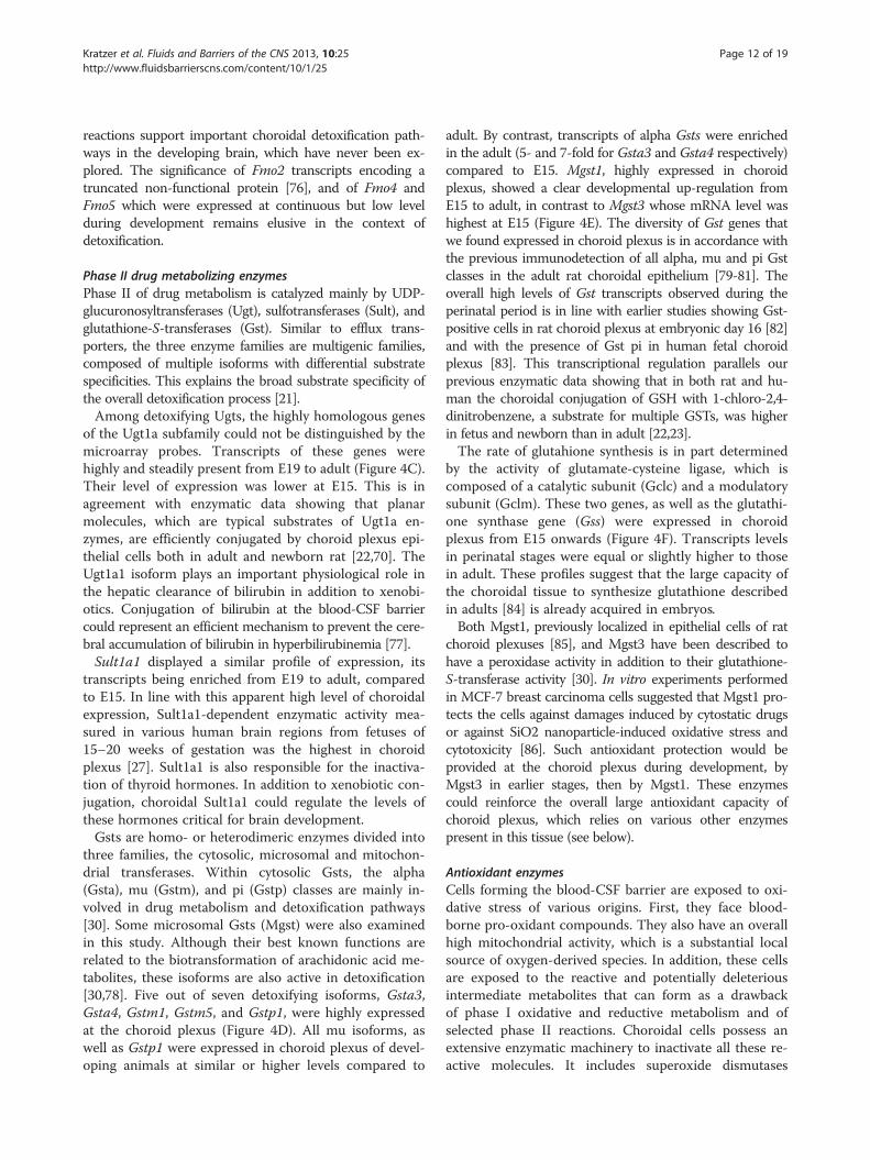
Phase II drug metabolizing enzymesPhase II of drug metabolism is catalyzed mainly by UDP-glucuronosyltransferases (Ugt), sulfotransferases (Sult), andglutathione-S-transferases (Gst). Similar to efflux trans-porters, the three enzyme families are multigenic families,composed of multiple isoforms with differential substratespecificities. This explains the broad substrate specificity ofthe overall detoxification process [21].Among detoxifying Ugts, the highly homologous genes
of the Ugt1a subfamily could not be distinguished by themicroarray probes. Transcripts of these genes werehighly and steadily present from E19 to adult (Figure 4C).Their level of expression was lower at E15. This is inagreement with enzymatic data showing that planarmolecules, which are typical substrates of Ugt1a en-zymes, are efficiently conjugated by choroid plexus epi-thelial cells both in adult and newborn rat [22,70]. TheUgt1a1 isoform plays an important physiological role inthe hepatic clearance of bilirubin in addition to xenobi-otics. Conjugation of bilirubin at the blood-CSF barriercould represent an efficient mechanism to prevent the cere-bral accumulation of bilirubin in hyperbilirubinemia [77].Sult1a1 displayed a similar profile of expression, its
transcripts being enriched from E19 to adult, comparedto E15. In line with this apparent high level of choroidalexpression, Sult1a1-dependent enzymatic activity mea-sured in various human brain regions from fetuses of15–20 weeks of gestation was the highest in choroidplexus [27]. Sult1a1 is also responsible for the inactiva-tion of thyroid hormones. In addition to xenobiotic con-jugation, choroidal Sult1a1 could regulate the levels ofthese hormones critical for brain development.Gsts are homo- or heterodimeric enzymes divided into
three families, the cytosolic, microsomal and mitochon-drial transferases. Within cytosolic Gsts, the alpha(Gsta), mu (Gstm), and pi (Gstp) classes are mainly in-volved in drug metabolism and detoxification pathways[30]. Some microsomal Gsts (Mgst) were also examinedin this study. Although their best known functions arerelated to the biotransformation of arachidonic acid me-tabolites, these isoforms are also active in detoxification[30,78]. Five out of seven detoxifying isoforms, Gsta3,Gsta4, Gstm1, Gstm5, and Gstp1, were highly expressedat the choroid plexus (Figure 4D). All mu isoforms, aswell as Gstp1 were expressed in choroid plexus of devel-oping animals at similar or higher levels compared to
adult. By contrast, transcripts of alpha Gsts were enrichedin the adult (5- and 7-fold for Gsta3 and Gsta4 respectively)compared to E15. Mgst1, highly expressed in choroidplexus, showed a clear developmental up-regulation fromE15 to adult, in contrast to Mgst3 whose mRNA level washighest at E15 (Figure 4E). The diversity of Gst genes thatwe found expressed in choroid plexus is in accordance withthe previous immunodetection of all alpha, mu and pi Gstclasses in the adult rat choroidal epithelium [79-81]. Theoverall high levels of Gst transcripts observed during theperinatal period is in line with earlier studies showing Gst-positive cells in rat choroid plexus at embryonic day 16 [82]and with the presence of Gst pi in human fetal choroidplexus [83]. This transcriptional regulation parallels ourprevious enzymatic data showing that in both rat and hu-man the choroidal conjugation of GSH with 1-chloro-2,4-dinitrobenzene, a substrate for multiple GSTs, was higherin fetus and newborn than in adult [22,23].The rate of glutahione synthesis is in part determined
by the activity of glutamate-cysteine ligase, which iscomposed of a catalytic subunit (Gclc) and a modulatorysubunit (Gclm). These two genes, as well as the glutathi-one synthase gene (Gss) were expressed in choroidplexus from E15 onwards (Figure 4F). Transcripts levelsin perinatal stages were equal or slightly higher to thosein adult. These profiles suggest that the large capacity ofthe choroidal tissue to synthesize glutathione describedin adults [84] is already acquired in embryos.Both Mgst1, previously localized in epithelial cells of rat
choroid plexuses [85], and Mgst3 have been described tohave a peroxidase activity in addition to their glutathione-S-transferase activity [30]. In vitro experiments performedin MCF-7 breast carcinoma cells suggested that Mgst1 pro-tects the cells against damages induced by cytostatic drugsor against SiO2 nanoparticle-induced oxidative stress andcytotoxicity [86]. Such antioxidant protection would beprovided at the choroid plexus during development, byMgst3 in earlier stages, then by Mgst1. These enzymescould reinforce the overall large antioxidant capacity ofchoroid plexus, which relies on various other enzymespresent in this tissue (see below).
Antioxidant enzymesCells forming the blood-CSF barrier are exposed to oxi-dative stress of various origins. First, they face blood-borne pro-oxidant compounds. They also have an overallhigh mitochondrial activity, which is a substantial localsource of oxygen-derived species. In addition, these cellsare exposed to the reactive and potentially deleteriousintermediate metabolites that can form as a drawbackof phase I oxidative and reductive metabolism and ofselected phase II reactions. Choroidal cells possess anextensive enzymatic machinery to inactivate all these re-active molecules. It includes superoxide dismutases

Kratzer et al. Fluids and Barriers of the CNS 2013, 10:25 Page 13 of 19http://www.fluidsbarrierscns.com/content/10/1/25
(Sod), which inactivate superoxide anions, catalase (Cat),which cleaves hydrogen peroxide, and glutathioneperoxidases (Gpx), which metabolize a large spectrum ofperoxide species. Some isoforms of the latter enzymesrequire glutathione in their catalytic cycle, and thusfunction in conjunction with glutathione reductase(Gsr), which regenerates the reduced form of the thiol-containing molecule (reviewed in [87]).Five out of eight Gpx genes identified in mammals
(Gpx1, -3, -4, -7, and −8) and Gsr were detected in
Figure 4 Developmental profiles of drug metabolizing enzymes. (A) sand epoxide hydrolases in the lateral ventricle choroid plexus. Cyp2u1 anddevelopmental profile overlap on the graph. No expression for Cyp2u1 wasdependent monoxygenase. Fmo3 was undetectable at E15. (C) shows genesulfotransferases. Ugt2a1 was detected in adult, but not at E15. (D) showsfor microsomal glutathione-S-transferases. (F) shows expression profiles forisoforms. Other abbreviations, data analysis, and data expression as in Figur
choroid plexuses (Figure 5A). All were expressed atsimilar or higher levels during the perinatal period com-pared to adult. At E15, Log2Fcad absolute values werelower than 2, indicating only limited variation at thisearly embryonic stage. Gpx1 and Gpx4 were expressedat high levels at all stages. Gpx1 is mainly devoted to themetabolism of hydrogen peroxide when produced in ex-cess, as occurring in infections, while Gpx4 specificallyinactivates membrane-integrated hydroperoxy lipids. Itthereby counteracts the activity of lipoxygenases, and
hows gene expression profiles for different cytochrome P450 enzymesEphx1 display similar Log2 FCad during development, thus thedetected at E15. (B) shows gene expression profiles for flavin-expression profiles for UDP glucuronosyltransferases and
gene expression profiles for soluble glutathione-S-transferases, and (E)genes involved in GSH synthesis and metabolism. MI, multiplees 2 and 3.

Figure 5 Developmental profiles of other antioxidant enzymes. (A)shows gene expression profiles for different glutathione peroxidases andglutathione reductase, (B) shows gene expression profiles for superoxidedismutases and for catalase, and (C) for heme oxygenases and biliverdinreductases. Abbreviations, data analysis, and data expression as inFigures 2 and 3.
Kratzer et al. Fluids and Barriers of the CNS 2013, 10:25 Page 14 of 19http://www.fluidsbarrierscns.com/content/10/1/25
can modulate inflammatory responses [87]. Very highGpx activities have been measured in rat choroid plex-uses isolated from adults [88]. It is expected from ourdata that these enzymes are already functional at earliertimes and therefore contribute to protect the developingbrain against oxidative and inflammatory insults. Thesoluble Cu/Zn superoxide dismutase genes Sod1 andSod3, as well as the cat gene were also highly expressedin choroid plexus both during the perinatal period andin adult (Figure 5B). Variations in expression of Sodgenes were more heterogeneous in choroid plexuses inE15 animals than in subsequent developmental stages. Astrong immunoreactivity for catalase was previouslydetected in rat choroid plexus as early as at E14.5 [89].Mammalian cells display several other means of
protection against reactive species, including the hemeoxygenase (Hmox) pathway. Hmox proteins catalysethe degradation of heme, resulting in the formation ofbiliverdin, which in turn can be converted via biliver-din reductases (Bvr) into bilirubin. Intracellular un-conjugated bilirubin acts as antioxidant, and thebiliverdin/bilirubin redox cycle is considered as an im-portant constituent of the cellular antioxidant defensesystem acting mainly towards lipophilic reactive spe-cies [77]. Moderate hyperbilirubinemia was foundneuroprotective against focal ischemia to which neo-nates are especially susceptible [90]. Both stress-inducible Hmox1 and constitutive Hmox2 genes, aswell as the Bvra and Bvrb genes were expressed inchoroid plexus (Figure 5C). For all four genes, tran-scripts were detected at similar or slightly higher levelsduring development than in adults (log2 FCad valuesbetween −0.92 to 2.44). In line with our data, immuno-reactivity of Hmox-2 usually considered as a neuronalisoform, has been reported in choroid plexuses of adultrats [91]. The steady expression of Hmox2 and the con-current enriched levels of Hmox1 transcripts in embry-onic stages represent a potent heme catabolic systemable to generate biliverdin, not only in normal develop-mental conditions, but also in response to perinatal in-sults. In contrast to the liver, in which bvra and bvrbshowed opposite age-dependent profiles of expression[92], both genes were expressed at similar levels inchoroid plexuses from E19 to the adult stage. Thus thebilirubin/biliverdin antioxidant redox cycle is likely tobe efficient in choroidal cells throughout development.
Transcription factors involved in the regulation ofdetoxifying genesLigand and non-ligand activated transcription factorsinvolved in the coordinated regulation of drug-metabolizing enzymes and transporters were incorpo-rated in our study. They include the aryl hydrocarbonreceptor (Ahr), the pregnane X receptor (Nr1i2), the

Figure 6 Developmental profiles of transcription factorsinvolved in regulation of detoxifying genes. Gene expressionprofiles for transcription factors that regulate drug-metabolizingenzymes and efflux transporters, and for their co-regulators areshown. Abbreviations, data analysis, and data expression as inFigure 2.
Kratzer et al. Fluids and Barriers of the CNS 2013, 10:25 Page 15 of 19http://www.fluidsbarrierscns.com/content/10/1/25
constitutive androstane/activated receptor (Nr1i3), theperoxisome proliferator-activated receptor α (Ppara),the glucocorticoid receptor (Nr3c1), and the CCAAT/enhancer binding protein beta (Cebpb) reviewed in[29], and [30]. Ahr dimerizes with the Ahr nucleartranslocator (Arnt, also known as Hif1b). Nr1i2, Nr1i3and Ppara heterodimerize with the retinoid X receptorα (Rxra). The tumor protein p53 (Tp53) was added tothe study as it was shown to directly mediate the tran-scriptional induction of Abcb1 induced by genotoxiccompounds [93]. The oxidative stress sensor nuclearfactor erythroid 2-related factor 2 (Nfe2l2) and thehypoxia-inducible factor 1 alpha (Hif1a), all transcrip-tion factors involved in inductive processes mediatedby redox imbalance and whose activation leads to theinduction of detoxifying genes, are also reported. Fourof these genes, Rxra, Nfe2l2, Hif1a, and Nr3c1 wereexpressed at high levels in choroid plexuses. Apartfrom Nr1i2, whose expression was clearly upregulatedfrom E15 to adult stage (p < 0.001), the choroidal ex-pression of all transcription factors was remarkablysteady throughout development (Figure 6). Absolutelog2 FCad values determined at E19 were between 0and 0.9.Induction of choroidal drug metabolizing enzymes in
choroid plexuses has been demonstrated only in adultrats, following treatment with polycyclic aromatic hydro-carbons, indicating that the AhR/Arnt-dependent path-way is functional at the blood-CSF barrier [72,94]. Iffunctionality proves true for all factors throughout de-velopment, the panel of transcription factors that areexpressed in choroid plexus suggests that following ex-posure to a large range of chemical xenobiotics the ac-tivity of drug metabolizing enzymes and transporters inthe blood-CSF barrier can be induced in adult as well asin developing animals. Of note, the lower choroidalNr1i2 expression at E15 could be compensated by Nr1i3transcripts, as these two transcription factors regulate anumber of common genes well expressed at the choroidplexus, such as Ugt1a, Abcc and cytosolic Gsts [95].Similarly, the steady expression of Nfe2l2, Hif1a, andArnt suggests that the neuroprotective functions of theblood-CSF barrier can be modulated in response to hyp-oxia and oxidative stress, whether occurring at early de-velopmental age or in adult.
Developmental profile of enzymes forming a barrier toneurotransmittersA function of enzymatic barrier to monoamine neuro-transmitters, that involves three enzymes, has beenascribed to the choroidal epithelium [96]. The Dopa-decarboxylase (Ddc) catalyzes the decarboxylation ofL-3,4-dihydroxyphenylalanine (L-Dopa) to dopamine, L-5-hydroxytryptophan to serotonin, and L-tryptophan to
tryptamine. Monoamine oxidases (Mao) in turnmetabolize and degrade the monoamine neurotransmit-ters dopamine, serotonin, and adrenalin. Catechol-O-methyltransferase (Comt) catalyzes the O-methylation ofcatecholamine neurotransmitters and catechol hor-mones, leading to their inactivation. It also shortens thebiological half-lives of certain neuroactive drugs, like L-Dopa, alpha-methyl Dopa, and isoproterenol [97]. Chor-oidal Ddc expression was 4- to 5.5-fold higher in thechoroid plexus of developing animals as compared toadult, while levels of Maoa, Maob, and Comt transcriptsvaried very little between E15 and adult (Figure 7).These data suggest that a choroidal enzymatic barrier to-wards neurotransmitters is already established beforebirth. If functional, as observed in adult [96], it couldrepresent a protective mechanism preventing blood-borne monoamines from interfering with centralneurotransmission.
General commentsThe combination of Affymetrix microarrays, RNA-Seqand qRT-PCR has enabled a comprehensive analysisof expression of choroid plexus genes involved inneuroprotection from embryonic through postnatal de-velopment compared with the adult. By their nature,microarrays are not comprehensive, but the additionaluse of RNA-Seq and qRT-PCR allowed the detectionand quantification of genes not represented in themicroarray. On the other hand some genes detected inthe Affymetrix arrays were below or close to thresholdin RNA-Seq. The quantitative estimates of changes inexpression level of individual genes are more secure, be-cause of the use of three independent methods.

Figure 7 Developmental profiles of enzymes forming a barrierto neurotransmitters. Gene expression profiles for monoamineoxidases, dopa decarboxylase and catechol-O-methyltransferaseare shown. Abbreviations, data analysis, and data expression as inFigure 2.
Table 2 Average amplitude of fold changes inneuroprotective gene expression levels in E15, E19 andP2 animals relative to adults in the choroid plexus
TJ proteins Transporters Enzymes
E15 5.9 ± 1.4 23.5 ± 9.2 5.1 ± 1.1
E19 2.5 ± 0.6 3.6 ± 0.9 2.1 ± 0.2
P2 1.9 ± 0.3 3.2 ± 0.7 1.9 ± 0.2
E15 vs. E19 * * **
E15 vs. P2 ** ** ***
E19 vs. P2 ns ns ns
Data are expressed as mean ± SEM of trend-independent FC calculated backfrom Log2FCad “absolute” values for all the genes (but three), analyzed in thisstudy and illustrated in Figures 2, 3, 4, 5, 7. Slc22a12, Fmo3 and Ugt2a1 wereexcluded because FCad values at E15 could not be calculated (see figurelegends). “Absolute” values of Log2FCad correspond to the value reported inthe different figures, except that negative signs were removed. This table thusillustrates the FC amplitude independently of the variation trend, whetherexpression levels are higher or lower in earlier stages compared to the adult.*, **, ***Statistical differences between developmental stages, analyzed byKruskal-Wallis followed by Dunns multiple comparison test, p < 0.05, 0.01,0.001, respectively. ns: not statistically different.
Kratzer et al. Fluids and Barriers of the CNS 2013, 10:25 Page 16 of 19http://www.fluidsbarrierscns.com/content/10/1/25
The choroidal tissue microdissected from brain ispredominantly constituted by the choroidal epitheliallayer that forms the blood-CSF barrier, it also containsin smaller numbers fibroblasts and some myeloid cellsforming the stromal core of the choroid plexus, andendothelial cells forming the choroidal vessels. Ourstudy does not discriminate between these different celltypes. However, apart from Cldn5 and Cyp1a1 whichare localized in choroidal vessels [15,71], all otherneuroprotective gene products for which immunohisto-chemical or functional analyses have been performed atthe level of the choroid plexus have been identified inthe choroidal epithelium. These include Cldn1, Cldn2,Cldn3, Cldn9, Cldn19, Abcc1, Abcc4, Slco1a4, Slco1a5,Slc22a6, Slc22a8, Slc15a2, concentrative and equilibrativenucleoside transporters, Ephx1, Ugt1a, several cytosolic andmembrane-bound Gsts, cat, and enzymes bearing a barrierfunction to neurotransmitters [17-19,22,23,25,46,56,57,67,69,75,80,85,89,96]. The present gene expression analysistherefore supports the concept that the blood-CSF barrierpossesses an efficient neuroprotective machinery during thepre- and postnatal stages of development. This is reflectedby the overall limited variation in the choroidal expressionof efflux transporters and metabolizing/antioxidant en-zymes between E19, P2 and adult animals (Table 2). Only alimited number of genes displayed a large age-dependentup or down regulation within this timeframe. The 16-foldhigher transcript levels for Abcc9 and Slco2a1 detected atE19 compared to adult likely reflect differences betweenthe perinatal and adult functions of the blood-CSF barrier.These specificities remain to be investigated. The variationin gene expression relative to adult levels is more pro-nounced at E15 than at perinatal stages of development
(Table 2). Thirty percent of the neuroprotective genes ana-lyzed in the present work were expressed at levels at least4-fold lower at E15 compared to E19, reflecting some de-gree of maturation of the functions associated with thesegenes between early embryonic and late fetal stages. Bycontrast, transcript levels were 4-fold higher or more inE15 compared to E19 for only ten percent of the genes.Given the growing evidence for the important role of chor-oid plexus in early stages of brain development [4,98] theseparticular choroidal transporters and enzymes may be in-volved in brain development processes rather than inneuroprotection per se.
ConclusionsThe present data provide an overview of the multipleenzymatic and transport systems expressed in choroidplexus, which in conjunction with the epithelial tightjunctional complexes, contribute to the protective bar-rier functions of the blood-CSF interface. However, it re-mains to be confirmed by in vivo testing with drugs andother xenobiotics if these systems are as functionally ef-ficient in the developing brain as in the adult. Furthercharacterization of these proteins to determine theirlocalization and efficiency during development will en-able more reliable risk assessment for drugs used to treatperinatal diseases. Evidence for the choroidal expressionof various transcription factors involved in the inductionof neuroprotective genes opens new pharmacologicalperspectives to improve neuroprotection at the inter-faces between the blood and the central nervous systemin the context of both perinatal injuries and environ-mental toxicity.

Kratzer et al. Fluids and Barriers of the CNS 2013, 10:25 Page 17 of 19http://www.fluidsbarrierscns.com/content/10/1/25
Endnotesa Gene official symbols (in italic), as defined on the
NCBI website, other common gene or protein names,and accession numbers (NM; number mRNA) for allgenes analyzed in this study are listed in Table 1.Symbols referring to human genes or proteins arewritten in capital letters.
Additional file
Additional file 1: List of primers, amplicon sizes and MgCl2-concentrations used for qRT-PCR.
AbbreviationsABC: ATP-binding cassette; CSF: Cerebrospinal fluid; FC: Fold changes; qRT-PCR: Quantitative real-time polymerase chain reaction; Slc: Solute carrier;TJ: Tight junctions.
Competing interestsThe authors declare that they have no competing interests.
Authors’ contributionsIK and NRS carried out the Affymetrix and qRT-PCR analyses. SAL and KScarried out the Illumina analysis. IK, SAL and JFGE performed the statisticalanalysis. NS, NRS and JFGE conceived and designed the study. IK and JFGEdrafted the manuscript. All authors read and approved the final manuscript.
AcknowledgementsThe research leading to these results has received funding from theEuropean Union Seventh Framework Program (FP7/2007- 2013) under grantagreement n° HEALTH-F2-2009-241778 and from the National Health andMedical Research Council, Australia. SAL is funded by the American-Australian Association. The authors thank Catherine Rey and the ProfilXpertplatform (Lyon) for their technical help.
Author details1Inserm U1028, Lyon Neuroscience Research Center, Neurooncology &Neuroinflammation Team, Lyon-1 University, Lyon F-69000, France.2Department of Pharmacology & Therapeutics, University of Melbourne,Parkville, Victoria 3010, Australia. 3Department of Neurobiology, StanfordUniversity, Stanford, CA 94305, USA. 4Brain-i, Lyon, France.
Received: 23 April 2013 Accepted: 10 July 2013Published: 1 August 2013
References1. Dziegielewska KM, Ek J, Habgood MD, Saunders NR: Development of the
choroid plexus. Microsc Res Tech 2001, 52:5–20.2. Shuangshoti S, Netsky MG: Choroid plexus and paraphysis in lower
vertebrates. J Morphol 1966, 120:157–187.3. Strazielle N, Ghersi-Egea JF: Choroid plexus in the central nervous system:
biology and physiopathology. J Neuropathol Exp Neurol 2000, 59:561–574.4. Liddelow SA, Temple S, Mollgard K, Gehwolf R, Wagner A, Bauer H, Bauer
HC, Phoenix TN, Dziegielewska KM, Saunders NR: Molecularcharacterisation of transport mechanisms at the developing mouseblood-CSF interface: a transcriptome approach. PLoS One 2012, 7:e33554.
5. Damkier HH, Praetorius J: Genetic ablation of Slc4a10 alters theexpression pattern of transporters involved in solute movement in themouse choroid plexus. Am J Physiol Cell Physiol 2012, 302:C1452–C1459.
6. Catala M: Embryonic and fetal development of structures associated withthe cerebro-spinal fluid in man and other species. Part I: The ventricularsystem, meninges and choroid plexuses. Arch Anat Cytol Pathol 1998,46:153–169.
7. Johansson PA, Dziegielewska KM, Liddelow SA, Saunders NR: The blood-CSF barrier explained: when development is not immaturity. BioEssays2008, 30:237–248.
8. Strazielle N, Ghersi-Egea JF: Physiology of blood–brain interfaces inrelation to brain disposition of small compounds and macromolecules.Mol Pharm 2013, 10:1473–1491.
9. Brightman MW, Reese TS: Junctions between intimately apposed cellmembranes in the vertebrate brain. J Cell Biol 1969, 40:648–677.
10. Mollgard K, Malinowska DH, Saunders NR: Lack of correlation betweentight junction morphology and permeability properties in developpingchoroid plexus. Nature 1976, 264:293–294.
11. Wolburg H, Wolburg-Buchholz K, Liebner S, Engelhardt B: Claudin-1,claudin-2 and claudin-11 are present in tight junctions of choroid plexusepithelium of the mouse. Neurosci Lett 2001, 307:77–80.
12. Johansson PA, Dziegielewska KM, Ek CJ, Habgood MD, Liddelow SA, PotterAM, Stolp HB, Saunders NR: Blood-CSF barrier function in the rat embryo.Eur J Neurosci 2006, 24:65–76.
13. Ek CJ, Dziegielewska KM, Stolp H, Saunders NR: Functional effectiveness ofthe blood–brain barrier to small water-soluble molecules in developingand adult opossum (Monodelphis domestica). J Comp Neurol 2006,496:13–26.
14. Ek CJ, Habgood MD, Dziegielewska KM, Saunders NR: Structuralcharacteristics and barrier properties of the choroid plexuses indeveloping brain of the opossum (Monodelphis Domestica). J CompNeurol 2003, 460:451–464.
15. Kratzer I, Vasiljevic A, Rey C, Fevre-Montange M, Saunders N, Strazielle N,Ghersi-Egea JF: Complexity and developmental changes in theexpression pattern of claudins at the blood-CSF barrier. Histochem CellBiol 2012, 138:861–879.
16. Wijnholds J, DeLange EC, Scheffer GL, Van Den Berg DJ, Mol CA, Van DerValk M, Schinkel AH, Scheper RJ, Breimer DD, Borst P: Multidrug resistanceprotein 1 protects the choroid plexus epithelium and contributes to theblood-cerebrospinal fluid barrier. J Clin Invest 2000, 105:279–285.
17. Leggas M, Adachi M, Scheffer GL, Sun D, Wielinga P, Du G, Mercer KE,Zhuang Y, Panetta JC, Johnston B, et al: Mrp4 confers resistance totopotecan and protects the brain from chemotherapy. Mol Cell Biol 2004,24:7612–7621.
18. Kusuhara H, Sugiyama Y: Efflux transport systems for organic anions andcations at the blood-CSF barrier. Adv Drug Deliv Rev 2004, 56:1741–1763.
19. Nagata Y, Kusuhara H, Endou H, Sugiyama Y: Expression and functionalcharacterization of rat organic anion transporter 3 (rOat3) in the choroidplexus. Mol Pharmacol 2002, 61:982–988.
20. Smith DE, Hu Y, Shen H, Nagaraja TN, Fenstermacher JD, Keep RF:Distribution of glycylsarcosine and cefadroxil among cerebrospinal fluid,choroid plexus, and brain parenchyma after intracerebroventricularinjection is markedly different between wild-type and Pept2 null mice.J Cereb Blood Flow Metab 2011, 31:250–261.
21. Strazielle N, Khuth ST, Ghersi-Egea JF: Detoxification systems, passive andspecific transport for drugs at the blood-CSF barrier in normal andpathological situations. Adv Drug Deliv Rev 2004, 56:1717–1740.
22. Strazielle N, Ghersi-Egea JF: Demonstration of a coupled metabolism-efflux process at the choroid plexus as a mechanism of brain protectiontoward xenobiotics. J Neurosci 1999, 19:6275–6289.
23. Ghersi-Egea JF, Strazielle N, Murat A, Jouvet A, Buenerd A, Belin MF: Brainprotection at the blood-cerebrospinal fluid interface involves aglutathione-dependent metabolic barrier mechanism. J Cereb Blood FlowMetab 2006, 26:1165–1175.
24. Ek CJ, Wong A, Liddelow SA, Johansson PA, Dziegielewska KM, Saunders NR:Efflux mechanisms at the developing brain barriers: ABC-transporters inthe fetal and postnatal rat. Toxicol Lett 2010, 197:51–59.
25. Gazzin S, Strazielle N, Schmitt C, Fevre-Montange M, Ostrow JD, Tiribelli C,Ghersi-Egea JF: Differential expression of the multidrug resistance-relatedproteins ABCb1 and ABCc1 between blood–brain interfaces. J CompNeurol 2008, 510:497–507.
26. Daood M, Tsai C, Ahdab-Barmada M, Watchko JF: ABC transporter (P-gp/ABCB1, MRP1/ABCC1, BCRP/ABCG2) expression in the developing humanCNS. Neuropediatrics 2008, 39:211–218.
27. Richard K, Hume R, Kaptein E, Stanley EL, Visser TJ, Coughtrie MW: Sulfationof thyroid hormone and dopamine during human development:ontogeny of phenol sulfotransferases and arylsulfatase in liver, lung, andbrain. J Clin Endocrinol Metab 2001, 86:2734–2742.
28. Stolp HB, Turnquist C, Dziegielewska KM, Saunders NR, Anthony DC, MolnarZ: Reduced ventricular proliferation in the foetal cortex followingmaternal inflammation in the mouse. Brain 2011, 134:3236–3248.

Kratzer et al. Fluids and Barriers of the CNS 2013, 10:25 Page 18 of 19http://www.fluidsbarrierscns.com/content/10/1/25
29. Aleksunes LM, Klaassen CD: Coordinated regulation of hepatic phase Iand II drug-metabolizing genes and transporters using AhR-, CAR-, PXR-,PPARalpha-, and Nrf2-null mice. Drug Metab Dispos 2012, 40:1366–1379.
30. Higgins LG, Hayes JD: Mechanisms of induction of cytosolic andmicrosomal glutathione transferase (GST) genes by xenobiotics and pro-inflammatory agents. Drug Metab Rev 2011, 43:92–137.
31. Okada K, Shoda J, Taguchi K, Maher JM, Ishizaki K, Inoue Y, Ohtsuki M,Goto N, Sugimoto H, Utsunomiya H, et al: Nrf2 counteracts cholestaticliver injury via stimulation of hepatic defense systems. Biochem BiophysRes Commun 2009, 389:431–436.
32. Strazielle N, Ghersi-Egea JF: Drug metabolism in newborn rat choroidplexus from lateral, third and fourth ventricle. Dev Anim Vet Sci 1997,27:895–901.
33. Saunders NR, Ek CJ, Habgood MD, Johansson P, Liddelow S, DziegielewskaKM: Assessing blood-cerebrospinal fluid barrier permeability in the ratembryo. Methods Mol Biol 2011, 686:247–265.
34. Liddelow S, Dziegielewska KM, Ek CJ, Habgood MD, Bauer H, Bauer H-C, LindsayH, Wakefield MJ, Strazielle N, Kratzer I, et al: Mechanisms that determine theinternal environment of the developing brain: a transcriptomic, functionaland ultrastructural approach. PLoS One 2013, 8:e65629.
35. Fang Z, Cui X: Design and validation issues in RNA-seq experiments.Brief Bioinform 2011, 12:280–287.
36. Griffith M, Griffith OL, Mwenifumbo J, Goya R, Morrissy AS, Morin RD,Corbett R, Tang MJ, Hou YC, Pugh TJ, et al: Alternative expression analysisby RNA sequencing. Nat Methods 2010, 7:843–847.
37. Bottomly D, Walter NA, Hunter JE, Darakjian P, Kawane S, Buck KJ, SearlesRP, Mooney M, McWeeney SK, Hitzemann R: Evaluating gene expression inC57BL/6J and DBA/2J mouse striatum using RNA-Seq and microarrays.PLoS One 2011, 6:e17820.
38. Marioni JC, Mason CE, Mane SM, Stephens M, Gilad Y: RNA-seq: anassessment of technical reproducibility and comparison with geneexpression arrays. Genome Res 2008, 18:1509–1517.
39. Leslie EM, Deeley RG, Cole SP: Multidrug resistance proteins: role of P-glycoprotein, MRP1, MRP2, and BCRP (ABCG2) in tissue defense. ToxicolAppl Pharmacol 2005, 204:216–237.
40. Redzic ZB, Malatiali SA, Grujicic D, Isakovic AJ: Expression and functionalactivity of nucleoside transporters in human choroid plexus.Cerebrospinal Fluid Res 2010, 7:2.
41. Choudhuri S, Cherrington NJ, Li N, Klaassen CD: Constitutive expression ofvarious xenobiotic and endobiotic transporter mRNAs in the choroidplexus of rats. Drug Metab Dispos 2003, 31:1337–1345.
42. Hoque MT, Robillard KR, Bendayan R: Regulation of breast cancer resistantprotein by peroxisome proliferator-activated receptor alpha in humanbrain microvessel endothelial cells. Mol Pharmacol 2012, 81:598–609.
43. Hagenbuch B, Meier PJ: The superfamily of organic anion transportingpolypeptides. Biochim Biophys Acta 2003, 1609:1–18.
44. Ohtsuki S, Takizawa T, Takanaga H, Terasaki N, Kitazawa T, Sasaki M, Abe T,Hosoya K, Terasaki T: In vitro study of the functional expression of organicanion transporting polypeptide 3 at rat choroid plexus epithelial cellsand its involvement in the cerebrospinal fluid-to-blood transport ofestrone-3-sulfate. Mol Pharmacol 2003, 63:532–537.
45. Abe T, Kakyo M, Sakagami H, Tokui T, Nishio T, Tanemoto M, Nomura H,Hebert SC, Matsuno S, Kondo H, et al: Molecular characterization andtissue distribution of a new organic anion transporter subtype (oatp3)that transports thyroid hormones and taurocholate and comparisonwith oatp2. J Biol Chem 1998, 273:22395–22401.
46. Gao B, Stieger B, Noe B, Fritschy JM, Meier PJ: Localization of the organicanion transporting polypeptide 2 (Oatp2) in capillary endothelium andchoroid plexus epithelium of rat brain. J Histochem Cytochem 1999,47:1255–1264.
47. Roth M, Obaidat A, Hagenbuch B: OATPs, OATs and OCTs: the organicanion and cation transporters of the SLCO and SLC22A genesuperfamilies. Br J Pharmacol 2012, 165:1260–1287.
48. Berbel P, Navarro D, Auso E, Varea E, Rodriguez AE, Ballesta JJ, Salinas M,Flores E, Faura CC, de Escobar GM: Role of late maternal thyroidhormones in cerebral cortex development: an experimental model forhuman prematurity. Cereb Cortex 2010, 20:1462–1475.
49. Sugiyama D, Kusuhara H, Taniguchi H, Ishikawa S, Nozaki Y, Aburatani H,Sugiyama Y: Functional characterization of rat brain-specific organicanion transporter (Oatp14) at the blood–brain barrier: high affinitytransporter for thyroxine. J Biol Chem 2003, 278:43489–43495.
50. Morreale De Escobar G, Obregon MJ, Escobar Del Rey F: Role of thyroidhormone during early brain development. Eur J Endocrinol 2004,151(3):25–37.
51. Huber RD, Gao B, Sidler Pfandler MA, Zhang-Fu W, Leuthold S, HagenbuchB, Folkers G, Meier PJ, Stieger B: Characterization of two splice variants ofhuman organic anion transporting polypeptide 3A1 isolated fromhuman brain. Am J Physiol Cell Physiol 2007, 292:795–806.
52. Fung WP, Thomas T, Dickson PW, Aldred AR, Milland J, Dziadek M, Power B,Hudson P, Schreiber G: Structure and expression of the rat transthyretin(prealbumin) gene. J Biol Chem 1988, 263:480–488.
53. Monk JA, Sims NA, Dziegielewska KM, Weiss RE, Ramsay RG, Richardson SJ:Delayed development of specific thyroid hormone-regulated events intransthyretin null mice. Am J Physiol Endocrinol Metab 2013, 304:E23–E31.
54. Jacobsson JA, Haitina T, Lindblom J, Fredriksson R: Identification of sixputative human transporters with structural similarity to the drugtransporter SLC22 family. Genomics 2007, 90:595–609.
55. Tamai I: Pharmacological and pathophysiological roles of carnitine/organic cation transporters (OCTNs: SLC22A4, SLC22A5 and Slc22a21).Biopharm Drug Dispos 2013, 34:29–44.
56. Pritchard JB, Sweet DH, Miller DS, Walden R: Mechanism of organic aniontransport across the apical membrane of choroid plexus. J Biol Chem1999, 274:33382–33387.
57. Strazielle N, Belin MF, Ghersi-Egea JF: Choroid plexus controls brainavailability of anti-HIV nucleoside analogs via pharmacologicallyinhibitable organic anion transporters. AIDS 2003, 17:1473–1485.
58. Burckhardt G: Drug transport by Organic Anion Transporters (OATs).Pharmacol Ther 2012, 136:106–130.
59. Nehlig A, de Pereirax Vasconcelos A: Glucose and ketone body utilizationby the brain of neonatal rats. Prog Neurobiol 1993, 40:163–221.
60. Rauchova H, Vokurkova M, Koudelova J: Hypoxia-induced lipidperoxidation in the brain during postnatal ontogenesis. Physiol Res 2012,61(Suppl 1):S89–S101.
61. Ayala CA: Stimulation of choline acetyl transferase activity by L-carnitine andD-carnitine in brain areas of neonate rats. J Neurosci Res 1995, 41:403–408.
62. Bennett KM, Liu J, Hoelting C, Stoll J: Expression and analysis of two novelrat organic cation transporter homologs, SLC22A17 and SLC22A23. MolCell Biochem 2011, 352:143–154.
63. Gupta K, Shukla M, Cowland JB, Malemud CJ, Haqqi TM: Neutrophilgelatinase-associated lipocalin is expressed in osteoarthritis and forms acomplex with matrix metalloproteinase 9. Arthritis Rheum 2007,56:3326–3335.
64. Strazielle N, Khuth ST, Murat A, Chalon A, Giraudon P, Belin MF, Ghersi-EgeaJF: Pro-inflammatory cytokines modulate matrix metalloproteinasesecretion and organic anion transport at the blood-cerebrospinal fluidbarrier. J Neuropathol Exp Neurol 2003, 62:1254–1264.
65. Ip JP, Nocon AL, Hofer MJ, Lim SL, Muller M, Campbell IL: Lipocalin 2 in thecentral nervous system host response to systemic lipopolysaccharideadministration. J Neuroinflammation 2011, 8:124.
66. Keep RF, Smith DE: Choroid plexus transport: gene deletion studies.Fluids Barriers CNS 2011, 8:26.
67. Shu C, Shen H, Teuscher NS, Lorenzi PJ, Keep RF, Smith DE: Role of PEPT2in peptide/mimetic trafficking at the blood-cerebrospinal fluid barrier:studies in rat choroid plexus epithelial cells in primary culture.J Pharmacol Exp Ther 2002, 301:820–829.
68. Mackey JR, Mani RS, Selner M, Mowles D, Young JD, Belt JA, Crawford CR,Cass CE: Functional nucleoside transporters are required for gemcitabineinflux and manifestation of toxicity in cancer cell lines. Cancer Res 1998,58:4349–4357.
69. Redzic ZB, Biringer J, Barnes K, Baldwin SA, Al-Sarraf H, Nicola PA, Young JD,Cass CE, Barrand MA, Hladky SB: Polarized distribution of nucleosidetransporters in rat brain endothelial and choroid plexus epithelial cells.J Neurochem 2005, 94:1420–1426.
70. Ghersi-Egea JF, Leninger-Muller B, Suleman G, Siest G, Minn A: Localizationof drug-metabolizing enzyme activities to blood–brain interfaces andcircumventricular organs. J Neurochem 1994, 62:1089–1096.
71. Brittebo EB: Metabolism-dependent binding of the heterocyclic amineTrp-P-1 in endothelial cells of choroid plexus and in large cerebral veinsof cytochrome P450-induced mice. Brain Res 1994, 659:91–98.
72. Morse DC, Stein AP, Thomas PE, Lowndes HE: Distribution and inductionof cytochrome P450 1A1 and 1A2 in rat brain. Toxicol Appl Pharmacol1998, 152:232–239.

Kratzer et al. Fluids and Barriers of the CNS 2013, 10:25 Page 19 of 19http://www.fluidsbarrierscns.com/content/10/1/25
73. Granberg L, Ostergren A, Brandt I, Brittebo EB: CYP1A1 and CYP1B1 inblood–brain interfaces: CYP1A1-dependent bioactivation of 7,12-dimethylbenz(a)anthracene in endothelial cells. Drug Metab Dispos 2003,31:259–265.
74. Dauchy S, Miller F, Couraud PO, Weaver RJ, Weksler B, Romero IA,Scherrmann JM, De Waziers I, Decleves X: Expression and transcriptionalregulation of ABC transporters and cytochromes P450 in hCMEC/D3human cerebral microvascular endothelial cells. Biochem Pharmacol 2009,77:897–909.
75. Marowsky A, Burgener J, Falck JR, Fritschy JM, Arand M: Distribution ofsoluble and microsomal epoxide hydrolase in the mouse brain and itscontribution to cerebral epoxyeicosatrienoic acid metabolism.Neuroscience 2009, 163:646–661.
76. Janmohamed A, Hernandez D, Phillips IR, Shephard EA: Cell-, tissue-, sex- anddevelopmental stage-specific expression of mouse flavin-containingmonooxygenases (Fmos). Biochem Pharmacol 2004, 68:73–83.
77. Gazzin S, Strazielle N, Tiribelli C, Ghersi-Egea JF: Transport and metabolismat blood–brain interfaces and in neural cells: relevance tobilirubin-induced encephalopathy. Front Pharmacol 2012, 3:89.
78. Jakobsson PJ, Scoggan KA, Yergey J, Mancini JA, Ford-Hutchinson AW:Characterization of microsomal GST-II by western blot and identificationof a novel LTC4 isomer. J Lipid Mediat Cell Signal 1997, 17:15–19.
79. Cammer W, Tansey F, Abramovitz M, Ishigaki S, Listowsky I: Differentiallocalization of glutathione-S-transferase Yp and Yb subunits inoligodendrocytes and astrocytes of rat brain. J Neurochem 1989,52:876–883.
80. Philbert MA, Beiswanger CM, Manson MM, Green JA, Novak RF, Primiano T,Reuhl KR, Lowndes HE: Glutathione S-transferases and gamma-glutamyltranspeptidase in the rat nervous systems: a basis for differentialsusceptibility to neurotoxicants. NeuroToxicology 1995, 16:349–362.
81. Johnson JA, el Barbary A, Kornguth SE, Brugge JF, Siegel FL: GlutathioneS-transferase isoenzymes in rat brain neurons and glia. J Neurosci 1993,13:2013–2023.
82. Senjo M, Ishibashi T, Terashima T, Inoue Y: Successive appearance ofglutathione S-transferase-positive cells in developing rat brain: choroidplexus, pia mater, ventricular zone and astrocytes. Neurosci Lett 1986,66:131–134.
83. Carder PJ, Hume R, Fryer AA, Strange RC, Lauder J, Bell JE: GlutathioneS-transferase in human brain. Neuropathol Appl Neurobiol 1990, 16:293–303.
84. Anderson ME, Underwood M, Bridges RJ, Meister A: Glutathionemetabolism at the blood-cerebrospinal fluid barrier. FASEB J 1989,3:2527–2531.
85. Johansson K, Ahlen K, Rinaldi R, Sahlander K, Siritantikorn A, Morgenstern R:Microsomal glutathione transferase 1 in anticancer drug resistance.Carcinogenesis 2007, 28:465–470.
86. Shi J, Karlsson HL, Johansson K, Gogvadze V, Xiao L, Li J, Burks T,Garcia-Bennett A, Uheida A, Muhammed M, et al: Microsomal glutathionetransferase 1 protects against toxicity induced by silica nanoparticles butnot by zinc oxide nanoparticles. ACS Nano 2012, 6:1925–1938.
87. Toppo S, Flohe L, Ursini F, Vanin S, Maiorino M: Catalytic mechanisms andspecificities of glutathione peroxidases: variations of a basic scheme.Biochim Biophys Acta 2009, 1790:1486–1500.
88. Tayarani I, Cloez I, Clement M, Bourre JM: Antioxidant enzymes andrelated trace elements in aging brain capillaries and choroid plexus.J Neurochem 1989, 53:817–824.
89. Nardacci R, Falciatori I, Moreno S, Stefanini S: Immunohistochemicallocalization of peroxisomal enzymes during rat embryonic development.J Histochem Cytochem 2004, 52:423–436.
90. Kitamura Y, Ishida Y, Takata K, Mizutani H, Kakimura J, Inden M, Nakata J,Taniguchi T, Tsukahara T, Akaike A, et al: Hyperbilirubinemia protectsagainst focal ischemia in rats. J Neurosci Res 2003, 71:544–550.
91. Sutherland BA, Rahman RM, Clarkson AN, Shaw OM, Nair SM, Appleton I:Cerebral heme oxygenase 1 and 2 spatial distribution is modulatedfollowing injury from hypoxia-ischemia and middle cerebral arteryocclusion in rats. Neurosci Res 2009, 65:326–334.
92. Yamaguchi T, Komoda Y, Nakajima H: Biliverdin-IX alpha reductase andbiliverdin-IX beta reductase from human liver. Purification andcharacterization. J Biol Chem 1994, 269:24343–24348.
93. Mathieu MC, Lapierre I, Brault K, Raymond M: Aromatic hydrocarbonreceptor (AhR).AhR nuclear translocator- and p53-mediated induction of
the murine multidrug resistance mdr1 gene by 3-methylcholanthreneand benzo(a)pyrene in hepatoma cells. J Biol Chem 2001, 276:4819–4827.
94. Leininger-Muller B, Ghersi-Egea JF, Siest G, Minn A: Induction andimmunological characterization of the uridine diphosphate-glucuronosyltransferase conjugating 1-naphthol in the rat choroidplexus. Neurosci Lett 1994, 175:37–40.
95. Tolson AH, Wang H: Regulation of drug-metabolizing enzymes byxenobiotic receptors: PXR and CAR. Adv Drug Deliv Rev 2010,62:1238–1249.
96. Lindvall M, Hardebo JE, Owman C: Barrier mechanisms forneutrotransmitter monoamines in the choroid plexus. Acta Physiol Scand1980, 108:215–221.
97. Kurogi K, Alazizi A, Liu MY, Sakakibara Y, Suiko M, Sugahara T, Liu MC:Concerted actions of the catechol O-methyltransferase and the cytosolicsulfotransferase SULT1A3 in the metabolism of catecholic drugs.Biochem Pharmacol 2012, 84:1186–1195.
98. Saunders NR, Liddelow SA, Dziegielewska KM: Barrier mechanisms in thedeveloping brain. Front Pharmacol 2012, 3:46.
doi:10.1186/2045-8118-10-25Cite this article as: Kratzer et al.: Developmental changes in thetranscriptome of the rat choroid plexus in relation to neuroprotection.Fluids and Barriers of the CNS 2013 10:25.
Submit your next manuscript to BioMed Centraland take full advantage of:
• Convenient online submission
• Thorough peer review
• No space constraints or color figure charges
• Immediate publication on acceptance
• Inclusion in PubMed, CAS, Scopus and Google Scholar
• Research which is freely available for redistribution
Submit your manuscript at www.biomedcentral.com/submit