research open access canpmr syndrome and chromosome …
TRANSCRIPT
RESEARCH Open Access
CANPMR syndrome and chromosome1p32-p31 deletion syndrome coexistin two related individuals affected bysimultaneous haplo-insufficiency ofCAMTA1 and NFIA genesEmanuele G. Coci1*, Udo Koehler2, Thomas Liehr3, Armin Stelzner1, Christian Fink4, Hendrik Langen1
and Joachim Riedel1
Abstract
Background: Non-progressive cerebellar ataxia with mental retardation (CANPMR, OMIM 614756) and chromosome1p32-p31 deletion syndrome (OMIM 613735) are two very rare inherited disorders, which are caused by mono-allelicdeficiency (haplo-insufficiency) of calmodulin-binding transcription activator 1 (CAMTA1) and, respectively, nuclearfactor 1 A (NFIA) genes. The yet reported patients affected by mono-allelic CAMTA1 dysfunction presented withneonatal hypotonia, delayed and ataxic gait, cerebellar atrophy, psychological delay and speech impairment, whileindividuals carrying a disrupted NFIA allele suffered from agenesis/hypoplasia of the corpus callosum, ventriculomegaly,developmental delay and urinary tract abnormalities. Both disorders were not seen in one patient together before.
Results: In this study two related individuals affected by a complex clinical syndrome, characterized by cognitive,neurological and nephrological features were studied for the underlying genetic disorder(s) by molecular cytogenetics.The two individuals present dysmorphic facies, macrocephaly, generalized ataxia, mild tremor, strabismus, mild mentalretardation and kidney hypoplasia. Moreover, neuro-radiological studies showed hypoplasia of corpus callosum.Genetic investigations revealed a paracentric inversion in the short arm of one chromosome 1 with breakpoints withinCAMTA1 and NFIA coding sequences.
Conclusions: To the best of our knowledge, this is the first report of two patients harboring the simultaneousmono-allelic disruptions and consequent haplo-insufficiencies of two genes due to an inversion event. Disruption ofCAMTA1 and NFIA genes led to neuro-psychological and nephrological dysfunctions, which comprised clinical featuresof CANPMR syndrome as well as chromosome 1p32-p31 deletion syndrome.
Keywords: NFIA, Chromosome 1p32-p31 deletion syndrome, CAMTA1, CANPMR syndrome, Paracentric inversion onshort arm of chromosome 1
* Correspondence: [email protected] of Social Pediatrics and Pediatric Neurology, General Hospital ofCelle, 29221 Celle, GermanyFull list of author information is available at the end of the article
Coci et al. Molecular Cytogenetics (2016) 9:10 DOI 10.1186/s13039-016-0219-y
© 2016 Coci et al., corrected publication 2021. Open Access This article is distributed under the terms of the Creative Commons Attribution 4.0 International License (http://creat iveco mmons .org/licen ses/by/4.0/), which permits unrestricted use, distribution, and reproduction in any medium, provided you give appropriate credit to the original author(s) and the source, provide a link to the Creative Commons license, and indicate if changes were made. The Creative Commons Public Domain Dedication waiver (http://creat iveco mmons .org/publi cdoma in/zero/1.0/) applies to the data made available in this article, unless otherwise stated.
BackgroundNon-progressive cerebellar ataxia with mental retard-ation (CANPMR, OMIM 614756) is a very rare neuro-developmental disorder, belonging to the heterogeneousfamily of genetically determined cerebellar ataxias [1, 2]with recessive [3] and dominant [4] pattern of inheritance.The affected patients present with ataxic gait, dysmetries,variable mental retardation, cerebellar abnormalities anddysmorphic facies with heterogeneous penetrance. Todate few genes/loci have been associated to autosomalrecessive forms of cerebellar ataxias: ATCAY [5], chromo-some 20q11-q13 locus [6], VLDLR [7], ZNF592 [8],SPTBN2 [9], CWF19L1 [10], PMPCA [11]. Calmodulin-binding transcription activator 1 (CAMTA1) maps onchromosome 1p36, carries 23 exons and encodes 2 pro-tein isoforms in mammalians. The brain-specific tran-scription factor CAMTA1 functions as homodimericcomplex binding to gene promoters’ CGCG box thoroughCG-1 domain, supporting the assembly of other transcrip-tion factors (e.g. Nkx2-5) and enhancing transcription ofeffector genes (e.g. Fbxl4) [12–14]. CAMTA1 dysfunctionhas been associated to human pathology by Thevenonet al. [15], who reported five patients affected by CAMTA1haploinsufficiency due to deletions or duplications in thegene region coding for CG-1 domain, which plays a piv-otal role in the whole CAMTA1 function. As mentionedabove, the reported patients suffered from ataxia, broad-based gait, tremor, intellectual impairment and speechdelay, cerebellar abnormalities (atrophy of lobes and/orvermis) and facial dysmorphisms (strabismus, large fore-head, wide and broad nose, small ears).The clinical features of chromosome 1p32-p31 deletion
syndrome (OMIM 613735) were firstly described byCampbell et al. [16]. Some years later, Lu et al. proposedthe causal association between this malformation syn-drome and Nuclear Factor 1 A (NFIA) haplo-insufficiency[17]. NFIA maps on 1p31.2, carries 11 exons and producesat least 9 different protein isoforms [18–20]. The proteinis functionally divided in two sections: a 200 amino acidlong N-terminal DNA binding and dimerization domain,mainly encoded by exon 2, and C-terminal transactivationand repression domains, mainly encoded by exons 3 to 11.The first one binds to the nucleotide consensussequence TTGGC(N)5GCCAA within the promoterregion of several genes. The latter ones operate bydirectly activating basal transcription factors at tran-scription start sites, by displacing repressive histonesfrom target genes, by interacting with other co-activator proteins [20]. All five individuals reported byLu et al. (three of which previously described byCampbell et al.) presented with hypoplastic or absentcorpus callosum, ventriculomegaly with or withoutrelevant hydrocephalus and development delay; someof them carried urinary tract abnormalities (3 patients),
epileptic seizures (3 patients), tethered spinal cord (4 pa-tients) and Chiari malformation (3 patients). Although thechromosomal abnormalities [a translocation t(1;20), atranslocation t(1;2), a 2.2 Mb deletion in 1p31-p32, a12 Mb deletion in 1p31-p32] differed and the deleted re-gions comprised different genes among the five patients,only NFIA gene was either disrupted or fully deleted in allfive patients, thus underpinning the association betweenNFIA haplo-insufficiency and the common CNS abnor-malities (hypoplasia of corpus callosum, ventriculomegalyand hydrocephalus). A strong confirmation on their patho-physiological hypothesis was given by the clinical andhistopathological findings of Nfia-/- mouse model [17, 21].A detailed study of the genome expression profile inmurine Nfia-/- brain at embryonic and post-natal stagesshowed a very strong imbalance in time-related expressionof several genes playing a pivotal role in oligodendrocytedifferentiation (e.g. Mag, Mal, Mobp, Mog, Sox2, Sox4,Sox11, Dio2, Myef2) as well as in axonal growth/guidance(e.g. Clusterin, aFGF, Ndrg2, EphrinB2, Crmp1) [22]. Theessential role of transcription factor Nfib and Nfic in brain,tooth and lung development was already described in thecorresponding mouse models [23, 24]. Few further reportsdescribed heterogeneous clinical findings associated withdeletions mapping on chromosome 1p31 and 1p32 andencompassing several other genes [25, 26].We describe the first family (Fig. 1) with two related in-
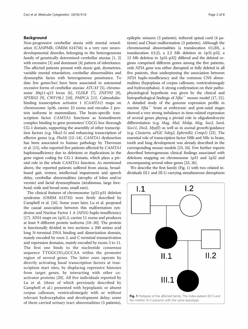
dividuals (II.1 and III.1) carrying simultaneous disruptions
Fig. 1 Pedigree of the affected family. The index-patient (III.1) andher mother (II.1) presents with the same karyotype
Coci et al. Molecular Cytogenetics (2016) 9:10 Page 2 of 8
and consequent haplo-insufficiencies of NFIA andCAMTA genes due to a paracentric inversion in the shortarm of chromosome 1 and presenting with clinical signsof CANPMR syndrome as well as chromosome 1p32-p31deletion syndrome.
Results and discussionPatient 1Since the first months of life the index-patient (III.1)presented with generalized hypotonia, reduced muscularstrength, particularly evident at trunk und pelvis, normaltendon reflexes, large head (> P90), prominent forehead,strabismus divergens and light bilateral ptosis. The pa-tient could sit at 11 months and walk at 23 months ofage. Gowers’ sign was through-out positive. Light dyski-nesias and ataxia appeared at 3 years and, respectively,6 years of age. She could speak the first words at about30 months of age.She slowly improved her motor, cognitive and language
skills. Now, at the age of 7 years she can walk unassisted,climb stairs and speak short sentences. Her daily skills arecompromised and she attends a special care kindergarten.Her intelligence quotient (IQ) score of 51 (Snijders-Oomen Non-Verbal test, SON-R) indicates a severe intel-lectual impairment (Table 1).The electroencephalogram (EEG) did not show epilep-
tic discharges. The brain MR investigations at 2 and 6 ½years of age revealed hypoplasia of corpus callosum,while the cerebellum was structurally normal (Fig. 2).Organ ultrasound did not show any structural abnormal-ities of kidneys and urinary tract, although the patientsuffered from recurrent urine infections starting from18 months of age.
Patient 2 and other individuals of pedigreeThe mother (II.1) of the index patient (28 years) pre-sents with a similar clinical constellation of III.1,whereby she suffered from neonatal hypotonia, but satunassisted at 8 months, walked at 14 months and spokethe first words at 12 months of age. She presents with amoderate ataxia, gait instability and dysmetria, whichdoes not strongly impair the daily skills. Her IQ score of65 (SON-R test) revealed a mental retardation. She hasnot completed any job training course and is currentlyunemployed. The brain MR scan, performed at 28 yearsof age, revealed hypoplasia of corpus callosum (Fig. 2).Since the early childhood, she suffered from recurrent
urinary tract infections due to vesicoureteral reflux (VUR)and from hypoplasia of the right kidney. The father (II.2)of the index patient carries a normal male karyotype andis a healthy individual. The maternal grandparents (I.1and I.2) are healthy individuals; nevertheless their karyo-type could not be analyzed due to missing compliance.
Cytogenetic investigationsThe index-patient III.1 harbors a paracentric inversion inthe short arm of chromosome 1 with breakpoints withinCAMTA1 (1p36.31) and NFIA (1p31.3) genes (Fig. 3).In correspondence of both breakpoints, two deletions
occurred affecting CAMTA1 exon 5 (deletion length 211Kb; position 6,936,272 - 7,146,519 Mb, GRCh37/hg19)
Table 1 Clinical, psychological, radiological features of theaffected patients II.1 and III.1
II.1 (28 years) III.1 (6 ½ years)
Development parameters
Sitting age (months) 8 11
Walking age(months)
14 23
Speaking age(months)
12 30
Clinical findings
Macrocephaly Yes (P 97) No (P 90)
Muscle tonus Normal Normal
Seizures No No
Facies
Forehead Large Large
Strabismus Yes (divergens) Yes (divergens)
Nasal bridge Broad Broad
Ears form andposition
Normal Normal
Mouth form andocclusion
Normal Normal
Eye distance 2.5 cm (intercantal)and 6 cm (interpupillar)
2.5 cm (intercantal)and 5.5 cm (interpupillar)
Cerebellar symptoms
Ataxic gait Yes Yes
Instability Yes Yes
Dysmetry Yes Yes
Dysartria Yes Yes
SARA score 6/40 11/40
Kidney and urinarytract defects
Recurrent infections,hypoplasia of the rightkidney
Recurrent infections
Intelligencequotient (SON-Rscale)
65 51
Brain MRI findings
Corpus callosumhypoplasia
Hypoplastic Hypoplastic
Ventriculomegalyor hydrocephalus
No No
Cerebellarabnormalities
No No
SARA Scale for the Assessment and Rating of Ataxia
Coci et al. Molecular Cytogenetics (2016) 9:10 Page 3 of 8
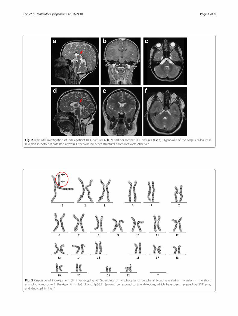
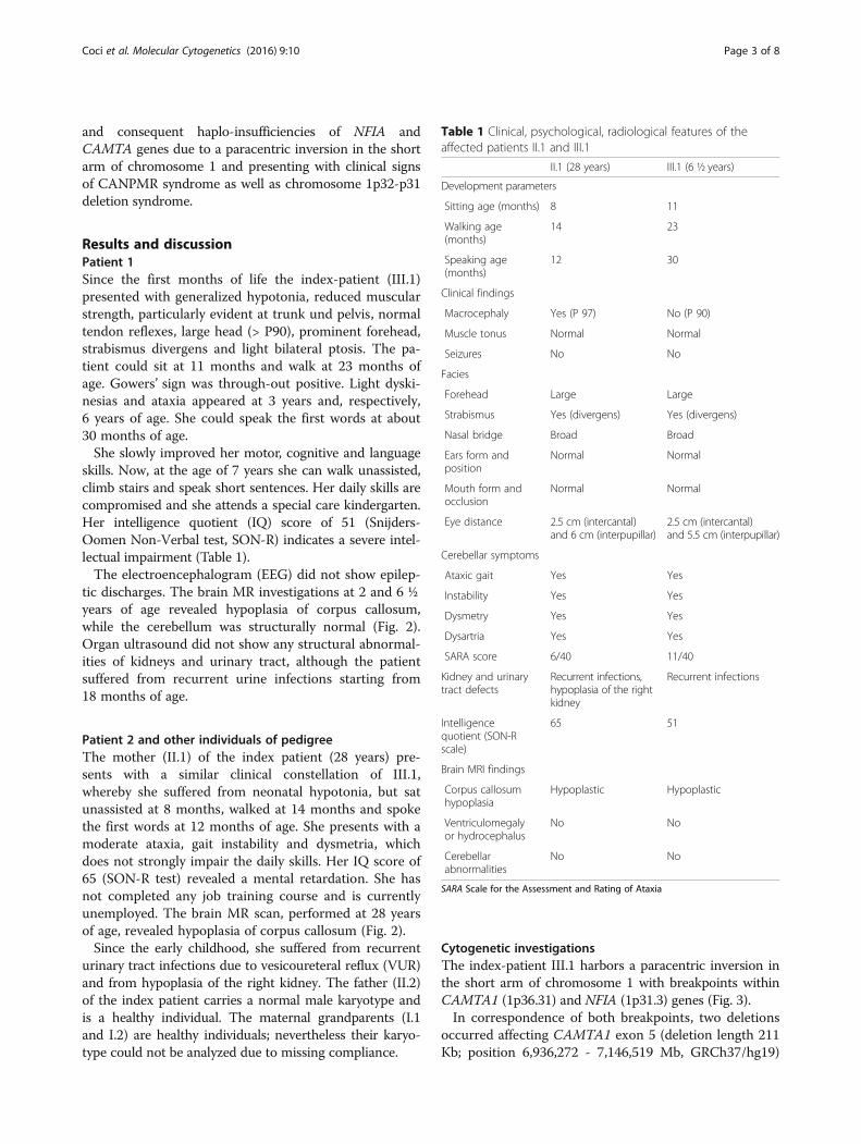
Fig. 2 Brain MR investigation of index-patient (III.1, pictures a, b, c) and her mother (II.1, pictures d, e, f). Hypoplasia of the corpus callosum isrevealed in both patients (red arrows). Otherwise no other structural anomalies were observed
Fig. 3 Karyotype of index-patient (III.1). Karyotyping (GTG-banding) of lymphocytes of peripheral blood revealed an inversion in the shortarm of chromosome 1. Breakpoints in 1p31.3 and 1p36.31 (arrows) correspond to two deletions, which have been revealed by SNP arrayand depicted in Fig. 4
Coci et al. Molecular Cytogenetics (2016) 9:10 Page 4 of 8
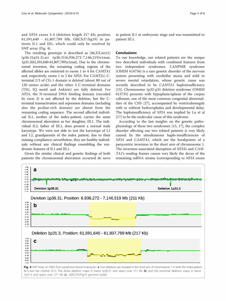
and NFIA exons 3-4 (deletion length 217 Kb; position61,591,640 - 61,807,789 Mb, GRCh37/hg19) in pa-tients II.1 and III1, which could only be resolved bySNP array (Fig. 4).The resulting genotype is described as [46,XX,inv(1)
(p36.31p31.3).arr 1p36.31(6,936,272-7,146,519)x1mat,1p31.3(61,591,640-61,807,789)x1mat]. Due to the chromo-somal inversion, the remaining coding regions of theaffected alleles are restricted to exons 1 to 4 for CAMTA1and, respectively, exons 1 to 2 for NFIA. For CAMTA1, C-terminal 2/3 of CG-1 domain is deleted (about 80 out of120 amino acids) and the other 3 C-terminal domains(TIG, IQ motif and Ankirin) are fully deleted. ForNFIA, the N-terminal DNA binding domain (encodedby exon 2) is not affected by the deletion, but the C-terminal transactivation and repression domains (includingalso the proline-rich domain) are absent from theremaining coding sequence. The second affected individ-ual II.1, mother of the index-patient, carries the samechromosomal aberration as her daughter, III.1. The indi-vidual II.2, father of III.1, does present a normal malekaryotype. We were not able to test the karyotype of I.1and I.2, grandparents of the index patient, due to theirmissing compliance; nevertheless, they are healthy individ-uals without any clinical findings resembling the syn-dromic features of II.1 and III.1.Given the similar clinical and genetic findings of both
patients the chromosomal aberration occurred de novo
in patient II.1 at embryonic stage and was transmitted topatient III.1.
ConclusionsTo our knowledge, our related patients are the uniquetwo described individuals with combined features fromtwo independent syndromes. CANPMR syndrome(OMIM 614756) is a rare genetic disorder of the nervoussystem presenting with cerebellar ataxia and mild tosevere mental retardation, whose genetic cause wasrecently described to be CAMTA1 haploinsufficiency[15]. Chromosome 1p32-p31 deletion syndrome (OMIM613735) presents with hypoplasia/aplasia of the corpuscallosum, one of the most common congenital abnormal-ities of the CNS [27], accompanied by ventriculomegalywith or without hydrocephalus and developmental delay.The haploinsufficiency of NFIA was implied by Lu et al[17] to be the molecular cause of this syndrome.According to the last insights on the genetic patho-
physiology of these two syndromes [15, 17], the complexdisorder affecting our two related patients is very likelycaused by the simultaneous haplo-insufficiencies ofNFIA and CAMTA1, which are the breakpoints of aparacentric inversion in the short arm of chromosome 1.The inversion-associated disruption of NFIA’s and CAM-TA1’s reading frames causes very likely the decay of theremaining mRNA strains (corresponding to NFIA exons
Fig. 4 SNP Array on DNA from peripheral blood leukocytes. a Two deletions are revealed in the short arm of chromosome 1 in both the index-patient(III.1) and her mother (II.1). The distal deletion maps in band 1p36.31 and spans over 211 Kb (b) and the proximal deletion maps in band1p31.3 and spans over 271 Kb (c). (GRC37h/hg19 genome build)
Coci et al. Molecular Cytogenetics (2016) 9:10 Page 5 of 8
1–2 and, respectively CAMTA1 exons 1–4), which aretranscribed from the affected alleles of both genes.If a truncated NFIA protein is synthesized, the C-
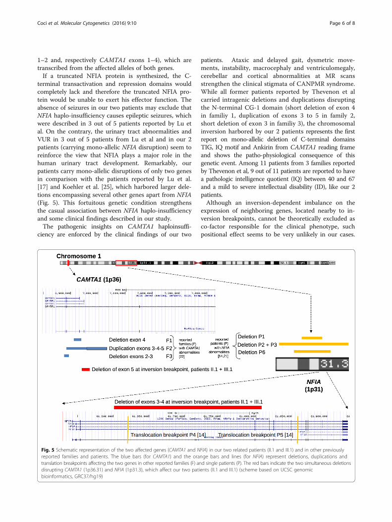
terminal transactivation and repression domains wouldcompletely lack and therefore the truncated NFIA pro-tein would be unable to exert his effector function. Theabsence of seizures in our two patients may exclude thatNFIA haplo-insufficiency causes epileptic seizures, whichwere described in 3 out of 5 patients reported by Lu etal. On the contrary, the urinary tract abnormalities andVUR in 3 out of 5 patients from Lu et al and in our 2patients (carrying mono-allelic NFIA disruption) seem toreinforce the view that NFIA plays a major role in thehuman urinary tract development. Remarkably, ourpatients carry mono-allelic disruptions of only two genesin comparison with the patients reported by Lu et al.[17] and Koehler et al. [25], which harbored larger dele-tions encompassing several other genes apart from NFIA(Fig. 5). This fortuitous genetic condition strengthensthe casual association between NFIA haplo-insufficiencyand some clinical findings described in our study.The pathogenic insights on CAMTA1 haploinsuffi-
ciency are enforced by the clinical findings of our two
patients. Ataxic and delayed gait, dysmetric move-ments, instability, macrocephaly and ventriculomegaly,cerebellar and cortical abnormalities at MR scansstrengthen the clinical stigmata of CANPMR syndrome.While all former patients reported by Thevenon et alcarried intragenic deletions and duplications disruptingthe N-terminal CG-1 domain (short deletion of exon 4in familiy 1, duplication of exons 3 to 5 in family 2,short deletion of exon 3 in familiy 3), the chromosomalinversion harbored by our 2 patients represents the firstreport on mono-allelic deletion of C-terminal domainsTIG, IQ motif and Ankirin from CAMTA1 reading frameand shows the patho-physiological consequence of thisgenetic event. Among 11 patients from 3 families reportedby Thevenon et al, 9 out of 11 patients are reported to havea pathologic intelligence quotient (IQ) between 40 and 67and a mild to severe intellectual disability (ID), like our 2patients.Although an inversion-dependent imbalance on the
expression of neighboring genes, located nearby to in-version breakpoints, cannot be theoretically excluded asco-factor responsible for the clinical phenotype, suchpositional effect seems to be very unlikely in our cases.
Fig. 5 Schematic representation of the two affected genes (CAMTA1 and NFIA) in our two related patients (II.1 and III.1) and in other previouslyreported families and patients. The blue bars (for CAMTA1) and the orange bars and lines (for NFIA) represent deletions, duplications andtranslation breakpoints affecting the two genes in other reported families (F) and single patients (P). The red bars indicate the two simultaneous deletionsdisrupting CAMTA1 (1p36.31) and NFIA (1p31.3), which affect our two patients (II.1 and III.1) (scheme based on UCSC genomicbioinformatics, GRC37/hg19)
Coci et al. Molecular Cytogenetics (2016) 9:10 Page 6 of 8
In fact, no gene mapping in the proximity of deletion1p36.31 (e.g. DNAJC11, THAP3, PHF13, VAMP3, PER3,UTS2) or in the proximity of the deletion 1p31.3 (e.g.CYP2J2, HOOK1, FGGY, TM2D1, INADL, KANK) was as-sociated with brain and urinary tract abnormalities to date.Taken together, in our two patients the study of the
mono-allelic and simultaneous disruption of genesCAMTA1 and NFIA, whose haploinsufficiencies werealready associated to two independent pathologicalphenotypes [15, 17], sheds further light on the clinicaland genetic features of the rare developmental disor-ders CANPMR syndrome and chromosome 1p32-p31deletion syndrome.
MethodsUpon extraction of DNA from total peripheral blood leu-cocytes, conventional karyotyping was performed usingG-banding techniques on stimulated blood lymphocyteswith standard cytogenetic methods and analyzed at 500–550 band resolution. Karyotypes were described accordingto the International System for Human CytogeneticNomenclature (ISCN 2013).SNP array was performed using an Infinium CytoSNP-
850 K microarray (Illumina, San Diego, CA, USA) with anaverage resolution of 18Kb and a practical resolution ingenes CAMTA1 and NFIA of 1 Kb according to the man-ufacturer’s protocol. The data analysis was done usingBlueFuse V4.2 software. The gene alignment was doneusing the University of California Santa Cruz (UCSC) gen-omic bioinformatics browser. The two deleted regionswere mapped using the Genome Research ConsortiumBuild 37 human/human genome 19 (GRCh37/hg19). TheGenBank, Ensembl and OMIM browser accession num-bers for CAMTA1 are NM_015215, ENST00000303635,ENSG00000171735, MIM 611501 and for NFIA areNM_001134673, ENST00000403491, ENSG 00000162599,MIM 600727.Cerebral MRI was performed on clinical 1.5 T MRI
systems (Magnetom Avanto and Aera, Siemens Medical,Germany) using standardized MRI protocols includingmultiplanar T1 and T2-weighted MR-sequences.
Patient consentThe authors obtained the patient consent for the publi-cation of the data.
AbbreviationsCANPMR: cerebellar ataxia non progressive with mental retardation; SARA: scalefor the assessment and rating of ataxia; SON-R: Snijders-Oomen non-verbal test.
Competing interestsThe authors declare that they have no competing interests.
Authors’ contributionsEC, TL, AS, HL, JR participated in the design of the study and analyzed thepatients’ phenotype. EC and UK perfomed the genetic studies. CF performed
the neuro-radiological investigations. All authors drafted and approved thefinal manuscript.
AcknowledgementsThe authors express our deep gratitude to the family for the full collaborationthroughout the project.
Author details1Center of Social Pediatrics and Pediatric Neurology, General Hospital ofCelle, 29221 Celle, Germany. 2Medizinisch Genetisches Zentrum, 80335Munich, Germany. 3Institute of Human Genetics, Friedrich Schiller University,Jena University Hospital, 07743 Jena, Germany. 4Department of Radiology,General Hospital of Celle, 29223 Celle, Germany.
Received: 5 November 2015 Accepted: 26 January 2016
References1. Steinlin M, Zangger B, Boltshauser E. Non-progressive congenital ataxia with
or without cerebellar hypoplasia: a review of 34 subjects. Dev Med Child Neurol.1998;40:148–54.
2. Yapici Z, Eraksoy M. Non-progressive congenital ataxia with cerebellarhypoplasia in three families. Acta Paediatr. 2005;94:248–53.
3. Guzzetta F, Mercuri E, Bonanno S, Longo M, Spano M. Autosomal recessivecongenital cerebellar atrophy. A clinical and neuropsychological study.Brain Dev. 1993;15:439–45.
4. Imamura S, Tachi N, Oya K. Dominantly inherited early-onset non-progressive cerebellar ataxia syndrome. Brain Dev. 1993;15:372–6.
5. Bomar JM, Benke PJ, Slattery EL, Puttagunta R, Taylor LP, Seong E, et al. Mutationsin a novel gene encoding a CRAL-TRIO domain cause human Cayman ataxiaand ataxia/dystonia in the jittery mouse. Nat Genet. 2003;35:264–9.
6. Tranebjaerg L, Teslovich TM, Jones M, Barmada MM, Fagerheim T, Dahl A, etal. Genome-wide homozygosity mapping localizes a gene for autosomalrecessive non-progressive infantile ataxia to 20q11-q13. Hum Genet. 2003;113:293–5.
7. Boycott KM, Flavelle S, Bureau A, Glass HC, Fujiwara TM, Wirrell E, et al.Homozygous deletion of the very low density lipoprotein receptor genecauses autosomal recessive cerebellar hypoplasia with cerebral gyralsimplification. Am J Hum Genet. 2005;77:477–83.
8. Nicolas E, Poitelon Y, Chouery E, Salem N, Levy N, Mégarbané A, et al.CAMOS, a nonprogressive, autosomal recessive, congenital cerebellar ataxia,is caused by a mutant zinc-finger protein, ZNF592. Eur J Hum Genet. 2010;18:1107–13.
9. Lise S, Clarkson Y, Perkins E, Kwasniewska A, Sadighi Akha E, SchnekenbergRP, et al. Recessive mutations in SPTBN2 implicate β-III spectrin in bothcognitive and motor development. PLoS Genet. 2012;8(12):e1003074.doi:10.1371/journal.pgen.1003074.
10. Burns R, Majczenko K, Xu J, Peng W, Yapici Z, Dowling JJ, et al. Homozygoussplice mutation in CWF19L1 in a Turkish family with recessive ataxia syndrome.Neurology. 2014;83:2175–82.
11. Jobling RK, Assoum M, Gakh O, Blaser S, JRaiman JA, Mignot C, et al. PMPCAmutations cause abnormal mitochondrial protein processing in patientswith non-progressive cerebellar ataxia. Brain. 2015;138:1505–17.
12. Finkler A, Ashery-Padan R, Fromm H. CAMTA1s: calmodulin-binding transcriptionactivators from plants to human. FEBS Lett. 2007;581:3893–8.
13. Han J, Gong P, Redding K, Mitra M, Guo P, Li HS. The fly CAMTA transcriptionfactor potentiates deactivation of rhodopsin, a G protein-couples lightreceptor. Cell. 2006;127:847–58.
14. Gong P, Han L, Redding K, Li HS. A potential dimerization region of dCAMTA iscritical termination of fly visual response. J Biol Chem. 2007;282:21253–8.
15. Thevenon J, Lopez E, Keren B, Heron D, Mignot C, Altuzarra C, et al.Intragenic CAMTA1 rearrangements cause non-progressive congenital ataxiawith or without intellectual disability. J Med Genet. 2012;49:400–8.
16. Campbell CG, Wang H, Hunter GW. Interstitial microdeletion of chromosome1p in two siblings. Am J Med Genet. 2002;111:289–94.
17. Lu W, Quintero-Rivera F, Fan Y, Alkuraya FS, Donovan DJ, Xi Q, et al. NFIAhaploinsufficiency is associated with a CNS malformation syndrome andurinary tract defects. PLoS Genet. 2007;3:830–43.
18. Qian F, Kruse U, Lichter P, Sippel AE. Chromosomal localization of the fourgenes (NFIA, B, C, and X) for the human transcription factor nuclear factorI by FISH. Genomics. 1995;28:66–73.
Coci et al. Molecular Cytogenetics (2016) 9:10 Page 7 of 8
19. Grunder A, Qian F, Ebel TT, Mincheva A, Lichter P, Kruse U, et al. Genomicorganization, splice products and mouse chromosomal localization of genesfor transcription factor Nuclear Factor One. Gene. 2003;304:171–81.
20. Gronostajski RM. Roles of the NFI/CTF gene family in transcription anddevelopment. Gene. 2000;249:31–45.
21. Das Neves L, Duchala CS, Godinho F, Haxhiu MA, Colmenares C, MacklinWB, et al. Disruption of the murine nuclear factor I-A gene (Nfia) results inperinatal lethality, hydrocephalus, and agenesis of the corpus callosum.PNAS. 1999;96:11946–51.
22. Wong YW, Schulze C, Streichert T, Gronostajski RM, Schachner M, Tilling T.Gene expression analysis of nuclear factor I-A deficient mice indicatesdelayed brain maturation. Genome Biol. 2007;8:R72.
23. Steele-Perkins G, Butz KG, Lyons GE, Zeichner-David M, Kim HJ, Cho MI,et al. Essential role of NFI-C/CTF transcription-replication factor in toothroot development. Mol Cell Biol. 2003;23:1075–84.
24. Steele-Perkins G, Plachez C, Butz KG, Yang G, Bachurski CJ, Kinsman SL, et al.The transcription factor gene Nfib is essential for both lung maturation andbrain development. Mol Cell Biol. 2005;25:685–98.
25. Koehler U, Holinski-Feder E, Ertl-Wagner B, Kunz J, von Moers A, von Voss H,et al. A novel 1p31.3p32.2 deletion involving the NFIA gene detected byarray CGH in a patient with macrocephaly and hypoplasia of the corpuscallosum. Eur J Pediatr. 2010;169:463–8.
26. Nyboe D, Kreiborg S, Kirchhoff M, Hove HB. Familial craniosynostosis associatedwith a microdeletion involving the NFIA gene. Clin Dysmorphol. 2015;24:109–12.
27. Paul LK, Brown WS, Adolphs R, Tyszka JM, Richards LJ, Mukherjee P, et al.Agenesis of the corpus callosum: genetics, development and functionalaspects of the connectivity. Nat Rev Neurosci. 2007;8:287–99.
• We accept pre-submission inquiries
• Our selector tool helps you to find the most relevant journal
• We provide round the clock customer support
• Convenient online submission
• Thorough peer review
• Inclusion in PubMed and all major indexing services
• Maximum visibility for your research
Submit your manuscript atwww.biomedcentral.com/submit
Submit your next manuscript to BioMed Central and we will help you at every step:
Coci et al. Molecular Cytogenetics (2016) 9:10 Page 8 of 8