research open access active demethylation in mouse zygotes
TRANSCRIPT

Santos et al. Epigenetics & Chromatin 2013, 6:39http://www.epigeneticsandchromatin.com/content/6/1/39
RESEARCH Open Access
Active demethylation in mouse zygotes involvescytosine deamination and base excision repairFátima Santos1*, Julian Peat1, Heather Burgess1,3, Cristina Rada2, Wolf Reik1,3 and Wendy Dean1
Abstract
Background: DNA methylation in mammals is an epigenetic mark necessary for normal embryogenesis. Duringdevelopment active loss of methylation occurs in the male pronucleus during the first cell cycle after fertilisation.This is accompanied by major chromatin remodelling and generates a marked asymmetry between the paternaland maternal genomes. The mechanism(s) by which this is achieved implicate, among others, base excision repair(BER) components and more recently a major role for TET3 hydroxylase. To investigate these methylation dynamicsfurther we have analysed DNA methylation and hydroxymethylation in fertilised mouse oocytes by indirectimmunofluorescence (IF) and evaluated the relative contribution of different candidate factors for activedemethylation in knock-out zygotes by three-dimensional imaging and IF semi-quantification.
Results: We find two distinct phases of loss of paternal methylation in the zygote, one prior to and anothercoincident with, but not dependent on, DNA replication. TET3-mediated hydroxymethylation is limited to thereplication associated second phase of demethylation. Analysis of cytosine deaminase (AID) null fertilised oocytesrevealed a role for this enzyme in the second phase of loss of paternal methylation, which is independent fromhydroxymethylation. Investigation into the possible repair pathways involved supports a role for AID-mediatedcytosine deamination with subsequent U-G mismatch long-patch BER by UNG2 while no evidence could befound for an involvement of TDG.
Conclusions: There are two observable phases of DNA demethylation in the mouse zygote, before andcoincident with DNA replication. TET3 is only involved in the second phase of loss of methylation. Cytosinedeamination and long-patch BER mediated by UNG2 appear to independently contribute to this second phaseof active demethylation. Further work will be necessary to elucidate the mechanism(s) involved in the first phaseof active demethylation that will potentially involve activities required for early sperm chromatin remodelling.
Keywords: Epigenetic reprogramming, DNA methylation, Hydroxymethylation, AID, BER, UNG2, TET3
BackgroundDNA methylation is an important epigenetic mark in-volved in gene silencing, X chromosome and transposoninactivation, genomic imprinting, and chromosome stabi-lity. DNA methylation is subject to reprogramming duringdevelopment, involving both demethylation (active andpassive) and de novo methylation phases. To date, themost clear examples of active DNA demethylation takeplace during the very early steps of mammalian develop-ment, namely in the zygote where the paternal genomeundergoes a massive wave of loss of 5-methylcytosine(5mC) right after fertilisation [1-4].
* Correspondence: [email protected] Programme, The Babraham Institute, Cambridge CB22 3AT, UKFull list of author information is available at the end of the article
© 2013 Santos et al.; licensee BioMed CentralCommons Attribution License (http://creativecreproduction in any medium, provided the or
Within 1 h of fertilisation, the paternal genome goesthrough major chromatin remodelling, loses protaminesand is re-packaged by maternal nucleosomal histones,forming the paternal pronucleus [5,6]. Post-fertilisationdevelopment can be defined by the pronuclear stagesPN0/1 to PN5; PN0-PN2 embryos are in the G1 phase,PN3 and PN4 embryos are largely in S phase, replicatingboth the paternal and maternal genomes, and PN5 em-bryos are mostly in the post-replicative G2 phase [3,7-9].Several reports have shown that the paternal genomeundergoes genome-wide DNA demethylation via an ac-tive mechanism before replicating its DNA [1-4]. Thesearch for enzymes responsible for this demethylationhas produced numerous candidates and reaction mecha-nisms [10-13]. These fall within three main groups: (1)
Ltd. This is an open access article distributed under the terms of the Creativeommons.org/licenses/by/2.0), which permits unrestricted use, distribution, andiginal work is properly cited.
Santos et al. Epigenetics & Chromatin 2013, 6:39 Page 2 of 12http://www.epigeneticsandchromatin.com/content/6/1/39
direct removal of the methyl group from the 5-C pos-ition of cytosine; (2) DNA repair, either base excision re-pair (BER) or nucleotide excision repair (NER); and (3)iterative enzymatic oxidation leading to conversion of 5mCto 5- hydroxymethylcytosine (5hmC), 5-formylcytosine(5fC), and 5-carboxylcytosine (5caC).The methyl-CpG-binding domain protein 2 (MBD2)
was reported to possess direct demethylase activity [14],but the result could not be reproduced by others. DNAglycosylases have been described in plants that can remove5mC, leaving an abasic site that is repaired by the BER ma-chinery but mammalian glycosylases (e.g., thymine DNAglycosylase (TDG) and methyl-CpG-binding domain pro-tein 4 (MBD4)) show weak activity on 5mC in vitro [15].However, both MBD2 and MBD4 null fertilised oocytesundergo paternal loss of DNA methylation indistinguish-able from matched controls [16].An alternative to direct removal of 5mC by a DNA
glycosylase is enzymatic deamination of 5mC to thymine,followed by T-G mismatch specific BER that replaces thy-mine with cytosine [17]. Two classes of enzymes havebeen proposed to be capable of carrying out the first stepin this process: cytosine deaminases and DNA methyl-transferases (reviewed in [11]). Cytosine DNA deaminasesconvert cytosine to uracil in nucleic acids and are wellknown from their roles in RNA editing, viral defence andantibody diversification [18]. Recently a series of resultshave pointed to an involvement of activation-induced de-aminase (AID) mediated cytosine deamination in DNAdemethylation in primordial germ cells (PGCs) and in-duced pluripotent stem (iPS) cell reprogramming, in can-cer and embryonic stem (ES) cell gene expression [19-21].Furthermore, overexpression of AID and MBD4 havebeen described to cause general demethylation of the zeb-rafish embryo genome, suggesting that deamination of5mC followed by BER of T-G mismatches results in de-methylation [22]. Gadd45, a p53-inducible gene involvedin a variety of cellular processes, seems to facilitate thisprocess and it has also been shown to interact withnucleotide excision repair (NER) components [22,23].Models have been proposed for Gadd45 mediated de-methylation of DNA either by deamination followedby BER or NER, or even by a combination involvingconsecutive NER and BER mechanisms (reviewed in[24]). AID's best defined activity is in B lymphocytes,where deamination of cytosines leading to uracil initi-ates both somatic hypermutation and immunoglobinclass switch recombination [25-27]. However, its ex-pression in mouse oocytes as well as in ES cells andPGCs [28], make it a potential candidate for perfor-ming global demethylation. In vitro assays have shownAID has 5mC deaminase activity, resulting in thymineand, therefore, T-G mismatches in DNA, which can beeffectively repaired through the BER pathway [28].
Cytosine and 5-methylcytosine can also be enzymaticallydeaminated by DNA methyltransferases (DNMTs), pri-marily known as enzymes that transfer a methyl groupto the C-5 position of cytosine from the methyl donorS-adenosylmethionine (SAM- reviewed in [29]). Recentwork in mammalian cell lines has led to the proposalthat deamination by the Dnmt3a and Dnmt3b DNAmethyltransferases could be a means of achieving fast,active DNA demethylation at promoters undergoingtranscriptional cycling, by generating thymine, whichis repaired via TDG and other enzymes [12,30-32].More recently, studies have suggested that loss ofDNA methylation in the paternal genome in the zygoteis primarily dependent on TET3 [4,33], a member ofthe ten-eleven translocation (TET) family of DNAdioxygenases, which are capable of converting 5mC to5- hydroxymethylcytosine (5hmC), 5-formylcytosine (5fC)and 5-carboxylcytosine (5caC) through iterative oxidation[34], suggesting that the main mechanism involved in ac-tive genome-wide demethylation is via oxidation of 5mC.Although these various models for active loss of DNAmethylation from the paternal pronucleus have been pro-posed, they have, for the most part, overlooked the evi-dence that this process occurs in two phases - before andcoincident with DNA replication. To investigate this DNAmethylation dynamics we have analysed fertilised mouseoocytes, by indirect immunofluorescence (IF) of DNAmethylation and hydroxymethylation, and evaluated therelative contribution of different activities to active de-methylation, by three-dimensional (3D) imaging and IFsemi-quantification.
Results and discussionWe have previously characterised the dynamics of DNAmethylation loss during the first cell cycle in the mouseby indirect immunofluorescence [3,35]. Since then, sev-eral similar studies have been published, more recentlyincluding other modifications of DNA methylation, namelyhydroxymethylation, and attempts have been made tosemi-quantify the immunofluorescence signals in order toget a sense of the kinetics of demethylation [4,8,33,36,37].These approaches have provided clear evidence for the oxi-dation of 5mC in the zygote, specifically in the paternalpronucleus, seemingly explaining the concomitant de-crease in DNA methylation.We have established a protocol that allows us to repro-
ducibly semi-quantify the staining levels of both DNAmethylation and hydroxymethylation in pre-implantationembryos (see Methods and Additional files 1 and 2) andhave used it to validate qualitative immunofluorescenceresults suggesting that, by the time the fertilised oocyteenters G1 a substantial amount of DNA methylationsignal has already been lost from the paternal pro-nucleus, prior to the increase in the observed levels of

Santos et al. Epigenetics & Chromatin 2013, 6:39 Page 3 of 12http://www.epigeneticsandchromatin.com/content/6/1/39
hydroxymethylation (Figure 1A). To probe the kineticsof these two processes (loss of 5mC and increase in5hmC) we have captured 3D data of fertilised oocytessimultaneously stained for 5mC and 5hmC. The signalintensity of both modifications was semi-quantifiedand the ratio between the paternal and maternal pro-nuclei in each individual one-cell embryo (from PN1to PN5) calculated. The initial values for the two com-plements cannot be measured using this protocol ascondensed sperm is notably impervious to immuno-staining due to its highly compacted chromatin state[6], resulting in unreliable semi-quantification valuesfor PN0. Based on the levels of DNA methylation for
2
1
1.5
0.5
5mC
5mC male/female
5hmC male/female
5mC
5hmC
B6x
B6
PN0 PN1A
Meiosis II completion G1
B
pb
m
f
m
fpb
m
f
pb
m
f
pb
pb
pb
Phase I
Figure 1 Paternal loss of DNA methylation occurs in two phases. (Aprojections of Z-stack images of control (B6 x B6) pronuclear stage embryos (hydroxymethylation (5hmC) clearly showing two phases of paternal loss ofno observable change in DNA hydroxymethylation, and Phase II, when Dincrease in DNA hydroxymethylation in the paternal pronucleus takes plpolar body. (B) The dynamic changes of DNA methylation and hydroxymof the total immunofluorescence signal (3D imaging) between the materespectively. Values at time of fertilisation are hypothetical (dashed lines), calcoocytes (see text for explanation). Values plotted for stages between PN1 and
mouse sperm and oocytes, reported by genome-wideor reduced representation bisulphite-sequencing, anddespite variability in the quantification methods, there is ageneral agreement that DNA methylation levels in spermare twice that observed in the oocyte [38,39]. It is there-fore possible to calculate a conservative (using the lowestestimate based on whole-genome bisulphite-sequencing,[38]) theoretical paternal to maternal ratio of DNA methy-lation of 2 for PN0 (Figure 1B). Functional grouping of thedata according to cell cycle [8], corresponding to embryosin G1 (Early; PN1-PN2) and in S-G2 phase (Mid-Late;PN3-PN5), was used to compare our data with that col-lected by others. A summary of paternal to maternal ratios
6
4.5
3
1.5
5hmC
PN2 PN3 PN4 PN5
S G2
m
f
m
f
m
fpb
m
fpb
mf
pb
mf
pb
Phase II
) Diagrammatic illustration and representative two-dimensional (2D)PN0 to PN5) simultaneously stained for DNA methylation (5mC) andmethylation, Phase I, corresponding to a pre-replicative state withNA replication is taking place, and during which a very significantace. Scale bar 25 μm. f, female pronucleus; m, male pronucleus; pb,ethylation during the first cell cycle can be represented by the ratiornal and paternal pronuclei (male/female ratio) for 5mC and 5hmC,ulated considering a minimum initial total 5mC and 5hmC for sperm andPN5 (minimum 10 embryos per stage). Bars indicate standard deviation.

Table 2 DNA hydroxymethylation (5hmC) average paternalto maternal ratios (indirect immunofluorescence)
Study Early(PN1-PN2)
Mid-Late(PN3-PN5)
Mouse strain
Iqbal et al., 20111 0.78 2.82 FVB
Wossidlo et al., 20111 0.85 2.44 BDF1 (C57BL/6 x DBA)
Inoue and Zhang, 2011 1.89 4.98 BDF1 (C57BL/6 x DBA)
Salvaing et al., 20121 0.9 2.23 F1 (C57BL/6 x CBA)
This study 1.334 4.01 C57BL/6 J1DNA staining was used for normalisation of paternal/maternal ratios.
Santos et al. Epigenetics & Chromatin 2013, 6:39 Page 4 of 12http://www.epigeneticsandchromatin.com/content/6/1/39
of 5mC levels based on reports from the literature, as wellas this work, is presented in Table 1. From its examinationit becomes clear that, in all cases and consistentlythroughout the data generated by multiple independentlabs, the paternal/maternal ratio of DNA methylation inG1 is never close to the theoretical initial PN0 value of 2,predicted from the 5mC levels reported for the gametes.There is thus an observable first loss of paternal DNAmethylation levels of approximately 60% by the time thezygote enters G1 (average ratio drop from 2 to 0.8) and afurther loss of 20% (average ratio drop from 0.8 to 0.5)thereafter. This supports the distinction between twophases of active demethylation, one in G1 and the other inS-G2 (Figure 1A, B). In contrast, the levels of hydroxy-methylation, evaluated the same way, show a rather differ-ent profile. While the paternal/maternal ratio remainsrelatively stable during Phase I, there is a remarkable in-crease during Phase II with a marked rise in the 5hmCsignal in the paternal pronucleus (Figure 1A, B). Althoughconcomitant with S-phase, this increase in paternal hydro-xymethylation is not dependent on DNA replication ([4]and Additional file 3). This is again in agreement with ouranalysis of results reported in the literature, which showsa significant increase in the 5hmC paternal/maternal ratiooccurring between Early (Phase I) and Mid-Late (Phase II)one-cell embryos (Table 2). The changes in 5hmC signalpoint to the involvement of TET-mediated 5mC oxidationduring Phase II, but not Phase I, of paternal loss of DNAmethylation. In order to test this hypothesis, we used aconditional (Zp3-Cre) TET3 knock-out mouse generatedin the lab. TET3 null oocytes were fertilised by C57Bl/6 J(B6) sperm and the same simultaneous 5mC and 5hmCstaining and semi-quantification analysis was performed(Figure 2). The results confirmed the dependency of pater-nal hydroxymethylation on the maternally derived TET3protein [33] with no observable increase in 5hmC stainingin Mid-Late one-cell embryos (Figure 2A) and consequentlya very significant difference in the paternal/maternal ratiosbetween TET3 MAT KO and control B6 fertilised oocytes(Figure 2B). The absence of paternal 5hmC signal increasewas accompanied by an equally significant rise in paternalDNA methylation staining levels in PN3-PN5 one-cell
Table 1 DNA methylation (5mC) average paternal tomaternal ratios (indirect immunofluorescence)
Study Early(PN1-PN2)
Mid-Late(PN3-PN5)
Mouse strain
Iqbal et al., 20111 0.9 0.47 FVB
Wossidlo et al., 20111 0.53 0.34 BDF1 (C57BL/6 x DBA)
Inoue and Zhang, 2011 0.85 0.61 BDF1 (C57BL/6 x DBA)
Salvaing et al., 20121 0.72 0.47 F1 (C57BL/6 x CBA)
This study 0.84 0.62 C57BL/6 J1DNA staining was used for normalisation of paternal/maternal ratios.
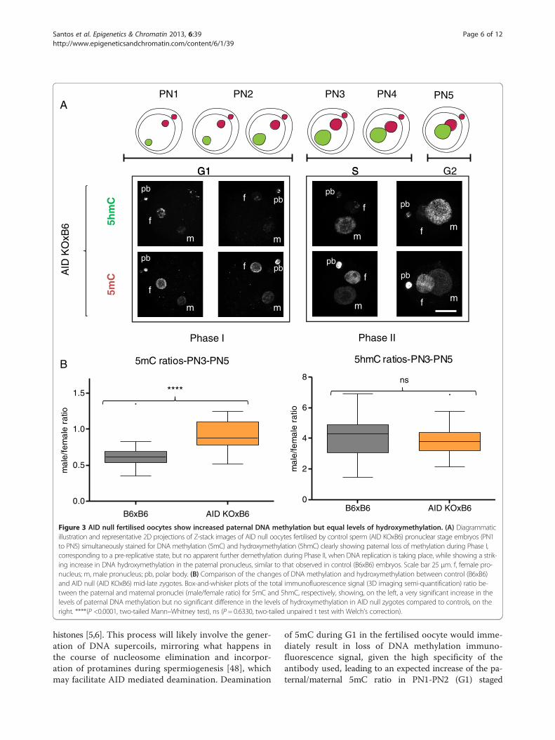
embryos (Figure 2B), supporting a role for TET3-mediatedhydroxymethylation in the loss of paternal DNA methyla-tion at this stage. We then concentrated our attention onthe first phase of paternal DNA methylation loss that seemsto be independent of hydroxymethylation. DNA repair hasbeen proposed as a mechanism to explain active DNA de-methylation and there is evidence for DNA repair pathwaysbeing involved in paternal DNA methylation loss in the zyg-ote, particularly BER [11,40,41]. Moreover, deamination hasbeen implicated as a possible upstream event initiating theBER mediated demethylation [42]. Activation-inducedcytidine deaminase has also been shown to be capableof deaminating 5mC and is expressed in pluripotenttissues, including mouse oocytes [28]. AID was initiallythought to be only relevant in B-lymphocytes, where itis essential for somatic hypermutation and class-switchrecombination [26,27]. Recently AID has been shownto be involved in dynamic methylation changes in avariety of tissues ranging from PGCs [21] to ES andiPS cells [20,43]. The presence of AID protein in con-trol mouse oocytes was confirmed by indirect im-munofluorescence (Additional file 4). Making use ofthe same AID knock-out mouse model that showed al-tered DNA methylation levels in PGCs [21], AID nulloocytes were fertilised by either AID (Additional file 5)or B6 (Figure 3) sperm and the same simultaneous5mC and 5hmC staining and semi-quantification ana-lysis was performed. A significant difference was foundin the paternal/maternal ratio of DNA methylation be-tween AID and control (B6) fertilised oocytes, with noapparent effect on the levels of hydroxymethylation(Figure 3B). Notably, this difference was only detect-able in PN3-PN5 (Phase II) embryos and no differencecould be seen in PN1-PN2 (Phase I) zygotes (Figure 3Aand Additional file 5) despite reports indicating thatAID works during G1 [44]. AID deaminates cytosines atimmunoglobulin genes on single-stranded DNA thoughtto be made available during transcription [45], howeverpost-fertilisation mouse oocytes are transcriptionally silent[46] thus necessitating other means of generating single-stranded DNA substrates. As such, it is reasonable to ex-pect that AID might be able to deaminate immediately

5mC ratios-PN3-PN5
mal
e/fe
mal
era
tio
B6xB6 TET3 MAT KOxB6
0.0
0.5
1.0
1.5
A
G2G1G1 SS
Phase I Phase II
5mC
5hmC
TE
T3
MA
TK
OxB
6
PN1 PN2 PN3 PN4 PN5
B
m
f
pb
5hmC ratios-PN3-PN5
mal
e/fe
mal
era
tio
B6xB6 TET3 MAT KOxB60
2
4
6
8****
****
m
f
pb
m
f
pb
m
f
pb
m
f
pb
m
f
pb
m
f
pb
m
f
pb
Figure 2 TET3 null fertilised oocytes show increased paternal DNA methylation and reduced hydroxymethylation. (A) Diagrammaticillustration and representative 2D projections of Z-stack images of TET3 maternally deleted oocytes fertilised by control sperm (TET3 MAT KOxB6)pronuclear stage embryos (PN1 to PN5) simultaneously stained for DNA methylation (5mC) and hydroxymethylation (5hmC) clearly showingpaternal loss of methylation during Phase I, corresponding to a pre-replicative state, but no apparent further demethylation during Phase II,when DNA replication is taking place, and during which there is complete failure in TET3 null oocytes to generate DNA hydroxymethylationin the paternal pronucleus. Scale bar 25 μm. f, female pronucleus; m, male pronucleus; pb, polar body. (B) Comparison of the changes ofDNA methylation and hydroxymethylation between control (B6xB6; Figure 1A) and TET3 maternally deleted (TET3 MAT KOxB6) mid-latezygotes. Box-and-whisker plots of the total immunofluorescence signal (3D imaging semi-quantification) ratio between the paternal andmaternal pronuclei (male/female ratio) for 5mC and 5hmC, respectively, showing, on the left, a very significant increase in the levels ofpaternal DNA methylation and, on the right, an equally significant decrease in the levels of hydroxymethylation in TET3 maternally deleted zygotescompared to controls. ****(P <0.0001, two-tailed Mann–Whitney test).
Santos et al. Epigenetics & Chromatin 2013, 6:39 Page 5 of 12http://www.epigeneticsandchromatin.com/content/6/1/39
after fertilisation during nucleoprotamine exchange, a timewhen single-stranded DNA would be accessible. Recentlyit has been described that, in both class-switching B-cellsand E. coli, negative DNA supercoiling, and hence
generation of single-stranded DNA, enhances AID muta-genesis [47]. Within 1 h after fertilisation the paternalchromatin suffers a complete remodelling resulting in anexchange of the sperm protamines by maternally derived
5hmC ratios-PN3-PN5
mal
e/fe
mal
era
tio
B6xB6 AID KOxB60
2
4
6
8
5mC ratios-PN3-PN5
mal
e/fe
mal
era
tio
B6xB6 AID KOxB6
0.0
0.5
1.0
1.5
A
****ns
G2G1G1 SS
Phase I Phase II
5mC
5hmC
AID
KO
xB6
PN N2 PN1 P 3 PN4 PN5
B
m
f
pb
m
f
pb
m
f pb
m
f pb
m
f
pb
m
f
pb
mf
pb
mf
pb
Figure 3 AID null fertilised oocytes show increased paternal DNA methylation but equal levels of hydroxymethylation. (A) Diagrammaticillustration and representative 2D projections of Z-stack images of AID null oocytes fertilised by control sperm (AID KOxB6) pronuclear stage embryos (PN1to PN5) simultaneously stained for DNA methylation (5mC) and hydroxymethylation (5hmC) clearly showing paternal loss of methylation during Phase I,corresponding to a pre-replicative state, but no apparent further demethylation during Phase II, when DNA replication is taking place, while showing a strik-ing increase in DNA hydroxymethylation in the paternal pronucleus, similar to that observed in control (B6xB6) embryos. Scale bar 25 μm. f, female pro-nucleus; m, male pronucleus; pb, polar body. (B) Comparison of the changes of DNA methylation and hydroxymethylation between control (B6xB6)and AID null (AID KOxB6) mid-late zygotes. Box-and-whisker plots of the total immunofluorescence signal (3D imaging semi-quantification) ratio be-tween the paternal and maternal pronuclei (male/female ratio) for 5mC and 5hmC, respectively, showing, on the left, a very significant increase in thelevels of paternal DNA methylation but no significant difference in the levels of hydroxymethylation in AID null zygotes compared to controls, on theright. ****(P <0.0001, two-tailed Mann–Whitney test), ns (P = 0.6330, two-tailed unpaired t test with Welch’s correction).
Santos et al. Epigenetics & Chromatin 2013, 6:39 Page 6 of 12http://www.epigeneticsandchromatin.com/content/6/1/39
histones [5,6]. This process will likely involve the gener-ation of DNA supercoils, mirroring what happens inthe course of nucleosome elimination and incorpor-ation of protamines during spermiogenesis [48], whichmay facilitate AID mediated deamination. Deamination
of 5mC during G1 in the fertilised oocyte would imme-diately result in loss of DNA methylation immuno-fluorescence signal, given the high specificity of theantibody used, leading to an expected increase of the pa-ternal/maternal 5mC ratio in PN1-PN2 (G1) staged
Santos et al. Epigenetics & Chromatin 2013, 6:39 Page 7 of 12http://www.epigeneticsandchromatin.com/content/6/1/39
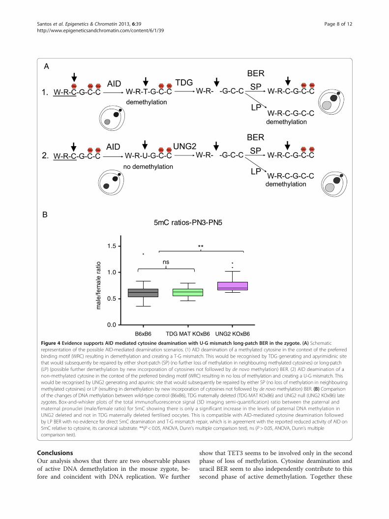
zygotes compared to B6 controls. The fact that no signifi-cant difference could be found suggests that no such de-amination has occurred in the control zygotes. The resultsobtained were therefore surprising. Investigating theknown mechanisms for DNA repair following AID-mediated deamination provided a possible explanation(Additional file 6). Repair can be achieved by differentmechanisms, both error-prone and error-free [49]. De-amination of 5mC generates T creating a T-G mismatchwhich is the preferred substrate for TDG and MBD4 gly-cosylases [28,50]. On the other hand, deamination of Cgenerates U and uracil residues in DNA are also largelyresolved by BER, with the uracil being removed by a DNAglycosylase [50]. Uracyl glycosylase (UNG2) has been im-plicated as the major glycosylase responsible for repair ofC to U mismatches following deamination [51]. BER en-ables the repair of damaged DNA via two general path-ways, short-patch and long-patch [50,52,53]. Short-patch(SP) BER replaces a single nucleotide by polymerase β andthe newly synthesized DNA is sealed by DNA ligase III/X-ray cross-complementing group 1 (XRCC1) heterodimer.Long-patch (LP) BER inserts two to 13 nucleotides by co-ordinate action of polymerase δ, proliferating cell nuclearantigen (PCNA), flap endonuclease 1 and DNA ligase I.Although at face value only the deamination of 5mC couldresult in demethylation, in the case of cytosine deamin-ation followed by LP BER, there would be the possibilityfor any methylated cytosines adjacent to the U-G mis-match to be replaced by newly incorporated non-modifiedcytosines and the original methylated state would be lost,resulting in demethylation (Additional file 6). It is thuspossible to achieve DNA methylation loss through a C toU deamination and subsequent replacement of adjacentmethylated cytosines by LP BER (Figure 4A). To resolvethese potential alternative downstream pathways, analysisof knock-out mouse oocytes for either TDG [54] orUNG2 [55], fertilised by B6 sperm, was performed(Figure 4B). The results support a role for AID mediatedcytosine deamination with subsequent U-G mismatch LPBER and no evidence could be found for direct 5mC de-amination and T-G mismatch repair. This is in agreementwith the reported reduced activity of AID on 5mC whencompared to cytosine, its canonical substrate [10,28].Furthermore, PARP1, a hallmark of LP BER [52,53](Additional file 6), has been found to be predominantlyconfined to the paternal pronucleus in PN3-PN5 stagedzygotes [40,41] and its specific inhibition caused a signifi-cant increase in the paternal/maternal ratio of DNAmethylation compared to controls [40]. In this scenario,AID-mediated cytosine deamination can still occur in G1(PN1-PN2), without causing any immediate 5mC loss,and the resulting U-G mismatches could later be repairedby LP BER, presumably causing loss of adjacent methyl-ated cytosines with consequent demethylation in PN3-
PN5 staged zygotes. Although speculative, this model canfully account for the results obtained.According to recent reports, deamination of cytosine
and 5-methylcytosine could also be mediated by DNAmethyltransferases, namely the Dnmt3a and Dnmt3bde novo methyltransferases (reviewed in [11,12]) andmore recently that DNA could be directly demethylatedby the same enzymes, at least in an in vitro system [56].Moreover, results from ES cells suggest that TET andDnmt3 enzymes may interact at common binding sites[57]. These prompted us to investigate whether Dnmt3anull oocytes were capable of demethylating a control(B6) sperm using a conditional (Zp3-Cre) Dnmt3aknock-out mouse model [58]. Oocytes null for Dnmt3aare severely depleted of DNA methylation [59] whichprecluded the use of the paternal/maternal ratio as ameasure of demethylation as the maternal pronucleusshowed only residual levels of 5mC staining (Additionalfile 7A); instead we directly compared the total levels ofimmunofluorescence signal in the paternal pronuclei ofDnmt3a null, control (B6) and AID null oocytes ferti-lised by B6 sperm (Additional file 7B). No significant dif-ference, in either 5mC or 5hmC levels, between paternalpronuclei in control and Dnmt3a null fertilised oocyteswas observed, whereas both are significantly differentfrom AID null in 5mC but not in 5hmC levels (Additionalfile 7B). These results do not support a role for Dnmt3ain paternal active demethylation in the mouse one-cellembryo.As a whole, our results support a role for both 5mC
hydroxymethylation and cytosine deamination, followedby LP BER, as demethylation mechanisms in the mousezygote. Both these mechanisms seem to contribute in-dependently to a decrease in paternal DNA methyla-tion coincident, but not reliant, on DNA replication.Furthermore, an initial loss of paternal DNA methyla-tion, prior to S-phase, seems to take place, for whichnone of the activities investigated in this work seem tosupply an explanation. It is likely that this early de-methylation is related to the need for major chromatinremodelling of the sperm within the first hours postfertilisation and may involve other pathways of DNArepair. NER has been suggested as a candidate forDNA demethylation (reviewed in [10,11]), removesbulky DNA lesions and is a multistep process involvingthe action of as many as 20 to 30 proteins working in awell-defined sequence [60]. Other repair pathways,such as non-homologous end joining (NHEJ) and hom-ologous recombination (HR), could also play a role inthis first phase of paternal loss of methylation [61].The complexity, and possible redundancy, of these al-ternative pathways will require considerable researcheffort in order to elucidate this early DNA methylationloss.
A
B5mC ratios-PN3-PN5
mal
e/fe
mal
e ra
tio
B6xB6 TDG MAT KOxB6 UNG2 KOxB6
0.0
0.5
1.0
1.5
ns
**
Figure 4 Evidence supports AID mediated cytosine deamination with U-G mismatch long-patch BER in the zygote. (A) Schematicrepresentation of the possible AID-mediated deamination scenarios. (1) AID deamination of a methylated cytosine in the context of the preferredbinding motif (WRC) resulting in demethylation and creating a T-G mismatch. This would be recognised by TDG generating and apyrimidinic sitethat would subsequently be repaired by either short-patch (SP) (no further loss of methylation in neighbouring methylated cytosines) or long-patch(LP) (possible further demethylation by new incorporation of cytosines not followed by de novo methylation) BER. (2) AID deamination of anon-methylated cytosine in the context of the preferred binding motif (WRC) resulting in no loss of methylation and creating a U-G mismatch. Thiswould be recognised by UNG2 generating and apurinic site that would subsequently be repaired by either SP (no loss of methylation in neighbouringmethylated cytosines) or LP (resulting in demethylation by new incorporation of cytosines not followed by de novo methylation) BER. (B) Comparisonof the changes of DNA methylation between wild-type control (B6xB6), TDG maternally deleted (TDG MAT KOxB6) and UNG2 null (UNG2 KOxB6) latezygotes. Box-and-whisker plots of the total immunofluorescence signal (3D imaging semi-quantification) ratio between the paternal andmaternal pronuclei (male/female ratio) for 5mC showing there is only a significant increase in the levels of paternal DNA methylation inUNG2 deleted and not in TDG maternally deleted fertilised oocytes. This is compatible with AID-mediated cytosine deamination followedby LP BER with no evidence for direct 5mC deamination and T-G mismatch repair, which is in agreement with the reported reduced activity of AID on5mC relative to cytosine, its canonical substrate. **(P < 0.05, ANOVA, Dunn’s multiple comparison test), ns (P > 0.05, ANOVA, Dunn’s multiplecomparison test).
Santos et al. Epigenetics & Chromatin 2013, 6:39 Page 8 of 12http://www.epigeneticsandchromatin.com/content/6/1/39
ConclusionsOur analysis shows that there are two observable phasesof active DNA demethylation in the mouse zygote, be-fore and coincident with DNA replication. We further
show that TET3 seems to be involved only in the secondphase of loss of methylation. Cytosine deamination anduracil BER seem to also independently contribute to thissecond phase of active demethylation. Together these

Santos et al. Epigenetics & Chromatin 2013, 6:39 Page 9 of 12http://www.epigeneticsandchromatin.com/content/6/1/39
findings allow us to conclude that demethylation isachieved by at least two parallel mechanisms whichmay or may not be partially redundant. Our resultshighlight the dynamic nature of DNA demethylation,with two apparent distinct stages (Phase I and Phase II).The major increase in 5hmC (Phase II) seems to beuncoupled from the initial loss of DNA methylation(Phase I) which was not previously acknowledged. Still,further work will be necessary to elucidate the mechan-ism(s) involved in the first phase of demethylation,likely to involve activities required for early chromatinremodelling on fertilisation and perhaps other types ofDNA repair.To our knowledge, this is the first report of possible
involvement of LP BER in DNA demethylation, openingnew avenues of investigation not formerly considered.
MethodsMice and sample collectionAll experimental procedures were approved by theAnimal Welfare, Experimentation and Ethics Committee(AWEEC) at the Babraham Institute and were per-formed under licenses by the Home Office (UK) inaccordance with the Animals (Scientific Procedures)Act 1986.Fertilised mouse oocytes were collected on the day of
plugging from naturally mated inbred C57BL/6 J (B6)mice supplied from breeding colonies in the BiologicalSupport Unit at the Babraham Institute. Mechanistic in-vestigation into the loss of DNA methylation was madepossible through the generation of conditional deletionof key activities, Dnmt3a [62] and TDG [54] (rescued bya floxed TDG minigene), derived by breeding femalemice homozygous for floxed alleles together with theZp3 Cre recombinase transgene [63]. Conditionally de-leted oocytes generated in this way were referred to asmaternal knock-outs (MAT KO). In all cases these MATKO generating females were bred to wild-type B6 con-trol males. Constitutively deleted activities were derivedfor AID [26] and UNG2 [51,55] from homozygous fe-males null for the respective enzymes.Individual zygote pronuclear staging was performed as
previously described [3]. The cell-cycle state of these dif-ferent stages has been characterised according to theliterature, establishing that zygotes between PN1 andPN2 will be in G1 and from PN3 to PN5 in S-G2 [8].
Generation of TET3 conditional deletionC57BL/6 N Tac ES cells (TaconicArtemis) were targetedwith a vector introducing LoxP sites around exon 5 ofTet3 RefSeq NM_183138.2 (sequence: CCGGACCTGTGCTTGCCAAGGCAAAGACCCTAACACCTGCGGTGCCTCCTTCTCCTTCGGCTGTTCCTGGAGCATGTACTTCAACGGCTGCAAATATGCTCGGAGCAAGACGCCA
CGAAAGTTCCGCCTCACGGGAGACAATCCGAAGGAG) which encodes residues required for chelation ofFe(II) and is upstream of exons containing other keycatalytic residues [64]. Expression of Cre recombinaseresults in excision of this region and a frame-shift fromexon 6 affecting all downstream exons until a prema-ture stop codon in exon 7. Animals heterozygous forthe floxed allele were bred to a transgenic mouse linecontaining Zp3 Cre on a B6 background [63] andhomozygous mice were generated by inter-crossing togive females of the appropriate genotype.
Immunofluorescence and confocal microscopyAntibody staining of DNA methylation (Eurogentec, BI-MECY) and hydroxymethylation (Active Motif, 39769)was performed as previously described [9] with modifi-cations. Briefly, zygotes were fixed with 4% PFA for 15min and, after permeabilisation with 0.5% Triton X-100,the samples were treated with 4 N HCl for 10 min atroom temperature, washed in PBS/Tween and blockedovernight; simultaneous incubation with both primaryantibodies followed by simultaneous secondary detec-tion (AlexaFluor, Molecular probes, Invitrogen) wasused. To allow for full 3D sample capture the sampleswere mounted in fibrin clots [65]. Image acquisitionwas performed with a LSM 510 Meta confocal laserscanning microscope (Carl Zeiss) equipped with a ‘Plan-Apochromat’ 63x/1.40 DIC oil-immersion objective and anOlympus FV1000 equipped with a UPLSAPO 60x/1.35DIC oil-immersion objective. DNA was counterstainedwith YOYO1™ (Molecular Probes, Life Technologies). Z-stacks of 20 to 65 optical sections were collected fromeach zygote (700x700, pixel size; z-step, 0.50 μm). Atleast 10 zygotes of each group were imaged from at leasttwo biological replicates. Images were pseudo-colouredusing Adobe Photoshop CS4.
Semi-quantification and statisticsFluorescence semi-quantification analysis (total sum, 3Drendering) was performed as follows, 3D reconstructionof confocal image stacks was performed using Volocity5.5 (Improvision), after which regions of interest (ROIs)were defined around each pronucleus and total voxelcount signal intensity (SUM) for each channel was com-puted. Examples of each of these steps can be found inAdditional file 1. The data were then exported into Exceland the individual maternal to paternal ratios (male/female ratios) for each zygote calculated as this meas-ure is widely used in the field and allows for a gooddegree of comparison between studies. Statistical ana-lysis (analysis of variance (ANOVA), Mann–Whitney orunpaired t test) and whisker-plot graphs were performedwith GraphPad Prism 5 and 6.

Santos et al. Epigenetics & Chromatin 2013, 6:39 Page 10 of 12http://www.epigeneticsandchromatin.com/content/6/1/39
Additional files
Additional file 1: 3D reconstruction of confocal image stacks andtotal fluorescence semi-quantification. Volocity 5.5 (Improvision) wasused for 3D rendering and signal semi-quantification of each individualembryo Z-stack. (A) Screen-shot showing an XYZ view of a representativeZ-stack. (B) Screen-shot showing the 3D rendering of the same Z-stack.(C) Screen-shot showing the regions of interest (ROIs) defined aroundeach of the objects inside the sample, paternal pronucleus (red), maternalpronucleus (green) and polar body (blue). (D) Screen-shot showing theprotocol to define the ROIs and subsequent computation of severalmeasurements, including Sum signal intensity for each of the channels,used as a measure of the total signal for 5hmC (Channel: 2) and 5mC(Channel: 3).
Additional file 2: Fluorescence semi-quantification protocol valid-ation data. (A) Four independent samples of B6xB6 generated fertilisedoocytes between PN3 and PN5 were evaluated using the optimizedprotocol for simultaneous staining of 5mC and 5hmC, 3D imageacquisition and semi-quantification. Statistical analysis (ANOVA) shows nosignificant differences can be found between the four replicates. (B)Mouse embryonic stem cells (E14) were cultured in both serum and 2iconditions [57] and analysed for global DNA methylation levels by usingthe optimised immunofluorescence semi-quantification protocol (left)or by mass-spectrometry (right). Both methods are in agreement bothqualitatively (E14 serum > E14 2i) and quantitatively (E14 serum 40% to50% more methylated than E14 2i).
Additional file 3: Replication inhibition does not affect DNAmethylation or hydroxymethylation paternal/maternal ratios in thezygote. Two independent replicates of at least 15 B6xB6 early fertilisedoocytes were collected and incubated in M16 medium (M-7292-SIGMA)supplemented with either 2.5 μL/mL DMSO (control) or 2.5 μL/mLAphidicolin (A4487-SIGMA) and cultured (37°C; 5%CO2) for 5 h. Forreplication analysis both groups (control-DMSO and Aphidicolin) were thentransferred to a fresh same composition medium drop, to which 20μM EdU (Click-iT™ EdU Alexa Fluor® 488, Invitrogen) was added, for afurther 1 h (detection according to the manufacturer’s instructions).(A) Representative images of control (DMSO) and replicationinhibited (Aphidicolin) embryos. Single optical slices. EdU-green;5hmC-red; 5mC-white. Scale bar 25 μm. f, female pronucleus; m, malepronucleus; pb, polar body. (B) Comparison of the changes of DNAmethylation and hydroxymethylation between control (DMSO) andreplication inhibited (Aphidicolin) mid-late zygotes. Box-and-whiskerplots of the total immunofluorescence signal (3D imaging semi-quantification) ratio between the paternal and maternal pronuclei (male/fe-male ratio) for 5mC and 5hmC, respectively, showing no significant differ-ence (unpaired t test) in the levels of methylation (left, P = 0.7842) orhydroxymethylation (right, P = 0.0748) in Aphidicolin-treated zygotescompared to controls (DMSO).
Additional file 4: AID is expressed in mouse oocytes and localisesto the pronuclei. Wild-type controls (B6xB6) and AID null (AID KO x(C57Bl/6JxCBA)-F1) zygotes were stained with an antibody against AID (A-15, Santa Cruz Biotechnology) and DAPI. The control zygotes show a typicalpredominantly cytoplasmic localisation of AID protein, as has beendescribed for B-cells (reviewed in [66]), but there is visible signal in bothpronuclei that is completely absent in the AID null fertilised oocytes. Scalebar 25 μm. f, female pronucleus; m, male pronucleus; pb, polar body.
Additional file 5: AID null zygotes show no significant 5mCdifference during Phase I of paternal demethylation. Comparison ofthe changes of DNA methylation and hydroxymethylation betweenwild-type control (B6xB6) and AID null (AIDxAID) Phase I (PN1-PN2)and Phase II (PN3-PN5) zygotes. Box-and-whisker plots of the totalimmunofluorescence signal (3D imaging semi-quantification) ratio be-tween the paternal and maternal pronuclei (male/female ratio) for5mC and 5hmC, respectively, showing, on the left, a very significant increasein the levels of paternal DNA methylation in Phase II but no significantdifference in Phase I fertilised oocytes. No significant differences couldbe found in the levels of hydroxymethylation in AID null compared towild-type in either Phase I or Phase II zygotes (on the right). ****(P <0.0001,Mann–Whitney test), ns (P >0.05, Unpaired t test).
Additional file 6: AID-mediated deamination and BER SP and LPpathways. Diagram of the SP and LP BER pathways for AID-mediatedDNA deamination. AID has been shown in vitro and in E. coli to becapable of deaminating 5-methylcytosine (5mC), generating T-G mismatchesand thus directly removing methylation from DNA (left hand-side), althoughthe preferred substrate is cytosine, very efficiently generating U-G mismatchesbut having no direct effect on DNA methylation loss (right hand-side).After removal of the mismatched base (black circles) by a DNA glycosylase(TDG in the case of a T-G mismatch and UNG2 in the case of an U-Gmismatch) and incision by apurinic/apyrimidinic endonuclease (APE1),BER may proceed by the SP repair or by the LP repair. SP BER replacesa single nucleotide by polymerase β and the newly synthesized DNAsealed by DNA ligase III/X-ray cross-complementing group 1 (XRCC1)heterodimer. LP BER inserts two to 13 nucleotides by concordant action ofpolymerase δ, proliferating cell nuclear antigen (PCNA), flap endonuclease 1and DNA ligase I. In this case, any methylated cytosines adjacent to thegenerated U-G mismatch would be replaced by new cytosines and, ifnot subsequently de novo methylated, the original methylated statewould be lost resulting in demethylation. Poly ADP Ribose Polymerase1 (PARP1), which binds to and is activated by DNA strand breaks, hasbeen implicated in LP repair promoting the rapid recruitment of PAR-bindingproteins to the site of DNA damage, which is important for efficient damagerepair (modified from [52]).
Additional file 7: No evidence for Dnmt3a mediated de novo DNAmethylation in the paternal pronucleus. (A) Representative 2Dprojections of Z-stack images of control (B6xB6) and Dmnt3a maternallydeleted oocytes fertilised by control sperm (Dnmt3a MAT KOxB6) latepronuclear stage embryos (PN3) simultaneously stained for DNA methylation(5mC- red) and hydroxymethylation (5hmC-green) showing no difference inboth paternal loss of methylation and acquisition of hydroxymethylation, buta very obvious lack of maternal DNA methylation. Inset, merge with DNAstaining (YOYO1) - blue. Scale bar 25 μm. f, female pronucleus; m, malepronucleus; pb, polar body. (B) Comparison of the changes of DNAmethylation and hydroxymethylation between control (B6xB6),Dmnt3a maternally deleted (Dnmt3a MAT KOxB6) and AID null(AIDxB6) mid-late (PN3-PN5) zygotes. Box-and-whisker plots of thetotal paternal (male) pronucleus indirect immunofluorescence signal(3D imaging semi-quantification) for 5mC and 5hmC show no signifi-cant difference in either the levels of paternal DNA methylation (5mC)in Dnmt3a MAT KO fertilised oocytes relative to controls, on the left,or of hydroxymethylation (5hmC), on the right. ****(P <0.0001,ANOVA); ns (P >0.05, ANOVA).
Abbreviations2D: Two-dimensional; 3D: Three-dimensional; 5caC: 5-carboxylcytosine;5fC: 5-formylcytosine; 5hmC: 5-hydroxymethylcytosine; 5mC: 5-methylcytosine;AID: Activation-induced deaminase; ANOVA: Analysis of variance;APE1: Apurinic-apyrimidinic endonuclease 1; B6: C57BL/6 J mouse strain;BER: Base excision repair; C: Cytosine; DNA: Deoxyribonucleic acid; EScells: Embryonic stem cells; Dnmt3a: DNA (cytosine-5)-methyltransferase 3a;Dnmt3b: DNA (cytosine-5)-methyltransferase 3b; G: Guanosine;HCl: Hydrochloric acid; HR: Homologous recombination;IF: Immunofluorescence; iPS cells: induced pluripotent stem cells; LP: Long-patch; MAT KO: Maternal knock-out; MBD2: Methyl-CpG binding Domain 2;MBD4: Methyl-CpG binding Domain 4; NER: Nucleotide excision repair;NHEJ: Non-homologous end joining; PARP1: poly-ADP-ribose polymerase 1;PCNA: Proliferating cell nuclear antigen; PGCs: Primordial germ cells;ROIs: Regions of interest; SP: Short-patch; T: Thymine; TDG: Thymine DNAglycosylase; TET3: Ten-eleven translocation 3; U: Uracil; UNG2: Uracil-DNAglycosylase 2; XRCC1: X-ray cross-complementing group 1.
Competing interestsThe authors declare that they have no competing interests.
Authors’ contributionsFS, WD and WR generated the main idea of the work and developed thestudy design, both conceptually and methodologically. WD organised andcollected the samples. FS acquired and analysed the data. FS, WD and WRcontributed to analysis and interpretation of the data. CR contributedmaterials. JP and HB were responsible for mouse genotyping of TET3 and
Santos et al. Epigenetics & Chromatin 2013, 6:39 Page 11 of 12http://www.epigeneticsandchromatin.com/content/6/1/39
TDG strains, respectively. FS, WD and WR wrote the manuscript. FS, WD, JP,HB, CR and WR made comments, suggested appropriate modifications andmade corrections. All authors read and approved the final manuscript.
Authors’ informationWolf Reik and Wendy Dean are senior authors.
AcknowledgementsThis work was supported by funding from the Biotechnology and BiologicalSciences Research Council (BBSRC), the Medical Research Council (MRC) andThe Wellcome Trust. The authors would like to thank Dr Hiro Sasaki forproviding the Dnmt3a mice used in this study, Dr Primo Schär for the TDGmaterials and Dr Gabriella Ficz for the initial design of the TET3 knock-out.
Author details1Epigenetics Programme, The Babraham Institute, Cambridge CB22 3AT, UK.2Laboratory of Molecular Biology, Francis Crick Avenue, Cambridge CB2 0QH,UK. 3Centre for Trophoblast Research, University of Cambridge, CambridgeCB2 3EG, UK.
Received: 22 August 2013 Accepted: 30 October 2013Published: 14 November 2013
References1. Mayer W, Niveleau A, Walter J, Fundele R, Haaf T: Demethylation of the
zygotic paternal genome. Nature 2000, 403:501–502.2. Oswald J, Engemann S, Lane N, Mayer W, Olek A, Fundele R, Dean W,
Reik W, Walter J: Active demethylation of the paternal genome in themouse zygote. Curr Biol 2000, 10:475–478.
3. Santos F, Hendrich B, Reik W, Dean W: Dynamic reprogramming of DNAmethylation in the early mouse embryo. Dev Biol 2002, 241:172–182.
4. Wossidlo M, Nakamura T, Lepikhov K, Marques CJ, Zakhartchenko V,Boiani M, Arand J, Nakano T, Reik W, Walter J: 5-Hydroxymethylcytosine inthe mammalian zygote is linked with epigenetic reprogramming.Nat Commun 2011, 2:241.
5. McLay DW, Clarke HJ: Remodelling the paternal chromatin at fertilizationin mammals. Reproduction 2003, 125:625–633.
6. van der Heijden GW, Dieker JW, Derijck AA, Muller S, Berden JH, Braat DD,van der Vlag J, de Boer P: Asymmetry in histone H3 variants and lysinemethylation between paternal and maternal chromatin of the earlymouse zygote. Mech Dev 2005, 122:1008–1022.
7. Adenot PG, Mercier Y, Renard JP, Thompson EM: Differential H4 acetylationof paternal and maternal chromatin precedes DNA replication anddifferential transcriptional activity in pronuclei of 1-cell mouse embryos.Development 1997, 124:4615–4625.
8. Salvaing J, Aguirre-Lavin T, Boulesteix C, Lehmann G, Debey P, Beaujean N:5-Methylcytosine and 5-hydroxymethylcytosine spatiotemporal profilesin the mouse zygote. PloS one 2012, 7:e38156.
9. Santos F, Peters AH, Otte AP, Reik W, Dean W: Dynamic chromatinmodifications characterise the first cell cycle in mouse embryos. Dev Biol2005, 280:225–236.
10. Franchini DM, Schmitz KM, Petersen-Mahrt SK: 5-Methylcytosine DNAdemethylation: more than losing a methyl group. Annu Rev Genet 2012,46:419–441.
11. Gehring M, Reik W, Henikoff S: DNA demethylation by DNA repair.Trends Genet 2009, 25:82–90.
12. Ooi SK, Bestor TH: The colorful history of active DNA demethylation.Cell 2008, 133:1145–1148.
13. Wu SC, Zhang Y: Active DNA demethylation: many roads lead to Rome.Nat Rev Mol Cell Biol 2010, 11:607–620.
14. Bhattacharya SK, Ramchandani S, Cervoni N, Szyf M: A mammalian proteinwith specific demethylase activity for mCpG DNA. Nature 1999, 397:579–583.
15. Zhu JK: Active DNA demethylation mediated by DNA glycosylases.Annu Rev Genet 2009, 43:143–166.
16. Santos F, Dean W: Epigenetic reprogramming during early developmentin mammals. Reproduction 2004, 127:643–651.
17. Wiebauer K, Jiricny J: Mismatch-specific thymine DNA glycosylase andDNA polymerase beta mediate the correction of G.T mispairs in nuclearextracts from human cells. Proc Natl Acad Sci U S A 1990, 87:5842–5845.
18. Conticello SG: The AID/APOBEC family of nucleic acid mutators.Genome Biol 2008, 9:229.
19. Isobe T, Song SN, Tiwari P, Ito H, Yamaguchi Y, Yoshizaki K: Activation-induced cytidine deaminase auto-activates and triggers aberrant geneexpression. FEBS Lett 2013, 587:2487–2492.
20. Kumar R, DiMenna L, Schrode N, Liu TC, Franck P, Munoz-Descalzo S,Hadjantonakis AK, Zarrin AA, Chaudhuri J, Elemento O, Evans T: AID stabilizesstem-cell phenotype by removing epigenetic memory of pluripotencygenes. Nature 2013, 500:89–92.
21. Popp C, Dean W, Feng S, Cokus SJ, Andrews S, Pellegrini M, Jacobsen SE,Reik W: Genome-wide erasure of DNA methylation in mouse primordialgerm cells is affected by AID deficiency. Nature 2010, 463:1101–1105.
22. Rai K, Huggins IJ, James SR, Karpf AR, Jones DA, Cairns BR: DNAdemethylation in zebrafish involves the coupling of a deaminase, aglycosylase, and gadd45. Cell 2008, 135:1201–1212.
23. Barreto G, Schafer A, Marhold J, Stach D, Swaminathan SK, Handa V,Doderlein G, Maltry N, Wu W, Lyko F, Niehrs C: Gadd45a promotesepigenetic gene activation by repair-mediated DNA demethylation.Nature 2007, 445:671–675.
24. Niehrs C, Schafer A: Active DNA demethylation by Gadd45 and DNArepair. Trends Cell Biol 2012, 22:220–227.
25. Di Noia JM, Neuberger MS: Immunoglobulin gene conversion in chickenDT40 cells largely proceeds through an abasic site intermediategenerated by excision of the uracil produced by AID-mediateddeoxycytidine deamination. Eur J Immunol 2004, 34:504–508.
26. Muramatsu M, Kinoshita K, Fagarasan S, Yamada S, Shinkai Y, Honjo T: Classswitch recombination and hypermutation require activation-inducedcytidine deaminase (AID), a potential RNA editing enzyme. Cell 2000,102:553–563.
27. Petersen-Mahrt S: DNA deamination in immunity. Immunol Rev 2005,203:80–97.
28. Morgan HD, Dean W, Coker HA, Reik W, Petersen-Mahrt SK: Activation-inducedcytidine deaminase deaminates 5-methylcytosine in DNA and is expressedin pluripotent tissues: implications for epigenetic reprogramming. J Biol Chem2004, 279:52353–52360.
29. Bestor TH: The DNA methyltransferases of mammals. Hum Mol Genet2000, 9:2395–2402.
30. Kangaspeska S, Stride B, Metivier R, Polycarpou-Schwarz M, Ibberson D,Carmouche RP, Benes V, Gannon F, Reid G: Transient cyclical methylation ofpromoter DNA. Nature 2008, 452:112–115.
31. Li YQ, Zhou PZ, Zheng XD, Walsh CP, Xu GL: Association of Dnmt3a andthymine DNA glycosylase links DNA methylation with base-excisionrepair. Nucleic Acids Res 2007, 35:390–400.
32. Metivier R, Gallais R, Tiffoche C, Le Peron C, Jurkowska RZ, Carmouche RP,Ibberson D, Barath P, Demay F, Reid G, Benes V, Jeltsch A, Gannon F,Salbert G: Cyclical DNA methylation of a transcriptionally activepromoter. Nature 2008, 452:45–50.
33. Gu TP, Guo F, Yang H, Wu HP, Xu GF, Liu W, Xie ZG, Shi L, He X, Jin SG,Iqbal K, Shi YG, Deng Z, Szabo PE, Pfeifer GP, Li J, Xu GL: The role of Tet3DNA dioxygenase in epigenetic reprogramming by oocytes. Nature 2011,477:606–610.
34. Wu H, Zhang Y: Tet1 and 5-hydroxymethylation: a genome-wide view inmouse embryonic stem cells. Cell Cycle 2011, 10:2428–2436.
35. Dean W, Santos F, Stojkovic M, Zakhartchenko V, Walter J, Wolf E, Reik W:Conservation of methylation reprogramming in mammaliandevelopment: aberrant reprogramming in cloned embryos. Proc NatlAcad Sci USA 2001, 98:13734–13738.
36. Inoue A, Zhang Y: Replication-dependent loss of 5-hydroxymethylcytosine in mouse preimplantation embryos. Science 2011,334:194.
37. Iqbal K, Jin SG, Pfeifer GP, Szabo PE: Reprogramming of the paternalgenome upon fertilization involves genome-wide oxidation of 5-methylcytosine. Proc Natl Acad Sci USA 2011, 108:3642–3647.
38. Kobayashi H, Sakurai T, Imai M, Takahashi N, Fukuda A, Yayoi O, Sato S,Nakabayashi K, Hata K, Sotomaru Y, Suzuki Y, Kono T: Contribution ofintragenic DNA methylation in mouse gametic DNA methylomes toestablish oocyte-specific heritable marks. PLoS Genet 2012, 8:e1002440.
39. Smith ZD, Chan MM, Mikkelsen TS, Gu H, Gnirke A, Regev A, Meissner A: Aunique regulatory phase of DNA methylation in the early mammalianembryo. Nature 2012, 484:339–344.
40. Hajkova P, Jeffries SJ, Lee C, Miller N, Jackson SP, Surani MA: Genome-widereprogramming in the mouse germ line entails the base excision repairpathway. Science 2010, 329:78–82.

Santos et al. Epigenetics & Chromatin 2013, 6:39 Page 12 of 12http://www.epigeneticsandchromatin.com/content/6/1/39
41. Wossidlo M, Arand J, Sebastiano V, Lepikhov K, Boiani M, Reinhardt R,Scholer H, Walter J: Dynamic link of DNA demethylation, DNA strandbreaks and repair in mouse zygotes. EMBO J 2010, 29:1877–1888.
42. Teperek-Tkacz M, Pasque V, Gentsch G, Ferguson-Smith AC: Epigeneticreprogramming: is deamination key to active DNA demethylation?Reproduction 2011, 142:621–632.
43. Bhutani N, Brady JJ, Damian M, Sacco A, Corbel SY, Blau HM:Reprogramming towards pluripotency requires AID-dependent DNAdemethylation. Nature 2010, 463:1042–1047.
44. Schrader CE, Guikema JE, Linehan EK, Selsing E, Stavnezer J:Activation-induced cytidine deaminase-dependent DNA breaks inclass switch recombination occur during G1 phase of the cell cycleand depend upon mismatch repair. J Immunol 2007, 179:6064–6071.
45. Storck S, Aoufouchi S, Weill JC, Reynaud CA: AID and partners: for betterand (not) for worse. Curr Opin Immunol 2011, 23:337–344.
46. Clarke HJ: Post-transcriptional control of gene expression during mouseoogenesis. Results Probl Cell Differ 2012, 55:1–21.
47. Parsa JY, Ramachandran S, Zaheen A, Nepal RM, Kapelnikov A, Belcheva A,Berru M, Ronai D, Martin A: Negative supercoiling creates single-strandedpatches of DNA that are substrates for AID-mediated mutagenesis.PLoS Genet 2012, 8:e1002518.
48. Boissonneault G: Chromatin remodeling during spermiogenesis: apossible role for the transition proteins in DNA strand break repair.FEBS Lett 2002, 514:111–114.
49. Chahwan R, Wontakal SN, Roa S: Crosstalk between genetic andepigenetic information through cytosine deamination. Trends Genet 2010,26:443–448.
50. Krokan HE, Bjoras M: Base excision repair. Cold Springs Harb Perspect Biol2013, 5:a012583.
51. Rada C, Williams GT, Nilsen H, Barnes DE, Lindahl T, Neuberger MS:Immunoglobulin isotype switching is inhibited and somatic hypermutationperturbed in UNG-deficient mice. Curr Biol 2002, 12:1748–1755.
52. Barnes DE, Lindahl T: Repair and genetic consequences of endogenousDNA base damage in mammalian cells. Annu Rev Genet 2004, 38:445–476.
53. Robertson AB, Klungland A, Rognes T, Leiros I: DNA repair in mammaliancells: Base excision repair: the long and short of it. Cell Mol Life Sci 2009,66:981–993.
54. Cortazar D, Kunz C, Selfridge J, Lettieri T, Saito Y, MacDougall E, Wirz A,Schuermann D, Jacobs AL, Siegrist F, Steinacher R, Jiricny J, Bird A, Schar P:Embryonic lethal phenotype reveals a function of TDG in maintainingepigenetic stability. Nature 2011, 470:419–423.
55. Nilsen H, Rosewell I, Robins P, Skjelbred CF, Andersen S, Slupphaug G,Daly G, Krokan HE, Lindahl T, Barnes DE: Uracil-DNA glycosylase (UNG)-deficient mice reveal a primary role of the enzyme during DNAreplication.Mol Cell 2000, 5:1059–1065.
56. Chen CC, Wang KY, Shen CK: DNA 5-methylcytosine demethylationactivities of the mammalian DNA methyltransferases. J Biol Chem2013, 288:9084–9091.
57. Ficz G, Hore TA, Santos F, Lee HJ, Dean W, Arand J, Krueger F, Oxley D,Paul YL, Walter J, Cook SJ, Andrews S, Branco MR, Reik W: FGF signalinginhibition in ESCs drives rapid genome-wide demethylation to theepigenetic ground state of pluripotency. Cell Stem Cell 2013, 13:351–359.
58. Kaneda M, Hirasawa R, Chiba H, Okano M, Li E, Sasaki H: Genetic evidencefor Dnmt3a-dependent imprinting during oocyte growth obtained byconditional knockout with Zp3-Cre and complete exclusion of Dnmt3bby chimera formation. Genes Cells 2010 [Epub ahead of print].
59. Shirane K, Toh H, Kobayashi H, Miura F, Chiba H, Ito T, Kono T, Sasaki H:Mouse oocyte methylomes at base resolution reveal genome-wideaccumulation of non-CpG methylation and role of DNA methyltransferases.PLoS Genet 2013, 9:e1003439.
60. Wood RD, Mitchell M, Sgouros J, Lindahl T: Human DNA repair genes.Science 2001, 291:1284–1289.
61. Derijck A, van der Heijden G, Giele M, Philippens M, de Boer P: DNAdouble-strand break repair in parental chromatin of mouse zygotes, thefirst cell cycle as an origin of de novo mutation. Hum Mol Genet 2008,17:1922–1937.
62. Kaneda M, Okano M, Hata K, Sado T, Tsujimoto N, Li E, Sasaki H: Essentialrole for de novo DNA methyltransferase Dnmt3a in paternal andmaternal imprinting. Nature 2004, 429:900–903.
63. Lewandoski M, Wassarman KM, Martin GR: Zp3-cre, a transgenic mouseline for the activation or inactivation of loxP-flanked target genesspecifically in the female germ line. Curr Biol 1997, 7:148–151.
64. Tahiliani M, Koh KP, Shen Y, Pastor WA, Bandukwala H, Brudno Y, Agarwal S,Iyer LM, Liu DR, Aravind L, Rao A: Conversion of 5-methylcytosine to5-hydroxymethylcytosine in mammalian DNA by MLL partner TET1.Science 2009, 324:930–935.
65. Probst AV, Santos F, Reik W, Almouzni G, Dean W: Structural differences incentromeric heterochromatin are spatially reconciled on fertilisation inthe mouse zygote. Chromosoma 2007, 116:403–415.
66. Patenaude AM, Di Noia JM: The mechanisms regulating the subcellularlocalization of AID. Nucleus 2010, 1:325–331.
doi:10.1186/1756-8935-6-39Cite this article as: Santos et al.: Active demethylation in mouse zygotesinvolves cytosine deamination and base excision repair. Epigenetics &Chromatin 2013 6:39.
Submit your next manuscript to BioMed Centraland take full advantage of:
• Convenient online submission
• Thorough peer review
• No space constraints or color figure charges
• Immediate publication on acceptance
• Inclusion in PubMed, CAS, Scopus and Google Scholar
• Research which is freely available for redistribution
Submit your manuscript at www.biomedcentral.com/submit