research open access a critical role for stim1 in ......research open access a critical role for...
TRANSCRIPT
Shim et al. Molecular Brain 2013, 6:51http://www.molecularbrain.com/content/6/1/51
RESEARCH Open Access
A critical role for STIM1 in filopodial calcium entryand axon guidanceSangwoo Shim1,2,3,4, James Q Zheng1,2* and Guo-li Ming3,4,5
Abstract
Background: Stromal interaction molecule 1 (STIM1), a Ca2+ sensor in the endoplasmic reticulum, regulatesstore-operated Ca2+ entry (SOCE) that is essential for Ca2+ homeostasis in many types of cells. However, if andhow STIM1 and SOCE function in nerve growth cones during axon guidance remains to be elucidated.
Results: We report that STIM1 and transient receptor potential channel 1 (TRPC1)-dependent SOCE operates inXenopus spinal growth cones to regulate Ca2+ signaling and guidance responses. We found that STIM1 workstogether with TRPC1 to mediate SOCE within growth cones and filopodia. In particular, STIM1/TRPC1-dependentSOCE was found to mediate oscillatory filopodial Ca2+ transients in the growth cone. Disruption of STIM1 functionabolished filopodial Ca2+ transients and impaired Ca2+-dependent attractive responses of Xenopus growth cones tonetrin-1. Finally, interference with STIM1 function was found to disrupt midline axon guidance of commissuralinterneurons in the developing Xenopus spinal cord in vivo.
Conclusions: Our data demonstrate that STIM1/TRPC1-dependent SOCE plays an essential role in generatingspatiotemporal Ca2+ signals that mediate guidance responses of nerve growth cones.
Keywords: Axon guidance, STIM1, SOCE, TRPC1, Calcium, Netrin-1, Filopodial Ca2+ entry, Ca2+ oscillation,Calcium homeostasis
BackgroundGuided axonal growth and regeneration depend on themotile growth cone at the tip of axons to extend andnavigate through a complex environment to reach specifictargets for neuronal connections. It is well established thatthe nerve growth cone needs to maintain an optimal rangeof intracellular Ca2+ concentration ([Ca2+]i) for its motilityand responses to extracellular cues [1]. The cytoplasmicCa2+ homeostasis is regulated by Ca2+ entry from theextracellular environment, internal Ca2+ release and re-plenishment of the intracellular store [2,3]. However, howneuronal growth cones coordinate guidance cue-inducedCa2+ influx, internal Ca2+ release and Ca2+ store replen-ishment to maintain proper guidance behaviors is un-known. Store-operated Ca2+ entry (SOCE) was originallycharacterized in non-excitable cells as an indispensable
* Correspondence: [email protected] of Cell Biology and Neurology, Emory University School ofMedicine, Atlanta, GA 30078, USA2Center for Neurodegenerative Diseases, Emory University School ofMedicine, Atlanta, GA 30322, USAFull list of author information is available at the end of the article
© 2013 Shim et al.; licensee BioMed Central LtCommons Attribution License (http://creativecreproduction in any medium, provided the orwaiver (http://creativecommons.org/publicdomstated.
Ca2+ influx mechanism to replenish internal stores [2,3].It is triggered by Ca2+ depletion from ER through the ERCa2+ sensor protein, stromal interacting molecule 1(STIM1). In response to Ca2+ depletion, STIM1 oligo-merizes and translocates to ER and plasma membranejunctions, where it interacts with and activates store-operated Ca2+ (SOC) channels that include TRPC1 andOrai1 proteins [2,3].In the nervous system, SOCE has been seen to exist in a
number of cell types [4-7] and implicated in synapticplasticity, axon branching, neuropathic pain and fly motorcircuit function [6-10]. However, the existence of SOCEand STIM1, and their potential contribution to the intra-cellular Ca2+ homeostasis and signaling in axon guidanceis not well established. Axonal growth cones are high-lighted by two types of actin-based motile membrane pro-trusions, filopodia and lamellipodia [11]. Of these twostructures, lamellipodia are considered to be responsiblefor growth cone locomotion, whereas filopodia are be-lieved to function in sensing of the environment duringaxon pathfinding [11-13]. Interestingly, rapid Ca2+ tran-sients in growth cone filopodia have been shown to be
d. This is an Open Access article distributed under the terms of the Creativeommons.org/licenses/by/2.0), which permits unrestricted use, distribution, andiginal work is properly cited. The Creative Commons Public Domain Dedicationain/zero/1.0/) applies to the data made available in this article, unless otherwise
Shim et al. Molecular Brain 2013, 6:51 Page 2 of 14http://www.molecularbrain.com/content/6/1/51
involved in growth cone responses to extracellular cues[14,15]. But how Ca2+ signals are generated in filopodiaand whether SOCE is involved in this process remain un-known. Here we report that SOCE operates in Xenopusspinal growth cones and depends on STIM1 and TRPC1.Importantly, we find that SOCE mediates spontaneousand netrin-1-potentiated filopodial Ca2+ entries withingrowth cones. We further provide evidence that STIM1-and TRPC1-dependent SOCE is required for attractiveguidance responses of growth cones to netrin-1. Finally,we show that STIM1 is required for midline axon guid-ance of commissural interneurons in the developingXenopus spinal cord in vivo. Our data suggest that SOCE isan essential component of intracellular Ca2+ homeostasisand signaling that regulate neuronal growth cone guidance.
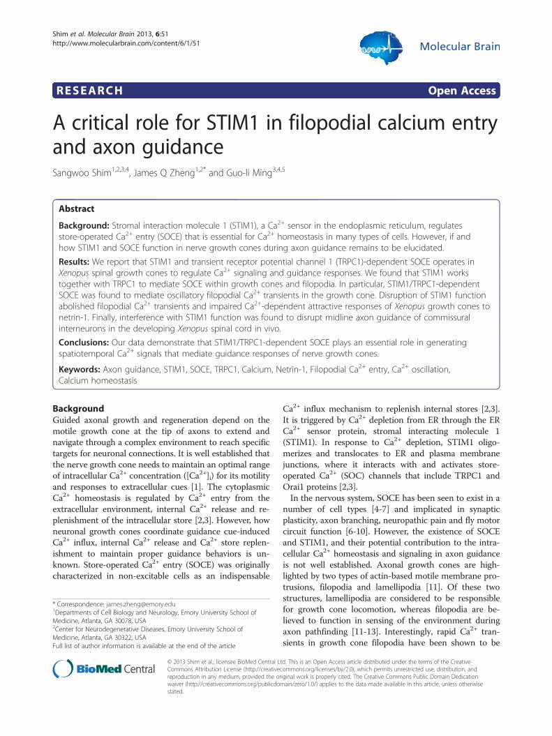
ResultsCloning and expression of Xenopus STIM1We first cloned Xenopus STIM1 (XSTIM1; 668 a.a.),which exhibited 72.8% identity to human STIM1 (685 a.a.;Additional file 1: Figure S1). Whole-mount in situ hy-bridization of developing Xenopus embryos showed thatSTIM1 is highly expressed in the dorsal part of the devel-oping embryo, including the neural tube at stages whenactive axon guidance occurs (Figure 1A, top panels). Cor-onal sections of Xenopus embryos confirmed the expres-sion of STIM1 mRNA in the neural tube, as well as in thenotochord and somites (Figure 1A, bottom panels). RT-PCR analysis from dissected tissues further confirmed thatXSTIM1 mRNA is expressed in neural tube and noto-chord of early tailbud Xenopus embryos (Figure 1B). Im-munofluorescence analysis using anti-STIM1 antibodyand fluorescent phalloidin to stain F-actin showed thatSTIM1 protein is ubiquitously distributed in Xenopusspinal neuron, including soma, neurites and growth cones(Figure 1C). Robust expression of XSTIM1 throughout thegrowth cone and in filopodia is better seen at higher mag-nifications (Figure 1D). These results show that XenopusSTIM1 is expressed in developing neural tissues and neur-onal growth cones of developing axons.
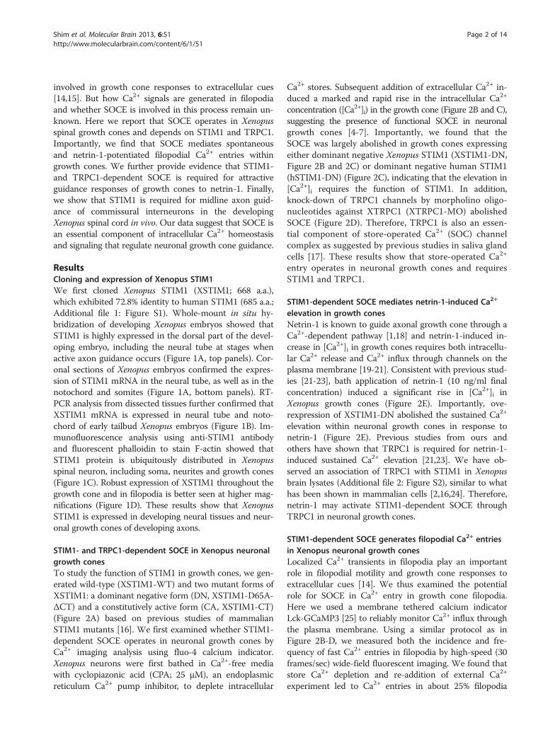
STIM1- and TRPC1-dependent SOCE in Xenopus neuronalgrowth conesTo study the function of STIM1 in growth cones, we gen-erated wild-type (XSTIM1-WT) and two mutant forms ofXSTIM1: a dominant negative form (DN, XSTIM1-D65A-ΔCT) and a constitutively active form (CA, XSTIM1-CT)(Figure 2A) based on previous studies of mammalianSTIM1 mutants [16]. We first examined whether STIM1-dependent SOCE operates in neuronal growth cones byCa2+ imaging analysis using fluo-4 calcium indicator.Xenopus neurons were first bathed in Ca2+-free mediawith cyclopiazonic acid (CPA; 25 μM), an endoplasmicreticulum Ca2+ pump inhibitor, to deplete intracellular
Ca2+ stores. Subsequent addition of extracellular Ca2+ in-duced a marked and rapid rise in the intracellular Ca2+
concentration ([Ca2+]i) in the growth cone (Figure 2B and C),suggesting the presence of functional SOCE in neuronalgrowth cones [4-7]. Importantly, we found that theSOCE was largely abolished in growth cones expressingeither dominant negative Xenopus STIM1 (XSTIM1-DN,Figure 2B and 2C) or dominant negative human STIM1(hSTIM1-DN) (Figure 2C), indicating that the elevation in[Ca2+]i requires the function of STIM1. In addition,knock-down of TRPC1 channels by morpholino oligo-nucleotides against XTRPC1 (XTRPC1-MO) abolishedSOCE (Figure 2D). Therefore, TRPC1 is also an essen-tial component of store-operated Ca2+ (SOC) channelcomplex as suggested by previous studies in saliva glandcells [17]. These results show that store-operated Ca2+
entry operates in neuronal growth cones and requiresSTIM1 and TRPC1.
STIM1-dependent SOCE mediates netrin-1-induced Ca2+
elevation in growth conesNetrin-1 is known to guide axonal growth cone through aCa2+-dependent pathway [1,18] and netrin-1-induced in-crease in [Ca2+]i in growth cones requires both intracellu-lar Ca2+ release and Ca2+ influx through channels on theplasma membrane [19-21]. Consistent with previous stud-ies [21-23], bath application of netrin-1 (10 ng/ml finalconcentration) induced a significant rise in [Ca2+]i inXenopus growth cones (Figure 2E). Importantly, ove-rexpression of XSTIM1-DN abolished the sustained Ca2+
elevation within neuronal growth cones in response tonetrin-1 (Figure 2E). Previous studies from ours andothers have shown that TRPC1 is required for netrin-1-induced sustained Ca2+ elevation [21,23]. We have ob-served an association of TRPC1 with STIM1 in Xenopusbrain lysates (Additional file 2: Figure S2), similar to whathas been shown in mammalian cells [2,16,24]. Therefore,netrin-1 may activate STIM1-dependent SOCE throughTRPC1 in neuronal growth cones.
STIM1-dependent SOCE generates filopodial Ca2+ entriesin Xenopus neuronal growth conesLocalized Ca2+ transients in filopodia play an importantrole in filopodial motility and growth cone responses toextracellular cues [14]. We thus examined the potentialrole for SOCE in Ca2+ entry in growth cone filopodia.Here we used a membrane tethered calcium indicatorLck-GCaMP3 [25] to reliably monitor Ca2+ influx throughthe plasma membrane. Using a similar protocol as inFigure 2B-D, we measured both the incidence and fre-quency of fast Ca2+ entries in filopodia by high-speed (30frames/sec) wide-field fluorescent imaging. We found thatstore Ca2+ depletion and re-addition of external Ca2+
experiment led to Ca2+ entries in about 25% filopodia
Shim et al. Molecular Brain 2013, 6:51 Page 3 of 14http://www.molecularbrain.com/content/6/1/51
(Figure 3; Additional file 3: Movie 1). High frequencytime-lapse traces of the integrated intensity of the Lck-GCaMP3 fluorescence and a kymograph representationclearly demonstrated that filopodial Ca2+ entries are inde-pendent of growth cone Ca2+ transients (Figure 3A-C;Additional file 3: Movie 1). Importantly, both the inci-dence and frequency of these filopodial Ca2+ entries werelargely abolished by inhibition of XSTIM1 with XSTIM1-DN overexpression (Figure 3D and E; Additional file 4:Movie 2) or TRPC1 knockdown by XTRPC1-MO (Figure 3Dand E; Additional file 5: Movie 3). These results thus in-dicate that STIM1- and TRPC1-dependent SOCE is in-dispensable for filopodial Ca2+ entries within neuronalgrowth cones.
Figure 1 Xenopus STIM1 is expressed in developing neural tissues anand cross-section (bottom) in situ hybridization analysis of the mRNA expressiprobe. Dotted lines delineate the boundaries of neural tube and notochord. (state 25-26 Xenopus neural tube and notochord tissues. –RT lane is the negatreverese transcriptase. (C) Representative immunofluorescence images of(phalloidin: green). Scale bar: 20 μm. (D) Representative immunofluoresce(green). Negative control processed without STIM1 antibody (without STIM
Previous studies have revealed that Xenopus growthcones exhibit spontaneous filopodial Ca2+ transients thatare closely associated with growth cone motility [14,15,26].Using Lck-GCaMP3, we also observed robust periodic,spontaneous calcium entries in filopodia of Xenopus growthcones in a Modified Ringer solution that contains 1 mMextracellular Ca2+ (Figure 4A; Additional file 6: Movie 4).Importantly, both the incidence and frequency of the en-tries were also substantially reduced by inhibition of STIM1and TRPC1 function (Figure 4B and C; Additional file 7:Movie 5 and Additional file 8: Movie 6), suggesting thatSTIM1/TRPC1-dependent SOCE is involved in generatingand maintaining oscillatory patterns of spontaneous filo-podial Ca2+ transients.
d neuronal growth cones. (A) Sample images of whole-mount (top)on of XSTIM1 in developing Xenopus embryos. Left, antisense; right, senseB) RT-PCR detection of XSTIM1 mRNA from RNA samples extracted fromive control of the RT-PCR on neural tube tissue RNA in the absence of acultured Xenopus spinal neurons labeled for STIM1 (red) and F-actinnce images of growth cones labeled for STIM1 (red) and F-actin1, bottom) shows absence of immunolabeling. Scale bar: 10 μm.
Shim et al. Molecular Brain 2013, 6:51 Page 4 of 14http://www.molecularbrain.com/content/6/1/51
Bath application of netrin-1 (10 ng/ml final concentra-tion) was found to potentiate both the incidence andfrequency of filopodial Ca2+ entries of Xenopus spinalgrowth cones (Figure 4D-F; Additional file 9: Movie 7),consistent with previous study using Fluo-4 [15]. Wefound that this increase in filopodial Ca2+ entries bynetrin-1 was abolished when STIM1 function was inhi-
Figure 2 STIM1-dependent SOCE operates and mediates netrin-1-induschematic diagram of full-length wild-type (WT) and mutant constructs ofof growth cones of Xenopus spinal neurons from the uninjected or mcherrmedia, before and after the re-addition of 1.5 mM Ca2+ bath solution. Pseuthe lowest. Scale bar: 10 μm. (C) Summary of internal Ca2+ store depletion-and after re-addition of 1.5 mM Ca2+. The fluorescence intensity was normprior to Ca2+ re-addition. Values represent mean ± s.e.m. (n = 25 for controlBootstrap-test). (D) XTRPC1 is required for store depletion-evoked Ca2+ entdepletion-induced Ca2+ entry in growth cones from the control-MO or XTRre-addition of 1.5 mM Ca2+. Values represent mean ± s.e.m. (n = 12 for cont(E) XSTIM1 is required for netrin-1-induced Ca2+ elevation in growth cone.from uninjected or mCherry-XSTIM1-DN mRNA injected embryos. The fluorof 2 min baseline levels prior to the netrin-1 application (10 ng/ml). Values* indicates P < 0.05; Bootstrap-test).
bited by XSTIM1-DN (Figure 4E and F). Over-expressionof morpholino against XTRPC1 (XTRPC1-MO) also com-promised the potentiation of filopodial Ca2+ entries bynetrin-1 (Figure 4E and F). Therefore, STIM1/TRPC1-dependent SOCE mediates the netrin-1-dependent po-tentiation of oscillatory filopodial Ca2+ entries in neuronalgrowth cones.
ced Ca2+ elevation in Xenopus neuronal growth cones. (A) AXSTIM1. (B) Bright field and pseudocolor images of fluo-4 fluorescencey-XSTIM1-DN injected embryos in the presence of CPA in Ca2+-freedocolors indicate Ca2+ levels, with white as the highest and black asinduced Ca2+ entry in growth cones at different time points beforealized to the average fluorescence intensity of 2 min baseline levels, n = 10 for XSTIM1-DN and n = 19 for hSTIM1-DN; * indicates P < 0.01;ry in neuronal growth cones. Summary of internal Ca2+ storePC1-MO injected embryos at different time points before and after therol, and n = 13 for XTRPC1-MO; * indicates P < 0.01; Bootstrap-test).Summary of time course of Ca2+ changes in neuronal growth conesescence intensity was normalized to the average fluorescence intensityrepresent mean ± s.e.m. (n = 6 for control and n = 9 for XSTIM1-DN;

Figure 3 STIM1-dependent SOCE generates filopodial Ca2+ entries in Xenopus neuronal growth cones. (A) A pseudocolored Lck-GCaMP3fluorescent Ca2+ image of growth cone showing rectangular ROIs (region of interest) used to measure fluorescent intensities over time. Pseudocolorsindicate Ca2+ levels, with white as the highest and black as the lowest. Scale bar, 10 μm. (B) Representative traces of Lck-GCaMP3 fluorescentCa2+ signals profile measured in two filopodia (F1, F2) and a growth center (F3) over 7 min period of store-depletion and re-addition of extracellularCa2+. Images were captured at 200 milliseconds intervals. #, indicates filopodial and global Ca2+ transients that are shown in (C). Right imagesare kymographs generated from a segmented line along the filopodia from the tip to the base using NIH ImageJ. The arrowheads denote tipand base of filopodia. (C) Representative pseudocolored Lck-GCaMP3 fluorescent Ca2+ images at the time point as indicated by # in B. The arrowsshow the initiation of filopodial Ca2+ transients. (D-E) The incidence (D) and frequency (E) of filopodial Ca2+ transients were determined incontrol (n = 21), XSTIM1-DN (n = 12), XTRPC1-MO (n = 10) expressing filopodia. *P < 0.005 and **p < 0.05 compared with control conditionusing t-test. Values represent mean ± s.e.m.
Shim et al. Molecular Brain 2013, 6:51 Page 5 of 14http://www.molecularbrain.com/content/6/1/51
The membrane tethered calcium indicator lck-GCaMP3also provides an opportunity to map the entry sites ofCa2+ in filopodia. We thus analyzed the sites of initialfilopodial Ca2+ entry in Xenopus growth cones bykymography analysis. We found that, although the ini-tial sites of Ca2+ entry distributed throughout the lengthof a filopodium, a large portion of the Ca2+ entry sites(42-59%) were found at the filopodial tip (Figure 4G).When filopodial Ca2+ entries under different conditions(store-operated, spontaneous, and netrin-1-induced)were examined, no difference was seen on the locationof Ca2+ entry sites in filopodia (Figure 4G). Therefore,
the tip of the filopodia appears to be the primary site ofSOCE-mediated Ca2+ entry in nerve growth cones.STIM1 proteins reside predominantly in the ER, and
undergo rapid and reversible translocation into ER-plasmamembrane junctions to interact with and activate SOCchannels following store depletion in non-excitable cells[27]. Live cell imaging of Xenopus neurons expressing YFPtagged STIM1 showed that YFP-XSTIM1-WT appearedto translocate into filopodia after store Ca2+ depletion, asrevealed by pseudocolored images and phase overlay im-ages with mCherry (Figure 5; Additional file 10: Movie 8).Together with the presence of STIM1 in Xenopus growth

Figure 4 STIM1/TRPC1-dependent SOCE mediates the spontaneous and netrin-1-potentiated filopodial Ca2+ entries. (A) Left panel; aLck-GCaMP3 fluorescent Ca2+ image of a Xenopus spinal growth cone showing three ROIs (F1, F2 and F3) encompassing the filopodia used tomeasure fluorescent intensities over time (right panels). Scale bar, 10 μm. The incidence (B) and the frequency (C) of the spontaneous filopodialCa2+ transients were significantly attenuated by XSTIM1-DN (n = 30) or XTRPC1-MO (n = 30), when compared to the control (n=34; *P < 0.001and **p < 0.005, Student’s t-test). Values represent mean ± s.e.m. (D) A Lck-GCaMP3 fluorescent Ca2+ image of a growth cone showing three ROIs(left panel) and their representative traces of Ca2+ signals in three filopodia (F1, F2, F3) before and after bath application of netrin-1 (10 ng/ml)(right panel) in the presence of Sp-cAMP (25 μM). Scale bar, 10 μm. (E-F) Netrin-1 potentiated the incidence (E) and the frequency (F) of filopodial Ca2+
transients in spinal growth cones (control; n = 14) and this potentiation was abolished by XSTIM1-DN (n = 8) and XTRPC1-MO (n = 10). **P < 0.005 and***p < 0.05 (Student’s t-test). Values represent mean ± s.e.m. (G) Filopodia tips are the major site of initiation of filopodial Ca2+ entry as revealed bykymographs of Ca2+ signals in filopodia using Lck-GCaMP3 in modified Ringers saline (MR; n = 67), netrin-1 exposure (n = 27) and Ca2+ re-addition afterdepletion (SOCE; n = 43). The y axis represents the path distance along the filopodia divided into 10 portions and the x axis represents time. The arrowsdenote the tip and base of filopodia.
Shim et al. Molecular Brain 2013, 6:51 Page 6 of 14http://www.molecularbrain.com/content/6/1/51

Shim et al. Molecular Brain 2013, 6:51 Page 7 of 14http://www.molecularbrain.com/content/6/1/51
cones and their filopodia by immunostaining (Figure 1D),these results suggest an intriguing possibility that STIM1proteins may become spatially reorganized into growthcone filopodia after activation by store-depletion to fur-ther activate SOC channels that may include TRPC1.
XSTIM1 is required for growth cone guidanceTo test whether STIM1-dependent SOCE is required forgrowth cone guidance in response to netrin-1, we employeda well-established in vitro growth cone turning assay[19,20,23,28]. Previous studies have shown that netrin-1, a classical guidance cue, induces growth cone turningresponses that are mediated by Ca2+ from both extracellu-lar and intracellular sources [19-21,29]. In a microscopicgradient of netrin-1 (5 μg/ml in the pipette, ~5 ng/mlreaching the growth cone), Xenopus growth cones of over-night culture (12-20 hrs) without laminin coating exhib-ited robust chemoattractive turning within 30 minutes(Figure 6A). Importantly, expression of XSTIM1-DN orXSTIM1-CA in Xenopus spinal neurons completelyabolished netrin-1-induced attraction, and interestinglyresulted in repulsion (Figure 6A and B). Expression ofwild-type STIM1 (WT) produced no effect on netrin-1-induced attractive turning (Figure 6A and B). Over-expression of the dominant negative human STIM1(hSTIM1-DN) [16] also eliminated netrin-1-induced at-traction and converted it to repulsion (Figure 6B). Theneurite extension rate in a netrin-1 gradient was not
Figure 5 Dynamic translocation of STIM1 into neuronal filopodia in reimages of growth cone expressing YFP-XSTIM1 before (1 mM Ca2+) and aftto mark filopodia and growth cone. Images were pseudocolored to enhancetranslocated XSTIM1 proteins into neuronal filopodia. Scale bars: 10 μm.
significantly different under these conditions [29], ex-cept XSTIM1-CA which slightly reduced the growthrate (Figure 6C). Thus, proper function of XSTIM1 isessential for netrin-1-induced growth cone turningresponses of Xenopus spinal neurons in vitro. Togetherwith the previous studies showing that TRPC1 knock-down abolished the netrin-1-induced attractive growthcone turning responses [20,21], the results indicate thatSTIM1/TRPC1-dependent SOCE may play a criticalrole in growth cone guidance.To assess whether STIM1 is required for axon guidance
in vivo, we examined the midline axon guidance of com-missural interneurons in the developing Xenopus spinalcord, which is known to require netrin-1 signaling [20,23].Commissural interneuron axons in developing Xenopusembryos were specifically identified with the 3A10 mo-noclonal antibody [20]. In stage 25-26 embryos, 3A10-positive commissural axons extend toward and across themidline in a highly organized manner (Figure 7A and B).In contrast, a significant percentage of commissural axonsderived from YFP-XSTIM1-DN injected embryos won-dered around and failed to cross the midline with someeven went out of the spinal cord (Figure 7C). For simpli-city, we scored both types of guidance defects as the onenot crossing and presented and quantified the percentageof crossed axons (Figure 7F). XSTIM1-DN markedly re-duced the percentage of crossed axons to about 50% ofthe control groups (uninjected and GFP injection only)
sponse to store-depletion. Representative time-lapse fluorescenter store Ca2+ depletion (0 mM Ca2+/CPA). mCherry was co-expressedthe observation of intensity changes. The arrows indicate newly
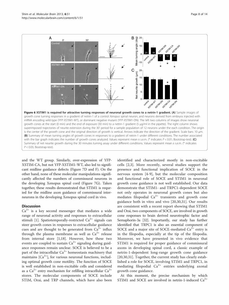
Figure 6 XSTIM1 is required for attractive turning responses of neuronal growth cones to a netrin-1 gradient. (A) Sample images ofgrowth cone turning responses in a gradient of netrin-1 of a control Xenopus spinal neuron, and neurons derived from embryos injected withmRNA encoding wild-type (YFP-XSTIM1-WT), or dominant negative mutant (YFP-XSTIM1-DN). The left two columns of images show neuronalgrowth cones at the start (0 min) and the end of exposure (30 min) to a netrin-1 gradient (5 μg/ml in the pipette). The right column showssuperimposed trajectories of neurite extension during the 30' period for a sample population of 12 neurons under the each condition. The originis the center of the growth cone and the original direction of growth is vertical. Arrows indicate the direction of the gradient. Scale bars: 10 μm.(B) Summary of mean turning angles of growth cones in responses to a gradient of netrin-1 under different conditions. The number associatedwith the bar graph indicates the number of growth cones analyzed. Values represent mean ± s.e.m. (* indicates P < 0.01; Bootstrap-test). (C)Summary of net neurite growth during the 30 minutes turning assay under different conditions. Values represent mean ± s.e.m. (* indicatesP < 0.05; Bootstrap-test).
Shim et al. Molecular Brain 2013, 6:51 Page 8 of 14http://www.molecularbrain.com/content/6/1/51
and the WT group. Similarly, over-expression of YFP-XSTIM-CA, but not YFP-XSTIM1-WT, also led to signifi-cant midline guidance defects (Figure 7D and F). On theother hand, none of these molecular manipulations signifi-cantly affected the numbers of commissural neurons inthe developing Xenopus spinal cord (Figure 7G). Takentogether, these results demonstrated that STIM1 is requr-ied for the midline axon guidance of commissural inter-neurons in the developing Xenopus spinal cord in vivo.
DiscussionCa2+ is a key second messenger that mediates a widerange of neuronal activity and responses to extracellularstimuli [1]. Spatiotemporally-restricted Ca2+ signals cansteer growth cones in responses to extracellular guidancecues and are thought to be generated from Ca2+ influxthrough the plasma membrane as well as Ca2+ releasefrom internal store [1,18]. However, how these twoevents are coupled to sustain Ca2+ signaling during guid-ance responses remain unclear. SOCE is believed to be apart of the intracellular Ca2+ homeostasis machinery thatmaintains [Ca2+]i for various neuronal functions, includ-ing optimal growth cone motility. The function of SOCEis well established in non-excitable cells and consideredas a Ca2+ entry mechanism for refilling intracellular Ca2+
stores. The molecular components of SOCE includeSTIM, Orai, and TRP channels, which have also been
identified and characterized mostly in non-excitablecells [2,3]. More recently, several studies support thepresence and functional implication of SOCE in thenervous system [4-9], but the molecular compositionand functional role of SOCE and STIM1 in neuronalgrowth cone guidance is not well established. Our datademonstrate that STIM1- and TRPC1-dependent SOCEnot only operates in neuronal growth cones but alsomediates filopodial Ca2+ transients and growth coneguidance both in vitro and vivo [20,30,31]. Our resultsare consistent with a recent report showing that STIM1and Orai, two components of SOCE, are involved in growthcone responses to brain derived neurotrophic factor andSemaphorin-3a [32]. Importantly, our study has furtheridentified that TRPC1 is also an essential component ofSOCE and a major site of SOCE-mediated Ca2+ entry isin the filopodia, especially at the tip of the filopodia.Moreover, we have presented in vivo evidence thatSTIM1 is required for proper guidance of commissuralaxons in developing spinal cord, a classic example ofnetrin-1-dependent long-range growth cone guidance[20,30,31]. Together, the current study has clearly estab-lished a role for SOCE, involving STIM1 and TRPC1, inmediating filopodial Ca2+ entries underlying axonalgrowth cone guidance.At this moment, the precise mechanism by which
STIM1 and SOCE are involved in netrin-1-induced Ca2+
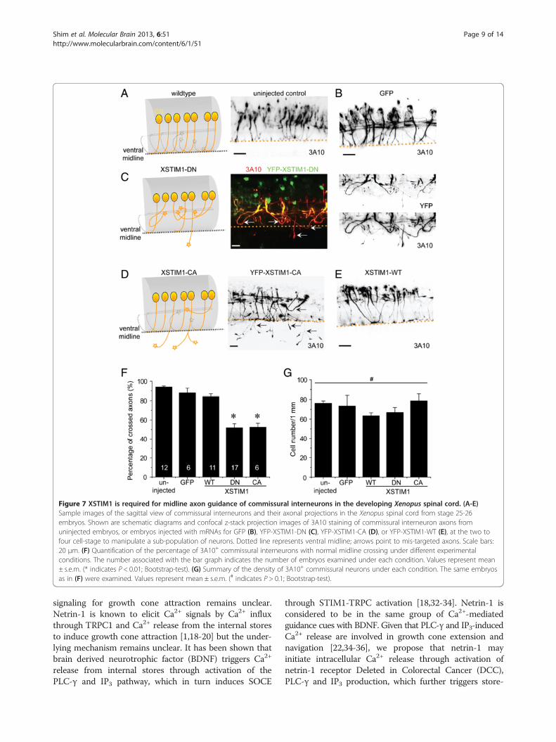
Figure 7 XSTIM1 is required for midline axon guidance of commissural interneurons in the developing Xenopus spinal cord. (A-E)Sample images of the sagittal view of commissural interneurons and their axonal projections in the Xenopus spinal cord from stage 25-26embryos. Shown are schematic diagrams and confocal z-stack projection images of 3A10 staining of commissural interneuron axons fromuninjected embryos, or embryos injected with mRNAs for GFP (B), YFP-XSTIM1-DN (C), YFP-XSTIM1-CA (D), or YFP-XSTIM1-WT (E), at the two tofour cell-stage to manipulate a sub-population of neurons. Dotted line represents ventral midline; arrows point to mis-targeted axons. Scale bars:20 μm. (F) Quantification of the percentage of 3A10+ commissural interneurons with normal midline crossing under different experimentalconditions. The number associated with the bar graph indicates the number of embryos examined under each condition. Values represent mean± s.e.m. (* indicates P < 0.01; Bootstrap-test). (G) Summary of the density of 3A10+ commissural neurons under each condition. The same embryosas in (F) were examined. Values represent mean ± s.e.m. (# indicates P > 0.1; Bootstrap-test).
Shim et al. Molecular Brain 2013, 6:51 Page 9 of 14http://www.molecularbrain.com/content/6/1/51
signaling for growth cone attraction remains unclear.Netrin-1 is known to elicit Ca2+ signals by Ca2+ influxthrough TRPC1 and Ca2+ release from the internal storesto induce growth cone attraction [1,18-20] but the under-lying mechanism remains unclear. It has been shown thatbrain derived neurotrophic factor (BDNF) triggers Ca2+
release from internal stores through activation of thePLC-γ and IP3 pathway, which in turn induces SOCE
through STIM1-TRPC activation [18,32-34]. Netrin-1 isconsidered to be in the same group of Ca2+-mediatedguidance cues with BDNF. Given that PLC-γ and IP3-inducedCa2+ release are involved in growth cone extension andnavigation [22,34-36], we propose that netrin-1 mayinitiate intracellular Ca2+ release through activation ofnetrin-1 receptor Deleted in Colorectal Cancer (DCC),PLC-γ and IP3 production, which further triggers store-
Shim et al. Molecular Brain 2013, 6:51 Page 10 of 14http://www.molecularbrain.com/content/6/1/51
depletion, STIM1 activation, and Ca2+ influx throughTRPC1 for replenishing ER Ca2+. This notion is furthersupported by the findings that both netrin-1 and BDNFactivate PLC-γ and Phosphatidylinositol 4,5-bisphosphate(PIP2) hydrolysis in neurite elongation [37,38]. Therefore,our results provide additional evidence for the conservedsignaling pathways among Ca2+-mediated guidance cuesand between netrin-1 and neurotrophins.The role of TRPC channels as SOC has been contro-
versial, but multiple lines of evidences support TRPC asa strong candidate component of store-operated Ca2+
channels. For example, TRPC1 has been shown to bebound and activated by STIM1 and contribute to SOCEin some cells [16,17,39-41]. We found that STIM1 inter-acts with TRPC1 in embryonic neural tissues (Additionalfile 2: Figure S2) and that TRPC1 knockdown inhibitsSTIM1-mediated SOCE within growth cones and filo-podia (Figures 2D, 3D and 3E), suggesting that TRPC1 isan essential component of SOCE. As STIM1 is also re-quired for netrin-1-induced Ca2+ elevation and growthcone attraction which was shown to be mediated byTRPC1, our data support a role for STIM1 in activatingTRPC1. However, we cannot rule out the possibility thatSTIM1 may affect Ca2+ signaling and growth cone guid-ance by other mechanisms, such as its effects on cAMPsignaling or ER remodeling [42,43]. Recent studies alsoshowed biochemical assembly of STIM1-TRPCs-Oraicomplex and functional connections between TRPC chan-nels and Orai1 [41,44,45]. STIM1-Orai1 co-localization inresponse to Ca2+ depletion was reported in neuronalgrowth cones [32]. Therefore, it is possible that Orai alsoplays a role in netrin-1 signaling and guidance.It should be noted that Lck-GCaMP3 was successfully
used in distinguishing the Ca2+ signals from membraneentry from internal release from the stores [25,46]. Ourdata with Lck-GCaMP3 showing the presence of filopodialCa2+ transients and its potentiation by netrin-1 is consist-ent with the previous reports using Fluo-4 [14,15,26].However, when compared with previous studies usingFluo-4, the incidence and frequency of filopodial Ca2+
transients observed in our study appear to be lower thanthose seen in the previous reports. The difference may beattributed to two possibilities. First, Lck-GCaMP3 detectsCa2+ entry events only at near-plasma membrane regions.However, fluo-4 could detect cytosol Ca2+ changes fromother sources such as intracellular stores, which will likelybe missed by Lck-GCaMP3. In this regard, Lck-GCaMP3fluorescence Ca2+ signals may be better called “filopodialCa2+ entries” rather than filopodial Ca2+ transients. Sec-ond, we did not count the Ca2+ transients propagatedfrom the growth cone proper and only counted the Ca2+
entry events generated within the filopodium independ-ently of Ca2+ transients from the growth cone proper.Therefore, our data do not contradict the previous work.
It is of interest to see that the initial site of filopodialCa2+ entry is largely localized to the tip of filopodia.Filopodia are considered to be the sensory apparatus forgrowth cones as they extend farther distance to detect theenvironmental signals. Therefore, it makes sense to havethe sensory molecules accumulated at the tip for signaltransduction initiation. However, the lack of quality anti-bodies prevented us from convincingly detecting thelocalization of STIM1/TRPC1 and other SOCE compo-nents at the filopodial tip. On the other hand, it has beenreported that several receptors such as integrins, TRPC1and DCC [14,26,47] and signaling molecules such as Src,PAK, PKA [48,49] are enriched at the tip of filopodia a-long with many other cytoskeleton regulatory molecules[11,50,51]. Therefore, it is conceivable that STIM1 andTRPC1 could function at the filopodial tip as an effectiveway to sense the environment and initiate Ca2+ signalingduring growth cone guidance.We report fast, highly localized and periodic spontan-
eous filopodial Ca2+ entries initiated independently ofgrowth cone Ca2+ transients, which was consistent withthe previous reports of oscillatory pattern of spontaneousCa2+ transients within growth cone and filopodia duringaxonal growth [14,15,52]. The critical role for TRPC1 ingeneration of filopodial Ca2+ entry and its potentiation bynetrin-1 is also consistent with the previous reports[14,15,26]. A further unexpected result was that STIM1-DN mutant blocked not only the SOCE-induced filopodialCa2+ entries that depend on STIM1 but also spontaneousand netrin-1-potentiated oscillatory filopodial Ca2+ en-tries, suggesting that STIM1-dependent SOCE mediatesthe generation and maintenance of filopodial Ca2+ entries.Thus, our visualization of oscillatory patterns of spon-taneous filopodial Ca2+ entries and their inhibition bySTIM1- or TRPC1-knockdown is the first demonstrationof a role of STIM/TRPC1-dependent SOCE in regulatingCa2+ oscillation in neurons, which is consistent with previ-ous findings in other cell types [53-55]. It is plausible thatstore Ca2+ release, transient drop in ER Ca2+, and Ca2+
entry through TRPC1 triggered by transient STIM1 acti-vation may underlie the Ca2+ oscillations seen in growthcone filopodia. Thus, considering the functional correl-ation between the frequency of filopodial Ca2+ transientsand growth cone outgrowth and turning [14,26], disrup-tion of STIM1 or TRPC1 function is likely to result in thebreakup of Ca2+ cycling for oscillations and subsequent at-tenuated frequency of filopodial Ca2+ entries, which mayfurther cause the suppression of growth cone guidance inresponse to netrin-1.
ConclusionsOur data demonstrate a role for STIM1/TRPC1-dependentSOCE in mediating oscillatory patterns of spontaneous and

Shim et al. Molecular Brain 2013, 6:51 Page 11 of 14http://www.molecularbrain.com/content/6/1/51
netrin-1-potentiated filopodial Ca2+ entries that underlieaxonal growth cone guidance both in vitro and in vivo.
MethodsMolecular constructsXenopus STIM1 (XSTIM1) [GenBank: BC126011] wasidentified by BLAST searches of the GenBank databaseusing human STIM1 cDNA sequence. The coding regionof XSTIM1 gene was isolated by RT-PCR, sequenced andcloned into the pCS2 vector (gift of D. Turner, Universityof Michigan). The following constructs of XSTIM1 and itsmutants were generated by site-directed mutagenesis(Strategene) or by PCR based on previous studies ofmammalian STIM1 mutants [16]: YFP-XSTIM1-WT,YFP-XSTIM1-DN, YFP-XSTIM1-CA, and mCherry-XSTIM1-DN (Figure 2A). Different XSTIM constructs werein vitro transcribed with the mMESSAGE mMACHINESP6 kit (Ambion). pN1-Lck-GCaMP3 plasmid was ob-tained from Addgene (plasmid #26974, [25]), cloned intopCS2 vector using BamHI and XbaI sites and in vitrotranscribed with mMESSAGE mMACHINE SP6 kit(Ambion).
Xenopus embryo injection and spinal neuron cultureBlastomere injections of mRNAs or morpholinos intoearly stages of Xenopus embryos and culturing of spinalneurons from these injected embryos were performed aspreviously described [20,23,29,56]. Briefly, fertilized em-bryos were injected at the two- or four-cell stage withmRNA (2-3 ng/embryo). A control morpholinos or mor-pholinos specific for Xenopus TRPC1 (XTRPC1-MO)was previously described [20]. Uninjected or injectedembryos at stage 22 were used for cultures of spinalneurons as previous described [20,23]. All the proce-dures involving Xenopus frogs and embryos were carriedout in accordance to the NIH guideline for animal useand have been approved by the Institutional AnimalCare and Use Committee (IACUC) of Emory University.
RT-PCR and Whole-mount in situ hybridization ofXenopus embryosNeural tube and notochord were isolated from the dorsalsection of the stage 25-26 Xenopus embryos after dissec-tion with microsurgical scissors and incubation with colla-genase (type I, Sigma). Total RNA was prepared by usingTRIzol Reagent (invitrogen) and treated with the RNase-free DNAse I (Roche) to remove genomic DNA. Theextracted RNA was reverse transcribed by using M-MLVreverse transcriptase (Invitrogen) and random hexamers(Roche). PCR amplification was performed using Taqpolymerase (Fermentas). The –RT lane is the negativecontrol of the RT-PCR on neural tube tissue RNA in theabsence of a reverese transcriptase. The PCR primers areas follows; XSTIM1-forward, 5′ CCAGAACCTTGGAA
GAGGTG 3′, XSTIM1-reverse, 5′ GACTGAATGGTACCGGCTGT 3′; XODC-forward, 5′ CAGCTAGCTGTGGTGTGG 3′, XODC-rev, 5′ CAACATGGAAACTCA-CACC 3′. For whole-mount in situ hybridization, thedigoxigenin (DIG)-UTP-labelled antisense RNA was usedas previously described [23,57]. The C-terminal region ofXSTIM1 corresponding to amino acid 192-668 was usedfor the specific anti-sense and sense probes. The labelledprobe was detected with alkaline phosphatase-conjugatedanti-DIG antibody (Fab fragments) and visualized with theBM purple AP substrate (Roche Applied Science). Se-lected embryos from whole-mount in situ hybridizationwere embedded in a sucrose and Tissue-Tek O.C.Tmedium, completely frozen and cross-sectioned at 40 μmwith a cryostat (Leica CM1850).
ImmunocytochemistryXenopus spinal neuron cultures were fixed in 4% para-formaldehyde in a cacodylate buffer (0.1 M sodium caco-dylate, 0.1 M sucrose, pH 7.4) for 30 minutes andpermeabilized with Triton X-100 (0.1%) for 10 minutes.The cells were incubated with a rabbit polyclonal anti-body against full length human STIM1 (MyBioScource)at a dilution of 1:50 after blocking with 5% goat serumand labelled with Alexa Fluor 546 goat anti-rabbit sec-ondary (Invitrogen). Fluorescent imaging was capturedon an inverted microscope (Nikon Eclipse Ti-E).
Growth cone turning assayMicroscopic gradients of netrin-1 (5 μg/ml in the pipette)were produced as previously described [29,56,58,59].Xenopus spinal neurons derived from injected blastomereswere identified under fluorescent microscope and used forturning assay at the room temperature 14 to 20 hrs afterplating as previously described [20,23,29,56]. The culturewas plated on glass coverslip without any coating. Theturning angle was defined by the angle between the ori-ginal direction of neurite extension and a straight lineconnecting the positions of the center of the growth coneat the onset and the end of the 30 min period. The ratesof neurite extension were calculated based on the netneurite extension during the turning assay. Only thosegrowth cones of isolated neurons with a net neurite ex-tension > 5 μm over the 30-min period were included foranalysis. Statistical significance was assessed using theBootstrap-test.
Ca2+ imaging of cultured Xenopus spinal neuronsCa2+ imaging of cultured growth cones of Xenopus spinalneurons was performed as previously described [23,29,56].Specifically, isolated Xenopus spinal neurons were cul-tured on glass coverslip without coating, loaded withFluo-4 AM (2 μM, Molecular Probes) for 30 minutes,rinsed with the Modified Ringer used for growth cone

Shim et al. Molecular Brain 2013, 6:51 Page 12 of 14http://www.molecularbrain.com/content/6/1/51
turning assay, and imaged after bath-application of netrin-1 (10 ng/ml). For store-operated Ca2+ entry experiment,neurons were bathed in Ca2+ -free media with CPA(25 μM) to deplete intracellular Ca2+ stores, and imagedafter re-addition of extracellular Ca2+ (1.5 mM). Growthcones expressing mCherry-XSTIM1-DN proteins wereidentified under fluorescent microscope and selected forfurther Ca2+ imaging. Imaging was carried out using aZeiss 510 META system equipped with a 20X objective(NA 0.8). Excitation was at 488 nm by argon laser and theemitted fluorescence was collected at 500-560 nm. Fluor-escence and bright-field images were simultaneously ac-quired at every 30 seconds with a frame scan. The meanfluorescence intensity of each time point was measuredover a fixed circular region of interest that covers the en-tire growth cone and normalized to the average fluores-cence intensity that was measured during a 2 minutesbaseline period (prior to the netrin-1 application oraddition of 1.5 mM Ca2+ solution).For filopodial Ca2+ imaging, Lck-GCaMP3 mRNA was
injected into early staged embryos without or with othermRNAs or morpholino. Spinal neurons were cultured onthe glass coverslip coated with poly-D-lysine and laminin,which increases the number and length of filopodia [60],in serum-free culture medium. In our netrin-1-inducedfilopodial Ca2+ entries experiments, the spinal neuronswere incubated in MR solution with the addition of cAMPanalog Sp-cAMP (25 μM) to counterbalance the laminin’seffect of reducing cAMP levels in growth cones [61] andmimic the condition of our in vitro turning assay wherelaminin coating on the glass culture dish was omitted. Livecell imaging of Ca2+ transients was performed on aninverted microscope (Nikon Eclipse Ti-E) equipped with a60X Apo TIRF objective (NA 1.49), and EMCCD camera(Photometrics) using NIS-Elements software (Nikon).Excitation was at 488 nm and the emitted fluorescencewas collected at 520 nm and Lck-GCaMP3 fluorescenceimages were acquired at every 200 milliseconds. To deter-mine several characteristics of filopodial Ca2+ entries, in-cluding the incidence, frequency and initiation sites oftransients, the Kymographs (spatio-temporal map) werecreated from the images of the user-defined segmentedline one pixel in width spanning the filopodium from thetime-lapse movies with NIH ImageJ software. Grayscalevalues for this linear region of interest (ROI) for eachframe of the time series were transformed into pseudoco-lored images to show time-dependent changes in intracel-lular Ca2+ concentration ([Ca2+]i) along the length of theROI (y-axis) over time (x-axis).
Immunoprecipitation and immunoblottingFor co-immunoprecipitation, protein lysates were pre-pared from the Xenopus brain explants including spinalcords dissected from the embryos at stage 26-28 using
lysis buffer containing 1% Triton X-100, 150 mM NaCl,10 mM Tris-Cl (pH 7.4), 1 mM EDTA, 1 mM EGTA,0.5% Nonidet P-40, 0.2 mM Na-orthovanadate, and prote-ase inhibitor cocktail. They were incubated with the ap-propriate antibody for 3-4 hours at 4°C, followed byincubation with protein A/G agarose beads (Pierce) forovernight at 4°C. Mouse anti-c-Myc monoclonal antibody(Roche Applied Science) and rabbit anti-GFP polyclonalantibody (Abcam) were used for immunoprecipitationand immunoblotting.
Whole-mount immunocytochemistryEmbryos at stage 25-26 were fixed and processed for im-munocytochemistry as previously described [20,23,62].Monoclonal antibody 3A10, specific for commissural in-terneurons [23,63], was obtained from the DevelopmentalStudies Hybridoma Bank at the University of Iowa andused at a dilution of 1:100. Secondary antibodies wereused at a dilution of 1:250. Confocal images of sagittalviews of embryos were taken with a Zeiss LSM 510 METAsystem and Z-series reconstructions were processed withthe Zeiss LSM image acquisition program as previouslydescribed [23]. Statistical significance was assessed usingthe Bootstrap-test.
Additional files
Additional file 1: Figure S1. Alignment of STIM1 amino acidsequences of Xenopus laevis, Xenopus tropicalis and human. Identicalamino acid residues are highlighted in dark. Dashes indicate gapsinserted for maximal alignment score. The red box indicates the aminoacid (D65, within the EF hand motif) that was mutated in the dominantnegative form of XSTIM1.
Additional file 2: Figure S2. Association of XSTIM1 with TRPC1. Shownare sample westernblots for co-immunoprecipitation of human TRPC1(myc-hTRPC1-WT) and wild-type (YFP-XSTIM1-WT) or constitutively activeSTIM1 (YFP-XSTIM1-CA) expressed in Xenopus embryonic neural tissues.
Additional file 3: Movie 1. Filopodial Ca2+ entries are generated bySOCE. A pseudocolored Lck-GCaMP3 fluorescent Ca2+ images of Xenopusneuronal growth cones were captured at 200 milliseconds intervals over7 min period of the store-depletion and re-addition of extracellular Ca2+
as shown in Figure 4A.
Additional file 4: Movie 2. SOCE-induced filopodial Ca2+ entries areabolished by inhibition of XSTIM1 with XSTIM1-DN overexpression. Apseudocolored Lck-GCaMP3 fluorescent Ca2+ images of Xenopus neuronalgrowth cones expressing XSTIM1-DN were captured at 200 millisecondsintervals over 7 min period of the store-depletion and re-addition ofextracellular Ca2+.
Additional file 5: Movie 3. SOCE-induced filopodial Ca2+ entries areattenuated by inhibition of XTRPC1 with XTRPC1-MO overexpression. Apseudocolored Lck-GCaMP3 fluorescent Ca2+ images of Xenopus neuronalgrowth cones expressing XTRPC1-MO were captured at 200 millisecondsintervals over 7 min period of the store-depletion and re-addition ofextracellular Ca2+.
Additional file 6: Movie 4. Oscillatory spontaneous filopodial Ca2+
entries. A pseudocolored Lck-GCaMP3 fluorescent Ca2+ images of Xenopusneuronal growth cones were captured at 200 milliseconds intervals over7 min period in Modified Ringer solutions (1 mM extracellular Ca2+) asshown in Figure 5A.
Shim et al. Molecular Brain 2013, 6:51 Page 13 of 14http://www.molecularbrain.com/content/6/1/51
Additional file 7: Movie 5. Spontaneous filopodial Ca2+ entries areabolished by inhibition of XSTIM1 with XSTIM1-DN overexpression. Apseudocolored Lck-GCaMP3 fluorescent Ca2+ images of Xenopus neuronalgrowth cones expressing XSTIM1-DN were captured at 200 millisecondsintervals over 7 min period in Modified Ringer solutions.
Additional file 8: Movie 6. Spontaneous filopodial Ca2+ entries areabolished by inhibition of XTRPC1 with XTRPC1-MO overexpression. Apseudocolored Lck-GCaMP3 fluorescent Ca2+ images of Xenopus neuronalgrowth cones expressing XTRPC1-MO were captured at 200 millisecondsintervals over 7 min period in Modified Ringer solutions.
Additional file 9: Movie 7. Netrin-1-potentiated filopodial Ca2+ entries.A pseudocolored Lck-GCaMP3 fluorescent Ca2+ images of Xenopus neur-onal growth cone were captured at 200 milliseconds intervals in ModifiedRinger solution (1 mM extracellular Ca2+) with Sp-cAMP (25 μM) beforeand after bath application of netrin-1 (10 ng/ml) as shown in Figure 4D.
Additional file 10: Movie 8. Dynamic translocation of STIM1 intoneuronal filopodia in response to store-depletion. Time-lapse fluorescentimages of Xenopus neuronal growth cone expressing YFP-XSTIM1 before(1 mM Ca2+) and after store Ca2+ depletion (0 mM Ca2+/CPA). Imageswere captured at 2 seconds intervals.
Competing interestsThe authors declare that they have no competing interests.
Authors’ contributionsSS designed and performed all the experiments, and wrote the manuscript.JQZ oversaw the project, designed some of the experiments, and revised themanuscript with SS. GLM provided guidance to the project and contributedto the writing of the manuscript. All authors read and approved the finalmanuscript.
AcknowledgementsWe thank P.F. Worley for help and discussion. This work was supported inpart by grants from National Institutes of Health to JQZ (GM083889 andGM084363) and to GLM. (NS048271 and HD069184).
Author details1Departments of Cell Biology and Neurology, Emory University School ofMedicine, Atlanta, GA 30078, USA. 2Center for Neurodegenerative Diseases,Emory University School of Medicine, Atlanta, GA 30322, USA. 3Institute forCell Engineering, Johns Hopkins University School of Medicine, Baltimore,MD 21205, USA. 4Department of Neurology, Johns Hopkins University Schoolof Medicine, Baltimore, MD 21205, USA. 5The Solomon H. SnyderDepartment of Neuroscience, Johns Hopkins University School of Medicine,Baltimore, MD 21205, USA.
Received: 5 November 2013 Accepted: 23 November 2013Published: 1 December 2013
References1. Zheng JQ, Poo MM: Calcium signaling in neuronal motility. Annu Rev Cell
Dev Biol 2007, 23:375–404.2. Cahalan MD: STIMulating store-operated Ca(2+) entry. Nat Cell Biol 2009,
11:669–677.3. Lewis RS: Store-operated calcium channels: new perspectives on
mechanism and function. Cold Spring Harb Perspect Biol 2011, 3(12).doi: 10.1101/cshperspect.a003970.
4. Kachoei BA, Knox RJ, Uthuza D, Levy S, Kaczmarek LK, Magoski NS: Astore-operated Ca(2+) influx pathway in the bag cell neurons of Aplysia.J Neurophysiol 2006, 96:2688–2698.
5. Li D, Herault K, Oheim M, Ropert N: FM dyes enter via a store-operatedcalcium channel and modify calcium signaling of cultured astrocytes.Proc Natl Acad Sci U S A 2009, 106:21960–21965.
6. Gemes G, Bangaru ML, Wu HE, Tang Q, Weihrauch D, Koopmeiners AS,Cruikshank JM, Kwok WM, Hogan QH: Store-operated Ca2+ entry insensory neurons: functional role and the effect of painful nerve injury.J Neurosci 2011, 31:3536–3549.
7. Venkiteswaran G, Hasan G: Intracellular Ca2+ signaling and store-operatedCa2+ entry are required in Drosophila neurons for flight. Proc Natl AcadSci U S A 2009, 106:10326–10331.
8. Tang F, Kalil K: Netrin-1 induces axon branching in developing corticalneurons by frequency-dependent calcium signaling pathways. J Neurosci2005, 25:6702–6715.
9. Baba A, Yasui T, Fujisawa S, Yamada RX, Yamada MK, Nishiyama N, MatsukiN, Ikegaya Y: Activity-evoked capacitative Ca2+ entry: implications insynaptic plasticity. J Neurosci 2003, 23:7737–7741.
10. Steinbeck JA, Henke N, Opatz J, Gruszczynska-Biegala J, Schneider L, TheissS, Hamacher N, Steinfarz B, Golz S, Brustle O, et al: Store-operated calciumentry modulates neuronal network activity in a model of chronic epi-lepsy. Exp Neurol 2011, 232:185–194.
11. Geraldo S, Gordon-Weeks PR: Cytoskeletal dynamics in growth-cone steering.J Cell Sci 2009, 122:3595–3604.
12. Zheng JQ, Wan JJ, Poo MM: Essential role of filopodia in chemotropicturning of nerve growth cone induced by a glutamate gradient.J Neurosci 1996, 16:1140–1149.
13. Davenport RW, Dou P, Rehder V, Kater SB: A sensory role for neuronalgrowth cone filopodia. Nature 1993, 361:721–724.
14. Gomez TM, Robles E, Poo M, Spitzer NC: Filopodial calcium transientspromote substrate-dependent growth cone turning. Science 2001,291:1983–1987.
15. Nicol X, Hong KP, Spitzer NC: Spatial and temporal second messengercodes for growth cone turning. Proc Natl Acad Sci U S A 2011,108:13776–13781.
16. Huang GN, Zeng W, Kim JY, Yuan JP, Han L, Muallem S, Worley PF: STIM1carboxyl-terminus activates native SOC, I(crac) and TRPC1 channels.Nat Cell Biol 2006, 8:1003–1010.
17. Liu X, Cheng KT, Bandyopadhyay BC, Pani B, Dietrich A, Paria BC, SwaimWD, Beech D, Yildrim E, Singh BB, et al: Attenuation of store-operatedCa2+ current impairs salivary gland fluid secretion in TRPC1(-/-) mice.Proc Natl Acad Sci U S A 2007, 104:17542–17547.
18. Tojima T, Hines JH, Henley JR, Kamiguchi H: Second messengers andmembrane trafficking direct and organize growth cone steering. Naturereviews Neuroscience 2011, 12:191–203.
19. Hong K, Nishiyama M, Henley J, Tessier-Lavigne M, Poo M: Calcium signallingin the guidance of nerve growth by netrin-1. Nature 2000, 403:93–98.
20. Shim S, Goh EL, Ge S, Sailor K, Yuan JP, Roderick HL, Bootman MD, Worley PF,Song H, Ming GL: XTRPC1-dependent chemotropic guidance of neuronalgrowth cones. Nat Neurosci 2005, 8:730–735.
21. Wang GX, Poo MM: Requirement of TRPC channels in netrin-1-inducedchemotropic turning of nerve growth cones. Nature 2005, 434:898–904.
22. Tojima T: Intracellular signaling and membrane trafficking controlbidirectional growth cone guidance. Neurosci Res 2012, 73:269–274.
23. Shim S, Yuan JP, Kim JY, Zeng W, Huang G, Milshteyn A, Kern D, Muallem S,Ming GL, Worley PF: Peptidyl-prolyl isomerase FKBP52 controlschemotropic guidance of neuronal growth cones via regulation ofTRPC1 channel opening. Neuron 2009, 64:471–483.
24. Hogan PG, Lewis RS, Rao A: Molecular basis of calcium signaling inlymphocytes: STIM and ORAI. Annu Rev Immunol 2010, 28:491–533.
25. Shigetomi E, Kracun S, Khakh BS: Monitoring astrocyte calciummicrodomains with improved membrane targeted GCaMP reporters.Neuron Glia Biol 2010, 6:183–191.
26. Kerstein PC, Jacques-Fricke BT, Rengifo J, Mogen BJ, Williams JC, Gottlieb PA,Sachs F, Gomez TM: Mechanosensitive TRPC1 channels promote calpainproteolysis of talin to regulate spinal axon outgrowth. J Neurosci2013, 33:273–285.
27. Soboloff J, Rothberg BS, Madesh M, Gill DL: STIM proteins: dynamic calciumsignal transducers. Nature reviews Molecular cell biology 2012, 13:549–565.
28. Song H, Ming G, He Z, Lehmann M, McKerracher L, Tessier-Lavigne M, PooM: Conversion of neuronal growth cone responses from repulsion toattraction by cyclic nucleotides. Science 1998, 281:1515–1518.
29. Ming GL, Song HJ, Berninger B, Holt CE, Tessier-Lavigne M, Poo MM: cAMP-dependent growth cone guidance by netrin-1. Neuron 1997, 19:1225–1235.
30. Serafini T, Colamarino SA, Leonardo ED, Wang H, Beddington R, Skarnes WC,Tessier-Lavigne M: Netrin-1 is required for commissural axon guidance inthe developing vertebrate nervous system. Cell 1996, 87:1001–1014.
31. Kennedy TE, Serafini T, de la Torre JR, Tessier-Lavigne M: Netrins are diffusiblechemotropic factors for commissural axons in the embryonic spinal cord.Cell 1994, 78:425–435.
Shim et al. Molecular Brain 2013, 6:51 Page 14 of 14http://www.molecularbrain.com/content/6/1/51
32. Mitchell CB, Gasperini RJ, Small DH, Foa L: STIM1 is necessary for store-operatedcalcium entry in turning growth cones. J Neurochem 2012,122:1155–1166.
33. Li Y, Jia YC, Cui K, Li N, Zheng ZY, Wang YZ, Yuan XB: Essential role ofTRPC channels in the guidance of nerve growth cones by brain-derivedneurotrophic factor. Nature 2005, 434:894–898.
34. Ming G, Song H, Berninger B, Inagaki N, Tessier-Lavigne M, Poo M: PhospholipaseC-gamma and phosphoinositide 3-kinase mediate cytoplasmic signaling innerve growth cone guidance. Neuron 1999, 23:139–148.
35. Takei K, Shin RM, Inoue T, Kato K, Mikoshiba K: Regulation of nerve growthmediated by inositol 1,4,5-trisphosphate receptors in growth cones.Science 1998, 282:1705–1708.
36. Akiyama H, Matsu-ura T, Mikoshiba K, Kamiguchi H: Control of neuronalgrowth cone navigation by asymmetric inositol 1,4,5-trisphosphatesignals. Sci Signal 2009, 2:ra34.
37. Xie Y, Hong Y, Ma XY, Ren XR, Ackerman S, Mei L, Xiong WC:DCC-dependent phospholipase C signaling in netrin-1-induced neuriteelongation. J Biol Chem 2006, 281:2605–2611.
38. Xie Y, Ding YQ, Hong Y, Feng Z, Navarre S, Xi CX, Zhu XJ, Wang CL,Ackerman SL, Kozlowski D, et al: Phosphatidylinositol transfer protein-alphain netrin-1-induced PLC signalling and neurite outgrowth. Nat Cell Biol 2005,7:1124–1132.
39. Yuan JP, Kim MS, Zeng W, Shin DM, Huang G, Worley PF, Muallem S:TRPC channels as STIM1-regulated SOCs. Channels (Austin) 2009, 3:221–225.
40. Yuan JP, Zeng W, Huang GN, Worley PF, Muallem S: STIM1 heteromultimerizesTRPC channels to determine their function as store-operated channels.Nat Cell Biol 2007, 9:636–645.
41. Kim MS, Zeng W, Yuan JP, Shin DM, Worley PF, Muallem S: Native Store-operatedCa2+ Influx Requires the Channel Function of Orai1 and TRPC1. J Biol Chem 2009,284:9733–9741.
42. Grigoriev I, Gouveia SM, van der Vaart B, Demmers J, Smyth JT, Honnappa S,Splinter D, Steinmetz MO, Putney JW Jr, Hoogenraad CC, Akhmanova A:STIM1 is a MT-plus-end-tracking protein involved in remodeling of theER. Curr Biol 2008, 18:177–182.
43. Lefkimmiatis K, Srikanthan M, Maiellaro I, Moyer MP, Curci S, Hofer AM:Store-operated cyclic AMP signalling mediated by STIM1. Nat Cell Biol2009, 11:433–442.
44. Cheng KT, Liu X, Ong HL, Swaim W, Ambudkar IS: Local Ca(2) + entry viaOrai1 regulates plasma membrane recruitment of TRPC1 and controlscytosolic Ca(2) + signals required for specific cell functions. PLoS Biol2011, 9:e1001025.
45. Ong HL, Cheng KT, Liu X, Bandyopadhyay BC, Paria BC, Soboloff J, Pani B,Gwack Y, Srikanth S, Singh BB, et al: Dynamic assembly of TRPC1-STIM1-Orai1ternary complex is involved in store-operated calcium influx. Evidence forsimilarities in store-operated and calcium release-activatedcalcium channel components. J Biol Chem 2007, 282:9105–9116.
46. Shigetomi E, Tong X, Kwan KY, Corey DP, Khakh BS: TRPA1 channelsregulate astrocyte resting calcium and inhibitory synapse efficacythrough GAT-3. Nat Neurosci 2012, 15:70–80.
47. Shekarabi M, Kennedy TE: The netrin-1 receptor DCC promotes filopodiaformation and cell spreading by activating Cdc42 and Rac1. Mol CellNeurosci 2002, 19:1–17.
48. Robles E, Woo S, Gomez TM: Src-dependent tyrosine phosphorylation atthe tips of growth cone filopodia promotes extension. J Neurosci 2005,25:7669–7681.
49. Han J, Han L, Tiwari P, Wen Z, Zheng JQ: Spatial targeting of type IIprotein kinase A to filopodia mediates the regulation of growth coneguidance by cAMP. J Cell Biochem Suppl 2007, 176:101–111.
50. Mattila PK, Lappalainen P: Filopodia: molecular architecture and cellularfunctions. Nature reviews Molecular cell biology 2008, 9:446–454.
51. Dent EW, Gupton SL, Gertler FB: The growth cone cytoskeleton in axonoutgrowth and guidance. Cold Spring Harb Perspect Biol 2011, 3(3).doi: 10.1101/cshperspect.a001800.
52. Gomez TM, Snow DM, Letourneau PC: Characterization of spontaneouscalcium transients in nerve growth cones and their effect on growthcone migration. Neuron 1995, 14:1233–1246.
53. Wedel B, Boyles RR, Putney JW Jr, Bird GS: Role of the store-operated calciumentry proteins Stim1 and Orai1 in muscarinic cholinergic receptor-stimulatedcalcium oscillations in human embryonic kidney cells.J Physiol 2007, 579:679–689.
54. Bird GS, Hwang SY, Smyth JT, Fukushima M, Boyles RR, Putney JW Jr:1STIM1 is a calcium sensor specialized for digital signaling. Curr Biol 2009,19:1724–1729.
55. Di Capite J, Ng SW, Parekh AB: Decoding of cytoplasmic Ca(2+) oscillationsthrough the spatial signature drives gene expression. Curr Biol 2009,19:853–858.
56. Ming GL, Wong ST, Henley J, Yuan XB, Song HJ, Spitzer NC, Poo MM:Adaptation in the chemotactic guidance of nerve growth cones. Nature2002, 417:411–418.
57. Harland RM: In situ hybridization: an improved whole-mount method forXenopus embryos. Methods Cell Biol 1991, 36:685–695.
58. Lohof AM, Quillan M, Dan Y, Poo MM: Asymmetric modulation of cytosoliccAMP activity induces growth cone turning. J Neurosci 1992, 12:1253–1261.
59. Zheng JQ, Felder M, Connor JA, Poo MM: Turning of nerve growth conesinduced by neurotransmitters. Nature 1994, 368:140–144.
60. Rivas RJ, Burmeister DW, Goldberg DJ: Rapid effects of laminin on thegrowth cone. Neuron 1992, 8:107–115.
61. Hopker VH, Shewan D, Tessier-Lavigne M, Poo M, Holt C: Growth-coneattraction to netrin-1 is converted to repulsion by laminin-1. Nature1999, 401:69–73.
62. Gomez TM, Spitzer NC: In vivo regulation of axon extension and pathfindingby growth-cone calcium transients. Nature 1999, 397:350–355.
63. Phelps PE, Alijani A, Tran TS: Ventrally located commissural neuronsexpress the GABAergic phenotype in developing rat spinal cord.J Comp Neurol 1999, 409:285–298.
doi:10.1186/1756-6606-6-51Cite this article as: Shim et al.: A critical role for STIM1 in filopodialcalcium entry and axon guidance. Molecular Brain 2013 6:51.
Submit your next manuscript to BioMed Centraland take full advantage of:
• Convenient online submission
• Thorough peer review
• No space constraints or color figure charges
• Immediate publication on acceptance
• Inclusion in PubMed, CAS, Scopus and Google Scholar
• Research which is freely available for redistribution
Submit your manuscript at www.biomedcentral.com/submit