research findings of smoking of abused substances, 99 · pyrolytic degradation of ... research...
TRANSCRIPT

NationalInstitute on
Research Findingson Smoking ofAbused Substances
U.S. DEPARTMENT OF HEALTH AND HUMAN SERVICESPublic Health Service Alcohol, Drug Abuse, and Mental Health Administration
Research Findings onSmoking of AbusedSubstances
Editor:
C. Nora Chiang, Ph.D.Richard L. Hawks, Ph.D.Division of Preclinical ResearchNational Institute on Drug Abuse
NIDA Research Monograph 991990
U.S. DEPARTMENT OF HEALTH AND HUMAN SERVICESPublic-Health ServiceAlcohol, Drug Abuse, and Mental Health Administration
National Institute on Drug Abuse5600 Fishers LaneRockville, Maryland 20657
For sale by the Superintendent of Documents, U.S. Government Printing OfficeWashington, D.C. 20402


Research Findings on Smoking ofAbused Substances
ACKNOWLEDGMENT
This monograph is based on the papers and discussion from a technicalreview on “Research Findings on Smoking of Abused Substances” held onNovember 10, 1988, in Rockville, MD. The review meeting was sponsoredby the Office of Science and the Division of Preclinical Research, NationalInstitute on Drug Abuse.
COPYRIGHT STATUS
The National Institute on Drug Abuse has obtained permission from thecopyright holders to reproduce certain previously published material as notedin the text. Further reproduction of this copyrighted material is permittedonly as part of a reprinting of the entire publication or chapter. For anyother use, the copyright holder’s permission is required. All other materialin this volume except quoted passages from copyrighted sources is in thepublic domain and may be used or reproduced without permission from theInstitute or the authors. Citation of the source is appreciated.
Opinions expressed in this volume are those of the authors and do notnecessarily reflect the opinions or official policy of the National Institute onDrug Abuse or any other part of the U.S. Department of Health and HumanServices.
The U.S. Government does not endorse or favor any specific commercialproduct or company. Trade, proprietary, or company names appearing inthis publication are used only because they are considered essential in thecontext of the studies reported herein.
DHHS publication number (ADM)90-1690Printed 1990
NIDA Research Monographs are indexed in the Index Medicus. They areselectively included in the coverage of American Statistics Index,BioSciences Information Service, Chemical Abstracts, Current Contents,Psychological Abstracts, and Psychopharmacology Abstracts.
i v

Contents
Page
Introduction and Overview . . . . . . . . . . . . . . . . . . . . . . . . . . . . 1C. Nora Chiang
Current Patterns of Drug Abuse That Involve Smoking . . . . . . . . . 5Donald R. Wesson and Peter Washburn
Clinical Pharmacology of Inhaled Drugs of Abuse:Implications in Understanding Nicotine Dependence . . . . . . . . . . . 12
Neal L. Benowitz
The Pharmacology of Cocaine Smoking in Humans . . . . . . . . . . . 30Reese T. Jones
Marijuana Smoking: Factors That Influence theBioavailability of Tetrahydrocannabinol . . . . . . . . . . . . . . . . . . . 42
Mario Perez-Reyes
Effects of Habitual Use of Marijuana and/or Cocaineon the Lung . . . . . . . . . . . . . . . . . . . . . . . . . . . . . . . . . . . . 63
Donald P. Tashkin, Suzanne Fligiel, Tzu-Chin Wu,Henry Gong, Jr., Richard G. Barbers, Anne H. Coulson,Michael S. Simmons, and Theodore F. Beals
Marijuana Effects and Urinalysis After Passive Inhalationand Oral Ingestion . . . . . . . . . . . . . . . . . . . . . . . . . . . . . . . . 88
Edward J. Cone
v
Pyrolytic Degradation of Heroin, Phencyclidine, andCocaine: Identification of Products and SomeObservations on Their Metabolism . . . . . . . . . . . . . . . . . . . . . . 97
C. Edgar Cook and A. Robert Jeffcoat
Chemical and Biological Analysis of MarijuanaSmoke Condensate . . . . . . . . . . , . . . . . . . . . . . . . . . . . . . . . 121
Charles M. Sparacino, Patricia A. Hyldburg,and Thomas J. Hughes
Pyrolysis and Inhalation Studies With Phencyclidineand Cocaine . . . . . . . . . . , . . . . . . . . . . . . . . . . . . . . . . . . . 141
Billy R. Martin and Joseph Boni
Animal Models of Drug Self-Administration by Smoking . . . . . . . . 159Ronald W. Wood
List of NIDA Research Monographs . . . . . . . . . . . . . . . . . . . . . 172
v i
Introduction and OverviewC. Nora Chiang and Richard L. Hawks
For centuries, smoking has been a popular route for the self-administrationof recreational drugs such as tobacco, marijuana, and opium. Recently,smoking has also gained popularity for the use of phencyclidine (PCP), co-caine, and methamphetamine. It is likely that smoking as a route of admin-istration is being more frequently chosen by drug abusers as a “safer” routebecause of the dangers of intravenous (IV) transmission of the humanimmunodeficiency virus. This route of administration is a major contribut-ing factor to the current epidemic of cocaine and methamphetamine abuse.The toxicity resulting from this route of administration is complex. Notonly the drug itself but also the resulting degradation products contribute tothe pharmacological and toxicological effects. In addition, there are uniquepharmacokinetic and behavioral characteristics associated with smoking as aroute of administration. To understand the particular toxicity associatedwith smoking, it is necessary to integrate research findings in several areas.These include chemistry, pharmacology and toxicology, pharmacokinetics,and behavioral and clinical observations. A technical review concerningresearch finding on smoking of abused substances was held by the NationalInstitute on Drug Abuse on November 10, 1988.
The first presentation by Dr. Donald Wesson on the pattern as well as thetechniques for the self-administration of abused drugs revealed that cocaineand methamphetamine smoking is on the rise and that the methods forsmoking abused drugs are quite variable. Often, combinations of drugs aresmoked. This information is important for the interpretation of clinicalobservations and for the design of research experiments for the investigationof various aspects of smoking of abused drugs.
Dr. Neal Benowitz discus& the unique pharmacokinetic and pharmaco-dynamic aspects of the smoked route of drug administration that may con-tribute to the abuse of tobacco cigarettes. Using nicotine as an example,Dr. Benowitz demonstrated that the inhalation route provides a very rapiddelivery of the drug into the brain. The smoker is therefore able to titratethe level of the drug in the brain to achieve a desired mental state. Thisuser control would be expected to enhance the reinforcing nature of the
1
drug and thus contribute to the preferred use of inhalation for abusedsubstances.
The pharmacokinetic and pharmacodynamic data on cocaine presented byDr. Reese Jones showed rapid peak effects and peak plasma levels fromsmoked and IV doses as compared with those from intranasal and oraldoses. He explained that the rapidly developed tolerance, which occurseven with the first dose of cocaine, is a contributing factor to the muchless intense drug effects perceived from the oral and intranasal doses. Asthe intense peak effect is important to drug abusers, the differences betweenaddictive potential and lethality of cocaine from different routes of adminis-tration undoubtedly could be partly due to the pharmacokinetic differences.
The dynamics of smoking marijuana was presented by Dr. Mario Perez-Reyes. A great portion of tetrahydrocannabinol (THC) is either degradedduring pyrolysis or lost in sidestream smoke. Therefore, the manner inwhich marijuana cigarettes are smoked plays an important role in determin-ing the amount of THC inhaled or the resultant plasma concentrations.Consequently, great variability in both plasma levels and observed effectshas been reported for the smoked dose. The assumption that smokers havecontrol over the rate of THC inhaled is also evidenced by the observationthat the smoke volume inhaled per puff during smoking progressivelydecreases.
Lungs are the principal organ exposed to the smoke and pyrolysis productsof abused drugs. The long-term effects of such smoking on the lung werediscussed by Dr. Donald Tashkin. Chronic smoking of only a few mari-juana cigarettes a day has been shown to have long-term adverse effects onthe lung. A comparison of habitual smoking of marijuana and tobacco indi-cates that both cause functional alterations in the respiratory tract but ondifferent sites. The adverse effects on the respiratory tract and the lungcaused by marijuana plus tobacco are additive. Preliminary data fromstudies with free-base cocaine smokers revealed a high proportion of sub-jects with acute cardiopulmonary symptoms temporally related to cocainesmoking. Further research should be directed at the effect of cocaine smok-ing on lung function and structure.
Health concerns about passive exposure to tobacco smoke have led to simi-lar concerns about marijuana smoke. Additionally, passively inhaled THChas been raised as an issue in urine testing. As summarized by Dr. EdwardCone, the duration of a positive test for urine for cannabinoids and theintensity of behavioral and pharmacological effects depend upon the amountof THC that actually gets into the body. Passive inhalation of greatamounts of smoke could no doubt result in significant behavioral responsesas well as positive urine analyses. However, exposures that occur in socialsituations are unlikely to result in levels that could produce noticeablepharmacological effects or positive results for cannabinoids in urinalysis.
2
On the other hand, oral ingestion of marijuana in a brownie containing theequivalent of one 2.8 percent cigarette was shown to result in measurablepharmacological effects and positive urines for about a week. It is con-ceivable that such ingestion could be inadvertent.
Since all the pyrolysis products of abused substances in the mainstreamsmoke are inhaled and could contribute to the pharmacological effects andtoxicities associated with smoking, the identification and quantificationof these products are important for understanding the drug’s effects.Drs. Edgar Cook and Billy Martin presented comprehensive data obtainedfrom both in vivo and in vitro studies of smoked cocaine, PCP, and heroin.Some of the pyrolytic products from heroin, identified by Dr. Cook, such as6-acetylmorphine and N-acetylnormorphine, might be expected to contributeto pharmacological and toxicological effects observed with smoking. Inaddition, the contaminants or adulterants in an abused drug could also posehealth risks when smoked. Quantitative determination of the constituents ofthe smoke actually inhaled by a drug abuser on the street is very difficult.The amounts of the intact drug and its degradation products in mainstreamsmoke are quite variable and highly dependent upon the physicochemicalproperties of the drug and the smoking conditions employed. These includetemperature, flow rate, and the presence or absence of other materials. Thedecomposition of a compound generally is much more extensive if it is inits acid-salt form rather than its free-base form. This explains why cocainefree base is the predominant form for smoked cocaine abuse. An exceptionto this is methamphetamine for which the hydrochloride is typically thesmoked form (ice), as this form is extensively vaporized on heating.
When plant materials or abused drugs mixed with plant materials aresmoked, the resultant pyrolysis byproducts can be even more complex.Studies by Dr. Charles Sparacino indicate that marijuana smoke condensatecontains several thousand compounds. So far, several hundred compoundshave been identified, and a significant portion of these are nitrogencontaining aromatic compounds. Dr. Sparacino showed that fractions ofsmoke condensate that contain such amine compounds are mutagenic.
The use of proper animal models has made significant contributions to ourunderstanding of the behavioral pharmacokinetic and pharmacologic effectsof abused drugs. Studies by Dr. Martin in rats have shown that behavioraleffects and biodisposition for PCP were similar following both smoking andintraperitoneal administration of PCP.
Dr. Ronald Wood explained that animal models for studying self-administra-tion of abused drugs by smoking have been very difficult to devise. Inaddition to the fact that animals cannot be trained to inhale like humans, thepattern of particulate deposition and absorption is dependent on both speciesand particle size. Research is currently attempting to develop a special gen-erator to produce reliable exposure concentrations, stable particle size
3
distribution, and less irritation. This system will be used for studies ofbehavioral and pharmacological effects from self-administration of smokeddrugs
The conclusion can be easily drawn from this technical review that smokingof abused drugs is a very complex issue. So far, a limited body of infor-mation has accumulated on some of the unique characteristics of thesmoked dose. Much additional research is needed in this area and willinvolve the collaboration of both basic and clinical researchers to understandsufficiently the fundamental mechanisms, toxicities, and behaviors, a col-laboration that can lead to the design of better treatment and preventionstrategies.
AUTHORS
C. Nora Chiang, Ph.D.Research Technology Branch
Richard L. Hawks, Ph.D.Chief, Research Technology Branch
Division of Preclinical ResearchNational Institute on Drug AbuseRockville, MD 20857
4
Current Patterns of Drug AbuseThat Involve SmokingDonald R. Wesson and Peter Washburn
INTRODUCTION
Smoking is a method of drug use that is acceptable to many people, includ-ing many for whom injection of street drugs would not be acceptable. Fa-miliarity with smoking is, of course, an important factor. Because of exten-sive tobacco and marijuana smoking in this culture, most people are familiarwith drug use by smoking, and they do not perceive smoking to be as cul-turally deviant as self-administration of drugs by intravenous injection.Most people associate intravenous injection of drugs with “hardcore drugabusers.”
Smoking is perceived as safer than intravenous injection; however, the safe-ty is somewhat illusionary. Although smoking does not produce many ofthe medical complications that occur with intravenous drug abuse, e.g.,human immunedeficiency virus (HIV) infection, infective endocarditis, orserum hepatitis, the toxicity of some drugs that are smoked is substantial.Cocaine smokers, for example, can ingest lethal quantities of cocaine bysmoking. Of particular public health concern is cocaine’s toxicity for thefetuses of pregnant women who smoke crack.
The willingness of many people to smoke drugs played a major role in theemergence of the crack cocaine epidemic of the eighties. Table 1 showsthe sources of commonly smoked drugs. The introduction of cocaine insmall lots ready for smoking opened a large new marketplace for cocaineamong people whose former drug choices were alcohol, marijuana, andnicotine.
For drugs that volatilize at low temperature, smoking can be an easy, effi-cient method of consumption, and large amounts of a drug can be rapidlydelivered to the brain. Most psychoactive drugs exert the maximum sub-jective effects when blood levels of the drug are rapidly increasing. De-cause smokable drugs enter the blood stream rapidly through the lungs, an
5

inhalation can produce a sharp increase in arterial blood concentration of thedrug. The drug is carried by the arterial blood directly to the brain. Thehigh concentration of drug in the arterial blood can produce an intense psy-choactive drug effect, which can be qualitatively different from the drug’ssteady state effect. The intense psychoactive drug effect, often called a“rush,” is the effect desired by the abuser. This is why, for example, drugabusers prefer to smoke or inject methamphetamine rather than take itorally. Methamphetamine is bioavailable orally; however, when absorbedfrom the gastrointestinal tract, blood levels rise slowly and do not producethe desired “rush.”
TABLE 1. Drugs that are smoked and their source
Drug Source
Freebase cocaine(freebase crystals and crack)
OpiumFreebase heroinPhencyclidine (PCP)MethamphetamineDelta-9-tetrahydrocannabinol (THC)
marijuana (.1-9.5%) -9-THC*hashish (1040%) -9-THC*
Coca plant
Poppy plantOpiumIllicit synthesisIllicit synthesisCannabis indica,Cannabis sativa
*Range of THC values from Mikuriya and Aldrich (1988).
COCAINE
In the United States, cocaine is sold as either the hydrochloride salt or asfree base. Cocaine hydrochloride, which is water soluble, is used by snort-ing or by intravenous injection. Some cocaine abusers smoke cocainehydrochloride; however, cocaine hydrochloride volatilizes at a high tempera-ture and most of it is destroyed by burning. Most cocaine abusers whosmoke cocaine use free-base cocaine. Cocaine free base volatilizes at alower temperature, and a greater percentage of it is spared during the smokingprocess. The form of free base called crack has become the most pop-ular form of cocaine that is smoked in the United States.
Figure 1 shows the number of emergency room mentions of cocaine in theDrug Abuse Warning Network (DAWN) between 1982 and 1987. The totalnumber of cocaine mentions per year have increased for all methods ofamine use; however, the largest increase has ocurred in the mentions ofsmoked cocaine-which is now primarily crack.
6
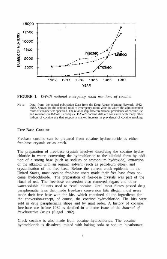
FIGURE 1. DAWN national emergency room mentions of cocaine
Note: Data from the annual publication Data from the Drug Abuse Warning Network, 1982-1987. Shown are the national total of emergency room visits in which the administrationroute of cocaine was specified. The relationship between national prevalence of cocaine useand mentions in DAWN is complex. DAWN cocaine data are consistent with many otherindices of cocaine use that support a marked increase in prevalence of cocaine smoking.
Free-Base Cocaine
Freebase cocaine can be prepared from cocaine hydrochloride as eitherfree-base crystals or as crack.
The preparation of free-base crystals involves dissolving the cocaine hydro-chloride in water, converting the hydrochloride to the alkaloid form by addi-tion of a strong base (such as sodium or ammonium hydroxide), extractionof the alkaloid with an organic solvent (such as petroleum ether), andcrystallization of the free base. Before the current crack epidemic in theUnited States, most cocaine free-base users made their free base from co-caine hydrochloride. The preparation of free-base crystals was part of theritual of use. The free-base conversion also removed sugars and otherwater-soluble diluents used to “cut” cocaine. Until most States passed drugparaphernalia laws that made free-base conversion kits illegal, most usersmade their free base with the kits, which contained all the ingredients forthe conversion-except, of course, the cocaine hydrochloride. The kits weresold in drug paraphernalia shops and by mail order. A history of cocainefree-base use before 1982 is detailed in a theme issue of the Journal ofPsychoactive Drugs (Siegel 1982).
Crack cocaine is also made from cocaine hydrochloride. The cocainehydrochloride is dissolved, mixed with baking soda or sodium bicarbonate,
7

and heated. While the mixture is being heated, the “cooker” gently swirlsor rotates the container. The freebase cocaine precipitates and coalescesinto a soft mass that becomes hard when it is dried. Sugars and otherwater-soluble diluents remain in the water, which is then poured off; leavingthe solid mass. Now, most crack users buy crack in the form of pellets or“rocks” that are already in the free-base form. Some users prefer to maketheir own crack from the cocaine hydrochloride because they feel moreconfident of its purity.
Several years before the current crack epidemic, smoking cocaine paste, anextract of coca leaves containing 40 to 85 percent cocaine sulfate, became amajor public health problem in Peru. Subsequently, paste smoking becamecommon in Bolivia, Columbia, and Ecuador (Jeri 1984). Cocaine pasteabusers there are mostly people in the lower socioeconomic classes. Thepaste is generally smoked in marijuana or tobacco cigarettes.
Crack and cocaine free-base crystals are often smoked in glass pipes. Theglass pipe has a bowl fitted on the bottom with one or more fine-meshcopper screens, which support the cocaine. The user heats the side of thebowl with a small butane torch or lighter. The free base vaporizes and isdrawn through the pipe. A pipe used in this manner gets hot and usersoften bum fingers or lips. Some free-base smokers have burns on theirgroins from suddenly hiding hot free-base pipes in their pockets.
Cocaine and Marijuana Combinations
Cocaine free base and occasionally cocaine hydrochloride are mixed withmarijuana and smoked. A marijuana cigarette laced with crack is common-ly called a “grimmie.” Crack is crushed between two coins, mixed withmarijuana, and rolled into a “joint.” Although smokers of grimmies aresmoking crack, they seem to view it as a less intense and leas severe formof use than using crack in a pipe. Many users of grimmies do not realizethat they are free basing as they associate free basing with using a pipe.Some free-base abusers report using grimmies as a means of controllingtheir cocaine use. When cocaine is smoked in marijuana, users report thatthere is less compulsion for continued use and less difficulty sleeping afteruse. In the San Francisco Bay area, grimmies are particularly popularamong black youth.
Cocaine and Tobacco Combinations
The combination of crack cocaine and a tobacco cigarette is known as cav-iar (or cavies). Cocaine hydrochloride is also used in this manner. Theuser makes the combination by reducing the crack to a powder and usingthe cigarette as a straw, sucking the cocaine inside the cigarette. The ad-vantage of this dosage form is that it can be smoked in public without itsbeing apparent that the person is using cocaine.
8

Phencyclidine and Cocaine
Phencyclidine (PCP) and cocaine are sometimes mixed together. The com-bination is known as “whack” or “spacebase.” Some users seek the combi-nation, but PCP is more often included because it is a less expensivesubstitute for cocaine. Thus, some cocaine users who believe they areusing only cocaine are also using PCP.
Cocaine Substitutes
Cocaine substitutes, most often procaine, are still used either as drugs ofdeception or intentionally as a “poor man’s cocaine substitute.” Sold undernames such as Snowcaine or Rock, procaine am be purchased in drug para-phernalia shops or by mail order. Sellers escape drug regulation by sellingthe substitute as incense. The label disclaimer gives directions for use:“Do not inhale vapors, as it may cause stimulation.”
Acquired Immunodeficiency Syndrome and Crack
Although smoking avoids the intravenous drug abuser’s risk of being in-fected with HIV by contaminated needles, smoking crack cocaine is stillassociated with behavior that puts some abusers at risk. Increasingly,women offer sex in exchange for crack.
PCP
Most users of PCP use it by smoking, usually mixing it with parsley,tobacco, or marijuana. The PCP may be dissolved and a cigarette dipped inthe solution. A dark-brown colored cigarette is often used for this purposeas the discoloration produced by PCP is not as obvious as it would be on awhite cigarette. Sherman cigarettes, because they are dark brown, are oftenused, thus the street name of this combination is known as “Shermans” or“shermies.”
METHAMPHETAMlNE (ICE)
Methamphetamine can be smoked in either the hydrochloride salt or freebase form. Smoking of the d-isomer of methamphetamine, called “ice,” hasbecome popular in Hawaii. Drug Enforcement Agency (DEA) seizures ofice in Hawaii began in 1985. Ice sold in Hawaii probably originates inKorea; however, mainland U.S. manufacture is beginning. In 1990, theDEA seized ice crystals manufactured in California.
Methamphetamine can be smoked in one of several ways:
1. Methamphetamine is placed on aluminum foil, the aluminum foil heatedfrom below, and the methamphetamine vapors inhaled. (As with heroin,this mode of use is sometimes called “chasing the dragon.“)
9
2. Methamphetamine is smoked in small, straight glass pipes. (Unlikefree-base cocaine, which is not water soluble, methamphetamine cannotbe smoked in water pipes, as the methamphetamine is water soluble andwould be trapped in the water.)
3. Methamphetamine is smoked in combination with tobacco or marijuana.
HEROIN AND OPIUM SMOKING
Smoking heroin is a dominant form of opiate use in Asia, where the formof freebase heroin prepared for smoking is designated by the U.S. DrugEnforcement Agency as heroin #3. Smoking free-base heroin also occurs inthe United States and England.
In the early seventies, Persian heroin, a freebase form of heroin, wasfavored by cocaine free-base users on the West Coast, who used it to atten-uate cocaine-induced anxiety, agitation, or paranoia. With continued, fre-quent use, some cocaine users who used Persian heroin as a secondary drugbecame physically dependent on the heroin, much to their surprise, as theythought that physical dependency could occur only with intravenousinjection.
Smoking opium is a primary and stable mode of use among some youthfulheroin abusers in England (Gossop et al. 1988). Opium smoking alsooccurs in the United States, particularly among immigrants from SoutheastAsia.
STIMULANT AND OPIATE COMBINATIONS
Drug abusers have long used combinations of stimulants and opiates intra-venously. The combination is commonly called a “speedball.” Cocainefreebase abusers also smoke cocaine free base or methamphetamine mixedwith freebase heroin. The street term for smoking a mixture of cocaineand heroin is “chasing and basing.”
RECOMMENDATIONS FOR FUTURE RESEARCH
Future research in pyrolytic chemistry should examine both street drugs anddrug combinations, emulating as closely as possible the way abusers actuallysmoke the drugs. Much pyrolytic chemistry research on drugs of abuseinvolves the use of pure drugs. This is important in delineating the toxicityof specific drugs, but does not completely address clinicians’ need to under-stand the toxicity of abused drugs combined with street impurities, adulter-ants, and other drugs. Much of the toxicity information currently availableto clinicians is based on case reports that lacked adequate analysis of thedrug.
10
The role of cigarette smoking in drug abuse relapse needs systematic study.Some patterns of drug abuse involve combining drugs with cigarettes. Inaddition, many patients report that they smoke more cigarettes when usingcocaine. The role of continued cigarette smoking in inducing cravings forother drug of abuse and relapse to other drug abuse deserves careful clini-cal study. If researchers should find that smoking cigarettes contributes torelapse, treatment programs should incorporate smoking cessation as part ofdrug abuse treatment.
REFERENCES
Gossop, M.; Griffiths, P.; and Strang, J. Chasing the dragon: Characteris-tics of heroin chasers. Br J Addict 83:1159-1162, 1988.
Jeri, F.R. Coca-paste smoking in some Latin American countries: Asevere and unabated form of addiction. Bull Narc 36(2):15-31, 1984.
Mikuriya, T.H., and Aldrich, M.R. Cannabis 1988: Old drug, newdangers: The potency question. J Psychoactive Drugs 20(1):47-55, 1988.
Siegel, R.K. Cocaine smoking. J Psychoactive Drugs 14(4):271-358, 1982.
AUTHORS
Donald R. Wesson, M.D.Associate Clinical Professor of PsychiatryUniversity of California, San FranciscoandStaff PsychiatristSan Francisco Veterans Administration Medical Center4150 Clement Street, 116FSan Francisco, CA 94121
Peter Washburn, M.D.Medical DirectorMerritt Peralta InstituteOakland, CA
11
Clinical Pharmacology of InhaledDrugs of Abuse: Implications inUnderstanding NicotineDependenceNeal L. Benowitz
INTRODUCTION
Why do people abuse drugs by the smoked or inhaled route? How doesthe pharmacology of inhaled drugs differ from that of drug administered byother routes? This chapter will discuss these and other aspects of the clini-cal pharmacology of inhaled drugs of abuse using nicotine as an example.
Pharmacokinetic and pharmacodynamic characteristics of a drug are impor-tant determinants of dependence liability, the temporal patterns of drug use,and the level of drug use. ultimately, an understanding of pharmacokineticsand pharmacodynamics may be useful in developing effective treatmentstrategies. Several pharmacokinetic and pharmacodynamic characteristicsappear to be necessary or optimal for a drug to produce dependence:(1) the drug must be effectively absorbed into the blood stream; (2) thedrug must rapidly enter into the brain; and (3) the drug must be psycho-active and that psychoactivity related to levels of the drug in the brain.These characteristics allow for the drug abuser to manipulate the dose of hisor her drug to optimize mood and psychological functioning and are mostlikely to result in the behavior described as criteria for drug dependence,
Inhalation of a drug provides a unique delivery system that is conducive toproducing drug dependence. Inhalation of a drug facilitates rapid absorptioninto the circulation which, in turn, results in rapid delivery to the brain.Studies on the clinical pharmacology of inhaled drugs of abuse presentsome particular research problems. For example, the bioavailability of in-haled drugs is highly variable from person to person and difficult to quanti-tate. Inhaled drugs are delivered and distributed to target organs rapidly.This means that pharmacokinetic and pharmacodynamic behaviors are in
12
disequilibrium conditions. This makes it difficult to study actions of a drugby the usual techniques of comparing venous concentrations with effects.Finally, tolerance develops rapidly to many or all effects of drug of abuse.Thus, even if the concentration in the brain could be known, the effect willvary over time and as a function of previous drug exposure history.
Approaches to studying these and other research problems concerning thepharmacology of inhaled drugs will be presented.
THE LUNGS AND DRUG ACTION
Inhaled drugs are taken into the lungs, from which they are absorbed intothe systemic circulation (Bend et al. 1985) (table 1). Before discussing thepharmacology of inhaled drugs, it is useful to consider how the anatomyand physiology of the lung may influence drug action.
TABLE 1. Lungs and drug action
Pharmacologic Function Action
Absorption of Inhaled Drugs Rapid transfer of unionizedlipophilic drugs from alveolarspaces to circulation
High pulmonary blood flow
Accumulation (Uptake) of Drugs Retention of drugs after inhalationFirst pass uptake after IV dosingLocal toxicity
Metabolism Clearance of drugs and endogenouschemicals
Oxidation, reduction, hydrolysis,biosynthesis
The lung may be conceived as having three major pharmacologic actions.The first is the absorption of inhaled drugs. Absorption of drugs from thelungs is facilitated by a huge alveolar surface area, thin alveolar epithelialand endothelial layers, and an extensive capillary bed. As a result of theseanatomical factors, many drugs, particularly un-ionized lipophilic drugs(which includes all of the common drugs of abuse), move rapidly from thealveolar spaces into the systemic circulation. Also of importance is thatpulmonary capillary blood flow is high, representing passage of the entireblood volume through the lung every minute. Thus, drugs that are absorbedare carried quickly to various parts of the body and to target organs.
13

A second function of the lung is the uptake of drugs by the lung paren-chyma. Such uptake may involve extracellular binding, intracellular bind-ing, or both to the various proteins, including receptors, and in some casesenergy-dependent active transport systems. The results of such uptake mayinclude retention of drugs after inhalation or during the first pass throughthe pulmonary circulation after intravenous dosing. In these cases, the lungbecomes a reservoir for slow and persistent release of drugs into the circu-lation. Pulmonary uptake may also result in high local concentrations of adrug, which could result in local toxicity, such as is the case with paraquat.
A third pharmacologic function of the lung is the metabolism of some drugsand endogenous chemicals. The lungs have the potential to metabolizedrugs by the same pathways as the liver. Although the concentrations ofdrug-metabolizing enzymes in lung tissues are lower than that in liver, theblood flow to the lungs (and hence the amount of a drug brought into con-tact with drug-metabolizing enzymes) is much greater. Therefore, metabolism by the lung can contribute substantially to total drug metabolism.Cytochrome P450 activity is primarily in nonciliated bronchial epithelial(Clara) and Type 2 alveolar cells. These cells are most vulnerable to injuryas a result of metabolism of certain drugs and chemicals to reactive metabo-lites. Examples of how these various lung processes contribute to thepharmacology of inhaled drugs, in particular nicotine, are discussed in sub-sequent sections.
ABSORPTION AND BIOAVAILABILITY
Nicotine is a tertiary amine composed of a pyridine and a pyrrolidine ring.Nicotine is a weak base with a pKa of 8.0 soluble both in water and inlipids. At physiological pH, about 31 percent of nicotine is non-ionizedsuch that it readily crosses cell membranes. The pH of tobacco smoke isimportant in determining the absorption of nicotine from different sites with-in the body. The pH of smoke from flue-cured tobaccos found in mostcigarettes is acidic (Brunneman and Huffman 1974). At this pH, the nico-tine is primarily ionized. In its ionized state, such as in acidic environ-ments, nicotine does not rapidly cross membranes. As a consequence, thereis little buccal absorption of nicotine from cigarette smoke, even when it isheld in the mouth. The pH of smoke from air-cured tobaccos, such as inpipes, cigars, and a few European cigarettes, is alkaline, and nicotine isprimarily unionized. Smoke from these products is well absorbed throughthe mouth (Armitage et al. 1978).
When tobacco smoke reaches the small airways and alveoli of the lung, thenicotine is rapidly absorbed independent of pH of the smoke. Blood con-centrations of nicotine rise quickly during and peak at the completion ofcigarette smoking (figure 1). The rapid absorption of nicotine from ciga-rette smoke through the lungs, presumably because of the huge surface area
14
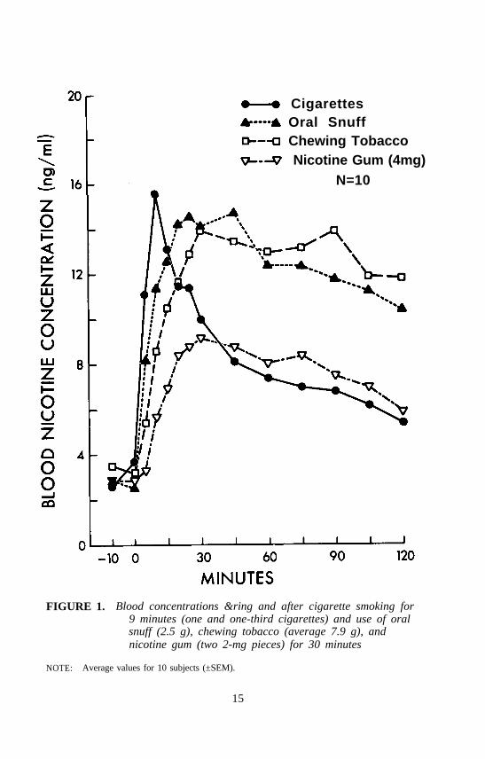
CigarettesOral SnuffChewing TobaccoNicotine Gum (4mg)
N=10
FIGURE 1. Blood concentrations &ring and after cigarette smoking for9 minutes (one and one-third cigarettes) and use of oralsnuff (2.5 g), chewing tobacco (average 7.9 g), andnicotine gum (two 2-mg pieces) for 30 minutes
NOTE: Average values for 10 subjects (±SEM).
15
of the alveoli and small airways and dissolution of nicotine into fluid of pHin the human physiologic range, facilitates transfer across cell membranes.
Blood or plasma concentrations of nicotine sampled in the afternoon insmokers generally range from 10 to 50 ng/ml (0.6 to 3x10⁻⁷ M). The incre-ment in blood nicotine concentration after smoking a single cigarette rangesfrom 5 to 30 ng/ml, depending on how the cigarette is smoked. Smokingis commonly considered to be a process of intermittent dosing of nicotine,which is in turn rapidly eliminated from the body. There is considerablepeak to trough oscillation from cigarette to cigarette. However, consistentwith a half-life of 2 hours, nicotine accumulates over 6 to 8 hours of regu-lar smoking and nicotine levels persist overnight, even as the smoker sleeps(Benowitz et al. 1982b). Thus, smoking results not in intermittent exposurebut in exposure to nicotine that lasts 24 hours of each day.
Smoking behavior is complex, and the dose of a drug delivered to the cir-culation is influenced by how the smoker smokes. Thus, the intensity,duration and number of puffs, depth of inhalation, degree of mixing ofsmoke with air, and other factors influence the dose. Estimation of thedose of nicotine has been attempted by setting up standardized proceduresof machine testing, where a smoking machine puffs a cigarette in a patternsimilar to that of human smokers. This test is the basis for the cigaretteyield data that were formerly published by the United States Federal TradeCommission, and that appear in many cigarette advertisements. However,human smokers smoke differently from machines, and machine deliveries donot correlate very well with biochemical markers of nicotine absorption inpeople (Benowitz et al. 1983).
Bioavailability can be assesed by methods analogous to those used in otherforms of drug testing. For example, we infused nicotine intravenouslyeither on a separate day or, using stableisotope-labeled nicotine, on thesame day as ad libitum cigarette smoking. Using the clearance estimatedfrom infusion data and the area under the blood nicotine concentration timecurve while smoking, absolute intake of nicotine from cigarette smoking,could be estimated (Jacob et al 1988; Benowitz and Jacob 1984). Theintake of nicotine is quite variable, both on the basis of daily intake (range10 to 80 mg) and on a per cigarette basis (range 0.4 to 1.6 mg per ciga-rette), among people.
The experimental situation under which smokers are studied may influenceself-determined intake of an inhaled drug. For example, we compared theintake of nicotine per cigarette in smokers during restriction of cigaretteavailability (Benowitz et al. 1986). When smoking was restricted from anaverage of 37 to 5 cigarettes per day, intake of nicotine per cigaretteincreased 2.7-fold. It is likely for other drugs that the time since the lastuse of the drug and other environmental factors will influence the inhaleddose of the drug and that smokers will vary the dose throughout the day.
16
The bioavailability of a drug from smoking material is in theory influencedby the amount of drug in the material. For tobacco, we examined thisrelationship by measuring the amount of nicotine in unsmoked tobacco andcomparing that to biological markers of absorption of nicotine in smokers(Benowitz et al 1983). On average, modem American cigarettes contain9 mg nicotine per cigarette. The average dose inhaled into the systemiccirculation by a smoker is 1 mg. Therefore, the bioavailability averages11 percent. However, there was very little correlation between the amountof nicotine in the tobacco or the amount of tobacco in the cigarette burnedwith the amount of nicotine absorbed by smokers (Benowitz et al. 1983;Benowitz and Jacob 1984). The bioavailability of nicotine can be system-atically changed by engineering features such as placement of ventilationholes to allow dilution of tobacco smoke with room air.
In summary, the absorption and bioavailability of inhaled drugs is highlyvariable from person to person and potentially from smoke to smoke.Smoking behavior is complex and permits the smoker considerable latitudein adjusting the dose to desired levels. Pharmacologic studies of inhaleddrugs of abuse need to consider the extent and sources of individual varia-bility in bioavailability in developing dose-response and dose-toxicity forsuch drugs.
DISTRlBUTION KINETICS
Smoking is a unique form of systemic drug administration in that entry intothe circulation is through the pulmonary rather than the portal or systemicvenous circulations. Entry via the lung influences the rate and pattern ofdelivery of drugs to body organs. For example, nicotine can be expectedto move quickly from inhaled cigarette smoke to the brain. It is estimatedthat it takes 11 seconds or less from the start of a puff to the deliveryof nicotine to the brain and 19 seconds to complete transit through the brain(table 2). This estimation assumes a 2-second puff, negligible time fordiffusion of nicotine across alveolar membranes, and negligible time for
TABLE 2. Nicotine effects after inhalation: Temporal considerations
Action Seconds
Puff time 2.0
Pulmonary circulation time 7.5
Left ventricle to cerebral circulation time -1.0
Brain transit time 8.5
Total circulation time 19.0
NOTE: Estimates derived from Mapleson (1973).
17

movement from the arterial blood into the brain. With respect to the latter,nicotine is rapidly and extensively taken up from the blood in its first passthrough the brains of rats (figure 2). Likewise, intravenously injected C14
nicotine is immediately taken up by the brains of mice, reaching a maxi-mum concentration within 1 minute after injection (Stalhandski and Slanina1970). Similar findings based on positron emission tomography of the brainwere seen after injection of 11C nicotine in monkeys (Maziere et al. 1976).The time from the start of a puff to delivery of nicotine to the brain issimilar to the time from intravenous injection to arterial circulation,estimated as from 9 to 16 seconds in healthy people.
FIGURE 2. Concentrations of nicotine in the brain and arterial plasmaof rats after IV injection of 0.1 mg/kg nicotine over30 seconds
NOTE: Concentration of nicotine peak in the brain at or bcfore the earliest sampling time (30 sec-onds after the time of injetion). Brain concentrations are much highger than arterial concen-trations, particularly in the early minutes after injection.
18
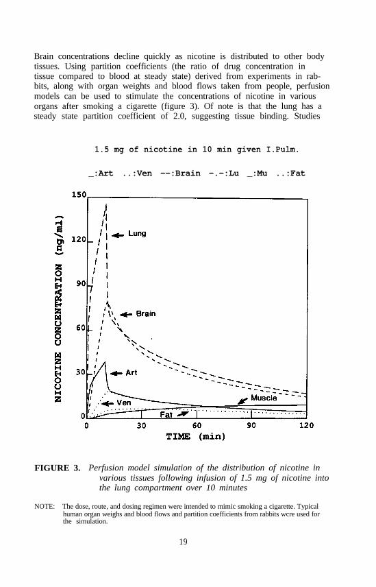
Brain concentrations decline quickly as nicotine is distributed to other bodytissues. Using partition coefficients (the ratio of drug concentration intissue compared to blood at steady state) derived from experiments in rab-bits, along with organ weights and blood flows taken from people, perfusionmodels can be used to stimulate the concentrations of nicotine in variousorgans after smoking a cigarette (figure 3). Of note is that the lung has asteady state partition coefficient of 2.0, suggesting tissue binding. Studies
1.5 mg of nicotine in 10 min given I.Pulm.
_:Art ..:Ven --:Brain -.-:Lu _:Mu ..:Fat
FIGURE 3. Perfusion model simulation of the distribution of nicotine invarious tissues following infusion of 1.5 mg of nicotine intothe lung compartment over 10 minutes
NOTE: The dose, route, and dosing regimen were intended to mimic smoking a cigarette. Typicalhuman organ weighs and blood flows and partition coefficients from rabbits wcre used forthe simulation.
19
with lung slices demonstrate lung binding, which is somewhat dose depend-ent (Ludden et al. 1976). There was 50 percent more binding of nicotineto lung slices at low concentrations than at high concentrations. As a resultof high local concentrations and binding, concentrations of nicotine in thelung are predicted to be extremely high after intrapulmonary arterial injec-tion and would be expected to be similarly high after tobacco, smoking.Such high concentrations could conceivably cause local pharmacologiceffects on the lung, such as neutrophil chemcattraction (Totti et al. 1984) atconcentrations that would not be expected to be achieved based on the rela-tively low concentrations of nicotine in the blood.
Concentrations of nicotine in arterial blood and the brain are seen to in-crease sharply following exposure, then to decline over 20 to 30 minutes asnicotine is redistributed to other body tissues, particularly skeletal muscle.Venous blood concentrations, reflecting outflow of nicotine from body tis-sues, are predicted to be considerably lower than arterial concentrations forthe duration of the infusion and for several minutes afterward. This dis-crepancy has been observed in rabbits following rapid intravenous injectionof nicotine (Porchet et al. 1988b) (figure 4). It is seen from figure 3 thatthe ratio of nicotine in the brain to that in venous blood is highest duringand at the end of the infusion and gradually decreases as the eliminationphase is entered. The importance of this disequilibrium between venousand arterial concentrations with respect to pharmacodynamic studies is dis-cussed in a later section.
In summary, inhalation of drugs allows for rapid transfer into the arterialcirculation and into the brain. Rapid passage into the brain provides for thepossibility of rapid behavioral reinforcement from smoking and allows thesmoker to precisely control the concentrations of nicotine in the brain,hence to modulate pharmacologic effects. To better understand this process,the relationship between the level of a drug and its effects on the brainmust be examined.
PHARMACODYNAMICS OF NICOTlNE IN TEE BRAIN
Nicotine binds to specific receptors in the brain and affects cerebral metabo-lism in the areas of specific binding (London et al. 1985). Nicotine isknown to have a complex dose-response relationship. In experimental prep-arations, nicotine in low doses causes ganglionic stimulation and in highdoses causes ganglionic blockade following brief stimulation. This biphasicresponse pattern is observed in the intact organism as well, although themechanisms are far more complex. With regard to central nervous systemeffects, Ashton has provided evidence that nicotine exerts biphasic actions inhumans (Ashton et al. 1980). When contingent negative variation (CNV, acomponent of the evoked EEG potential) was examined, it was seen thatnicotine in low doses increased while high doses decreased the amplitude ofthe CNV. It was proposed that low doses produced arousal or increased
20

Femoral VeinFemoral Artery
Internal Jugular Vein
FIGURE 4. Mean blood concentrations from three vascular sites after anIV bolus of 25 ug/kg of nicotine given over 1 minute in sixrabbits
KEY: Squares=femoral vein; inverted trianguls=femoral artery; triangles=internal jugular vein.
NOTE: Arterial and internal jugular vcnous concentrations (the latter proportional to brainconcentrations) were much higher than those in femoral venous blood during the first5 minutes.
SOURCE: Porchet et al. 1988b, copyright 1988. Rockefeller University Press.
attention and vigilance, while higher doses produced relaxation and reducedstress. Presumably, the smoker, by titrating the level of nicotine in thebrain, can achieve the desired mental state.
The time course of nicotine absorption strongly influences the actions ofnicotine, although it is not clear whether this is a result of a more rapid
21
rate of rise or higher absolute levels of nicotine in the brain. We andothers have found that heart rate acceleration, a reflection of the level ofsympathetic neural discharge and a sensitive physiologic response, tends tocorrelate with the magnitude of subjective effect (Rosenberg et al 1980;West and Russell 1987). Heart rate acceleration appears to be mediated bythe central nervous system either through actions on chemoreceptor afferentpathways or via direct effects on the brain stem. With repeated intravenousinjections of nicotine, dosed to simulate cigarette smoking, heart rate accel-eration and subjective effects were greatest with the first series of injectionsand then declined over time with successive injections (Rosenberg et al.1980).
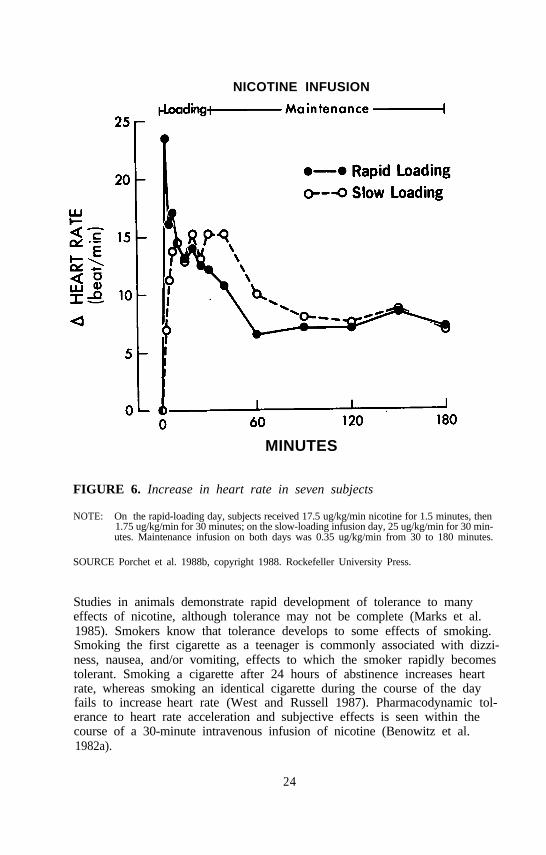
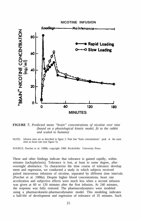
To examine the importance of rate of dosing of nicotine and resultantactions and the relationship between effects and brain levels of nicotine, wecompared the consequences of rapid- and slow-loading infusions of nicotine(Porchet et al. 1988b). The study was performed in seven volunteer smok-ers who had abstained from smoking overnight. Venous blood concentra-tions were not markedly different for the two infusion rates except for thefirst 5 minutes, when the increase in blood concentration was greater withrapid infusion (figure 5). Peak concentrations were seen at 30 minutes andwere similar for both infusions. However, the heart rate increased muchmore quickly and to a greater level and was associated with transient dizzi-ness and euphoria after rapid infusion, compared to slow infusion (figure 6).During the slower infusion, heart rate increased slowly, peaking at 30 min-utes, and subjective effects were minimal. Using kinetic data derived fromsubjects of this study and studies in rabbits and with the use of a perfusionmodel, we estimated levels of nicotine in the human brain during the rapidand slow infusions. Predicted brain concentrations rose much more rapidlyand to a higher level after rapid infusion, with a concentration time coursesimilar to that seen for heart rate acceleration (figure 7). Brain concentra-tions versus heart rate acceleration tended to change in parallel These datasuggest that effects of nicotine track brain concentrations over time.
In summary, for drugs that rapidly enter and act upon the brain, drug dos-ing such as by inhalation results in higher brain concentrations and greaterpsychoactive (and possibly cardiovascular) effects than when the same doseis given more slowly. The possibility of titrating the level of a drug in thebrain to achieve a desired mental state is also facilitated by inhalation asthe route of dosing. These factors may explain why smoking a drug maybe preferred over the oral or nasal routes of administration. In addition, asmentioned previously in the discussion of drug distribution, venous bloodconcentrations substantially underestimate arterial and brain levels after rapiddosing, and such data cannot (without mathematical transformations) be usedto study concentration-effect relationships.
22

NICOTINE INFUSION
MINUTES
FIGURE 5. Average blood nicotine concentration in seven subjects
NOTE: (On the rapid-loading day, subjects received 17.5 ug/kg/min nicotine for 1.5 minutes, then1.75 ug/kg/min for 30 minutes; on the slow-loading infusion day, 25 ug/kg/min for 30 min-utes. Maintenance infusion on both days was 0.35 ug/kg/min from 30 to 180 minutes.
SOURCE: Porchet et al. 1988b. copyright 1988. Rockefeller University Press.
TOLERANCE
An important issue in drug dependence is the development of tolerance.Tolerance is defined as the tendency for a given dose of a drug, afterrepeated doses, to produce less of an effect. In some cases, increasing thedose of a drug in the presence of tolerance may achieve the same effectthat was obtained after the first dose. Pharmacodynamic tolerance can befurther defined as the condition that exists when a particular concentrationof a drug at a receptor site (in the intact organism, approximated by theconcentration in the blood) produces less of an effect than it did after aprior exposure.
23
NICOTINE INFUSION
MINUTES
FIGURE 6. Increase in heart rate in seven subjects
NOTE: On the rapid-loading day, subjects received 17.5 ug/kg/min nicotine for 1.5 minutes, then1.75 ug/kg/min for 30 minutes; on the slow-loading infusion day, 25 ug/kg/min for 30 min-utes. Maintenance infusion on both days was 0.35 ug/kg/min from 30 to 180 minutes.
SOURCE Porchet et al. 1988b, copyright 1988. Rockefeller University Press.
Studies in animals demonstrate rapid development of tolerance to manyeffects of nicotine, although tolerance may not be complete (Marks et al.1985). Smokers know that tolerance develops to some effects of smoking.Smoking the first cigarette as a teenager is commonly associated with dizzi-ness, nausea, and/or vomiting, effects to which the smoker rapidly becomestolerant. Smoking a cigarette after 24 hours of abstinence increases heartrate, whereas smoking an identical cigarette during the course of the dayfails to increase heart rate (West and Russell 1987). Pharmacodynamic tol-erance to heart rate acceleration and subjective effects is seen within thecourse of a 30-minute intravenous infusion of nicotine (Benowitz et al.1982a).
24
NICOTINE INFUSION
MINUTES
FIGURE 7. Predicted mean “brain” concentrations of nicotine over time(based on a physiological kinetic model, fit to the rabbitand scaled to humans)
NOTE: lnfusion rates are as described in figure 3. Note that “brain concentrations" peak at the sametime as heart rate (see figure 5).
SOURCE: Porchet et al. 1988b, copyright 1988. Rockefeller University Press.
These and other findings indicate that tolerance is gained rapidly, withinminutes (tachyphylaxis). Tolerance is lost, at least to some degree, afterovernight abstinence. To characterize the time course of tolerance development and regression, we conducted a study in which subjects receivedpaired intravenous infusions of nicotine, separated by different time intervals(Porchet et al. 1988a). Despite higher blood concentrations, heart rateacceleration and subjective effects were much less when a second infusionwas given at 60 or 120 minutes after the first infusion. At 240 minutes,the response was fully restored. The pharmacodynamics were modeledusing a pharmacokinetic-pharmacodynamic model. This modeling indicatesa half-life of development and regression of tolerance of 35 minutes. Such
25
modeling suggests that at three half-lives or 1 1/2 hours after a cigarette,nearly full sensitivity should have been regained.
Because tolerance develops so quickly, the rate of drug dosing influencesthe magnitude of effect. That is, the faster the administration, the less timethere is to develop tolerance and the greater the effect for any given doseor maximal level. The interval at which cigarette smokers smoke cigarettesmay be determined by two factors: the rate of distribution out of the brainafter smoking a cigarette and the kinetics of regression of tolerance toeffects of nicotine.
In summary, tolerance develops rapidly to the effects of nicotine as it doesto effects of other inhaled drugs of abuse. The kinetics of tolerance aresuch that tolerance may develop and regress in cycles throughout the day.The interval at which users smoke their drugs may be influenced by twofactors: the rate of distribution of drug out of the brain after a particulardose of the drug and the kinetics of regression of tolerance to effects ofthat drug. Rapid rate of dosing, as by inhalation, also allows the maximaldrug effect with the least extent of development of tolerance.
PHARMACOKINETICS AND PHARMACODYNAMICS OFDRUG DEPENDENCE
The reinforcing properties of a drug are expected to be strongest when apsychoactive effect, usually a pleasant one, follows in close temporal prox-imity to the self-administration of a drug. In pharmacokinetic or pharmaco-dynamic terms, after dosing, the drug should enter the bloodstream rapidlyand move rapidly from the bloodstream into the brain. The appearance ofthe drug in the brain should be associated with the desired psychoactiveeffects. If the effect of a drug is delayed after appearance of the drug inthe brain or if tolerance to the drug effect is already present, the drug isless likely to be reinforcing.
That the user can easily control the dose of the drug and modulate the re-sultant psychoactivity would be expected to further strengthen the reinforc-ing nature of the drug. As discussed previously, nicotine obtained bycigarette smoking demonstrates these characteristics, as do other drugs ofabuse that are inhaled, such as crack cocaine or marijuana, and drugs thatare intravenously injected, such as heroin and cocaine.
Tolerance is most likely to develop to the effects of a drug when its recep-tors are continuously exposed to the drug. Frequent and sustained dosing, along half-life of a drug, or a combination of these factors, would favor de-velopment of tolerance. The half-life of nicotine in the brain following asingle dose exposure is short, probably about 10 minutes in humans, due toredistribution out of the brain to other body tissues. Some degree of toler-ance does develop rapidly (tachyphylaxis), even after brief exposures. Such
26

tolerance also regresses relatively quickly (half-life, 35 minutes) such that asmoker may learn that smoking a cigarette at particular intervals is more re-inforcing than smoking more frequently.
With repetitive dosing, levels of nicotine build up in the body (and in thebrain) in accordance with the terminal half-life of 2 hours. Thus, there issubstantial accumulation of nicotine in the brain and an increasing generallevel of tolerance throughout the day. Presumably, however, smoking indi-vidual cigarettes still results in peaks of nicotine in the brain, which mayovercome the underlying level of tolerance and produce some of the desiredeffects.
Withdrawal symptoms are mast likely to occur when there has been a sus-tained effect in the brain and the drug is then rapidly removed. Thus, rapidexit from specific brain regions would favor more severe withdrawal symp-toms. Nicotine, as it is dosed by cigarette smokers, exhibits both of thesecharacteristics. Due to repetitive dosing, the brain is exposed to nicotinefor prolonged periods of time, so that neuroadaptation occurs during thesmoking day. When exposure is terminated, nicotine rapidly exits the brainand withdrawal symptoms are experienced. It is likely, therefore, that reliefof withdrawal symptoms plays an increasingly important role in determiningsmoking behavior as the day progress.
NICOTINE AND THE DAILY SMOKING CYCLE
The pharmacokinetic and pharmacodynamic considerations discussed thus farhelp us understand the development of nicotine dependence and humancigarette-smoking behavior, as well as adverse effects of cigarette smoking.The daily smoking cycle can be conceived as follows. The first cigarette ofthe day produces substantial pharmacologic effects, primarily arousal, but atthe same time tolerance begins to develop. A second cigarette may besmoked at a later time, at which the smoker has learned there is some re-gression of tolerance. With subsequent cigarettes, there is accumulation ofnicotine in the body, resulting in a greater level of tolerance, and with-drawal symptoms become more pronounced between cigarettes. Transientlyhigh brain levels of nicotine following smoking individual cigarettes maypartially overcome tolerance. But the primary (euphoric) effects of indi-vidual cigarettes tend to lessen throughout the day. Overnight abstinenceallows considerable resensitization to actions of nicotine. Because of thedose-response and dose-tolerance characteristics, habitual smokers need tosmoke at least 15 cigarettes and consume 20 to 40 mg nicotine per day toachieve the desired effects of cigarette smoking and minimize withdrawaldiscomfort throughout the day.
27
REFERENCES
Armitage, A.; DoIlery, C.; Houseman, T.; Kohnes, E.; Lewis, P.J.; andTurner, D.M. Absorption of nicotine from small cigars. Clin PharmacolTher 23:143-150, 1978.
Ashton, H.; Marsh, V.R.; Millman, J.E.; Rawlins, M.D.; Telford, R.; andThompson, J.W. Biphasic dose-related responses of the CNV (contingentnegative variation) to i.v. nicotine in man. Br J Clin Pharmacol 10:579-589,1980.
Bend, J.R.; Serabjit-Singh, C.J.; and Philpot, R.M. The pulmonary uptake,accumulation, and metabolism of xenobiotics. Annu Rev Pharmacol Toni-col 25:97-125, 1985.
Benowitz, N.L.; Hall, SM.; Herning, R.I.; Jacob, P., III; Jones, R.T.; andOsman, A.L. Smokers of low yield cigarettes do not consume leas nico-tine. N Engl J Med 309:139-142, 1983.
Benowitz, N.L., and Jacob, P., III. Daily intake of nicotine during cigarettesmoking. Clin Pharmacol Ther 35:499-504, 1984.
Benowitz, N.L.; Jacob, P., III; Jones, R.T.; and Rosenberg, J. Interindivid-ual variability in the metabolism and cardiovascuIar effects of nicotine inman. J Pharmacol Exp Ther 221:368-372, 1982a.
Benowitz, N.L; Jacob, P., III; Kozlowski, L.; and Yu, L. Influence ofsmoking fewer cigarettes on exposure to tar, nicotine, and carbon monox-ide exposure. N Engl J Med 314:1310-1313, 1986.
Benowitz, N.L.; Kuyt, F.; and Jacob, P., III. Circadian blood nicotine con-centrations during cigarette smoking. Chin Phamacol Ther 32:758-764,1982b.
Brunneman, K.D., and Huffman, D. The pH of tobacco smoke. Food Cos-metics Toxicol 12:115-124, 1974.
Jamb, P., III; Benowitz, N.L.; and Shulgin, A.T. Recent studies of nicotinemetabolism in humans. Pharmacol Biochem Behav 30:249-253, 1988.
London, E.D.; Connolly, R.J.; Szikszay, M.; and Wamsley, J.K. Distribu-tion of cerebral metabolic effects of nicotine in the rat. Eur J Pharmacol110:391-392, 1985.
Ludden, T.M.; Schanker, L.S.; and Lanman, R.C. Binding of organic com-pounds to rat liver and lung. Drug Metab Dispos 4:8-16, 1976.
Mapleson, W.W. Circulation-time models of the uptake of inhaled anesthe-tics and data for quantifying them. Br J Anaesth 45:319-334, 1973.
Marks, M.J.; Stitzel, J.A.; and Collins, A.C. Time course study of theeffects of chronic nicotine infusion on drug response and brain receptors.J Pharmacol Exp Ther 235:619-628, 1985.
Maziere, M.; Comar, D.; Marazano, C.; and Berger, G. Nicotine-11C: Syn-thesis and distribution kinetics in animals. Eur J Nucl Med 1:255-258,1976.
Porchet, H.C.; Benowitz, N.L.; and Sheiner, LB. Pharmacodynamic modelof tolerance: Application to nicotine. J Pharmacol Exp Ther 244:231-236, 1988a
28
Porchet, H.C.; Benowitz, N.L.; Sheiner, L.B.; and Copeland, J.R. Apparenttolerance to the acute effect of nicotine results in part from distributionkinetics. J Clin Invest 80:1466-1471, 1988b.
Rosenberg, J.; Benowitz, N.L.; Jacob, P., III; and Wilson, K.M. Dispositionkinetics and effects of intravenous nicotine. Clin Pharmacol Ther 28:516-522, 1980.
Stalhandski, T., and Slanina, P. Effect of nicotine treatment on the metabo-lism of C14-labeled nicotine in mice and rats. Acta Pharmacol Toxicol(Copenh) 28:75-80, 1970.
Totti, N., III; McCusker, K.T.; Campbell, E.J.; Griffin, G.L.; and Senior,R.M. Nicotine is chemotactic for neutrophils and enhances neutrophilresponsiveness to chemotactic peptides. Science 223:169-173, 1984.
West, R.J., and Russell, M.A.H. Cardiovascular and subjective effects ofsmoking before and after 24 h of abstinence from cigarettes. Psycho-pharmacology 92:118-121, 1987.
ACKNOWLEDGMENTS
Supported in part by U.S. Public Health Service grants DA 02277,CA 32389, and DA 016%. These studies were carried out in part in theGeneral Clinical Research Center (RR-00083) with support of the Divisionof Research Resources, National Institutes of Health.
AUTHOR
Neal L. Benowitz, M.D.Professor of Medicine and PsychiatryUniversity of California, San FranciscoSan Francisco General Hospital Medical CenterBuilding 30, Fifth FloorSan Francisco, CA 94110
29

The Pharmacology of CocaineSmoking in HumansReese T. Jones
INTRODUCTION
Smoking crack is smoking cocaine. Why would people go to the trouble ofsmoking cocaine when they can so easily snuff it, take it orally, or inject itparenterally? For many of the same reasons that nicotine is pleasurable toa tobacco addict and is so much more addicting when smoked in the formof tobacco cigarettes than when snuffed or chewed, so smoking the alkaloidcocaine is even more addicting than use by other routes (Benowitz, this vol-ume). For similar reasons, most cannabis-dependent people choose tosmoke marijuana or hashish even though tetrahydrocannabinol (THC) is psy-choactive by oral ingestion. Of course, cocaine, nicotine, and THC pharma-cology differ, but the aspects of their pharmacology that lead to preferencefor the smoked route of ingestion as well as some of the consequences ofthat choice are similar. If what is known of other inhaled drugs is kept inmind (Benowitz, this volume), it may be easier to understand certain consequences of cocaine smoking and, perhaps more important, to design properexperiments to study cocaine smoking.
Smoking cocaine has become popular only in recent years, mostly in NorthAmerica and some Latin American countries (Jeri 1984; Siegel 1985). Likemost current patterns of psychoactive drug use, smoking cocaine was notcompletely unknown to the ancients. For example, in the United States,during the early part of this century, the Parke, Davis & Company pharma-ceuticals catalog listed, among other forms of coca and cocaine, cocacheroots and coca cigarettes. The Parke, Davis coca cigars and cigarettesprobably contained between 0.5 and 1.0 percent cocaine and were recom-mended for therapeutic use. A similar product known as the Coca LeafBall was advertised in England during the same period with the suggestionthat “families, mothers and children” would benefit from the therapeuticeffects of smoked coca. Famous people from England and Europe testifiedas to its effectiveness and must have engaged in cocaine smoking, althoughthere is no indication that addiction to coca smoking was common. In
30
some ways, smoking coca leaf might be analogous to smoking tobacco orcannabis leaf in that a relatively small concentration (0.5 to 2 percent) ofactive substance (cocaine, nicotine, THC) is contained in a relatively largeamount of plant material. Smoking coca leaf never became as popular assmoking tobacco leaf. Chewing coca leaf remained common only in rela-tively isolated groups, where it had long been part of their culture (Hannaand Hornick 1977).
In more recent times, smoking cocaine in the form of a crude plant extractsometimes called paste and containing about 80 percent cocaine base be-came popular in Peru and in adjacent countries (Almeida 1978; Jeri 1984;Flores 1986). Up to then, both in medical applications and illicitly, thewater-soluble salt, usually cocaine hydrochloride, was the form more com-monly used. Currently in North America, a readily available and widelydiscussed form is cocaine base, usually converted from the cocaine salt bymixing cocaine hydrochloride and a solution containing an excess of sodiumbicarbonate usually baking soda) and heating and evaporating off the fluid.This leaves mostly cocaine base in a bicarbonate crystalline mixture. Inmost large cities in the United States, the availability of illicit ready-to-smoke base facilitates smoking cocaine. Cocaine base melts at a much low-er temperature than the salt (about 80 °C instead of 180 ºC) and then boils,producing an inhalable aerosol (Snyder et al. 1988; Wood, this volume).Whether forces of the illicit marketplace led to changes in cocaine chemis-try and packaging or whether the availability of high concentration, relative-ly pure cocaine base shaped the market forces to increase consumer demandis not entirely clear. In any case, a cocaine user who chooses to smokecan now obtain material containing close to 100 percent cocaine rather thanusing coca plant material containing 1 percent of cocaine base. Considerthe likely consequences if THC was readily available to add to smokingdevices or if nicotine addicts began adding additional nicotine to tobaccocigarettes. Although not a perfect analogy, considering such events mayhelp to put cocaine smoking in perspective.
PROBLEMS IN STUDYING COCAINE SMOKING
A variety of devices are used for cocaine smoking: pipes of all sorts, orcigarettes made of admixtures of cocaine base with tobacco, with marijuana,or with other plant material. The use of nonstandardized smoking devicescontributes to a situation similar to that seen during the early days ofcannabis-smoking research. When programs of controlled laboratory experi-ments with smoked cocaine are begun, it is not obvious what the typicallaboratory smoking conditions should be, what constraints on smoking be-havior are proper, and what physical and chemical considerations will maxi-mize the likelihood that laboratory experiments will be relevant to the realworld of illicit smoked cocaine, but will maintain adequate research subjectsafety. In contrast, a researcher of tobacco smoking has the advantages ofgreat standardization of dose, dosage form, and delivery systems developed
31
by the tobacco industry. Even with that information, smoking behavior pro-cedural issues in tobacco-smoking research, particularly dose regulation, arecomplex, and not all are resolved (Biding 1987; Kozlowski et al. 1982).
Relatively little has been published describing the human pharmacology ofcocaine smoking under controlled or semicontrolled laboratory conditions(Perez-Reyes et al. 1982; Jeffcoat et al. 1989). Experiments in whichexperienced volunteers smoked from a glass apparatus heated to 260 ºC andcontaining 50 mg of cocaine base demonstrated the rapid increase in plasmacocaine levels during smoking, as compared to intravenous (IV) cocaineadministration, that would be expected from what is known of the smokingprocess with nicotine and cannabis (Benowitz, this volume; Perez-Reyes,this volume). Other than more rapid onset, the resulting cardiovascular andpsychologic changes were not unlike those seen after equivalent IV or nasalcocaine doses. Other experiments with monkeys (Siegel et al. 1976) andwith smokers of crude extracts of cocaine often called paste noted a gener-ally similar pattern of effect (Paly. et al. 1982).
Laboratory studies with any smoked drug present great methodologic chal-lenges to the investigator. Proper pharmacologic studies must involve con-trol of dose or at least accurate specification of the dose actually absorbed.The nature of smoking behavior makes dose difficult to regulate and, undermany conditions, difficult even to estimate. Although animals can be madeto smoke under some conditions (Siegel et al. 1976), animals seem reluctantto smoke the way humans do; thus, human studies are necessary.
Cocaine may present problems that have not been important in tobacco orcannabis smoking research. The rate of cocaine degradation is very temper-ature dependent, hence, subject to variability because of smoking-apparatusdesign and variation in conditions. Temperature in most cocaine-smokingdevices varies with air flow. Air flow, in the case of humans smokingunder natural conditions, depends on both puff and inhalation volumes andduration. The importance of puff and inhalation volume in determiningdose is well recognized by tobacco and cannabis researchers (Heming et al.1983; Benowitz 1986). How to control or regulate dose in psychopharma-cologic studies involves compromises, however, and optimal solutions arenot evident yet. When cocaine is smoked, the amount of cocaine pyrolyzedto other compounds will be related to the temperature in the smoking de-vice. The temperature, in turn, will be related to the smoker’s behavior,making for a complex equation to determine drug delivery.
When researching nicotine smoking, an investigator can depend on a degreeof industry- and government-derived standardization of the smoking devices(cigarettes, pipes, etc.). In marijuana smoking research over the years,some consensus has developed as to smoking material and apparatus, atleast in the laboratory. Cocaine smoking research, however, is nowherenear that point. The usual and expected “natural” or typical pattern (or
32

more likely, the range of patterns) of cocaine smoking is not at all estab-lished; hence, the relevance of laboratory paradigms is still open to ques-tion. For example, typical puff and inhalation parameters obtained duringrelatively unconstrained cocaine smoking have not been determined as theyhave with nicotine and with marijuana.
THE PHARMACOLOGY OF COCAINE SMOKING
In experiments trying to describe the pharmacology of cocaine smoking, weattempted to control some aspects of smoking behavior but, in these begin-ning experiments, left options open for the subjects, all of whom claimed tobe experienced cocaine smokers. What is apparent in the laboratory datacollected thus far is great variability; both variability in cocaine smokingbehavior and, under conditions where smoking behavior parameters ate tosome extent constrained, considerable variability in cocaine plasma levelsand effects.
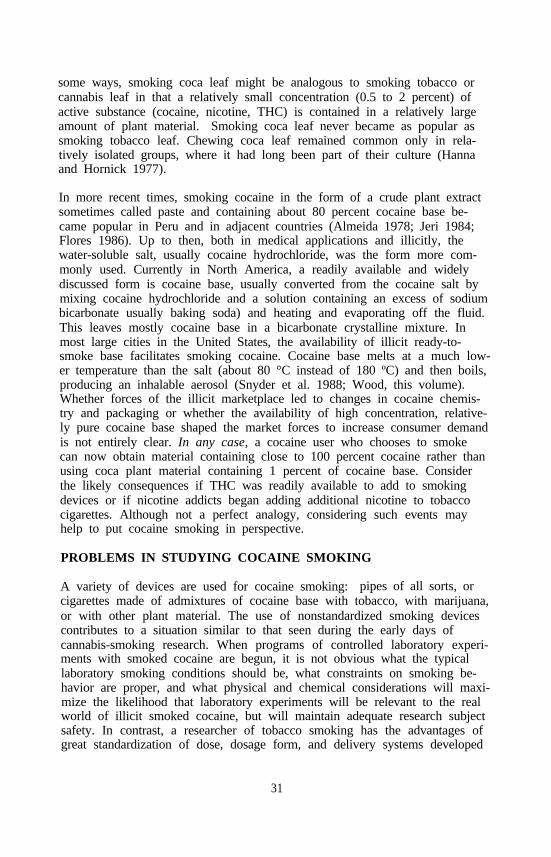
For example, in figure 1 the plasma concentrations are shown from 10 dif-ferent research subjects smoking cocaine base under similar conditions.Peak venous cocaine levels ranged from less than 100 ng/ml to approxi-mately 500 ng/ml, with considerable variability in time of peak as well.These cocaine smokers were allowed to take one or two and occasionallythree inhalations spaced 1 minute apart from an electrically heated l-literglass flask, beginning about 45 seconds after 100 mg of finely powderedcocaine base was dropped into the flask. At that time the cocaine base hadmelted, and a fine white aerosol was forming in the flask. The clear pud-dle of melted cocaine at the bottom of the flask was just beginning to yel-low in the process that would turn it into a black tarry mass just a fewminutes later. The temperature of the glass surface at the bottom of theflask was 260 °C.
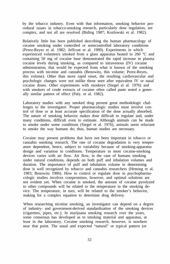
The variation in plasma levels is contrasted with those from the samesubjects given cocaine IV at a dose of 0.6 mg/kg of body weight injectedby infusion pump over 1 minute (figure 2). Perhaps noteworthy is theconsiderable variability in peak plasma levels of cocaine even withcontrolled IV administration. Variability in venous plasma levels is notunexpected with a rapidly distributed, relatively rapidly metabolized drug,although it should give pause for thought to those attempting to predictprecise relationships between drug effect and blood levels from a particularcocaine dose. It should be kept in mind that, particularly by the smokedroute, arterial level of cocaine is more likely to be a much better indicatorof brain level than is venous blood level. As is the case after nicotinesmoking, venous cocaine levels will underestimate brain levels.
When the mean plasma cocaine levels after IV or smoked doses are com-pared to those of nasal or oral doses of cocaine hydrochloride, reasons forsome of the appeal of the smoked route are evident (figure 3). The time of
33
MINUTES AFTER FIRST INHALATION
FIGURE 1. Plasma levels of cocaine after smoking
MINUTES AFTER DOSE
FIGURE 2. Plasma levels of cocaine: IV administration
34

peak level is significantly earlier after dose in the smoked or IV route.Similarly, the time of peak subjective effect is earlier by the smoked route(figure 4). Figure 4 illustrates the peak subjective-high rating in which zerois defined as a normal, sober state, and 100 is as intoxicated as ever experi-enced after cocaine administration.
MINUTES AFTER DOSE
FIGURE 3. Plasma levels of cocaine
DEVELOPMENT OF TOLERANCE TO COCAINE
Why should a shorter interval to peak effect be important to a drug user?probably because neuroadaptation (tolerance) to many psychoactive drugeffects is a rapidly developing process better timed in seconds and minutesthan in hours or days. The slower any drug effect arises, other thingsbeing equal the less intense the effect, because tolerance to the effect is ina real sense developing concurrently with the appearance of the drug effect.
Rapidly developing tolerance to cocaine is demonstrated in a hysteresis plotof plasma level vs. subjective intoxication rating in figure 5. If little or notolerance (neuroadaptation) developed over the 4 hours summarized in figure5, the plot following the ascending and descending phases of the time/plasma level function would be more or less a straight line. One index ofthe magnitude of tolerance is the divergence of those ascending anddescending functions (Holford and Sheiner 1981). Note that the subjectshad the option of rating their level of dysphoria with a minus value, and
35

MINUTES AFTER DOSE
FIGURE 4. Subjective high ratings
some did so as the cocaine effects wore off. Figure 6 shows the clockwisehysteresis plots of subjective intoxication levels vs. cocaine concentrations inplasma after smoking cocaine and after an IV dose (0.6 mg/kg) in the same10 research patients whose time vs. concentration data are shown in figures1 and 2. Quantitative comparisons between the smoking and the IV hyster-esis loops involve many assumptions, but a variety of semiparametric meth-ods seem useful for a quantitative pharmacokinetic-pharmacodynamicdescriptive model (Verotta et al. 1989). The similarity to the rapid toler-ance that develops to many effects of nicotine is striking (Benowitz, thisvolume).
PHYSIOLOGIC AND SUBJECTIVE EFFECTS
Assuming that most of cocaine’s subjective effects are related to its braineffects, the more rapid transit time from lung to brain (5 to 10 seconds)after inhalation of cocaine smoke would more likely provide a steeper andhigher gradient of cocaine brain levels than could oral, nasal, or even IVadministration. Table 1 summarizes the magnitude and time of some peakeffects after cocaine was given to the same 10 experienced cocaine users byvarious routes, in balanced order, with about 3 days between doses.
36

PLASMA CONCENTRATION (NG/ML)
FIGURE 5. “High” vs. cocaine levels in plasma after nasal cocaine(20% solution)
PLASMA CONCENTRATION (NG/ML)
FIGURE 6. “High” vs. cocaine levels in plasma after IV andsmoked cocaine
37

That both the peak venous plasma cocaine levels and the peak intoxicationlevels are greater when cocaine is taken IV, compared with the smokedroute, might suggest more advantage to the IV route. To make propercomparisons between smoked dose and effect, the dose actually delivered tothe individual subject must be considered. Therein lies one of the problem-atic considerations in human research with any smoked material. Tradition-al procedures to characterize bioavailability do not precisely fit drug-smoking situations. How much of that 100 mg of cocaine base placed inthe pipe actually was absorbed by the subject?
TABLE 1. Mean physiologic and subjective changes after cocaine
Route (Dose)IV Smoked* Nasal Oral
Changes (0.6 mg/kg) (0.4 mg/kg) (2mg/kg) (2 mg/kg)
Heart rate increase, BPM 46 (10) 32 (2) 26 (40) 20 (55)(time to max, minutes)
Systolic BP increase 28 (10) 32 (2) 24 (25) 19 (70)mmHg (time to max)
Diastolic BP increase 16 (10) 22 (1) 11 (25) 14 (75)mmHg (time to max)
Pupil diameter increase 0.8 (4) 1.1 (5) 0.6 (45) 0.5 (90)mm (time to max)
Skin temperature decrease -2.8 (30) -1.8 (20) -4.7 (30) -5.1 (75)°C (time to max)
Subjective high 1-100 48 (4) 35 (1) 18 (20) 18 (70)scale (time to max)
*Smoked dose is estimate, see text for discussion.
On average, 70 (range 10 to 85) mg of the cocaine base remained in theflask and was recrystallized on the surface of the tubing after smoking.Thus, on average, a maximum of about 30 mg of cocaine base was deliv-ered to the mouth of the inhaling research subject. An indeterminantamount of cocaine was broken down to poorly characterized pyrolytic prod-ucts in the smoking process. The biological activity of those products isnot well established. Thus, in fact, considerably less than 30 mg of cocainebase was probably delivered to the mouth of some smokers. Our electrical-ly heated l-liter flask was, of course, a rather special and not a typical free-base pipe, but experience with cocaine pipes brought into the laboratory byresearch subject volunteers is similar. Much, perhaps even most, of the
38
cocaine base placed in the pipe does not reach the mouth of the smoker.How much cocaine aerosol actually reaches lung alveoli depends on heatingconditions, air flow, number of inhalations, and other things determinedlargely by the smoker, who only reluctantly yields control to the researcher,if cocaine smoking is like other smoking behavior.
If all the cocaine not remaining in the pipe was delivered to the lungalveoli and thus probably completely absorbed, cocaine dose could be betterestimated. However, pyrolysis occurs at variable rates during the smokingprocess. Although breakdown is not completely quantified yet, there isstrong evidence that anhydroecgonine methyl ester is one of the majorbreakdown products under our smoking conditions. Thus, under the smok-ing conditions leading to the plasma levels in figures 1 and 2, only a smallfraction of the 100 mg of cocaine base in the smoking apparatus actuallywas delivered to the alveolar surface, where rapid absorption is most likely.
The relative percentages of cocaine base delivered, what remains in thepipe, and what is destroyed vary greatly with the smoking conditions en-countered with various smoking apparatus. Although all the 10 subjects inthe experiment discussed claimed to be experienced and reasonably con-firmed cocaine base smokers, they exhibited a varied repertoire of smokingstyles ranging from very big puffs with very little and brief inhalation to, atthe other extreme, maximum and rapid forced inhalation and valsalva man-euver with little or no intermediate puff behavior. The variability in smok-ing behavior we have encountered among cocaine smokers is much greaterthan the variability commonly seen in otherwise similar groups of marijuanaor tobacco smokers.
CONCLUSION
Inferring what is likely to be happening in the real world of crack smokingfrom laboratory studies is risky until the range of possible cocaine-smokingbehavior in real-life situations is better understood and better described.Much can be gleaned from what we know of nicotine and THC smoking.The principles that guide the smoking of those drugs and the unresolvedproblems of researching nicotine and THC smoking apply to cocaine aswell.
REFERENCES
Almeida, M. Natural history of dependence of cocaine base paste. RevNeuropsiquiatr 41:44-55, 1978.Benowitz, N.L. Clinical pharmacology of nicotine. Annu Rev Med 37:21-
32, 1986.Diding, N. Machine smoking results compared to human uptake of ciga-
rette smoke. Int J Clin Pharmacol Ther Toxicol 25:143-147, 1987.
39

Flores, A.R. Drug abuse problems in countries of the Andean subregion.Bull Narc 38:27-36, 1986.
Hanna, J.M., and Hot-nick, C.A. Use of coca leaf in southern Peru:Adaptation a addiction. Bull Narc 29:63-74, 1977.
Herning, R.I.; Jones, R.T.; Benowitz, N.L.; and Mines, A.H. How acigarette is smoked determines blood nicotine levels. Clin PharmacolTher 33:84-90, 1983.
Holford, N.H.G., and Sheiner, L.B. Understanding the dose-effectrelationship: Clinical application of pharmacokinetic-pharmacodynamicmodels. Clin Pharmacokinet 6:429-453, 1981.
Jeffcoat, AR.; Perez-Reyes, M.; Hill, J.M.; Sadler, B.M.; and Cook, C.E.Cocaine disposition in humans after intravenous injection, nasalinsufflation (snorting), or smoking. Drug Metab Dispos 17:153-159,1989.
Jeri, F.R. Coca-paste smoking in some Latin American countries: Asevere and unabated form of addiction. Bull Narc 36:15-31, 1984.
Kozlowski, L.T.; Rickert, W.S.; Pope, MA.; Robinson, J.C.; and Frecker,R.C. Estimating the yield to smokers of tar, nicotine, and carbonmonoxide from the ‘lowest yield’ ventilated filter-cigarettes. Br J Addict77:159-165, 1982.
Paly, D.; Jatlow, P.; Van, D.C.; Jeri, F.R.; and Byck, R. Plasma cocaineconcentrations during cocaine paste smoking. Life Sci 30:731-738, 1982.
Perez-Reyes, M.; Di, G.S.; Ondrusek, G.; Jeffcoat, A.R.; and Cook, C.E.Free-base cocaine smoking. Clin Pharmacol Ther 32:459-465, 1982.
Siegel, R.K. New patterns of cocaine use: Changing doses and routes. In:Kozel, N.J., and Adams, E.H., eds. Cocaine Use in America: Epidemio-logical and Clinical Perspectives. National Institute on Drug AbuseResearch Monograph 61. DHHS Pub. No. (ADM)85-1414. Washington,D.C: Supt. of Docs., U.S. Govt. Print. Off., 1985. pp. 204-220.
Siegel, R.K.; Johnson, C.A.; Brewster, J.M.; and Jarvik, M.E. Cocaine self-administration in monkeys by chewing and smoking. Pharmacol BiochemBehav 4:461-467, 1976.
Snyder, CA.; Wood, R.W.; Graefe, J.F.; Bowers, A.; and Magar, K.“Crack smoke” is a respirable aerosol of cocaine base. PharmacolBiochem Behav 29:93-95, 1988.
Verotta, D.; Beal, S.L.; and Sheiner, LB. Semiparametric approach topharmacokinetic-pharmacodynamic data. Am J Physiol 256(4, Part2):R1005-1010, 1989.
ACKNOWLEDGMENTS
Supported in part by U.S. Public Health Service grants DA 00053,DA 02829, DA 04419, DA 01696, and contract no. 271-87-8123.
40

AUTHOR
Reese T. Jones, M.D.Professor of PsychiatryLangley Porter Psychiatric InstituteUniversity of California, San FranciscoSan Francisco, CA 94143
41

Marijuana Smoking: Factors ThatInfluence the Bioavailability ofTetrahydrocannabinolMario Perez-Reyes
INTRODUCTION
Marijuana smoking is the most commonly used method for the self-administration of delta-9-tetrahydrocannabinol (THC) the active principle ofmarijuana. The inhalation of marijuana smoke induces subjective effects(“high”) that are rapidly perceived by the smokers. The perception of theseeffects allows smokers the capacity to achieve, within certain limits, theirdesired level of high by changing the puff volume, the interval betweenpuffs, and the number of puffs taken. Therefore, the manner in whichmarijuana cigarettes are smoked (smoking dynamics) is probably the mastimportant factor in determining the bioavailability of THC. However, thefollowing factors are also important: (1) the potency of the marijuanasmoked; (2) the amount of unchanged THC present in the smoke inhaled(i.e., the amount of THC not destroyed by pyrolysis); (3) the amount ofTHC lost in side-stream smoke; (4) the method of smoking (cigarette vs.pipe smoking); and (5) the amount of THC trapped in the mucosa of theupper respiratory tract. A review of the experiments we have conductedthat permit evaluation of the relative importance of these factors ispresented in this chapter.
METHODS COMMON TO ALL CLINICAL STUDIES
Subjects
Male and female, healthy, paid volunteers who used marijuana no more thantwo times per week participated in the studies. They were thoroughlyinformed about the experimental procedures, the purpose of investigation,and the potential risks. All signed an informed consent approved by theCommittee on the Protection of the Rights of Human Subjects of theUniversity of North Carolina at Chapel Hill.
42

Experimental Variables
Subjective Ratings of Intoxition. The major psychologic effect of mari-juana is a temporary euphoric effect with diversely perceived sensory,somatic, affective, and cognitive changes that are commonly described asa high. To measure this effect, we asked the subjects to rate their degreeof high at frequent intervals during the experiment on a graph form pro-vided for them. For this rating, the subjects were asked to estimate theirlevel of high on a scale of 0 to 100; zero represented no effect and 100represented the highest they had ever been after smoking marijuana. Everytime a rating was to be made, subjects were given their previous rating touse for comparison. This technique allowed the subjects to rate themselvesas experiencing relatively more, less, or the same effects as those rated inthe previous time interval.
Physiologic Effects. Since the most consistent physiologic effect of mari-juana in humans is cardiac acceleration, the ECG was continuously recordedon a polygraph before the beginning of smoking (baseline) and throughoutthe duration of the experiment. The results are reported as. the percentageof heart rate acceleration over baseline values.
Determination of the THC Plasma Concentration. Blood samples weredrawn through an indwelling needle before the initiation of smoking and atfrequent intervals thereafter. The plasma obtained was analyzed by standardradioimmunoassay procedures (Owens et al. 1981; Cook et al. 1982).
Statistical Analyses
For comparison, the means of the data plus or minus the standard errors ofthe mean (SEM) were calculated and are reported as such throughout thetext. Areas under the curve (AUCs) were calculated by the trapezoidal rulemethod. The results were analyzed statistically by means of regressionanalysis and t test for either dependent paired data or independent nonpaireddata as appropriate.
MARIJUANA POTENCY
To investigate the influence that marijuana potency per se has on the peakTHC plasma concentrations produced, a regression analysis was done on theresults of experiments in which National Institute on Drug Abuse (NIDA)marijuana cigarettes of eight different potencies (range 1 percent to6.24 percent THC) were smoked. The marijuana potencies investigated, thenumber of subjects tested, the mean peak THC plasma concentrations, andthe range of individual peak THC plasma concentrations are illustrated infigure 1. The results indicate the presence of a statistically significantcorrelation between the potency of the marijuana smoked and the peak THCplasma concentrations (r=.269, r=2.27, one-tailed p<.025). However, as
43
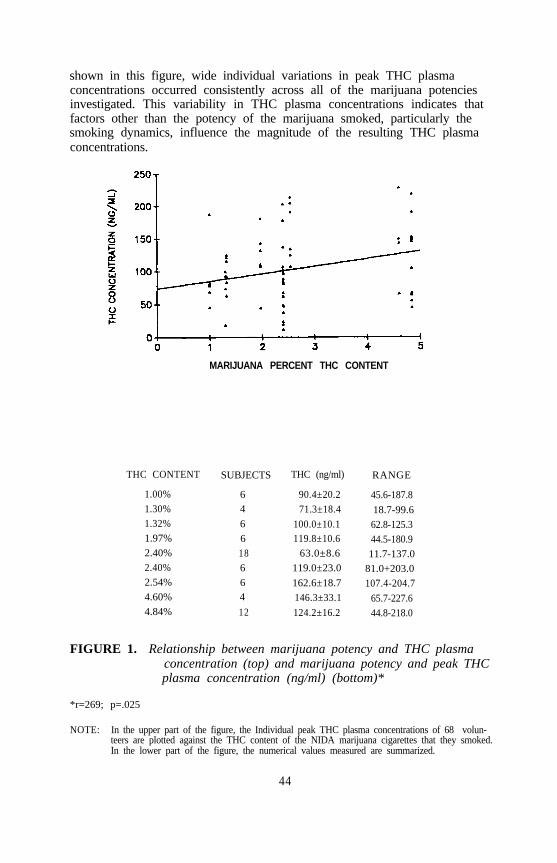
shown in this figure, wide individual variations in peak THC plasmaconcentrations occurred consistently across all of the marijuana potenciesinvestigated. This variability in THC plasma concentrations indicates thatfactors other than the potency of the marijuana smoked, particularly thesmoking dynamics, influence the magnitude of the resulting THC plasmaconcentrations.
MARIJUANA PERCENT THC CONTENT
THC CONTENT SUBJECTS THC (ng/ml) RANGE
1.00%1.30%1.32%1.97%2.40%2.40%2.54%4.60%4.84%
6 90.4±20.24 71.3±18.46 100.0±10.16 119.8±10.618 63.0±8.66 119.0±23.06 162.6±18.74 146.3±33.112 124.2±16.2
45.6-187.8
18.7-99.662.8-125.344.5-180.911.7-137.0
81.0+203.0107.4-204.7
65.7-227.644.8-218.0
FIGURE 1. Relationship between marijuana potency and THC plasmaconcentration (top) and marijuana potency and peak THCplasma concentration (ng/ml) (bottom)*
*r=269; p=.025
NOTE: In the upper part of the figure, the Individual peak THC plasma concentrations of 68 volun-teers are plotted against the THC content of the NIDA marijuana cigarettes that they smoked.In the lower part of the figure, the numerical values measured are summarized.
44

PYROLYTIC DESTRUCTION
When marijuana is smoked, a portion of its THC content is destroyed bypyrolysis before reaching the mouth of the smoker. Information regardingthe amount of THC destroyed by pyrolysis is derived from both cigarettesand water-pipe smoking.
Cigarette Smoking Study
Davis and colleagues (1984) conducted a study in which standard NIDAmarijuana cigarettes containing 1.6 percent and 3.1 percent THC weresmoked by a cigarette-smoking machine in a constant draft mode, i.e., thecigarettes were artificially inhaled in a single puff until nothing was left.The percent of the original THC not recovered in the smoke condensatecollected and probably lost to pyrolysis was 30.6±1.4 and 30.9±1.2 for the1.6 percent and the 3.1 percent THC content cigarettes, respectively. Sinceno measurement of the THC trapped in the smoking apparatus was done,the total amount actually lost to pyrolysis, under their experimental condi-tions, was probably less than 30 percent.
Water-Pipe Smoking Study
Marijuana water-pipe smoking replicates artificial single-draft smokingbecause the amount of plant material placed in the bowl of the pipe isentirely combusted, and the smoke produced is inhaled in a single puff.One milligram of Vitiated THC was added to 70 mg of placebo marijuana(0.004 percent THC) and placed in the bowl of a commercially availablewater pipe. One bowl was smoked every 2.5 minutes, for a total of 10bowls. The six volunteers who participated in this study were instructed todraw air until the total amount of marijuana placed in the pipe’s bowl wascombusted and to inhale the total amount of smoke produced in a singlepuff. A Cambridge filter, to trap the THC content of the smoke produced,was placed between the mouth of the pipe and the mouth of the subjects.The amount of THC recovered from the pipe, the plant material residues,and the Cambridge filters was measured by gas liquid chromatography. Itwas found, in this unpublished study, that 40.4±1.5 percent of the originalamount of THC was trapped in the Cambridge filters (i.e., approximatelythe amount of THC that would have reached the mouth of the subjects ifthe marijuana smoke had not been filtered), and that 40.2±4.5 percent of theoriginal amount of THC remained in the pipe and plant material residues(figure 2). Therefore, since approximately 80 percent of the originalamount of THC was recovered, it can be concluded that 20 percent wasdestroyed by pyrolysis.
The results of these smoking studies, given the large margin of error intro-duced by differences in methodology, compare favorably and indicate thatbetween 20 percent and 30 percent of the THC contained in the plant
45
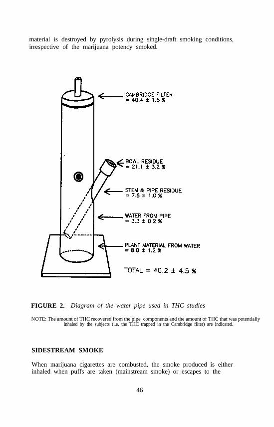
material is destroyed by pyrolysis during single-draft smoking conditions,irrespective of the marijuana potency smoked.
FIGURE 2. Diagram of the water pipe used in THC studies
NOTE: The amount of THC recovered from the pipe components and the amount of THC that was potentiallyinhaled by the subjects (i.e. the THC trapped in the Cambridge filter) are indicated.
SIDESTREAM SMOKE
When marijuana cigarettes are combusted, the smoke produced is eitherinhaled when puffs are taken (mainstream smoke) or escapes to the
46
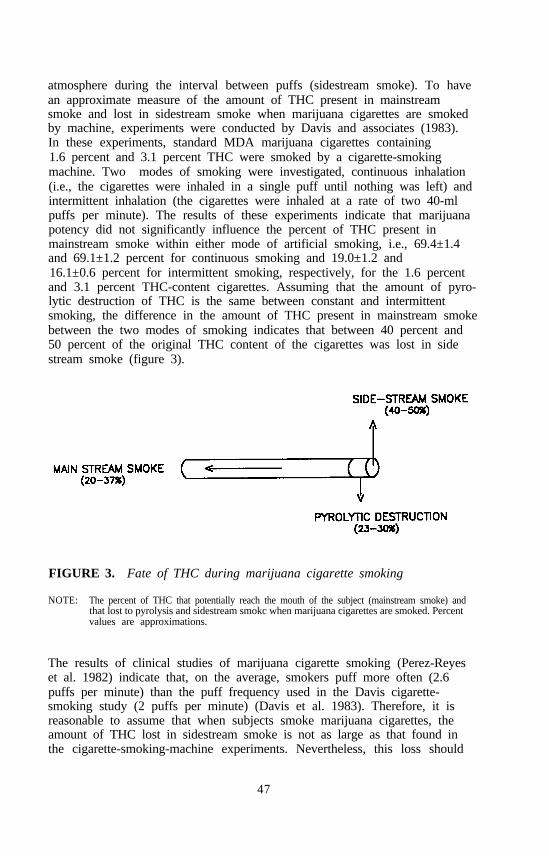
atmosphere during the interval between puffs (sidestream smoke). To havean approximate measure of the amount of THC present in mainstreamsmoke and lost in sidestream smoke when marijuana cigarettes are smokedby machine, experiments were conducted by Davis and associates (1983).In these experiments, standard MDA marijuana cigarettes containing1.6 percent and 3.1 percent THC were smoked by a cigarette-smokingmachine. Two modes of smoking were investigated, continuous inhalation(i.e., the cigarettes were inhaled in a single puff until nothing was left) andintermittent inhalation (the cigarettes were inhaled at a rate of two 40-mlpuffs per minute). The results of these experiments indicate that marijuanapotency did not significantly influence the percent of THC present inmainstream smoke within either mode of artificial smoking, i.e., 69.4±1.4and 69.1±1.2 percent for continuous smoking and 19.0±1.2 and16.1±0.6 percent for intermittent smoking, respectively, for the 1.6 percentand 3.1 percent THC-content cigarettes. Assuming that the amount of pyro-lytic destruction of THC is the same between constant and intermittentsmoking, the difference in the amount of THC present in mainstream smokebetween the two modes of smoking indicates that between 40 percent and50 percent of the original THC content of the cigarettes was lost in sidestream smoke (figure 3).
FIGURE 3. Fate of THC during marijuana cigarette smoking
NOTE: The percent of THC that potentially reach the mouth of the subject (mainstream smoke) andthat lost to pyrolysis and sidestream smokc when marijuana cigarettes are smoked. Percentvalues are approximations.
The results of clinical studies of marijuana cigarette smoking (Perez-Reyeset al. 1982) indicate that, on the average, smokers puff more often (2.6puffs per minute) than the puff frequency used in the Davis cigarette-smoking study (2 puffs per minute) (Davis et al. 1983). Therefore, it isreasonable to assume that when subjects smoke marijuana cigarettes, theamount of THC lost in sidestream smoke is not as large as that found inthe cigarette-smoking-machine experiments. Nevertheless, this loss should
47
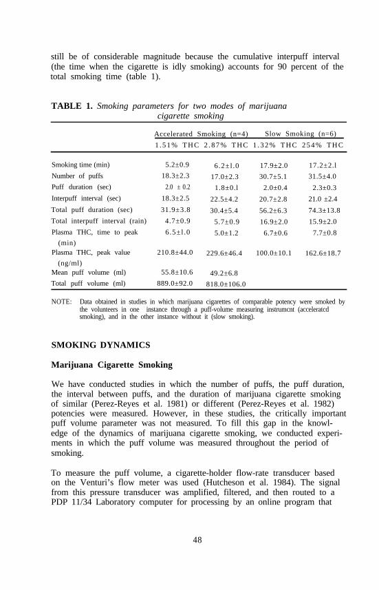
still be of considerable magnitude because the cumulative interpuff interval(the time when the cigarette is idly smoking) accounts for 90 percent of thetotal smoking time (table 1).
TABLE 1. Smoking parameters for two modes of marijuanacigarette smoking
Accelerated Smoking (n=4) Slow Smoking (n=6)
1 . 5 1 % T H C 2 . 8 7 % T H C 1 . 3 2 % T H C 2 5 4 % T H C
Smoking time (min) 5.2±0.9
Number of puffs 18.3±2.3
Puff duration (sec) 2.0 ± 0.2
Interpuff interval (sec) 18.3±2.5
Total puff duration (sec) 31.9±3.8
Total interpuff interval (rain) 4.7±0.9
Plasma THC, time to peak 6.5±l.0
(min)Plasma THC, peak value 210.8±44.0
(ng/ml)Mean puff volume (ml) 55.8±10.6
Total puff volume (ml) 889.0±92.0
6.2±l.0
17.0±2.3
1.8±0.l
22.5±4.2
30.4±5.4
5.7±0.9
5.0±1.2
229.6±46.4
49.2±6.8
818.0±106.0
17.9±2.0
30.7±5.1
2.0±0.4
20.7±2.8
56.2±6.3
16.9±2.0
6.7±0.6
100.0±10.1
17.2±2.l
31.5±4.0
2.3±0.3
21.0 ±2.4
74.3±13.8
15.9±2.0
7.7±0.8
162.6±18.7
NOTE: Data obtained in studies in which marijuana cigarettes of comparable potency were smoked bythe volunteers in one instance through a puff-volume measuring instrumcnt (acceleratcdsmoking), and in the other instance without it (slow smoking).
SMOKING DYNAMICS
Marijuana Cigarette Smoking
We have conducted studies in which the number of puffs, the puff duration,the interval between puffs, and the duration of marijuana cigarette smokingof similar (Perez-Reyes et al. 1981) or different (Perez-Reyes et al. 1982)potencies were measured. However, in these studies, the critically importantpuff volume parameter was not measured. To fill this gap in the knowl-edge of the dynamics of marijuana cigarette smoking, we conducted experi-ments in which the puff volume was measured throughout the period ofsmoking.
To measure the puff volume, a cigarette-holder flow-rate transducer basedon the Venturi’s flow meter was used (Hutcheson et al. 1984). The signalfrom this pressure transducer was amplified, filtered, and then routed to aPDP 11/34 Laboratory computer for processing by an online program that
48
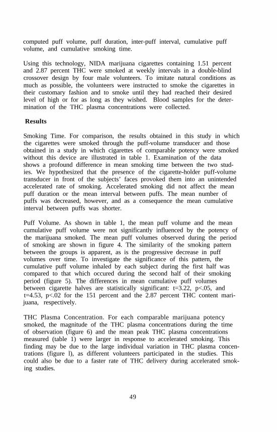
computed puff volume, puff duration, inter-puff interval, cumulative puffvolume, and cumulative smoking time.
Using this technology, NIDA marijuana cigarettes containing 1.51 percentand 2.87 percent THC were smoked at weekly intervals in a double-blindcrossover design by four male volunteers. To imitate natural conditions asmuch as possible, the volunteers were instructed to smoke the cigarettes intheir customary fashion and to smoke until they had reached their desiredlevel of high or for as long as they wished. Blood samples for the deter-mination of the THC plasma concentrations were collected.
Results
Smoking Time. For comparison, the results obtained in this study in whichthe cigarettes were smoked through the puff-volume transducer and thoseobtained in a study in which cigarettes of comparable potency were smokedwithout this device are illustrated in table 1. Examination of the datashows a profound difference in mean smoking time between the two stud-ies. We hypothesized that the presence of the cigarette-holder puff-volumetransducer in front of the subjects’ faces provoked them into an unintendedaccelerated rate of smoking. Accelerated smoking did not affect the meanpuff duration or the mean interval between puffs. The mean number ofpuffs was decreased, however, and as a consequence the mean cumulativeinterval between puffs was shorter.
Puff Volume. As shown in table 1, the mean puff volume and the meancumulative puff volume were not significantly influenced by the potency ofthe marijuana smoked. The mean puff volumes observed during the periodof smoking are shown in figure 4. The similarity of the smoking patternbetween the groups is apparent, as is the progressive decrease in puffvolumes over time. To investigate the significance of this pattern, thecumulative puff volume inhaled by each subject during the first half wascompared to that which occured during the second half of their smokingperiod (figure 5). The differences in mean cumulative puff volumesbetween cigarette halves are statistically significant: t=3.22, p<.05, andt=4.53, p<.02 for the 151 percent and the 2.87 percent THC content mari-juana, respectively.
THC Plasma Concentration. For each comparable marijuana potencysmoked, the magnitude of the THC plasma concentrations during the timeof observation (figure 6) and the mean peak THC plasma concentrationsmeasured (table 1) were larger in response to accelerated smoking. Thisfinding may be due to the large individual variation in THC plasma concen-trations (figure l), as different volunteers participated in the studies. Thiscould also be due to a faster rate of THC delivery during accelerated smok-ing studies.
49
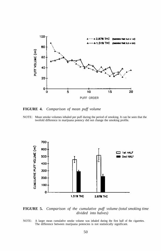
PUFF ORDER
FIGURE 4. Comparison of mean puff volume
NOTE: Mean smoke volumes inhaled per puff during the period of smoking. It can be seen that thetwofold difference in marijuana potency did not change the smoking profile.
FIGURE 5. Comparison of the cumulative puff volume (total smoking timedivided into halves)
NOTE: A larger mean cumulatlve smoke volume was inhaled during the first half of the cigarettes.The difference between marijuana potencies is not statistically significant.
50
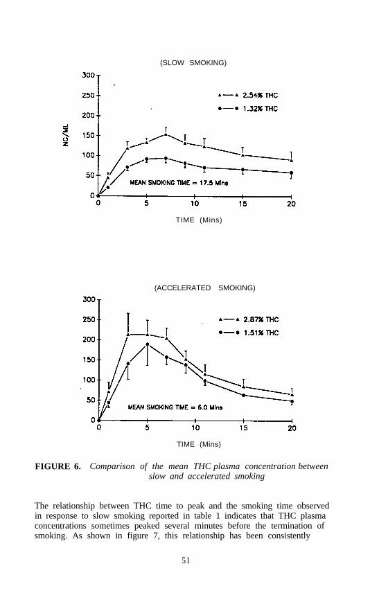
(SLOW SMOKING)
TIME (Mins)
(ACCELERATED SMOKING)
TIME (Mins)
FIGURE 6. Comparison of the mean THC plasma concentration betweenslow and accelerated smoking
The relationship between THC time to peak and the smoking time observedin response to slow smoking reported in table 1 indicates that THC plasmaconcentrations sometimes peaked several minutes before the termination ofsmoking. As shown in figure 7, this relationship has been consistently
51

observed in the many marijuana-smoking experiments that we have previ-ously conducted. ThiS observation was always intriguing because it wascontrary to the reasonable expectation that for as long as the smoke pro-duced by the combustion of marijuana cigarettes was inhaled, the resultingTHC plasma concentrations should have continued to increase until smokingwas terminated. Therefore, it was unexpected to find that, during acceler-ated smoking, the THC plasma concentrations peaked at approximately thesame time that smoking was terminated. We hypothesized that rapid inhala-tion of marijuana smoke resulted in a rate of delivery of THC to the blood-stream that was faster than the drug’s rate of disappearance into the extra-vascular compartment. In contrast, when the rate of inhalation of marijuanasmoke was slow, the drug’s rate of disappearance into the extravascularcompartment was faster than the rate of delivery of THC to thebloodstream.
MEAN THC TIME TO PEAK
MEAN SMOKING TIME
FIGURE 7. Relationship between smoking time and THC time to peak
NOTE: The results of eight separate studies in which marijuana cigarettes were smoked in thevolunteers customary manner are shown. It can be seen that peak THC plasmaconcentrations cosistently occurred before the termination of smoking.
52

Marijuana Water-Pipe Smoking
Marijuana is frequently smoked in pipes of different sixes and configura-tions, particularly in water pipes, because they cool the smoke and facilitateits inhalation. This method of smoking has become more popular in recentyears because it permits the combustion of smalI portions of high-potencymarijuana (sometimes as high as 13 percent THC in Sinsemilla plantmaterial).
As discussed, marijuana cigarette smoking of identical THC content resultsin large interindividual variations in THC plasma concentrations. This isbecause the number of puffs taken, the interval between puffs, and thevolume of smoke inhaled per puff vary greatly from person to person. Incontrast, marijuana pipe smoking permits control of the number of puffstaken, the interval between puffs, and the volume of smoke inhaled perpuff. Moreover, at least theoretically, the amount of plant material placedin the bowl of the pipe should be entirely combusted, all of the smoke pro-duced should be inhaled, and practically no THC should be lost in side-stream smoke. We hypothesized that under these controlled smoking cir-cumstances, less interindividual variation in the magnitude of THC plasmaconcentrations should occur.
To verify this hypothesis, we conducted three dose-response studies, using asingle-blind crossover design in which marijuana of different potencies wassmoked in a 250-ml capacity water pipe (figure 2). A wire screen wasplaced at the bottom of the pipe’s bowl to prevent uncombusted plantmaterial from dropping into the pipe. A constant amount of water wasused to fill the pipe, which provided a consistent 200-ml volume of airspace between the mouth of the subject and the water level (the puffvolume).
The volunteers were instructed to inhale the total amount of smoke pro-duced by the combustion of the marijuana placed in the pipe’s bowl(“toke”) and to hold the smoke in their lungs for 10 seconds. Smoke in-halation was facilitated by the drawing of atmospheric air through a smallhole located above the water level. Tokes were taken at 0, 2.5, 5, 7.5, and10 minutes. This rate of administration was selected because in our pre-vious studies in which radiolabeled ’H-THC was smoked in a water pipe,the resulting THC plasma concentrations were sustained during the period ofsmoking.
Experimental Variables. To determine the peak THC plasma concentra-tions produced, blood samples were drawn through an indwelling needle atappropriate intervals. The subjective rating of marijuana high was measuredat frequent intervals throughout the experiments, and the heart rate wasrecorded before and after marijuana smoking in only the second and thirdstudies.
53
Drug Dosage. In our initial study, 0.625 mg/kg of marijuana containing1.68 percent or 2.51 percent THC was combusted per toke. The amount ofsmoke produced by the combustion of this amount of marijuana was large,and some of the six volunteers who participated in the study had difficultiescombusting the tokes completely or inhaling the total amount of smoke gen-erated. For this reason, a second dose-response study was conducted inwhich the amount of plant material combusted per toke was reduced to0.5 mg/kg for marijuana containing 2.51 percent THC and to 0.4 mg/kg formarijuana containing 3.12 percent THC. The latter marijuana potency wasobtained by mixing equal parts of 6.24 percent and 0.004 percernt THC.The amount of smoke produced by the combustion of tokes containing0.5 mg/kg was still large enough that some of the six volunteers who par-ticipated in this study had difficulty inhaling the smoke completely. How-ever, the amount of smoke generated by the combustion of the 0.4 mg/kgtokes was completely inhaled without difficulty. The pharmacologic effectsobtained in this study were of greater magnitude than those obtained in thefirst study, but they were leas pronounced than those produced by the smok-ing of marijuana cigarettes. In an effort to obtain larger pharmacologiceffects, a third study was conducted in which 0.4 mg/kg of marijuana con-taining 3.12 percent and 6.24 percent THC was combusted per toke. Origi-nally, as in the previous studies, we planned to teat six volunteers. How-ever, the smoking of the high-potency marijuana induced postural hypoten-sion and bradycardia (vasovagal syndrome) in the third and fourth subjectstested. Because of this undesirable side effect, the study was terminatedafter testing only three subjects. We are perplexed by the occurrence ofthis syndrome because we have never observed it in response to marijuanacigarette smoking. We have, however, observed the occurrence this syn-drome in some subjects after the oral administration of high doses of THC(35 mg) or after the intravenous infusion of THC.
Results
THC Plasma Concentrations. As illustrated in figure 8, the THC plasmaconcentrations increased progressively for as long as tokes were taken. Asa result, peak THC concentrations occurred in close proximity to the end ofthe smoking period (table 2). This finding suggests that the inhalation of alarge volume of smoke (approximately 200 ml) every 2.5 minutes deliversenough THC to the blood stream to balance the drug’s rate of disappear-ance into the extravascular compartment. In this respect, the results ofmarijuana water-pipe smoking are comparable to the results of acceleratedmarijuana cigarette smoking.
Contrary to our expectations, peak THC plasma concentrations varied widelyamong the subjects who participated in the studies (table 2). This result isdisappointing because it demonstrates our failure to reduce the variability inpeak THC plasma concentrations, despite controlling the amount of mari-juana combusted per toke, the number of tokes taken, and the interval
54

FIGURE 8. Comparison of the magnitude of THC plasma concentrationsproduced by marijuana water-pipe smoking over time
between tokes. It should be noted, however, that smoking factors otherthan those controlled in this study may have influenced the magnitude ofthe THC plasma concentrations observed. For example, although the sub-jects were instructed to hold the smoke inhaled for a period of 10 seconds,this requirement was not enforced. Therefore, according to their custom,
55
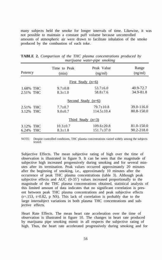
many subjects held the smoke for longer intervals of time. Likewise, it wasnot possible to maintain a constant puff volume because uncontrolledamounts of atmospheric air were drawn to facilitate inhalation of the smokeproduced by the combustion of each toke.
TABLE 2. Comparison of the THC plasma concentrations produced bymarijuana water-pipe smoking
PotencyTime to Peak Peak Value Range
(min) (ng/ml) (ng/ml)
1.68% THC2.51% THC
First Study (n=6)
9.7±0.8 53.7±6.08.3±1.0 58.8±7.6
40.9-72.734.9-81.8
2.51% THC3.12% THC
Second Study (n=6)
7.7±0.7 79.7±10.89.0±0.7 114.5±10.4
39.0-116.080.8-150.0
3.12% THC6.24% THC
Third Study (n=3)
10.3±0.7 109.6±20.88.3±1.8 151.7±37.0
81.0-150.090.2-218.0
NOTE: Despite controlled conditions, THC plasma concentrations varied widely among the subjectstested.
Subjective Effects. The mean subjective rating of high over the time ofobservation is illustrated in figure 9. It can be seen that the magnitude ofsubjective high increased progressively during smoking and for several min-utes after its termination. Peak values occurred approximately 20 minutesafter the beginning of smoking, i.e., approximately 10 minutes after theoccurrence of peak THC plasma concentrations (table 3). Although peaksubjective effects and AUC (0-35’) values increased proportionally to themagnitude of the THC plasma concentrations obtained, statistical analysis ofthis limited amount of data indicates that no significant correlation is pres-ent between peak THC plasma concentrations and peak subjective effects(r=.153, t=0.82, p NS). This lack of correlation is probably due to thelarge intersubject variations in both plasma THC concentrations and sub-jective effects.
Heart Rate Effects. The mean heart rate acceleration over the time ofobservation is illustrated in figure 10. The changes in heart rate producedby marijuana pipe smoking mimic in all respects the subjective rating ofhigh. Thus, the heart rate accelerated progressively during smoking and for
56
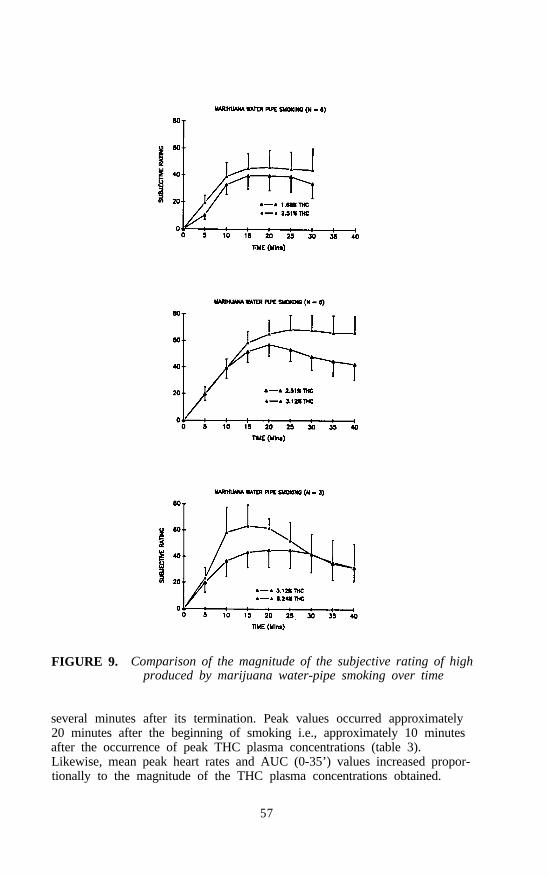
FIGURE 9. Comparison of the magnitude of the subjective rating of highproduced by marijuana water-pipe smoking over time
several minutes after its termination. Peak values occurred approximately20 minutes after the beginning of smoking i.e., approximately 10 minutesafter the occurrence of peak THC plasma concentrations (table 3).Likewise, mean peak heart rates and AUC (0-35’) values increased propor-tionally to the magnitude of the THC plasma concentrations obtained.
57

TABLE 3. Comparison of the pharmacologic effects produced bymarijuana after pipe smoking
Time to Peak Peak Value AUC (0-35’)
18.3±2.518.3±1.7
Subjective Rating of High
First Study (n=6)
41.7±10.548.3±11.9
20.0±2.228.3±4.0
Second Study (n=6)
57.5±8.973.0±10.4
Third Study (n=3)
2.0±5.0 50.7±12.913.3±3.3 68.3±13.6
980±2211,071±265
1,457±2491,754±282
1,270±3681,595±409
20.8±2.721.7±2.1
Percent Heart Rate Acceleration
Second Study (n=6)
34.4±6.253.6±5.5
492±151862±96
14.0±0.018.0±2.0
Third Study (n=3)
7 0 . 4 ± 3 . 074.9±18.0
1,377±2931,589322
NOTE: The heart rate was not measured in the first study.
Nonetheless, statistical analysis indicates that no significant correlation ispresent between peak THC plasma concentrations and peak heart rate accel-eration (r=.385, t=1.67, p NS). This is probably due to the large intersub-ject variations in plasma THC concentrations and heart rate accelerations.
AMOUNT OF THC TRAPPED IN TEE UPPER RESPIRATORYTRACT MUCOSA
The results of several independent studies indicate that THC is present insaliva and that its concentration declines with time after marijuana smoking(Hawks 1984; Cross et al. 1987; Maseda et al. 1986; Thompson and Cone1987). The presence of THC in saliva after marijuana smoking is not dueto its secretion by the salivary glands. This is demonstrated by the resultsof experiments in which we administered radioactively labeled THC orally
58

FIGURE 10. Comparison of magnitude of mean hem rate accelerationproduced by marijuana water-pipe smoking over time
and intravenously to normal volunteers (Perez-Reyes et al. 1972; Perez-Reyes et al. 1973). In these experiments we collected timed saliva samplesafter drug administration. No radioactivity was detected in any of the salivasamples assayed. This observation indicates that neither THC nor its metabolities are excreted by the salivary glands. Therefore, the THC present insaliva derives from that sequestered in the buccal mucosa during smoking.
The amount of THC trapped in the buccal mucosa or present in mixed sali-va samples is time and dose dependent. This is demonstrated by a study inwhich 30 volunteers smoked NIDA marijuana cigarettes containing either
59

2.49 percent (18 subjects) or 4.84 percent (12 subjects) THC blindly. Themean amount of THC present in swabs of the buccal mucosa and the meanamount of THC present in 25 microliters of mixed saliva samples areshown in table 4. Because the THC present in the swabs cannot be accu-rately related to a volume of saliva, we have reported them as amountsinstead of concentrations. The largest amounts of THC in swab and saliva,irrespective of the marijuana potency smoked, were observed at the firsttime of measurement (i.e., 1 hour after the initiation of smoking). Afterthis time, mean THC amounts declined progressively to minimal but stilldetectable values at the 4-hour end of the study.
TABLE 4. Comparison of the mean amounts of THC (ng) measured in25 microliters of saliva and swabs of the roof of themouth and tongue
2.40% THC 4.84% THCSwab Saliva Swab Saliva
1 Hour 16.9±7.2 20.9±8.2 59.8±27.5 60.5±24.92 Hours 7.8±3.7 5.8±1.8 26.5±12.3 26.7±14.83 Hours 7.1±3.8 4.1±1.3 16.6±8.9 6.9±2.7
4 Hours 3.8±1.7 1.9 ±0.8 13.5±7.4 4.2±1.7
Since THC is trapped in the buccal mucosa, it is reasonable to assume thatTHC is also trapped in the mucosa of the other parts of the upper respira-tory tract, i.e., pharynx, larynx, trachea, and bronchi. Obviously, it is notpossible to know the amount of THC that is trapped in the combined sur-face. area of these anatomical structures during marijuana smoking. How-ever, judging by the relatively small amounts of THC trapped in the buccalmucosa (i.e., less than 100 ng in either swabs or 25 microliters of saliva1 hour after smoking) the total amount of THC trapped in the mucosa ofthe upper respiratory tract can be considered to be a factor of minor impor-tance with regard to the bioavailability of THC
CONCLUSION
Although smoking is a practical method for the delivery of marijuana’sactive principle (THC), many factors inherent in the smoking processdecrease the drug’s bioavailability. For example, whether marijuana issmoked in pipe or cigarette form, 23 to 30 percent of its THC content isdestroyed by pyrolysis; if marijuana is smoked in cigarette form, as muchas 40 to 50 percent of THC can be lost in sidestream smoke.
60
There is a significant correlation between the potency of the marijuanasmoked and the magnitude of the THC plasma concentrations obtained.However, large interindividual variations in peak THC plasma concentrationsoccurred across all of the marijuana potencies investigated. These resultswere obtained in experiments in which the volunteers smoked marijuanacigarettes in their customary fashion (uncontrolled smoking). To reduce thisvariability, experiments were conducted in which marijuana was smoked ina water pipe. This method of smoking permits control of the amount ofmarijuana combusted per toke, the number of tokes taken, and the intervalbetween tokes. Despite controlling these important smoking factors, largeinterindividual variations in the magnitude of the THC plasma concentra-tions obtained continued to be observed.
During marijuana smoke inhalation, THC is trapped in the mucosa of theupper respiratory tract, thereby reducing the drug’s bioavailability. Judgingby the amounts of THC measured in saliva or swabbings of the buccalmucosa, however, the total loss of THC by this factor can be considered ofminor importance.
In summary, marijuana smoking is a complex process that does not permitcontrolled dosing. Therefore, for research or therapeutic purposes, it is indi-spensable to measure the THC plasma concentrations to know how efficient-ly a given individual smoked.
REFERENCES
Cook, C.E.; Seltzman, H.H.; Schindler, V.H.; Tallent, C.R.; Chin, KM.; andPitt, C.G. Radioimmunoassays for cannabinoids. In: Hawks, R.L., andWillette, R.E., eds. The Analysis of Cannabinoids in Biological Fluids.National Institute on Drug Abuse Research Monograph 42. NTIS Pub.No. 83-136044. Springfield, VA: U.S. Dept. of Commerce, 1982.pp. 19-32.
Davis, K.H.; McDaniel, I.A.; Caddell, L.W.; and Moody, P.L. Some smok-ing characteristics of marijuana cigarettes. In: Agurell, S.; Dewey, W.L.;and Willette, R.E., eds. The Cannabinoids: Chemical, Pharmacologic,and Therapeutic Aspects. New York: Academic Press, 1984. pp. 97-109.
Gross, S.J.; Worthy, T.E.; Nerder, L.; Zimmerman, E.G.; Soares, J.R.; andLomax, P. Detection of recent cannabis use by saliva 9-THC radioimmunoassay. J Anal Toxicol 9:91-97, 1987.
Hawks, R.L. Developments in cannabinoid analyses of body fluids: Impli-cations for forensic applications. In: Agurell, S.; Dewey, W.L.; andWillette, R.E., eds. The Cannabinoids: Chemical, Pharmacologic, andTherapeutic Aspects. New York: Academic Press, 1984. pp. 123-134.
61
Hutcheson, J.S.; Perez-Reyes, M.; and Di Guiseppi, S. An automatedcomputer-based system for the study of marijuana smoking dynamics.In: Agurell, S.; Dewey, W.L.; and Willette, R.E., eds. The Cannabi-noids: Chemical, Pharmacologic, and Therapeutic Aspects. New York:Academic Press, 1984. pp. 111-121.
Maseda, C.; Hama, K.; Fukui, Y.; Matsubara, K.; Takahashi, S.; and Akane,A. Detection of 9-THC in saliva by capillary GC/ECD after marihuanasmoking. Forensic Sci Int 32:259-266, 1986.
Owens, S.M.; McBay, A.J.; Reisner, H.M.; and Perez-Reyes, M. 125I Radio-immunoassay of 9-tetrahydrocannabino1 in blood and plasma with a solid-phase second-antibody separation method. Clin Chem 27:619-624, 1981.
Perez-Reyes, M.; Di Guiseppi, S.; Davis, H.H.; Schindler, V.H.; and Cook,C.E. Comparison of effects of marihuana cigarettes of three differentpotencies. Clin Pharmacol Ther 31:617-624, 1982.
Perez-Reyes, M.; Lipton, M.A.; Timmons, M.C.; Wall, M.E.; Brine, D.R.;and Davis, K.H. Pharmacology of orally administered 9-tetrahydrocannab-inol. Clin Pharmacol Ther 14:48-55, 1973.
Perez-Reyes, M.; Owens, S.M.; and Di Guiseppi, S. The clinical pharma-cology and dynamics of marihuana cigarette smoking. J Clin Pharmacol21:201S-207S, 1981.
Perez-Reyes, M.; Timmons, M.C.; Lipton, M.A.; Davis, K.H.; and Wall,M.E. Intravenous injection in man of 9-tetrahydrocannabinol and 11-OH-9-tetrahydrocannabinol. Science 177:633-635, 1972.
Thompson, L.K., and Cone, E.J. Determination of 9-tetrahydrocannabinol inhuman blood and saliva by high-performance liquid chromatography withamperometric detection. J Chromatogr 421:91-97, 1987.
ACKNOWLEDGMENTS
Supported by National Institute on Drug Abuse contracts 271-80-3705 and271-87-8126 and grant DA 04484.
AUTHOR
Mario Perez-Reyes, M.D.ProfessorDepartment of PsychiatryThe University of North Carolina at Chapel HillCB #7175, Medical Research Building AChapel Hill, NC 27599-7175
62
Effects of Habitual Use ofMarijuana and/or Cocaine onthe LungDonald P. Tashkin, Suzanne Fligiel, Tzu-Chin Wu,Henry Gong, Jr., Richard G. Barbers, Anne H. Coulson,Michael S. Simmons, and Theodore F. Beals
INTRODUCTION
Marijuana remains one of the most widely abused substances in our society(Johnstone et al. 1987). As with tobacco, smoking is the preferred route ofadministration of marijuana, so the lungs are the principal organ exposed toits combustion products. The latter include a number of known respiratory‘irritants and carcinogens (Hoffman et al. 1975; Novotny et al. 1982), aswell as numerous other compounds the pulmonary effects of which are un-known. Experimental animal (Roy et al. 1976; Fleischman et al. 1975;Fleischman et al. 1979) and in vitro (Huber et al. 1975; Leuchtenberger andLeuchtenberger 1976) studies have demonstrated that exposure to marijuanasmoke produces significant toxic, inflammatory, and even carcinogeniceffects on lung tissue and cells. Few studies in man, however, have sys-tematically addressed the long-term effect of real-life habitual marijuanasmoking on the lung. Moreover, the few published clinical studies of thepulmonary effects of regular marijuana smoking (Rubin and Comitas 1975;Cruickshank 1976; Coggins et al. 1976; Boulougouris et al. 1976) haveyielded conflicting results, possibly because of small sample sixes, failure tocontrol adequately for confounding factors, such as concomitant tobaccosmoking, and the cross-sectional nature of these studies, which may not besensitive to intraindividual changes in respiratory status. In 1983, a longi-tudinal study was initiated at the University of California, Los Angeles(UCLA) in an attempt to determine (1) whether habitual smoking of mari-juana alone and with tobacco leads to pulmonary symptoms, lung dysfunc-tion, or respiratory illness and (2) whether such harmful effects persist orprogress with continued (or increased) use and decline with cessation ofuse. Another objective of this study was to evaluate the effect of regularsmoking of marijuana with and without tobacco on tracheobronchial
63
histopathology, alveolar cell populations, and the structure and function ofalveolar macrophages, key cells in the lung’s defense against infection andother noxious insults. Results of this continuing study are presented below.
PULMONARY EFFECTS OF HABITUAL MARIJUANA USE:RESPIRATORY SYMPTOMS AND LUNG FUNCTION
Cross-Sectional Findings
In 1983 and 1984 a convenience sample of Caucasian men and women(ages 25 to 49 years) who reported smoking 10 or mote marijuana joints(or joint-equivalents) per week, on the average, over the previous 5 years ormore was recruited from the greater Los Angeles area by means of news-paper and radio advertisements. Exclusionary criteria included intravenousdrug abuse more than 2 times per lifetime, smoking of illicit substancesother than cannabis more than 12 times per lifetime or within the previous6 months, significant occupational exposure to dusts or fumes potentiallyhazardous to the lungs, a history of chronic respiratory disease, such asasthma, cystic fibrosis, kyphoscoliosis, or tuberculosis, or previous chest sur-gery. A comparison sample of 167 nonmarijuana-smoking control subjectsof similar age was recruited from the same general population.
All subjects underwent a detailed respiratory and drug use questionnaire,modified from the American Thoracic Society/National Heart, Lung, andBlood Institute questionnaire (Ferris 1978) and the National Institute onDrug Abuse National Survey on Drug Abuse questionnaire (Fishburne et al.1980), which included questions concerning respiratory symptoms, generalhealth, residence, occupational history, socioeconomic status, and current andpast history of use of alcohol, tobacco, marijuana, and other illicit drugs.In addition, an extensive battery of pulmonary function tests was performed,including maximal expiratory flow-volume curves breathing air and 80 per-cent helium:20 percent oxygen, subdivisions of lung volume, single-breathdiffusing capacity for carbon monoxide (DLCO), single-breath nitrogenwashout, plethysmographically determined airway resistance, and specificairway conductance and methacholine inhalation challenge. Peripheralvenous blood was obtained for measurement of hemoglobin and carboxy-hemoglobin (for correction of DLC0 measurements) and for determination ofserum cotinine and 9-tetrahydrocannabinol (THC) and 11-nor-9-carboxy-THC for verification of self-reported daily smoking of tobacco andmarijuana.
Characteristics of the study population are shown in table 1. The smokersof marijuana (MS) and the control subjects who did not smoke tobacco(NS) were of similar age, while the tobacco-smoking control subjects (TS)were 3.4 years older than the smokers of both marijuana and tobacco(MTS). The MS smoked only slightly more marijuana than the MTS(p>.6), while the TS smoked significantly more tobacco than MTS (p<.01).
64
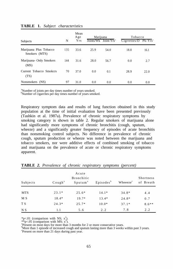
TABLE 1. Subject characteristics
Subjects
MeanAge
NMarijuana Tobacco
Yrs Joints/Wk Joint-Yrs’ Cigrrettes/D Pk-Yrs'
Marijuana Plus TobaccoSmokers (MTS)
135 33.6 25.9 54.0 18.8 16.1
Marijuana- Only Smokers 144 31.6 28.0 56.7 0.0 2.7(MS)
Current Tobacco Smokcrs 70 37.0 0.0 0.1 28.9 22.0(TS)
Nonsmokers (NS) 97 31.0 0.0
aNumber of joints per day times number of years smoked.bNumber of cigarcttes per day times number of years smoked.
0.0 0.0 0.0
Respiratory symptom data and results of lung function obtained in this studypopulation at the time of initial evaluation have been presented previously(Tashkin et al. 1987a). Prevalence of chronic respiratory symptoms bysmoking category is shown in table 2. Regular smokers of marijuana alonehad significantly more symptoms of chronic bronchitis (cough, sputum,wheeze) and a significantly greater frequency of episodes of acute bronchitisthan nonsmoking control subjects. No difference in prevalence of chroniccough, sputum production or wheeze was noted between the marijuana andtobacco smokers, nor were additive effects of combined smoking of tobaccoand marijuana on the prevalence of acute or chronic respiratory symptomsapparent.
TABLE 2. Prevalence of chronic respiratory symptoms (percent)
A c u t eBronchi t ic Shor tness
Subjec ts C o u g h a S p u t u m a Episodes b Wheeezec of Brea th
MTS 23 .1* 25 .6* 14.1* 34 .8* 4 . 4
M S 18.4* 19.7* 13.4* 24 .8* 0 . 7
T S 24 .3* 25 .7* 10.0* 37 .1* 8.6**
N S 1.1 5 . 6 2 . 2 7 . 8 2 . 2
*p<.01 (comparison with NS; x2).**p<.05 (comparison with MS; x2).aPresent on most days for more than 3 months for 2 or more consecutive years.bMore than 1 episode of increased cough and sputum lasting more than 3 weeks within past 3 years. cPresent on more than 21 days during past year.
65

Results of analysis of lung function are presented in tables 3 and 4. To-bacco smokers, irrespective of concomitant marijuana smoking, showedsignificantly poorer values and/or a significantly greater frequency of abnor-mality for several tests of lung function, including DLCO (a measure ofalveolar gas transfer, which is deranged in early emphysema) and Vmax50,
Vmax50, Visoflow, N2/L, CV, and CC (teats reflecting mainly small air-ways function), in comparison with nonsmokers or with smokers of mari-juana alone. On the other hand, marijuana smokers, regardless of
TABLE 3• Significantly different between-group comparisons of lungfunction tests from covariance analysis*
TestMen Women
(n=259) (n=128)
VisoV MTS>NSMTS>MS
N2L** MTS>NS TS>NS MTS>NSMTS>MS TS>MS
CV** TS>MS MTS>MS TS>NSTS>MTS MTS>NSTS>NS
CC** TS>MS NS>MS TS>NSTS>MTS TS>MS
TS>MTS
DLCO** MTS<NS TS<NS T S < N S MTS<NSM T S < M S TS<MS
R a w+ MS>NS MTS>NS
MS>TS MTS>TS
SGaw+ MTS<NS MS<NS
*p<.05 (least significant difference testing using adjusted group means from ANCOVA).**Two-way ANCOVA, with age and/or height as covariates: significant tabacco effect (p<.03); no
adverse marijuana effect.Two-way ANCOVA: significant marijuana effect, males only (p<.03); no tobacco effect.
KEY: VisoV=volume of isoflow; N2/L=change in nitrogen concentration per liter in Phase III ofsingle-breath nitrogen washout curve; CV=closing volume.; CC=closing capacity; DLCO=single-breath diffusing capacity of the lung for carbon monoxide; Raw=airway resistance SGaw=specific airway conductance.
SOURCE: Tashkin et al. 1987a.
66
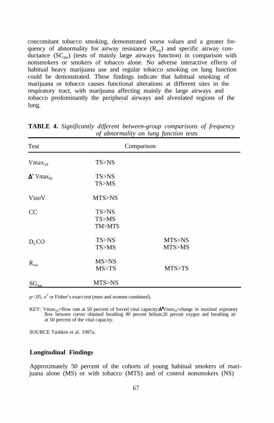
concomitant tobacco smoking, demonstrated worse values and a greater fre-quency of abnormality for airway resistance (Raw) and specific airway con-ductance (SCaw) (tests of mainly large airways function) in comparison withnonsmokers or smokers of tobacco alone. No adverse interactive effects ofhabitual heavy marijuana use and regular tobacco smoking on lung functioncould be demonstrated. These findings indicate that habitual smoking ofmarijuana or tobacco causes functional alterations at different sites in therespiratory tract, with marijuana affecting mainly the large airways andtobacco predominantly the peripheral airways and alveolated regions of thelung.
TABLE 4. Significantly different between-group comparisons of frequencyof abnormality on lung function tests
Test Comparison
Vmax50
Vmax50
VisoV
TS>NS
TS>NSTS>MS
MTS>NS
CC TS>NSTS>MSTM>MTS
DLCO TS>NS MTS>NSTS>MS MTS>MS
RawMS>NSMS>TS MTS>TS
SGaw MTS>NS
p<.05, x2 or Fisher’s exact test (men and women combined).
KEY: Vmax50=flow rate at 50 percent of forced vital capacity; Vmax50=change in maximal expiratoryflow between curves obtained breathing 80 percent helium:20 percent oxygen and breathing airat 50 percent of the vital capacity.
SOURCE Tashkin et al. 1987a.
Longitudinal Findings
Approximately 50 percent of the cohorts of young habitual smokers of mari-juana alone (MS) or with tobacco (MTS) and of control nonsmokers (NS)
67

and smokers of tobacco alone (TS) who were initially tested in 1983 and1984 (Time 1) returned for retesting in 1985 and 1986 (Time 2) after a 2to 3 year interval. On repeat testing, the following proportion of retestedsubjects remained in the same smoking category at both Time 1 and Time2: MTS 40 of 54; MS 60 of 71; TS 30 of 32; and NS 56 of 58. Table 5shows the prevalence, in percent, of abnormal respiratory symptoms at Time1 and Time 2 in the retested participants, by smoking group, for individualswho remained in the same smoking category. prevalence of cough, sputum,and wheeze was significantly higher in all smoking groups (MS, TS, andMTS) compared to nonsmokers at both Time 1 and Time 2 (p<.05, Chisquare). Among smokers of tobacco or marijuana alone (TS and MS), theprevalence of all respiratory symptoms tended to decrease in the intervalbetween the two examinations; in contrast, among the dual smokers (MTS),frequency of reported cough, sputum, wheeze and shortness of breath tendedto increase. Symptom prevalence was not statistically different, however,between Time 1 and Time 2 among participants in any smoking categorywho remained in the same category (p<.05; McNemar’s test of symmetry).
TABLE 5. Prevalence (percent) of respiratory symptoms at Time 1 (1983to 1984) and Time 2 (1985 to 1986)
SmokingGroup
AcuteBronchitic Shortness
Cough Sputum EpisodesT 1 T 2 T 1 T 2 T 1 T 2
Wheeze of BreathT1 T2 T 1 T 2
MTS 28* 33*+ 23* 33* 13* 13* 32* 40*+ 10 13*
MS 26* 16* 21* 15* 12* 6 28* 19* 4 0TS 37* 33* 33* 23* 23* 17 37* 20* 17* 10NS 0 4 4 4 0 0 5 4 2 2
*Significantly greater than values in NS; p<.05; x2 or Fisher’s exact test.+Significantly greater than values in MS; p<.05; x2 or Fisher's exact test.
Table 6 shows the prevalence, in percent, of abnormality in several lungfunction measures in the retested cohort at both Time 1 and Time 2 forsubjects in each smoking group who did not change smoking categories.These findings show persistent or increasing prevalence of abnormality inmeasures mainly of small airways function (e.g., FEF25-75, CC) and in DLCO(an indicator of early emphysema) among smokers of tobacco, especiallythose who also smoked marijuana, compared to nonsmokers. Over thesame time interval, the higher prevalence of abnormality in measuresreflecting large airways function (e.g., Vmax, Raw among marijuana smokerscompared to tobacco-only smokers and nonsmokers increased even further.
68
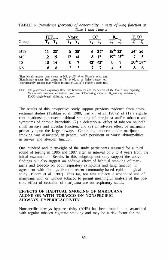
TABLE 6. Prevalence (percent) of abnormality in tests of lung function atTime 1 and Time 2
Group
MTS
MSTSNS
aSignificantly greater than values in NS; p<.05, x² or Fisher’s exact test.bSignificantly greater than values in TS; p<.05, x2 or Fisher’s exact test.cSignificantly greater than values in MS; p<.05, x2 a Fisher’s exact test.
KEY: FEF25-75=forced expiratory flow rate between 25 and 75 percent of the forced vital capacity;Vmax=peak maximal expiratory flow rate; CC=closing capacity; Raw=airway resistance;DLCO=single-breath diffusing capacity.
The results of this prospective study support previous evidence from cross-sectional studies (Tashkin et al. 1980; Tashkin et al. 1987a) of (1) a signifi-cant relationship between habitual smoking of marijuana and/or tobacco andsymptoms of chronic bronchitis, (2) a deleterious- effect of tobacco on bothsmall airways and alveolar function, and (3) an adverse effect of marijuanaprimarily upon the large airways. Continuing tobacco and/or marijuanasmoking was associated, in general, with persistent or worse abnormalitiesin airway and alveolar function.
One hundred and thirty-eight of the study participants returned for a thirdround of testing in 1986 and 1987 after an interval of 3 to 4 years from theinitial examination. Results in this subgroup not only support the abovefindings but also suggest an additive effect of habitual smoking of mari-juana and tobacco on both respiratory symptoms and lung function, inagreement with findings from a recent community-based epidemiologicalstudy (Bloom et al. 1987). Thus far, too few subjects discontinued use ofmarijuana with or without tobacco to permit meaningful analysis of the pos-sible effect of cessation of marijuana use on respiratory status.
EFFECTS OF HABITUAL SMOKING OF MARIJUANAALONE OR WITH TOBACCO ON NONSPECIFICAIRWAYS HYPERREACTIVITY
Nonspecific airways hyperreactivity (AHR) has been found to be associatedwith regular tobacco cigarette smoking and may be a risk factor for the
69
development of chronic obstructive pulmonary disease in tobacco smokers(Barter and Campbell 1976; Kanner 1984; Vollmer et al. 1985). Broncho-dilator drugs, by inhibiting airway reactivity to nonspecific inhaledspasmogens, could provide protection against chronic obstructive pulmonarydisease. Marijuana smoke contains many of the Same irritants found intobacco smoke (Hoffman et al. 1975; Novotny et al. 1982), in addition toTHC and other cannabinoids. THC causes dose-dependent bronchodilation(Vachon et al. 1973; Tashkin et al. 1973; Tashkin et al. 1974) and reversesexperimentally induced bronchospasm (Tashkin et al. 1975). THC is also abronchial irritant, however, at least when inhaled as an aerosol (Tashkinet al. 1977). Therefore, because of its bronchodilator or irritant properties,THC could inhibit or augment the tendency of other noxious components inmarijuana smoke (or in tobacco smoked in addition to marijuana) to causeAHR. The net influence was therefore examined of heavy, habitualsmoking of marijuana, alone or with tobacco, on nonspecific AHR incomparison with nonsmoking or smoking of tobacco alone (Tashkin et al.1988).
Airway reactivity to methacholine was measured in 281 subjects (67 MTS,116 MS, 34 ‘IS, and 64 NS) using a breath-activated dosimeter and a pro-tocol modified from that of Chai et al. (1975). Nonspecific AHR was con-sidered to be present if the forced expired volume in 1 second (FEV1)declined by 20 percent or more from the postdiluent control value afterinhalation of five deep breaths of methacholine in a concentration equal toor less than 25 mg/ml. The following proportion of subjects in eachsmoking category exhibited AHR: MTS 37.3 percent; MS 23.3 percent;‘IS 29.4 percent; and NS 17.2 percent. Although a greater proportionof smokers of marijuana or tobacco only exhibited AHR compared to non-smokers, these differences were not statistically significant. On the otherhand, the proportion of dual smokers of both marijuana and tobacco whohad AHR was more than twice that of nonsmokers, and this difference wassignificant (p<.01, x²). While the present small sample of tobacco-onlysmokers did not have a significantly higher prevalence of AHR compared tothe nonsmoking control subjects, two-way ANOVA did show a statisticallysignificant tobacco effect on the degree of airways activity, most pro-nounced in smokers of both marijuana and tobacco (F=10.14; p=.002).Therefore synergism between the effects of tobacco and marijuana was sug-gested, but the interaction did not achieve statistical significance.
It was concluded that the bronchodilator properties of THC do not protectagainst the development of AHR in smokers of both tobacco and marijuana.In contrast, these data suggest that respiratory irritants within marijuanasmoke (including THC and possibly other cannabinoids) augment the effectof tobacco in increasing airways reactivity. These findings are consistentwith histopathologic evidence of an additive effect of marijuana plus tobac-co in causing tracheobronchial epithelial injury.
70

PULMONARY EFFECTS OF HABITUAL MARIJUANA USE:TRACHEOBRONCHIAL HISTOPATHOLOGY AND ALVEOLARMACROPHAGE ULTRASTRUCTURE
Bronchial mucosal biopsies and alveolar macrophages were obtained byfiberoptic bronchoscopy and bronchoalveolar lavage (BAL) from young,heavy habitual smokers of marijuana alone (MS, n=30), marijuana plustobacco (MTS, n=17), tobacco alone (TS, n=15), and control nonsmokers(NS, n=11) selected at random from participants in the above study of thepulmonary effects of habitual marijuana use (Fligiel et al. 1988). For sub-jects in each smoking category undergoing bronchoscopy, smoking historiesand prevalence of respiratory symptoms and lung function abnormality werecomparable to those for all participants in the main study who belonged tothe same smoking group. During bronchoscopy, mucosa from the primarycarina and randomly selected secondary or tertiary carinae of the rightmiddle or lower lobe was biopsied (Gong et al. 1987). Biopsy specimensfrom each subject were processed for light microscopy and examined by asingle pathologist unaware of the subject’s smoking category. Histopatho-logic features were assessed according to the criteria suggested by Auerbachand colleagues (1957; 1961), as indicated in table 7. Results are shown intable 8. Histopathological alterations in the bronchial mucosa were signifi-cantly more common and severe in all groups of smokers than in nonsmok-ers. Furthermore, most of the mucosal abnormalities (with the exception ofsquamous metaplasia and the presence of mitotic figures) were more fre-quently observed in marijuana-only than in tobacco-only smokers. Mostnotable, however, was the finding that virtually all of the epithelial andbasement membrane alterations were most prevalent in subjects who habitu-ally smoked both marijuana and tobacco.
Bronchial biopsies were also examined using scanning electron microscopy(SEM) and transmission electron microscopy (TEM) for further, indepthcharacterization of mucosal morphology. TEM also permitted a thoroughexamination of the basement membrane region. The light microscopic find-ings were confirmed and expanded by both SEM and TEM. TEM revealedfrequent and early squamous metaplastic changes in all types of smokers ascompared to nonsmokers. Moreover, approximately one-third of all themarijuana smokers (MS and MTS) showed disruption of basement mem-brane continuity, which most likely occurred as a result of basementmembrane permeation by inflammatory cells from submucosal blood vessels,a finding not observed in tobacco-only smokers.
The findings are important for several reasons. First, all subjects studiedwere young adults, most of whom were asymptomatic with no or minimallung function abnormality, indicating that habitual marijuana smoking fre-quently causes significant, albeit clinically silent, airway injury manifestedby extensive morphologic changes at a relatively early age when symptoms
71
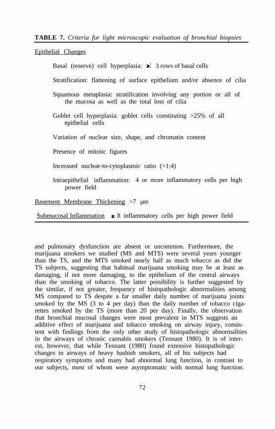
TABLE 7. Criteria for light microscopic evaluation of bronchial biopsies
Epithelial Changes
Basal (reserve) cell hyperplasia: 3 rows of basal cells
Stratification: flattening of surface epithelium and/or absence of cilia
Squamous metaplasia: stratification involving any portion or all ofthe mucosa as well as the total loss of cilia
Goblet cell hyperplasia: goblet cells constituting >25% of allepithelial cells
Variation of nuclear size, shape, and chromatin content
Presence of mitotic figures
Increased nuclear-to-cytoplasmic ratio (>1:4)
Intraepithelial inflammation: 4 or more inflammatory cells per highpower field
Basement Membrane Thickening >7 µm
Submucosal Inflammation 8 inflammatory cells per high power field
and pulmonary dysfunction are absent or uncommon. Furthermore, themarijuana smokers we studied (MS and MTS) were several years youngerthan the TS, and the MTS smoked nearly half as much tobacco as did theTS subjects, suggesting that habitual marijuana smoking may be at least asdamaging, if not more damaging, to the epithelium of the central airwaysthan the smoking of tobacco. The latter possibility is further suggested bythe similar, if not greater, frequency of histopathologic abnormalities amongMS compared to TS despite a far smaller daily number of marijuana jointssmoked by the MS (3 to 4 per day) than the daily number of tobacco ciga-rettes smoked by the TS (more than 20 per day). Finally, the observationthat bronchial mucosal changes were most prevalent in MTS suggests anadditive effect of marijuana and tobacco smoking on airway injury, consis-tent with findings from the only other study of histopathologic abnormalitiesin the airways of chronic cannabis smokers (Tennant 1980). It is of inter-est, however, that while Tennant (1980) found extensive histopathologicchanges in airways of heavy hashish smokers, all of his subjects hadrespiratory symptoms and many had abnormal lung function, in contrast toour subjects, most of whom were asymptomatic with normal lung function.
72
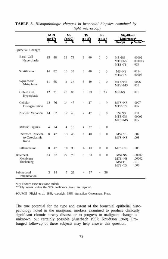
TABLE 8. Histopathologic changes in bronchial biopsies examined bylight microscopy
Epithelial Changes
Basal CellHyperplasia
Stratification
SquamousMetaplasia
Goblet CellHyperplasia
CellularDisorganization
Nuclear Variation
Mitotic Figures
Increased Nuclear-to-CytoplasmicRatio
Inflammation
BasementMembraneThickening
SubmucosalInflammation
15 88
14 82
11 65
12 71
13 76
14 82
4 24
8 47
8 47
14 82
3 18
22 73
16 53
8 27
25 83
14 47
12 40
4 13
13 43
10 33
22 73
7 23
6 40
6 40
6 40
8 53
4 27
7 47
4 27
6 40
6 40
5 33
4 27
0 0
0 0
0 0
3 2 7
1 9
0 0
0 0
0 0
0 0
0 0
4 36
MS>NS .00002MTS>NS .000003MTS>TS .005
MS>NS .0014MTS>TS .00002
MTS>NS .0006MTS>MS .010
MS>NS .001
MTS>NS .0007MTS>TS .006
TS>NS .010MTS>NS .00002MTS>MS .005
MS>NS .007MTS>NS .008
MTS>NS .008
MS>NS .00002MTS>NS .00002MS>TS .010
MTS>TS .006
*By Fisher's exact test (one-tailed).**Only values within the 99% confidence levels are reported.
SOURCE :FIigiel et al. 1988, copyright 1988, Australian Government Press.
The true potential for the type and extent of the bronchial epithelial histo-pathology noted in the marijuana smokers examined to produce clinicallysignificant chronic airway disease or to progress to malignant change isunknown, but certainly possible (Auerbach 1957; Knudtson 1960). Pro-longed followup of these subjects may help answer this question.
73
Alveolar macrophages obtained by BAL from the same three groups ofsmokers (MS, n=15; MTS, n=8; TS, n=7) and nonsmokers (NS, n=7) werecompared with respect to morphologic and morphometric ultrastructural cel-lular features (including an evaluation of cell membrane, nucleus, and cellplasma contents, especially cytoplasmic inclusions). Compared to NS, bothTS and MS had frequently larger cytoplasmic inclusions, most of whichcontained needlelike structures. While TS showed an increased nuclearcontour index and decreased cell membrane ruffling, MS demonstratedincreased blebbing and an increased ruffling index. Macrophages of NSshowed the greatest ruffling index and frequently contained surfactant ma-terial. These findings indicate definite ultrastructural differences betweenalveolar macrophages of smokers in general and nonsmokers, as well as between the macrophages of MS and TS. These morphological differencesmay indicate differences in functional activity of these important cells.
The numbers of alveolar macrophages and other inflammatory cells (lym-phocytes, neutrophils, and eosinophils) recovered in BAL fluid from16 MTS, 14 MS, 13 TS, and 19 NS were determined from Giemsa-stainedcytopreps (Barbers et al. 1987). Results are indicated in table 9. Thesefindings indicate that smoking of marijuana or tobacco alone induces aninflammatory cellular response in the alveoli consisting mainly of increasednumbers of alveolar macrophages and that habitual smoking of both mari-juana and tobacco causes a much greater inflammatory response, implyingan adverse effect of marijuana smoking on the lung that is independent ofand additive to that of tobacco. ‘The mechanism of the inflammatoryresponse in the lungs of marijuana and/or tobacco smokers is not known butmay include cellular stimulation by physicochemical irritants or antigenicsubstances within the smoke leading to release of mediators with chemo-attractant properties, release of enzymes, or generation of toxic oxygenradicals.
TABLE 9. Mean number ± SEM of each type of white blood cell(WBC/mlx104) recovered in bronchoalveolar lavage fromsmokers of marijuana and/or tobacco and from nonsmokers
Subject AlveolarGroup Macrophages Lymphocytes Neutrophils Eosinophils
MTS 57.9±6.4abc 3.0±0.7a 1.16±0.42a 0.11±0.11MS 27.1±5.1a 1.9±0.7 0.50±0.18 0.04±0.03TS 33.3±4.2a 2.0±0.5 0.28±0.05 0.02±0.02NS 10.6±1.5 0.9±0.1 0.07±0.03 0.04±0.02
aSignificantly different from NS; p<.01, unpaired Student’s t test.bSignificantly different from MS; p<.01, unpaired Student’s t testcSignificantly different from TS; p<.01, unpaired Student’s t teat.
74

RELATIONSHIP BETWEEN SMOKING DYNAMICS ANDRESPIRATORY DELIVERY OF INSOLUBLE PARTICULATES(TAR) DURING THE SMOKING OF MARIJUANA VS. TOBACCO
As already noted, the frequency of chronic respiratory symptoms and trache-obronchial epithelial histopathology observed in young smokers of only 3 to4 marijuana joints a day was similar to that of smokers of more than 20tobacco cigarettes a day (Tashkin et al. 1987a). One possible explanation for these findings is that more particulates and gaseous irritants may bedelivered to the respiratory tract in the smoke from marijuana than in thesmoke from a comparable amount of tobacco, perhaps due, in part, to dif-ferences in the dynamics of smoking these two substances. To evaluate thispossibility, the dynamics of smoking a single tobacco cigarette were com-pared in 11 habitual smokers of both marijuana and tobacco, 9 habitualsmokers of marijuana only, and 10 regular smokers of tobacco only. At thesame time, the amount of particulates delivered to and retained by thesmoker following the smoking of each type of cigarette was quantified. Wuand colleagues previously reported results obtained in dual smokers of bothmarijuana and tobacco in relation to smoking marijuana of low potency(0.004 and 1.24 percent THC) (Wu et al. 1988). Since “street” marijuanais currently estimated to be of higher potency, further experiments wereperformed involving the smoking of marijuana containing 2.74 percent THC.In addition, these studies were extended to habitual smokers of marijuanaonly or tobacco only to ascertain whether or not concomitant tobacco ormarijuana smoking affects the smoking dynamics and delivery of smokeparticulates for each of the other types of smoked substance.
Subjects were studied on one or two separate days at the same time of day.They were asked to refrain from smoking tobacco for at least 2 hours andcannabis for at least 6 hours before visiting the laboratory. During the firstvisit, the dual smokers of marijuana and tobacco initially smoked their ownbrand of tobacco, followed, in a single-blind fashion, by a placebo mari-juana joint (0.004 percent THC) and then by a marijuana cigarette contain-ing 1.24 percent THC. On the second visit., subjects smoked placebomarijuana followed by a joint containing 2.74 percent THC. An interval ofabout 30 minutes separated the smoking of each cigarette. The habitualsmokers of marijuana only followed the same protocol except that they didnot smoke tobacco. The regular smokers of tobacco only were studied in asingle session during which they smoked a tobacco cigarette of their ownbrand. All subjects were asked to smoke each cigarette in a manner assimilar as possible to their customary method of smoking each type ofsubstance.
Characteristics of each type of cigarette smoked during these sessions areshown in table 10. Tobacco cigarettes were invariably filter tipped andconsisted of different brands with varying FTC-estimated yields rangingfrom low to high nicotine and tar contents. The tobacco cigarettes on the
75
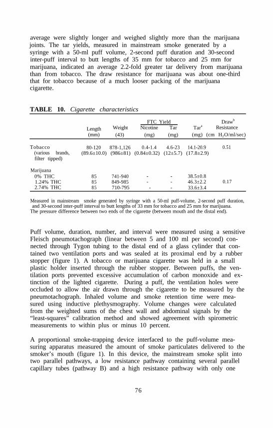
average were slightly longer and weighed slightly more than the marijuanajoints. The tar yields, measured in mainstream smoke generated by asyringe with a 50-ml puff volume, 2-second puff duration and 30-secondinter-puff interval to butt lengths of 35 mm for tobacco and 25 mm formarijuana, indicated an average 2.2-fold greater tar delivery from marijuanathan from tobacco. The draw resistance for marijuana was about one-thirdthat for tobacco because of a much looser packing of the marijuanacigarette.
TABLE 10. Cigarette characteristics
FTC Yield Drawb
Length Weight Nicotine Tar Tara Resistance(mm) (43) (mg) (mg) (mg) (cm H2O/ml/sec)
Tobacco(various brands,filter tipped)
80-120 878-1,126 0.4-1.4 4.6-23 14.1-20.9 0.51(89.6±10.0) (986±81) (0.84±0.32) (12±5.7) (17.8±2.9)
Marijuana0% THC 85 741-940 - - 38.5±0.81.24% THC 85 849-985 - - 46.3±2.2 0.172.74% THC 85 710-795 - - 33.6±3.4
Measured in mainstream smoke generated by syringe with a 50-ml puff-volume, 2-second puff duration,and 30-second inter-puff interval to butt lengths of 33 mm for tobacco and 25 mm for marijuana.
The pressure difference between two ends of the cigarette (between mouth and the distal end).
Puff volume, duration, number, and interval were measured using a sensitiveFleisch pneumotachograph (linear between 5 and 100 ml per second) con-nected through Tygon tubing to the distal end of a glass cylinder that con-tained two ventilation ports and was sealed at its proximal end by a rubberstopper (figure 1). A tobacco or marijuana cigarette was held in a smallplastic holder inserted through the rubber stopper. Between puffs, the ven-tilation ports prevented excessive accumulation of carbon monoxide and ex-tinction of the lighted cigarette. During a puff, the ventilation holes wereoccluded to allow the air drawn through the cigarette to be measured by thepneumotachograph. Inhaled volume and smoke retention time were mea-sured using inductive plethysmography. Volume changes were calculatedfrom the weighted sums of the chest wall and abdominal signals by the“least-squares” calibration method and showed agreement with spirometricmeasurements to within plus or minus 10 percent.
A proportional smoke-trapping device interfaced to the puff-volume mea-suring apparatus measured the amount of smoke particulates delivered to thesmoker’s mouth (figure 1). In this device, the mainstream smoke split intotwo parallel pathways, a low resistance pathway containing several parallelcapillary tubes (pathway B) and a high resistance pathway with only one
76
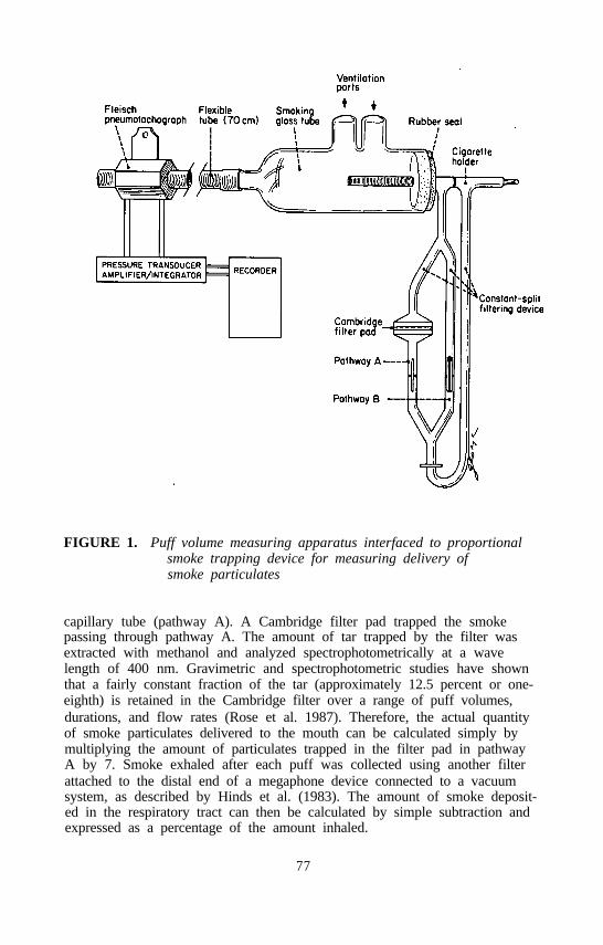
FIGURE 1. Puff volume measuring apparatus interfaced to proportionalsmoke trapping device for measuring delivery ofsmoke particulates
capillary tube (pathway A). A Cambridge filter pad trapped the smokepassing through pathway A. The amount of tar trapped by the filter wasextracted with methanol and analyzed spectrophotometrically at a wavelength of 400 nm. Gravimetric and spectrophotometric studies have shownthat a fairly constant fraction of the tar (approximately 12.5 percent or one-eighth) is retained in the Cambridge filter over a range of puff volumes,durations, and flow rates (Rose et al. 1987). Therefore, the actual quantityof smoke particulates delivered to the mouth can be calculated simply bymultiplying the amount of particulates trapped in the filter pad in pathwayA by 7. Smoke exhaled after each puff was collected using another filterattached to the distal end of a megaphone device connected to a vacuumsystem, as described by Hinds et al. (1983). The amount of smoke deposit-ed in the respiratory tract can then be calculated by simple subtraction andexpressed as a percentage of the amount inhaled.
77

Mean data for smoking dynamics are shown in table 11 for MIS. Com-pared to tobacco, marijuana smoking was associated with a 30 to 40 percentsmaller number of puffs, a 30 to 78 percent greater puff volume, a 33 to50 percent longer puff duration, a 40 to 54 percent greater inhaled volumeand a 3.9- to 5.2-fold longer retention time. Similar differences were notedcomparing MS with TS, suggesting that among habitual MTS, the dynamicsof smoking one type of substance does not influence smoking topographyfor the other substance. It is also noteworthy that, despite marked differ-ences in the degree of intoxication, the different strengths of marijuana wereassociated with a similar smoking profile, except for puff volume, whichwas smaller during the smoking of 2.74 percent marijuana compared to thelower strengths of marijuana, but still larger than the puff volume associatedwith tobacco smoking.
TABLE 11. Mean smoking topographic data for smokers of marijuanaplus tobacco (n=11)
Mar i juanaParamete r T o b a c c o 0%THC 1.24% THC 274% THC
Puff No. 12.6±4.7 7.4±2.2b 8.3±3.1b 8.4±3.2b
Puff Volume, ml 47.8±14.4 84.9±34.0b , c 82.6±20.4b , c 62.4±23.1
Puff Duration, sec 2 .4± l . l 3.6±2.1b 4.4±2.2b,c 3.1±2.1
Puff Interval, sec 32.1±16.9 38.8±14.2 43.1±18.4 36.6±12.6
Inhaled Volume, 1 1.24±0.31 1.91±0.74b 1.74±0.60b 1 .73±0.72b
Retention lime, sec 3.0±0.8 14.0±11.1b 15.7±11.6b 11.6±8.0b
No Smoking Interval, sec 28.9±17.4 22.2±9.5 2 4 5 1 2 . 3 22.7±5.7
Butt Length, mm 38±14 28±17 29±17 27±10
“High,” % (maximum 100%) - 17±14 69±17d 74±18d
aSignificant differences across tobacco and marijuana cigarettes (0-2.74 percent THC); p<.006,two-way ANOVA.
bSignificantly different from tobacco; p<.03.cSignificantly different from 2.74 percent THC; p<.02.dSignificantly different from 0 percent THC; p<.001.
For the dual smokers, the mean quantity of inhaled particulates measured inoptical density (OD) units and the percentage of inhaled particulates deposit-ed in the respiratory tract for each type of cigarette are shown in table 12.Compared to tobacco, marijuana was associated with the delivery of anaverage of 2.9 times more particulates and a 40 percent greater retention ofthe inhaled particulates in the respiratory tract. Similar differences werenoted comparing MS with TS.
In summary, in smokers of both marijuana and tobacco, a nearly threefoldgreater delivery of particulate matter to the smoker’s mouth was found
78

during the smoking of a single marijuana joint of varying potency up to27.74 percent THC compared to a single tobacco cigarette of the smoker’sown brand. We also found the respiratory retention of particulates inhaledfrom marijuana smoke to be 40 percent greater than that of particulatesinhaled from the smoke of tobacco. Therefore, the net respiratory burdenof particulates was approximately fourfold greater during marijuana thantobacco smoking. Similar differences were noted comparing smokers ofmarijuana alone with smokers of tobacco alone.
TABLE 12. Mean quantity of inhaled particulates and percent of inhaledparticulates retained in the respiratory tract &ring thesmoking of marijuana or tobacco by habitual smokers ofboth substances (n=11)
Mar i juanaParamete r T o b a c c o O%THC 1.24% THC 274% THC
Inhaled Particulates, 4.8±2.l 12.9±5.8* 14.9±5.8* 13.7±4.6*Optical Density
Particulate Deposition 59.3±19.6 84.3±15.6+ 84.4±17.4+ 86.7±13.2+
* Significantly different from tobacco, p<.001, ANOVA multiple comparison test.
Factors that might account for the greater particulate burden to the lungsfrom smoked marijuana than from a comparable quantity of smoked tobaccoinclude differences in the combustion characteristics of the two plantsubstances and a greater filtration of the tobacco cigarettes, which werefilter tipped and more densely packed than the marijuana joints. These dif-ferences could largely account for the more than twofold greater tar yieldfrom marijuana than tobacco that was measured using syringe-simulatedpuffs of similar volume and duration (table 10). Another possible factorinvolves consumption of a larger proportion of the marijuana than tobaccocigarettes, leaving a smaller butt length. Nonetheless, although marijuanawas smoked to a smaller average butt length (28 mm) than tobacco(38 mm), the tobacco cigarettes were longer and heavier to begin with, sothat a similar quantity of the two types of cigarette was consumed. Afourth factor, differences in smoking dynamics, probably contributed signifi-cantly to the observed differences in tar delivery to and retention by thesmoker. Differences in smoking dynamics included a 30 to 78 percentlarger puff volume, a 40 to 54 percent greater inhaled volume, and a 3.9 to5.2 times longer retention time for marijuana than for tobacco, whichappeared to more than offset any effect of the 30 to 40 percent smallernumber of puffs of marijuana than of tobacco. The deeper inhaled volumesand, in particular, the much longer breathholding times during marijuana vs.tobacco smoking could have accounted for the greater fractional deposition
79

of inhaled marijuana smoke particulates in the respiratory tract due to gravi-tational sedimentation and interception of surfaces in the lower airways andair spaces during molecular diffusion.
In conclusion, the fourfold greater respiratory burden of particulatesobserved during the smoking of a single marijuana cigarette compared to asingle tobacco cigarette may partly explain earlier findings that daily smok-ing of only a few joints of marijuana without tobacco was associated withchronic respiratory symptoms and tracheobronchial epithelial histopathologyof frequency and extent similar to that noted in daily smokers of more than20 cigarettes of tobacco without marijuana. These observations provide fur-ther support for the concept that the habitual smoking of only a few mari-juana cigarettes a day can have adverse long-term effects on the lung.
On the other hand, the mechanism of the relatively greater impact of mari-juana than tobacco smoking on central airway function and the apparentlygreater frequency of some histopathologic changes in the epithelium of thecentral airways among marijuana-only than tobacco-only smokers, in con-trast to the predominant effect of tobacco smoking on the peripheral air-ways, remains unclear. Possible explanations for these differences could bea relatively greater fractional deposition of marijuana smoke particulates inthe central airways due to inertial impaction, greater absorption of irritatinggases within marijuana smoke by the mucosa of the central airways leadingto proportionally less particulate deposition or gaseous absorption moreperipherally in the respiratory tract, or a combination of these factors.Since particle size is an important determinant of the mechanism and rate ofdeposition of particulates in the respiratory tract (Brain and Valberg 1974),the aerodynamic size of particulates was measured in the smoke of filteredand unfiltered tobacco cigarettes (Marlboro) and marijuana joints containing0.004, 1.24, and 2.75 percent A9-THC using a multistage cascade impactor(Andersen, Series 290). Puff volumes and durations of air similar to thoseobserved in smokers of marijuana and tobacco (100 ml, 4 sec and 50 ml,2 sec, respectively, at a flow rate of 25 ml/sec) were delivered through themarijuana or tobacco cigarette in a combustion chamber. The cigarettesmoke was immediately diluted 10 to 30 times by a high volume ofambient air at 21 °C and 15 percent relative humidity in a mixing chamberto prevent agglomeration of smoke particulates. The smoke was drawn intothe cascade impactor at a flow rate of 4.0 liters per minute. The excesssmoke mixed with air was exhausted to the atmosphere through a vent.Methanol extracts of the impaction plates were assayed spectrophotometric-ally at 400 nm. Measurements were performed in quintuplicate for eachtype of cigarette. Mass mean aerodynamic diameter (MMAD) and geo-metric standard deviation (GSD) were calculated based on the assumption ofa log normal distribution of particle sixes by the least squares method.Results are shown in table 13.
80
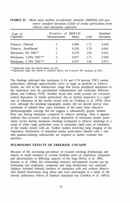
#TABLE 13. Mean mass median aerodynamic diameter (MMAD) and geo-metric standard deviation (GSD) of smoke particulates fromtobacco and marijuana cigarettes
Type of N u m b e r o f M M A DCigarettes Measurements Mean GSD
StandardDeviation
Tobacco, Filtered 5 0.490 1.71 0.039Tobacco, Nonfiltered 5 0.539 1.79 0.061Marijuana, 0% THC* 5 0.579 1.85 0.032
Marijuana, 1.24% THC** 5 0.927 1.71 0.039Marijuana, 2.74% THC** 5 0.957 1.66 0.072
* Significantly larger than filtered tobacco (p<.05).**Significantly larger than filtered or unfiltered tobacco and 0 percent ‘MC marijuana (p<.001).
The findings indicated that marijuana (1.24 and 2.74 percent THC) smokeparticulates, although approximately twice as large as particles in tobaccosmoke, are still in the submicronic range that favors peripheral deposition inthe respiratory tract by gravitational sedimentation and molecular diffusion(Brain and Valberg 1974). Another factor that could account for excessivecentral deposition of smoke particulates due to inertial impaction is a rapidrate of inhalation of the smoke mixed with air (Valberg et al. 1978). How-ever, although the smoking topography studies did not permit precise mea-surements of inhaled flow rates, estimates of the latter from inductiveplethysmographic tracings did not suggest a substantially greater inhaledflow rate during marijuana compared to tobacco smoking. Thus it appearsunlikely that excessive central airway deposition of marijuana smoke partic-ulates occurs during marijuana smoking (compared to tobacco smoking) as aresult of either large particulate sixes or unusually rapid rates of inhalationof the smoke mixed with air. Further studies involving lung imaging of therespiratory distribution of deposited smoke particulates labeled with a suit-able gamma-emitting radionuclide are required to further evaluate thisquestion.
PULMONARY EFFECTS OF FREEBASE COCAINE
Because of the increasing prevalence of cocaine smoking (freebasing) andreports in small numbers of cocaine freebase users of respiratory symptomsand abnormalities in diffusing capacity of the lung (Weiss et al. 1981;Itkonen et al. 1984), the relationship between self-reported cocaine use byfreebasing and respiratory symptoms and lung dysfunction was analyzed.Subjects included habitual smokers of marijuana with and without tobaccowho denied intravenous drug abuse and were participants in a study of thechronic pulmonary effects of habitual marijuana use (Tashkin et al. 1987a).
81

Of 356 marijuana smokers in the study sample, 84 (67 males, 17 females,mean age 30 years) reported freebasing cocaine from 1 to 992 times intheir lifetimes. Comparisons were made between tobacco-smoking cocainefreebase users (CTS, n=36) and tobacco-smoking, cocaine nonusers (TS,n=136) and between tobacco-nonsmoking, cocaine-freebase users (CS, n=48)and total nonusers (NS, n=36). “Moderate” (more than 10 times, mean 131times) and “mild” (fewer than 10 times, mean approximately 3 times)cocaine users were also analyzed separately. Marijuana use was similar (52to 53 joint-years) but tobacco use less (12 vs. 17 pack-years) in CTS thanT’S, marijuana use was less in CS than NS (43 vs. 62 joint-years), andcocaine smoking was comparable in CTS and CS (lifetime use frequency 56vs. 57 times). No significant differences in frequency of chronic cough,sputum, wheeze, or shortness of breath were noted across any of the abovegroups. However, moderate freebase cocaine users reported hemoptysis(14 percent), black sputum (43 percent) and chest pain (78 percent) aftersmoking cocaine. Also, moderate freebase cocaine use had an adverseeffect on several tests of airway function (FEV1, FEV1/FVC, Vmax, Viso-flow, N2/1, closing volume, and RV), which was significantly greater thanthat which could be attributed to tobacco (p<.05). On the other hand,moderate freebase cocaine use, unlike tobacco, had no demonstrable effecton DLCO. These findings suggest that, among habitual marijuana smokers,moderate cocaine smoking (1) does not appear to have an effect on chronicrespiratory symptoms that is additive to that of marijuana alone; (2) notuncommonly causes acute respiratory symptoms shortly after smokingcocaine; (3) irritates large and small airways, as reflected by functionalchanges that are not obscured by concomitant marijuana use and appear tohe at least additive to those produced by tobacco; and (4) has no demon-strable effect on the pulmonary microcirculation.
Preliminary data in 39 additional cocaine smokers who reported heavy useof cocaine (mean of 11.7 grams per week) with, a history of regular free-base smoking for an average of 34 months reveal a high proportion of sub-jects with acute cardiopulmonary symptoms temporally related to cocainesmoking. Of this group of heavy cocaine smokers, 44 percent reported
. coughing up black sputum from their chests and 48 percent noted hemopty-sis within 12 hours after smoking cocaine, while 60 percent reported chestpain (mostly pleuritic in quality) and 40 percent experienced palpitationswithin 1 hour after freebasing.
Of the 272 non-freebase users in the sample of heavy, habitual marijuanasmokers, 242 admitted to “snorting” cocaine. However, analysis of respira-tory symptoms and lung function in nonsmokers of cocaine classified intogroups of “snorters” and “nonsnorters” of cocaine who did and did notsmoke tobacco failed to show any significant relationship between snortingcocaine and either respiratory symptoms or lung dysfunction among eithersmokers or nonsmokers of tobacco. More detailed studies are needed toexamine the impact of frequent cocaine smoking on lung structure and
82

function and the relationship between frequency, intensity, recency, andmethod of cocaine freebase use on respiratory alterations, adjusting foreffects of concomitant use of other drugs that can have a damaging effecton the lungs.
REFERENCES
Auerbach, O.; Gere, J.B.; Forman, J.B.; Petrick, T.G.; Smolin, H.J.;Muehsam, G.E.; and Kassouny, D.Y. Changes in the bronchial epitheli-um in relation to smoking and cancer of the lung. N Engl J Med256:97-104, 1957.
Auerbach, O.; Stout, A.P.; Hammond, E.D.; and Garfinkel, L Changes inbronchial epithelium in relation to cigarette smoking and its relation tolung cancer. N Engl J Med 265:253-267, 1961.
Barbers, R.G.; Gong, H., Jr.; Tashkin, D.P.; Oishi, J.; and Wallace, J.M.Differential examination of bronchoalveolar lavage cells in tobacco ciga-rette and marijuana smokers. Am Rev Respir Dis 135:1271-1275, 1987.
Barter, C.E., and Campbell, AH. Relationship of constitutional factors andcigarette smoking to decrease in l-second forced expiratory volume. AmRev Respir Dis 113:305-314, 1976.
Bloom, J.W.; Kaltenbom, W.T.; Paoletti, P.; Camilli, A.; and Lebowitz,M.D. Respiratory effects of non-tobacco cigarettes. Br Med J [Clin Res]295:1516-1518, 1987.
Boulougouris, J.C.; Panayiotopoulos, C.P.; Antypas, E.; Liakos, A.; andStefanis, C. Effects of chronic hashish use on medical status in 44 userscompared with 38 controls. Ann NY Acad Sci 282:168-172, 1976.
Brain J.D., and Valberg, P.A. Models of lung retention based on the ICRPTask Group. Arch Environ Health 28:1, 1974.
Chai, H.; Farr, R.S.; Froehlich, L.A.; Mathison, D.A.; McLean, J.A.;Rosenthal, R.R.; Sheffer, A.L.; Spector, S.L.; and Townley, R.G. Stan-dardization of bronchial inhalation challenge procedures. J Allergy ClinImmunol 56:323-327, 1975.
Coggins, W.J.; Swenson, E.W.; Dawson, W.W.; Fernandez-Salas, A.;Hernandez-Bolanos, J.; Jiminez-Antillon, C.F.; Vinocour, R.; and Faerron-Valdez, F. Health status of chronic heavy cannabis users. Ann NY AcadSci 282:148-161, 1976.
Cruickshank, E.K. Physical assessment of 30 chronic cannabis users and 30matched controls. Ann NY Acad Sci 282:148-161, 1976.
Ferris, B.G. Epidemiology Standardization Project. Am Rev Respir Dis118/6(Part 2): 1-88, 1978.
Fishburne, P.M.; Abelson, H.I.; and Cisin, I. National Survey on DrugAbuse: Main Findings, 1979. DHHS Pub. No. (ADM)80-976. Rock-ville, MD: National Institute on Drug Abuse, 1980.
Fleischman, R.W.; Baker, J.R.; and Rosenkrantz, H. Pulmonary pathologicchanges in rats exposed to marijuana smoke for 1 year. Toxicol ApplPharmacol 47:557-566, 1979.
83
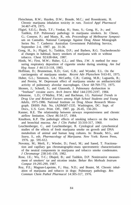
Fleischman, R.W.; Hayden, D.W.; Braude, M.C.; and Rosenkrantz, H.Chronic marijuana inhalation toxicity in rats. Toxicol Appl Pharmacol34:467-478, 1975.
Fligiel, S.E.G.; Beals, T.F.; Venkat, H.; Stutz, S.; Gong, H., Jr.; andTashkin, D.P. Pulmonary pathology in marijuana smokers. In: Cheser,G.; Consroe, P.; and Musty, R., eds. Proceedings of Melbourne Symposi-um on Cannabis. National Campaign Against Drug Abuse MonographSeries No. 7. Canberra: Australian Government Publishing Service,September 2-4, 1987. pp. 31-36.
Gong, H., Jr.; Fligiel, S.; Tashkin, D.P.; and Barbers, R.G. Tracheobronchi-al changes in habitual, heavy smokers of marijuana with or withouttobacco. Chest 92:638-664, 1987.
Hinds, W.; First, M.W.; Huber, G.L.; and Shea, J.W. A method for mea-suring respiratory deposition of cigarette smoke during smoking. Am IndHyg Assoc J 44:113-118, 1983.
Hoffman, D.; Brunneman, K.D.; Gori, G.B.; and Wynder, E.L. On thecarcinogenity of marijuana smoke. Recent Adv Phytochem 9:63-81, 1975.
Huber, G.L.; Simmons, GA.; McCarthy, C.R.; Cutting, M.B.; Laguarda, R.;and Pereira, W. Depressant effect of marijuana smoke on antibactericidalactivity of pulmonary alveolar macrophages. Chest 68:769-773, 1975.
Itkonen, J.; Schnoll, S.; and Glassroth, J. Pulmonary dysfunction in“freebase” cocaine users. Arch Intern Med 144:2195-2197, 1984.
Johnstone, L.D.; O’Malley, P.M.; and Bachman, J.G. National Trends inDrug Use and Related Factors among High School Students and YoungAdults, 1975-1986. National Institute on Drug Abuse Research Mono-graph. DHHS Pub. No, (ADM)87-1535. Washington, DC: Supt. ofDocs., U.S. Govt. Print. Off., 1987. pp. 26-45, 156-201.
Kanner, R.E. The relationship between airways responsiveness and chronicairflow limitation. Chest 86:54-57, 1984.
Knudtson, K.P. The pathologic effects of smoking tobacco on the tracheaand bronchial mucosa. Am J Clin Pathol 33:310-317, 1960.
Leuchtenberger, C., and Leuchtenberger, R. Cytological and cytochemicalstudies of the effects of fresh marijuana smoke on growth and DNAmetabolism of animal and human lung cultures. In: Braude, M.G., andSzara, S., eds. Pharmacology of Marijuana. New York: Raven Press,1976. pp. 595-612.
Novotny, M.; Merli, F.; Wiesler, D.; Fencl, M.; and Saeed, T. Fractiona-tion and capillary gas chromatographic-mass spectrometric characterizationof the neutral components in marijuana and tobacco smoke condensates.J Chromatogr 238:141-150, 1982.
Rose, J.E.; Wu, T-C.; Dhajed, B.; and Tashkin, D.P. Noninvasive measure-ment of smokers’ tar and nicotine intake. Behav Res Methods InstrumComput 19:295-299, 1987.
Roy, P.E.; Magnan-Lapointe, F.; Huy, N.D.; and Boutet, M. Chronic inhal-ation of marijuana and tobacco in dogs: Pulmonary pathology. ResCommun Chem Pathol Pharmacol 14:305-317, 1976.
84

Rubin, V., and Comitas, L. Respiratory function and hematology. In:Ganja in Jamaica: A Medical Anthropological Study of Chronic Mari-huana Use. The Hague, Paris: Mouton, 1975. pp. 88-102.
Tashkin, D.P.; Calvarese, B.M.; Simmons, M.S.; and Shapiro, B.J. Respira-tory status of seventy-four habitual marijuana smokers. Chest 78:699-706,1980.
Tashkin, D.P.; Coulson, AH.; Clark, VA; Simmons, M.; Bourque, L.B.;Duann, S.; Spivey, G.H.; and Gong, H. Respiratory symptoms and lungfunction in habitual heavy smokers of marijuana alone, smokers of mari-juana and tobacco, smokers of tobacco alone, and nonsmokers. Am RevRespir Dis 135:209-216, 1987a.
Tashkin, D.P.; Reiss, S.; Shapiro, B.J.; Calvarese, B.; Olsen, J.L.; andLodge, J.W. Bronchial effects of aerosolized 9-tetrahydrocannabinol inhealthy and asthmatic subjects. Am Rev Respir Dis 115:57-65, 1977.
Tashkin, D.P.; Shapiro, B.J.; and Frank, I.M. Acute pulmonary physiologiceffects of smoked marijuana and oral 9-tetrahydrocannabinol in healthyyoung men. N Engl J Med 289:336-341, 1973.
Tashkin, D.P.; Shapiro, B.J.; and Frank, I.M. Acute effects of smokedmarijuana and oral 9-tetrahydrocannabinol on specific airway conduct-ance in asthmatic subjects. Am Rev Respir Dis 109:420-428, 1974.
Tashkin, D.P.; Shapiro, B.J.; Lee, Y.F.; and Harper, C.E. Effects ofsmoked marijuana in experimentally induced asthma. Am Rev Respir Dis112377386, 1975.
Tashkin, D.P.; Simmons, M.; and Clark, V. Effect of habitual smoking ofmarijuana alone and with tobacco on nonspecific airways hyperreactivity.J Psychoactive Drugs 28:21-25, 1988.
Tashkin, D.P.; Simmons, M.S.; Coulson, A.H.; Clark, V.A.; and Gong, H.,Jr. Respiratory effects of cocaine “freebasing” among habitual users ofmarijuana with or without tobacco. Chest 92:638-644, 1987b.
Tennant, F.S., Jr. Histopathologic and clinical abnormalities of the respira-tory system in chronic hashish smokers. Subst Alcohol Actions Misuse1:93-100, 1980.
Vachon, L.; Fitzgerald, M.X.; Solliday, N.H.; Gould, LA.; and Gaensler,E.A. Singledose effect of marijuana smoke: Bronchial dynamics andrespiratory-center sensitivity in normal subjects. N Engl J Med 288:985-989, 1973.
Valberg, P.A.; Brain, J.D.; Le Mott, S.R.; Sneddon, S.R.; and Vinegar, A.Dependence of aerosol deposition site on breathing pattern. Am RevRespir Dis 117:262A, 1978.
Vollmer, W.M.; Johnson, L.R.; and Buist, A.S. Relationship of response toa bronchodilator and decline in’ forced expiratory volume in one second inpopulation studies. Am Rev Respir Dis 132:1186-1193, 1985.
Weiss, R.D.; Goldenheim, P.D.; Mirin, S.M.; Hales, C.A.; and Mendelsohn,J.H. Pulmonary dysfunction in cocaine smokers. Am J Psychiatry138:1110-1112, 1981.
85

Wu, T-C.; Tashkin, D.P.; Djahed, B.; and Rose, J.E. Pulmonary hazards ofsmoking marijuana as compared with tobacco. N Engl J Med 318:347-351, 1988.
ACKNOWLEDGMENTS
This work was supported by United States Public Health Service grantRO1 DA 03018 from the National Institute on Drug Abuse.
AUTHORS
Donald P. Tashkin, M.D.Professor of MedicineUCLA School of MedicineUniversity of California, Los Angeles10833 Le Conte AvenueLos Angeles, CA 90024-1736
Suzanne Fligiel, M.D.Head, Electron MicroscopyDepartment of Laboratory MedicineVeterans Administration Medical CenterAllen Park, MI 48101
andAssociate Professor of PathologyDepartment of PathologyWayne State University Medical CenterDetroit, Ml 48202
Tzu-Chin Wu, M.D.UCLA School of Medicine
Henry Gong, Jr., M.D.UCLA School of Medicine
Richard G. Barbers, M.D.Assistant Professor of MedicineUniversity of Massachusetts School of MedicineWorcester, MA 01605
Anne H. CoulsonUCLA School of Public Health
Michael S. SimmonsUCLA School of Medicine
86

Theodore F. Beals, M.D.Director, Clinical Electron MicroscopyVeterans Administration Medical CenterAnn Arbor, MI 48105
andAssistant Professor of PathologyUniversity of Michigan Medical SchoolAnn Arbor, Ml 48109
87

Marijuana Effects and UrinalysisAfter Passive Inhalation and OralIngestionEdward J. Cone
INTRODUCTION
The psychoactive properties of marijuana in humans have been the basis ofconsiderable scientific scrutiny. Over the last two decades, numerousstudies have been performed on the pharmacokinetic and pharmacodynamicproperties of marijuana and tetrahydrocannabinol (THC), the major activeconstituent of marijuana. The majority of these studies involve drug admin-istration by subjects smoking marijuana plant material or orally ingestingpure THC. Recently, the effects of passive exposure to marijuana smokeand the oral ingestion of marijuana plant material have been the subject ofconsiderable speculation as a result of extensive urine testing of individualswho claim exposure by these alternate routes of administration.
Absorption of sufficient THC by oral ingestion of marijuana plant materialto produce behavioral effects and detectable concentrations of urine metabo-lites was considered likely if free THC was present or conversion of THCprecursor acids in the plant material to THC occurred. Wall and colleagues(1983) found that the bioavailability of orally administered THC was 10 to20 percent for male and female college students. Also, Law and associates(1984) reported detection of cannabinoid metabolites in blood and urineafter administration of cannabis resin to human subjects in a meat sandwich.Studies of subjects who were passively exposed to marijuana smoke alsoindicated the possibility for absorption of sufficient THC to test positive byurinalysis for cannabinoid metabolites (Perez-Reyes et al. 1983; Mason et al.1983; Morland et al. 1935; Law et al. 1984; Ferslew et at 1983).
However, two basic pharmacologic questions remained essentially unan-swered regarding the effects of marijuana administered by these two routesof administration. These questions were:
88
1. Ate behavioral effects produced and are they dose related?
2. What is the urine excretion profile of cannabinoid metabolites?
Clinical studies at the Addiction Research Center, in collaboration with theU.S. Navy, were designed to answer these and related pharmacologic andtoxicologic questions. In separate studies, male subjects either were pas-sively exposed to marijuana smoke under highly controlled conditions oringested marijuana-laced brownies. Behavioral measures and all urines werecollected and analyzed. This chapter summarizes the results from thesestudies (Cone and Johnson 1986; Cone et al. 1987; Cone et al. 1988).
METHODS
Subjects
Subjects were healthy, drug-free males with a history of marijuana use.Criteria to enter the study included a documented period of cannabinoid-negative urines (EMIT dau, 20 ng/ml cutoff). Four negative days wererequired for the brownie study and 14 days for the passive-inhalation study.During the study, subjects were housed on a closed research ward underclose surveillance. Two additional male subjects, from the staff, with nohistory of marijuana use participated in one phase of the passive-exposurestudy. The study was conducted under the guidelines for the protection ofhuman subjects (45CRF46), and each subject gave informed consent.
Passive Marijuana-Exposure Conditions
Five subjects were passively exposed under double-blind conditions to sidestream smoke from 16 marijuana cigarettes (2.8 percent THC) for 1 houreach day (0830 to 0930) for 6 consecutive days. Preceding and followingthe days of marijuana smoke exposure, the subjects were exposed for2 days to the smoke of 16 placebo cigarettes (0 percent THC). Duringexposure, the subjects wore goggles and sat quietly in a small, unventilatedroom (2.1 m x 2.5 m x 2.4 m). The approximate room volume afteradjustment for contents was 12,225 L. Right of the cigarettes were burnedduring the first 12 minutes, and the remaining eight were burned 30 to42 minutes into the exposure session. The cigarettes were burned in aconstant-draft smoking apparatus designed to release only sidestream smoke.Flow rates for the smoking apparatus were adjusted for an average cigarettebum time of 12 minutes. Room air samples were withdrawn at timed inter-vals and analyzed for THC content. A second exposure study was per-formed with the subjects for 6 consecutive days under similar conditions butwith exposure to the smoke of four marijuana cigarettes (2.8 percent THC).This was preceded and followed by only 1 day of placebo smoke exposure.A third study was performed with two marijuana-naive subjects who wereexposed for 6 consecutive days to the smoke of 16 marijuana cigarettes
89
(2.8 percent THC) without blind conditions to placebo smoke. Individualurine specimens were collected from all subjects throughout the entire studyand were frozen until analyzed.
Marijuana-Laced Brownie Study
Each of the five subjects who participated in the study received single treat-ments of the following three doses of marijuana brownies, assigned in ran-dom order under double-blind conditions: two brownies containing anamount of cannabis plant material equivalent to two marijuana cigarettes(2.8 percent THC); two brownies containing the equivalent of one marijuanacigarette (2.8 percent THC) and one placebo marijuana cigarette (0 percentTHC); and two brownies containing the equivalent of two placebo marijuanacigarettes (0 percent THC). The brownies were consumed within 5 minutesstarting at 0830. All individual urine specimens were collected and frozenuntil analyzed.
Behavioral, Physiological, and Chemical Measures
Subjects completed subscales of the Addiction Research Center Inventory(Haertzen 1974) as follows: marijuana scale (Mar 15); morphine-benzedrinegroup (MBG) scale, which reflects feelings of euphoria and well-being;pentobarbital-chlorpromazine-alcohol group (PCAG) scale, which reflectssedation and intoxication; and the lysergic acid diethylamide (LSD)-specificscale. Additional measures collected included scales from the Single-DoseQuestionnaire (Feel Drug and Liking) and a visual analog scale (VAS) formeasurement of good feelings and physiological effects (pulse, bloodpressure). All measures were taken at 0730 and 0800 before each testsession (0830 to 0930) and thereafter at 0930, 1030, 1130, and 1230. Forthe marijuana brownie study, additional measures also were collected at1430,1630, and 2030.
Urinalysis
All urines were analyzed for cannabinoids by EMIT dau with a 20 ng/mLcutoff and by Abuscreen radioimmunoassay (RIA) with a 10 ng/mL level ofquantitation. Urines also were analyzed quantitatively for ll-nor- 9-THC-9-carboxylic acid (9-carboxy-THC) by gas chromatography/mass spectrometry(GC/MS) with a 5 ng/mL level of quantitation.
RESULTS
Behavioral Effects and Urinalysis After Passive Inhalation ofMarijuana Smoke
Mean responses after passive exposure to the smoke of 16 marijuana ciga-rettes were significantly (p<.05) elevated when compared to placebo smokeexposure (day 2) on the MAR 15, LSD, VAS, Feel Drug and Liking scales
90

(figure 1). Responses appeared to be elevated on the PCAG scale, but werenot significantly different from placebo and were unchanged on the MBGscale (not shown). Modest elevations also were obtained on these scaleswith the same subjects after exposure to the smoke of four marijuana ciga-rettes. The mean response on these scales by the same subjects afteractively smoking one marijuana cigarette (2.8 percent THC) is included infigure 1 for comparison.
FIGURE 1. Subjective effects induced by passive inhalation ofmarijuana smoke
*Significant difference (p<.05) compared with day 2 response.**Significant difference (p<.05) compared with day 3 response.
NOTE: Mean AUC data (n=5) for the response (minus control) over time are presented after exposureto the smoke of marijuana (MJ) or placebo (P). Mean (±SE) drug (D)AUC data in the same subjects also are shown after active smoking of one marijuana and oneplacebo cigarette and after active smoking of two placebo cigarettes (A).
Source: Cone and Johnson 1986, copyright 1986, C.V. Mosby Company.
91
The behavioral effects after passive exposure to marijuana smoke were timerelated, with peak behavioral responses being obtained immediately at theend of the smoke-exposure session. Responses were diminished thereafterand generally were completely dissipated within 3 hours. Generally, neutralor negative responses were obtained to the placebo-smoke-exposure sessions.
Assay of urine specimens by immunoassay for cannabinoids and by GC/MSfor 9-carboxy-THC provided information on the amount of THC absorbedduring the passiveexposure sessions with marijuana smoke and the timecourse of excretion of marijuana metabolites. Following exposure to thesmoke of 16 marijuana cigarettes, 6 of the 7 subjects in the 2 separateexperiments produced positive EMIT urine specimens after the first expo-sure session. The remaining subject produced his first positive after thesecond exposure session. A high percentage (50 to 100 percent) of allspecimens produced thereafter during the 6 consecutive passive-exposuredays were positive for cannabinoids. Following exposure to the smoke offour marijuana cigarettes, the results were more variable, with only two sub-jects producing positive EMIT specimens after the first exposure. Severalmore positive specimens were produced during the remaining sessions by allsubjects with the exception of one subject who produced no positive speci-mens throughout exposure at the lower passive-smoke-exposure dose. Theranges in concentration of 9-carboxy-THC in urines collected during theconsecutive smoke-exposure days, determined by GC/MS assay, are shownin table 1 in comparison to those determined for the same subjects afteractively smoking one or two marijuana cigarettes. Following the 6 consecu-tive days of passive marijuana-smoke exposure, subjects continued toexcrete cannabinoids in urine for varying periods, as shown in table 2.
Behavioral Effects and Urinalysis After Oral Ingestion of Marijuana-Laced Brownies
After the ingestion of brownies containing cannabis plant material equivalentto one or two marijuana cigarettes (2.8 percent THC content), subjectresponses on behavioral measures for Feel Drug, Liking, and the VASscales were elevated over that of placebo (figure 2). Heart rate was elevat-ed after administration of the two marijuana-cigarette-equivalent brownies.The AUC measures for Feel Drug and Liking scales were significantly(p<.05) different from placebo drug administration. The onset of behavioralresponses was somewhat slow and variable, with some subjects reportingelevated responses after 30 minutes, but peak responses generally occurred2.5 to 3.5 hours after dosing. The responses were prolonged and continuedto be elevated above placebo through 11.5 hours.
Cannabinoid metabolites were detected in the first urine voids after oralingestion of marijuana plant material and continued to be detectable for 3 to14 days. Peak concentrations of 9-carboxy-THC were equivalent to or
92

higher than those measured after smoking (table 1). Mean detection timesto the last positive specimen after dosing are shown in table 2.
TABLE 1. Individual ranges in peak urinary concentration of 9-carboxy-THC by GC/MS assay after marijuana exposure
Marijuana Number of 9-carboxy-THCExposure Dose1 Subjects (ng/ml)
Marijuana brownie2 1 5 108-325(Oral) 2 5 177-436
Marijuana smoke3 4 5 0-12(Passive inhalation) 16 5 15-35
16 1;1 10;87
Marijuana cigarettes3 1 5 9-140(Active smoking) 2 5 19-152
1Dose is expressed in marijuana cigarette equivalents; each cigarette contained 2.8 percent THC. Theaverage cigarette weight was 875 mg.
2Data are from Cone et al. 1988.3Data are from Cone et al. 1987.
TABLE 2. Hours to detection of last positive urine specimen forcannabinoid metabolites after marijuana exposure
Marijuana Number of Mean Hours + SEWExposure Dose’ Subjects EMIT (20) RIA (10) GUMS (5)
Marijuana brownie’ 1 5 121.6±27.9 140.3±31.9 149.0±36.2(Oral) 2 5 142.6±34.3 190.3±42.7 156.3±49.0
Marijuana smoke’ 4 5 1.7±1.3 30.9±10.4 9.4±5.8(Passive inhalation) 16 5 44.2±6.0 129.4±27.5 65.2±15.4
16 1;1 25.4;141.0 69.2;144.6 25.4;141.0
Marijuana cigarettes’ 1 10(Active smoking) 2 10
53.0±12.269.1±13.5
1Dose is expressed in marijuana cigarette equivalents; each cigarette contained 2.8 percent THC. Theaverage cigarette weight was 875 mg.
2The cutoffs (ng/ml) for each assay are shown in parentheses3Data are from Cone et al. 1988.4Data are from Cone et al. 1987.Unpublished data.
93

FIGURE 2. Mean behavioral and physiological effects produced in jivesubjects after consumption of marijuana-laced brownies
NOTE: Marijuana brownie content is shown in inset. 1MJ CIG=brownie dose that contained plantmaterial equivalent to one marijuana cigarette (2.8 percent THC) and one placebo cigarette(0 percent THC); 2MJ CIG=brownie dose that contained plant material equivalent to twomarijuana cigarettes (2.8 percent THC).
SOURCE Cone et al. 1988 copyright 1988, Preston Publications.
CONCLUSION
Dose-Related Behavioral Effects Produced After Passive Marijuana-Smoke Exposure and After Ingestion of Marijuana Plant Material
Behavioral responses on the Feel Drug and Liking scales were significantlyelevated over placebo responses after passive exposure to the smoke of 16marijuana cigarettes and after the oral ingestion of the equivalent marijuanaplant material of 1 or 2 cigarettes. Responses on these measures afterlower doses were elevated, but did not differ significantly from placebo. Itis clear from these studies that as the level of exposure to marijuana in-creased, so did the behavioral response. In addition, the behavioralresponse observed after passive exposure to the smoke of 16 marijuanacigarettes in a small, unventilated room was both qualitatively and quantita-tively equivalent to that observed after active smoking of 1 marijuanacigarette. Behavioral response after oral ingestion was both qualitativelyand quantitatively similar to that observed after active smoking, but with adelay of 1 to 3 hours.
94

Urine Excretion Profile of Cannabinoid Metabolites After PassiveMarijuana-Smoke Exposure and After Ingestion of Marijuana PlantMaterial
Passive exposure to the smoke of 16 marijuana cigarettes led to excretion ofsubstantial amounts of cannabinoid metabolites in urine after each exposurewith some evidence of accumulation after multiple exposures. Occasionally,levels in urine were sufficiently concentrated to exceed the 100 ng/mL cut-off calibrator commonly used in drug testing. After passive exposure to thesmoke of 4 marijuana cigarettes, an exposure level that seems more likelyto be encountered in social environments than is the extreme exposure tothe smoke of 16 marijuana cigarettes for 1 hour, urines tested positive forcannabinoid metabolites at levels only slightly above the 20 ng/mL cutoff.None of the urines collected after exposure to the smoke of four marijuanacigarettes tested positive by the 75 ng/mL EMIT calibrator.
Oral ingestion of marijuana plant material in cooked brownies led to excre-tion of highly concentrated amounts of cannabinoid in urine, as seen intable 1. Although marijuana smokers potentially can titrate their drug effectand thus their amount of marijuana exposure, the effects of oral ingestion ofmarijuana are slow in onset and do not allow the subject sufficiently rapidfeedback for dosage adjustment. Hence, the amounts of 9-carboxy-THC inurine after oral ingestion were, at times, substantially higher than thoseobserved after smoking equivalent doses.
REFERENCES
Cone, E.J., and Johnson, R.E. Contact highs and urinary cannabinoid excre-tion after passive exposure to marijuana smoke. Clin Pharmacol ExpTher 40(3):247-256, 1986.
Cone, E.J.; Johnson, R.E.; Darwin, W.D.; Yousefnejad, D.; Mell, L.D.;Paul, B.D.; and Mitchell, J. Passive inhalation of marijuana smoke: Uri-nalysis and room air levels of delta-9tetrahydrocannabinol. J Anal Toxi-col 11:89-96, 1987.
Cone, E.J.; Johnson, R.E.; Paul, B.D.; Mell, L.D.; and Mitchell, J. Mari-juana-laced brownies: Behavioral effects, physiologic effects, and urinaly-sis in humans following ingestion. J Anal Toxicol 12:169-175, 1988.
Ferslew, K.E.; Manno, J.E.; and Manno, B.R. Determination of urinarycannabinoid metabolites following incidental exposure to marijuana smoke.Res Commun Subst Abuse 4:289-300, 1983.
Haertzen, C.A. An overview of Addiction Research Center InventoryScales (ARCI): An appendix and manual of scales. DHEW Pub. No.(ADM)74-92. Washington, DC: Supt. of Docs., U.S. Govt. Print. Off.,1974.
Law, B.; Mason, P.A.; Moffat, AC.; Gleadle, R.I.; and Ring, L.J. Forensicaspects of the metabolism and excretion of cannabinoids following oralingestion of cannabis resin. J Pharm Pharmacol 36:289-294, 1984.
95
Mason, A.P.; Perez-Reyes, M.; McBay, A.J.; and Foltz, R.L. Cannabinoidconcentrations in plasma after passive inhalation of marijuana smoke.J Anal Toxicol 7:172-174, 1983.
Morland, J.; Bugge, A.; Skuterud, B.; Steen, A.; Wethe, G.H.; and Kjedlsen,T. Cannabinoids in blood and urine after passive inhalation of cannabissmoke. J Forensic Sci 30:997-1002, 1985.
Perez-Reyes, M.; Di Guiseppi, S.; Mason, A.P.; and Davis, K.H. Passiveinhalation of marijuana smoke and urinary excretion of cannabinoids.Clin Pharmacol Ther 34:36-41, 1983.
Wall, M.E.; Sadler, B.M.; Brine, D.; Taylor, H.; and Perez-Reyes, M.Metabolism, disposition, and kinetics of delta-9-tetrahydrocannabinol inmen and women. Clin Pharmacol Ther 34:352-363, 1983.
ACKNOWLEDGMENTS
Rolley E. Johnson, William D. Darwin, and David Yousefnejad of theAddiction Research Center assisted in the design and execution of the study,and Leroy D. Mell, Buddha D. Paul, and John Mitchell of the Navy DrugScreening Laboratory assisted with GC/MS analysis. Partial financial sup-port was provided by the U.S. Navy.
AUTHOR
Edward J. Gone, Ph.D.Laboratory of Chemistry and Drug MetabolismAddiction Research CenterNational Institute on Drug AbuseBaltimore, MD 21224
96

Pyrolytic Degradation of Heroin,Phencyclidine, and Cocaine:Identification of Products and SomeObservations on Their MetabolismC. Edgar Cook and A. Robert Jeffcoat
INTRODUCTION
Smoking of drugs offers certain advantages to the user, Purity of the drugis less important for smoking than it is for intravenous use, the extensivecapiIlary network in the lungs facilitates rapid absorption, and the blood cir-culation pathway ensures that absorbed substances will be rapidly transport-ed to the brain, where they can exert their effects. Thus, rapid onset ofaction (immediate, positive feedback) combined with some ability to exertcontrol over the dosage and rate of onset of activity combine to producepowerful incentives for smoking of abused drugs. These incentives havelong been obvious to smokes of marijuana, opium, and tobacco, but havein recent years also led to smoking of other drugs, such as phencyclidine(PCP) and cocaine. As drug users become more aware of the dangers ofacquired immunodeficiency syndrome (AIDS) associated with intravenousdrug use, it is possible that smoking will increase in popularity as a meansof drug taking.
The advantages enumerated above are also accompanied by disadvantagesthat may not be so readily apparent to the smoker. Many organic com-pounds undergo pyrolytic or oxidative reactions when exposed to high tem-peratures in the presence of air. The resulting compounds may have acutetoxicities differing from those of the parent drug, as well as having morelong-term effects, such as the induction of lung cancer associated withtobacco smoking. It thus becomes necessary to learn as much as possibleabout’ the nature of these pyrolytic products, their structures, their absorptioninto the body, and their routes of metabolism as well as toxicity associatedwith them. Over the past few years, our laboratory has been involved instudies of this type. In this chapter we summarize our work on the
97
pyrolysis of three drugs of abuse-heroin, cocaine, and PCP. This workdemonstrates the diversity of structures that can be obtained and illustratesthat smoking of drugs can lead to demonstrable absorption of pyrolysisproducts.
PYROLYSIS PRODUCTS OF HEROIN
Opium has been smoked for centuries in the Middle and Far East. There-fore, when in the early 1900s heroin was introduced into China and startedreplacing opium as a narcotic, it also was usually smoked (Huizer 1987).Although in western countries intravenous injection is a frequent mode ofheroin abuse, there are still many reports of its continued use by smokeinhalation. These have been reviewed by Cook and Brine (1985) and byHuizer (1987). Smoking of heroin-suffused marijuana or tobacco cigaretteshas been reported, but apparently the inhalation of smoke from heroinheated on aluminum foil (chasing the dragon) is currently the most popularmethod of administration (Huizer 1987). A number of cases of leukoen-cephalopathy have been attributed to a toxic compound or compoundsobtained from heroin smoking (Walters et al. 1981; Walters et al. 1982),but no specific compound has yet been implicated. The problem couldhave arisen from either pyrolysis products of heroin or of diluents used inthe particular heroin smoked. Since relatively little was known about thepyrolysis of heroin or its salts, we investigated the structure of some of themajor products of this process (Cook and Brine 1985).
Materials and Methods
Chemicals. Heroin hydrochloride and not-morphine hydrochloride wereobtained from the National Institute on Drug Abuse. Free heroin wasobtained by base conversion of the hydrochloride and extraction into butylchloride solution. The resulting product was shown to be pure by high per-formance liquid chromatography (HPLC). Normorphine was similarlyobtained. It was acetylated with acetic anhydride in pyridine to N-acetyl-nor heroin. Codeine, pseudocodeine, and isocodeine were acetylated in asimilar manner. Methylation was carried out by use of diazomethane inether containing 10 percent methanol. Allopseudocodeine acetate was pre-pared from ß-bromocodide by refluxing in acetic acid (Cook and Brine1985).
Pyrolysis. Pyrolysis was carried out in a quartz boat placed in a quartzfurnace tube and connected to a dry trap and two bubbler traps containing100 mL of absolute ethanol, each at -70 “C. Air was passed through thesystem at 30 mL per minute. A tube furnace was heated to the desiredtemperature and placed quickly around the quartz tube. Pyrolysis productswere isolated from the various parts of the apparatus by dissolving insolvent and were chromatographed on HPLC. Products were isolated on aPartisil-10/PAC column, eluted with a linear gradient of hexane/ethanol.Like fractions were combined and analyzed by HPLC on Partisil-10/ODS-2
98
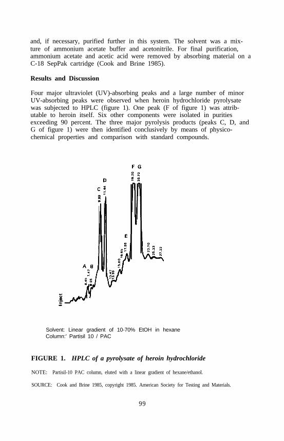
and, if necessary, purified further in this system. The solvent was a mix-ture of ammonium acetate buffer and acetonitrile. For final purification,ammonium acetate and acetic acid were removed by absorbing material on aC-18 SepPak cartridge (Cook and Brine 1985).
Results and Discussion
Four major ultraviolet (UV)-absorbing peaks and a large number of minorUV-absorbing peaks were observed when heroin hydrochloride pyrolysatewas subjected to HPLC (figure 1). One peak (F of figure 1) was attrib-utable to heroin itself. Six other components were isolated in puritiesexceeding 90 percent. The three major pyrolysis products (peaks C, D, andG of figure 1) were then identified conclusively by means of physico-chemical properties and comparison with standard compounds.
Solvent: Linear gradient of 10-70% EtOH in hexaneColumn:’ Partisil 10 / PAC
FIGURE 1. HPLC of a pyrolysate of heroin hydrochloride
NOTE: Partisil-10 PAC column, eluted with a linear gradient of hexane/ethanol.
SOURCE: Cook and Brine 1985, copyright 1985. American Society for Testing and Materials.
99
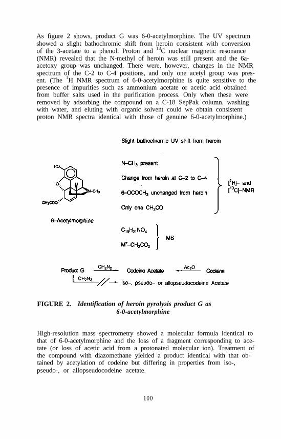
As figure 2 shows, product G was 6-0-acetylmorphine. The UV spectrumshowed a slight bathochromic shift from heroin consistent with conversionof the 3-acetate to a phenol. Proton and 13C nuclear magnetic resonance(NMR) revealed that the N-methyl of heroin was still present and the 6a-acetoxy group was unchanged. There were, however, changes in the NMRspectrum of the C-2 to C-4 positions, and only one acetyl group was pres-ent. (The 1H NMR spectrum of 6-0-acetylmorphine is quite sensitive to thepresence of impurities such as ammonium acetate or acetic acid obtainedfrom buffer salts used in the purification process. Only when these wereremoved by adsorbing the compound on a C-18 SepPak column, washingwith water, and eluting with organic solvent could we obtain consistentproton NMR spectra identical with those of genuine 6-0-acetylmorphine.)
FIGURE 2. Identification of heroin pyrolysis product G as6-0-acetylmorphine
High-resolution mass spectrometry showed a molecular formula identical tothat of 6-0-acetylmorphine and the loss of a fragment corresponding to ace-tate (or loss of acetic acid from a protonated molecular ion). Treatment ofthe compound with diazomethane yielded a product identical with that ob-tained by acetylation of codeine but differing in properties from iso-,pseudo-, or allopseudocodeine acetate.
100
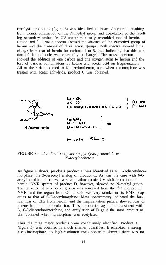
Pyrolysis product C (figure 3) was identified as N-acetylnorheroin resultingfrom formal elimination of the N-methyl group and acetylation of the result-ing secondary amine. Its UV spectrum closely resembled that of heroin.Proton and 13C NMR spectra showed the absence of the N-methyl group ofheroin and the presence of three acetyl groups. Both spectra showed littlechange from that of heroin for carbons 1 to 8, thus indicating that this por-tion of the molecule was essentially unchanged. The mass spectrumshowed the addition of one carbon and one oxygen atom to heroin and theloss of various combinations of ketene and acetic acid on fragmentation.All of these data pointed to N-acetylnorheroin, and, when not-morphine wastreated with acetic anhydride, product C was obtained.
FIGURE 3. Identification of heroin pyrolysis product C asN-acetylnorheroin
As figure 4 shows, pyrolysis product D was identified as N, 6-0-diacetylnor-morphine, the 3-desacetyl analog of product C. As was the case with 6-0-acetylmorphine, there was a small bathochromic UV shift from that ofheroin. NMR spectra of product D, however, showed no N-methyl group.The presence of two acetyl groups was observed from the 13C and protonNMR, and the region from C-l to C-8 was very similar in its NMR prop-erties to that of 6-O-acetylmorphine. Mass spectrometry indicated the for-mal loss of CH2 from heroin, and the fragmentation pattern showed loss ofketene from the molecular ion. These properties again are consistent withN, 6-0-diacetylnormorphine, and acetylation of D gave the same product asthat obtained when normorphine was acetylated.
Thus the three major products were conclusively identified. Product A(figure 5) was obtained in much smaller quantities. It exhibited a strongUV chromophore. Its high-resolution mass spectrum showed there was no
101

Small Bathochromic UV shift from heroin
FIGURE 4. Identification of heroin pyrolysis product D asN, 6-0-diacetylnormorphine
nitrogen in the molecular ion and indicated a formula of C18H14O4. Themass spectrum also indicated fragmentation involving the loss of twoCH2CO residues, consistent with the presence of two acetate functions. Thedegree of unsaturation revealed by mass spectrometry could be accommodat-ed by three aromatic rings together with two acetoxy groups, leading us totentatively identify this product as 3, 4-diacetoxyphenanthrene. A search ofthe literature showed that this compound had been reported in 1886 byFischer and Gerichten from heating of heroin methiodide with silver acetatein acetic acid; and it appears that this indeed is product A, although anauthentic sample was not available for comparison. (Dr. Fischer, unfortu-nately, was not in a position to provide us with such a sample.)
Strong UV chromophore
FIGURE 5. Tentative identification of heroin pyrolysis product A as3,4-diacetoxphenanthrene
102

The pryrolysis products identified are consistent with anticipated pyrolyticmechanisms shown in figure 6. In this figure, we see the initial formationof a quaternary N-acetyl intermediate by interaction of two molecules ofheroin. Attack of a nucleophile is most likely to occur on the N-methylgroup leading to its loss and the generation of the N-acetyl compound.Although nucleophilic attack could also take place on the other carbon sub-stituents of the nitrogen, it is less likely to occur there for steric reasons.Alternatively, removal of a proton in the position ß to the nitrogen wouldlead to formation of a double bond and opening of the piperidine ring.Alternatively, in the case of heroin hydrochloride, nucleophilic attack on themethyl group of the protonated amine would lead to the formation of nor-heroin, which could undergo acetylation by reaction with other molecules ofheroin.
FIGURE 6. Possible pyrolytic mechanisms for heroin
103

That some of the reactions involving ß elimination are also occurring isindicated by the data for pyrolysis products B and E. Product E had a for-mula indicating formal addition of CH2, to heroin. The structures of figure7, where R=CH3, would fit this molecular formula. Such structures couldderive from N-methylation followed by olefin formation. Since product Edid not have a styrene nor a conjugated diene chromophore, opening of thepiperidine ring to give the nonconjugated diene appears the most likely ex-planation at this time. Pyrolysis product B has a molecular ion at m/z 411,which is consistent with the N-acetyl N-methyl formulas shown in figure 7.
FIGURE 7. Possible structures for heroin pyrolysis products B and E
Products involving rearrangements or inversions of the allylic acetate systemdid not appear to play a major role in the pyrolysis, although such com-pounds could certainly exist as some of the minor products shown in theHPLC chromatogram. Finally, a large amount of unidentified tarry materialwas produced, which could have resulted from further reaction of some ofthe initially formed pyrolysis products.
The pyrolysis of heroin hydrochloride and heroin free base was studied atseveral temperatures ranging from 200 to 800 °C (Jones and Davis, personalcommunication). As figure 8 shows, decomposition was much more exten-sive with the hydrochloride salt. Less than 10 percent of the heroinremained as the parent compound after 10 minutes at 300 “C. At this timeand temperature, almost two-thirds of the heroin free base was recovered asthe parent compound. 6-0-Acetylmorphine was the major product at250 °C in the pyrolysis of heroin hydrochloride. About 20 percent of theoriginal heroin was converted to this product, together with 14 percent ofN-acetylnorheroin and about 6 percent of N,6-0-diacetylnormorphine. Pyrol-ysis of heroin free base at somewhat higher temperatures yielded similarpatterns and amounts of these products.
104
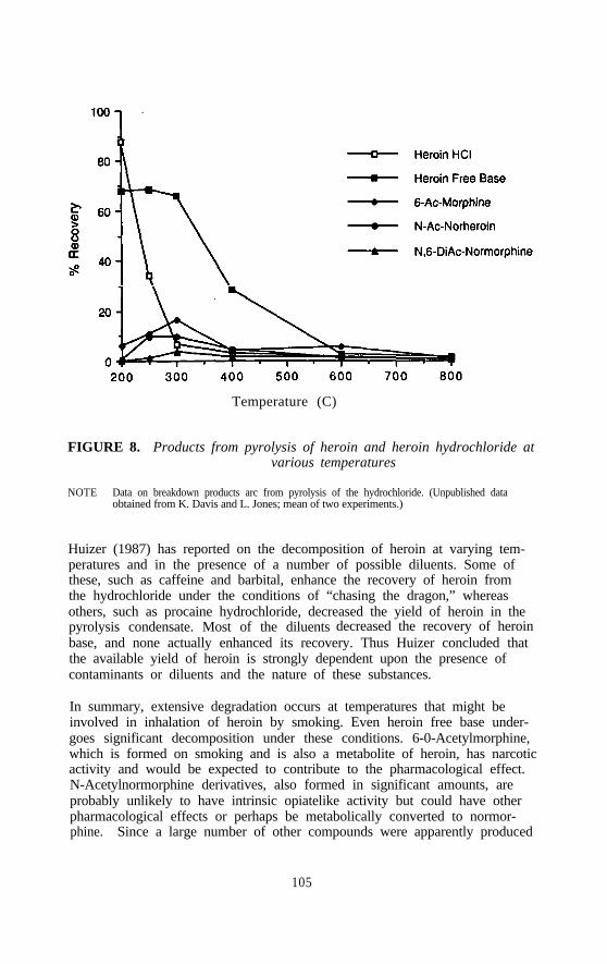
Temperature (C)
FIGURE 8. Products from pyrolysis of heroin and heroin hydrochloride atvarious temperatures
NOTE Data on breakdown products arc from pyrolysis of the hydrochloride. (Unpublished dataobtained from K. Davis and L. Jones; mean of two experiments.)
Huizer (1987) has reported on the decomposition of heroin at varying tem-peratures and in the presence of a number of possible diluents. Some ofthese, such as caffeine and barbital, enhance the recovery of heroin fromthe hydrochloride under the conditions of “chasing the dragon,” whereasothers, such as procaine hydrochloride, decreased the yield of heroin in thepyrolysis condensate. Most of the diluents decreased the recovery of heroinbase, and none actually enhanced its recovery. Thus Huizer concluded thatthe available yield of heroin is strongly dependent upon the presence ofcontaminants or diluents and the nature of these substances.
In summary, extensive degradation occurs at temperatures that might beinvolved in inhalation of heroin by smoking. Even heroin free base under-goes significant decomposition under these conditions. 6-0-Acetylmorphine,which is formed on smoking and is also a metabolite of heroin, has narcoticactivity and would be expected to contribute to the pharmacological effect.N-Acetylnormorphine derivatives, also formed in significant amounts, areprobably unlikely to have intrinsic opiatelike activity but could have otherpharmacological effects or perhaps be metabolically converted to normor-phine. Since a large number of other compounds were apparently produced
105
in minor amounts by heating of heroin, and since diluents used in streetsamples may also undergo pyrolysis to potentially toxic products, it appearsthat the smoking of heroin could have significant toxicological conse-quences. Further information on the pharmacological and toxicologicalimplications of this type of heroin administration is needed.
PYROLYSIS STUDIES OF COCAINE
The smoking of cocaine free base has become an increasingly popularmethod of self-administration. The ready availability of crack (a form ofcocaine free base) in particular has raised major concerns about this routeof administration. Since esters undergo pyrolytic elimination reactions, itseemed plausible to expect cocaine to be degraded by heat. We, therefore,undertook a study to identify the pyrolysis products of this compound (Cooket al. 1985; Jeffcoat et al. 1985).
Materials and Methods
Unlabeled cocaine and cocaine containing tritium in the N-methyl positionor on the C-4 methylene group were obtained from the National Instituteon Drug Abuse and diluted with unlabeled cocaine. The radiochemicalpurity was greater than 92 percent by HPLC analysis.
Pyrolysis. Tritiated cocaine in quantities of 25 to 200 mg was pyrolyzedby an apparatus similar to that used for the pyrolysis of morphine. Pyroly-sis products in the quartz tube and those collected in the U-tube wererecovered by dissolving them in acetonitrile.
Human Studies. Cocaine was administered to human subjects by the intra-venous, smoking, and nasal insufflation routes. The clinical and analyticalprocedures have been described (Jeffcoat et al. 1989; Perez-Reyes et al.1982).
Results and Discussion
Cocaine hydrochloride is almost completely destroyed at 800 °C (the tem-perature of the glowing end of a tobacco cigarette) and cocaine itself isextensively degraded with only 16 percent of the pyrolysis product being in-tact cocaine. At lower temperatures, however, cocaine is much more stable,although the percentage of intact cocaine recovered appears to be stronglydependent on exact experimental conditions. Separation of pyrolysis productmixtures by HPLC showed four major peaks of radiolabeled material. Thethird of these peaks had the same retention time as cocaine, and its massspectrum was identical with authentic cocaine.
Since the materials in the first two peaks were too volatile to permit theirisolation from the HPLC solvents, the crude pyrolysis product mixture was
106

examined by combined capillary gas chromatography/mass spectrometry(GC/MS). Three major types of pyrolysis products in addition to cocainewere observed. One set of three peaks with similar retention times relativeto cocaine had almost identical mass spectra, which were also identical tothe published spectra of methyl ecgonidine (Lowry et al. 1979; Lukaszewskiand Jeffery 1980). Although this compound is reported to be formed whencocaine hydrochloride is introduced into a heated injection port of a gaschromatograph, we found no such occurrence when cocaine free base wasinjected at the temperature used in this work The presence of three gaschromatography peaks indicates that other isomeric products are formed inaddition to methyl ecgonidine. One of these could be the ß, y isomer.Internal ring opening could also lead to compounds isomeric with methylecgonidine (Cook et al. 1985).
A second set of four peaks with similar but not equivalent mass spectra wasalso observed. The presence of molecular ions at m/z 150 was consistentwith identification of these as the doublebond isomers of methylcyclohepta-trienecarboxylate. This assignment was further supported by a base peak atm/z 91 corresponding to benzyl or cycloheptatrienyl ion with loss of carb-oxymethyl residue and transfer of a hydrogen. The compounds also under-went loss of methyl and methoxy residues (Cook et al. 1985).
In addition to these major peaks, significant amounts of methyl benzoate,benzoic acid, and N-methylbenzamide were also observed (Cook et al.1985). These products are explained by elimination of benzoic acid and bymechanisms involving acylation of the tertiary nitrogen of cocaine to formintermediates, which undergo nucleophilic attack much as was described inthe case of morphine.
The effectiveness of smoking as a route of administration is readily seen bycomparison of plasma concentrations of cocaine in human subjects whoeither smoked the drug or were given an intravenous injection of cocainehydrochloride (Jeffcoat et al. 1989). The who curves are essentially com-parable, and it was observed that the plasma levels of cocaine had reacheda maximum by approximately 6 minutes after the beginning of smoking(figure 9). Table 1 shows calculations of pharmacokinetic parameters andbioavailability calculated after different routes of administration in studiesfrom our laboratory (Jeffcoat et al. 1989). Average elimination half-livesranged from 56 minutes for the subjects who smoked cocaine to 80 minutesfor those who snorted the drug. Bioavailability based on the amount ofradiolabeled material actually inhaled showed a range of 32 to 77 percent.Since simulated smoking studies indicated that only about 44 percent of thematerial inhaled was cocaine, this suggests that most of the cocaine actuallyinhaled reaches the circulation unchanged. However, marked interindividualvariation in the amount of cocaine obtained by smoking is likely.
107

FIGURE 9. Cocaine and benzoylecgonine concentrations in the plasma ofsubjects who received cocaine by smoking or intravenousinjection
SOURCE Cook et al. 1985, copyright 1985, Biomedical publications.
A somewhat smaller proportion of the inhaled cocaine was excreted in theurine than was the case for intravenous cocaine. This suggests that some ofthe pyrolysis products are eliminated in the bile and feces. Examination ofthe urinary excretion of cocaine metabolites (figure 10) showed little dif-ference in the average amount of ecgonine methyl ester excreted whetherthe drug was given intravenously, by smoke inhalation, or by snorting; butthe amount of benzoyl ecgonine appeared significantly less in the case ofthe smoking route. Figure 11 shows the ratio of benxoyl ecgonine to ecgo-nine methyl ester in the urine. During the period 0 to 24 hours, there is asignificantly (p<.05) lower average ratio for the smoking route than for theother two routes of administration. This difference is even more pro-nounced in the period from 24 to 72 hours after administration. However,little (about 14 percent) of the total urinary excretion occurred during thislatter period, with 86 percent of the excretion occurring in the 0- to 24-hourperiod. Furthermore, there was considerable overlap in the ranges in the 0-to 24-hour period. Data in a recent paper by Ambre et al. (1988) show arange of ratios of benzoylecgonine to ecgonine methyl ester excretion of 0.8to 8.7 after intravenous administration. Thus, although an average differ-ence in metabolism based on route appeared to be present in our populationof cocaine users, this ratio could not be used as a means of distinguishingwhether a given individual had smoked or snorted cocaine.
108
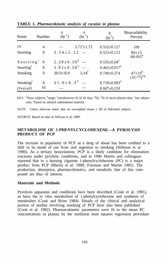
TABLE 1. Pharmacokinetic analysis of cocaine in plasma
a aRoute Number (hr-1) (hr-1)
B(hr-1)
BioavailabilityPercent
IV 4 — 3.71a±1.73 0.532±0.127 100Snorting 6 3 . 5 4 ± 2 . 1 2 — 0.523±0.113 80±13
68-l03b
S n o r t i n g c 6 2 . 2 8 ± 0 . 3 9 d — 0.535±0.04d
Snortingc 6 3 . 9 2 ± 0 . 3 8 d — 0.465±0.017d
Smoking 6 38.O±16.0 3.14f 0.740±0.274 47±198
(32-77)b,g
Smokingc 6 3 1 . 0 ± 8 . 3 d — 0.718±0.083d
Overall 16 — — 0.607±0.210
KEY: aThree subjects; brange; csimultaneous fit of all data; dSE; efit of mean plasma data; fone subjectonly; gbased on inhaled radiolabeled material.
NOTE Unless otherwise noted, date are unweighted means ± SD of Individual subjects.
SOURCE: Based on data in Jeffcoat et al, 1989.
METABOLISM OF 1-PHENYLCYCLOHEXENE—A PYROLYSISPRODUCT OF PCP
The increase in popularity of PCP as a drug of abuse has been credited to ashift in its mode of use from oral ingestion to smoking (Stillman et at1980). As a tertiary benzylamine, PCP is a likely candidate for eliminationreactions under pyrolytic conditions, and in 1980 Martin and colleaguesreported that in a burning cigarette 1-phenylcyclohexene (PC) is a majorproduct from PCP (Martin et al. 1980; Freeman and Martin 1981). Theproduction, absorption, pharmacokinetics, and metabolic fate of this com-pound are thus of interest.
Materials and Methods
Pyrolysis apparatus and conditions have been described (Cook et al. 1981),as have the in vitro metabolism of 1-phenylcyclohexene and synthesis ofmetabolites (Cook and Brine 1984). Details of the clinical and analyticalportion of studies involving smoking of PCP have also been published(Cook et al. 1982). Pharmacokinetic parameters were fit to the mean PCconcentrations in plasma by the nonlinear least squares regression procedure
109

FIGURE 10. Percent of dose of cocaine excreted as parent drug ormetabolites in urine as a function of route ofadministration (average 3-day excretion)
FIGURE 11. Ratio of benzoylecgonine to ecgonine methyl ester excretedin urine as a function of route of administration and timefrom drug administration
NOTE: Error bars show ranges. Number of subjects four for intravenous injection (IV), six each fornasal insufflation (NI) and smoking (SI).
110
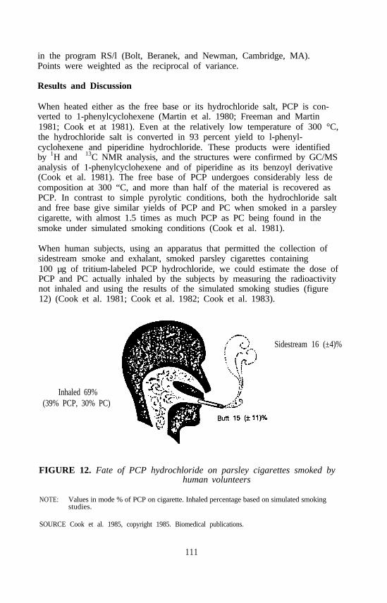
in the program RS/l (Bolt, Beranek, and Newman, Cambridge, MA).Points were weighted as the reciprocal of variance.
Results and Discussion
When heated either as the free base or its hydrochloride salt, PCP is con-verted to 1-phenylcyclohexene (Martin et al. 1980; Freeman and Martin1981; Cook et at 1981). Even at the relatively low temperature of 300 °C,the hydrochloride salt is converted in 93 percent yield to l-phenyl-cyclohexene and piperidine hydrochloride. These products were identifiedby 1H and 13C NMR analysis, and the structures were confirmed by GC/MSanalysis of 1-phenylcyclohexene and of piperidine as its benzoyl derivative(Cook et al. 1981). The free base of PCP undergoes considerably less decomposition at 300 “C, and more than half of the material is recovered asPCP. In contrast to simple pyrolytic conditions, both the hydrochloride saltand free base give similar yields of PCP and PC when smoked in a parsleycigarette, with almost 1.5 times as much PCP as PC being found in thesmoke under simulated smoking conditions (Cook et al. 1981).
When human subjects, using an apparatus that permitted the collection ofsidestream smoke and exhalant, smoked parsley cigarettes containing100 µg of tritium-labeled PCP hydrochloride, we could estimate the dose ofPCP and PC actually inhaled by the subjects by measuring the radioactivitynot inhaled and using the results of the simulated smoking studies (figure12) (Cook et al. 1981; Cook et al. 1982; Cook et al. 1983).
Sidestream 16 (±4)%
Inhaled 69%(39% PCP, 30% PC)
FIGURE 12. Fate of PCP hydrochloride on parsley cigarettes smoked byhuman volunteers
NOTE: Values in mode % of PCP on cigarette. Inhaled percentage based on simulated smokingstudies.
SOURCE Cook et al. 1985, copyright 1985. Biomedical publications.
111
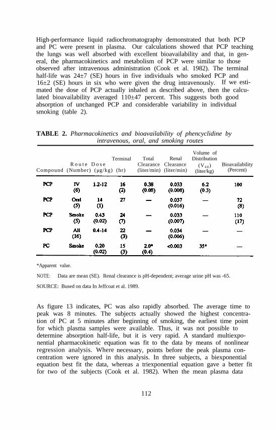
High-performance liquid radiochromatography demonstrated that both PCPand PC were present in plasma. Our calculations showed that PCP teachingthe lungs was well absorbed with excellent bioavailability and that, in gen-eral, the pharmacokinetics and metabolism of PCP were similar to thoseobserved after intravenous administration (Cook et al. 1982). The terminalhalf-life was 24±7 (SE) hours in five individuals who smoked PCP and16±2 (SE) hours in six who were given the drug intravenously. If we esti-mated the dose of PCP actually inhaled as described above, then the calcu-lated bioavailability averaged 110±47 percent. This suggests both goodabsorption of unchanged PCP and considerable variability in individualsmoking (table 2).
TABLE 2. Pharmacokinetics and bioavailability of phencyclidine byintravenous, oral, and smoking routes
Volume ofTerminal Total Renal Distribution
R o u t e D o s e (V4 8 )Compound (Number) (µg/kg) (hr)
Clearance Clearance Bioavailability(liter/min) (liter/min) (liter/kg) (Percent)
*Apparent value.
NOTE: Data are mean (SE). Renal clearance is pH-dependent; average urine pH was -65.
SOURCE: Bused on data In Jeffcoat et al. 1989.
As figure 13 indicates, PC was also rapidly absorbed. The average time topeak was 8 minutes. The subjects actually showed the highest concentra-tion of PC at 5 minutes after beginning of smoking, the earliest time pointfor which plasma samples were available. Thus, it was not possible todetermine absorption half-life, but it is very rapid. A standard multiexpo-nential pharmacokinetic equation was fit to the data by means of nonlinearregression analysis. Where necessary, points before the peak plasma con-centration were ignored in this analysis. In three subjects, a biexponentialequation best fit the data, whereas a triexponential equation gave a better fitfor two of the subjects (Cook et al. 1982). When the mean plasma data
112

FIGURE 13. Mean plasma concentrations (±SEM) of PC in five subjectswho smoked parsley cigarettes containing 100 µg of PCPhydrochloride
NOTE: Line is beat fit of a triexponential equation to data points from 10 minutes to 72 hours. Toppanal shows from 0 to 2 hours, bottom panel from 2 to 48 hours.
were used (figure 14), the data points from 10 minutes to 72 hours aftersmoking began were best fit by a triexponential equation (F test, p<.01)
113
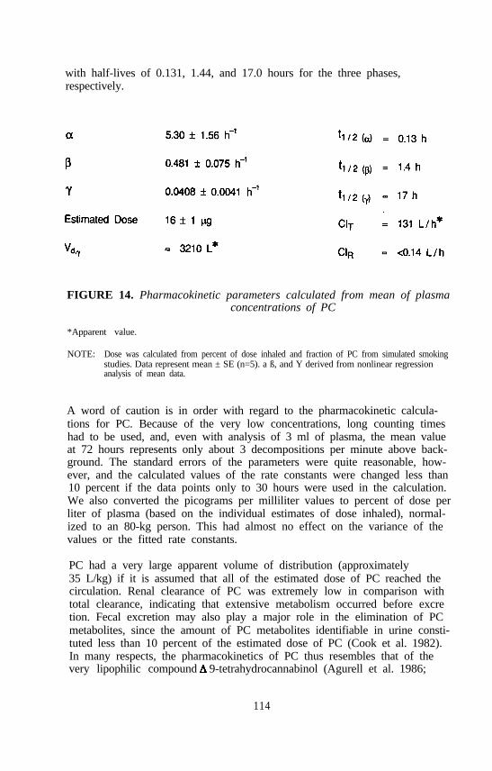
with half-lives of 0.131, 1.44, and 17.0 hours for the three phases,respectively.
FIGURE 14. Pharmacokinetic parameters calculated from mean of plasmaconcentrations of PC
*Apparent value.
NOTE: Dose was calculated from percent of dose inhaled and fraction of PC from simulated smokingstudies. Data represent mean ± SE (n=5). a ß, and Y derived from nonlinear regressionanalysis of mean data.
A word of caution is in order with regard to the pharmacokinetic calcula-tions for PC. Because of the very low concentrations, long counting timeshad to be used, and, even with analysis of 3 ml of plasma, the mean valueat 72 hours represents only about 3 decompositions per minute above back-ground. The standard errors of the parameters were quite reasonable, how-ever, and the calculated values of the rate constants were changed less than10 percent if the data points only to 30 hours were used in the calculation.We also converted the picograms per milliliter values to percent of dose perliter of plasma (based on the individual estimates of dose inhaled), normal-ized to an 80-kg person. This had almost no effect on the variance of thevalues or the fitted rate constants.
PC had a very large apparent volume of distribution (approximately35 L/kg) if it is assumed that all of the estimated dose of PC reached thecirculation. Renal clearance of PC was extremely low in comparison withtotal clearance, indicating that extensive metabolism occurred before excretion. Fecal excretion may also play a major role in the elimination of PCmetabolites, since the amount of PC metabolites identifiable in urine consti-tuted less than 10 percent of the estimated dose of PC (Cook et al. 1982).In many respects, the pharmacokinetics of PC thus resembles that of thevery lipophilic compound 9-tetrahydrocannabinol (Agurell et al. 1986;
114

Chiang and Barnett 1984; Wall et al. 1983; Hunt and Jones 1980; Barnettet al. 1982)—perhaps not a surprising conclusion.
The small fraction of PC metabolites excreted in urine and the low dosesused made identification of metabolites in humans very difficult. We there-fore made an initial study of the in vitro metabolism of this compound byrat liver. Our results (Cook et al. 1984) confirmed and extended the resultsof Martin et al. (1982) on metabolism by mouse liver. A large number ofmetabolites were identified by comparison of physical data with those ofstandards whose structures were established by synthesis and rigorous physi-cal methods (Cook et al. 1984). These studies showed that PCP undergoesoxidative metabolism at multiple sites on the molecule and that multipleoxidations also occur (figure 15). The double bond may be epoxidized andhydrolyzed to a diol. Oxidation may occur at the 3 position to yield analcohol or ketone. Further hydroxylation may occur at the 4 position, andoxidation may also occur at the 6 position. In several of the metabolites,an asymmetric center is present, thus raising the possibility of stereo-selective metabolism. Further research is needed on this feature of PCmetabolism.
Good separation of many of these metabolites may be achieved by HPLCon a Pattisil-PAC column, and reasonable separations are also achievedon a reverse phase chromatography system.
Extraction experiments showed that PC metabolites in urine were highlypolar and, therefore, likely consisted of conjugated substances. Thus, foridentification of metabolites, urine samples were incubated with a mixture ofB-glucuronidase and sulfatase. Extraction with butyl chloride then gave anorganic-soluble phase from which acid-soluble PCP metabolites could be ex-tracted with aqueous HCl. A subsequent extraction with aqueous sodiumhydroxide removed basesoluble PC metabolites, accompanied by some ofthe more water-soluble neutral metabolites. We found little evidence for thepresence of phenolic metabolites of PC. The neutral metabolites were puri-fied by small-scale preparative HPLC, generally on a reverse-phase column.Plasma extracts were also examined by HPLC in an attempt to identify PCmetabolites present in plasma. Metabolites were designated a, ß,in order of their polarity. Little, if any, of metabolite a was present inurine.
Only small amounts (generally less than a microgram) of these metabolitescould be isolated from urine of a single subject. Therefore, we relied ontechniques such as derivatization and isotope dilution experiments to identifythe metabolites. Thus, metabolite ß was suspected to be l-phenylcyclo-hexen-3-o1, based on its retention time on HPLC. To confirm this, theisolated metabolite was mixed with a known amount of standard compound,and the specific activity was determined by HPLC analysis. The weight ofthe material in the HPLC peak was determined by comparison of UV
115

FIGURE 15. In vitro metobolites of phenylcyclohexene
NOTE: Arrows represent chemically reasonable, but speculative, metabolic pathways. Broken andsolid lines show relative stereoechemical relationships, not absolute configuration. The positionof the hydroxyl at C-3 In PC-1,2,3-triol and BP-3-ol was not conclusively established. Mdesignates metabolites Identified from mouse (Martin et al. 1982); R designates ratmetabolites.
SOURCE Cook et al. 1984, copyright 1984, American Society for Pharmacology and ExperimentalTherapeutics.
absorption with a standard curve, and the radioactivity was measured byliquid scintillation spectrometry. Other aliquots of the mixture were deriva-tized and subjected to this same procedure. As derivatives, we chose theacetyl and p-nitrobenzoyl esters, to achieve a range of retention times onHPLC, thus maximizing the probability of separating closely related sub-stances. Since the specific activity of the two derivatives coincided veryclosely with that of the parent metabolite, we concluded that metabolite ß isindeed 1-phenylcyclohexen-3-o1.
Metabolite y on treatment with methoxylamine underwent a shift in reten-tion time on HPLC, thus indicating the presence of a keto group. Benzoy-lation also resulted in a change in retention and suggested the presence of a
116
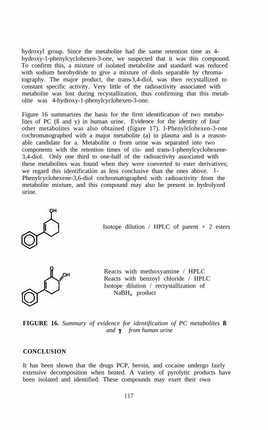
hydroxyl group. Since the metabolite had the same retention time as 4-hydroxy-1-phenylcyclohexen-3-one, we suspected that it was this compound.To confirm this, a mixture of isolated metabolite and standard was reducedwith sodium borohydride to give a mixture of diols separable by chroma-tography. The major product, the trans-3,4-diol, was then recystallized toconstant specific activity. Very little of the radioactivity associated withmetabolite was lost during recystallization, thus confirming that this metab-olite was 4-hydroxy-1-phenylcyclohexen-3-one.
Figure 16 summarizes the basis for the firm identification of two metabo-lites of PC (ß and y) in human urine. Evidence for the identity of fourother metabolites was also obtained (figure 17). l-Phenylclohexen-3-onecochromatographed with a major metabolite (a) in plasma and is a reason-able candidate for a. Metabolite o from urine was separated into twocomponents with the retention times of cis- and trans-1-phenylcyclohexene-3,4-diol. Only one third to one-half of the radioactivity associated withthese metabolites was found when they were converted to ester derivatives;we regard this identification as less conclusive than the ones above. l -Phenylcyclohexene-3,6-diol cochromatographed with radioactivity from themetabolite mixture, and this compound may also be present in hydrolyzedurine.
Isotope dilution / HPLC of parent + 2 esters
Reacts with methoxyamine / HPLCReacts with benzoyl chloride / HPLCIsotope dilution / recrystallization of
NaBH4 product
FIGURE 16. Summary of evidence for identification of PC metabolites ßand from human urine
CONCLUSION
It has been shown that the drugs PCP, heroin, and cocaine undergo fairlyextensive decomposition when heated. A variety of pyrolytic products havebeen isolated and identified. These compounds may exert their own
117

HPLC (plasma)
(c) HPLCHPLC of acetate (35% pure)
(t) HPLCHPLC of acetate and benzoate (47-50% pure)
HPLC
FIGURE 17. Summary of evidence for tentative identification of PCmetabolites a, o, and E in human subjects
pharmacological activity or may prove to have toxic effects. Much yetremains to be learned about these potential hazards of smoking of abuseddrugs.
REFERENCES
Agurell, S.; Halldin, M.; Lindgren, J.E.; Ohlsson, A.; Widman, M.;Gillespie, H.; and Hollister, L Pharmacokinetics and metabolism of
-tetrahydrocannabinol and other cannabinoids with emphasis on man.Pharmacol Rev 38:21-44,1986.
Ambre, J.; Ruo, T.I.; Nelson, J.; and Be&nap, S. Urinary excretion ofcocaine, benzoylecgonine, and ecgonine methyl ester in humans. J AnalToxicol 12:301-306, 1988.
Barnett, G.; Chiang, C.N.; Perez-Reyes, M.; and Owens, S.M. Kineticstudy of smoking marijuana. J Pharmacokinet Biopham 10:495-506,1982.
118

Chiang, C.N., and Barnett, G. Marijuana effect and delta-9-tetrahydrocan-nabinol plasma level. Clin Pharmacol Ther 36:234-238, 1984.
Cook, C.E., and Brine, D.R. Pyrolysis products of heroin. J Forensic Sci30(1):251-261, 1985.
Cook, C.E.; Brine, DR.; Quin, G.D.; Perez-Reyes, M.; and Di Guiseppi,S.R. Phencyclidine and phenylcyclohexene disposition after smokingphencyclidine. Clin Pharmacol Ther 31:635-641, 1982.
Cook, C.E.; Brine, D.R.; Quin, G.D.; Wall, M.E.; Perez-Reyes, M.; andDi Guiseppi, S.R. Smoking of phencyclidine: Disposition in man andstability to pyrolytic conditions. Life Sci 29:17-1972, 1981.
Cook, C.E.; Brine, D.R.; and Tallent, C.R. Identification of in vitro ratmetabolites of l-phenylcyclohexene. Drug Metab Dispos 12(2):186-192,1984.
Cook, C.E.; Jeffcoat, A.R.; and Perez-Reyes, M. Pharmacokinetic studies ofcocaine and phencyclidine in man. In: Barnett, G., and Chiang, C.N.,eds. Pharmacokinetics and Pharmacodynamics of Psychoactive Drugs.Foster City, CA: Biomedical Publications, 1985. pp. 48-74.
Cook, C.E.; Perez-Reyes, M.; Jeffcoat, AR.; and Brine, D.R. Phencyclidinedisposition in humans after small doses of radiolabeled drug. Fed Proc42(9):2566-2569, 1983.
Fischer, O., and Gerichten, E.V. Zur Kenntniss des morphins. ChemischeBerichten 19:792-794, 1886.
Freeman, A.S., and Martin, B.R. Quantification of phencyclidine in main-stream smoke and identification of phenylcyclohex-l-ene as pyrolysisproduct. J Pharm Sci 70:1002-1004, 1981.
Huizer, Henk. Analytical studies on illicit heroin. V. Efficacy of volatili-zation during heroin smoking. Pharmaceutisch Weekblad Scientific Edi-tion 9:203-211, 1987.
Hunt, C.A., and Jones, R.T. Tolerance and disposition of tetrahydro-cannabinol in man. J Pharmacol Exp Ther 215:35-44, 1980.
Jeffcoat, A.R.; Cook, C.E.; and Perez-Reyes, M. Psychoactive drugs. In:Barnett, G., and Chiang, C.N., eds. Pharmacokinetics and Pharmaco-dynamics of Psychoactive Drugs. Foster City, CA: Biomedical Publica-tions, 1985. pp. 441-455.
Jeffcoat, A.R.; Perez-Reyes, M.; Hill, J.M.; Sadler, B.M.; and Cook., C.E.Cocaine disposition in humans after intravenous injection, nasal insuffla-tion (snorting) or smoking. Drug Metab Disp 17:153-159, 1989.
Lowry, W.T.; Lomonte, J.N.; Hatchett, D.; and Garriott, J.C. Identificationof two novel cocaine metabolites in bile by gas chromatography/massspectrometry in a case of acute intravenous cocaine overdose. J Anal Tox3:91, 1979.
Lukaszewski, T., and Jeffery, W.K. Impurities and artifacts of illicitcocaine. J Forensic Sci 25:499-507, 1980.
Martin, B.R.; Bailey, B.B.; Awaya, H.; May, E.L.; and Narasimhachari, N.In vitro metabolism of I-phenyl-l-cyclohexene, a pyrolysis product ofphencyclidine. Drug Metab Dispos 10:585-689, 1982.
119
Martin, B.R.; Freeman, A.S.; Kelly, K.L.; Clayton, V.M.; Vincek, W.C.; andHarris, L.S. Fate of phencyclidine in a burning cigarette. Fed Proc39:302 (A189), 1980.
Perez-Reyes, M.; DiGuiseppi, S.; Ondrusek, G.; Jeffcoat, AR.; andCook, C.E. Free-base cocaine smoking. Pharmacol Ther 32(4):459,1982.
Stillman, R.; Barnett, G.; and Petersen, R. Phencyclidine (PCP): Epidemi-ology, pharmacology, and pharmacokinetics. In: Mello, N.K., ed. Ad-vances in Substance Abuse: Behavioral and Biological Research. Vol. 1.Greenwich, CT: JAI Press, 1980. pp. 289-303.
Wall, M.E.; Sadler, B.M.; Brine, D.; Taylor, H.M.; and Perez-Reyes, M.Metabolism, disposition and kinetics of 9-tetrahydmcannabinol in menand women. Clin Pharmacol Ther 34:352-363, 1983.
Wolters, E.Ch.; Van Wyngaarden, G.K.; Stam, EC.; Rengelink, H.;Lousberg, R.J.; Schipper, M.E.; and Verbeeten, B. Leucoencephalopathyafter inhaling ‘heroin’ pyrolysate. Lancet 2:1233-1237, 1982.
Wolters, E.Ch.; Van Wyngaarden, G.K.; Stam, EC.; Rengelink, H.;Lousberg, R.J.; Schipper, ME.; and Verbeeten, B. ‘Heroin’ leuco-encephalopathy; spongiform leuco-myelo-encephalopathy after inhalation ofthe pyrolysate of contaminated heroin. Ned Tijdschr Geneeskd 126:508-514, 1981.
ACKNOWLEDGMENTS
Work was carried out under National Institute on Drug Abuse contract271-80-3705. Dr. Mario Perez-Reyes carried out the clinical portion of thedescribed studies. Dolores Brine and Judith Hill provided technical assist-ance with the pyrolysis and analytical studies. Ken Davis and Larry Jonesprovided the quantitative data on pyrolysis of heroin.
AUTHORS
C. Edgar Cook, Ph.D.A. Robert Jeffcoat, Ph.D.
chemistry and Life SciencesResearch Triangle InstituteP.O. Box 12194Research Triangle Park, NC 277092194
Chemical and Biological Analysisof Marijuana Smoke Condensate
Charles M. Sparacino, Patricia A. Hyldburg, andThomas J. Hughes
INTRODUCTION
The various products derived from Cannabis sativa L., generally lumpedtogether under the descriptive name “marijuana,” are ingested for recreation-al purposes by a sizeable portion of the population. Most popularly, theleaves and flowering parts of the plant are smoked, producing a variety ofphysical and psychomimetic effects commonly referred to as being “high.”Owing to the large number of people using marijuana, an intensive efforthas been under way since about 1968 to define its chemistry, the metabo-lism of its major active ingredients, and the possible health effects of itsuse. Although much knowledge has been gained about the chemistry,pharmacology, metabolism, and pharmacokinetics of pure compounds fromCannabis, an insufficient amount is known about these subjects as theyrelate to marijuana smoke because of its complex chemistry.
The general goal of our work was the development of methodology for thecomprehensive analysis of the biologically active fractions of marijuanasmoke condensate (MSC). Biological activity was assessed through the useof the Ames Salmonella mutagenesis assay (Maron and Ames 1983). This“mutagenesis directed” research was designed to focus the analytical efforton the most biologically active fraction(s) to increase the possibility of iden-tifying potential mutagens or carcinogens in marijuana smoke condensate.Chemical and biological screening of smoke condensate fractions was con-ducted for both marijuana and tobacco to allow for comparisons.
The complex chemistry of marijuana smoke involves not only the majorcannabinoids, but also a large number of combustion and pyrolytic products.Many compounds found in marijuana smoke are known to have adversebiological properties. For example, one study (Leuchtenbeyer et al. 1973)showed that exposure of human lung explants to fresh marijuana smokeresulted in alterations in DNA and chromosome complement. But-stein
121

et al. (1973; Burstein et a1. 1975) have presented evidence that certainphenolic components present in the extracts of Cannabis sativa L. are stronginhibitors of prostaglandin biosynthesis. Polycyclic aromatic hydrocarbons(PAH) are formed during any organic combustion process and have beenisolated from MSC and tobacco smoke condensate (TSC) (Lee et al. 1976).These compounds are among the most studied of any class with regard totheir mutagenic and carcinogenic properties. Novotny et al. (1980),Maskarinec et al. (1976), and Merli et al. (1981) have identified many toxi-cologically important species in various MSC fractions and have, in somecases, compared these data with similar information from TSC factors.Some studies on the mutagenicity of MSC have been reported (Busch et al.1979; Wehner et al. 1980), but no research has yet combined mutagenicstudies with a concerted, comprehensive effort to identify specific com-pounds responsible for the biological activity.
Historically, a tremendous amount of work has been done to determine thecomposition and pharmacological activity of tobacco smoke. Parameters(puff frequency, volume, duration, and holding time) governing the genera-tion of TSC are well documented in the literature (Ogg 1964). Although itis easy to assume that such methodology is directly applicable to the mari-juana situation, this assumption in fact is not correct (Peterson 1979).Information found in the literature partially describing the physicodynamicsof marijuana smoking and the observation of research subjects smokingmarijuana cigarettes indicate that people smoking marijuana tend to do so ina much more vigorous and aggressive manner than is pursued with tobacco.While this information illustrates that a single set of parameters describingthe physicodynamics of marijuana smoking cannot be defined easily, itclearly shows that marijuana smoking parameters are more rigorous thanthose describing the tobacco smoking process.
GENERATION OF SMOKE CONDENSATES
The particular smoking parameters used in governing the production ofmarijuana smoke are important since the composition of the resulting MSCwill vary, depending on the conditions under which it is produced. In aprevious study performed at Research Triangle Institute (Davis et al. 1984),the burning temperature and other values associated with the collection ofMSC in a constant draft apparatus operating at varying flow rates weredetermined. These data demonstrated that the total mass of smoke conden-sate produced, as well as the recovery of 9-THC, decreases with increas-ing flow rate. The cannabinoid profile, however, remained relativelyconstant, and the burning temperature of the marijuana did not appear to begreatly dependent on flow rate. Also, the burning temperature of standardNational Institute on Drug Abuse cigarettes being smoked by human sub-jects was measured. A range of burning temperatures of 730 to 800 °Cwas determined in these experiments.
122
Methods and Results
Selection of Marijuana Samples for Study. Several distinctly differenttypes of marijuana are available on the illicit market and thus were potentialsubjects for study under this program. With respect to 9-THC potency,these range from essentially nonpotent hemp-type marijuana, which is foundon rare occasions, to the common Colombian or Mexican marijuana of 3 to4 percent 9-THC content, to extremely potent sinsemilla of Colombian,Hawaiian, or Californian origin containing 6 to 12 percent 9-THC. Gov-erned by the practical limitations of this program, we chose to study two ofthese types, producing MSC from them and carrying out biological testingand analysis as described later. Although a number of pairs could bechosen from among the types listed above, we felt that samples of Mexicanmarijuana containing, respectively, 1.3 percent 9-THC (low dose) and4.4 percent 9-THC (high dose) represented the most practical choices,bracketing most marijuana available on the street in this country. Forcomparison, a typical cigarette tobacco was studied along with the twomarijuana samples. This tobacco was obtained as reference cigarettes fromthe Tobacco and Health Research Institute (University of Kentucky,Lexington, KY).
Constant Draft and Intermittent Puff Smoking Systems. Realizing thatthere is no smoking system available that exactly duplicates the wayhumans smoke marijuana, we felt that the best approach to the productionof MSC was to use two types of smoking systems: first, the constant draftapparatus to produce smoke condensate representative of the total smokeproduced at the burning temperature of marijuana cigarettes being smokedby human subjects; and, second, intermittent puff smoking systems operatingunder the less rigorous tobacco smoking parameters, equivalent to the leaststringent conditions under which marijuana might be smoked by humans.By using each extreme of actual smoke conditions, the evaluation of smokecondensates from these two systems could provide a qualitative and quanti-tative range within which the various components of the marijuana smokeactually experienced by human smokers might be found.
Cigarettes of 70 mm length were prepared from high-potency marijuana andlow-porosity “street” cigarette papers with a manual cigarette-rollingmachine. Low-potency marijuana and research tobacco cigarettes were ma-chine rolled, cut to 70 mm length, and rerolled in low-porosity papers.Each of the three cigarette types was cornbusted under puff and constantdraft modes on a 30-port cigarette smoking machine (figure 1) to producesix different smoke condensate types. The cigarette smoking was conductedat flow rates of 1,200 mL per minute for all constant draft combustion runs,40 mL per 2-second puff (1 puff per minute) for puff mode combustionruns with marijuana, and 35 mL per 2-second puff (1 puff per minute) forpuff mode combustion runs with tobacco cigarettes. Mainstream smoke wascollected in a three-element trapping system consisting of an ethanol
123

bubbler, a packed sand trap, and an impinger trap arranged in series. Thevolume of each smoke condensate was reduced in vacuo at room tempera-ture to less than 200 mL. At least 10 grams dry weight of each type ofsmoke condensate were produced. Six smoke condensates were thus pro-duced: MSC-low potency by puff and by constant draft mode, MSC-highpotency by puff and by constant draft, and TSC by puff and by constantdraft.
FIGURE 1. Cigarette smoking machine for generation ofsmoke condensates
FRACTIONATION OF SMOKE CONDENSATES
MSC and TSC are known to be highly complex matrices containing up toseveral thousand compounds at levels that may vary over several orders ofmagnitude (Gudzinowicz and Gudzinowicz 1980). Adequate characterization(chemical and biological) of these components requires prefractionation to(1) increase the relative mass of particular components and thereby the sen-sitivity of biological and analytical techniques for characterization, and(2) allow accurate assessment of biological and chemical characterization forsignificant constituents with minimum interference from other components inthe matrix. The latter becomes a particularly important consideration withrespect to the testing of mutagenic activity, since toxicity resulting from afew specific components in complex sample mixtures often becomes thelimiting factor.
Methods and Results
Fractionation of smoke condensates (MSC and TSC) was accomplishedusing an acid-base liquid to liquid partition sequence, Similar sequenceshave been used in many studies of smoke condensates (Merli et al. 1980;
124

Merli et al. 1981) as part of the fractionation methodology due to the facileseparation of major classes. Liquid-liquid partition provides a good meansof dealing with the relatively large mass of crude smoke condensate thatmust he used to obtain adequate levels of individual constituents for latercharacterization.
The acid-base. partition scheme used in the fractionation of smoke conden-sates is shown schematically in figure 2. The procedure is based on that ofPetersen et al. (1982) as applied to diesel soot extract.
FIGURE 2. Acid-base partition scheme
In summary, the smoke condensates were taken to dryness and redissolvedin methylene chloride (100 mL). Aliquots were removed for total massdetermination. Portions of the condensate solution were then extracted in a
125

separatory funnel three times with 5 percent aqueous sulfuric acid. Thethree combined aqueous fractions were back-extracted once with 20 mL ofmethylene chloride. This methylene chloride was returned to the originalsmoke condensate solution.
To recover bases from the aqueous phases, the pH of the aqueous phasewas adjusted to 13 with 40 percent aqueous potassium hydroxide. Thisaqueous phase was then extracted in a separatory funnel three times withmethylene chloride. The base fraction (methylene chloride) was dried withsodium sulfate.
Next, acids were recovered from the original condensate solution (now inmethylene chloride) by extraction with 5 percent aqueous sodium hydroxide.The combined aqueous base solutions were back-extracted with one aliquotof methylene chloride and the aliquot returned to the original methylenechloride smoke condensate. The acids were recovered by adjusting the pHof the sodium hydroxide solution to acidic values and extracting the solutionwith methylene chloride. Thus, methylene chloride solutions were obtainedfor the crudes (C), neutrals (N), acids (A), and bases (B). Aliquots wereremoved from all phases for total mass determination.
The neutral fraction (N) was further subfractionated by silica gel columnchromatography. Prior to subfractionation, a portion of the neutral fractionwas solventexchanged into hexane. A 20 g silica gel column was used.The neutral fraction was introduced at the top of the column and theneluted. Four eluting solvents were used to collect four fractions. Fraction1 was eluted with 60 mL of hexane, fraction 2 with 100 mL of 1:l tolueneand hexane, fraction 3 with 100 mL of methylene chloride, and fraction 4with 200 mL of methanol. Each fraction was collected separately; furtherfractionation has not been conducted.
The base fractions (B) of the smoke condensates were further fractionatedby normal phase, gradient HPLC. A cyanopropyl column with 5-micronspherical packing was used. Three solvent mixes were used:
Solvent A: 0.05 percent methanol/hexaneSolvent B: 0.05 percent methanol/methylene chlorideSolvent C: 20 percent methanol/methylene chloride
The gradient was as follows:
100 percent solvent A for 5 minutes100 percent solvent A to 100 percent solvent B over 20 minutes100 percent solvent B for 8.5 minutes100 percent solvent C for 6 minutes
126

The flow rate was maintained at 4.0 mL per minute. Seven subfractionswere collected based on the time elapsed from the start of the run. The cutpoints were selected arbitrarily, based on the appearance of thechromatogram. Fractions were collected as follows:
Fraction 1: 0 to 5 minutesFraction 2: 5 to 9 minutesFraction 3: 9 to 12 minutesFraction 4: 12 to 15.5 minutesFraction 5: 15.5 to 23.5 minutesFraction 6: 23.5 to 33 minutesFraction 7: 33 to 39 minutes
Multiple runs were performed and like fractions from each run werecombined.
CHARACTERIZATION OF SMOKE CONDENSATES
Biological Characterization
The objectives of the bioassay testing were (1) to determine and comparethe mutagenic activity of the chemical class fractions from TSC and MSCin the Ames Salmonella mutagenicity assay (Maron and Ames 1983), (2) todetermine the effect of smoking mode (puff vs. constant draft) and mari-juana batch on mutagenicity, and (3) to identify the most mutagenic frac-tions so that effective analytical chemical characterization techniques couldbe developed for identification of mutagenic and carcinogenic compounds.
The mutagenic potential of the various fractions was evaluated using thepreincubation modification of the standard plate incorporation technique ofthe Ames Salmonella assay. Fractions were tested for mutagenicity underthe following conditions: Salmonella typhimurium strains TA98 and TAlOOat five dose levels (10 to 2.50 µg per plate), triplicate plates per dose, withand without Aroclor 1254-induced hamster liver S9. Preincubation was per-formed for 20 minutes at 37 °C with shaking.
The following fractions were tested: crude (C), neutral (N) acid (A), andbase (B) fractions of the six smoke condensates, and the seven subfractions(base fractions 1 to 7) of the base fraction of the high-dose MSC generatedby constant draft (see above). Appropriate solvent and negative controlswere conducted concurrently with each ‘assay. Bioassay results were anal-yzed for mutagenic activity by slope analysis modeling. Slopes are reportedas revertants per µg of sample. Slope values were given a ranking for mu-tagenic activity based on slope analysis of an historical data base of muta-gens and nonmutagens (table 1). The higher the slope value, the moremutagenic the fraction was. Slope analysis for crude and chemical class
127

TABLE 1. Ranking of slope values from mutagenic fractions
Slope* Mutagenic Ranking(Revertants/µg) in Ames System
0.01 to 0.1 negative/equivocal0.1 to 1.0 weak
1.0 to 5.0 moderateabove 5.0 strong
* Nonlinear regression analysis of Myers et al. (1981).
fractions (A, B, N) of marijuana and tobacco smoke condensates is providedin table 2. Slope analysis of HPLC subfractions of the basic fraction isshown in table 3.
Chemical Characterization
Mass Determinations. Dry weight determinations were made on aliquotsof the various chemical class fractions and HPLC fractions of the crudecondensates. All weight determinations were made in triplicate by with-drawing 100 µL aliquots from the previously measured total volume of thefraction in solvent. These aliquots were placed in a tared container andtaken to dryness in a drying oven (50 “C). Each container was alternatelyweighed and dried until a constant weight was obtained.
Table 4 shows the mass distribution obtained from the chemical class frac-tions (A, B, N) of the crude condensates. Most of the material was foundin the neutral fraction.
Table 5 shows the mass distribution of the HPLC fractions of the base frac-tion of three of the crude condensates. As can be seen from the table, thebulk of the material was found in the last three fractions.
Thermogravimetric Analysis. The base fractions of the crude smoke con-densates were subjected to thermogravimetric analysis. In this technique,the weight of a sample is continuously recorded while the sample is heatedusing a specific temperature program. The rate and extent of physical andchemical changes that are accompanied by an increase or decrease in masscan be determined in this manner. Weight losses can be attributed to dry-ing, volatilization, or combustion. Weight losses occurring below 100 °Care generally due to drying.
Samples of 20 to 50 mg were heated, from ambient temperature, at2 °C per minute to approximately 600 °C at atmospheric pressure. A
128

TABLE 2. Slope analysis of chemical class fractions from tobacco andmarijuana smoke condensate in the Ames Assay
Fraction
Slope(Revertants/µg)TAlOO TA98 Mutagenic Ranking*
TSC-Puff-Crude 1.39TSC-Puff-Neutrals 0.03TSC-Puff-Acids 0.00TSC-Puff-Bases 0.00
weak
TSC-Constant-Crude 0.14 0.53 weakTSC-Constant-Neutrals 0.68 0.11 weakTSC-Ccnstant-Acids 0.04 0.08 equivocalTSC-Ccnstant-Bases 1.16 2.08 moderate
MSC-Low-Puff-Crude 0.16MSC-Low-Puff-Neutrals 0.21MSG-Low-Puff-Acids 0.12MSG-Low-Puff-Bases 0.19
MSG-Low-Constant-Crude 0.17 0.25MSG-Low-Constant-Neutrals 0.52 0.09MSG-Low-Constant-Acids 0.10 0.09MSG-Low-Constant-Bases 1.54 3.79
MSG-High-Puff-CrudeMSG-High-Puff-NeutralsMSG-High-Puff-AcidsMSG-High-Puff-Bases
MSG-High-Constant-CrudeMSC-High-Constant-NeutralsMSG-High-Constant-Acids
0.110.140.05
MSG-High-Constant-Bases 1.54
0.60 weak0.61 weak0.18 weak7.16 strong
*see table 1.
KEY: TSC=tobacco smoke condensate.MSC=marijuana smoke condensate.Puff/Constant smoking mode used in condensate generation.
NOTE: Salmonella typhimurium TA98 and TAl00 with Aroclor-induced hamster liver S9, five doselevels, triplicate plates per dose. preincubation with shaking for 20 minutes at 37 °C
0.280.150.001.92
0.660.170.310.92
A E0.230.89,
138 moderate0.36 weak0.46 weak7.04 strong
129
weakweakweak
weakweakweakweak
weakequivocalequivocalmoderate

thermogravimetric analyzer coupled to a data analyzer was used. A flow of50 mL per minute of inert gas was maintained through the sample chamberthroughout the test.
TABLE 3. Slope analysis of HPLC subfractions of base fractions fromtobacco and marijuana smoke condensate in the Ames assay
FractionSlope
(Revertants/µg)
TSC-Constant-Base Fraction 1 0.75TSC-Constant-Base Fraction 2 0.84TSC-Constant-Base Fraction 3 0.37TSC-Constant-Base Fraction 4 1.28TSC-Constant-Base Fraction 5 4.81TSC-Constant-Base Fraction 6 1.95TSC-Constant-Base Fraction 7 4.00
MSC-Puff-Base Fraction 1 1.89MSC-Puff-Base Fraction 2 3.36MSC-Puff-Base Fraction 3MSC-Puff-Base Fraction 4 12.89MSC-Puff-Base Fraction 5 14.98MSC-Puff-Base Fraction 6 16.48MSC-Puff-Base Fraction 7 18.68
MSC-Constant-Base Fraction 1 1.01MSC-Constant-Base Fraction 2 3.52MSC-Constant-Base Fraction 3 4.04MSC-Constant-Base Fraction 4 9.68MSC-Constant-Base Fraction 5 13.38MSC-Constant-Base Fraction 6 14.23MSC-Constant-Base Fraction 7 11.43
KEY: TSC=tobacco, smoke, and condensate.MSC=marijuana, smoke, condensate, high dose.Puff vs. Constant=smoking mode used in condensate generation.
NOTE: Salmonella typhimurium TA98 with Aroclor-induced hams&r lives S9, five dose levels,triplicate plates per dose, preincubation with shaking for 20 minutes at 37 °C.
Table 6 shows the results from this experiment. Minor weight losses dueto drying were observed in all samples. Most of the sample material wasvolatilized at temperatures below 300 °C. This indicates that the bulk ofthe material is amenable to gas chromatography (GC) or gas chromatogra-phy/mass spectrometry (GC/MS) analysis.
130
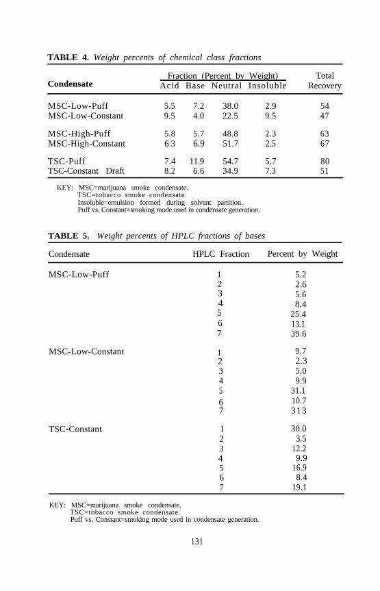
TABLE 4. Weight percents of chemical class fractions
CondensateFraction (Percent by Weight) Total
Acid Base Neutral Insoluble Recovery
MSC-Low-Puff 5.5 7.2 38.0 2.9 54MSC-Low-Constant 9.5 4.0 22.5 9.5 47
MSC-High-Puff 5.8 5.7 48.8 2.3 63MSC-High-Constant 6 3 6.9 51.7 2.5 67
TSC-Puff 7.4 11.9 54.7 5.7 80TSC-Constant Draft 8.2 6.6 34.9 7.3 51
KEY: MSC=marijuana smoke condensate.TSC=tobacco smoke condensate.Insoluble=emulsion formed during solvent partition.Puff vs. Constant=smoking mode used in condensate generation.
TABLE 5. Weight percents of HPLC fractions of bases
Condensate HPLC Fraction Percent by Weight
MSC-Low-Puff 1234567
MSC-Low-Constant 1234567
TSC-Constant 1 30.02 3.53 12.245
9.916.9
6 8.4
5.22.65.68.4
25.413.139.6
9.72.35.09.9
31.110.7313
7 19.1
KEY: MSC=marijuana smoke condensate.TSC=tobacco smoke condensate.Puff vs. Constant=smoking mode used in condensate generation.
131

TABLE 6. Thermogravimetric analysis of marijuana and tobacco smokecondensate bases
Condensate 70 110 150Percent Volatile (°C)190 230 270 310 350 600
MSC-Low-Puff a 53 66 71 75 78 79 80 82MSC-Low-Constant 3 13 22 37 51 63 71 75 83
MSG-High-Puff 2 10 25 44 59 68 74 18 82MSC-High-Constant 5 14 26 44 62 76 135 89 94
TSC-PUff 3 7 30 68 79 86 91 93 97TSC-Constant 11 37 66 76 84 87 90 93
KEY: MSC=Marijuana smoke condensate.TSC=Tobacco smoke condensate.Puff vs. Constant=smoking mode used in condensate generation.
Elemental Analysis. All crude smoke condensates were submitted to Gal-braith Laboratories, Inc., Knoxville, TN, for elemental analysis. Elementsdetermined were carbon, hydrogen, nitrogen, sulfur, and oxygen. Resultsare presented in table 7 as percent of total. No significant differences werefound between any of the condensates analyzed. The atomic carbon/hydro-gen ratios for all samples were low (~0.6), indicating a “paraffinic” sample,rather than an “aromatic” one (C/H ratios for aromatic systems are typicallygreater than 1.0).
TABLE 7. Elemental analysis results
Condensate %C %H %N %S %O
MSC-High-Puff 6653 8.43 6.48 033 17.84MSC-High-Constant 64.92 8.71 6.82 0.40 18.42
MSC-Low-Puff 63.18 9.14 4.17 0.26 20.65MSC-Low-Constant 61.78 8.37 4.29 031 23.77
Tobacco-Puff 63.11 8.33 4.70 0.25 19.48Tobacco-Constant 63.70 854 4.46 0.18 18.74
KEY: MSC=Marijuana smoke condensate.TSC=Tobacco smoke condensate.Puff vs. Constant=smoking mode used in condensate generation.
Gas Chromatography/Mass Spectrometry of Base HPLC Fractions. Thebase fraction of high-dose marijuana smoke condensate (constant draft) wasanalyzed by GC/MS using electron impact ionization. A 60 m DB-1701
132
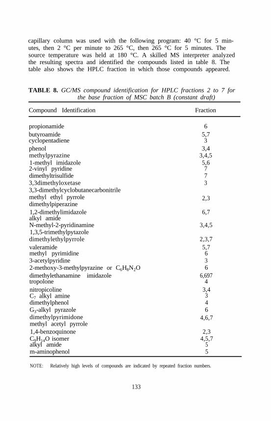
capillary column was used with the following program: 40 °C for 5 min-utes, then 2 °C per minute to 265 °C, then 265 °C for 5 minutes. Thesource temperature was held at 180 °C. A skilled MS interpreter analyzedthe resulting spectra and identified the compounds listed in table 8. Thetable also shows the HPLC fraction in which those compounds appeared.
TABLE 8. GC/MS compound identification for HPLC fractions 2 to 7 forthe base fraction of MSC batch B (constant draft)
Compound Identification Fraction
propionamidebutyroamidecyclopentadienephenolmethylpyrazine1-methyl imidazole2-vinyl pyridinedimethyltrisulfide3,3dimethyloxetase3,3-dimethylcyclobutanecarbonitrilemethyl ethyl pyrroledimethylpiperazine1,2-dimethylimidazolealkyl amideN-methyl-2-pyridinamine1,3,5-trimethylpytazoledimethylethylpyrrolevaleramidemethyl pyrimidine3-acetylpyridine2-methoxy-3-methylpyrazine or C6H8N2Odimethylethanamine imidazoletropolonenitropicolineC7 alkyl aminedimethylphenolG3-alkyl pyrazoledimethylpyrimidonemethyl acetyl pyrrole1,4-benzoquinoneC9H14O isomeralkyl amidem-aminophenol
65,73
3,43,4,55,6773
2,3
6,7
3,4,5
2,3,75,7636
6,6974
3,4346
4,6,7
2,34,5,7
55
NOTE: Relatively high levels of compounds are indicated by repeated fraction numbers.
133
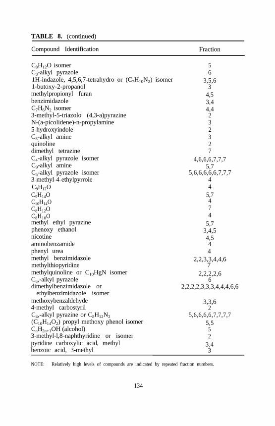
TABLE 8. (continued)
Compound Identification Fraction
C8H12O isomerC3-alkyl pyrazole1H-indazole, 4,5,6,7-tetrahydro or (C7H10N2) isomer1-butoxy-2-propanolmethylpropionyl furanbenzimidazoleC7H6N2 isomer3-methyl-5-triazolo (4,3-a)pyrazineN-(a-picolidene)-n-propylamine5-hydroxyindoleC8-alkyl aminequinolinedimethyl tetrazineC4-alkyl pyrazole isomerC9-alkyl amineC5-alkyl pyrazole isomer3-methyl-4-ethylpyrroleC9H12OC9H14OC10H14OC8H12OC8H10Omethyl ethyl pyrazinephenoxy ethanolnicotineaminobenzamidephenyl ureamethyl benzimidazolemethylthiopyridinemethylquinoline or C10HgN isomerC6,-alkyl pyrazoledimethylbenzimidazole or
ethylbenzimidazole isomermethoxybenzaldehyde4-methyl carbostyrilC4,-alkyl pyrazine or C8H12N2(C10H14O2) propyl methoxy phenol isomerCnH2n+1OH (alcohol)3-methyl-l,8-naphthyridine or isomerpyridine carboxylic acid, methylbenzoic acid, 3-methyl
56
3,5,63
4,53,44,4232327
4,6,6,6,7,7,75,7
5,6,6,6,6,6,7,7,744
5,7474
5,73,4,54,544
2,2,3,3,4,4,67
2,2,2,2,66
2,2,2,2,3,3,3,4,4,4,6,6
3,3,62
5,6,6,6,6,7,7,7,75,552
3,43
NOTE: Relatively high levels of compounds are indicated by repeated fraction numbers.
134
TABLE 8. (continued)
Compound Identification Fraction
phenyl pyrazoline3,4-dimethylbenzoic acidbenzyl acetateC1 0H1 6O1,2-dihydro-3-isobutyl-l-methylpyrazin-2-oneethyl hydroxy acetophenone2,4-dimethylquinazolinephenyl methyl ureaphenyl pyridinepropylbenzimidazoleaminoquinoline or C9H8N2,dimethyl naphthyridineN-phenyl acrylamidemethoxy propyl pyrazinephenyl alcoholethoxy benzaldehydetolyl azidephenyl methyl quanidineC6,-alkyl phenolC3,-alkyl benzimidazole1-decanolC5,-alkyl pyrazinealkyl amidedimethylbenzimidazone isomertrimethyl-2-oxo-1,2,3,4-tetrahydropyrimidinedimethoxybenzene isomeraminodimethylpyrimidinehydroxymethylquinolinemethylbenzoxazoletert-butyl-parahydroxybenzoateC10H1202 (ester)methyl-n(pyrid-2-yl)dihydropyrroleC12H18OmethylaminonaphthyridinediphenylamineC9H10O3tert-butylparahydroxybenzoateethoxyquinazoline or isomerdiethylphenylene diamineC5H5N5 isomerN,N-dimethyl-N-(p-methoxyphenyl) formamide
4347
3,54
2,33
5 5 34343
4,53
4,535
3,44
4,5,6,77
5,6,657577
7,7473
3,43
2,2,2,34,5
NOTE: Relatively high levels of compounds are indicated by repeated fraction numbers.
135
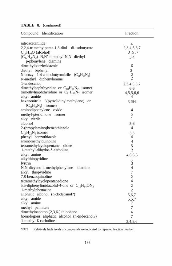
TABLE 8. (continued)
Compound Identification Fraction
nitroacetanilide 42,2,4-trimethylpenta-1,3-diol di-isobutyrate 2,3,4,5,6,7C11H16O (alcohol) 3 , 5 , 7(C12H20N2) N,N’-dimethyl-N,N’-diethyl- 3,4
p-phenylene diaminedimethylbenzimidazole 6diethyl biphenyl 2N-benzy 1-4-aminobutyronitrile (C11H14N2) 2N-methyl diphenylamine 21-undecanol 2,3,4,5,6,7dimethylnaphthyridine or C10H10N2, isomer 6,6trimethylnaphthyridine or C11H12N2 isomer 4,5,5,6,6alkyl amide 4hexanenitrile 3(pyrrolidinylmethylene) or 3,494
(C11H18N2) isomersaminodiphenylene oxide 4methyl-pteridinone isomer 5alkyl nitrile 4alcohol 5,62-(propylamino)benzothiazole 4C13H22N2 isomer 3,3phenyl benzothiazole 4aminomethylquinoline 4tetramethylcyclopentane dione 51-methyl-dihydro-ß-carboline 2alkyl amine 4,6,6,6alkylthiopyridine 6lystrin 3N,N-dicyano-4-methylphenylene diamine 4alkyl thiopyridine 77,8-benzoquinoline 2tetramethylcyclopentanedione 45,5-diphenylimidazolid-4-one or C15H14ON2 21-methylphenazine 2aliphatic alcohol (n-dodecanol?) 5,6,7alkyl amide 5,5,7alkyl amine 7methyl palmitate 7dimethylnaphtho (2,3,6-) thiophene 4homologous aliphatic alcohol (n-tridecanol?) 71-methyl-ß-carboline 3,4,5,6
NOTE: Relatively high levels of compounds are indicated by repeated fraction number.
136

TABLE 8. (continued)
Compound Identification Fraction
ß-carbolinep-cumyl phenoldibutyl phthalatebenzyl acetophenonehomologous aliphatic alcohol (n-tetradecanol?)diphenylpyridine isomeralkyl esterdihydroxymethyl phenyl quinazolineditolyl ethane1-azido naphthalene1-phenyl decanedimethyl-ß-carboline isomeralkyl amidephenylbenzimidazole2,6-diterbutylnaphthalene or isomerC14H10O3 isomerhomologous aliphatic alcohol (n-tetradecanol?)methylthiazolopyrimidine8-acetoxy-pyrazolobenzo-as-triazine or C11H8N2O4methyl stearatemethyl phenyl cinnoline or C15H12N2, isomer2-thiocyanatodiphenylaminemethylpyriloindolealcohol (n-pentadecanol?)naptho-sydinonen -hexadecanoln-C22H46 (docosane)alkyl amineC12H10N2O4 isomermethylphenyl quinoxaline or C15H12N2n-C23H48, tricosanehomologous aliphatic alcohol (n-heptadecanol?)n-C24H50 (tetracosane)DL-cannabichromenen-C25H52 (pentacosane)3-n-pentyl-delta-9-tetrahydrocannabinoldioctyl phthalaten-C26H54 (hexacosane)3-n-pentyl cannabinoln-C27H56 (heptacosane)alkyl amide
5,6,73
2,3,73
6,7224366
5,5,6633
3,43,5,6,7
2,32,3,63,7344
5,666
3,5,75,7
3,3,43
3,4,5,6,75
3,4,5,6,72,3
3,4,5,6,72,3,7
3,4,5,6,74,5,7
2,3,4,6,74,5,6,7
3,4,5,6,7
NOTE: Relatively high levels of compounds are indicated by repeated fraction numbers.
137

TABLE 8. (continued)
Compound Identification Fraction
n-C28H58 (octacosane)saturated hydrocarbonn-C29H60 (nonacosane)alkyl phthalatesaturated hydrocarbonsn-C 3 0 H 6 2
3,4,5,75,5
2,5,76
5,5,64,5,6
NOTE: Relatively high levels of compounds are indicated by repeated fraction numbers.
CONCLUSION
Bioassay results showed that (1) the crude fractions were weakly mutagenic,(2) the acidic fractions were not significantly mutagenic, (3) the neutralfractions were weakly mutagenic, (4) the base fractions were significantlymutagenic, (5) the marijuana high-dose base fraction was sevenfold moremutagenic than either the tobacco or the low-dose marijuana base fraction,(6) constant draft base fractions were more mutagenic than puff mode basicfractions for both marijuana and tobacco, and (7) the more polar subfrac-tions (numbers 4 to 7) of the base fraction were more mutagenic than theless polar subfractions.
Thermogravimetric analysis was performed on the base fractions of thecrude smoke condensates to provide an indication of the applicability of GCor GC/MS analysis to these fractions. Most of the material was found tovolatilize at temperatures below 300 “C, indicating GC as an appropriatetechnique for analysis of these fractions.
Elemental analysis of the crude smoke condensates for carbon, hydrogen,nitrogen, sulfur, and oxygen revealed no significant differences between thevarious condensates. The carbon/hydrogen ratios indicated that the crudesamples were “nonaromatic.”
Approximately 200 compounds were tentatively identified by mass spec-trometry. Of this total, about half were amines, with about half of thesebeing aromatic amines. Pyrazines, pyrimidines, pyrroles, pyridines, andisoxazoles were the predominant compound classes. Some alkylated pyra-zoles and pyrazines were detected in very large amounts, as was an alkyl-ated benzimidazole. Some THC derivatives were also detected. It shouldbe noted that the mutagenicity of the vast majority of these compounds hasnot been determined.
138
REFERENCES
Burstein, S.; Levin, E.; and VaranelIi, C. Prostaglandins and cannabis—II.Inhibition of biosynthesis by the naturally occurring cannabinoids. Bio-chem Pharmacol 22:2905-2910, 1973.
Burstein, S.; Varanelli, C.; and Slade, L Prostaglandins and cannabis—III.Inhibition of biosynthesis by essential oil components of marijuana. Bio-chem Pharmacol 24:1053-1054, 3975.
Busch, F.; Said, D.; and Wi, E. Mutagenic activity of marijuana smokecondensate. Cancer Lett 6:319-324, 1979.
Davis, K.H.; McDaniel, I.A.; Caddell, L.W.; and Moody, P.L. Some smok-ing characteristics of marijuana cigarettes. In: Willett, R.; Agurell, S.;and Dewey, W., eds. The Cannabinoids: Chemical, Pharmacologic andTherapeutic Aspects. Orlando, FL: Academic Press, 1984. pp. 97-109.
Gudzinowicz, B., and Gudzinowicz, M. Analysis of Drugs and Metabolitesby Gas Chromatography/Mass Spectrometry. Vol. 7. Natural, Pyrolyticand Metabolic Products of Tobacco and Marijuana. New York: MarcelDekker, 1980.
Lee, M.; Novotny, M.; and Battle, K. Gas chromatography/mass spectrome-try and nuclear magnetic resonance spectrometric studies of carcinogenicpolycyclic aromatic hydrocarbons in tobacco and marijuana smoke con-densate. Anal Chem 48:405-416, 1976.
Leuchtenbeyer, C.; Leuchtenbeyer, R.; Ritter, V.; and Inui, N. Effects ofmarijuana and tobacco smoke on DNA and chromosomal complement inhuman lung explants. Nature (London) 242:403-404, 1973.
Maron, D., and Ames, B. Revised methods for the Salmonella mutagenicitytest. Mutat Res 113:173-215, 1983.
Maskarinec, M.; Alexander, G.; and Novotny, M. Analysis of the acidicfraction of marijuana smoke condensate by capillary gas chromatogra-phy/mass spectrometry. J Chromatogr 126:559-568, 1976.
Merli, F.; Novotny, M.; and Lee, M. Fractionation and gas chromatogra-phic analysis of aza-arenas in complex mixtures, J Chromatogr 199:371-378, 1980.
Merli, F.; Wiester, D.; Maskarinec, M.; Novotny, M.; Vassilanos, D.; andLee, M. Characterization of the basic fraction of marijuana smoke con-densate by capillary gas chromatography/mass spectrometry. Anal Chem53:1929-1935, 1981.
Myers, L.; Sexton, N.; Southerland, L.; and Wolff, T. Regression analysison Ames test data. Environ Mutagen 3:575-586, 1981.
Novotny, M.; Kump, R.; Merli, F.; and Todd, L Capillary gas chromato-graphy/mass spectrometric determination of nitrogen aromatic compoundsin complex mixtures. Chem 52:401-406, 1980.
Ogg, C. Determination of particulate matter and alkaloids (as nicotine) incigarette smoke. JAOAC 47:357-362, 1964.
139
Petersen, B.; Nishioka, M.; and Chung, C. Nitroaromatics in air particulateextract. Presented at the 3rd Biannual Symposium on Application ofShort Term Bioassays to Complex Environmental Mixtures, Chapel Hill,NC, January 1982.
Peterson, R. Importance of inhalation patterns in determining effects ofmarihuana use. Lancet March:727-728, 1979.
Wehner, F.; VanRensburg, S.; and Thiel, P. Mutagenicity of marijuana andTranskei tobacco smoke condensate in the Salmonella/microsome assay.Mutat Res 77:135-142, 1980.
ACKNOWLEDGMENT
This work was supported by grant R01 DA 03417 from the NationalInstitute on Drug Abuse.
AUTHORS
Charles M. Sparacino, Ph.D.Assistant DirectorAnalytical and Chemical Sciences
Patricia A. Hyldburg, Ph.D.Research Analytical ChemistAnalytical and Chemical Sciences
Thomas J. Hughes, MS.Senior Research Genetic ToxicologistChemistry and Life Sciences
Research Triangle Institute3040 Comwallis RoadResearch Triangle Park, NC 27789
140

Pyrolysis and Inhalation StudiesWith Phencyclidine and CocaineBilly R. Martin and Joseph Boni
INTRODUCTION
Smoking has been the major mode of use for many substances (includingtobacco and marijuana) for centuries. In the past decade, the list of sub-stances has been enlarged to include phencyclidine (PCP) and cocaine freebase. A particularly important reason for the surge in smoking of thesesubstances is the rapid absorption that occurs with inhalation, which resultsin a rapid onset of behavioral effects that are deemed pleasurable. In thecase of PCP, it has been suggested that this rapid onset of effects allowsthe user to titrate his drug and therefore minimize serious side effects.However, smoking or inhalation of abused substances creates a spectrum ofproblems from minor behavioral problems to death. Despite the fact thatmany substances are abused either by smoking or inhalation, they are fre-quently administered by other routes of administration when they are char-acterized pharmacologically. Obviously, smoking or inhalation studies arefraught with difficulties that can be avoided by simply injecting the drug.The question then arises as to what factors must be considered when ex-trapolating from the drug injection studies to the human abuse situation.
It is essential to understand the fate of abused substances during the smok-ing process as well as the absorption, biodisposition, and metabolism ofthese substances by the inhalation route. Moreover, we must be able todetermine whether the pharmacological effects of the drugs are quantitative-ly and qualitatively similar after different routes of administration. Ourstrategy has been to develop an apparatus and procedures to (1) producesmoke or drug vapor in a way that mimics the drug abuser’s behavior, and(2) expose animals to this smoke or vapor in sufficient quantities to producea pharmacological effect. Initially, we determine the extent of pyrolysis thatoccurs and identify the pyrolysis products. Then, conditions are establishedfor exposing animals to the smoke or vapor, followed by biodispositionstudies of the parent compound and pyrolysis products. Finally, behavioralstudies are conducted to assess the correlation with the biodisposition
141
studies. Difficulties that are unique for each drug arise when conductinginhalation and smoking studies, although some of the problems are commonto all drug studies. One common difficulty is establishing dosimetry ininhalation and smoking experiments. This difficulty can be overcome byemploying radiolabeled drug and subsequent quantitation of total body radio-activity in the animal after drug exposure.
PYROLYSIS OF PHENCYCLIDINE
PCP has been abused by several routes of administration, which includesmoking, snorting, oral ingestion, and intravenous (IV) injection. However,it is thought that smoking became very popular because this route permitteda precise control of dosage and thus reduced the probability of overdose.PCP is frequently applied to parsley or marijuana dry leaf material to besmoked, possibly the most common method of abuse. The quantity of drugpresent in mainstream smoke and pyrolysis products is an important factorin determining pharmacological effects. That PCP was heat labile wasshown by the earlier work of Lin and colleagues (1975), who reported theformation of 1-phenyl-1-cyclohexene when PCP was analyzed by gas-liquidchromatography. Given the fact that the tip of a burning cigarette canreach temperatures of 600 it °C was predicted that PCP would be exten-sively pyrolyzed when smoked.
To investigate the pyrolysis of PCP, an alcoholic solution of 3H-PCP-HCI(radiolabeled in the phenyl ring) was injected into cigarettes made of com-mercially processed parsley leaves, along the longitudinal axis (Freeman andMartin 1981). The ethanol was allowed to evaporate by air drying at roomtemperature. The cigarettes, which contained 50 mg of 3H-PCP-HCl inmost experiments, were smoked in an apparatus designed to mimic humansmoking conditions. The apparatus, depicted in figure 1, consisted of acigarette holder connected to a series of traps to collect the constituents ofthe mainstream smoke. Air was drawn through the entire apparatus by awater aspirator or vacuum pump. In addition, puffing was simulated by avalve that opened and closed the system at designated intervals. Air flowedonly when the valve was closed. An air flow regulator, located betweenthe last trap and the switching valve, was set to deliver 45 ml of smokeduring a 7-second puff duration. At the end of the pyrolysis experiment,the entire apparatus was disassembled and washed with methanol. Radioactivity was quantitated in these washes of the apparatus and glass wooltrap, as well as the liquid traps. A cigarette requires approximately 6 to8 minutes to bum under these conditions. The major portion of radio-activity was collected on the glass wool trap, and only trace quantities ofradioactivity were found in the cigarette butt and the remainder of theapparatus. The glass wool trap has been found to be the most effectivemeans of collection of PCP and other drug vapors, including the organo-phosphates (Scimeca et al. 1985) and cocaine. Using liquid nitrogen trapsand bubbling the vapor or smoke through methanol traps proved to be less
142

effective. Analysis of the glass wool traps by thin-layer chromatographyand gas chromatography/mass spectrometry (GC/MS) revealed that the PCPwas readily converted to 1-phenyl-1-cyclohexene (PC). Actually, compar-able quantities of PCP and PC were delivered in mainstream smoke, andlittle material was lost in the side-stream smoke (Freeman and Martin1981). It was assumed that each mole of PCP that was cleaved to form PCresulted in the formation of an equal quantity of piperidine. However, thevolatile nature of piperidine and the fact that this portion of the PCPmolecule was not radiolabeled made quantitation difficult. Therefore,studies were conducted with 3H-PCP-HCl that was radiolabeled in thepiperidine ring. Analysis by both thin-layer chromatography and GC/MS ofthe glass wool extracts revealed that only 6 percent of the starting materialwas recovered as 3H-pipetidine. As in the previous experiments, 3H-PCPand PC accounted for the major portion of the material, while almost30 percent of the starting material could not be identified (Martin andFreeman 1983).
FIGURE 1. First generation PCP pyrolysis apparatus
One of the important questions was whether there was a constant deliveryof PCP during the smoking of a cigarette through which PCP was evenlydistributed. To assess the rate of delivery, the glass wool traps werechanged as each third of the cigarette was smoked. It was clear from thesestudies that the major portion of the PCP was delivered during the smokingof the last third of the cigarette, which suggested that the PCP wascondensing in the cigarette (Freeman and Martin 1981). Although weverified that the burning tip of the cigarettes reached temperatures in excessof 600 °C (Lue et al. 1986), it was thought that PCP was most likely beingvaporized before the burning tip of the cigarette reached it (figure 2). Cooket al. (1985) had demonstrated that PCP was completely destroyed when
143
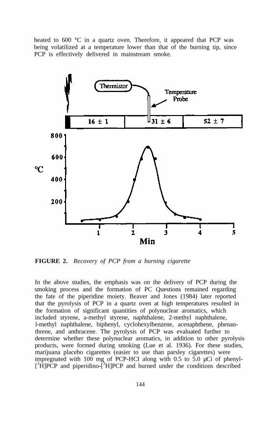
heated to 600 °C in a quartz oven. Therefore, it appeared that PCP wasbeing volatilized at a temperature lower than that of the burning tip, sincePCP is effectively delivered in mainstream smoke.
FIGURE 2. Recovery of PCP from a burning cigarette
In the above studies, the emphasis was on the delivery of PCP during thesmoking process and the formation of PC Questions remained regardingthe fate of the piperidine moiety. Beaver and Jones (1984) later reportedthat the pyrolysis of PCP in a quartz oven at high temperatures resulted inthe formation of significant quantities of polynuclear aromatics, whichincluded styrene, a-methyl styrene, naphthalene, 2-methyl naphthalene,l-methyl naphthalene, biphenyl, cyclohexylbenzene, acenaphthene, phenan-threne, and anthracene. The pyrolysis of PCP was evaluated further todetermine whether these polynuclear aromatics, in addition to other pyrolysisproducts, were formed during smoking (Lue et al. 1936). For these studies,marijuana placebo cigarettes (easier to use than parsley cigarettes) wereimpregnated with 100 mg of PCP-HCI along with 0.5 to 5.0 µCi of phenyl-[3H]PCP and piperidino-[3H]PCP and burned under the conditions described
144
above. Mainstream smoke was passed through glass wool filters as well asthrough acidic and basic traps. Greater than 90 percent of the startingmaterial could be accounted for, with the major portion (77 percent) in thefirst glass wool trap.
Methanolic extracts of the glass wool traps were analyzed by both highpressure liquid chromatography (HPLC) and GC/MS. For quantitativeGC/MS analysis, concentration curves were obtained for 1, 5, 7.5, and10 ng of each polynuclear aromatic standard using single-ion monitoringanalysis. The limit of detectability for each of the standards was 0.5 ng perinjection or 1 ug/10 ml of methanol extract. Because of the possibility thatthe polynuclear aromatics could be formed during smoking but might not becollected on the glass wool filters, control experiments were conducted inwhich the marijuana placebo cigarettes were treated with 4.8 mg of3H-PCP-HCl with and without 0.1 mg of each of the polynuclear aromaticstandards. The methanol extracts of the glass wool filter condensates fromthese cigarettes were analyzed by GC/MS using single-ion monitoring. Therecoveries of the polynuclear aromatic standards ranged from 33 to 100 per-cent. None of the polynuclear aromatics was detected in the condensatesfrom cigarettes impregnated with 3H-PCP-HCl alone. It appears that thesecompounds are formed from PCP only under conditions that do not mimicsmoking.
In addition to single-ion monitoring analysis, total ion chromatograms werealso obtained for all of the methanol extracts. Four peaks were observed inthe condensates from the 3H-PCP-HCl-spiked cigarettes that were not evi-dent in those from the placebo cigarettes. The spectra and retention timesof these compounds were coincident with those of PCP, l-phenyl-l-cyclo-hexene, piperidine, and N-acetylpiperidine, which represented 39, 46, 7, and9 percent, respectively, of the starting material (figure 3). These resultssuggest that PCP is not exposed to temperatures during smoking that arehigh enough to produce polynuclear aromatics.
PYROLYSIS OF 1-(1-PIPERIDINO) CYCLOHEXANECARBONITRILE(PCC)
PCC is a major contaminant in illicit PCP because the most common clan-destine synthesis of PCP employs PCC as the intermediate; the PCC is notalways removed from the final product (Helisten and Shulgin 19776; Gileset al. 1977). The presence of PCC in PCP represents a potential health riskgiven the fact that it is considerably more toxic than PCP (Baker 1982).PCC is readily degraded to 1-(1-piperidino)-l-cyclohexene, piperidine, andcyclohexanone during either GC or thin-layer chromatogmphic (TLC) analy-sis or on standing in solution (Helisten and Shulgin 1976; Giles et al.1977). As the stability of PCC during smoking had not been investigated,despite the potential contribution its pyrolysis products may make to thepharmacology and toxicology of illicit PCP, a study was conducted to
145

FIGURE 3. Pyrolytic fate of PCP during smoking
determine the quantity of PCC that is delivered in mainstream smoke and toidentify and quantify pyrolysis products (Lue et al. 1988).
[3H]Piperidino-[14C]cyano-PCC was synthesized to facilitate the quantitationof PCC in mainstream smoke and the identification of pyrolysis products.Marijuana placebo cigarettes were impregnated with this double-labeled PCCand smoked with the apparatus depicted in figure 1. The mainstreamsmoke was passed through the glass wool, acidic, and basic traps. Approxi-mately 75 percent of the tritium was collected in these traps, while 46, 11,and 5 percent of the starting 14C was found in the glass wool, H2SO4 andNaOH traps, respectively. The constituents of the glass wool trap wereanalyzed by GC/MS, which revealed the presence of l-(l-piperidino)-1-cyclohexene, PCC, piperidine, and N-acetylpiperidine. Cyanide ion wasdetected in the glass wool, acidic, and basic traps by a titration procedure.It appeared that approximately 47 percent of the PCC was delivered intactin mainstream smoke. Approximately 58 percent was cleaved to form cya-nide and 1-(1-piperidino)-1-cyclohexene. The latter was further brokendown to cyclohexanone (representing 21 percent of the starting material),piperidine (29 percent), and N-acetylpiperidine (7 percent); about 2 percentremained intact. The entire pyrolytic profile is shown in figure 4.
146

Cyclohexanone Piperidine N-Acetylpiperidine
FIGURE 4. Pyrolytic fate of PCC during smoking
PYROLYSIS OF COCAINE
Although the abuse of cocaine has generated considerable attention in thepast few years, cocaine has been used for religious, medicinal, and recrea-tional purposes for thousands of years. Inhalation of burning coca leaves orsmoking coca evolved gradually and remained major modes of medicinaland recreational use until enactment of the Pure Food and Drug Ad of1906 and the Harrison Act of 1914, which curtailed the distribution and useof coca leaves as well as coca products. It is ironic that these laws prob-ably contributed to the diversion of cocaine use from coca smoking to intra-nasal and IV administration of cocaine-HC1, because the latter productbecame more readily available. Cocaine-HCl has been smoked with mari-juana, tobacco, or herbs either as a cigarette or in a pipe. The relativelymodest effects that were derived from smoking the salt soon led to smokingcoca paste (crude extract containing free alkaloids) and finally to “freebase,” which is cocaine-HCl that has been converted to the free base (Jeriet al. 1978; Siegel 1985). The physical properties of the different prepara-tions have dictated their mode of use. Cocaine-HCl has a melting point of195 °C, is not very volatile, and decomposes with heat, but it is highlywater soluble, therefore readily absorbed via the mucous membranes. Con-versely, cocaine free base has a melting point of 95 °C making it readilyvolatile. As a result of these properties, cocaine-HCl is usually adminis-tered IV or intranasally, whereas cocaine free base is volatilized and
147
absorbed through the large surface area of the lungs after inhalation. Oneof the greatest problems facing us, as summarized by Siegel (1985), is theemergence of new patterns of use, such as the inhalation or smoking of freebase, which has increased the risks of dependency and toxicity.
The toxicity of cocaine depends upon several factors: the route of adminis-tration, dose, drug combinations, etc. (Cohen 1985). The general features ofacute cocaine intoxication include extensive central nervous system (CNS)stimulation progressing to convulsions followed by cardiovascular and respi-ratory failure (Jones 1984). Although cocaine has long been considered apotentially dangerous drug of abuse, the popularity of free basing has led tothe realization of these dangers. The recent upsurge of “crack” has in-creased the availability and decreased the cost of cocaine. Additionallycatastrophic is the fact that “crack” is a rapidly addicting form of cocainethat easily results in lethal overdose. At present, the underlying factors forthis increased toxicity resulting from free basing are unclear. The influenceof route of administration on addictive potential and lethality of cocaineundoubtedly involves pharmacokinetic differences. However, there may beother contributory factors. For example, Pearman (1979) suggested that fac-tors that increase the absorption of cocaine may be more important consid-erations for toxicity than simply overdose. Several investigators have foundthat the bioavailability and intensity of many cocaine effects are similarafter oral and nasal cocaine (Wilkinson et al. 1980; Van Dyke et al. 1978;Jones 1984), while the profiles of cocaine effects are similar after IV andsmoked routes. Bioavailability by the nasal route was found to be 30 to40 percent (Jones 1984) and 80 percent (Cook et al. 1985) of that after IV.Biodegradation of cocaine (through oral (PO), IV, and intranasal routes) hasbeen reported to form benzoylecgonine and ecgonine methyl ester (methyl-ecgonine) (Lukaszewski and Jeffery 1980; Chinn et al. 1980; Wallace et al.19776; Wallace et al. 1977; Jindal and Vestergaard 1978).
Although previously published studies have dealt with the pharmacologicaland pharmacodynamic properties of cocaine abuse, little attention has beenpaid to the volatilized products of free basing cocaine or crack (Gawin1980; Isner et al. 1986; Novak and Salemink 1984). Some of the importantaspects of cocaine inhalation include the purity of street cocaine, impreci-sion of dosing by variability in volatilization and pyrolysis of cocaine,smoking technique, rate of inhalation, naive vs. chronic use, target tissue,and underlying pathology. Previous studies have shown that cocaine can bethermally degraded to benzoic acid and methylecgonidine (Cook and Jeff-coat 1986; Cook et al. 1985) as well as methyl-4-(3-pytidinyl)butyrate(Novak and Salemink 1984). The species and quantities of volatilized prod-ucts of cocaine formed under conditions simulating free basing have notbeen thoroughly investigated, however, despite their pharmacological andtoxicological importance. Therefore, the present study was conducted toidentity and quantify the delivery of cocaine and to investigate the forma-tion of pyrolysis products during volatilization (Martin et al. 1989).
148

Protocol
The method of volatilizing 3Hcocaine (levo-(-)-[benzoyl-3,4-3H(N)]-cocaine)represented a modification of the procedure described above for PCP. AU-shaped pipe was constructed from glass tubing (6 mm inside diameterand 24 cm in length) and placed in a Lindberg Crucible Furnace filled withglass beads, as depicted in the bottom of figure 5. After the pipe hadreached the desired temperature, 50 mg of cocaine base containing 1 uCi of[3H-benzoyl]-cocaine was inserted into the pipe with the cocaine deliverysystem. Cocaine was placed between two glass wool plugs in a glass tube,which was inserted into the glass pipe at the start of the experiment. Thecocaine and glass wool plugs were pushed out of the tubing and into thepipe. Vapor was drawn through the volatilization apparatus at 8-secondpuff intervals with intermittent 4-second rest periods (no flow). Cocaineand its pyrolysis products were recovered from the pipe with ethanol(10 ml), and they were extracted from the glass wool with three l0-ml por-tions of dichloromethane. The pH of the acid trap was adjusted to approxi-mately 8.5 using sodium bicarbonate prior to extraction with three l0-mlportions of dichloromethane. The pH of the alkaline trap was adjusted to4.5 to 5.0 prior to extraction with three l0-ml portions of dichloromethane.The multiple extractions were combined and concentrated to 10 ml under agentle stream of nitrogen at room temperature. In some experiments, thevapor was sampled repetitively using l-ml air-tight syringes. The vaporwas added to ethanol, and the ethanol was analyzed by liquid scintillationspectrometry and by GC/MS.
Air Flow Rate
The rate of air flow through the system had a dramatic effect on volatiliza-tion of the cocaine. At a flow rate of 100 ml per minute, only 7 percentof the starting material was recovered in the glass wool trap, and only tracequantities were found in the other traps after a 5-minute volatilizationat 260 °C temperature. When the flow rate was increased to 400 ml perminute, approximately 40 percent of the radioactivity remained in the pipeand approximately 40 percent was recovered in the glass wool trap. Aswith the lower flow rate, only trace quantities of radioactivity wereobserved in the remaining traps. The contents in the pipe, glass wool, andtraps were analyzed by mass spectrometry either by acquiring the total ionchromatogram or by selected-ion monitoring. Both procedures revealed thepresence of only cocaine, methylecgonidine, and benzoic acid (figure 6).There was considerable pyrolysis of cocaine at the low flow rate, so onlysmall quantities of cocaine remained in the pipe and in the glass wool trap.The major component was methylecgonidine in both the pipe and the glasswool. At the higher flow rate of 400 ml per minute, there was little pyrol-ysis, as evidenced by the fact that high concentrations of cocaine werefound in the pipe as well as in the glass wool trap.
149

FIGURE 5. Smoking-exposure apparatus for PCP and cocaine
Temperature
The effect of temperature on pyrolysis was examined by maintaining theflow rate of 400 ml per minute during a IO-minute volatilization. Increas-ing the temperature from 260 °C to 400 °C produced a linear increase inthe formation of methylecgonidine and benzoic acid. In the temperaturerange of 350 to 400 °C, approximately 50 percent of the cocaine was degraded, whereas only trace quantities of cocaine remained intact at 650 °C
150

B e n z o i c a c i d M e t h y l e c g o n i d i n e
FIGURE 6. Pyrolytic profile of cocaine
Because pyrolysis and volatilization of cocaine are highly dependent uponthe experimental condition, studies were conducted to determine optimalconditions for delivery of a constant concentration of cocaine for subsequentanimal studies. The results, shown in figure 7, reveal the time courseof radioactivity in the air stream as the air flow rate is altered. Most of theradioactivity is delivered in the first minute at a flow rate of 400 ml perminute, whereas the delivery period is extended to approximately 3 minuteswhen the flow rate is reduced to 100 ml per minute. In a separate seriesof experiments, the flow rate was held constant at 400 ml per minute, andthe temperatures were varied between 200 and 260 °C. A relatively con-stant cocaine delivery of 5 minutes duration was obtained at 200 °C. Athigher temperatures, the duration of a plateau phase was shortened to thepoint where maximal cocaine delivery was observed within a l-minute timeframe (figure 8).
151
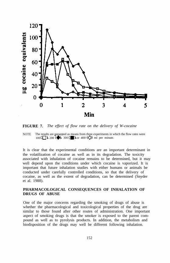
FIGURE 7. The effect of flow rate on the delivery of W-cocaine
NOTE The results are presented as means from three experiments in which the flow rates were100 200 300 or 400 ml per minute.
It is clear that the experimental conditions are an important determinant inthe volatilization of cocaine as well as in its degradation. The toxicityassociated with inhalation of cocaine remains to be determined, but it maywell depend upon the conditions under which cocaine is vaporized. It isimportant that future inhalation studies with either humans or animals beconducted under carefully controlled conditions, so that the delivery ofcocaine, as well as the extent of degradation, can be determined (Snyderet al. 1988).
PHARMACOLOGICAL CONSEQUENCES OF INHALATION OFDRUGS OF ABUSE
One of the major concerns regarding the smoking of drugs of abuse iswhether the pharmacological and toxicological properties of the drug aresimilar to those found after other routes of administration. One importantaspect of smoking drugs is that the smoker is exposed to the parent com-pound as well as to pyrolysis products. In addition, the metabolism andbiodisposition of the drugs may well be different following inhalation.
152

120
100
80
6 0
4 0
2 0
0
FIGURE 8. The effect of temperature on the delivery of 3H-cocaine
NOTE: The results are presented as means from three experiments in which the themperatures were2 0 0 2 2 0 2 4 0 o r 2 6 0 ° C .
Studies conducted in our laboratories have shown that 1-3H-pheny1-1-cyclohexene (3H-PC), the major pyrolysis product of PCP, is metabolizedextensively when incubated in vitro with crude microsomal membranes frommouse livers (Martin et al. 1982). The major routes of metabolism wereallylic hydroxylation, oxidation of the allytic alcohol, and epoxidation-hydrolysis. The following metabolites were identified: l-phenyl-1-cyclohexen-3-o1 (major metabolite), 1-phenyl-1-cyclohexen-3-one (PC-3-one) (major), 1-phenyl-1-cyclohexen-6-o1 (minor), l-phenyl-l-cyclohexen-6-one (minor), and l-phenylcyclohexane-1,2-diol (PC-1,2-diol) (minor).Pharmacological evaluation of PC clearly showed that it produced less be-havioral effect and was less toxic than PCP (Holsapple et al. 1982). TheED, and confidence limits for PCP in the inverted-screen test were 2.5 (1.6to 2.9) umo1/kg, with a duration of action of approximately 30 minutes afterIV administration. PC’s effects lasted less than 5 minutes after IV adminis-tration, and the ED, (confidence limits) was 325 (294 to 360) µmo1/kg.The behavioral activity of PC-3-ol, PC-3-one, and PC-1,2-diol (PC-6-ol andPC-6-one were not synthesized in quantities sufficient for testing) also lastedless than 5 minutes (Martin et al. 1982). The diol was considerably lessactive than PC, and PC-3-o1 and PC-3-one were somewhat mote active than
153

PC. Nonetheless, all the metabolites were considerably less active than PCPand undoubtedly contributed little to the latter’s behavioral activity.
To determine whether the behavioral properties of the drug are the sameafter smoking and other routes of administration, studies were carried out inour laboratories. Male Sprague-Dawley rats were trained to discriminateintraperitoneal (IP) injections of 3.0 mg/kg PCP from saline under a 2-leverfixed-ratio 32 schedule of food presentation (Wessinger et al. 1985). Afterreliable discriminative control of lever choice was established, a dose-dependent increase in PCP-lever selection and dose-dependent decreases inrates of responding were determined after IP injections. When doses ofPCP were administered by exposure to smoke from cigarettes containingPCP, a dose-dependent increase in PCP-lever responding was also observed.
Tetrahydrocannabinol (THC) administered via smoke exposure, up todoses that markedly suppressed response rates, did not result in PCP-appropriate responding, demonstrating the specificity of the PCP stimulus bythe inhalation route. Brain levels and distribution of 3H-PCP were deter-mined in rats administered doses calculated to result in 50 percent generali-zation by the IP injection or smoke inhalation route. By both routes ofadministration, roughly equivalent brain levels were attained, and the distri-bution was relatively even across the seven brain areas analyzed.
CO-ABUSE OF MARIJUANA AND PCP
There is also the possibility of drug interactions by the smoking route. Oneobvious interaction is PCP and -THC, since PCP is sometimes smoked inmarijuana cigarettes. The behavioral and pharmacological interactions between THC and PCP were studied following coadministration of thedrugs in smoke to mice (Freeman and Martin 1983). Although THC(25, 50, or 100 mg per cigarette) had little effect on spontaneous motoractivity, all doses attenuated the hyperactivity elicited by PCP•HCL (25 and50 mg per cigarette). THC produced a dose-related hypothermia, where-as PCP•HCL (50 mg per cigarette) had no effect on body temperature.However, PCP enhanced the hypothermia produced by smoke from ciga-rettes containing 25 mg of THC. THC (100 mg per cigarette) had noeffect on the biodisposition of 3H-PCP and its pyrolytic product, 3H-phenyl-cyclohexene (3H-PC), when examined immediately after 3H-PCP•HCl (50 mgper cigarette) exposure. At 30 minutes, however, brain, liver, lung, andplasma contained higher concentrations of ‘H-PC, and fat and plasma con-tained lower concentrations of 3H-PCP in the mice exposed to both drugs,compared to those that were exposed to 3H-PCPmHCl alone. It appears,therefore, that THC has the potential for altering the behavioral, pharma-cological, and pharmacokinetic sequelae of PCP abuse.
CO-ABUSE OF COCAINE AND PCP
Chitwood (1985) reported that most cocaine users are polydrug users whofrequently use cocaine in conjunction with CNS depressants. There is also
154

co-abuse with heroin, commonly known as “speedball,” which represents ahighly toxic combination (Finkle and McCloskey 1978). There appears tobe less co-abuse with stimulants. In one report, cocaine users reportedsimultaneous use of PCP and cocaine, albeit infrequently (Chitwood 1985).There have been recent reports in the media of intoxication with “spacebase,” which is a combination of PCP and cocaine. The behavioral andpharmacological profiles of cocaine and PCP are similar in many respects,as discussed by Cunningham and Appel (1982) and Bagachi (1985). Theyalso exert similar effects on the sympathomimetic and serotonergic systems,which may or may not suggest a common mechanism of action (Cunning-ham and Appel 1982). It is noteworthy that Colpaert et al. (1979) foundcocaine and PCP to have similar discriminative properties. Cunningham andAppel (1982) found PCP to partially substitute for cocaine in cocaine-treatedanimals. More important, Thompson and Winsauer (1985) found that co-caine potentiates the disruptive effects of PCP on complex operant behaviorin monkeys. Although the similarities and dissimilarities of cocaine andPCP remain to be established unequivocally, it is becoming evident that co-abuse of cocaine and PCP could have dire consequences. An understandingof the pharmacokinetics and pharmacological effects of combinations ofthese agents following inhalation would provide not only insight into thepotential dangers of such co-abuse but a basis for devising treatmentstrategies.
CONCLUSION
Inhalation represents one of the most effective modes of self-administrationof drugs that are relatively easily volatilized. In some cases, inhalationrepresents a relatively safe way to use some drugs, such as PCP, in that theuser is better able to titrate the dosage to achieve a desired behavioraleffect. Certainly fewer deaths are reported for smoking than for oral ad-ministration of PCP. Also, the user does not have to resort to IV injectionto achieve a rapid onset. Nonetheless, inhalation presents the user withdangers that are not associated with other modes of administration. Usuallythe drug must be volatilized by heating, which can result in degradation ofthe drug to products that may be dangerous. Inhalation that is achieved bysmoking leafy material results in the introduction of foreign material intothe lungs, the health consequences of which are well documented. A majorconcern is that the pharmacological profile of the drug obtained after smok-ing or inhalation may be different from that produced after other routes ofadministration. There is no doubt that far greater addiction is producedwhen cocaine is smoked than when coca leaves are chewed. The greaterpharmacological effects that are associated with inhalation of cocaine areaccompanied by greater toxicity. It has been well established that smokingand inhalation represent effective means of self-administering PCP andcocaine. It remains to be determined whether drug users will attempt touse other drugs by this mode of administration.
155

REFERENCES
Bagchi, S.P. Cocaine and phencyclidine: Heterogenous dopaminergic inter-actions with tetrabenzamine. Neuropharmacol 24:37-41, 1985.
Baker, J.K. Determination of l-piperidinocyclohexanecatbonitrile in illicitphencyclidine samples. Anal Chem 54:347-349, 1982.
Beaver, R.W., and Jones, LA. Pyrolysis products of 1-(l-phenylcyclo-hexyl) piperidine (PCP). Can J Chem 62:1022-1027, 1984.
Chinn, D.M.; Crouch, D.J.; Peat, MA; Finkle, B.S.; and Jennison, T.A.Gas chromatography-chemical ionization mass spectroscopy of cocaine andits metabolites in biological fluids. J Anal Toxicol 4:37-42, 1980.
Chitwood, D.D. Patterns and consequences of cocaine use. In: Kozel,N.J., and Adams, E.H., eds. Cocaine Use in America.- Epidemiologicand Clinical Perspectives. National Institute on Drug Abuse ResearchMonograph 61. DHHS Pub. No. (ADM)85-1414. Washington, DC: Supt.of Docs., U.S. Govt. Print. Off., 1985. pp. 111-129.
Cohen, S. Reinforcement and rapid delivery systems: Understanding ad-verse consequences of cocaine. In: Kozel, N.J., and Adams, E.H., eds.Cocaine Use in America: Epidemiologic and Clinical Perspectives.National Institute on Drug Abuse Research Monograph 61. DHHS Pub.No. (ADM)85-1414. Washington, DC: Supt. of Docs., U.S. Govt. Print.off., 1985. pp. 151-157.
Colpaert, F.C.; Niemegeers, CJ.E.; and Janssen, P.A.J. Discriminativestimulus properties of cocaine: Neuropharmacological characteristics asderived from stimulus generalization experiments. Pharmacol BiochemBehav 10:535-546, 1979.
Cook, C.E., and Jeffcoat, AR. Cocaine thermal degradation. Chem EngNews 64:4, 1986.
Cook, C.E.; Jeffcoat, AR.; and Perez-Reyes, M. Pharmacokinetic studies ofcocaine and phencyclidine in man. In: Barnett, G., and Chiang, C.N.,eds. Pharmacokinetics and Pharmacodynamics of Psychactive Drugs.Foster City, CA: Biomedical Publications, 1985. pp. 48-74.
Cunningham, K.A., and Appel, J.B. Discriminative stimulus properties ofcocaine and phencyclidine: Similarities in the mechanism of action. In:Colpaert, EC., and Slangen, J.L., eds. Drug Discrimination: Applica-tions in CNS Pharmacology. Amsterdam: Elsevier Biomedical Press,1982. pp. 181-192.
Finkle, B.S., and McCloskey, K.L. The forensic toxicology of cocaine(1971-1976). J Forensic Sci 23:173-189, 1978.
Freeman, AS., and Martin, B.R. Quantitation of phencyclidine in main-stream smoke and identification of phenylqclohex-1-ene as pyrolysisproduct. J Pharmaceut Sci 70:1002-12004, 1981.
Freeman, AS., and Martin, B.R. Interactions between phencyclidine and-tetrahydrocannabinol in mice following smoke exposure. Life Sci
32:1081-1089, 1983.Gawin, F.H. Neuroleptic reduction of cocaine-induced paranoia but not
euphoria? Psychopharmacol 190:142-143, 1980.
156

Giles, H.G.; Adamson, K.L; Marshman, J.A; and Sellers, E.M. An analysis of phencyclidine street drug preparations in Ontario Canada. J PharmSci 12:107-108, 1977.
Helisten, C., and Shulgin, AT. The detection of l-piperidinocyclohexane-carbonitrile contamination in illicit preparations of 1-(1-phenylcyclohexyl) piperidine and l-[l-(2-thienyl)-cyclohexyl] piperidine. J Chromatogr117:232-235, 1976.
Holsapple, M.P.; Munson, A.E.; Freeman, AS.; and Martin, B.R. Pharma-cological activity and toxicity of phencyclidine (PCP) and phenylcyclo-hexene (PC), a pyrolysis product. Life Sci 30:803-813, 1982.
Isner, J.M.; Esters, N.A.M. III, Thompson, P.D.; Costanza-Nordin, M.R.;Subramanian, R.; Miller, G.; Katsas, G.; Sweeney, K.; and Sturner, W.Q.Acute cardiac events temporally related to cocaine abuse. N Engl J Med315:1438-1443, 1986.
Jeri, F.R.; Sanchez, C.; de1 Dozo, T.; and Fernandez, M. The syndrome ofcoca paste: Observations in a group of patients in the Lima area, JPsychedelic Drugs 10:361-370, 1978.
Jindal, S.P., and Vestergaard, P. Quantitation of cocaine and its principlemetabolite, benzoylecgonine, by GLC-mass spectrometry using stable iso-tope labeled analogs as internal standards. J Pharm Sci 67:811-814,1978.
Jones, R.T. The pharmacology of cocaine. In: Grabowski, J., ed.Cocaine: Pharmacology, Effects and Treatment of Abuse. NationalInstitute on Drug Abuse Research Monograph 50. DHHS Pub. No.(ADM)84-1326. Washington, DC: Supt. of Docs., U.S. Govt. Print. Off.,1984. pp. 34-53.
Lin, D.C.K.; Fentiman, A.F., Jr.; Foltz, R.L; Forney, R.D., Jr.; andSunshine, I. Quantification of phencyclidine in body fluids by gas chro-matography chemical ionization mass spectrometry and identification oftwo metabolites. Biomed Mass Spectrom 2:206-214, 1975.
Lue, LP.; Scimeca, J.A.; Thomas, B.F.; and Martin, B.R. Identification andquantification of phencyclidine pyrolysis products formed during smoking.J Anal Toxicol 10:81-86, 1986.
Lue, LP.; Scimeca, J.A.; Thomas, B.F.; and Martin, B.R. Pyrolytic fate ofpiperidinocyclohexanecarbonitrile, a contaminant of phencyclidine, duringsmoking. J Anal Toxicol 12:57-61, 1988.
Lukaszewski, T., and Jeffery, W.K. Impurities and artifacts of illicitcocaine. J Foren Sci 25:499-507, 1980.
Martin, B.R.; Bailey, B.B.; Awaya, H.; May, E.L; and Narasimhachari, N.In vitro metabolism of 1-phenyl-l-cyclohexane, a pyrolysis product ofphencyclidine. Drug Metab Dispos 10:685-689, 1982.
Martin, B.R.; Boni, J.P.; and Lue, L.P. Pyrolysis and volatilization ofcocaine. J Anal Tox, 13:158-162, 1989.
Martin, B.R., and Freeman, AS. Disposition of phencyclidine and its pyro-lytic products in mice exposed to smoke. Fed Proc 42:2561-2565, 1983.
157

Novak, M., and Salemink, CA. A model experiment in the study ofcocaine base smoking , isolation of methyl 4-(3-pyridyl) butyrate fromcocaine pyrolysate. Bull Narc 36:79-82, 1984.
Pearman, K. Cocaine. J Laryngol Otol 93:1191-1199, 1979.Scimeca, J.A.; Little, P.J.; and Martin, B.R. Relationship between the phar-
macological effects and the biodisposition of [3H]diisopropylfluorophos-phate in mice after inhalation. Toxicol Appl Pharmacol 79:502-510,1985.
Siegel, R.K. New patterns of cocaine use: Changing doses and routes. In:Kozel, N.J., and Adams, E.H., eds. Cocaine Use in America: Epidemi-ologic and Clinical Perspectives. National Institute on Drug AbuseResearch Monograph 61. DHHS Pub. No. (ADM)85-1414. Washington,DC: Supt. of Docs., U.S. Govt. Print. Off., 1985. pp. 204-220.
Synder, CA.; Wood, R.W.; Graefe, J.F.; Bowers, A.; and Magar, K“Crack smoke” is a respirable aerosol of cocaine base. PharmacolBiochem Behav 29:93-95, 1988.
Thompson, D.M., and Winsauer, P.J. Cocaine potentiates the disruptiveeffects of phencyclidine on repeated acquisition in monkeys. PharmacolBiochem Behav 23:823-829, 1985.
Van Dyke, C.; Jatlow, P.; Barash, P.G.; and Byck, R. Oral cocaine:Plasma concentrations and central effects. Science 200:211-213, 1978.
Wallace, J.E.; Hamilton, H.E.; Christenson, J.G.; Shimek, E.L; Land, P.;and Harris, S.C. An evaluation of selected methods for determiningcocaine and benzoylecgonine in urine. J Anal Toxicol 1:20-26, 1977.
Wallace, J.E.; Hamilton, H.E.; King, E.E.; Bason, D.J.; Schwertner, H.A.;and Harris, S.C. Gas-liquid chromatographic determination of cocaineand benzoylecgonine in urine. Anal Chem 48:34-38, 1976.
Wessinger, W.D.; Martin, B.R.; and Balster, R.L. Discriminative stimulusproperties and brain distribution of phencyclidine in rats followingadministration by injection and smoke inhalation. Pharmacol BiochemBehav 23:607-612, 1985.
Wilkinson, P.; Van Dyke, C.; Jatlow, P.; Barash, P.; and Byck, R. Intra-nasal and oral cocaine kinetics. Clin Pharmacol Ther 27:386-394. 1980.
ACKNOWLEDGMENTS
Mr. Edward Dimen designed and constructed the inhalation apparatus. Thisresearch was supported by the U.S. Public Health Service grant DA 023%from the National Institute on Drug Abuse and the Virginia CommonwealthCenter on Drug Abuse Research.
AUTHORS
Billy R. Martin, Ph.D.Joseph Boni, Ph.D.
Department of Pharmacology and ToxicologyMedical College of VirginiaVirginia Commonwealth UniversityRichmond, VA 23298-0613
158
Animal Models of Drug Self-Administration by SmokingRonald W. Wood
INTRODUCTION
Animal models of drug self-administration have made major contributions tounderstanding the behavioral and pharmacologic determinants of substanceabuse practices. Intravenous (IV) administration has been the predominantroute employed in such studies; the limited self-administration literature onorganic solvents and volatile anesthetics demonstrates that the inhalationroute is useful and practical. The literature on animal models of smoking isless persuasive because the experimental designs have been limited andbecause the technical challenges offered by these experiments can be sub-stantial. Description of the dose delivered by smoking may be complexbecause of the number of compounds present and the differences in deposi-tion and absorption associated with gases and particulates. The pattern ofparticulate deposition is species and particle-size dependent. Combustionproducts may be aversive because of eye or airway stimulation; behavior ofthe user may minimize aversive stimulation. In some species, the trigemi-nal reflex limits exposure to acutely aversive atmospheres. Combustion orpyrolysis need not occur if cocaine base is heated gently; the smokeobserved under these conditions is a condensation aerosol that may not beaversive. Special generation techniques will be required to generate:(1) reliable exposure concentrations with stable particle size distributions;(2) test atmospheres of low irritancy that are appropriate for the speciesunder study; and (3) exposures tailored specifically for studies of self-administration, behavioral and physiological effects, pharmacokinetics, oracute or chronic toxicity.
The chemicals subject to abuse by inhalation may be divided into twoclasses, those that are volatile at room temperature (inhalants) and those thatrequire heat for self-administration, i.e., smoking. This chapter willexamine the commonalities and differences between these substance abusepractices in a search for the major determinants of the process and for theimplications for developing useful animal models of the process.
159

The inhalation route offers a number of advantages; only the simplest equip-ment is needed, no needles are required, and the rapid onset of effects isunmatched. A wide variety of chemicals are self-administered by inhala-tion. Inhalant abuse continues as an unrelenting substance abuse problemthat is global in scope; organic solvents, fuels, gases, and volatile anesthe-tics continue to be abused to such an extent that the lifetime incidence ofinhalant abuse among high school seniors ranks fifth behind alcohol, tobac-co, marijuana, and stimulants. Smoked compounds include tobacco products(cigars, cigarettes, pipes), narcotics (opium, heroin), cannabis products (mari-juana leaf, hashish, extracts), hallucinogens (dimethyltryptamine, the aryl-cyclohexylamines), and cocaine base (crack, freebase). A drug or chemicalcan be “smoked” if it displays substantial vapor pressure below temperaturesat which pyrolysis occurs.
Forcible exposure to combustion products by chamber or tracheostomy areuseful for some toxicologic purposes, but they yield no information on self-administration, and they may vary in their ability to provide adequate identi-fication or characterization of the health consequences of smoking. The testatmospheres generated in such studies do not necessarily resemble the prod-ucts generated by human smoking topographies, because the products of aburning cigarette are complexly determined, as are the properties of theaerosol (Davies 1988). In such studies, animals are exposed either continu-ously or during a fixed proportion of breaths, and the spectrum of toxiceffects has been observed to differ from that in humans (Larson and Silvette1971; Auerbach 1967; Hammond et al. 1970).
There are many anecdotal reports of animals that smoke. Darwin (1892)reports
Many kinds of monkeys will, as I have myself seen,smoke tobacco with pleasure . . . The same tastes arecommon to some animals lower in the scale. Mr. ANichols informs that he kept in Queensland in Australiathree individuals of the Phaseolarstus cinereus [koala]that . . . acquired a strong taste for rum and smokingtobacco. (Darwin 1892, p. 7)
Tracheostomized dogs have been used extensively in studies of lungdiseases induced by cigarette smoke, and the animals seem to adapt to theseprocedures quite well:
After a few weeks, the [tracheostomized] dogs becamehabituated to cigarette smoking and seemed to enjoy it asindicated by tail wagging and jumping into the ‘smokingbox’ voluntarily. Thereafter, the use of the pump was dis-continued and the dogs voluntarily inhaled smoke by draw-ing on the cigarettes . . . (Hammond et al. 1970, p. 742)
160

By and large as he becomes more used to the smoking, inhumanoid fashion, he seems to relish the habit. (Cahan andKirman 1968, p. 573)
The scientific literature on drug self-administration by smoking, however, isrestricted and of limited value. There is no doubt that performances can beengendered that resemble human smoking topographies; monkeys can betrained to puff on cigarettes to obtain a liquid reinforcer, and this has beenused as an exposure technique to study the various consequences of expo-sure to the test smoke atmosphere. Although these puffing performancescan be characterized as responseproduced exposures, they are not necessari-ly demonstrations of self-administration, i.e., behavior maintained by thedrug as a reinforcer. Few studies have conducted any of the control proce-dures necessary to document self-administration (Pickens and Thompson1968; Wood 1979). These control procedures include
acquisition of an arbitrary response with access to the agent as thereinforcer,
decease in the frequency of the response when access to the purportedreinforcer is discontinued;
generation, by schedules of reinforcement, of typical patterns ofbehavior;
maintenance by the agent of frequency of responding that is aninverted-U-shaped function of concentration; and
discrimination by the subject between those responses that produce theagent and those that do not, that is, the behavior must not result from ageneral rate-increasing effect.
The last control procedure is a critical requirement, since a response-produced exposure to a psychoactive substance may increase its own fre-quency of occurrence. For example, an animal may develop a stereotypyon or have a seizure near the response sensor; in the case of puffing, thedrug exposure may alter the frequency of oral activities.
Further information supports inhalation, but not self-administration. Patentcompounds or metabolites in body fluids, such as, in cigarette smoking, thepresence of large amounts of cotinine or nicotine in the urine, indicatesinhalation, because the acidic smoke is not absorbed through the oralmucosa (Armitage 1970). For puffing response topographies, the volume ofsmoke drawn past the subjects lips should be greater than that of the buccalcavity and/or typical puffing responses. Finally, exposure-related physio-logical changes support inhalation, but provide no evidence of self-adiministration.
161
TOBACCO SMOKING MODELS
Several investigators have produced smoke puffing by encouraging animalsto drink through a straw, i.e., to pull a column of water up from a reser-voir, intermittently switching from water to smoke and subsequently requir-ing puffing for liquid reinforcers. Ratner et al. (1974) generated puffing ina cebus monkey in this manner. Jarvik (1967) was able to train fewer thanhalf of his monkeys to suck water through a tube. To improve this percen-tage, he obtained a group of maternally deprived and semi-isolated monkeyswith a high rate of sucking their digits. They puffed cigarettes freely with-out any other reinforcing consequences; they also puffed on formaldehyde,glacial acetic acid, and isobutyric acid. These early studies relied on obser-vation, lip contact, or water bubbling for response definition; the investi-gators occasionally observed nasal expulsion of smoke. The monkeyspreferred cigarette smoke over hot air, tobacco smoke over tobacco vapor,and very fragrant pipe tobacco over cigarette smoke. No preference wasdemonstrated between high- and low-nicotine cigarettes, or between cigarand cigarette smoke; one would expect differential control by nicotine if itis the reinforcer maintaining the puffing behavior. In the case of cigarsmoking, Armitage et al. (1970) demonstrated that nicotine in cigar smokeis readily absorbed through the oral mucosa, whereas it is not from cigarettesmoke, and that this absorption was associated with the high pH of cigarsmoke. Jarvik attributed the lack of preference to the duration of the drugeffect, which may have made it difficult to discriminate between the conse-quences of the two smoke sources, assuming that nicotine was maintainingthe puffing. Jarvik also demonstrated that water reinforcement could pro-duce tenfold to twentyfold increases in the rate of puffing.
Glick et al. (1970) used lip contact on either of two mouthpieces as a mea-sure of puffing and maintained this performance on a fixed ratio (up to 30lip contacts per water reward); this baseline was used to evaluate the effectsof drug treatment on puffing rates. Mecamylamine, a ganglionic blockingagent, irreversibly eliminated smoke puffing in two animals, had no effecton a third, and produced reduction followed by recovery of the smoke puf-fing preference in a fourth. The third and fourth animals were then admin-istered varying doses of pentobarbital, hexamethonium, scopolamine, andamphetamine, in that order. Pentobarbital decreased the rate of puffing butdid not alter the preference for smoke over air. Hexamethonium, anotherganglionic blocking agent, reversed the preference while decreasing rate atlarge doses. Scopolamine and d-amphetamine reversed the preferences andgreatly decreased the fixed ratio puffing rates. The pentobarbital effectsindicated that preference reversal was not simply an artifact of rate reduc-tion. A further experiment suggested that the scopolamine and amphetamineeffects might be attributable to increased thirst, since water deprivation mayincrease the aversiveness of tobacco smoke. The hexamethonium and mec-amylamine reversals of puffing preferences suggest that the reinforcingproperties of cigarette smoke might represent pharmacologic effects other
162

than olfactory and gustatory effects. A subsequent study (Robinson et al.1974) utilizing radioactive smoke indicated that some smoke was inhaledinto the lungs with this training procedure.
Rucker (1970) maintained cigarette puffing in monkeys with a water re-inforcer and examined the effects of fixed ratio size on puff volume, peakflow rate, and puff duration. An appendix to this thesis documents numer-ous attempts and strategies to produce inhalation. He instituted a progres-sive puff volume requirement that increased by 10 percent with each puffand decreased by 10 percent after four failures. He then changed therequirement to an increase equal to 10 percent of the amount by which themonkey exceeded the preceding requirement, with no decrement in therequirement. These procedures produced puffs that would not exceed 100milliseconds in duration; the animals were sucking vigorously. Water depri-vation was increased; attempts were made to reinforce blowing and, bymanipulating the characteristics of the spout, to make puffing impossible.He differentially reinforced longer puff durations, which rarely exceeded 300milliseconds and added resistance to the air flow, again without successfulproduction of inhalation, as judged by the volume of the response. Themonkeys failed to continue puffing in the absence of liquid reinforcement.
A baboon model of cigarette smoking also has been developed, again byusing water as a reinforcer for maintaining a minimum negative pressureand duration on a mouthpiece (McGill et al. 1978). The criterion forsmoke inhalation was elevated blood carbon monoxide levels in comparisonto a presmoking baseline; CO levels in nonsmoking animals in the smokingroom were not reported. Animals obtained their entire water ration in thismanner, puffing on up to 48 cigarettes per day at spaced intervals through-out a 12-hour day. One attendant could maintain 20 animals on thisregime, making Large chronic studies feasible.
This preparation has been used to examine the effects of nicotine content onpuffing performance (Rogers et al. 1985), and to study bronchial reactivityfollowing cigarette (Roehrs et al. 1981) or nicotine aerosol puffing (Walliset al. 1982). The preparation has also been used for an extensive series ofstudies on thyroid hormone levels (Sepkovic et al. 1988), on atherosclerosisinduction (Rogers et al. 1980, Rogers et al. 1988), on urinary mutagen for-mation (Marshall et al. 1983), on alveolar macrophage migration (Fine et al.1981), and on bronchoalveolar lavage fluids (Rogers et al. 1981; Kolb et al.1981; Radhakrishnamurthy et al. 1983).
Ando and Yanagita (1981) trained 14 rhesus monkeys to suck liquidsthrough a tube; they then maintained air sucking at a tube using sweetfluids as a reinforcer at an adjacent spout. Animals were given prolongedperiods of schedule-controlled puffing, where either a progressively increas-ing puff duration was required to produce liquid, or a tandem fixed-intervalfixed-ratio schedule of puffing was in effect; in either circumstance, the
163
animals were given access to cigarettes in the afternoon without any otherconsequence for periods that were gradually lengthened to 20 hours. At thetime of their report, only 2 of the 14 animals had developed sustained “vol-untary smoking” that lasted for 2 or more years; in the face of great varia-bility, the authors reported that low-nicotine cigarettes seemed to lead todecreases in smoking. In two animals that did not sustain voluntary smok-ing, schedule-controlled smoking was established using a sweet reinforcer,and control of the performance by the schedule of reinforcement wasdemonstrated. Schedule-controlled smoking was associated with elevatedblood levels of nicotine. Yanagita et al. (1983) reported that 10 of the 14monkeys continued to puff for a month or so after termination of thereinforcement contingency, and that the two chronic voluntary smokers con-tinued to smoke from 1976 through the time of the later report.
Because the success rate was low with these procedures, Ando et al. (1986)tried two other techniques. In the first experiment, with two monkeys,smoking was established with a progressive puff duration requirement; theanimals were then shifted to see if smoking could be maintained adjunctive-ly by reinforcing nozzle licking at a separate tube with different values of arandom interval schedule. One animal’s puffing waned and did not recover;in the other animal, performance was maintained fairly well at shortrandom-interval values. In the second experiment, four other monkeys wereshifted from a random-interval smoking contingency with a 0.2 second mini-mum response duration to a modified Sidman avoidance schedule. Whenthis performance stabilized at a low shock-delivery rate, the avoidance con-tingency was terminated; under these extinction conditions, two quit within15 sessions, but the remaining two persisted for 50 and 80 sessions.
CANNABIS (MARIJUANA, DELTA-9-THC, AND HASHISH)
Cole et al. (1971) attempted to study the effects of delta-9-tetrahydrocanna-binol (THC) on spaced responding performance by engendering smoking behavior in two chimpanzees and an orangutan. These great apes were rein-forced for successively longer puff durations on unlighted cigarettes, theduration ranging from 0.2 to 5.4 seconds. Lights served as feedback forpuffing, and M&M’s were used as reinforcers. The reinforcer was present-ed at the time the criterion was exceeded (Pieper and Cole 1973). Criteriawere increased between sessions in approximately 250 millisecond incre-ments. When stable, long-duration puffing was established, smoke wasgradually introduced into the air stream by reducing the number of holespunched at the base of the lit cigarette. The consistent increases in the rateof the spaced responding performance that occurred immediately followingthe smoking sessions with THC in the cigarette were consistent with drugintake, but the dose effect functions were equivocal. IV self-administrationof delta-9-THC is difficult to engender as evidenced by the heroic but un-successful effort of Harris et al. (1974). Pickens et al. (1973) induced IV
164
delta-9-THC self-administration in animals with a recent history of phency-clidine self-administration.
Pickens and Thompson (1972) reported being able to obtain fixed-ratioschedule control of puffing, maintained by presentation of hashish smoke.Performance was not maintained by heated air. In disagreement with thisearly report, Pickens et al. (1973) subsequently reported being unable tomaintain hashish smoking in the absence of food delivery. Hashish wasremoved from the burning chamber and the animal was placed on a concur-rent (FR3 sucking) (FR5 lever pulling) schedule of food presentation.These values were selected because they produced approximately equal re-sponse frequencies in the absence of hashish. When the animal was givenhashish for sucking, the frequency of sucking increased (resembling Jarvik’searlier finding with tobacco). An ADA reversal design was executed fortwo animals. In this design, hashish was available, removed, and madeavailable again, demonstrating a reversal in preference; that is, in theabsence of smoke, the animals preferred lever pressing maintained by food.The authors interpreted these finding cautiously, indicating that a decreasein the probability of lever pressing during hashish availability may havebeen an effect of hashish smoke on lever pressing, rather than a true prefer-ence; taste was also a complicating factor.
DlMETHYLTRYPTAMINE
Siegel and Jarvik (1980), using animals with tobacco- and cocaine-puffinghistories, established puffing on lettuce cigarettes by requiring a l-secondpuff duration for access to 1.5 ml water, the monkeys earned all of theirdaily water in this manner in a 1-hour session. Termination of the waterdeprivation eliminated puffing. The subject was then. placed in a darksoundproof chamber with free access to food and water; this did notincrease lettuce puffing. Animals were then removed from the chamberand puffed on dimethyltryptamine (DMT) cigarettes for access to water;“ . . . after a few puffs on DMT cigarettes, monkeys frequently exhibitedaggressive displays, threats, and barks directed at the smoking tube . . .”(Siegel and Jarvik 1980, p. 120). Termination of water deprivation elimi-nated DMT puffing. Animals were then placed on free access to food andwater in the dark chamber, and DMT puffing increased “dramatically”; oneanimal puffed greatly during the first session, displayed convulsions andspasms, and did not approach the spout again. The other two animals grad-ually commenced puffing across several days, and clusters of puffs occurredat intervals of about 1 half-hour, approximating DMT’s duration of action.Observers recorded increases in behaviors similar to those observed follow-ing injection of DMT to animals in darkened environments. Puffingproduced some illumination of the chamber under both lettuce and DMTconditions, but was only sustained in the presence of DMT. The authorssuggested that “. . . more complete tests of DMT’s reinforcing properties inisolation sessions would require either challenges with forced injections of
165
DMT or choice trials with cigarettes containing short-acting nonhallucino-genic stimulants . . .” (Siegel and Jarvik 1980, p. 120). Since some puffingoccurred spontaneously in the absence of drug or the water reinforcementcontingency, DMT puffing may have been due to an agonistic property ofthe drug on puffing, rather than to a reinforcing property of the drug. If anarbitrary response had been required to produce the opportunity to puff on alighted cigarette, then a comparison of the frequency of this response with aresponse with no drug consequence would argue against an agonistic effectof the drug. A subsequent reversal of which arbitrary operant was effectivein producing drug access would demonstrate discriminative control of thisbehavior. A demonstration of control of the frequency of this respondingby some parameter of delivered dose would then conclusively demonstratereinforcing properties of DMT by inhalation. The truism that hallucinogensdo not engender self-administration by laboratory animals apparently hasarisen because nonarylcyclohexylamine hallucinogens have received limited.attention, and those that have been studied tend to have delayed onset orprolonged duration of action. Thus, studies of DMT self-administration byeither the intravenous or inhalation route would be of interest.
COCAINE
Siegel at al. (1976) prepared lettuce cigarettes with cocaine base. Threemonkeys were trained to puff as in the Jarvik (1967) study, and then werepermitted for an hour to smoke lettuce or lettuce-cocaine base cigaretteswhile under water deprivation or not, and were finally given a choicebetween a lettuce or a lettuce-cocaine base cigarette under conditions ofwater deprivation. Although the authors did not perform the t-tests of mostinterest, under nonwater-deprived conditions the monkeys, by any measure,smoked more cocaine than lettuce cigarettes. Whether this performance wasinduced by a rate-increasing effect of cocaine or by a reinforcing effect ofthe drug is not known.
Siegel and Jarvik (1980) repeated these observations in a different order,finishing the experiment with nonwater-deprived cocaine smoking; this finalcondition provided 23-hour access to cocaine cigarettes rather than just1 hour. Two monkeys tended to distribute puffing fairly evenly across thefirst few hours and then quit, only to resume after the daily l-hour breakfor service. The third animal displayed high rates of puffing that was notassociated with increased urinary benzoylecgonine. The authors reportedthat the rate of puffing did not attenuate across this block of 20 sessions.
THE INFLUENCE OF AEROSOL PHENOMENA ON THE DESIGNOF SUITABLE ANIMAL MODELS
The pattern and efficiency of airway aerosol deposition are dependent onthe size of the aerosol and the species exposed (Schlesinger 1985;Schlesinger 1988). We recently characterized the size of crack smoke
166
(cocaine base aerosol) to be about 2.3 microns with a geometric standarddeviation ranging from 1.68 to 2.22 (Snyder et al. 1988). As one wouldexpect from human experience, these particles are small enough to achievealveolar deposition following oral inhalation. These observations on cocaineparticle size have important implications for the development of animalmodels if comparable patterns of absorption are to be achieved in otherspecies. Large particles impact on surfaces in the upper respiratory tract;because of their high velocity through and the tortuosity of the upper air-ways of the rodent, even relatively small particles impact in the noses ofthese species. The rat, for example, is an obligate nose breather, and toachieve penetration to the lung with this species will require the generationof cocaine aerosols with a much smaller particle size.
Smoke, as we think of it in this context, is a condensation aerosol of thedrug of interest. As the drug is heated, it vaporizes, only to cool relativelyrapidly; as it cools, the vapor condenses spontaneously or upon particles(condensation nuclei) too small to be seen with optical techniques. In thecase of drug smoking using a torch and pipe, the torch provides very largenumbers of these nuclei. Given a constant airborne drug concentration, thenumber of nuclei are the predominant determinant of the size of the result-ant particles. At the butt of a tobacco cigarette, one might expect 3x10⁹
particles per cubic centimeter (Davies 1988). At such high particle num-bers, the likelihood of particle collision and subsequent coagulation is greatand proceeds very rapidly; Smoluchowski (1917) was the first to assert thatthe rate of particle coagulation is proportional to the square of the particlecount. Thus, particles grow as the smoke ages, and the number of smallerparticles decreases by as much as 40 percent in 1 second (Davies 1988).
It should be apparent that substantial technical problems are associated withthe rapid generation and delivery of aerosols that are small enough to reachthe lungs of small animals at the high air concentrations associated withdrug smoking. The higher the desired concentration, the larger the particletends to be. To decrease the size of the particle, the number of condensa-tion nuclei can be increased; as the number of either condensation nuclei orcondensed particles increases, the duration that the particle exists becomesdramatically shorter. Thus, there are practical and engineering limits towhat can be achieved. Fortunately, a number of condensation aerosol gen-erators have been designed since the initial LaMer-Sinclair generators ofWorld War II (Muir 1965; Swift 1967). All consist of the same basicdesign: condensation nuclei flowing through a heated vapor generator intoa reheating zone, which assures complete volatilization and subsequent uni-form aerosol condensation in a downward flow chimney. Prodi (1972)demonstrated the feasibility of generating solid particles ranging in sizefrom 0.22 to 2.8 microns from materials with melting points in the samerange as cocaine base; his innovation provided independent control of vaporconcentration and nucleus count. His condensation nucleus generator, how-ever, is not capable of generating high enough counts to maintain these
167

small particle sixes at high airborne concentrations. Other condensationnucleus generation techniques may be suitable (Swift 1967).
Once such an atmosphere is made., characterizing the material can com-mence. Most continuous-particle-sizing instruments are designed for moni-toring particles in the “clean” rooms of the semiconductor industry, andcannot accommodate the concentrations found at the end of a cigarette or ina burning building. Most such instruments rely on light scattering fromsingle particles and require extraordinarily small measuring volumes andprecise optics. Many have small physical paths that cannot accommodatehigh particle concentrations, bias particle measurement because of samplingartifacts (anisokinetic sampling), or may run at sufficiently high tempera-tures to alter the particle under measurement. Noncontinuous devices relyon particle impaction and subsequent recovery and quantitation of theimpacted material. This process can be complicated by the presence ofpyrolysis products; their presence may be difficult to accommodate withoutthe use of sophisticated analytical devices, extensive control calibrationswith the pyrolysate, or radioactive materials. The measurement of the airconcentration of the chemical of interest poses the same challenge in addi-tion to the special problems associated with trapping particles in contrast tovapors, e.g. cold fingers do not trap particulate materials. Thus, the initialgeneration and characterization of aerosol test atmospheres requires thescientific ingenuity of a multidisciplinary team; once this effort is undercontrol, the pharmacology and toxicology can commence.
REFERENCES
Ando, K.; Hironaka, N.; and Yanagita, T. Development of cigarette smok-ing in rhesus monkeys. In: Harris, L.S., ed. Problems of Drug Depend-ence 1985. National Institute on Drug Abuse Research Monograph 67.DHHS Pub. No. (ADM)861448. Washington, DC: Supt. of Docs., U.S.Govt. Print. Off., 1986. pp. 147-153.
Ando, K., and Yanagita, T. Cigarette smoking in rhesus monkeys. Psycho-pharmacology (Berlin) 72(2):117-127, 1981.
Armitage, A; Dollery, C.; Houseman, T.; Kohner, E.; Lewis, P.J.; andTuner, D. Absorption of nicotine from small cigars. Clin PharmacolTher 23:143-151, 1978.
Armitage, A.K., and Turner, D.M. Absorption of nicotine in cigarette andcigar smoke through the oral mucosa. Nature 226:1231-1232, 1970.
Auerbach, A.H.; Hammand, E.C.; Kirman, D.; and Garfinkel, L. Emphy-sema produced in dogs by cigarette smoking. JAMA 199:241-246, 1967.
Benowik, N.L; Hall, S.M.; Herning, R.I.; Jacob, P.; Jones, R.T.; andOsman, AL. Smokers of low yield cigarettes do not consume less nico-tine. N Engl J Med 309:139-142, 1983.
Cahan, W.G., and Kit-man, D. An effective system and procedure for ciga-rette smoking by dogs. J Surg Res 8:567-575, 1968.
168
Clarke, P.B. Nicotine and smoking: A perspective from animal studies.Psychopharmacology (Berlin) 92(2):135-143, 1987.
Cole, J.M.; Pieper, WA; and Rumbaugh, D.M. Effects of delta-9 tetra-hydrocannabinol on spaced responding in great apes. Commun BehavBiol 6:285-293, 1971.
Darwin, C.R. The Descent of Man and Selection in Relation to Sex (newed.) New York: D. Appleton and Co., 1892.
Davies, C.N. Cigarette smoke: Generation and properties of the aerosol.J Aerosol Sci 19:463-469, 1988.
Fine, R.; Shaw, J.O.; and Rogers, W.R. Effects of C5a on baboon alveolarmacrophage migration. Am Rev Respir Dis 123(1):110-114, 1981.
Glick, S.D.; Canfield, J.L; and Jarvik, M.E. A technique for assessingstrength of a smoking preference in monkeys. Psychol Rep 26:707-710,1970.
Glick, S.D.; Jarvik, M.E.; and Nakamura, R.K. Inhibition by drugs ofsmoking behavior in monkeys. Nature 227969971, 1970.
Hammond, E.C.; Auerbach, O.; Kirman, D.; and Garfinkel, L. Effects ofcigarette smoking on dogs. I. Design of experiment, mortality, andfindings in lung parenchyma. Arch Environ Health 21:740-753, 1970.
Harris, R.T.; Waters, W.; and McLendon, D. Evaluation of reinforcingcapability of delta-9-tetrahydrocannabinol in rhesus monkeys, Psycho-pharmacologia (Berlin) 37:23-29, 1974.
Jarvik, M.E. Tobacco smoking in monkeys. Ann NY Acad Sci 142:280-294, 1967.
Kolb, W.P.; Kolb, L.M.; Wetsel, R.A.; Rogers, W.R.; and Shaw, J.O.Quantitation and stability of the fifth component of complement (C5) inbronchoalveolar lavage fluids obtained from non-human primates.Am Rev Respir Dis 123(2):226-231; 1981.
Larson, P.S., and Silvette, H. Tobacco: Experimental Clinical Studies.Suppl. 2. Baltimore: Williams and Wilkins, 1971.
Marshall, M.V.; Noyola, A.J.; and Rogers, W.R. Analysis of urinary muta-gens produced by cigarette-smoking baboons. Mutat Res (Amsterdam)118(4):241-256, 1983.
McGill, H.C., Jr.; Rogers, W.R.; Wilbur, R.L.; and Johnson, D.E. Cigarettesmoking baboon model: Demonstration of feasibility. Proc Soc Exp BiolMed 157(4):672-676, 1978.
Muir, D.C.F The production of monodisperse aerosols by a LaMer-Sinclairgenerator. Ann Occup Hyg 8:233-238, 1965.
Pickens, R., and Thompson, T. Cocaine-reinforced behavior in rats:Effects of reinforcement magnitude and fixed-ratio size. J PharmacolExp Ther 161:122-129, 1968.
Pickens, R., and Thompson, T. Simple schedules of drug self-administra-tion in animals. In: Singh, J.M.; Miller, L; and Lal H., eds. DrugAddiction: Experimental Pharmacology. Mt. Kisco, NY: Futura, 1972.
Pickens, R.; Thompson, T.; and Muchow, D. Cannabis and phencyclidineself-administration by animals. In: Goldberg, L., and Hoffmeister, F.,eds. Psychic Dependence. Berlin: Springer-Verlag, 1973. pp. 78-86.
169
Pieper, WA., and Cole, J.M. Operant control of smoking in great apes.Behav Res Meth Inst 5:4-6, 1973.
Prodi, V.A. A condensation aerosol generator for solid monodisperse par-ticles. In: Mercer, T.; Morrow, P.E.; and Stober, W., eds. Assessmentof Airborne Particles. Springfield, IL: Charles C. Thomas, 1972.pp. 169-181.
Radhakrishnamurthy, B.; Smart, F.; Berenson, G.S.; and Rogers, W.R. Thecomposition of glycoproteins and activities of glycosidases in broncho-alveolar lavages from smoking baboons. Lung 161(3):165-172, 1983.
Ratner, S.C.; Katz, L; and Denny, M.R. Training a surrogate for evalua-tion of tobacco smoking of humans: Rationale and outcome. PsycholRec 24:365-372, 1974.
Robinson, G.D.; Siegel, R.K.; and Johnson, C.A. Evaluation of learned“puffing response” of monkeys with In-113 labeled smoke. J Nucl Sci15:528, 1974.
Roehrs, J.D.; Rogers, W.R.; and Johanson, W.G., Jr. Bronchial reactivity toinhaled methacholine in cigarette-smoking baboons. J Appl Physiol50(4):754-760, 1981.
Rogers, W.R.; Bass, R.L, III; Johnson, D.E.; Kruski, A.W.; McMahan,C.A.; Montiel, M.M.; Mott, G.E.; Wilbur, R.L; and McGill, H.C., Jr.Atherosclerosis-related responses to cigarette smoking in the baboon.Circulation 61(6):1188-1193, 1980.
Rogers, W.R.; Carey, K.D.; McMahan, CA.; Montiel, M.M.; et al. Ciga-rette smoking, dietary hyperlipidemia, and experimental atherosclerosis inthe baboon. Exp Mol Pathol 48(1):135-151, 1988.
Rogers, W.R.; McCullough, B.; and Caton, J.E. Cigarette smoking bybaboons: In vivo assessment of particulate inhalation using broncho-alveolar lavage to recover 14cdotriacontane. Toxicology 20(4):309-321,1981.
Rogers, W.R.; Wilbur, R.L.; Bass, R.L, III; and Johnson, D.E. Effects ofcigarette nicotine content on smoking behavior of baboons. Addict Behav10(3):225-233, 1985.
Rucker. Unpublished doctoral dissertation. University of Rochester, 1970.Sepkovic, D.W.; Marshall, M.V.; Rogers, W.R.; Cronin, P.A.; et al. Thy
roid hormone levels and cigarette smoking in baboons. Proc Soc ExpBiol Med 187(2):223-228, 1988.
Siegel, R.K. Cocaine smoking. J Psychoactive Drugs 14:307, 1982.Siegel, R.K., and Jarvik, M.E. DMT self-administration by monkeys in iso-
lation. Bull Psychonomic Soc 16:117-120, 1980.Siegel, R.K.; Johnson, CA.; Brewster, J.M.; and Jarvik, M.E. Cocaine self-
administration in monkeys by chewing and smoking. Pharmacol BiochemBehav 4(4):461-467, 1976.
Schlesinger, R.B. Comparative deposition of inhaled aerosols in experi-mental animals and humans: A review. J Toxicol Env Health 15:197-214, 1985.
170
Schlesinger, R.B. Biological disposition of airborne particles: Basicprinciples and application to vehicular emissions. In: Watson, A.Y.;Bates, R.B.; and Kennedy, D., eds. Air Pollution, the Automobile, andPublic Health. Washington: National Academy Press, 1988. pp. 239-298.
Smoluchowski, M. von. A mathematical theory of the kinetics of coagula-tion of colloidal solutions. Z Phys Chem 92:129-168 1917.
Snyder, CA.; Wood, R.W.; Graefe, J.F.; Bowers, A.; and Magar, K“Crack Smoke” is a respirable aerosol of cocaine base. PharmacolBiochem Behav 29:93-95, 1988. Erratum: 29:835.
Swift, D.L A study of the size and monodispersity of aerosols produced ina Sinclair-LaMer generator. Ann Occup Hyg 10:337-348, 1967.
Wallis, T.W.; Rogers, W.R.; and Johanson, W.G., Jr. Effects of acute andchronic exposure to nicotine aerosol on bronchial reactivity to inhaledmethacholine. J Appl Physiol 52(4):1071-1076, 1982.
Wood, R.W. Reinforcing properties of inhaled substances. NeurobehavToxicol [Suppl] 1:67-72, 1979. Errata: 2:63.
Yanagita, T.; Ando, K.; Kate, S.; and Takada, K. Psychopharmacologicalstudies on nicotine and tobacco smoking in rhesus monkeys. Psycho-pharmacol Bull 19(3):409-412, 1983.
ACKNOWLEDGMENTS
This work was supported by grants DA 00623, DA 04438, and DA 05080and Research Scientist Development Award DA 00117.
AUTHOR
Ronald W. Wood, Ph.D.Department of Environmental MedicineNew York University Medical Center550 First AvenueNew York, NY 10016
171
monograph seriesWhile limited supplies last, single copies of the monographs may beobtained free of charge from the National Clearinghouse for Alcohol andDrug Information (NCADI). Please contact NCADI also for informationabout availability of coming issues and other publications of the NationalInstitute on Drug Abuse relevant to drug abuse research.
Additional copies may be purchased from the U.S. Government PrintingOffice (GPO) and/or the National Technical Information Service (NTIS) asindicated. NTIS prices are for paper copy; add $3 handling charge for eachorder. Microfiche copies are also available from NTIS. Prices from eithersource are subject to change.
Addresses are:
NCADINational Clearinghouse for Alcohol and Drug Information
P.O. Box 2345Rockville, MD 20852
GPOSuperintendent of Documents
U.S. Government Printing OfficeWashington, DC 20402
NTISNational Technical Information Service
U.S. Department of CommerceSpringfield, VA 22161
(703) 487-4650
For information on availability of National Institute on Drug AbuseResearch Monographs 1 through 24 (1975-1979) and others not listedwrite to NIDA, Community and Professional Education Branch,Room 10A-54, 5600 Fishers Lane, Rockville, MD 20857.
172
25 BEHAVIORAL ANALYSIS AND TREATMENT OF SUBSTANCEABUSE. Norman A Krasnegor, Ph.D., ed.GPO out of stock NCADI out of stock
NTIS PB #80112428/AS $31
26 THE BEHAVIORAL ASPECTS OF SMOKING. Norman A. Krasnegor.Ph.D., ed. (Reprint from 1979 Surgeon General’s Report on Smoking andHealth.)GPO out of stock NTIS PB #80-118755/AS $23
30 THEORIES ON DRUG ABUSE: SELECTED CONTEMPORARYPERSPECTIVES. Dan J. Lettieri, Ph.D.; Mollie Sayers; and Helen W. Pearson,eds.GPO Stock #017-024-00997-l NCADI out of stock
Not available from NTIS
31 MARIJUANA RESEARCH FINDINGS: 1980. Robert C Petersen, Ph.D.,ed.GPO out of stock NTIS PB #80215171/AS $31
32 GUMS ASSAYS FOR ABUSED DRUGS IN BODY FLUIDS. Rodger LFolk, Ph.D.; Allison F. Fentiman, Jr., Ph.D.; and Ruth B. FoltzGPO out of stock NCADI out of stock
NTIS PB #81-133746/AS $31
36 NEW APPROACHES TO TREATMENT OF CHRONIC PAIN: AREVIEW OF MULTIDISCIPLINARY PAIN CLINICS AND PAIN CENTERS.Lorenz KY. Ng, M.D., ed.GPO out of stock NCADI out of stock
NTIS PB #81-240913/AS $31
37 BEHAVIORAL PHARMACOLOGY OF HUMAN DRUG DEPENDENCE.Travis Thompson, Ph.D., and Chris E. Johanson, Ph.D., eds.GPO out of stock NCADI out of stock
NTIS PB #82-136961/AS $39
38 DRUG ABUSE AND THE AMERICAN ADOLESCENT. Dan J. Lettieri.Ph.D., and Jacqueline P. Ludford. M.S., eds. A RAUS Review Report.GPO out of stock NCADI out of stock
NTIS PB #82-148198/AS $23
40 ADOLESCENT MARIJUANA ABUSERS AND THEIR FAMILIES.Herbert Hendin. M.D.; Ann Pollinger, Ph.D.; Richard UIman, Ph.D.; andArthur Can, Ph.D., eds.GPO out of stock NCADI out of stock
NTIS PB #82-133117/AS $23
42 THE ANALYSIS OF CANNABINOIDS IN BIOLOGICAL FLUIDS.Richard L Hawks, Ph.D., ed..GPO out of stock NTIS PB #83-136044/AS $23
173

44 MARIJUANA EFFECTS ON THE ENDOCRINE AND REPRODUCTIVESYSTEMS. Monique C. Braude, Ph.D., and Jacqueline P. Ludford, M.S.. eds.A RAUS Review Report.GPO out of stock NCADI out of stock
NTIS PB #85-150563/AS $23
45 CONTEMPORARY RESEARCH IN PAIN AND ANALGESIA, 1983.Roger M. Brown, Ph.D.; Theodore M. Pinkett, M.D., J.D.; and Jaqueline P.Ludford, M.S., eds. A RAUS Review Report.GPO out of stock NCADI out of stock
NTIS PB #84-184670/AS $17
46 BEHAVIORAL INTERVENTION TECHNIQUES IN DRUG ABUSETREATMENT. John Grabowski, Ph.D.; Maxine L Stitzer. Ph.D.; and Jack E.Henningfield, Ph.D., eds.GPO out of stock NCADI out of stock
NTIS PB #84-184688/AS $23
47 PREVENTING ADOLESCENT DRUG ABUSE: INTERVENTIONSTRATEGIES. Thomas J. Glynn. Ph.D.; Carl G. Leukefeld, D.S.W.; andJacqueline P. Ludford, M.S., eds. A RAUS Review Report.GPO Stock #017-024-01180-1 $5.50 NCADI out of stock
NTIS PB #85-159663/AS $31
48 MEASUREMENT IN THE ANALYSIS AND TREATMENT OFSMOKING BEHAVIOR. John Grabowski, Ph.D., and Catherine Bell, M.S.,eds.GPO Stock #0l7-024-01181-9 $4.58 NCADI out of stock
NTIS PB #84-145184/AS $23
50 COCAINE: PHARMACOLOGY, EFFECTS, AND TREATMENT OFABUSE. John Grabowski, Ph.D., ed.GPO Stock #017-024-01214-9 $4 NIX PB #85-150381/AS $23
51 DRUG ABUSE TREATMENT EVALUATION: STRATEGIES,PROGRESS, AND PROSPECTS. Frank M. Tims, Ph.D., ed.GPO Stock #017-024-01218-1 $450 NTIS PB #85-150365/AS $23
52 TESTING DRUGS FOR PHYSICAL DEPENDENCE POTENTIAL ANDABUSE LIABILITY. Joseph V. Brady, Ph.D., and Scott E. Lukas, Ph.D., eds.GPO Stock #017-024-01204-l $4.25 NTIS PB #85-150373/AS $23
53 PHARMACOLOGICAL ADJUNCTS IN SMOKING CESSATION.John Grabcwski, Ph.D., and Sharon M. Hall, Ph.D., eds.GPO Stork #017-024-01266-l $350 NCADI out of stock
NTIS PB #89-123186/AS $23
174
54 MECHANISMS OF TOLERANCE AND DEPENDENCE. Charles Wm.Sharp, Ph.D., ed.GPO out of stock NCADI out of stock
NTIS PB #89-103279/AS $39
55 PROBLEMS OF DRUG DEPENDENCE, 1984: PROCEEDINGS OFTHE 46TH ANNUAL SCIENTIFIC MEETING, THE COMMITTEE ONPROBLEMS OF DRUG DEPENDENCE, INC. Louis. S. Harris, Ph. D., ed.GPO out of stock NCADI out of stock
NTIS PB #89-123194/AS $45
56 ETIOLOGY OF DRUG ABUSE: IMPLICATIONS FOR PREVENTION.Coryl LeRue Jones, Ph.D., and Robert J. Battjes, D.S.W., eds.GPO Stock #017-024-01250-5 $6.50 NTIS PB #89-123160/AS $31
57 SELF-REPORT METHODS OF ESTIMATING DRUG USE: MEETINGCURRENT CHALLENGES TO VALIDITY. Beatrice A. Rouse, Ph.D.;Nicholas J. Kozel, M.S.; and Louise G. Richards, Ph.D., eds.GPO Stock #017-024-01246-7 $4.25 NTIS PB #88-248083/AS $23
58 PROGRESS IN THE DEVELOPMENT OF COST-EFFECTIVE TREAT-MENT FOR DRUG ABUSERS. Rebecca S. Ashery, D.S.W., ed.GPO Stock #017-024-01247-5 $4.25 NTIS PB #89-125017/AS $23
59 CURRENT RESEARCH ON THE CONSEQUENCES OF MATERNALDRUG ABUSE. Theodore M. Pinkert M.D., J.D., ed.GPO Stock #017-024-01249-1 $2.50 NTIS PB #89-125025/AS $23
60 PRENATAL DRUG EXPOSURE: KINETICS AND DYNAMICS.C Nora Chiang, Ph.D., and Charles C. Lee, Ph.D., eds.GPO Stock #017-024-01257-2 $3.50 NTIS PB #89-124564lAS $23
61 COCAINE USE IN AMERICA: EPIDEMIOLOGIC AND CLINICALPERSPECTIVES. Nicholas J. Kozel, MS., and Edgar H. Adams, M.S.. eds.GPO Stock #017-024-01258-l $5 NTIS PB #89-131866/AS $31
62 NEUROSCIENCE METHODS IN DRUG ABUSE RESEARCH. Roger M.Brown, Ph.D., and David P. Friedman, Ph.D., eds.GPO Stock #017-024-01260-2 $3.50 NCADI out of stock
NTIS PB #89-130660/AS $23
63 PREVENTION RESEARCH: DETERRING DRUG ABUSE AMONGCHILDREN AND ADOLESCENTS. Catherine S. Bell, M.S.. and Robert J.Battjes, D.S.W., eds.GPO Stock #017-024-01263-7 $5.50 NTIS PB #89-103287/AS $31
64 PHENCYCLIDINE: AN UPDATE. Doris H. Clouet. Ph.D., ed.GPO Stock #017-024-01281-5 $6.50 NTIS PB #89-131858/AS $31
175

65 WOMEN AND DRUGS: A NEW ERA FOR RESEARCH. Barbara A.Ray, Ph.D., and Monique C Braude, Ph.D., eds.GPO Stock #017-024-01283-l $3.50 NTIS PB #89-130637/AS $23
66 GENETIC AND BIOLOGICAL MARKERS IN DRUG ABUSE ANDALCOHOLISM. Monique C. Braude, Ph.D., and Helen M. Chao, Ph.D., eds.GPO Stock #017-024-01291-2 $3.50 NCADI out of stock
NTIS PB #89-134423/AS $23
68 STRATEGIES FOR RESEARCH ON THE INTERACTIONS OF DRUGSOF ABUSE. Monique C. Braude, Ph.D., and Harold M. Ginzburg, M.D., M.D..eds.GPO Stock #017-024-01296-3 $6.50 NCADI out of stock
NTIS PB #39-134936/AS $31
69 OPIOID PEPTIDES: MEDICINAL CHEMISTRY. Rao S. Rapaka, Ph.D.;Gene Bamett, Ph.D.; and Richard L Hawks, Ph.D., eds.GPO Stock #017-024-1297-l $11 NTIS PB #89-158422/AS $39
70 OPIOID PEPTIDES: MOLECULAR PHARMACOLOGY, BIOSYNTHE-SIS, AND ANALYSIS. Rao S. Rapaka, Ph.D., and Richard L Hawks, Ph.D.,
GPO Stock #017-024-12980 $12 NTIS PB #89-158430/AS $45
71 OPIATE RECEPTOR SUBTYPES AND BRAIN FUNCTION. Roger M.Brown, Ph.D.; Doris H. Clouet, Ph.D.; and David P. Friedman, Ph.D., eds.GPO Stock #017-024-01303-0 $6 NTIS PB #89-151955/AS $31
72 RELAPSE AND RECOVERY IN DRUG ABUSE. Frank M. Tims, Ph.D.,and Carl G. Leukefeld, D.S.W.. eds.GPO Stock #017-024-01302-l $6 NTIS PB #89-151963/AS $31
73 URINE TESTING FOR DRUGS OF ABUSE. Richard L Hawks, Ph.D.,and C Nora Chiang, Ph.D., eds.GPO Stock #017-024-01313-7 $3.75 NTIS PB #89-151971/AS $23
74 NEUROBIOLOGY OF BEHAVIORAL CONTROL IN DRUG ABUSE.Stephen I. Szara, M.D., D.Sc., ed.GPO Stock #017-024-1314-5 $3.75 NTIS PB #89-151989/AS $23
75 PROGRESS IN OPIOID RESEARCH. PROCEEDINGS OF THE 1986INTERNATIONAL NARCOTICS RESEARCH CONFERENCE. John W.Holaday, Ph.D.; Ping-Yee Law, Ph.D.; and Albert Hem, M.D., eds.GPO Stock #017-024-01315-3 $21 NCADI out of stock
Not available from NTIS
176

76 PROBLEMS OF DRUG DEPENDENCE, 1986. PROCEEDINGS OFTHE 48TH ANNUAL SCIENTIFIC MEETING, THE COMMITTEE ONPROBLEMS OF DRUG DEPENDENCE, INC. Louis S. Harris, Ph.D., ed.GPO Stock #017-024-01316-1 $16 NCADI out of stock
NTIS PB #?8-208111/AS $53
77 ADOLESCENT DRUG ABUSE: ANALYSES OF TREATMENTRESEARCH. Elizabeth R. Rahdert, Ph.D., and John Grabowski, Ph.D., eds.GPO Stock #017-024-01348-0 $4 NTIS PB #39-125488/AS $23
78 THE ROLE OF NEUROPLASTICITY IN THE RESPONSE TO DRUGS.David P. Friedman, Ph.D., and Doris H. Clouet, Ph.D., ads.GPO Stock #017-024-01330-7 $6 NTIS PB #88245683/AS $31
79 STRUCTURE-ACTIVITY RELATIONSHIPS OF THE CANNABINOIDS.Rao S. Rapaka. Ph.D., and Alexandros Makriyannis, Ph.D., eds.GPO Stock #017-024-01331-5 $6 NTIS PB #89-10920l/AS $31
80 NEEDLE SHARING AMONG INTRAVENOUS DRUG ABUSERS:NATIONAL AND INTERNATIONAL, PERSPECTIVES. Robert J. Battjes,D.S.W.. and Roy W. Pickens, Ph.D., ads.GPO Stock #017-024-01345-5 $5.50 NTIS PB #88-236138/AS $31
81 PROBLEMS OF DRUG DEPENDENCE, 1987. PROCEEDINGS OFTHE 49TH ANNUAL, SCIENTIFIC MEETING, THE COMMITTEE ONPROBLEMS OF DRUG DEPENDENCE, INC. Louis S. Harris, Ph.D., ed.GPO Stock #017-024-01354-4 $17 NTIS PB #89-109227/AS
Contact NTIS for price
82 OPIOIDS IN THE HIPPOCAMPUS. Jacqueline F. McGinty, Ph.D., andDavid P. Friedman, Ph.D., eds.GPO Stock #017-024-01344-7 $4.25 NTIS PB #88-245691/AS $23
83 HEALTH HAZARDS OF NITRITE INHALANTS. Harry W. Haverkos,M.D., and John A. Dougherty, Ph.D., eds.GPO Stock #017-024-01351-0 $3.25 NTIS PB #89-125496/AS $23
84 LEARNING FACTORS IN SUBSTANCE ABUSE. Barbara A. Ray,Ph.D., ed.GPO Stock #017-024-01353-6 $6 NTIS PB #89-125504/AS $31
85 EPIDEMIOLOGY OF INHALANT ABUSE: AN UPDATE. Raquel A.Crider, Ph.D., and Beatrice A. Rouse, Ph.D., eds.GPO Stock #017-024-01360-9 $5.50 NTIS PB #89-123178/AS $31
86 COMPULSORY TREATMENT OF DRUG ABUSE: RESEARCH ANDCLINICAL PRACTICE. Carl G. Leukefeld, D.S.W.. and Frank M. Tims,Ph.D., eds.GPO Stock #017-024-01352-8 $7.50 NTIS PB #89-151997/AS $31
177
87 OPIOID PEPTIDES: AN UPDATE. Rao S. Rapaka, Ph.D., and Bhola N.Dhawan, M.D., eds.GPO Stock #017-024-01366-8 $7 NTIS PB #89-158430/As $45
88 MECHANISMS OF COCAINE ABUSE AND TOXICITY. Doris H.Clouet, Ph.D.; Khutsheed Asghar, Ph.D.; and Roger M. Brown, Ph.D., eds.GPO Stock #017-024-01359-5 $11 NTIS PB #89-125512/AS $39
89 BIOLOGICAL VULNERABILITY TO DRUG ABUSE. Roy W. Pickens,Ph.D., and Dace S. Svikis, B.A., eds.GPO Stock #017-022-01054-2 $5 NTIS PB #89-125520/AS $23
90 PROBLEMS OF DRUG DEPENDENCE, 1988. PROCEEDINGS OFTHE 50TH ANNUAL SCIENTIFIC MEETING. THE COMMITTEE ONPROBLEMS OF DRUG DEPENDENCE, INC Louis S. Harris, Ph.D., ed.GPO Stock #017-024-01362-S $17
91 DRUGS IN THE WORKPLACE: RESEARCH AND EVALUATIONDATA. Steven W. Gost, Ph.D., and 1. Michael Walsh, Ph.D., eds.GPO Stock #017-024-01384-6 $10 NTIS PB #90-147257/AS $39
92 TESTING FOR ABUSE LIABILITY OF DRUGS IN HUMANS.Marian W. Fischman, Ph.D., and Nancy K. Mello, Ph.D., eds.GPO Stock #017-024-01379-0 $12 NTIS PB #90-148933/AS $45
93 AIDS AND INTRAVENOUS DRUG USE: FUTURE DIRECTIONS FORCOMMUNITY-BASED PREVENTION RESEARCH. C.G. Leukefeld. D.S.W.;R.J. Battjes, D.S.W.; and Z. Amsel, Ph.D., eds.GPO Stock #017-024-01388-9 $10 NTIS PB #90-148941/AS $39
94 PHARMACOLOGY AND TOXICOLOGY OF AMPHETAMINE ANDRELATED DESIGNER DRUGS. Khursheed Asghar, Ph.D.; Errol De Souza,Ph.D., eds.GPO Stock #017-024-01386-2 $11 NTIS PB #90-148958/AS $39
IN PRESS:
95 PROBLEMS OF DRUG DEPENDENCE, 1989. PROCEEDINGS OFTHE 51ST ANNUAL SCIENTIFIC MEETING. THE COMMITTEE ONPROBLEMS OF DRUG DEPENDENCE, INC. Louis S. Harris, Ph.D., ed.
96 DRUGS OF ABUSE: CHEMISTRY, PHARMACOLOGY, IMMUNOL-OGY, AND AIDS. Phuong Thi Kim Pham, Ph.D., and Kenner Rice, Ph.D.,eds.
97 NEUROBIOLOGY OF DRUG ABUSE: LEARNING AND MEMORY.Lynda Erinoff, Ph.D., ed.
98 THE COLLECTION AND INTERPRETATION OF DATA FROMHIDDEN POPULATIONS. Elizabeth Y. Lambert, M.S.. ed.
178
* U.S. -GOVERNMENT PRINTING OFFICE: 1990-273-199

DHHS Publication No. (ADM)90-1690Alcohol, Drug Abuse, and Mental Health AdministrationPrinted 1990