research article structural characterization and in vitro...
TRANSCRIPT

Research ArticleStructural Characterization and In Vitro AntioxidantActivity of Kojic Dipalmitate Loaded W/O/W MultipleEmulsions Intended for Skin Disorders
Maíra Lima Gonçalez, Diana Gleide Marcussi, Giovana Maria Fioramonti Calixto,Marcos Antonio Corrêa, and Marlus Chorilli
Department of Drugs and Medicines, School of Pharmaceutical Sciences, UNESP, Rodovia Araraquara-Jau, Km 1,Campus, 14801-902 Araraquara, SP, Brazil
Correspondence should be addressed to Maıra Lima Goncalez; [email protected] Marlus Chorilli; [email protected]
Received 24 November 2014; Accepted 16 January 2015
Academic Editor: Ajit S. Narang
Copyright © 2015 Maıra Lima Goncalez et al. This is an open access article distributed under the Creative Commons AttributionLicense, which permits unrestricted use, distribution, and reproduction in any medium, provided the original work is properlycited.
Multiple emulsions (MEs) are intensively being studied for drug delivery due to their ability to load and increase the bioavailabilityof active lipophilic antioxidant, such as kojic dipalmitate (KDP). The aim of this study was to structurally characterize developedMEs by determining the average droplet size (Dnm) and zeta potential (ZP), performing macroscopic and microscopic analysisand analyzing their rheological behavior and in vitro bioadhesion. Furthermore, the in vitro safety profile and antioxidant activityof KDP-loaded MEs were evaluated. The developed MEs showed a Dnm of approximately 1 micrometer and a ZP of −13mV, andno change was observed in Dnm or ZP of the system with the addition of KDP. KDP-unloaded MEs exhibited “shear thinning”flow behavior whereas KDP-loaded MEs exhibited Newtonian behavior, which are both characteristic of antithixotropic materials.MEs have bioadhesion properties that were not influenced by the incorporation of KDP.The results showed that the incorporationof KDP into MEs improved the safety profile of the drug. The in vitro antioxidant activity assay suggested that MEs presented ahigher capacity for maintaining the antioxidant activity of KDP. ME-based systems may be a promising platform for the topicalapplication of KDP in the treatment of skin disorders.
1. Introduction
Skin disorders such as skin aging and hyperchromia interferenegatively in the social behavior of people. These disordersare mainly caused by reactive oxygen species induced, forexample, by prolonged exposure toUVradiation, establishinga condition in which the oxidative attack of biomolecules isincreased [1–6].
The current treatments include laser procedures, der-mabrasion and microdermabrasion, and chemical peels.Nevertheless, these treatments present several disadvantagessuch as persistent erythema, hypopigmentation, keloids, andhypertrophic scars [7].
Another common treatment modality is through theuse of cosmetics with tretinoin and hydroquinone drugs;
however, many studies indicate that tretinoin can cause skinirritation (e.g., erythema, stinging, burning, and dryness) andhydroquinone can have carcinogenic and mutagenic effects[7].
Therefore, developing new formulations to reduce thesedegenerative processes and increase people’s self-esteem is anongoing goal. To this end, interest has grown in antioxidantdrugs that interfere with the generation of free oxygenradicals and the reactions they trigger [8]. Among theseantioxidant drugs, kojic acid (KA) has been highlightedbecause it presents antioxidant activity by chelating iron ions.KA also presents depigmentation activity by chelating thecopper ion present in the active site of tyrosinase, whichmediates the formation of melanin from the amino acidtyrosine [9–11].
Hindawi Publishing CorporationBioMed Research InternationalVolume 2015, Article ID 304591, 8 pageshttp://dx.doi.org/10.1155/2015/304591

2 BioMed Research International
OO
OO O
O
Figure 1: Structural formula of kojic dipalmitate (KDP) [12].
Nevertheless, KA is unstable at high temperatures (40∘C)and presents a labile oxidative property against light. There-fore, KA is used as kojic dipalmitate (KDP) (Figure 1), whichis hydrolyzed by esterases present in skin cells to promote thein situ release of kojic acid [12].
KDP is a liposoluble white powder that is heat- and light-stable over a wide pH range [12].
Due to difficulties in solubilizing fat-soluble drugs such asKDP, a search for strategies to convey the drug is still required.KDP can be incorporated into colloidal carrier systems, suchas multiple emulsions (MEs), a single colloidal system con-sisting of both water-in-oil and oil-in-water emulsion systemtypes. MEs are classified as oil-in-water-in-oil (O/W/O) orwater-in-oil-in-water (W/O/W) depending on the dispersedand external phase compositions [13].
Therefore, MEs are being intensively studied as a drugdelivery system due to their ability to load lipophilic drugs toincrease their bioavailability and protect such drugs againstbiological degradation and oxidation processes. This canprolong the drug release, which could possibly reduce therequired dosages and application time of the formulation [14–16].
Thereby, ME can increase the effectiveness of KDP, actingas a promising drug delivery system to prevent and treat skindisorders [17].
However, MEs contain large and polydisperse dropletsthat are extremely thermodynamically unstable due to theireasy and spontaneous separation into three distinct phases.MEs also tend to undergo coalescence, flocculation, orcreaming [18]. One way to obtain stable MEs is to reduce thedroplet size through altering the method of preparation andmonitoring the system over time to ensure that the dropletsize does not increase. Another way to obtain the MEs stableover long periods of time is to choose the correct surfactantsystem that conserves the droplet size through changes in theinterfacial tension between internal aqueous and oil phases inW/O/W emulsions [19].
The aim of this study was to perform the structuralcharacterization of MEs through determining the averagedroplet size and zeta potential (ZP), performingmacroscopicand microscopic analyses, and evaluating the rheologicalbehavior and in vitro bioadhesion of the MEs. Furthermore,the in vitro safety profile and antioxidant activity of ME-incorporated KDP was evaluated.
2. Materials and Methods
2.1. Materials. For preparing the formulations, Kojic dipal-mitate (KDP) from Via Pharmaceuticals, Brazil, sorbitanmonooleate (Span 80) from Bold, Brazil, liquid petrolatum
Table 1: Composition (%) of the KDP-unloaded ME formulationsused in this study.
FormulationsMilli-Qwater(%)
Span80(%)
Liquidpetrolatum
(%)
Tween 2040% (%)
Primaryemulsion
(%)Primaryemulsion 35 20 45 — —
Multipleemulsion 10 — — 10 80
from Teclab, Brazil, sorbitan monooleate ethoxylated 20OE (Tween 20) from Vetec, Brazil, and water (Milli-Q)were used. In determining the in vitro antioxidant activityof the formulations, 2,2-diphenyl-1-picrylhydrazyl (DPPH)purchased from Fluka, Buchs, Switzerland, was employed.
2.2. Methods
2.2.1. ME Development. The ME W/O/W system was devel-oped by a two-step process. Initially, the primaryW/O emul-sion composed of 20% Span 80 (hydrophobic surfactant, HLB(hydrophile-lipophile balance) = 4.3), 45% liquid petrolatum,and 35% water (Milli-Q) was obtained, in which the liquidpetrolatum and Span 80 were heated to approximately 40∘Cand dispersed in water heated to the same temperature. Theheat was maintained while the mixture was agitated usingmagnetic stirrer for 1 minute. The primary emulsion wasdispersed into an aqueous solution of Tween 20 (hydrophilicsurfactant−HLB= 15) to generate theW/O/WMEcomposedof 80% of the primary emulsion, 10% of the solution in 40%Tween 20, and 10% water (Milli-Q) [20].
Table 1 shows the components and their concentrationsused in the preparation of the KDP-unloaded MEs. Toobtain KDP-loaded MEs, 1.25% of the total quantity of liquidpetrolatum was reduced from the primary emulsion and anequivalent percentage of KDP was incorporated.
The particle size distribution of a dispersed systemdepends on the speed of agitation between the dispersed anddispersing phases during the emulsification process and therate of addition of one phase over another. These parameterswere adjusted to obtain a proper system [21].
2.2.2. Physicochemical Characterizations of ME
(1) Macroscopic and Microscopic Analysis. The macroscopicappearance, organoleptic properties, and homogeneity ofthe MEs were evaluated after 24 hours of preparation. TheMEs were also observed using an optical Leitz DM RXEmicroscope (LeicaTM) at 25±0.5∘C.The samples were placedon glass slides and covered with a cover slip, and images werecaptured at 100x magnification.
( 2) Droplets Distribution and Quantification
(A) Optical Microscope. MEs were placed on a glass slides,coveredwith a cover slip and observed under a LeitzDMRXEmicroscope (LeicaTM) coupled to a camera, which allowedfor the approximate count of droplets of the MEs samples.
BioMed Research International 3
Captured images were scanned and analyzed using the MoticImages Plus 2.0 software.
(B) Dynamic Light Scattering. The size and distributionof droplets were determined by an optical particle ana-lyzer (Zetasizer Nano-ZS ZEN3600, Malvern Instruments)through the technique of dynamic light scattering. Thesamples were diluted in Milli-Q water at a 1 : 20 ratio andplaced in the equipment. The analyses were performed intriplicate.
(3) Determination of pH. For the determination of pH, 1 g offormulation was diluted into 10mL of Milli-Q water. Then,the pH was checked using a digital pH meter (DM-Digimed23). This assay was performed in triplicate.
(4) Determination of Electrical Conductivity. After the con-ductivity (DigimedDM-32)was calibratedwithKCl 0.1N, theelectrical conductivity was determined by directly insertingthe electrode into the samples. This assay was performed intriplicate.
(5) Determination of the Zeta Potential. Zeta potential valueswere obtained from the derivation of the electrophoreticmobility using the Zetasizer Nano-ZS ZEN3600 opticalparticle analyzer (Malvern Instruments). The samples werediluted in distilled water at a 1 : 20 ratio and placed in theequipment to be analyzed. The analyses were performed intriplicate.
(6) Rheological Study. Continuous flow was analyzed at 32 ±0.1
∘Cand in triplicate on a controlled-stressAR2000 rheome-ter (TA Instruments, New Castle, DE, USA) equipped withparallel plate geometry (40mmdiameter) and a sample gap of200𝜇m. Samples of the systems were carefully applied to thelower plate, ensuring that sample shearing was minimized,and allowed to equilibrate for 3min prior to analysis.
Continual testing was performed using a controlled shearrate ranging from 0.01 to 100 s−1 and back. Each stagelasted 120 s, with an interval of 10 s between the curves. Theconsistency index and flow index were determined using thepower law described in (1) for quantitative analysis of flowbehavior:
𝜏 = 𝑘 ⋅ 𝛾
𝜂
, (1)
where “𝜏” is shear stress; “𝛾” is shear rate; “𝑘” is theconsistency index; and “𝜂” is the flow index.
(7) In Vitro Bioadhesion Test. In vitro bioadhesion of thesystems was evaluated using TA.XT Plus texture analyzer(Stable Micro Systems, Surrey, England). A “Hold UntilTime” test was used to evaluate the force required for theformulation to leave the animal skin.
The ear skin of domestic pigs was used as an experimentalmodel. The ear skin of domestic pigs purchased from a localslaughterhouse was used as experimental model. The skinwas separated from the cartilage with a scalpel and a 500 𝜇mthick layer stratum corneum and epidermis was separated
from the adipose tissuewith a dermatome (NouvagTCM300,Goldach, USA), prior to analysis. Before the test, the skinswere maintained in 0.9% NaCl solution for 10 minutes andwere affixed to the probe with the aid of rubber rings.
MEs (7.5 g) were placed in Falcon (BD) centrifuge tubeswith a capacity of 45mL and centrifuged at 3500 rpm (Sorval,DuPont Model TC 6) for 3 minutes to eliminate air bubblesand obtain smooth surfaces.The Falcon tube was fixed belowthe probe.The sample was placed 10mm below the analyticalprobe, which was lowered at a constant speed (1mm/s)until it reached the sample (detected by a 2mN triggeringforce) and remained in contact with the formulation for 60seconds. Then, the probe was removed slowly at a speed of0.5mm/s. The force exerted by the probe on the skin surfacewas determined using the Texture Exponent Lite software toobtain a result curve for the bioadhesive force versus time.Assays were performed in triplicate.
2.2.3. In Vitro Biological Assays
(1) Erythrocyte Hemolysis. The erythrocyte hemolysis assaywas performed using the experimental procedure describedby Jumaa et al., 1999 [22], and Huang et al., 2008 [23].Briefly, before use, freshly collected human blood (Opositive)was washed three times with a 7.4 pH solution of 0.01MTris-HCl containing 0.15M NaCl (Tris-saline). A suspensioncontaining 1% (v/v) erythrocytes was prepared with packedred blood cells resuspended in Tris-saline. KDP-loaded MEs,KDP-unloaded MEs, and free KDP were dissolved in Tris-saline to a final concentration of 27𝜇M. As a positive control(100% lysis), a 1% (v/v) Triton X-100 solution was used. Afterincubation for 1 hour at 37∘C, the samples were centrifuged at3000×g for 2 minutes. Aliquots of 100 𝜇L of the supernatantwere transferred to 96-well microplates and the absorbancewas determined at 405 nm using a BioRad Model 3550-UV (USA) microplate reader. The assay was performed intriplicate. The percentage of hemolysis was calculated usingthe following equation: % hemolysis = (absorbance of the testsample/absorbance at 100% lysis) × 100.
(2) In Vitro Nonspecific Cytotoxicity. In vitro testing wasperformed using J-774 mouse macrophages as a templateto analyze the safety profile of the formulations. Cells wereseeded in the bottoms of 96-well microplates (Nunclon) at adensity of 2.5–10.0 × 10⁵ cells/well and treated for 48 hourswith different doses of the KDP-unloaded MEs, KDP-loadedMEs, and free KDP of 1, 5, 10, or 18.6 𝜇M. The cells werewashed with PBS after removal of the compounds and cellviability was assessed by colorimetry using formazan (MTT).
The method of 3-[4,5-dimethylthiazol-2-yl]-2,5-diphen-yltetrazolium bromide (MTT) is a simple, reliable, and repro-ducible colorimetric method for measuring mitochondrialmetabolic reduction of yellow tetrazolium salt to insolubleformazan crystals in aqueous solutions of viable cells. Cellsand MTT (0.4mg/mL) were incubated at 37∘C for 3 hours.Subsequently, the supernatant was removed and formazancrystals were dissolved in DMSO (180mL). The plates wereagitated for 10 minutes and the optical density was measuredusing a multiwall spectrophotometer operating at 560 nm.
4 BioMed Research International
The concentrations were tested in triplicate using six addi-tional controls (cells inmedium). Cell viability was calculatedusing the following equation: Cell viability (%) = [OD
560
(sample)/OD560
(control)] × 100.
2.2.4. In Vitro Antioxidant Activity. Free radical scav-enging activity was evaluated using the 2,2-diphenyl-1-picrylhydrazyl (DPPH) test with modifications [24]. Onehundredmicroliters of free KDP,ME formulations, or control(ethanol; 10–60 𝜇g/mL) was added to 3.9mL of DPPH solu-tion in ethanol (60 𝜇M). After 30 minutes of storage in a darkplace, the absorbancewasmeasured using a spectrophotome-ter at 517 nm. All measurements were repeated three times.Free radical scavenging activity was calculated using thefollowing formula: % inhibition of DPPH = [(𝐴
0−𝐴
1)/𝐴
0×
100], where𝐴0= absorbance of control and𝐴
1= absorbance
of sample. The IC50
value was determined by plotting theconcentration of formulations versus the percentage of DPPHthat was maintained at a steady state [24].
2.2.5. Statistical Analyses. Data were analyzed using themeans and standard deviation and compared by analysis ofvariance (ANOVA). Tukey’s test was used to assess significantdifferences between samples, where values of 𝑃 < 0.05were considered statistically significant. The Origin 7.0 SROprogram was used for the treatment of the data.
3. Results and Discussion
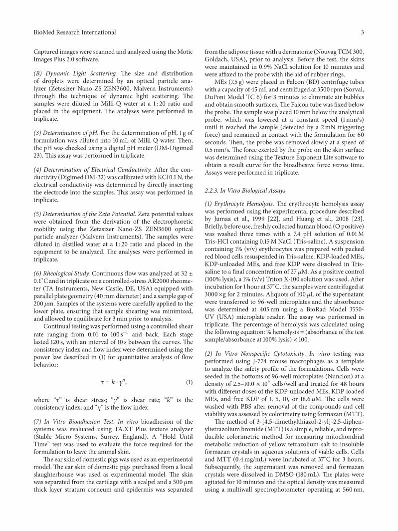
3.1. Physicochemical Characterization of ME. The MEs wereviscous, white, and opaque with a characteristic odor. Bymicroscopic analysis (Figure 2), larger droplets of less than10 𝜇m of diameter were observed. At certain times, smallerdroplets were observed within the larger droplets, character-istic of ME formulations. Moreover, there was no change inthe appearance of the ME formulations with the addition ofKDP.
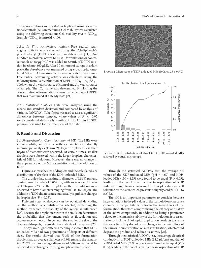
Figure 3 shows the size of droplets and the calculated sizedistribution of droplets of the KDP-unloaded MEs.
The droplets had a maximum diameter of 12.487𝜇m anda minimum diameter of 0.056 𝜇m, with an average diameterof 1.534 𝜇m. 72% of the droplets in the formulation wereobserved to have diameters ranging from 0.06 to 1.21 𝜇m.Theaddition of KDP did not cause statistically significant changesin droplet size (𝑃 > 0.05).
Different sizes of droplets can be obtained dependingon the method of emulsification selected, explaining themethod by which the stability of the system is influenced[25]. Because the droplet size within the emulsion determinesthe probability that phenomena such as flocculation andcoalescence will occur, in general, the smaller the size of thedispersed droplets, the greater the stability of the system [25].
Thedynamic light scattering technique showed that KDP-unloaded MEs had two populations of droplets of differentsizes. The results showed that 73.3% of the formulationdroplets had an average diameter of 1.016 𝜇mand the remain-ing 23.7% had an average diameter of 350 nm, as could beobserved morphologically using an optical microscope.
Figure 2: Microscopy of KDP-unloaded MEs (100x) at 25 ± 0.5∘C.
80
85
90
95
100
0102030405060708090
100
Freq
uenc
y (%
)
Cum
ulat
ive f
requ
ency
(%)
0.06
–1.21
1.21
–2.36
2.36
–3.51
3.51
–4.66
4.66
–5.81
5.81
–6.96
6.96
–8.11
8.11
–9.26
9.26
–10.41
10.41
–11.56
11
.56
–12
.71
Size distribution of multiple emulsion cells
Feret diameter (𝜇m)
Figure 3: Size distribution of droplets of KDP-unloaded MEsanalyzed by optical microscope.
Through the statistical ANOVA test, the average pHvalues of the KDP-unloaded MEs (pH = 4.62) and KDP-loaded MEs (pH = 4.55) were found to be equal (𝑃 > 0.05),leading to the conclusion that the incorporation of KDPinduced no significant change in pH.These pH values are welltolerated by the skin, which presents a slightly acid pH (4.5 to5.5) [20].
The pH is an important parameter to consider becauselarge variations in the pHvalues of the formulations can causechemical incompatibilities between the ingredients of theformulation, therefore compromising the efficacy and safetyof the active compounds. In addition to being a parameterrelated to the intrinsic stability of the formulation, it is essen-tial to control the pHof topical application products to ensurethat over time they do not cause changes in the microflora ofthe skin or induce irritation or skin sensitization, which coulddegrade the product and reduce its activity [21].
Through the statistical ANOVA test, the average electricalconductivity of KDP-unloadedMEs (34.2 𝜇S/cm) and that ofKDP-loaded MEs (31.90𝜇S/cm) were found to be equal (𝑃 >0.05), leading to the conclusion that the incorporation ofKDP

BioMed Research International 5
induced no significant change in the electrical conductivity ofthe ME system.
In terms of conductivity, the electrical properties of oiland water are extremely distinct and can be used to distin-guish whether the ME external phase is aqueous or oily. Inaccordance with the results obtained, electrical conductivitysuperior to 30 𝜇S/cm, it was confirmed that the external phaseis aqueous and that the ME formulation is a W/O/W type ofemulsion [26].
TheKDP-unloadedMEs showed an averageZPof−13mV.The addition of KDP did not cause statistically significantchanges in the ZP (𝑃 > 0.05).
The ZP results from the electric charges on the surfaceof the particle and is related to the physical stability of thecolloidal system [27]. At greater absolute values (more than+30mV or less than −30mV), the ZP of a system indicatesthe tendency of the particles to repel each other, which mayindicate that the system is stable. If, however, the absolutevalues for the ZP are low or zero, the particles tend toagglomerate and the system can easily flocculate [28]. In thiscase, the formulation should bemonitored over time to checkwhether the intermediate ZP values (−13mV) indicate anunstable system.
Semisolids products can exhibit a wide range of rheologi-cal behaviors. A good understanding of formulation rheologyis very important to the design, selection, and operation ofthe equipment involved in evaluating the release performanceand administration characteristics of the drug. Furthermore,rheological studies can provide useful information on the sta-bility and microstructure of emulsions. MEs can display flowcharacteristics depending on the fraction volume, droplet sizeand distribution, and the viscosity of the dispersed phase [29–32].
The relationship between the shear rate (Pa) and shearstress (1/s) of MEs is presented in Figure 4.
From the data obtained using (1) and shown in Table 2,KDP-unloaded MEs were demonstrated to exhibit “shearthinning” flow behavior (𝑛 < 1), whereas KDP-loaded MEsexhibited Newtonian behavior (𝑛 = 1).
The KDP-unloaded ME formulation behaved as a pseu-doplastic shear thinning fluid because its viscosity (𝜂∗)decreased with increasing stress. This may be due to thebreaking of organized structures that promote the formationof droplets, which are less organized structures [33].
However, the incorporation of the KDP drug had a stronginfluence on the formulation, leading the KDP-loaded MEformulation to behave as a low-viscosity Newtonian fluid.This phenomenon is related to the intense reduction in thedroplet size with the introduction of KDP, which thinned theflow and resulted in a higher diffusion coefficient than whenlarger oil droplets are present.
Furthermore, the rheogram showed that the up and downcurves did not overlap. The up curve was below the downcurve, which is typical and characteristic of antithixotropicmaterials.
Antithixotropic properties occur because the steadyapplication of shear stress increases the apparent viscosity,whereas cessation of the applied stress decreases the viscosityback to its initial value in a time-dependent manner [30].
0 20 40 60 80 1000
50
100
150
200
250
300
350
KDP-loaded-ME
KDP-unloaded ME
Shear rate (1/s)
Shea
r stre
ss (P
a),𝜂
∗(P
a·s)
𝜂∗ KDP-loaded ME𝜂∗ KDP-unloaded ME
Figure 4: Flow rheograms of KDP-unloaded ME (I) and KDP-loadedME (◻). Closed symbol represents up curve and open symbolrepresents down curve. 𝜂∗ (◊) is viscosity. Standard deviations havebeen omitted for clarity; however, in all cases, the coefficient ofvariation of triplicate analyses was less than 10%.Data were collectedat 32 ± 0.25∘C.
Table 2: Consistency index (𝑘) and flow index (𝑛) of KDP-loadedMEs and KDP-unloaded MEs.
Formulations 𝑘 𝑛
KDP-loaded ME 0.63 1.02KDP-unloaded ME 9.42 0.76
The increase in the apparent viscosity of the ME formu-lations with time at a constant shear was most likely dueto the reorganization of the droplets by strong forces. Thehigh viscosity of the MEs could retard the free motion ofthe droplets, thus delaying creaming, flocculation, and coa-lescence of the system [33].The conclusion from these resultsis that stable MEs can be obtained with the components usedin this study only under the application of vigorous stirringduring preparation so that viscosity is incorporated and thedestabilization of the system is avoided.
The KDP-unloaded MEs and KDP-loaded MEs wereevaluated for their changes in bioadhesion to determinewhether the addition of KDP alters the bioadhesive ability ofthe ME system.
Table 3 shows the bioadhesion test results of KDP-unloaded MEs and KDP-loaded MEs.
The results demonstrate that the incorporation of KDPdid not influence the bioadhesion of the MEs. Moreover, theformulations showed values very close to the bioadhesionof polyacrylic acid bioadhesive hydrogels used in previousstudies [32].
Thus, MEs have properties that favor the interactionbetween its components and the epithelial cells of the skin.
The investigation of the strength of bioadhesive pharma-ceutical formulations for topical use is extremely important

6 BioMed Research International
Table 3: Bioadhesion of KDP-unloadedMEs andKDP-loadedMEs.
Formulations Bioadhesivework (N⋅s) Bioadhesive force (N)
KDP-unloaded MEs 0.0166 ± 0.0016 0.0103 ± 0.0029KDP-loaded MEs 0.0163 ± 0.0025 0.0099 ± 0.0006
because bioadhesive formulations remain in contact withthe biological substrate for longer periods of time, whichmay lead to a gradient of drug concentration at the site ofaction and therefore improve the clinical performance of thetreatment.
3.2. In Vitro Biological Assays. Before exposing users to theproduct, the potential irritation of a new topical preparationshould be investigated to avoid inducing skin irritation. Forethical, legal, and financial reasons, in vivo tests were bannedfor testing cosmetics and formulations. Thus, in recent years,various in vitro assays have been conducted to test theproducts prior to realizing there in vivo safety profiles [34].
Erythrocyte-induced hemolysis in vitro is considered tobe a simple and reliablemeasure for estimating themembranedamage caused in vivo because erythrocytes are easily isolatedby centrifugation [35].
In vitro cytotoxicity assays using the hemolysis of redblood cells were used to assess the safety profile of theformulations and they are presented in Figure 5.
Free KDP caused lysis of 4.09 ± 0.13% of the erythrocytemembranes. KDP-unloaded MEs caused lysis of 1.57% ±0.47% of the erythrocyte membranes. The incorporationof KDP in ME was 2.98 ± 1.12% and showed decreasederythrocyte lysis compared to the free KDP.Thus, all systemsshowed a tolerable hemolysis of erythrocytes. The positivecontrol was represented by 100% hemolysis of erythrocytesusing Triton X-100, a known hemolytic agent. The studyresults indicate that the treatment developed using the MEsystem showed low toxicity and may therefore be a potentialalternative to current therapeutic applications [23].
In vitro cytotoxicity was performed using J-774 mousemacrophages as a cellular model. The data are shown as thepercentage of cell viability (Figure 6).
The cell viability assays showed that more than 93% ofthe normal macrophages treated with free KDP survived.Moreover, neither the KDP-unloaded MEs nor the KDP-loaded MEs showed toxic activity. Therefore, the resultsindicate the safety and biocompatibility of the formulationsand free KDP with eukaryotic cells [36–38].
3.3. In Vitro Antioxidant Activity. The formulations with orwithout the addition of KDP were evaluated for their in vitroantioxidant activity over a period of 28 days. The resultsobtained are shown in Figure 7.
The antioxidant activity was measured based on themethodology of Blois, 1958 [24], in which the stable radicalDPPH is reduced by antioxidants. The results of this studyare expressed as the percent inhibition of DPPH (%).
Over the period of 28 days, there was a decrease inthe antioxidant power of all the experimental groups, with
Hem
olys
is (%
)
0
5
80
90
100
110
Free KDPKDP-unloaded ME
KDP-loaded METriton X-100
Figure 5: Percentage of hemolysis of red blood cells after treatmentwith free KDP, KDP-unloaded MEs, and KDP-loaded MEs.
86
88
90
92
94
96
98
100
1 5 10 18.6
Cel
lula
r via
bilit
y (%
)
Free KDPKDP-unloaded MEKDP-loaded ME
Concentration (𝜇M)
Figure 6: Percentage of cell viability after treatment with free KDP,KDP-unloaded MEs, and KDP-loaded MEs.
the most marked decrease for the free KDP. Differencesbetween the samples were significant (𝑃 < 0.05) and thelesser destabilization of the samples is most likely due to theincreased stabilization of the KDP-loaded ME formulation.
Therefore, the ME formulation maintained the antiox-idant properties of KDP, which makes MEs a promisingvehicle for the incorporation of KDP to facilitate its topicalapplication in the treatment of skin aging.
4. Conclusion
Through physicochemical characterization assays, it wasconfirmed that the ME system developed was of the W/O/Wtype with a Dnm of approximately 1 𝜇m and a ZP of −13mV.No change in Dnm, ZP, or the stability of the system wasobserved after the addition KDP. KDP-unloaded MEs exhib-ited “shear thinning” flow behavior, and the incorporation of

BioMed Research International 7
0
10
20
30
40
50
60
70
0 10 20 30
DPP
H in
hibi
tion
(%)
Days
Free KDPKDP-loaded MEKDP-unloaded ME
Figure 7: Percent inhibition of the 2,2-diphenyl-1-picrylhydrazyl(DPPH) radical by the formulations over a period of 28 days.
the KDP drug strongly influenced the formulation, leadingthe KDP-loaded MEs to behave as a Newtonian fluid. Thisphenomenon can be related to the intense reduction inthe droplet size in the presence of KDP, which thinnedthe flow. However, both flow types are characteristic ofantithixotropic materials. The properties of MEs favor theinteraction between the ME system and the epithelial cells ofthe skin. KDP incorporation did not influence the bioadhe-sion of the MEs, and KDP-loaded MEs showed low toxicityand a good antioxidant activity. These results indicated thatthis is a promising system for using KDP in the treatment ofskin aging.
Conflict of Interests
The authors declare that there is no conflict of interestsregarding the publication of this paper.
Acknowledgments
This work was financially supported by Fundacao de Amparoa Pesquisa do Estado de Sao Paulo (FAPESP), Conselho Na-cional de Desenvolvimento Cientıfico e Tecnologico (CNPq),and Programa de Apoio ao Desenvolvimento Cientıfico(PADC-FCF).
References
[1] G. Benech, “Novo ativo clareador extraıdo de cıtricos,” Cosmet-ics & Toiletries, vol. 14, pp. 51–53, 2002.
[2] S. Iriyama, T.Ono,H.Aoki, and S.Amano, “Hyperpigmentationin human solar lentigo is promoted by heparanase-induced lossof heparan sulfate chains at the dermal-epidermal junction,”
Journal of Dermatological Science, vol. 64, no. 3, pp. 223–228,2011.
[3] K. Miyamoto, Y. Inoue, K. Hsueh et al., “Characterization ofcomprehensive appearances of skin ageing: an 11-year longitu-dinal study on facial skin ageing in Japanese females at Akita,”Journal of Dermatological Science, vol. 64, no. 3, pp. 229–236,2011.
[4] C. Longo, A. Casari, F. Beretti, A.M. Cesinaro, and G. Pellacani,“Skin aging: in vivo microscopic assessment of epidermal anddermal changes by means of confocal microscopy,” Journal ofthe American Academy of Dermatology, vol. 68, no. 3, pp. e73–e82, 2013.
[5] J. Labat-Robert andL. Robert, “Longevity and aging. Role of freeradicals and xanthine oxidase. A review,” Pathologie Biologie,vol. 62, no. 2, pp. 61–66, 2014.
[6] M. B. Oliveira, A. H. D. Prado, J. Bernegossi et al., “Topicalapplication of retinyl palmitate-loaded nanotechnology-baseddrug delivery systems for the treatment of skin aging,” BioMedResearch International, vol. 2014, Article ID 632570, 7 pages,2014.
[7] M. O. Visscher, B. S. Pan, and W. J. Kitzmiller, “Photodamage.Treatments and topicals for facial skin,” Facial Plastic SurgeryClinics of North America, vol. 21, no. 1, pp. 61–75, 2013.
[8] R. F. Pytel, L. V. N. Silva, A. S. Nunes, J.-L. Gesztesi, andA. da Costa, “Estudo in vivo de atividade anti-radicalar porquantificacao de peroxidos cutaneos,” Anais Brasileiros deDermatologia, vol. 80, pp. S323–S328, 2005.
[9] J.-M. Noh, S.-Y. Kwak, H.-S. Seo, J.-H. Seo, B.-G. Kim, and Y.-S.Lee, “Kojic acid-amino acid conjugates as tyrosinase inhibitors,”Bioorganic and Medicinal Chemistry Letters, vol. 19, no. 19, pp.5586–5589, 2009.
[10] M.Rendon and S.Horwitz, “Topical treatment of hyperpigmen-tation disorders,” Annales de Dermatologie et de Venereologie,vol. 139, supplement 4, pp. S153–S158, 2012.
[11] M. L. Goncalez,M. A. Correa, andM. Chorilli, “Skin delivery ofkojic acid-loaded nanotechnology-based drug delivery systemsfor the treatment of skin aging,” BioMed Research International,vol. 2013, Article ID 271276, 9 pages, 2013.
[12] A. Balaguer, A. Salvador, and A. Chisvert, “A rapid and reli-able size-exclusion chromatographic method for determinationof kojic dipalmitate in skin-whitening cosmetic products,”Talanta, vol. 75, no. 2, pp. 407–411, 2008.
[13] S. Y. Tang, M. Sivakumar, and B. Nashiru, “Impact of osmoticpressure and gelling in the generation of highly stable singlecore water-in-oil-in-water (W/O/W) nano multiple emulsionsof aspirin assisted by two-stage ultrasonic cavitational emulsifi-cation,” Colloids and Surfaces B: Biointerfaces, vol. 102, pp. 653–658, 2013.
[14] M. Chorilli, P. S. Prestes, R. B. Rigon et al., “Structural charac-terization and in vivo evaluation of retinyl palmitate in non-ionic lamellar liquid crystalline system,” Colloids and SurfacesB: Biointerfaces, vol. 85, no. 2, pp. 182–188, 2011.
[15] J. Kuntsche, J. C. Horst, andH. Bunjes, “Cryogenic transmissionelectron microscopy (cryo-TEM) for studying the morphologyof colloidal drug delivery systems,” International Journal ofPharmaceutics, vol. 417, no. 1-2, pp. 120–137, 2011.
[16] C.-X. Zhao, “Multiphase flow microfluidics for the productionof single or multiple emulsions for drug delivery,” AdvancedDrug Delivery Reviews, vol. 65, no. 11-12, pp. 1420–1446, 2013.
[17] M. Chorilli, G. R. Campos, and P. M. L. Bolfarini, “Desen-volvimento e estudo da estabilidade fısico-quımica de emulsoes

8 BioMed Research International
multiplas A/O/A e O/A/O acrescidas de filtros quımicos emanteiga de karite,” Acta Farmaceutica Bonaerense, vol. 28, no.6, pp. 936–940, 2009.
[18] M. C.Garcıa, J.Munoz,M. C. Alfaro, and J.M. Franco, “Physicalcharacterization of multiple emulsions formulated with a greensolvent and different HLB block copolymers,” Colloids andSurfaces A: Physicochemical and Engineering Aspects, vol. 458,pp. 40–47, 2014.
[19] T. Ohwaki, K. Nitta, H. Ozawa et al., “Improvement of the for-mation percentage of water-in-oil-in-water multiple emulsionby the addition of surfactants in the internal aqeous phase ofthe emulsion,” International Journal of Pharmaceutics, vol. 85,no. 1–3, pp. 19–28, 1992.
[20] G. Leonardi and P. M. Campos, “Estabilidade de formulacoescosmeticas,” International Journal of Pharmaceutical Com-pounding, vol. 3, pp. 154–156, 2001.
[21] S. A. Ansari, A. O. Barel, M. Paye, and H. I. Maibach, “Skin pHand skin flora,” inHandbook of Cosmetic Science andTechnology,pp. 221–231, 2009.
[22] M. Jumaa, P. Kleinebudde, and B. W. Muller, “Physicochemicalproperties and hemolytic effect of different lipid emulsionformulations using a mixture of emulsifiers,” PharmaceuticaActa Helvetiae, vol. 73, no. 6, pp. 293–301, 1999.
[23] Z.-R. Huang, S.-C. Hua, Y.-L. Yang, and J.-Y. Fang, “Develop-ment and evaluation of lipid nanoparticles for camptothecindelivery: a comparison of solid lipid nanoparticles, nanostruc-tured lipid carriers, and lipid emulsion,” Acta PharmacologicaSinica, vol. 29, no. 9, pp. 1094–1102, 2008.
[24] M. S. Blois, “Antioxidant determinations by the use of a stablefree radical,” Nature, vol. 181, no. 4617, pp. 1199–1200, 1958.
[25] M.-W. Jeong, S.-G. Oh, and Y. C. Kim, “Effects of amine andamine oxide compounds on the zeta-potential of emulsiondroplets stabilized by phosphatidylcholine,” Colloids and Sur-faces A: Physicochemical and Engineering Aspects, vol. 181, no.1–3, pp. 247–253, 2001.
[26] H. Masmoudi, Y. L. Dreau, P. Piccerelle, and J. Kister, “Theevaluation of cosmetic and pharmaceutical emulsions agingprocess using classical techniques and a new method: FTIR,”International Journal of Pharmaceutics, vol. 289, no. 1-2, pp. 117–131, 2005.
[27] W. M. Obeidat, K. Schwabe, R. H. Muller, and C. M. Keck,“Preservation of nanostructured lipid carriers (NLC),” Euro-pean Journal of Pharmaceutics and Biopharmaceutics, vol. 76,no. 1, pp. 56–67, 2010.
[28] C. Freitas and R. H. Muller, “Effect of light and temperature onzeta potential and physical stability in solid lipid nanoparticle(SLN) dispersions,” International Journal of Pharmaceutics, vol.168, no. 2, pp. 221–229, 1998.
[29] M. L. Bruschi, O. de Freitas, E. H. G. es Lara, H. Panzeri,M. P. D.Gremiao, andD. S. Jones, “Precursor systemof liquid crystallinephase containing propolis microparticles for the treatment ofperiodontal disease: development and characterization,” DrugDevelopment and Industrial Pharmacy, vol. 34, no. 3, pp. 267–278, 2008.
[30] R. Pal, “Rheology of simple and multiple emulsions,” CurrentOpinion in Colloid & Interface Science, vol. 16, no. 1, pp. 41–60,2011.
[31] F. C. Carvalho, G. Calixto, I. N. Hatakeyama, G. M. Luz, M.P. D. Gremiao, and M. Chorilli, “Rheological, mechanical, andbioadhesive behavior of hydrogels to optimize skin deliverysystems,” Drug Development and Industrial Pharmacy, vol. 39,no. 11, pp. 1750–1757, 2013.
[32] G. Calixto, A. C. Yoshii, H. Rocha e Silva, B. S. F. Cury, andM. Chorilli, “Polyacrylic acid polymers hydrogels intendedto topical drug delivery: preparation and characterization,”Pharmaceutical Development and Technology, 2014.
[33] G. B. R. F. da Silva, M. V. Scarpa, G. Rossanezi, E. S. T.do Egito, and A. G. de Oliveira, “Development and charac-terization of biocompatible isotropic and anisotropic oil-in-water colloidal dispersions as a new delivery system for methyldihydrojasmonate antitumor drug,” International Journal ofNanomedicine, vol. 9, no. 1, pp. 867–876, 2014.
[34] C. F. Zanatta, V. Ugartondo, M. Mitjans, P. A. Rocha-Filho,and M. P. Vinardell, “Low cytotoxicity of creams and lotionsformulated with Buriti oil (Mauritia flexuosa) assessed by theneutral red release test,” Food and Chemical Toxicology, vol. 46,no. 8, pp. 2776–2781, 2008.
[35] S. V. P. Malheiros, N. C. Meirelles, and E. de Paula, “Path-ways involved in trifluoperazine-, dibucaine- and praziquantel-induced hemolysis,”Biophysical Chemistry, vol. 83, no. 2, pp. 89–100, 2000.
[36] F. Kolenyak-Santos, R. N. de Oliveira, C. Garnero et al.,“Nanostructured lipid carriers as a strategy to improve thein vitro schistosomiasis activity of praziquantel,” Journal ofNanoscience and Nanotechnology, vol. 14, no. 1, pp. 1–12, 2014.
[37] M. H. Oyafuso, F. C. Carvalho, L. A. Chiavacci, M. P. D.Gremiao, and M. Chorilli, “Design and characterization ofsilicone and surfactant based systems for topical drug delivery,”Journal of Nanoscience and Nanotechnology, vol. 15, no. 1, pp.817–826, 2015.
[38] M. B. Oliveira, G. Calixto, M. Graminha, H. Cerecetto, M.Gonzalez, and M. Chorilli, “Development, characterization,and in vitro biological performance of fluconazole-loadedmicroemulsions for the topical treatment of cutaneous leish-maniasis,” BioMed Research International, vol. 2015, Article ID396894, 12 pages, 2015.

Submit your manuscripts athttp://www.hindawi.com
PainResearch and TreatmentHindawi Publishing Corporationhttp://www.hindawi.com Volume 2014
The Scientific World JournalHindawi Publishing Corporation http://www.hindawi.com Volume 2014
Hindawi Publishing Corporationhttp://www.hindawi.com
Volume 2014
ToxinsJournal of
VaccinesJournal of
Hindawi Publishing Corporation http://www.hindawi.com Volume 2014
Hindawi Publishing Corporationhttp://www.hindawi.com Volume 2014
AntibioticsInternational Journal of
ToxicologyJournal of
Hindawi Publishing Corporationhttp://www.hindawi.com Volume 2014
StrokeResearch and TreatmentHindawi Publishing Corporationhttp://www.hindawi.com Volume 2014
Drug DeliveryJournal of
Hindawi Publishing Corporationhttp://www.hindawi.com Volume 2014
Hindawi Publishing Corporationhttp://www.hindawi.com Volume 2014
Advances in Pharmacological Sciences
Tropical MedicineJournal of
Hindawi Publishing Corporationhttp://www.hindawi.com Volume 2014
Medicinal ChemistryInternational Journal of
Hindawi Publishing Corporationhttp://www.hindawi.com Volume 2014
AddictionJournal of
Hindawi Publishing Corporationhttp://www.hindawi.com Volume 2014
Hindawi Publishing Corporationhttp://www.hindawi.com Volume 2014
BioMed Research International
Emergency Medicine InternationalHindawi Publishing Corporationhttp://www.hindawi.com Volume 2014
Hindawi Publishing Corporationhttp://www.hindawi.com Volume 2014
Autoimmune Diseases
Hindawi Publishing Corporationhttp://www.hindawi.com Volume 2014
Anesthesiology Research and Practice
ScientificaHindawi Publishing Corporationhttp://www.hindawi.com Volume 2014
Journal of
Hindawi Publishing Corporationhttp://www.hindawi.com Volume 2014
Pharmaceutics
Hindawi Publishing Corporationhttp://www.hindawi.com Volume 2014
MEDIATORSINFLAMMATION
of