research article open access transient … article open access transient elevation of glycolysis...
TRANSCRIPT
Bhatt et al. BMC Cancer (2015) 15:335 DOI 10.1186/s12885-015-1368-9
RESEARCH ARTICLE Open Access
Transient elevation of glycolysis confersradio-resistance by facilitating DNA repair in cellsAnant Narayan Bhatt, Ankit Chauhan, Suchit Khanna, Yogesh Rai, Saurabh Singh, Ravi Soni, Namita Kalraand Bilikere S Dwarakanath*
Abstract
Background: Cancer cells exhibit increased glycolysis for ATP production (the Warburg effect) and macromolecularbiosynthesis; it is also linked with therapeutic resistance that is generally associated with compromised respiratorymetabolism. Molecular mechanisms underlying radio-resistance linked to elevated glycolysis remain incompletelyunderstood.
Methods: We stimulated glycolysis using mitochondrial respiratory modifiers (MRMs viz. di-nitro phenol, DNP;Photosan-3, PS3; Methylene blue, MB) in established human cell lines (HEK293, BMG-1 and OCT-1). Glucose utilizationand lactate production, levels of glucose transporters and glycolytic enzymes were investigated as indices of glycolysis.Clonogenic survival, DNA repair and cytogenetic damage were studied as parameters of radiation response.
Results: MRMs induced the glycolysis by enhancing the levels of two important regulators of glucose metabolismGLUT-1 and HK-II and resulted in 2 fold increase in glucose consumption and lactate production. This increase inglycolysis resulted in resistance against radiation-induced cell death (clonogenic survival) in different cell lines atan absorbed dose of 5 Gy. Inhibition of glucose uptake and glycolysis (using fasentin, 2-deoxy-D-glucose and3-bromopyruvate) in DNP treated cells failed to increase the clonogenic survival of irradiated cells, suggesting thatradio-resistance linked to inhibition of mitochondrial respiration is glycolysis dependent. Elevated glycolysis alsofacilitated rejoining of radiation-induced DNA strand breaks by activating both non-homologous end joining (NHEJ)and homologous recombination (HR) pathways of DNA double strand break repair leading to a reduction inradiation-induced cytogenetic damage (micronuclei formation) in these cells.
Conclusions: These findings suggest that enhanced glycolysis generally observed in cancer cells may beresponsible for the radio-resistance, partly by enhancing the repair of DNA damage.
Keywords: DNP, Respiratory modifiers, Glycolysis and radio-resistance
BackgroundIonizing radiation plays an important role in themanagement of a majority of malignancies [1], althoughmany tumors like glioma and several carcinomas areknown to be refractory to radiotherapy with marginalbenefits in survival. However, the molecular mechanismsunderlying this radio-resistance of cancer cells remainpoorly understood. One of the most common signaturesof highly malignant tumors is their capacity tometabolize more glucose to lactic acid than normal tis-sues, which confers a selective growth advantage [2].
* Correspondence: [email protected] and Cell Signaling Group, Institute of Nuclear Medicine and AlliedSciences, Brig SK Mazumdar Road, New Delhi, Delhi 110 054, India
© 2015 Bhatt et al.; licensee BioMed Central. TCommons Attribution License (http://creativecreproduction in any medium, provided the orDedication waiver (http://creativecommons.orunless otherwise stated.
Cells derived from hypoxic tumors typically maintaintheir metabolic phenotypes even under normoxic cultureconditions (Warburg effect), indicating that aerobicglycolysis is constitutively upregulated through stablegenetic or epigenetic changes [2]. It is also reported thatmitochondrial defect linked stabilization of HIF1α in-duces glycolytic phenotype in cancer cells and promotesaggressiveness of tumors [2,3]. On the other hand, effi-cient oxidative phosphorylation in cancer cells is re-quired for execution of apoptosis through the generationof reactive oxygen species (ROS) [4]. Therefore, meta-bolically reprogrammed and highly glycolytic cancercells can easily escape the death processes, conferringresistance to therapeutic modalities [5].
his is an Open Access article distributed under the terms of the Creativeommons.org/licenses/by/4.0), which permits unrestricted use, distribution, andiginal work is properly credited. The Creative Commons Public Domaing/publicdomain/zero/1.0/) applies to the data made available in this article,
Bhatt et al. BMC Cancer (2015) 15:335 Page 2 of 12
The phenotypic characteristics of enhanced glycoly-sis associated with tumors have been well exploitedfor the diagnosis of the disease using fluoro-deoxyglucose (FDG) based positron emission tomography(PET) imaging and the efficacy of glycolytic inhibi-tors as sensitizers to radiation and chemotherapeuticdrugs has established in pre-clinical studies, whileclinical trials are at different stages of evaluation[6-9]. Considerable amount of evidences suggest thatinhibition of glycolysis leads to compromised DNArepair, which is accompanied by the depletion of en-ergy [ATP and AXP (AMP and ADP)] in cells withhigh rates of glycolysis like the cancer cells, causingdeath [10-14]. However, the mechanisms underlyingenhanced resistance to radiation-induced cell deathin cells with high endogenous rates of glycolysis, like thecancer cells are not completely understood, although al-terations in pH and lactate levels have been implicated[2]. Therefore, present studies were undertaken toexamine if transient stimulation of glycolysis (beforeirradiation) using MRMs is sufficient to confer radio-resistance and also to unravel the underlying mechanisms.Results obtained in human malignant and non-malignantcell lines clearly show that stimulation of glycolysisusing MRMs reduces radiation-induced cell death byenhancing the repair of DNA damage leading to a re-duction in the mitotic death linked to cytogenetic damage(micronuclei formation).
MethodsMaterialsDulbecco’s Minimum Essential Medium (DMEM),Penicillin G, Streptomycin, Nystatin, Dimethyl sulfoxide(DMSO), Dichlorodihydrofluorescein diacetate (H2DCFH-DA), Potassium chloride (KCl), Magnesium chloride(MgCl2), Ethylene-di-amine-tetra acetate (EDTA),Protease inhibitor cocktail, HEPES, Hoechst-33258 (H258),propidium iodide (PI), RNAase, Di-nitrophenol andmethylene blue were procured from Sigma chemicalsCo. (St Louis, USA). Photosan (PS3) was procuredfrom See Lab, Germany. Bicinchoninic acid (BCA, Thermofisher scientific Rockford USA), primary and HRP conju-gated secondary antibodies were procured from SantaCruz, USA.
MethodsCell cultureThe cerebral glioma cell line (BMG-1; diploid, wild typep53) established by us earlier [12] whereas; OCT-1 (oralcarcinoma) and HEK293 (human embryonic kidney)cells were obtained from NCCS, Pune, India. BMG-1and OCT-1 cells were maintained as monolayers inDulbecco’s modified Eagles medium (DMEM) sup-plemented with fetal bovine serum (5% for BMG-1
and 10% for OCT-1), HEPES and antibiotics as de-scribed earlier [12]. HEK293 was grown in high glucose(4.5 g/L) DMEM supplemented with 10% fetal bovineserum (FBS). All experiments were carried out in expo-nentially growing cells.
TreatmentsCells were treated with MRMs at concentrations thatinduce glycolysis without any cytotoxicity (data notshown). The drug concentrations used in all the experi-ments were 1 μM for DNP, 25 μg/ml for PS3 and 25 μMfor MB. Cells were exposed to 5 Gy γ-ray irradiation inall experiments (unless specified otherwise in particularexperiments), using Bhabhatron-II, a teletherapy ma-chine from Panacea, Medical Technologies Pvt. Ltd.(Bangalore, India) at 80 cm SSD and 35x35 cm field witha dose rate of 2 Gy/min.
Glucose consumption and lactate production assayBMG-1, OCT-1 and HEK293 cells were incubated inHBSS or HBSS containing MRMs before irradiation.Cells were incubated for 4 hours; every hour HBSS sam-ple was taken from dishes and frozen immediately forfurther analysis. The amount of glucose remaining un-used and the lactate produced were estimated in theHBSS using enzymatic assays. Glucose was determinedby the glucose-oxidase method using Tecnicon RA-500auto analyzer. Lactate was estimated using lactate oxi-dase method based kit (Randox; Cat. No.-LC2389).Glucose consumption and lactate production were esti-mated at the end of every hour (up to four hours) andpresented as the average consumption or production perhour. The number of viable cells was counted onhemocytometer using trypan blue (0.4%) exclusion testand glucose consumption or lactate production was nor-malized with respect to the number of trypan blue nega-tive (viable) cells.
Hexokinase assayHexokinase enzymatic activity was measured as pre-viously described [15]. Briefly, cells were treated for4 hour with DNP followed by radiation beforewashed with PBS and lysed using the buffer containing50 mM Tris-HCl (pH-8.0), 1.5 mM MgCl2, 150 mMNaCl, 250mM sucrose along with protease inhibitor. Thecell lysate was incubated on ice for 30 min, followed bycentrifugation at 14,000 rpm at 4°C for 10 min. The pro-tein estimation was done by BCA method. An aliquot of20 μg protein from freshly lysed cell supernatant wasadded to 1 ml of reaction buffer containing 100 mMTris-HCl, pH 8.0, 0.5 mM EDTA, 10 mM ATP, 10 mMMgCl2, 2 mM glucose, 0.1 mM NADP, and 0.1 U/ml ofG6PD (Sigma). HK activity was determined by followingthe G6P-dependent conversion of NADP to NADPH

Bhatt et al. BMC Cancer (2015) 15:335 Page 3 of 12
spectrophotometrically at 340 nm at room temperature.The values were subtracted with respective blanks andthe relative enzymatic activity is presented as absorptionat 340 nm.
ATP measurementATP was measured using ATP bioluminescent assay kit(Sigma) following manufacturer’s protocol. Briefly, cellswere treated with MRMs followed by irradiation for4 hour before washed and scraped in cold PBS and pel-leted at 1000 rpm for 10 min. Further, cells were lysed in350 μl of lysis buffer (4mM EDTA and 0.2% TritonX-100). 100 μl of this lysate was loaded per well intriplicates with 100 μl of ATP mix in a 96-well whiteluminescence measuring plate. Luminescence of sam-ples along with standards was read at 562 nm andnormalized with the protein concentration. ATP concen-tration is depicted as ng/mg protein.
Measurement of mitochondrial content and membranepotentialCells were treated with MRMs followed with or withoutirradiation for various time points. Once the incubationtime was completed, cells were washed with PBS andstained for 30 minutes at 37°C in CO2 incubator witheither 100 nM mitotracker green or 100 nM DiOC6(both from Molecular Probes, Invitrogen) in respectivewells. After staining, cells were trypsinized and equalnumbers of cells were transferred to 96 well fluores-cence reading plate. The fluorescence was measured at490 nm excitation and 516 nm emission for mitotrackergreen and 484 nm excitation and 501 nm emission forDiOC6 in quadruplicates using Molecular devicefluorescence plate reader. The fluorescence intensityof mitotracker green is represented as relative mito-chondrial content and the DiOC6 fluorescence inten-sity is represented as relative mitochondrial membranepotential.
Macro colony assayClonogenic survival was performed using post platingmethod of macro colony assay. Cells were plated at adensity of 6,000 - 8,000 cells/cm2 in Petri dishes. 36 hlater, cells were washed with HBSS and treated with dif-ferent MRMs (viz. 1 μM di-nitro phenol, DNP; 25 μg/mlPhotosan-3, PS3; 25 μM Methylene blue, MB) in HBSSbefore irradiation. Following the radiation treatment,cells were incubated for 4h at 37°C in HBSS in CO2 in-cubator then washed twice with HBSS, trypsinized,counted and plated in triplicates in 60 mm petri dishat very low density (100 cells/dish) for control andMRMs treatment alone and relatively high cell dens-ity (500 cells/dish) for radiation (5 Gy) and MRMswith radiation. These dishes were incubated in fresh
media at 37°C in a humidified CO2 (5%) incubatorfor 7-10 days, depending on the cell line. Cultureswere terminated when macrocolonies were visible after7-10 days, were fixed with 10% methanol in PBS andstained with 1% crystal violet (dissolved in 7% metha-nol in PBS). Colonies of at least 50 cells (5 to 6doubling) were scored as survivors. Plating efficiencywas calculated as: PE = (No. of colonies counted/Noof cells plated) x 100. The surviving fraction was cal-culated as: SF = PET /PEC. Where PET is the platingefficiency of the treated group and PEC is the valueof the control.
Single cell gel electrophoresis (comet assay)Neutral comet assay was performed as described earlier[16,17]. For 0 hour time point (to obtain damage in-duction) cells were exposed on ice and processed im-mediately after exposure. Briefly, treated cells weretrypsinized and resuspended in the medium at re-spective time points. An aliquot of 30,000-40,000cells (~100 μl of medium) was mixed into a suspension of0.75% (~500μl) warm low gelling (gelling temp = 17°C)agarose (BDH Electran, England), and spread ontomicroscopic slide (pre-coated with 0.1% agarose) keptat 45°C. Slides were immediately transferred to a cool-ing plate at 4°C, and kept for 5 min to allow the agar-ose layer to gel along with the embedded cells.Following this, the slides carrying agarose gel with em-bedded cells were immersed in the SDS lysis buffer[2.5% sodium dodecyl sulphate (SDS), 1% sodium sar-cosinate and 25 mM ethylenediaminetetraacetic acid(EDTA)] for 15 min at room temperature (25-28°C).Slides were then washed in double distilled water for5 min at 4°C, electrophoresed at 2 V/cm for 5 min,and air-dried at 45°C. For analysis, the slides wererehydrated by immersing in distilled water at roomtemperature, and stained with 25 μg/ml propidiumiodide (PI) dye. Comet-like shape forms from the DNAmass of each individual cell following these treatments,the amount of DNA present in the tail directly cor-responding with the amount of DNA breaks inducedby radiation treatment. The comets were randomlyselected (n = 50) and images were acquired at 200xunder BX60 fluorescence microscope (Olympus, Japan).The comets were analyzed using OPTIMAS image ana-lysis software (calibrated for analyzing comets) and thetail moment was calculated from data generated by thesoftware.
Micronuclei assayCells were washed in PBS and fixed in carnoys fixa-tive (3:1 V/V, Methanol: Acetic acid). Fixed cellswere dropped on the chilled glass slides. Air-driedslides were stained with a DNA binding fluorochrome

Bhatt et al. BMC Cancer (2015) 15:335 Page 4 of 12
(bisbenzimidazole; hoechst-33258) at 10 μg/ml dissolvedin citric acid [0.01 M], disodium phosphate [0.45 M] buf-fer containing 0.05% Tween-20 detergent; pH 7.4 asdescribed earlier [6]. Slides were examined under fluores-cence microscope using UV excitation filter. Cells con-taining micronuclei were counted from 1000 cells asdescribed earlier [6,18].
Immuno-blotExpression of key proteins involved in glucose catabol-ism and DNA repair viz. glucose transporters GLUT-1,GLUT-4, hexokinase-II (HK-II), PFK-1, HIF1α, Rad51,Ku-70 and loading control β-Actin was determined incontrol and treated cells by immunoblot analysis. Cellswere harvested at various time points (showed in figure)and lysed in ice cold RIPA lysis buffer (Tris–HCl:50mM, pH 7.4, NP-40: 1%, Na-deoxycholate: 0.25%,NaCl: 150 mM, EDTA: 1 mM, PMSF: 2 mM, proteaseinhibitor cocktail, Na3VO4: 1 mM, NaF: 1 mM). Theprotein concentration in the lysates was measured usingBCA protein assay. Protein (40–50 μg) was resolved on10–15% SDS–PAGE (depending on the molecularweight) and electroblotted onto PVDF membrane(MDI). The membrane was then incubated in 4%skimmed milk for 1 h followed by primary antibody in-cubation GLUT-1 (1:300), GLUT-4 (1:300), HK-II(1:200), PFK-2 (1:200), Rad51 (1:200), Ku-70 (1:150)from Santa Cruz Biotechnology, HIF1α (1:1000) fromCell Signaling Tech. and β-Actin (1:5000) from BDBiosciences for overnight. Membrane was washed andincubated with the appropriate HRP conjugated second-ary antibody for 1h. After washing, the blots were devel-oped using ECL chemiluminescence detection reagent(Biological industries, Israel). The signal was detectedby ECL, and band intensities for each individual pro-tein were quantified by densitometry, corrected forbackground staining, and normalized to the signal forβ-Actin.
Statistical analysesAll the experiments were performed in triplicates orquadruplicates. Means and standard errors were com-puted. Student’s t test was performed to determinewhether a significant difference exists between thegroups.
ResultsMitochondrial respiratory modifiers induces glycolysisTo mimic the high glycolytic phenotype of cancer cells,we investigated the glycolysis stimulating potential offew mitochondrial respiratory modifiers (MRMs) thatare known to stimulate glycolysis as a compensatorymechanism [19]. At Treatment of exponentially growingcells with non-toxic concentrations MRMs such as
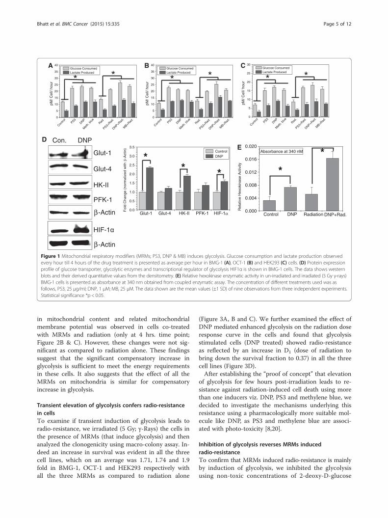
di-nitrophenol (DNP), porphyrin derivatives (photosan;PS3) and methylene blue (MB), which interfere with theoxidative phosphorylation at different stages in the elec-tron transport chain (ETC), was found to enhance theglycolysis (glucose utilization and lactate production) sig-nificantly (by approximately two folds) in both malignantcell lines BMG-1 and OCT-1 (Figure 1A and B), similarto our earlier results with KCN [11,12]. To test if com-promised oxidative phosphorylation can induce the com-pensatory increase in glycolysis in non-malignant cellsimilar to malignant cells, we treated HEK cell line(embryonic kidney) with MRMs under similar experi-mental conditions. Interestingly, MRMs induced the glu-cose uptake and lactate production in HEK cells also(Figure 1C). Further, we observed that irradiation alonealso marginally increased glycolysis (Figure 1A, B and C)as reported earlier [11], with further increase in presenceof MRMs (Figure 1A, B and C). It is pertinent to notethat compensatory increase in glycolysis due to inhibitionof oxidative phosphorylation appears to be not limitedonly to malignant cells.To unravel the contributing factors responsible for
MRM-induced enhancement in glycolysis, we examinedthe level of glycolytic enzymes and glucose transportersunder similar experimental conditions. Interestingly, wefound approximately 2.5 fold increased level of GLUT-1,while no significant change could be seen in GLUT-4(Figure 1D). A 2 fold increase was also seen in the levelof hexokinase-II, one of the first two regulatory kinases(HK-II and PFK-1) of glycolysis; however the level ofPFK-1 does not change appreciably (Figure 1D). DNPtreatment also showed increased level of hypoxia indu-cible transcription factor, HIF1α which is known to in-duce glycolysis. Further, the increase in hexokinaseexpression also correlated with nearly two fold increasein the total hexokinase activity (Figure 1E) induced byDNP under these experimental conditions. Interestingly,the hexokinase activity was increased further by nearly 4fold in cells treated with both DNP and radiation. Thesefindings suggest that inhibition of mitochondrial respir-ation stabilizes HIF1α which further induces glycolysisby up-regulating the level of glucose transporters viz.GLUT-1 and glucose phosphorylating enzyme HK-II toensure the increased flux and high retention of glucosein the cytoplasm.MRMs inhibit the process of electron transfer and
ATP generation from electron transport chain leading toincomplete respiration and reduced ATP generation.Therefore, we measured changes in ATP levels inducedby MRMs (Figure 2A), besides examining the mitochon-drial status by analyzing the membrane potential andmass (Figure 2B & C). Results (Figure 2) clearly showthat none of the three respiratory inhibitors caused anyappreciable changes in ATP (Figure 2A), while increase
A
0
5
10
15
20
25
30
35
40
MB+R
ad.
DNP+Rad
.
PS3+Rad
.Rad
.
Met
h. b
lueDNP
PS3
Contro
l
pM/ C
ell/
hour
Glucose ConsumedLactate Produced
* *
0
5
10
15
20
25
30
35
40
MB+R
ad.
DNP+Rad
.
PS3+Rad
.Rad
.
Met
h. b
lueDNP
PS3
Contro
l
pM/ C
ell/
hour
Glucose ConsumedLactate Produced
* *B
0
5
10
15
20
25
30
MB+R
ad.
DNP+Rad
.
PS3+Rad
.Rad
.
Met
h. b
lueDNP
PS3
Contro
l
pM/ C
ell/
hour
Glucose ConsumedLactate Produced
* *C
DE
0.000
0.004
0.008
0.012
0.016
0.020
DNP+Rad.RadiationDNPControl
Rel
ativ
e H
exok
inas
e A
ctiv
ity
Absorbance at 340 nM
*
*Glut-1
Glut-4
HK-II
PFK-1
-Actin
HIF-1
-Actin
Con. DNP
0.0
0.5
1.0
1.5
2.0
2.5
3.0
3.5
HIF-1PFK-1HK-IIGlut-4Glut-1
Control DNP
Fol
d C
hang
e (n
orm
aliz
ed w
ith-A
ctin
)
** *
Figure 1 Mitochondrial respiratory modifiers (MRMs; PS3, DNP & MB) induces glycolysis. Glucose consumption and lactate production observedevery hour till 4 hours of the drug treatment is presented as average per hour in BMG-1 (A), OCT-1 (B) and HEK293 (C) cells. (D) Protein expressionprofile of glucose transporter, glycolytic enzymes and transcriptional regulator of glycolysis HIF1α is shown in BMG-1 cells. The data shows westernblots and their derived quantitative values from the densitometry. (E) Relative hexokinase enzymatic activity in un-irradiated and irradiated (5 Gy γ-rays)BMG-1 cells is presented as absorbance at 340 nm obtained from coupled enzymatic assay. The concentration of different treatments used was asfollows, PS3, 25 μg/ml; DNP, 1 μM; MB, 25 μM. The data shown are the mean values (±1 SD) of nine observations from three independent experiments.Statistical significance *p < 0.05.
Bhatt et al. BMC Cancer (2015) 15:335 Page 5 of 12
in mitochondrial content and related mitochondrialmembrane potential was observed in cells co-treatedwith MRMs and radiation (only at 4 hrs. time point;Figure 2B & C). However, these changes were not sig-nificant as compared to radiation alone. These findingssuggest that the significant compensatory increase inglycolysis is sufficient to meet the energy requirementsin these cells. It also suggests that the effect of all theMRMs on mitochondria is similar for compensatoryincrease in glycolysis.
Transient elevation of glycolysis confers radio-resistancein cellsTo examine if transient induction of glycolysis leads toradio-resistance, we irradiated (5 Gy; γ-Rays) the cells inthe presence of MRMs (that induce glycolysis) and thenanalyzed the clonogenicity using macro-colony assay. In-deed an increase in survival was evident in all the threecell lines, which on an average was 1.71, 1.74 and 1.9fold in BMG-1, OCT-1 and HEK293 respectively withall the three MRMs as compared to radiation alone
(Figure 3A, B and C). We further examined the effect ofDNP mediated enhanced glycolysis on the radiation doseresponse curve in the cells and found that glycolysisstimulated cells (DNP treated) showed radio-resistanceas reflected by an increase in D1 (dose of radiation tobring down the survival fraction to 0.37) in all the threecell lines (Figure 3D).After establishing the “proof of concept” that elevation
of glycolysis for few hours post-irradiation leads to re-sistance against radiation-induced cell death using morethan one inducers viz. DNP, PS3 and methylene blue, wedecided to investigate the mechanisms underlying thisresistance using a pharmacologically more suitable mol-ecule like DNP, as PS3 and methylene blue are associ-ated with photo-toxicity [8,20].
Inhibition of glycolysis reverses MRMs inducedradio-resistanceTo confirm that MRMs induced radio-resistance is mainlyby induction of glycolysis, we inhibited the glycolysisusing non-toxic concentrations of 2-deoxy-D-glucose

0.0
0.5
1.0
1.5
2.0
Met
h.Blue
+Rad
.
DNP+Rad.
PS3+Rad
.Rad
.
Met
h. B
lueDNP
PS3
Contro
l
Rel
ativ
e M
ito. M
emb.
Pot
.
30 min 2 hrs. 4 hrs.
0
100
200
300
400
500
600
Met
h.Blue
+Rad.
DNP+Rad
.
PS3+Rad
.Rad
.
Met
h. B
lueDNP
PS3
Contro
l
AT
P [n
g/m
g pr
otei
n]
0.0
0.5
1.0
1.5
2.0
2.5
Met
h.Blue
+Rad.
DNP+Rad.
PS3+Rad
.Rad
.
Met
h. B
lueDNP
PS3
Contro
l
Rel
ativ
e M
ito. C
onte
nt
30 min 2 hrs. 4 hrs.
A
B
C
Figure 2 MRMs did not induce differential changes in energy andmitochondrial status in either un-irradiated or irradiated (5 Gy γ-rays)BMG-1 cells. (A) Shows MRMs induced glycolysis compensate theATP production, equally in all the modifiers. (B) & (C) shows that theeffects of MRMs on mitochondrial mass and membrane potential arealso similar. The concentrations of different MRMs used were asfollows, PS3, 25 μg/ml; DNP, 1 μM; MB, 25 μM.
Bhatt et al. BMC Cancer (2015) 15:335 Page 6 of 12
(2-DG, hexokinase inhibitor), 3-bromo pyruvate (3-BP,hexokinase inhibitor) and fasentin (Glut-1 inhibitor) inDNP treated BMG-1 cells before exposing to radiation.
All these known inhibitors of glycolysis failed to enhancethe clonogenicity in DNP treated and irradiated cells ascompared to radiation alone (Figure 4A). To further sub-stantiate that MRMs induced radio-resistance is facili-tated by induced glycolysis and not due to metabolicsignaling associated with inhibition of mitochondrial res-piration; we treated BMG-1 cells with non-toxic concen-tration of antimycin A (5 μg/ml), which inhibits oxidativephosphorylation but does not induce glycolysis [21]. Cellstreated with antimycin A before radiation exposure didnot show any increase in clonogenic survival (Figure 4B);lending further support to the notion that MRMs in-duced radio-resistance observed in cells is mediated bytransient stimulation of glycolysis.
Induced glycolysis facilitates DNA repair processSince DNA damage is one of the major contributing fac-tor for the loss of clonogenic survival at moderate levelsof absorbed radiation, we studied the effects of inducedglycolysis on the induction and repair of DNA damageusing single cell electrophoresis (Comet assay). We usedchanges in tail moment (increase and decrease) as a par-ameter to measure the damage induction and repair.Figure 5A shows the tail moment in BMG-1 cells, un-irradiated and immediately after irradiation. We ana-lyzed the data using derived equations to determine thepercentage of damage removed and presented in Table 1.Cells with stimulated glycolysis showed a faster kineticsof repair and the extent of damage removal at the end of30 minutes following irradiation. The extent of damageremoved was approximately 2.06 (93% / 45%) fold higheras compared to the un-stimulated cells (Figure 5B). Ana-lysis of the repair kinetics showed that almost all damagerepaired under conditions of stimulated glycolysis was han-dled by the fast component of repair (~93%) as comparedto the un-stimulated cells (~45%). These results suggestthat a higher rate as well as the extent of damage removalunder conditions of enhanced glycolysis is one of the im-portant contributing factors for enhanced resistance.Because, radiation predominantly causes mitotic catas-
trophe (an event in which a cell is destroyed duringmitosis) linked death at moderate doses [22], we investi-gated radiation induced cytogenetic damage, which is in-volved in mitotic catastrophe. Cytogenetic damage wasassessed by counting cells with micronucleus, whicharises from unrepaired/mis-repaired DNA double strandbreaks (DSBs) following irradiation and correlates withchanges in survival [23]. The kinetics of micronuclei ex-pression followed until 72 h post-irradiation clearlyshowed a significant decrease in the fraction of cells withmicronuclei in glycolysis stimulated BMG-1, OCT-1 andHEK293 cells at all time points (Figure 5C) suggestingreduced residual DNA damage, in line with the en-hanced repair observed (Figure 5B).

0.0
0.2
0.4
0.6
0.8
1.0
1.2
IrradiatedUnirradiated
Sur
vivi
ng fr
actio
n at
5 G
y
Control PS3 DNP Methylene blue
*
A B C
0 1 2 3 4 5 6 7 8 9 10
0.01
0.1
1 Vehicle DNP
Sur
vivi
ng F
ract
ion
Ionizing Radiation (Gy)
BMG-10 1 2 3 4 5 6 7 8 9 10
0.01
0.1
1 VehicleDNP
Sur
vivi
ng F
ract
ion
Ionizing Radiation (Gy)
OCT-1 HEK0 1 2 3 4 5 6 7 8 9 10
0.01
0.1
1 VehicleDNP
Sur
vivi
ng F
ract
ion
Ionizing Radiation (Gy)
D
*
0.0
0.2
0.4
0.6
0.8
1.0
1.2
IrradiatedUnirradiated
Sur
vivi
ng fr
actio
n at
5 G
y
Control PS3 DNP Methylene blue
*
0.0
0.2
0.4
0.6
0.8
1.0
1.2
IrradiatedUnirradiated
Sur
vivi
ng fr
actio
n at
5 G
y
Control PS3 DNP Methylene blue
Figure 3 MRMs induces radio-resistance. Enhanced radio-resistance (clonogenic survival; macro-colony assay) observed due to MRMs (PS3, DNPand MB) induced glycolysis in BMG-1 (A), OCT-1 (B) and HEK293 (C). The figure (D) represents the dose response curve of BMG-1, OCT-1 andHEK293 cells against radiation in DNP and vehicle treated cells. The concentrations of MRMs used were as follows, PS3, 25 μg/ml; DNP, 1 μM; MB,25 μM. After treatment cells were incubated for 4 hours in liquid holding before plating for macro colony formation. Surviving fraction of un-irradiatedand irradiated samples was calculated by considering the plating efficiency of un-irradiated control as 1. The data shown are the mean values (±1 SD)of nine observations from three independent experiments. Statistical significance *p < 0.05.
Bhatt et al. BMC Cancer (2015) 15:335 Page 7 of 12
Induced glycolysis facilitates NHEJ and HR pathways ofDNA repairDSBs are particularly deleterious to cells and its inef-ficient repair can lead to cell death. DSBs in themammalian genome are repaired through homologousrecombination (HR) and non-homologous end joining(NHEJ) repair pathways. Rad51 is a critical component ofthe HR pathway whereas; Ku-70 and Ku-80 are criticalcomponent of NHEJ pathway [24]. To examine if a par-ticular DSB repair pathway is facilitated under the condi-tions of induced glycolysis, we examined the status ofDSB repair initiating proteins, Rad51 and Ku-70 involvedin HR and NHEJ repair pathways, respectively. Westernblot analysis carried out in BMG-1 cells showed that thelevel of Rad51 protein increased approximately 1.5 foldat 4h following irradiation (5 Gy; Figure 6). However, inDNP treated cells, an early 2 fold increase was noted inthe level of Rad51 at 0.5 h after irradiation (Figure 6),suggesting a rapid induction of repair and faster removalof DSB leading to lesser residual DNA damage in glycoly-sis stimulated cells. Similar results were obtained for timedependent changes in Ku-70 levels, which was also foundto be up-regulated by 2 folds, 0.5 hr after irradiation, inDNP treated cells (Figure 6A). Although, increase inRad51 level correlates with the faster repair kinetics ob-served here, increase in the level of Rad51 following ir-radiation are at variance with earlier observations [25,26].These observations are in line with our observations of a
faster rate of DNA break rejoining (Figure 5B), whereinthe damage (tail moment) returned close to the basallevel within 0.5 h following irradiation in glycolysisstimulated cells (Figure 5B). Inhibition of glycolysis using2-DG in DNP stimulated cells did not show appreciableincrease in the level of these repair proteins Rad51and Ku70, lending support to the proposition thatstimulation of glycolysis enhances the kinetics of DNArepair by increasing the expression of repair proteinsRad51 and Ku70. These results suggest that both HRand NHEJ pathways of DNA repair are operationallyefficient following stimulation of glycolysis, therebyleading to a faster damage removal and enhanced sur-vival. Taken together, these observations suggest thatDNP induced transient elevation of glycolysis resultsin activation of both HR and NHEJ pathway to facili-tate the DNA repair thereby making the cells relativelymore resistant.
DiscussionIrradiation of cells causes macromolecular damage (vizDNA, protein etc) stimulating multiple signaling path-ways viz DNA repair, cell cycle check points, apoptosisand senescence that collectively determine the fateof cells [22]. It is well established that at moderateabsorbed doses of low LET radiation (like gamma raysand X-rays), the main contributing factor responsible forloss of clonogenecity (cell survival) is the mitotic death

0.0
0.2
0.4
0.6
0.8
1.0
1.2
IrradiatedUnirradiated
Sur
vivi
ng fr
actio
n at
5 G
y
Control Antimycin A
0.0
0.2
0.4
0.6
0.8
1.0
1.2
1.4
IrradiatedUnirradiated
Sur
vivi
ng fr
actio
n at
5 G
y
ControlDNP2-DG3-BPFasentinDNP>2-DGDNP>3-BPDNP>Fasentin
A
B
Figure 4 Glycolytic inhibitors reverse MRMs induced radio-resistance.Inhibition of glycolysis by 2-DG (5 mM), 3-BP (5 μM) and fasentin(25 μM) followed by DNP treatment before irradiation (5 Gy) reversesglycolysis induced radio-resistance in BMG-1 cells (A). Both DNP andinhibitors of glycolysis/ glucose transporter were added simultaneouslybefore irradiation. Inhibition of mitochondrial respiration withoutup-regulating glycolysis (using 5 μg/ ml Antimycin A) does notconfer radio-resistance in BMG-1 cells (B). The data shown are themean values (±1 SD) of nine observations from three independentexperiments. Differences were statistically significant (*p < 0.05).
Bhatt et al. BMC Cancer (2015) 15:335 Page 8 of 12
linked to cytogenetic damage that arises from residualDNA damage [22,27]. Incomplete repair and/or mis-repair of DNA strand breaks results in chromosomaldamage that can be observed as various chromosomalaberrations in the metaphase of the irradiated popula-tion and as micronuclei in the daughter cells [27],although mitotic spindle dysfunction and other distur-bances also lead to micronuclei formation and relatednuclear damage [28]. Therefore, alterations in the DNArepair processes that are operative for few hours follow-ing irradiation are expected to influence the cell sur-vival as well as the level of cytogenetic damage. In thepresent study, we observed an increase in the clono-genic survival following transient stimulation of glycoly-sis (Figure 3) that correlated with the decrease in thelevel of micronuclei expression (Figure 5C) suggesting areduction in the residual DNA damage under theseconditions. While stimulation of glycolysis did not sig-nificantly alter the level of induction of DNA damage
(Figure 5A), the rate of DNA strand break rejoining aswell as the extent of damage removal were clearlyhigher that resulted in a decrease in the residual DNAdamage at the end of 30 minutes after irradiation(Figure 5B).It will be interesting to see whether the mechanisms
underlying radio-resistance seen in cancer cells, where astable phenotype with enhanced glycolysis gradually de-velops during tumorigenesis will be identical to the tran-sient stimulation of glycolysis observed here. In thisrespect, it is pertinent to note that inhibition of mito-chondrial respiration [29] that leads to enhanced gly-colysis (as seen here; Figure 1) often results in thestimulation of HIF1α expression of many genes in theglycolytic pathway and up-regulated in malignantlytransformed cells and tumors [30,31]. Stimulation of gly-colysis seen as a compensatory mechanism following afall in the mitochondrial ATP production induced by re-spiratory inhibitors has been suggested to be due to theactivation of AMP kinase triggered by an increase in theAMP level (and AMP/ATP ratio) due to ATP breakdown[29]. Some recent evidences suggest that inhibition ofmitochondrial respiration leads to accumulation ofglycolytic end products like pyruvate and lactate whichcould dramatically increase HIF1α accumulation in can-cer cells by inhibiting the prolyl hydroxylase enzyme ac-tivity. Moreover, other mediators like the expression ofTKTL1 (Transketolase-like 1, an enzyme of pentosephosphate pathway) in head and neck carcinomas andgliomas leads to metabolic switch by stabilizing HIF1αfor improving energy yield from glucose via glycolysisand enhancing antioxidant defence against ROS via pen-tose phosphate pathway [32,33]. In this respect it is per-tinent to note that the increase in the protein levels ofregulators of glycolysis stimulated by DNP in BMG-1cells viz. Glut-1 and HK-II observed here (Figure 1D)are all regulated by HIF1α [9,27]. Therefore, radio-resistance following DNP induced transient elevation inglycolysis appears to partly involve similar mechanismsthat are reported as activators of glycolysis in cancercells generally associated with resistance to radiation andother drugs. Further, sensitization of BMG-1 cells to ra-diation in the presence of Antimycin A (Figure 4B),which inhibits mitochondrial respiration without com-pensatory increase in glycolysis and the glycolytic inhibi-tor 2-DG strongly suggests that MRMs (DNP, PS3 andMB) induced radio-resistance in cells is mainly due toincrease in glycolysis and not because of inhibition ofmitochondrial respiration.DNA double strand breaks, the most lethal lesions
widely considered to be responsible for radiation-induced cell death are repaired by both homologousrecombination (HR) and non-homologous end joining(NHEJ) in a proliferating mammalian cell population.

B
0 15 30 45 60 75 90 105 1208
10
12
14
16
18
20
22 Radiation DNP+Radiation
Tai
l Mom
ent
Time (Minute)
**
*
C
0 12 24 36 48 60 72
0
5
10
15
20
25 Radiation DNP+Radiation
Mic
ronu
clei
Fra
ctio
n (%
)
Time (hrs.)
**
**BMG-1
0 12 24 36 48 60 72
0
5
10
15
20
25 Radiation DNP+Radiation
Mic
ronu
clei
Fra
ctio
n (%
)
Time (hrs.)
** *
*OCT-1
0 12 24 36 48 60 72
0
10
20
30
40
50
60 Radiation DNP+Radiation
Mic
ronu
clei
Fra
ctio
n (%
)
Time (hrs.)
** *HEK
A
0
5
10
15
20
25
Irradiated
Tai
l Mom
ent
Unirradiated
ControlDNP
Figure 5 DNP induced glycolysis facilitates repair of radiation-induced DNA double strand breaks. Effects of DNP (1 μM) induced glycolysis onthe induction (A) and kinetics of repair (B) of radiation (5 Gy) induced DNA damage assayed using single-cell gel electrophoresis in BMG-1 cells.(C) Reduction in the radiation induced cytogenetic damage (micronuclei expression) observed in glycolysis stimulated (DNP treated) BMG-1,OCT-1 and HEK293 cells. The data shown are the mean values (±1 SD) of three independent experiments. Differences were statistically significant(*p < 0.05).
Bhatt et al. BMC Cancer (2015) 15:335 Page 9 of 12
Rad51 is a critical component of DNA DSB repair path-way [34], which is widely reported to redistribute withinthe nucleus following DNA damage suggesting the for-mation of repair foci involving this recombinase[25,26,35], although radiation induced elevated level ofthis protein enhances radio-resistance has also been re-ported [35]. Rapid increase in the level of Rad51 proteinat 30 minutes following irradiation in glycolysis stimu-lated (DNP treated) cells observed here (Figure 6) issimilar to the observations in the radio-resistant spher-oids of DU145 carcinoma cell line [36]; which also inter-estingly has a significant level of HIF1α with elevatedglycolysis and increased resistance to radiation [37]. In-creased level of Rad51 following irradiation seen here,particularly in DNP treated cells is at variance with
earlier reports [25,26,35] and may arise due to many rea-sons viz. proteasomal degradation, altered metabolic sta-tus of the cells [36,38] or changes in the interactionswith other members of the repair complex thereby alter-ing the immune reactivity, which needs further investi-gations. A profound increase in the level of Ku-70(Figure 6) in DNP treated cells also facilitates a faster re-pair by NHEJ conferring resistance against radiation inhigh glycolytic cells. Increased level of Ku-70 has beenreported to increase cellular tolerance against ischemicstress [39] and adaptation to ischemia provides hypoxiamediated in-vivo tumor radio-resistance [40]. Our re-sults also suggest that increased Ku-70 level may facili-tate NHEJ pathway of DSB repair in high glycolyticcells leading to reduced micronuclei and increased cell

Table 1 Table represents the DNA damage and repairparameters like damage induction and damage removalafter 30 minutes between treated and untreatedirradiated groups
Parameters Formula/derivation Result
Damage Induction, DI Tail moment, TM at 0 min. – TMin un-irradiated sample
DI without DNP, DIc 21.2 – 12.3 (a.u.) 8.9
(values obtained from Figure 5A)
DI for DNP treated, DId 19.6 – 12.0 (a.u.) 7.6
(values obtained from Figure 5A)
% Damage removed at30 min., DR30min.
[(TM0min. – TM30min)/DI] x 100
DR without DNP,DRc30min
[(21.2 – 17.2)/8.9] x 100 = [0.45] x 100 45%
(values obtained from Figure 5A & B)
DI for DNP treated,DRd30min
[(19.6 – 12.5)/7.6] x 100 = [0.93] x 100 93%
(values obtained from Figure 5A & B)
Fold change in damageremoved at 30 min.
DRd30min/ DRc30min
93/45 2.06
The values were obtained from Figure 5 A & B and applied in the derivedformulas to obtain the final fold change of damage removed.
Bhatt et al. BMC Cancer (2015) 15:335 Page 10 of 12
survival against radiation. Some recent evidences suggestthat DNA damage induced by adriamycin enhancesthe TIGAR and TKTL1 expression and knockingdown the TKTL1 or WRN complex both leads to re-duced glycolytic metabolism and accumulation ofDNA damage in cancer cells [33,41]. Although theseobservations strongly suggest enhanced repair ofradiation-induced DNA DSBs by stimulated glycolysis,
1.0 1.0 1.5 2.1 2.4 2
1.0 1.1 1.6 1.3 2.6 1
0.5 4
hrs. after IR
Control DNP
0.5
hrs. after
Figure 6 Time dependent changes in the levels of DNA repair proteins (Raeffects of DNP (1 μM) and DNP > 2-DG (5 mM) in BMG-1 cells. Numbers shnormalized with respective β-actin using densitometry on Imagequant 5.2control of each group.
underlying mechanisms responsible for this can only beunraveled using studies with DNA repair deficient andefficient cell systems following the stimulation of glycoly-sis. Pending this insight to be unraveled, results of thepresent studies lend support to our hypothesis thatenhanced glycolysis is a favorable metabolic change thatfacilitates DNA repair, which appears to be partly respon-sible for radio-resistance, which is often observed in can-cer cells.The transient increase in glycolysis conferring resist-
ance against radiation-induced cell death in normalHEK293 cells (clonogenic survival; Figure 3C) has im-portant implications in radiation countermeasure, aspharmacological agents that stimulate glycolysis at thesystemic level may act as radio-protective agents. Indeed,administration of low amounts of, DNP used in thisstudy for stimulating glycolysis has been shown to besafe for humans/canine [42]. Facilitated DNA repairleading to reduced cytogenetic damage and mitoticlinked cell death along with upregulated level of HK-IIand Ku-70 which are generally elevated with the increasein glycolysis is known to inhibit intrinsic pathway ofapoptosis [43] and therefore can protect both thehematopoietic and Gastro Intestinal system.
ConclusionIn the present study, we show that transient induction ofglycolysis by respiratory inhibitors gives rise to radio-resistance by activating both the NHEJ and HR DNA re-pair pathways, thereby reducing residual DNA damageand cytogenetic damage linked mitotic death. Furtherunderstanding of the mechanisms underlying glycolysisinduced facilitated DNA repair and radio-resistance may
.2 1.2 1.6 1.0
.4 1.4 1.0 2.2
Rad51
Ku 70
-Actin
DNP > 2-DG
4
IR
0.5 4
hrs. after IR
d51 and Ku-70) observed following irradiation (5 Gy) showing theown below the bands in the western blots represent the valuesprogram and represents the fold change relative to unirradiated

Bhatt et al. BMC Cancer (2015) 15:335 Page 11 of 12
help in unraveling critical molecular targets responsiblefor resistance and facilitate the design and/or identifica-tion of molecules/agents that specifically overcomeresistance linked to enhanced glycolysis, thereby enhan-cing the efficacy of radio- and chemotherapies.
Competing interestsThe authors declare that they have no competing interests.
Authors’ contributionsBSD and ANB conceived the study, designed the experiments and wrote themanuscript. ANB performed most of the experiments with help of AC, SK, YR,SS, RS and NK. AC conducted western blotting and helped in editing themanuscript. All authors read and approved the final manuscript.
AcknowledgementsThis work was supported by grants (INM 301, 311/1.4) funded by DefenceResearch and Development Organization, Government of India. Weacknowledge Dr. Sudhir Chandna for help in carrying out single cellelectrophoresis (Comet assay); Balvir, Kamal Bhardwaj and Vikas Bhuria forsupport during experiments; Director INMAS for constant support andencouragement. We thank Dr Viney Jain for his expert advice and helpfuldiscussions during the course of this study.
Received: 11 December 2014 Accepted: 24 April 2015
References1. Haffty BG, Glazer PM. Molecular markers in clinical radiation oncology.
Oncogene. 2003;22:5915–25.2. Gatenby RA, Gillies RJ. Why do cancers have high aerobic glycolysis? Nat
Rev Cancer. 2004;4:891–9.3. Simonnet H, Alazard N, Pfeiffer K, Gallou C, Beroud C, Demont J, et al. Low
mitochondrial respiratory chain content correlates with tumoraggressiveness in renal cell carcinoma. Carcinogenesis. 2002;23:759–68.
4. Santamaria G, Martinez-Diez M, Fabregat I, Cuezva JM. Efficient execution ofcell death in non-glycolytic cells requires the generation of ROS controlledby the activity of mitochondrial H-ATP synthase. Carcinogenesis.2006;27:925–35.
5. Schwaab J, Horisberger K, Ströbel P, Bohn B, Gencer D, Kähler G, et al. ErbenExpression of Transketolase like gene 1 (TKTL1) predicts disease-free survivalin patients with locally advanced rectal cancer receiving neoadjuvantchemoradiotherapy. BMC Cancer. 2011;11:363.
6. Dwarakanath BS, Jain VK. Enhancement of radiation damage by 2-Deoxy-D-glucose in organ cultures of brain tumors. Indian J Med Res. 1985;82:266–8.
7. Dwarakanath BS, Jain VK. In vitro radiation response of human intracranialmeningiomas and its modification by 2-deoxy-D-glucose. Indian J Med Res.1990;92:183–8.
8. Dwarakanath BS, Adhikari JS, Jain V. Hematoporphyrin derivatives potentiatethe radiosensitizing effects of 2-DG in cancer cells. Int J Radiat Oncol BiolPhys. 1999;43:1125–33.
9. Pelicano H, Martin DS, Xu RH, Huang P. Glycolysis inhibition for anticancertreatment. Oncogene. 2006;25:4633–46.
10. Dwarakanath BS, Zolzer F, Chandna S, Bauch T, Adhikari JS, Muller WU, et al.Heterogeneity in 2-deoxy-D-glucose induced modifications in energetic andradiation responses of human tumor cell lines. Int J Radiat Oncol Biol Phys.2001;51:1151–61.
11. Dwarakanath BS, Jain V. Effects of gamma-rays and glucose analogs on theenergy metabolism of a cell line derived from human cerebral glioma.Indian J Biochem Biophys. 1991;28(3):203–9.
12. Dwarkanath BS, Jain VK. Energy linked modifications of the radiationresponse in a human cerebral glioma cell line. Int J Radiat Oncol Biol Phys.1989;17:1033–40.
13. Kulkarni R, Reither A, Thomas RA, Tucker JD. Mitochondrial mutant cells arehypersensitive to ionizing radiation, phleomycin and mitomycin C. MutatRes. 2009;663:46–51.
14. Eguchi Y, Shimizu S, Tsujimoto Y. Intracellular ATP levels determine celldeath fate by apoptosis or necrosis. Cancer Res. 1997;57:1835–40.
15. Ahmad A, Ahmad S, Schneider BK, Allen CB, Chang LY, White CW. Elevatedexpression of hexokinase II protects human lung epithelial-like A549 cells againstoxidative injury. Am J Physiol Lung Cell Mol Physiol. 2002;283:L573–84.
16. Ostling O, Johanson KJ. Microelectrophoretic study of radiation-inducedDNA damages in individual mammalian cells. Biochem Biophys Res Commun.1984;123:291–8.
17. Chandna S. Single-cell gel electrophoresis assay monitors precise kinetics ofDNA fragmentation induced during programmed cell death. Cytometry A.2004;61:127–33.
18. Countryman PI, Heddle JA. The production of micronuclei fromchromosome aberrations in irradiated cultures of human lymphocytes.Mutat Res. 1976;41:321–32.
19. Loesberg C, Rooij HV, Nooijen WJ, Meijer AJ, Smets LA. Impairedmitochondrial respiration and stimulated glycolysis by m-iodobenzylguanidine(MIBG). Int J Cancer. 1990;46(2):276–81.
20. Sturmey RG, Wild CP, Hardie LJ. Removal of red light minimizes methyleneblue-stimulated DNA damage in oesophageal cells: implications forchromoendoscopy. Mutagenesis. 2009;24(3):253–8.
21. Dickman KG, Mandel LJ. Differential effects of respiratory inhibitors onglycolysis in proximal tubules. AJP-Renal Physiol. 1990;258(6):F1608–15.
22. Eriksson D, Stigbrand T. Radiation-induced cell death mechanisms. TumorBiol. 2010;31(4):363–72.
23. Midander J, Révész L. The frequency of micronuclei as a measure of cellsurvival in irradiated cell populations. Int J Radiat Biol Relat Stud Phys ChemMed. 1980;38(2):237–42.
24. Liu X, Han EK, Anderson M, Shi Y, Semizarov D, Wang G, et al. Acquiredresistance to combination treatment with temozolomide and ABT-888 ismediated by both base excision repair and homologous recombinationDNA repair pathways. Mol Cancer Res. 2009;7(10):1686–92.
25. Henson SE, Tsai S, Malone CS, Soghomonian SV, Ouyang Y, Wall R, et al.Pir51, a Rad51-interacting protein with high expression in aggressivelymphoma, controls mitomycin C sensitivity and prevents chromosomal breaks.Mutat Res. 2006;601:113–24.
26. Raderschall E, Stout K, Freier S, Suckow V, Schweiger S, Haaf T. Elevatedlevels of Rad51 recombination protein in tumor cells. Cancer Res.2002;62:219–25.
27. Murray D, Meyn RE. Differential repair of gamma-ray-induced DNA strandbreaks by various cellular subpopulations of mouse jejunal epithelium andbone marrow in vivo. Radiat Res. 1987;109(1):153–64.
28. Fenech M, Kirsch-Volders M, Natarajan AT, Surralles J, Crott JW, Parry J, et al.Molecular mechanisms of micronucleus, nucleoplasmic bridge and nuclearbud formation in mammalian and human cells. Mutagenesis.2011;26(1):125–32.
29. Airley RE, Loncaster J, Raleigh JA, Harris AL, Davidson SE, Hunter RD, et al.GLUT-1 and CAIX as intrinsic markers of hypoxia in carcinoma of the cervix:relationship to pimonidazole binding. Int J Cancer. 2003;104(1):85–91.
30. Marín-Hernández A, Gallardo-Pérez JC, Ralph SJ, Rodríguez-Enríquez S,Moreno-Sánchez R. HIF-1alpha modulates energy metabolism in cancer cellsby inducing over-expression of specific glycolytic isoforms. Med Chem.2009;9(9):1084–101.
31. Lu H, Forbes RA, Verma A. Hypoxia-inducible factor 1 activation by aerobicglycolysis implicates the warburg effect in carcinogenesis. J Biol Chem.2002;277:23111–5.
32. Sun W, Liu Y, Glazer CA, Shao C, Bhan S, Demokan S, et al. TKTL1 isactivated by promoter hypomethylation and contributes to head andneck squamous cell carcinoma carcinogenesis through increasedaerobic glycolysis and HIF1alpha stabilization. Clin Cancer Res.2010;16(3):857–66.
33. Wanka C, Steinbach JP, Rieger J. Tp53-induced glycolysis and apoptosisregulator (TIGAR) protects glioma cells from starvation-induced cell deathby up-regulating respiration and improving cellular redox homeostasis.J Biol Chem. 2012;287(40):33436–46.
34. Chen G, Yuan SS, Liu W, Xu Y, Trujillo K, Song B, et al. Radiation-inducedassembly of Rad51 and Rad52 recombination complex requires ATM andc-Abl. J Biol Chem. 1999;274:12748–52.
35. Vispe S, Cazaux C, Lesca C, Defais M. Overexpression of Rad51 proteinstimulates homologous recombination and increases resistance ofmammalian cells to ionizing radiation. Nucleic Acids Res. 1998;26:2859–64.
36. Taghizadeh M, Khoei S, Nikoofar AR, Ghamsari L, Goliaei B. The role ofRad51 protein in radioresistance of spheroid model of DU145 prostatecarcinoma cell line. Iran J Radiat Res. 2009;7(1):19–25.

Bhatt et al. BMC Cancer (2015) 15:335 Page 12 of 12
37. Khaitan D, Chandna S, Arya MB, Dwarakanath BS. Differential mechanisms ofradiosensitization by 2-deoxy-D-glucose in the monolayers and multicellularspheroids of a human glioma cell line. Cancer Biol Ther. 2006;5:1142–51.
38. Bennett BT, Knight KL. Cellular localization of human Rad51C and regulationof ubiquitin-mediated proteolysis of Rad51. J Cell Biochem. 2005;96:1095–109.
39. Sugawara T, Noshita N, Lewén A, Kim GW, Chan PH. Neuronal expression ofthe DNA repair protein Ku-70 after ischemic preconditioning corresponds totolerance to global cerebral ischemia. Stroke. 2001;32(10):2388–93.
40. Schwartz DL, Bankson J, Bidaut L, He Y, Williams R, Lemos R, et al. HIF-1-dependent stromal adaptation to ischemia mediates in vivo tumor radiationresistance. Mol Cancer Res. 2011;9(3):259–70.
41. Li B, Iglesias-Pedraz JM, Chen LY, Yin F, Cadenas E, Reddy S, et al.Downregulation of the Werner syndrome protein induces a metabolicshift that compromises redox homeostasis and limits proliferation ofcancer cells. Aging Cell. 2014;13(2):367–78.
42. Liang CS, Hood Jr WB. Companrson of cardiac output responses to2,4-Dinitrophenol-induced hypermetabolism and muscular work. J ClinInvestig. 1973;52:2283–92.
43. Sawada M, Sun W, Hayes P, Leskov K, Boothman DA, Matsuyama S. Ku-70suppresses the apoptotic translocation of Bax to mitochondria. Nat Cell Biol.2003;5:320–9.
Submit your next manuscript to BioMed Centraland take full advantage of:
• Convenient online submission
• Thorough peer review
• No space constraints or color figure charges
• Immediate publication on acceptance
• Inclusion in PubMed, CAS, Scopus and Google Scholar
• Research which is freely available for redistribution
Submit your manuscript at www.biomedcentral.com/submit