research article open access similar matrix alterations in
TRANSCRIPT

Eurlings et al. BMC Pulmonary Medicine 2014, 14:90http://www.biomedcentral.com/1471-2466/14/90
RESEARCH ARTICLE Open Access
Similar matrix alterations in alveolar and smallairway walls of COPD patientsIrene MJ Eurlings1, Mieke A Dentener1, Jack PM Cleutjens2, Carine J Peutz2, Gernot GU Rohde1,Emiel FM Wouters1 and Niki L Reynaert1*
Abstract
Background: Remodelling in COPD has at least two dimensions: small airway wall thickening and destruction ofalveolar walls. Recent studies indicate that there is some similarity between alveolar and small airway wall matrixremodelling. The aim of this study was to characterise and assess similarities in alveolar and small airway wall matrixremodelling, and TGF-β signalling in COPD patients of different GOLD stages.
Methods: Lung tissue sections of 14 smoking controls, 16 GOLD II and 19 GOLD IV patients were included andstained for elastin and collagens as well as hyaluronan, a glycosaminoglycan matrix component and pSMAD2.
Results: Elastin was significantly decreased in COPD patients not only in alveolar, but also in small airway walls.Interestingly, both collagen and hyaluronan were increased in alveolar as well as small airway walls. The matrixchanges were highly comparable between GOLD stages, with collagen content in the alveolar wall increasingfurther in GOLD IV. A calculated remodelling index, defined as elastin divided over collagen and hyaluronan, wasdecreased significantly in GOLD II and further lowered in GOLD IV patients, suggesting that matrix componentalterations are involved in progressive airflow limitation. Interestingly, there was a positive correlation presentbetween the alveolar and small airway wall stainings of the matrix components, as well as for pSMAD2. Nodifferences in pSMAD2 staining between controls and COPD patients were found.
Conclusions: In conclusion, remodelling in the alveolar and small airway wall in COPD is markedly similar andalready present in moderate COPD. Notably, alveolar collagen and a remodelling index relate to lung function.
BackgroundChronic obstructive pulmonary disease (COPD), currentlythe fourth leading cause of death, is characterised by re-duction in expiratory airflow that is not completely revers-ible [1]. The major risk factor of COPD is cigarettesmoking. Lung remodelling in COPD is marked by severalcharacteristics, like accumulation of mucous secretionsand bronchiolar fibrosis in the proximal airways and re-modelling of small airway and alveolar walls. Small air-ways are most important in the reduction of FEV1.Importantly, remodelling of small airways is mainly associ-ated with excess matrix deposition, whereas the neigh-bouring alveoli are hallmarked by destruction. Given these
* Correspondence: [email protected] and Toxicology Research Institute Maastricht (NUTRIM),Department of Respiratory Medicine, University Hospital Maastricht,P.O. Box 5800, 6202 AZ Maastricht, The NetherlandsFull list of author information is available at the end of the article
© 2014 Eurlings et al.; licensee BioMed CentraCommons Attribution License (http://creativecreproduction in any medium, provided the or
opposing remodelling processes, small airways and alveoliare mainly studied separately.Small airway remodelling affects the transition zones be-
tween airway and alveolar spaces with both air-conductingand gas-exchange functions. Small airways are usually de-fined as < 2 mm in internal diameter without cartilage.They have the characteristically folded respiratory epithe-lium surrounded by a smooth muscle layer and supportedby connective tissue without glands. Their thin walls offerlittle resistance to laminar airflow in healthy subjects [2].Small airways obstruction in COPD is associated with air-way wall thickening by remodelling related to tissue repairand accumulation of inflammatory exudates [3]. Evidencesuggests that small airway remodelling arises as a result ofepithelial abnormalities or from smooth muscle hyper-trophy/hyperplasia [4-6]. Most studies show airway wallthickening based on image analysis. Studies into the mo-lecular changes in extracellular matrix associated withsmall airway remodelling in COPD and mechanisms
l Ltd. This is an Open Access article distributed under the terms of the Creativeommons.org/licenses/by/2.0), which permits unrestricted use, distribution, andiginal work is properly credited.

Eurlings et al. BMC Pulmonary Medicine 2014, 14:90 Page 2 of 11http://www.biomedcentral.com/1471-2466/14/90
involved are few, and mainly focus on collagen, fibronectinand glycosaminoglycan deposition [7,8]. Also, thickeningof the subepithelial layer of the airway wall, by increasedexpression of collagen I and III precursors was shown inairway wall biopsies of COPD patients, however very se-vere patients showed decreased precursor levels and achanged collagen I to collagen III ratio [9,10].The main lesion in alveolar wall remodelling is emphy-
sema, defined as “a condition of the lung characterisedby abnormal, permanent enlargement of the airspacesdistal to terminal bronchioles, accompanied by destruc-tion of their walls and without obvious fibrosis” [11]. Animbalance between proteinases and their inhibitors isthought to account for the morphological changes [12].Recently, studies have implicated that there might be
more similarity between small airway and alveolar remod-elling as previously thought. For instance, elastic fibres notonly decreased in the alveolar but also in small airwaywalls in COPD [13]. Comparable extracellular matrixcomposition [14] and decreased levels of αSMA positivecells [15] in were found in parenchyma, small and largeairways of mild to moderate COPD patients. Furthermore,fragmentation of the reticular basement membrane inCOPD was shown as an increased number of clefts insmall airways [16]. In addition, the number of small air-ways was decreased in patients with COPD [17], which in-dicates emphysema-like destruction of the small airways.On the other hand some studies suggest that alveolar
septae show signs of fibrosis, by accumulation or in-creased production of collagen [18] and proteoglycans[19,20]. The glycosaminoglycan hyaluronan, involved inmaintaining the assembly of collagen fibrils and waterhomeostasis plays an important role in COPD. We previ-ously demonstrated higher levels of hyaluronan, in bothalveolar and airway walls of cigarette smoke exposed mice[21], and in sputum of COPD patients [22]. So far, none ofthese previously mentioned studies examined matrix re-modelling simultaneously in different compartments ofthe lung and in both moderate and severe COPD patients.TGF-β is an important regulator of extracellular matrix
deposition, as it controls expression of components of theextracellular matrix network and of protease inhibitors.These combined anabolic and anti-catabolic effects makeit a key growth factor in the development of tissue fibrosis.The main stimulatory signalling pathway involves associ-ation of pSmad2 and pSmad3, which in concert withSmad4 leads to activation of gene transcription of somemain extracellular matrix components including collagens.To characterise matrix remodelling and assess the role
of TGF-β signalling in the distal lung, we examinedsmall airway and remaining alveolar wall extracellularmatrix content, phosphorylation of SMAD2 and theirinterrelation within subjects. To this end, area fractionsof the major lung extracellular matrix components
elastin, collagen and hyaluronan, and pSMAD2 wereexamined in small airway and alveolar walls. Further-more the association between remodelling and lungfunction was assessed.
MethodsStudy subjects and tissue collectionLung tissue was obtained from the upper lobe subpleuralarea of 19 GOLD IV patients who underwent lung volumereduction surgery. From both 14 control subjects and 16GOLD II patients, who underwent resection for a solitaryperipheral tumour, tumour-free lung tissue in the sub-pleural area at appropriate distance from the tumour wastaken. All lung tissue was obtained at University hospitalMaastricht, the Netherlands, and tissue with a cross-sectional surface of approximately 2 cm2 was collected.Signs of a respiratory tract infection during 4 weeks pre-ceding the study and a history of respiratory diseases,other than lung cancer were considered exclusion criteria.The number of pack years and the smoking status wererecorded. People who stopped smoking at least 1 yearprior to recruitment was considered an ex- smoker. Allsubjects had smoked at least 10 pack-years.Lung function was determined by spirometry, and
post-bronchodilator forced expiratory volume in 1 sec(FEV1) and forced vital capacity (FVC) were calculatedfrom the flow-volume curve, and FEV1/FVC was calcu-lated. Patients with FEV1/FVC < 0.70 and FEV1 between50 and 80% were considered GOLD II, having FEV1
lower than 30% of normal were considered GOLD IV.All patients were prescribed combination therapy of in-haled corticosteroids and long-acting β2-agonists, tiotro-pium/ipratropiumbromide and salbutamol, on demand.This study was conducted in compliance with the
Helsinki Declaration. Lung tissue was obtained from theMaastricht Pathology Tissue Collection (MPTC). Collec-tion, storage and use of tissue and patient data were per-formed in agreement with the “Code for ProperSecondary Use of Human Tissue in the Netherlands”.The scientific board of the MPTC approved the use ofmaterials for this study under MPTC 2009–22.
StainingFour μm paraffin sections were cut from formalin fixedand paraffin embedded tissue.For elastin staining, slides were incubated for 20 mi-
nutes in Weigert’s resorcin-fuchsin (Chroma, Muenster,Germany) at 60–70°C and counterstained in a 0.25% tar-trazine (Chroma) in 0.25% acetic acid solution.Collagen was stained by incubation for 90 minutes in
0.1% Picro Sirius Red, known to stain collagen I as wellas II and III, in saturated aqueous picric acid, pH = 1.5(Klinipath, Duiven, the Netherlands). Thereafter, sectionswere counterstained with haematoxylin.
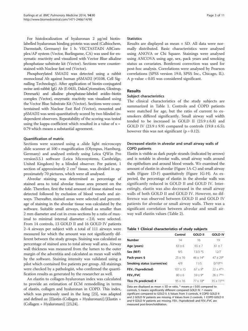
Table 1 Clinical characteristics of study subjects
Control GOLD II GOLD IV
Number 14 16 19
Age (years) 63 ± 6 65 ± 7 61 ± 7
Sex (M/F) 9/5 13/3 *$ 12/7
Pack-years § 25 ± 16 46 ± 14* 47 ± 29*
Smoking status (current/ex) 4/9 11/5 0/19*^
FEV1 (%predicted) 107 ± 15 67 ± 9* 22 ± 4*^
FEV1/FVC 80 ± 6 59 ± 9* 26 ± 7*^
Tlco (% predicted) # 95 ± 16 77 ± 18* 35 ± 15*^
Data are displayed as mean ± SD or ratio, * means p < 0.05 compared tocontrols, $ means significantly different compared GOLD IV. ^ meanssignificant compared to GOLD II. § Values from 3 controls, 4 COPD GOLD IIand 2 GOLD IV patients are missing. # Values from 2 controls, 1 COPD GOLD IIand 9 GOLD IV patients are missing. FEV1 (%predicted) and FEV1/FVC aremeasured post-broncholdilation.
Eurlings et al. BMC Pulmonary Medicine 2014, 14:90 Page 3 of 11http://www.biomedcentral.com/1471-2466/14/90
For histolocalization of hyaluronan 2 μg/ml biotin-labelled hyaluronan binding protein was used (Calbiochem,Darmstadt, Germany) for 1 h. VECTASTAIN ABCom-plex/AP system (Vector, Burlingame, CA) was used for en-zymatic reactivity and visualized with Vector Blue alkalinephosphatase substrate kit (Vector). Sections were counter-stained with Nuclear fast red (Vector).Phosphorylated SMAD2 was detected using a rabbit
monoclonal Ab against human pSMAD2 (#3108, Cell Sig-nalling Technology). After application of biotin-conjugatedswine anti-rabbit IgG Ab (E-0431, DakoCytomation, Glostrup,Denmark) and alkaline phosphatase-labeled avidin–biotincomplex (Vector), enzymatic reactivity was visualized usingthe Vector Blue Substrate Kit (Vector). Sections were coun-terstained with Nuclear Fast Red (Vector), mounted andpSMAD2 was semi-quantitatively scored by two blinded in-dependent observers. Repeatability of the scoring was testedusing the kappa coefficient which resulted in a value of κ =0.79 which means a substantial agreement.
Quantification of matrixSections were scanned using a .slide light microscopyslide scanner at 100 ×magnification (Olympus, Hamburg,Germany) and analysed entirely using Leica QWin Proversion3.5.1 software (Leica Microsystems, Cambridge,United Kingdom) by a blinded observer. Per patient, 1section of approximately 2 cm2 tissue, was divided in ap-proximately 70 pictures, which were all analysed.Alveolar staining was determined as percentage of
stained area to total alveolar tissue area present on theslide. Therefore, first the total amount of tissue stained wasdetected followed by exclusion of blood vessels and air-ways. Thereafter, stained areas were selected and percent-age of staining in the alveolar tissue was calculated by thesoftware. Suitable small airways, defined as smaller than2 mm diameter and cut in cross-sections by a ratio of max-imal to minimal internal diameter < 2.0, were selected.From 14 controls, 13 GOLD II and 16 GOLD IV patients2–4 airways per subject with a total of 111 airways weremeasured for which the amount was not significantly dif-ferent between the study groups. Staining was calculated aspercentage of stained area to total airway wall area. Airwaywall thickness was measured from the lumen to the outermargin of the adventitia and calculated as mean wall widthby the software. Staining intensity was validated using apilot which contained five patients per group. All stainingswere checked by a pathologist, who confirmed the quanti-fication results as generated by the researcher as well.An elastin to collagen-hyaluronan index was calculated
to provide an estimation of ECM remodelling in termsof elastin, collagen and hyaluronan in COPD. This index,which was previously used in the lung [23], was adaptedand defined as: [Elastin-(Collagen + Hyaluronan)]/[Elastin +(Collagen + Hyaluronan)] [23,24].
StatisticsResults are displayed as mean ± SD. All data were nor-mally distributed. Basic characteristics were analysedusing ANOVA or Chi Square. Stainings were analysedusing ANCOVA using age, sex, pack years and smokingstatus as covariates. Bonferoni correction was used forpost-hoc analysis. Correlations were analysed by Pearsoncorrelations (SPSS version 19.0, SPSS Inc., Chicago, IL).A p-value < 0.05 was considered significant.
ResultsSubject characteristicsThe clinical characteristics of the study subjects aresummarized in Table 1. Controls and COPD patientswere matched for age, but the ratio of current to ex-smokers differed significantly. Small airway wall widthtended to be increased in GOLD II (23.9 ± 6.8) andGOLD IV (23.9 ± 9.9) compared to controls (19.8 ± 6.5);however this was not significant (p = 0.12).
Decreased elastin in alveolar and small airway walls ofCOPD patientsElastin is visible as dark purple strands (indicated by arrows)and is notable in alveolar walls, small airway walls aroundthe epithelium and around blood vessels. We examined theamount of elastin in alveolar (Figure 1A-C) and small airwaywalls (Figure 1D-F) quantitatively (Figure 1G-H). As ex-pected, the percentage of elastin in the alveolar walls wassignificantly reduced in GOLD II and GOLD IV. Inter-estingly, elastin was also decreased in the small airwaywalls of both GOLD II and GOLD IV. However, no dif-ference was observed between GOLD II and GOLD IVpatients for alveolar or small airway walls. There was asignificant correlation between alveolar and small air-way wall elastin values (Table 2).

Figure 1 Decreased elastin in alveolar and small airway walls of COPD patients. Photomicrographs of elastin in alveolar walls of onerepresentative control subject (A), GOLD II patient (B), GOLD IV patient (C) and small airway walls of a control (D), a GOLD II patient(E) and a GOLD IV patient (F). Quantitative measured elastin content in alveolar walls (G) and small airway walls (H). Data were analysed usingANCOVA and results are expressed as means + SD; *P < 0.05 compared to control.
Eurlings et al. BMC Pulmonary Medicine 2014, 14:90 Page 4 of 11http://www.biomedcentral.com/1471-2466/14/90
Deposition of collagen in the alveolar walls in addition tosmall airway wallsDeposition of collagens (which could be either colla-gen I, II and/or III) indicated by arrows was observedin the pleura, perivascular, peribronchiolar in the basal
Table 2 Correlations between alveolar matrix and smallairway matrix in the whole study group
R p
Elastin 0.558 < 0.001
Collagen 0.612 < 0.001
Hyaluronan 0.523 < 0.001
Remodelling index 0.830 < 0.001
pSMAD2 0.656 < 0.001
Data were analysed using Pearson correlations.
membrane and connective tissue and in the alveolarwalls (Figure 2A-F). Interestingly, in the alveolar wallcollagen increased in GOLD II patients, which furtherprogressed in GOLD IV (Figure 2G). As expected, itis shown that there is excess collagen deposition inthe small airway walls of GOLD II and GOLD IV pa-tients compared to controls; however no significantdifference between GOLD stages was found (Figure 2H).In addition a significant correlation between alveolar andsmall airway collagen deposition was observed (Table 2).
Increased hyaluronan in both alveolar and small airwaywalls of COPD patientsHistochemical localization of hyaluronan was seen in thealveolar walls affiliated to capillaries; more prominentstaining was observed in small airway walls and blood

Figure 2 Deposition of collagen in the alveolar walls in addition to small airway walls. Photomicrographs of collagen in alveolar walls ofone representative control subject (A), GOLD II patient (B), GOLD IV patient (C) and small airway walls of a control (D), a GOLD II patient (E) anda GOLD IV patient (F). Quantitative measured collagen content in alveolar walls (G) and small airway walls (H). Data were analysed usingANCOVA and results are expressed as means + SD; *P < 0.05 compared to control, **P < 0.05 compared to control and GOLD II.
Eurlings et al. BMC Pulmonary Medicine 2014, 14:90 Page 5 of 11http://www.biomedcentral.com/1471-2466/14/90
vessels (indicated by arrows), which was highly compar-able to the localization of collagen (Figure 3A-F). Quan-titatively enhanced hyaluronan was shown in alveolar(Figure 3G) and small airway walls (Figure 3H) of GOLDII and GOLD IV patients. No differences were found be-tween GOLD II and GOLD IV patients. Again, a signifi-cant correlation between alveolar and small airwayhyaluronan deposition was observed (Table 2).
Elastin to collagen-hyaluronan index is decreased inCOPD patients and correlates with lung functionparametersNext the relationship between individual matrix com-pounds and FEV1 was shown to be significant (Table 3).Correlations were present for all matrix components in al-veolar and small airway walls with FEV1. However, in theCOPD patients group alone, no significant correlations
were found with exception of the inverse relation betweenalveolar collagen and FEV1.An elastin-to-collagen-hyaluronan index was calcu-
lated to provide an estimation of the extent of ECM re-modelling in COPD. Figure 4 shows that the index islower in both GOLD II and GOLD IV patients than incontrols. Furthermore, this index in contrast to the indi-vidual matrix components was significantly different be-tween GOLD II and GOLD IV patients for both alveolarand small airway walls. Importantly, the remodellingindex correlated with FEV1 as well as FEV1/FVC inCOPD (Figure 5).
Correlation between alveolar and small airway wallpSMAD2, despite absence of a relationship with COPDTo assess if the described remodelling is associated withincreased TGF-β signalling pSMAD2 was assessed by

Figure 3 Increased hyaluronan in both alveolar and small airway walls of COPD patients. Photomicrographs of hyaluronan in alveolar wallsof one representative control subject (A), GOLD II patient (B), GOLD IV patient (C) and small airway walls of a control (D), a GOLD II patient(E) and a GOLD IV patient (F). Quantitative measured hyaluronan content in alveolar walls (G) and small airway walls (H). Data were analysedusing ANCOVA and results are expressed as means + SD; *P < 0.05 compared to control.
Eurlings et al. BMC Pulmonary Medicine 2014, 14:90 Page 6 of 11http://www.biomedcentral.com/1471-2466/14/90
staining. Nuclear staining of pSMAD2 was seen through-out the lung, it was positive in alveolar macrophages andit was also observed associated with blood vessels, namelyin the endothelial layer, in the smooth muscle cells of themedia and in fibroblasts in the adventitia. For this study
Table 3 Correlations between matrix and FEV1
Whole study group n
R
Elastin alveolar 0.734
Elastin small airway 0.599
Collagen alveolar −0.757
Collagen small airway −0.694
Hyaluronan alveolar −0.656
Hyaluronan small airway −0.434
Data were analysed using Pearson correlations.
we focused on the staining which was observed in bron-chial epithelium and its submucosa, as well as in fibro-blasts, endothelial and type II epithelial cells in alveolarwalls (Figure 6A-B). No difference was seen between con-trols and COPD patients in both small airway and alveolar
= 49 COPD patients n = 35
p R P
< 0.001 0.107 0.553
< 0.001 0.318 0.106
< 0.001 −0.511 0.003
< 0.001 −0.219 0.262
< 0.001 −0.232 0.201
< 0.001 −0.194 0.333

Figure 4 Elastin to collagen-hyaluronan index is decreased in COPD patients. Elastin to Collagen-Hyaluronan index of controls, GOLD II andGOLD IV patients in alveolar walls (A) and small airway walls (B). Data were analysed using ANCOVA and results are expressed as means + SD;*P < 0.05 compared to control, **P < 0.05 compared to control and GOLD II.
Eurlings et al. BMC Pulmonary Medicine 2014, 14:90 Page 7 of 11http://www.biomedcentral.com/1471-2466/14/90
walls (Figure 6C-D). Interestingly, a significant correlationbetween alveolar and small airway pSMAD2 staining wasfound (Table 2).
Discussion and conclusionThis study was performed to compare matrix content inthe distal lung of GOLD II patients, GOLD IV patientsand controls. The data obtained show marked similar-ities in elastin, collagen and hyaluronan content changesbetween the alveolar and small airway compartment. Re-markably, only alveolar wall collagen was increased fur-ther in highly emphysematous patients compared topatients with moderate COPD and negatively correlatedwith lung function. All other matrix alterations were in-dependent of lung function.
This study showed decreased elastin not only in alveolarbut also in small airway walls of COPD patients. These re-sults are in contrast with a study in severe GOLD IV pa-tients showing increased elastic fiber density in alveolarwalls [25]. This discrepancy could be caused by differencein control groups between these studies, which in our casewere matched for age and smoking behaviour to the pa-tient groups. In addition the data presented in this studyshowed a positive correlation of elastin content with lungfunction parameters using the whole study group. InCOPD patients alone no significant correlations were de-termined. Previously, a study showed comparable correla-tions with lung function parameters, in the whole groupincluding controls [13]. Combined with the lack of differ-ence between GOLD II and GOLD IV patients, this
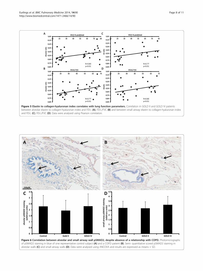
Figure 5 Elastin to collagen-hyaluronan index correlates with lung function parameters. Correlation in GOLD II and GOLD IV patientsbetween alveolar elastin to collagen-hyaluronan index and FEV1 (A), FEV1/FVC (B) and between small airway elastin to collagen-hyaluronan indexand FEV1 (C), FEV1/FVC (D). Data were analysed using Pearson correlation.
Figure 6 Correlation between alveolar and small airway wall pSMAD2, despite absence of a relationship with COPD. Photomicrographsof pSMAD2 staining in blue of one representative control subject (A) and a COPD patient (B). Semi- quantitative scored pSMAD2 staining inalveolar walls (C) and small airway walls (D). Data were analysed using ANCOVA and results are expressed as means + SD.
Eurlings et al. BMC Pulmonary Medicine 2014, 14:90 Page 8 of 11http://www.biomedcentral.com/1471-2466/14/90

Eurlings et al. BMC Pulmonary Medicine 2014, 14:90 Page 9 of 11http://www.biomedcentral.com/1471-2466/14/90
attenuation of small airway elastin content is thus morelikely a marker of disease, then of severity. A possibleunderlying mechanism for the decreased elastin contentin the small airway wall is the increased number ofneutrophils [26], especially neutrophil elastase [27]. Inaddition to loss of elastin, the number of small airwayswas reduced in mild and severe emphysematous patients[17]. The question remains whether the loss of elastin canlead to collapse or disappearance of small airways.Deposition of collagen and hyaluronan was increased
in small airway walls of COPD patients. This is an im-portant finding as a number of studies showed small air-way wall fibrosis, but there has been less research on thematrix composition. It was shown that there is an in-crease of collagen I and III precursors in the small air-ways of GOLD II patients; whereas in GOLD IV patientsthese precursors decreased [10], which can explain thelack of progression of collagen in the small airways asour data indicate. The current study shows elevated hya-luronan content in in addition to collagen. Within thelung collagen and elastin fibers are embedded in a hy-drated gel of which glycosaminoglycans are the majorconstituents. The composition of the matrix and thefiber to gel ratio changes during maturation and diseasestate [28]. Hyaluronan is the major glycosaminoglycan inlung tissue and has diverse functions in lung homeosta-sis and pulmonary disease. We previously showed thathyaluronan was increased in sputum of COPD patients[22]. Furthermore in cigarette smoke exposed mice therewas an increase in hyaluronan deposition in the smallairway wall after 4 weeks of exposure, which also wasnot progressive in the 6 month exposure model of em-physema [21].Increased collagen and hyaluronan deposition in the
alveolar wall indicates that besides airway fibrosis thereare fibrotic matrix changes in the alveolar walls whichare also present in the remaining walls in severelyobstructed patients with advanced emphysema. This isin line with previous research showing thickened alveo-lar walls in COPD patients and a decreased elastin-to-collagen index [23].In this study two different disease phenotypes were
analysed, a GOLD IV group with advanced emphysemaand a moderately obstructed GOLD II group. We ob-served no difference between these groups for the indi-vidual matrix components, showing that changes inmatrix composition even occur in patients with moder-ate airflow limitation. Only collagen in alveolar walls wasfurther increased in severely emphysematous patients. Inaddition, collagen area fraction in the alveolar walls wasthe only measure which correlated with TLCO withinthe COPD group (R = −0.493, p = 0.02). This indicatesthat next to destruction of alveolar walls, increased colla-gen content in remaining walls contributes to reduction of
gas transfer. Unfortunately, correction for the amount ofalveolar tissue left in these patients was impossible. In anelastase model it was shown that decreased elastin causedincreased alveolar collagen without inflammation. Fur-thermore, it appears that remodelled collagen fibres areweaker and break under the influence of mechanical stress[29]. The mechanism underlying remodelling of elastinand collagen, including subtyping composition and ultra-structure, should be investigated to support their involve-ment in emphysema development.In addition, a significantly decreased remodelling index
in COPD patients was found. This index was also signifi-cantly different for both small airway and alveolar walls ofCOPD patients versus controls, in line with individualmatrix components. More interestingly there was a signifi-cant difference in the remodelling index between GOLDII and GOLD IV patients which positively correlated withFEV1 and FEV1/FVC in the patient group. Although thedistribution was high, possibly due to the heterogeneousGOLD II population, the correlations were strong and sig-nificant. The individual matrix components on the otherhand did not differ significantly between GOLD stagesand did not correlate with lung function, indicating thatalthough the individual components are markers of dis-ease, even in early stages, it is the combination of matrixcomponent alterations that is related to progressive air-flow limitation.The amount of pSMAD2 staining in alveolar and small
airway walls was not different between controls andCOPD patients. Literature on the role of TGF-β inCOPD is conflicting; some studies found increased ex-pression of TGF-β in bronchial and alveolar epithelialcells [30,31] whereas others observed no differences inTGF-β levels in sputum, BALF and bronchial epithelialcells by immunostaining [32-36]. The lack of differencein pSMAD2 staining is in line with these latter publica-tions [35]. In the smoking mouse model of COPD thereis evidence for increased TGF-β expression and SMADsignaling [37]. Furthermore increased levels in TGF-βexpression were shown between never and currentsmokers [32] while no difference between smokers andCOPD patients was present. In this study using smokingcontrols, no differences were found in relation to thedisease. An effect of smoking per se cannot be excludedgiven the lack of a never smoking control group.The observed correlation between alveolar and small
airway wall matrix components invalidates previousthoughts that alveolar and small airway remodelling aretwo distinct processes which is in concert with some re-cent publications [13,16,17]. The finding that elastin isdecreased in both alveolar and small airway walls andthat collagen and hyaluronan are increased in both com-partments provides evidences that there are similarmatrix adaptations occurring simultaneously in alveoli

Eurlings et al. BMC Pulmonary Medicine 2014, 14:90 Page 10 of 11http://www.biomedcentral.com/1471-2466/14/90
and small airways. Furthermore, a positive correlationwas found in pSMAD2 staining between alveolar andsmall airway walls, indicating that not only remodellingin the small airways and alveolar walls can be seen asone unit but that also TGF-β signalling as shown bypSMAD2 is similar. The current study indicates that re-modelling and TGF-β signalling in the alveolar and smallairway wall in COPD are markedly similar and that re-modelling is already present in moderate COPD. Not-ably, alveolar collagen and a remodelling index relate toprogressive airflow limitation.
Competing interestsThe authors declare that they have no competing interests.
Authors’ contributionsIE performed immunohistochemical stainings, quantitative scoring andstatistical analysis, and drafted the manuscript. MD supervised experiments,participated in semi-quantitative immunohistochemical analysis and draftedthe manuscript. JC designed the programs for quantitative scorings. CPvalidated and assessed immunohistichemical stainings. GR participated inthe design of the study and drafted the manuscript. EW participated inthe design of the study, provided general supervision and drafted themanuscript. NR participated in the study design, interpretation of data andmanuscript preparation. All authors read and approved the final manuscript.
AcknowledgementsThis study was supported by the Weijerhorst foundation.
Author details1Nutrition and Toxicology Research Institute Maastricht (NUTRIM),Department of Respiratory Medicine, University Hospital Maastricht,P.O. Box 5800, 6202 AZ Maastricht, The Netherlands. 2Cardiovascular ResearchInstitute Maastricht (CARIM), Department of Pathology, Maastricht UniversityMedical Centre, Maastricht, the Netherlands.
Received: 12 July 2013 Accepted: 20 May 2014Published: 26 May 2014
References1. Mannino DM, Buist AS: Global burden of COPD: risk factors, prevalence,
and future trends. Lancet 2007, 370:765–773.2. Ranga V, Kleinerman J: Structure and function of small airways in health
and disease. Arch Pathol Lab Med 1978, 102:609–617.3. Hogg JC, Chu F, Utokaparch S, Woods R, Elliott WM, Buzatu L, Cherniack RM,
Rogers RM, Sciurba FC, Coxson HO, Pare PD: The nature of small-airwayobstruction in chronic obstructive pulmonary disease. N Engl J Med 2004,350:2645–2653.
4. Araya J, Cambier S, Markovics JA, Wolters P, Jablons D, Hill A, Finkbeiner W,Jones K, Broaddus VC, Sheppard D, Barzcak A, Xiao Y, Erle DJ, Nishimura SL:Squamous metaplasia amplifies pathologic epithelial-mesenchymalinteractions in COPD patients. J Clin Invest 2007, 117:3551–3562.
5. Bolton SJ, Pinnion K, Oreffo V, Foster M, Pinkerton KE: Characterisation ofthe proximal airway squamous metaplasia induced by chronic tobaccosmoke exposure in spontaneously hypertensive rats. Respir Res 2009,10:118.
6. Dekkers BG, Schaafsma D, Nelemans SA, Zaagsma J, Meurs H: Extracellularmatrix proteins differentially regulate airway smooth muscle phenotypeand function. Am J Physiol Lung Cell Mol Physiol 2007, 292:L1405–L1413.
7. Kranenburg AR, Willems-Widyastuti A, Moori WJ, Sterk PJ, Alagappan VK, deBoer WI, Sharma HS: Enhanced bronchial expression of extracellularmatrix proteins in chronic obstructive pulmonary disease. Am J ClinPathol 2006, 126:725–735.
8. Papakonstantinou E, Karakiulakis G: The ‘sweet’ and ‘bitter’ involvement ofglycosaminoglycans in lung diseases: pharmacotherapeutic relevance.Br J Pharmacol 2009, 157:1111–1127.
9. van Straaten JF, Coers W, Noordhoek JA, Huitema S, Flipsen JT, Kauffman HF,Timens W, Postma DS: Proteoglycan changes in the extracellular matrix of
lung tissue from patients with pulmonary emphysema. Mod Pathol 1999,12:697–705.
10. Harju T, Kinnula VL, Paakko P, Salmenkivi K, Risteli J, Kaarteenaho R:Variability in the precursor proteins of collagen I and III in differentstages of COPD. Respir Res 2010, 11:165.
11. Vestbo J, Hurd SS, Rodriguez-Roisin R: An overview of global strategy forthe diagnosis, management and prevention of chronic obstructivepulmonary disease (GOLD) (revised 2011). Zhonghua Yi Xue Za Zhi 2012,92:937–938.
12. Janoff A: Elastases and emphysema. Current assessment of the protease-antiprotease hypothesis. Am Rev Respir Dis 1985, 132:417–433.
13. Black PN, Ching PS, Beaumont B, Ranasinghe S, Taylor G, Merrilees MJ:Changes in elastic fibres in the small airways and alveoli in COPD.Eur Respir J 2008, 31:998–1004.
14. Annoni R, Lancas T, Yukimatsu Tanigawa R, de Medeiros Matsushita M, deMorais Fernezlian S, Bruno A, Fernando Ferraz da Silva L, Roughley PJ,Battaglia S, Dolhnikoff M, Hiemstra PS, Sterk PJ, Rabe KF, Mauad T:Extracellular matrix composition in COPD. Eur Respir J 2012, 40:1362–1373.
15. Karvonen HM, Lehtonen ST, Harju T, Sormunen RT, Lappi-Blanco E, Makinen JM,Laitakari K, Johnson S, Kaarteenaho RL: Myofibroblast expression in airwaysand alveoli is affected by smoking and COPD. Respir Res 2013, 14:84.
16. Sohal SS, Reid D, Soltani A, Ward C, Weston S, Muller HK, Wood-Baker R,Walters EH: Reticular basement membrane fragmentation and potentialepithelial mesenchymal transition is exaggerated in the airways ofsmokers with chronic obstructive pulmonary disease. Respirology 2010,15:930–938.
17. McDonough JE, Yuan R, Suzuki M, Seyednejad N, Elliott WM, Sanchez PG,Wright AC, Gefter WB, Litzky L, Coxson HO, Pare PD, Sin DD, Pierce RA,Woods JC, McWilliams AM, Mayo JR, Lam SC, Cooper JD, Hogg JC: Small-airway obstruction and emphysema in chronic obstructive pulmonarydisease. N Engl J Med 2011, 365:1567–1575.
18. Lang MR, Fiaux GW, Gillooly M, Stewart JA, Hulmes DJ, Lamb D: Collagencontent of alveolar wall tissue in emphysematous and non-emphysematous lungs. Thorax 1994, 49:319–326.
19. Merrilees MJ, Ching PS, Beaumont B, Hinek A, Wight TN, Black PN: Changesin elastin, elastin binding protein and versican in alveoli in chronicobstructive pulmonary disease. Respir Res 2008, 9:41.
20. Hallgren O, Nihlberg K, Dahlback M, Bjermer L, Eriksson LT, Erjefalt JS,Lofdahl CG, Westergren-Thorsson G: Altered fibroblast proteoglycanproduction in COPD. Respir Res 2010, 11:55.
21. Bracke KR, Dentener MA, Papakonstantinou E, Vernooy JH, Demoor T,Pauwels NS, Cleutjens J, van Suylen RJ, Joos GF, Brusselle GG, Wouters EF:Enhanced deposition of low-molecular-weight hyaluronan in lungs ofcigarette smoke-exposed mice. Am J Respir Cell Mol Biol 2010, 42:753–761.
22. Dentener MA, Vernooy JH, Hendriks S, Wouters EF: Enhanced levels ofhyaluronan in lungs of patients with COPD: relationship with lungfunction and local inflammation. Thorax 2005, 60:114–119.
23. Abraham T, Hogg J: Extracellular matrix remodeling of lung alveolar wallsin three dimensional space identified using second harmonic generationand multiphoton excitation fluorescence. J Struct Biol 2010, 171:189–196.
24. Lin SJ, Wu R Jr, Tan HY, Lo W, Lin WC, Young TH, Hsu CJ, Chen JS, Jee SH,Dong CY: Evaluating cutaneous photoaging by use of multiphotonfluorescence and second-harmonic generation microscopy. Opt Lett 2005,30:2275–2277.
25. Deslee G, Woods JC, Moore CM, Liu L, Conradi SH, Milne M, Gierada DS, Pierce J,Patterson A, Lewit RA, Battaile JT, Holtzman MJ, Hogg JC, Pierce RA: Elastinexpression in very severe human COPD. Eur Respir J 2009, 34:324–331.
26. Baraldo S, Turato G, Badin C, Bazzan E, Beghe B, Zuin R, Calabrese F, Casoni G,Maestrelli P, Papi A, Fabbri LM, Saetta M: Neutrophilic infiltration within theairway smooth muscle in patients with COPD. Thorax 2004, 59:308–312.
27. Nahori MA, Renesto P, Vargaftig BB, Chignard M: Activation and damageof cultured airway epithelial cells by human elastase and cathepsin G.Eur J Pharmacol 1992, 228:213–218.
28. Hukins DW, Aspden RM, Yarker YE: Fibre reinforcement and mechanicalstability in articular cartilage. Eng Med 1984, 13:153–156.
29. Kononov S, Brewer K, Sakai H, Cavalcante FS, Sabayanagam CR, Ingenito EP,Suki B: Roles of mechanical forces and collagen failure in thedevelopment of elastase-induced emphysema. Am J Respir Crit Care Med2001, 164:1920–1926.
30. Takizawa H, Tanaka M, Takami K, Ohtoshi T, Ito K, Satoh M, Okada Y,Yamasawa F, Nakahara K, Umeda A: Increased expression of transforming

Eurlings et al. BMC Pulmonary Medicine 2014, 14:90 Page 11 of 11http://www.biomedcentral.com/1471-2466/14/90
growth factor-beta1 in small airway epithelium from tobacco smokersand patients with chronic obstructive pulmonary disease (COPD).Am J Respir Crit Care Med 2001, 163:1476–1483.
31. de Boer WI, van Schadewijk A, Sont JK, Sharma HS, Stolk J, Hiemstra PS,van Krieken JH: Transforming growth factor beta1 and recruitment ofmacrophages and mast cells in airways in chronic obstructivepulmonary disease. Am J Respir Crit Care Med 1998, 158:1951–1957.
32. Aubert JD, Dalal BI, Bai TR, Roberts CR, Hayashi S, Hogg JC: Transforminggrowth factor beta 1 gene expression in human airways. Thorax 1994,49:225–232.
33. Buhling F, Tholert G, Kaiser D, Hoffmann B, Reinhold D, Ansorge S, Welte T:Increased release of transforming growth factor (TGF)-beta1, TGF-beta2,and chemoattractant mediators in pneumonia. J Interferon Cytokine Res1999, 19:271–278.
34. Kokturk N, Tatlicioglu T, Memis L, Akyurek N, Akyol G: Expression oftransforming growth factor beta1 in bronchial biopsies in asthma andCOPD. J Asthma 2003, 40:887–893.
35. Zandvoort A, Postma DS, Jonker MR, Noordhoek JA, Vos JT, van der GeldYM, Timens W: Altered expression of the Smad signalling pathway:implications for COPD pathogenesis. European Respir J 2006, 28:533–541.
36. Ziora D, Dworniczak S, Kaczmarczyk G, Jastrzebski D, Krzywiecki A, Kozielski J:Correlation of exhaled nitric oxide with nitrogen oxides and selectedcytokines in induced sputum of chronic obstructive pulmonary diseasepatients. J Physiol Pharmacol 2007, 58(Suppl 5):791–799.
37. Mortaz E, Givi ME, Da Silva CA, Folkerts G, Redegeld FA: A relation betweenTGF-beta and mast cell tryptase in experimental emphysema models.Biochim Biophys Acta 1822, 2012:1154–1160.
doi:10.1186/1471-2466-14-90Cite this article as: Eurlings et al.: Similar matrix alterations in alveolarand small airway walls of COPD patients. BMC Pulmonary Medicine2014 14:90.
Submit your next manuscript to BioMed Centraland take full advantage of:
• Convenient online submission
• Thorough peer review
• No space constraints or color figure charges
• Immediate publication on acceptance
• Inclusion in PubMed, CAS, Scopus and Google Scholar
• Research which is freely available for redistribution
Submit your manuscript at www.biomedcentral.com/submit