research article open access safety and efficacy of the
TRANSCRIPT

RESEARCH ARTICLE Open Access
Safety and efficacy of the immunosuppressiveagent 6-tioguanine in murine model of acute andchronic colitisMiloslav Kverka1*, Pavel Rossmann1, Helena Tlaskalova-Hogenova1, Klara Klimesova1, Bindia Jharap2,Nanne K de Boer2, Rene M Vos3, Adriaan A van Bodegraven2, Milan Lukas4 and Chris J Mulder2
Abstract
Background: Oral thiopurines are effective and widely used in treatment of inflammatory bowel disease (IBD) inhumans, although their use is limited due the development of adverse events. Here, we examine the efficacy andtoxicity of oral treatment with 6-tioguanine (6-TG) and azathioprine (AZA) in a murine model of IBD.
Methods: We induced acute or chronic colitis in BALB/c mice by one or four cycles of 3% dextran sulphatesodium (DSS), respectively. Mice were treated by daily gavages of various dosages of 6-tioguanine, azathioprine, orby phosphate buffered saline (PBS) starting the first day of DSS or after two cycles of DSS, respectively. Wemonitored the efficacy and toxicity by measuring the weight change and serum alanine aminotransferase (ALT)activity and by disease severity and histology, at the end of the experiment. Moreover, we measured cytokineproduction after colon fragment cultivation by enzyme-linked immunoabsorbent assay and numbers of apoptoticcells in the spleen by flow cytometry.
Results: 6-TG is effective in the treatment of acute DSS-induced colitis in a dose-dependent manner and 40 μg of6-TG is significantly more effective in the treatment of acute colitis than both AZA and PBS. This effect isaccompanied by decrease of IL-6 and IFN-g production in colon. We did not observe histological abnormalities inliver samples from control (PBS) or 6-TG treated mice. However, liver samples from most mice treated with AZAshowed mild, yet distinct signs of hepatotoxicity. In chronic colitis, all thiopurine derivatives improved colitis, 20 μgof 6-TG per dose was superior. High doses of 6-TG led to significant weight loss at the end of the therapy, butnone of the thiopurine derivatives increased levels of serum ALT. Both thiopurine derivatives reduced theproportion of apoptotic T helper cells, but a high production of both IL-6 and TGF-b was observed only in colonof AZA-treated mice.
Conclusions: Use of 6-TG in the treatment of experimental colitis in mice appears superior to AZA administrationand placebo. In contrast to 6-TG, the use of AZA resulted in histological liver abnormalities.
BackgroundThe immune-modulating thiopurines, 6-mercaptopurine(6-MP), and its pro-drug azathioprine (AZA), are widelyused in inflammatory bowel diseases (IBD) treatment[1-3]. Both AZA and 6-MP require extensive metaboli-sation before the pharmacologicaly active metabolites, 6-thioguaninenucleotides (6-TGN), are generated.
Their mechanism of action is ascribed to both cyto-toxic and apoptototic pathways. Owing to their struc-tural similarity to endogenous purine bases, 6-TGN areincorporated into DNA or RNA as fraudulent bases,ultimately leading to cytotoxicity. Activity of the specificmetabolite, 6-thioguanine-triphosphate was recentlyfound to contribute to the overall molecular immuno-suppressive effect. This end-metabolite induces apopto-sis and decreases the expression of proinflammatorymolecules in activated T cells [4,5].
* Correspondence: [email protected] of Immunology and Gnotobiology, Institute of Microbiology,Academy of Sciences of the Czech Republic, Videnska 1083, 14220 Prague 4,Czech RepublicFull list of author information is available at the end of the article
Kverka et al. BMC Gastroenterology 2011, 11:47http://www.biomedcentral.com/1471-230X/11/47
© 2011 Kverka et al; licensee BioMed Central Ltd. This is an Open Access article distributed under the terms of the Creative CommonsAttribution License (http://creativecommons.org/licenses/by/2.0), which permits unrestricted use, distribution, and reproduction inany medium, provided the original work is properly cited.
Although thiopurine derivatives are considered to be arelatively safe maintenance therapy, several studiesreport discontinuation of thiopurine derivatives in up to50% of patients during long-term therapy, mainly due tothe development of adverse events [6-8].Another thiopurine, 6-tioguanine (6-TG), has been
proposed as a rescue drug for IBD patients failing totolerate or respond to AZA and 6-MP [9,10]. However,this suggestion has been discouraged, when histologicalliver abnormalities, in particular nodular regenerativehyperplasia (NRH), were found in 6-TG treated IBDpatients [11,12]. These findings were verified by Ger-man and Austrian studies [13], but not by Irish andDutch studies with a follow-up of 3-5 years [14-18].Debate is ongoing whether 6-TG is potentially morehepatotoxic than other thiopurine derivatives such asconventional AZA or 6-MP, or whether its allegedhepatotoxicity is dose-dependent [19]. We thereforedesigned an AZA- and placebo-controlled study usingdifferent doses of 6-TG to assess the therapeutic effi-cacy and (hepato) toxicity in the murine model ofacute and chronic dextran sulphate sodium (DSS)-induced colitis.
MethodsMiceWe used conventional female, 3-month-old BALB/cmice (Institute of Physiology AS CR, Prague, CzechRepublic) in this study. The experiments were approvedby the Institutional animal care and use committee atthe Academy of Sciences of the Czech Republic.
Experimental designAcute colitis was induced by 3% (weight/volume) dex-tran sulfate sodium (DSS) (molecular weight 36-50 kDa;MP Biomedicals, Inc.) dissolved in drinking water for 9days. Starting from the first day of DSS administration,we administered 6-TG in a daily dosage of 10 μg, 20 μgor 40 μg (obtained from Mosadex C.V., Elsloo, TheNetherlands), or AZA (Imuran® Glaxo-SmithKline) in adaily dosage of 30 μg or 60 μg. Both agents were dis-solved in 100 μl of sterile phosphate buffered saline(PBS) and administered by daily gavage. Control groupwas treated only with PBS. Taking into account theaverage mice weight, the dose per kilogram for 10 μg,20 μg, 40 μg of 6-TG, 30 μg or 60 μg AZA was 0.45mg/kg, 0.91 mg/kg, 1.82 mg/kg, 1.37 mg/kg or 2.73 mg/kg, respectively.Chronic colitis was induced by four cycles of 3% DSS
(5 days DSS, 9 days water). The treatment with dailyoral dose of either PBS, 20 μg of 6-TG, 40 μg of 6-TGor 60 μg AZA started after the 2nd cycle of DSS, oncethe chronic colitis was established. Each mouse there-fore received during these 22 doses a cumulative dose of
either 19.8 or 39.3 mg/kg of 6-TG, or 58.1 mg/kg ofAZA, respectively.
6-TG metabolite monitoringTo analyse the concentration of 6-tioguanine nucleo-tides in red blood cells, we collected blood samples atthe last day of experiment in EDTA. The samples werecentrifuged to isolate erythrocytes and after washingwith PBS, erythrocyte counts were done. Samples werethan stored at -80°C until analysis, performed asdescribed previously [20].
Assessment of colitis severityColitis was evaluated on the last day of the experiment byusing a disease activity index (DAI), colon length, andhistological scoring system. The DAI scores body weightloss, stool consistency, and the presence of the blood inthe stool, as previously described by Cooper et al. [21].Occult blood in faeces was evaluated with Faecal OccultBlood Test (Okult-viditest Rapid; Vidia, Vestec, CzechRepublic). No deaths occurred during the experiment.Following sacrificing, the entire colon was removed
(from caecum to anus) and placed without tension on aruler. Colon length was measured as an indirect markerof inflammation. The descending colon was fixed in 4%formalin, and stained with haematoxylin and eosin (HE)to evaluate mucosal damage during acute colitis or withSirius red (Sigma-Aldrich) and Haematoxylin to evaluatemucosal damage in chronic colitis model. The micro-scopic findings in acute colitis were assessed semi-quan-titatively and weighted score developed in ourlaboratory for each section was obtained, ranging from 0(no signs of colitis) to 3 (severe colitis) as described pre-viously [22]. To describe mucosal damage in chroniccolitis model we used different scoring system todescribe, based on the scoring system validated by Die-leman et al. [23]. Additionally, Sirius red staining wasadded to score collagen deposition. Briefly, we scoredthe grade of five parameters (inflammation, extent,regeneration, crypt damage and collagen deposition).Each of these changes was also multiplied by the per-centage quantifying the disease involvement on crosssections of colon: (1) 1-25%; (2) 26-50%; (3) 51-75%; (4)76-100%. The final score was calculated by the sum ofthe scores for all five parameters.We used mouse haptoglobin enzyme-linked immu-
noabsorbent assay (ELISA; Alpco diagnostic, Salem, NH,USA) to analyse the concentration of acute phase pro-tein haptoglobin, a marker of inflammation, in serum ofmice with chronic colitis.
Histological assessment of liver toxicityLivers were fixed in 4% formalin, embedded in paraffinand stained with haematoxylin and eosin or with
Kverka et al. BMC Gastroenterology 2011, 11:47http://www.biomedcentral.com/1471-230X/11/47
Page 2 of 9

Gomori’s silver impregnation for reticulin (4 tissue sec-tions per mice were evaluated). Two expert liver pathol-ogists, unaware of the treatment of the mice, were askedto meticulously evaluate the liver samples independentlywith special focus on the presence of sinusoidal dilata-tion, veno-occlusive disease, fibrosis, cirrhosis, NRH,steatosis, necrosis and cholestasis.
Serological assessment of liver toxicityTo monitor the hepatotoxic effects in thiopurine treatedanimals, we measured the alanine transaminase (ALT)enzyme activity in serum by MaxDiscovery AlanineTransaminase Enzymatic Assay Kit (Bio Scientific, Aus-tin, TX, USA) according to manufacturer’s recommen-dations. We measured ALT activity at sacrifice and/orat the time of the 7th, 14th and 22nd dose during chroniccolitis therapy.
Analysis of cell apoptosis by Flow cytometrySingle-cell suspensions of spleen was prepared andstained for apoptotic cells using following fluorochromelabelled anti-mouse mAbs: CD3-FITC (BD Biosciences,San Jose, CA, USA), CD28-PE, CD8-PerCP.Cy5.5 (allfrom eBioscience, San Diego, CA, USA), CD4-Qdot®
605 and annexin V-Alexa Fluor® 647 (both InvitrogenCorp., Carlsbad, CA, USA) according to the manufac-turer’s recommendation. Hoechst 33258 (Sigma-Aldrich,St. Louis, MO, USA) was added just before analysis tostain for dead cells. Flow cytometric analysis was per-formed on LSRII (BD Biosciences), and data were ana-lyzed using FlowJo software (Tree Star Inc., Ashland,OR, USA).
Cytokine productionAt the end of the experiment, sections of mouse colonwere obtained, cut open longitudinally, washed in PBScontaining penicillin and streptomycin and weighed.
These tissue fragments were then cultivated for 48 h in ahumidified incubator at 37°C and 5% CO2 in RPMI-1640(Sigma-Aldrich) containing 10% fetal bovine serum (Bio-chrom AG, Berlin, Germany) and 1% Antibiotic-Antimy-cotic solution (Sigma-Aldrich). The supernatants werecollected and stored at -20°C until analysis for cytokineproduction. Levels of selected cytokines were determinedusing commercially available ELISA sets purchased fromInvitrogen (TNF-a, TGF-b, IL-10; Invitrogen Corp.) orR&D Systems (IFN-g, IL-6; R&D Systems Inc., Minnea-polis, MN, USA). All tests were performed according tothe manufacturers’ recommendations.
Statistical analysisValues are presented as mean ± standard deviation (SD).Differences in colon length, DAI and histological score,cell populations, ALT activity and haptoglobin levels ofmultiple groups were compared with one-way analysisof variance (ANOVA) with Tukey’s multiple comparisontest. Differences in weight change during time of multi-ple groups were compared to the control group (PBS/DSS) by two-way repeated measure ANOVA with Bon-ferroni’s post-hoc test. Differences were considered sta-tistically significant at P < 0.05. GraphPad Prismstatistical software (version 5.03, GraphPad Software,Inc. La Jolla, CA, USA) was used for analyses.
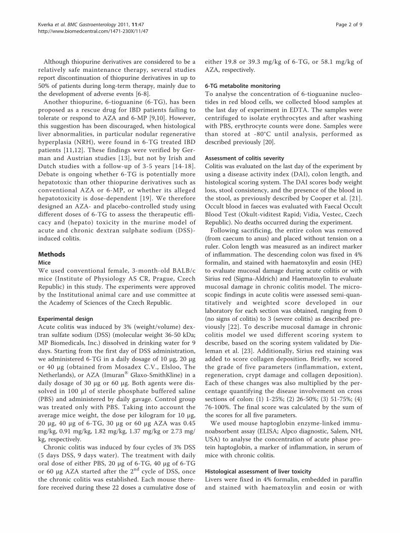
Results6-TG is more effective and less toxic than AZA in acutecolitis modelThe dosage of 40 μg of 6-TG daily was significantlymore effective in the treatment of acute DSS colitis thaneither AZA 30 or AZA 60 μg or PBS (Figure 1A-C andFigure 2A-C). The efficacy of 6-TG therapy correlateswith 6-TG dosage, the mice treated with 40 μg of 6-TGhad significantly longer colons and lower DAI thanthose treated with 10 μg of 6-TG per day.
Figure 1 Evaluation of treatment efficacy in acute DSS-induced colitis. Values are expressed as mean (bar) ± standard deviation (whisker).(A) DAI; (B) Colon length (cm) and (C) Histological grade. The differences among experimental groups are analyzed with one-way analysis ofvariance (ANOVA) with Tukey’s multiple comparison test (*P < 0.05; **P < 0.01; ***P < 0.001). Data are pool of four independent experiments;numbers in bars indicate number of animals per group.
Kverka et al. BMC Gastroenterology 2011, 11:47http://www.biomedcentral.com/1471-230X/11/47
Page 3 of 9
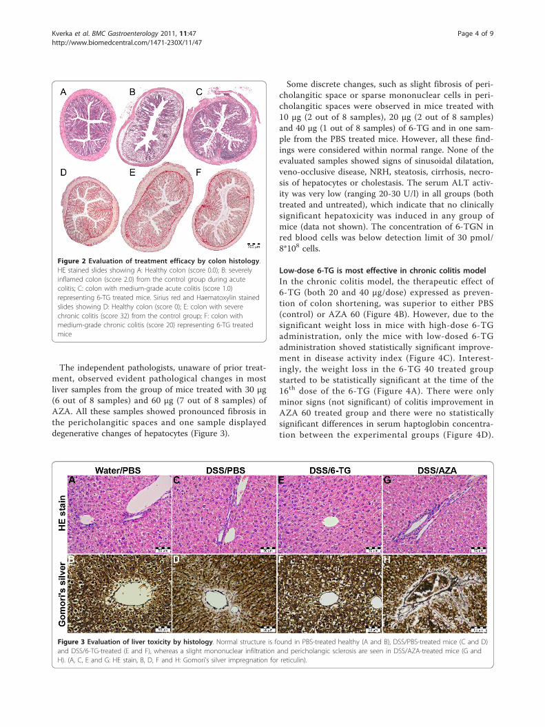
The independent pathologists, unaware of prior treat-ment, observed evident pathological changes in mostliver samples from the group of mice treated with 30 μg(6 out of 8 samples) and 60 μg (7 out of 8 samples) ofAZA. All these samples showed pronounced fibrosis inthe pericholangitic spaces and one sample displayeddegenerative changes of hepatocytes (Figure 3).
Some discrete changes, such as slight fibrosis of peri-cholangitic space or sparse mononuclear cells in peri-cholangitic spaces were observed in mice treated with10 μg (2 out of 8 samples), 20 μg (2 out of 8 samples)and 40 μg (1 out of 8 samples) of 6-TG and in one sam-ple from the PBS treated mice. However, all these find-ings were considered within normal range. None of theevaluated samples showed signs of sinusoidal dilatation,veno-occlusive disease, NRH, steatosis, cirrhosis, necro-sis of hepatocytes or cholestasis. The serum ALT activ-ity was very low (ranging 20-30 U/l) in all groups (bothtreated and untreated), which indicate that no clinicallysignificant hepatoxicity was induced in any group ofmice (data not shown). The concentration of 6-TGN inred blood cells was below detection limit of 30 pmol/8*108 cells.
Low-dose 6-TG is most effective in chronic colitis modelIn the chronic colitis model, the therapeutic effect of6-TG (both 20 and 40 μg/dose) expressed as preven-tion of colon shortening, was superior to either PBS(control) or AZA 60 (Figure 4B). However, due to thesignificant weight loss in mice with high-dose 6-TGadministration, only the mice with low-dosed 6-TGadministration shoved statistically significant improve-ment in disease activity index (Figure 4C). Interest-ingly, the weight loss in the 6-TG 40 treated groupstarted to be statistically significant at the time of the16th dose of the 6-TG (Figure 4A). There were onlyminor signs (not significant) of colitis improvement inAZA 60 treated group and there were no statisticallysignificant differences in serum haptoglobin concentra-tion between the experimental groups (Figure 4D).
Figure 2 Evaluation of treatment efficacy by colon histology.HE stained slides showing A: Healthy colon (score 0.0); B: severelyinflamed colon (score 2.0) from the control group during acutecolitis; C: colon with medium-grade acute colitis (score 1.0)representing 6-TG treated mice. Sirius red and Haematoxylin stainedslides showing D: Healthy colon (score 0); E: colon with severechronic colitis (score 32) from the control group; F: colon withmedium-grade chronic colitis (score 20) representing 6-TG treatedmice
Figure 3 Evaluation of liver toxicity by histology. Normal structure is found in PBS-treated healthy (A and B), DSS/PBS-treated mice (C and D)and DSS/6-TG-treated (E and F), whereas a slight mononuclear infiltration and pericholangic sclerosis are seen in DSS/AZA-treated mice (G andH). (A, C, E and G: HE stain, B, D, F and H: Gomori’s silver impregnation for reticulin).
Kverka et al. BMC Gastroenterology 2011, 11:47http://www.biomedcentral.com/1471-230X/11/47
Page 4 of 9

Nevertheless, all thiopurine derivatives reduced muco-sal damage (Figure 4E). Samples of liver after 22 thio-purine-doses were considered normal. None of thesamples showed signs of sinusoidal dilatation, veno-occlusive disease, NRH, steatosis, cirrhosis, or necrosisof hepatocytes or cholestasis. Activity of ALT in serumwas normal (between 20-30 U/l) and did not increasein any group throughout the whole treatment period,suggesting that there was no hepatotoxicity in anygroup (data not shown). The concentration of 6-TGNin red blood cells was below detection limit of 30pmol/8*108 cells.
6-TG and AZA changes the cytokine production in thecolonThere is a statistically significantly lower production ofIL-6 and IFN-g in DSS/6-TG 40 treated mice, as com-pared to DSS/PBS- or DSS/AZA 60 treated mice in theacute colitis model (Figure 5A). These changes in pro-inflammatory cytokines correlated with severity of coli-tis, which was much milder in DSS/6-TG treated ani-mals. In the chronic colitis model, a decreasedproduction of IL-6 in DSS/6-TG 20 treated mice ascompared to either DSS/PBS- or DSS/AZA 60-treatedmice was observed (Figure 5B). Production of IL-6 in
Figure 4 Evaluation of treatment efficacy in chronic DSS-induced colitis. Values are expressed as mean (dot or bar) ± SD (whisker). (A)Weight change during therapy. Body weight change was calculated for each mouse by dividing its body weight on the specified day by bodyweight at day 0 (body weight at the beginning of therapy) and expressed as percentage. (B) DAI (C) Colon Length (cm) (D) Serum haptoglobinconcentration (mg/ml) and (E) Histological grade. The differences among experimental groups are analyzed with either two-way analysis ofvariance (ANOVA) with Bonferroni post-hoc test (Weight change; *P < 0.05; **P < 0.01; ***P < 0.001 vs. PBS-treated) or one-way ANOVA withTukey’s multiple comparison test (*P < 0.05; **P < 0.01; ***P < 0.001). Data are pooled from two independent experiments, each with 5 mice pergroup.
Kverka et al. BMC Gastroenterology 2011, 11:47http://www.biomedcentral.com/1471-230X/11/47
Page 5 of 9
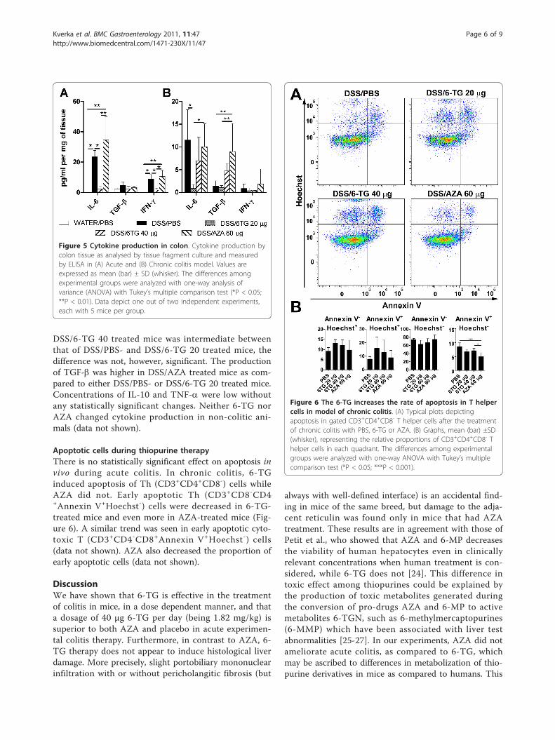
DSS/6-TG 40 treated mice was intermediate betweenthat of DSS/PBS- and DSS/6-TG 20 treated mice, thedifference was not, however, significant. The productionof TGF-b was higher in DSS/AZA treated mice as com-pared to either DSS/PBS- or DSS/6-TG 20 treated mice.Concentrations of IL-10 and TNF-a were low withoutany statistically significant changes. Neither 6-TG norAZA changed cytokine production in non-colitic ani-mals (data not shown).
Apoptotic cells during thiopurine therapyThere is no statistically significant effect on apoptosis invivo during acute colitis. In chronic colitis, 6-TGinduced apoptosis of Th (CD3+CD4+CD8-) cells whileAZA did not. Early apoptotic Th (CD3+CD8-CD4+Annexin V+Hoechst-) cells were decreased in 6-TG-treated mice and even more in AZA-treated mice (Fig-ure 6). A similar trend was seen in early apoptotic cyto-toxic T (CD3+CD4-CD8+Annexin V+Hoechst-) cells(data not shown). AZA also decreased the proportion ofearly apoptotic cells (data not shown).
DiscussionWe have shown that 6-TG is effective in the treatmentof colitis in mice, in a dose dependent manner, and thata dosage of 40 μg 6-TG per day (being 1.82 mg/kg) issuperior to both AZA and placebo in acute experimen-tal colitis therapy. Furthermore, in contrast to AZA, 6-TG therapy does not appear to induce histological liverdamage. More precisely, slight portobiliary mononuclearinfiltration with or without pericholangitic fibrosis (but
always with well-defined interface) is an accidental find-ing in mice of the same breed, but damage to the adja-cent reticulin was found only in mice that had AZAtreatment. These results are in agreement with those ofPetit et al., who showed that AZA and 6-MP decreasesthe viability of human hepatocytes even in clinicallyrelevant concentrations when human treatment is con-sidered, while 6-TG does not [24]. This difference intoxic effect among thiopurines could be explained bythe production of toxic metabolites generated duringthe conversion of pro-drugs AZA and 6-MP to activemetabolites 6-TGN, such as 6-methylmercaptopurines(6-MMP) which have been associated with liver testabnormalities [25-27]. In our experiments, AZA did notameliorate acute colitis, as compared to 6-TG, whichmay be ascribed to differences in metabolization of thio-purine derivatives in mice as compared to humans. This
Figure 5 Cytokine production in colon. Cytokine production bycolon tissue as analysed by tissue fragment culture and measuredby ELISA in (A) Acute and (B) Chronic colitis model. Values areexpressed as mean (bar) ± SD (whisker). The differences amongexperimental groups were analyzed with one-way analysis ofvariance (ANOVA) with Tukey’s multiple comparison test (*P < 0.05;**P < 0.01). Data depict one out of two independent experiments,each with 5 mice per group.
Figure 6 The 6-TG increases the rate of apoptosis in T helpercells in model of chronic colitis. (A) Typical plots depictingapoptosis in gated CD3+CD4+CD8- T helper cells after the treatmentof chronic colitis with PBS, 6-TG or AZA. (B) Graphs, mean (bar) ±SD(whisker), representing the relative proportions of CD3+CD4+CD8- Thelper cells in each quadrant. The differences among experimentalgroups were analyzed with one-way ANOVA with Tukey’s multiplecomparison test (*P < 0.05; ***P < 0.001).
Kverka et al. BMC Gastroenterology 2011, 11:47http://www.biomedcentral.com/1471-230X/11/47
Page 6 of 9

may be also the reason behind our inability to detect 6-TGN in red blood cells of mice treated with thiopurinederivates.Although all key enzymes are present in mice, it is dif-
ficult to compare mice pharmacology with that inhuman, which makes the comparison of dosages used inthis experiment difficult compared to studies in humans.Nevertheless, the use of AZA in mice clearly resulted inhistological liver abnormalities, while the use of 6-TGdid not. Several studies about hepatological abnormal-ities induced by AZA or 6-MP have been recently pub-lished, which confirmed the results we observed in thismouse model [28-31].The prevalence of NRH in the rest of the population
without IBD is 2.6%, while it is 6% in the thiopurine-naïve IBD patients, 0-7% in IBD patients treated withlow dose (approx. 20 mg/day) of 6-TG and in 18%-62%of patients with 40-80 mg/day of 6-TG [13,19,32,33].These findings suggest that IBD itself and high dose of6-TG, but not 6-TG in general, could be considered asa risk factor for NRH. The treatment duration shouldbe also mentioned, because in human IBD, thiopurinesare often administered for years, and although it waslong enough to cause liver injury in AZA treated mice,nine days may not be a relevant time for induction ofNRH.Despite the fact that there were no signs of liver
damage in chronic colitis, the mice with high dose of 6-TG had statistically significant decrease in body weightat the end of the experiment. This is in agreement withthe fact that high doses of 6-TG can have general toxiceffects, not related to the liver in these mice. We havenot found any pathology in liver, lungs or bone marroweven with extreme dosage of 100 μg of 6-TG daily, how-ever, we found hypocelularity in thymus and less acti-vated follicles with wide mantle zones and arrest ofhaematopoiesis and deposition of hemosiderin in spleen(data not shown). These changes were mostly associatedwith high dose of 6-TG, and less in low dose of 6-TGand in AZA and could represent the consequence of theimmunomodulatory properties of thiopurines.We observed a decrease in the number of early apop-
totic cells in the spleen of AZA-treated mice during thecourse of chronic colitis. This decrease was apparent inall thiopurine-treated geoups, when we gated to (CD3+CD4+CD8-) Th cells. Since the induction of apoptosisin activated Th cells is considered to be mechanism ofthiopurine anti-inflammatory action in vitro [4,5], ourfindings suggest the existence of compensatory mechan-isms in vivo.Both 6-TG and AZA changed cytokine production in
the colon of mice during inflammation. High levels ofIL-6 and IFN-g are produced in BALB/c mice duringDSS-induced inflammation as we reported previously
[22]. Since we observed a decrease in pro-inflammatorycytokines after 6-TG therapy in the acute and, to someextent, also in the chronic colitis model, this may corro-borate the anti-inflammaory potential of thiopurine deri-vatives. Increase in TGF-b production in AZA-treatedmice with chronic colitis could be caused by severalmechanisms. TGF-b has many functions in gut mucosahomeostasis, it serves as growth factor for both epithe-lial and mesenchymal cells facilitating repair of mucosalinjury and collagen deposition in IBD patients [34,35].TGF-b has also important immunomodulatory func-tions, because it can dampen the inflammation by indu-cing Treg cells. In contrast to this, in a presence of highlevels of IL-6, TGF-b might switch T cells to Th17 cells,thus promoting the protective immune response andinflammation [36,37].Since the vast majority of AZA and 6-MP-intolerant
IBD patients are able to tolerate maintenance treatmentwith 6-TG, the 6-TG was proposed as an alternativetreatment of IBD in humans in case of intolerance orlack of therapeutic effect of AZA or 6-MP[10,14,18,38,39]. Our findings provide additional evi-dence that low dose 6-TG might still be considered astherapeutic option in those IBD patients who are intol-erant or refractory to AZA or 6-MP.
ConclusionsIn conclusion, the use of 6-TG in the treatment ofexperimental colitis in mice appears superior to AZAadministration and placebo. Beneficial effect of 6-TGtreatment was associated with decrease in pro-inflam-matory cytokines in colons of treated mice. Counterin-tuitively, the thiopurines decreased the number of earlyapoptotic T helper cells. Interestingly, in contrast to 6-TG, the use of AZA resulted in histological liverabnormalities.
AbbreviationsIBD: inflammatory bowel disease; 6-TG: 6-tioguanine; AZA: azathioprine; DSS:dextran sulphate sodium; PBS: phosphate buffered saline; ALT: alanineaminotransferase; 6-MP: 6-mercaptopurine; 6-TGN: 6-thioguaninenucleotides;NRH: nodular regenerative hyperplasia; DAI: disease activity index; ANOVA:analysis of variance; 6-MMP: 6-methylmercaptopurines; HE: haematoxylin andeosin; ELISA: enzyme-linked immunoabsorbent assay
AcknowledgementsWe thank Professor F. J. W. ten Kate (Academic Medical Center, Departmentof Pathology, Amsterdam, The Netherlands) who independently scored theliver slides.The study was supported by grants KJB500200904 from the Academy ofSciences of Czech Republic; 310/08/H077 from the Czech ScienceFoundation; 2B06155 and MSM0021620812 from the Czech Ministry ofEducation, Youth and Sports and Institutional Research Concept GrantAV0Z50200510.
Author details1Department of Immunology and Gnotobiology, Institute of Microbiology,Academy of Sciences of the Czech Republic, Videnska 1083, 14220 Prague 4,
Kverka et al. BMC Gastroenterology 2011, 11:47http://www.biomedcentral.com/1471-230X/11/47
Page 7 of 9
Czech Republic. 2Department of Gastroenterology and Hepatology, VUUniversity Medical Center, P.O. Box 7057, 1007 MB Amsterdam, TheNetherlands. 3Department of Clinical Pharmacology and Pharmacy, VUUniversity Medical Center, P.O. Box 7057, 1007 MB Amsterdam, TheNetherlands. 4Clinical and Research Center for Inflammatory Bowel DiseaseISCARE-Lighthouse, Jankovcova 1569/2c, 170 04 Prague 7, Czech Republic.
Authors’ contributionsMK, HTH, ML and CJM designed the study; MK, PR, KK and RMV performedexperiments; MK, PR, BJ, NKB, AAB and RMV critically analyzed andinterpreted data; MK, HTH, BJ and NKB wrote the manuscript and KK, AAB,ML and CJM revised it critically for important intellectual content. All authorsread and approved the final manuscript.
Competing interestsThe authors declare that they have no competing interests.
Received: 2 July 2010 Accepted: 5 May 2011 Published: 5 May 2011
References1. Candy S, Wright J, Gerber M, Adams G, Gerig M, Goodman R: A controlled
double blind study of azathioprine in the management of Crohn’sdisease. Gut 1995, 37:674-678.
2. Fraser AG, Orchard TR, Jewell DP: The efficacy of azathioprine for thetreatment of inflammatory bowel disease: a 30 year review. Gut 2002,50:485-489.
3. Present DH, Korelitz BI, Wisch N, Glass JL, Sachar DB, Pasternack BS:Treatment of Crohn’s disease with 6-mercaptopurine. A long-term,randomized, double-blind study. N Engl J Med 1980, 302:981-987.
4. Tiede I, Fritz G, Strand S, Poppe D, Dvorsky R, Strand D, Lehr HA, Wirtz S,Becker C, Atreya R, et al: CD28-dependent Rac1 activation is themolecular target of azathioprine in primary human CD4+ Tlymphocytes. J Clin Invest 2003, 111:1133-1145.
5. Thomas CW, Myhre GM, Tschumper R, Sreekumar R, Jelinek D, McKean DJ,Lipsky JJ, Sandborn WJ, Egan LJ: Selective inhibition of inflammatorygene expression in activated T lymphocytes: a mechanism of immunesuppression by thiopurines. J Pharmacol Exp Ther 2005, 312:537-545.
6. de Jong DJ, Derijks LJ, Naber AH, Hooymans PM, Mulder CJ: Safety ofthiopurines in the treatment of inflammatory bowel disease. Scand JGastroenterol Suppl 2003, 69-72.
7. Jharap B, Seinen ML, de Boer NK, van Ginkel JR, Linskens RK,Kneppelhout JC, Mulder CJ, van Bodegraven AA: Thiopurine therapy ininflammatory bowel disease patients: Analyses of two 8-year interceptcohorts. Inflamm Bowel Dis 2010, 19:1541-1549.
8. Pearson DC, May GR, Fick GH, Sutherland LR: Azathioprine and 6-mercaptopurine in Crohn disease. A meta-analysis. Ann Intern Med 1995,123:132-142.
9. de Boer NK, van Bodegraven AA, Jharap B, de Graaf P, Mulder CJ: DrugInsight: pharmacology and toxicity of thiopurine therapy in patientswith IBD. Nat Clin Pract Gastroenterol Hepatol 2007, 4:686-694.
10. Herrlinger KR, Deibert P, Schwab M, Kreisel W, Fischer C, Fellermann K,Stange EF: Remission maintenance by tioguanine in chronic activeCrohn’s disease. Aliment Pharmacol Ther 2003, 17:1459-1464.
11. Dubinsky MC, Vasiliauskas EA, Singh H, Abreu MT, Papadakis KA, Tran T,Martin P, Vierling JM, Geller SA, Targan SR, Poordad FF: 6-thioguanine cancause serious liver injury in inflammatory bowel disease patients.Gastroenterology 2003, 125:298-303.
12. Geller SA, Dubinsky MC, Poordad FF, Vasiliauskas EA, Cohen AH, Abreu MT,Tran T, Martin P, Vierling JM, Targan SR: Early hepatic nodular hyperplasiaand submicroscopic fibrosis associated with 6-thioguanine therapy ininflammatory bowel disease. Am J Surg Pathol 2004, 28:1204-1211.
13. Seiderer J, Zech CJ, Reinisch W, Lukas M, Diebold J, Wrba F, Teml A,Chalupna P, Stritesky J, Schoenberg SO, et al: A multicenter assessment ofliver toxicity by MRI and biopsy in IBD patients on 6-thioguanine. JHepatol 2005, 43:303-309.
14. de Boer NK, Derijks LJ, Gilissen LP, Hommes DW, Engels LG, de-Boer SY, denHartog G, Hooymans PM, Makelburg AB, Westerveld BD, et al: Ontolerability and safety of a maintenance treatment with 6-thioguanine inazathioprine or 6-mercaptopurine intolerant IBD patients. World JGastroenterol 2005, 11:5540-5544.
15. Ansari A, Elliott T, Fong F, Arenas-Hernandez M, Rottenberg G, Portmann B,Lucas S, Marinaki A, Sanderson J: Further experience with the use of 6-thioguanine in patients with Crohn’s disease. Inflamm Bowel Dis 2008,14:1399-1405.
16. Derijks LJ, Gilissen LP, de Boer NK, Mulder CJ: 6-Thioguanine-relatedhepatotoxicity in patients with inflammatory bowel disease: dose orlevel dependent? J Hepatol 2006, 44:821-822.
17. Gilissen LP, Derijks LJ, Driessen A, Bos LP, Hooymans PM, Stockbrugger RW,Engels LG: Toxicity of 6-thioguanine: no hepatotoxicity in a series of IBDpatients treated with long-term, low dose 6-thioguanine. Some evidencefor dose or metabolite level dependent effects? Dig Liver Dis 2007,39:156-159.
18. Qasim A, McDonald S, Sebastian S, McLoughlin R, Buckley M, O’Connor H,O’Morain C: Efficacy and safety of 6-thioguanine in the management ofinflammatory bowel disease. Scand J Gastroenterol 2007, 42:194-199.
19. De Boer NK, Tuynman H, Bloemena E, Westerga J, Van Der Peet DL,Mulder CJ, Cuesta MA, Meuwissen SG, Van Nieuwkerk CM, VanBodegraven AA: Histopathology of liver biopsies from a thiopurine-naiveinflammatory bowel disease cohort: prevalence of nodular regenerativehyperplasia. Scand J Gastroenterol 2008, 43:604-608.
20. Derijks LJ, Gilissen LP, Engels LG, Bos LP, Bus PJ, Lohman JJ, Curvers WL,Van Deventer SJ, Hommes DW, Hooymans PM: Pharmacokinetics of 6-mercaptopurine in patients with inflammatory bowel disease:implications for therapy. Ther Drug Monit 2004, 26:311-318.
21. Cooper HS, Murthy SN, Shah RS, Sedergran DJ: Clinicopathologic study ofdextran sulfate sodium experimental murine colitis. Lab Invest 1993,69:238-249.
22. Kverka M, Zakostelska Z, Klimesova K, Sokol D, Hudcovic T, Hrncir T,Rossmann P, Mrazek J, Kopecny J, Verdu EF, Tlaskalova-Hogenova H: Oraladministration of Parabacteroides distasonis antigens attenuatesexperimental murine colitis through modulation of immunity andmicrobiota composition. Clin Exp Immunol 2011, 163:250-259.
23. Dieleman LA, Palmen MJ, Akol H, Bloemena E, Pena AS, Meuwissen SG, VanRees EP: Chronic experimental colitis induced by dextran sulphatesodium (DSS) is characterized by Th1 and Th2 cytokines. Clin ExpImmunol 1998, 114:385-391.
24. Petit E, Langouet S, Akhdar H, Nicolas-Nicolaz C, Guillouzo A, Morel F:Differential toxic effects of azathioprine, 6-mercaptopurine and 6-thioguanine on human hepatocytes. Toxicol In Vitro 2008, 22:632-642.
25. Colombel JF, Ferrari N, Debuysere H, Marteau P, Gendre JP, Bonaz B,Soule JC, Modigliani R, Touze Y, Catala P, et al: Genotypic analysis ofthiopurine S-methyltransferase in patients with Crohn’s disease andsevere myelosuppression during azathioprine therapy. Gastroenterology2000, 118:1025-1030.
26. Lennard L, Gibson BE, Nicole T, Lilleyman JS: Congenital thiopurinemethyltransferase deficiency and 6-mercaptopurine toxicity duringtreatment for acute lymphoblastic leukaemia. Arch Dis Child 1993, 69:577-579.
27. Dubinsky MC, Yang H, Hassard PV, Seidman EG, Kam LY, Abreu MT,Targan SR, Vasiliauskas EA: 6-MP metabolite profiles provide abiochemical explanation for 6-MP resistance in patients withinflammatory bowel disease. Gastroenterology 2002, 122:904-915.
28. Gisbert JP, Luna M, Gonzalez-Lama Y, Pousa ID, Velasco M, Moreno-Otero R,Mate J: Liver injury in inflammatory bowel disease: long-term follow-upstudy of 786 patients. Inflamm Bowel Dis 2007, 13:1106-1114.
29. Vernier-Massouille G, Cosnes J, Lemann M, Marteau P, Reinisch W,Laharie D, Cadiot G, Bouhnik Y, De Vos M, Boureille A, et al: Nodularregenerative hyperplasia in patients with inflammatory bowel diseasetreated with azathioprine. Gut 2007, 56:1404-1409.
30. Holtmann M, Schreiner O, Kohler H, Denzer U, Neurath M, Galle PR,Hohler T: Veno-occlusive disease (VOD) in Crohn’s disease (CD) treatedwith azathioprine. Dig Dis Sci 2003, 48:1503-1505.
31. Russmann S, Kaye JA, Jick SS, Jick H: Risk of cholestatic liver diseaseassociated with flucloxacillin and flucloxacillin prescribing habits in theUK: cohort study using data from the UK General Practice ResearchDatabase. Br J Clin Pharmacol 2005, 60:76-82.
32. de Boer NK, Zondervan PE, Gilissen LP, den Hartog G, Westerveld BD,Derijks LJ, Bloemena E, Engels LG, van Bodegraven AA, Mulder CJ: Absenceof nodular regenerative hyperplasia after low-dose 6-thioguaninemaintenance therapy in inflammatory bowel disease patients. Dig LiverDis 2008, 40:108-113.
Kverka et al. BMC Gastroenterology 2011, 11:47http://www.biomedcentral.com/1471-230X/11/47
Page 8 of 9

33. Wanless IR: Micronodular transformation (nodular regenerativehyperplasia) of the liver: a report of 64 cases among 2,500 autopsiesand a new classification of benign hepatocellular nodules. Hepatology1990, 11:787-797.
34. Roberts AB, Sporn MB, Assoian RK, Smith JM, Roche NS, Wakefield LM,Heine UI, Liotta LA, Falanga V, Kehrl JH, et al: Transforming growth factortype beta: rapid induction of fibrosis and angiogenesis in vivo andstimulation of collagen formation in vitro. Proc Natl Acad Sci USA 1986,83:4167-4171.
35. Lawrance IC, Maxwell L, Doe W: Inflammation location, but not type,determines the increase in TGF-beta1 and IGF-1 expression and collagendeposition in IBD intestine. Inflamm Bowel Dis 2001, 7:16-26.
36. Bettelli E, Carrier Y, Gao W, Korn T, Strom TB, Oukka M, Weiner HL,Kuchroo VK: Reciprocal developmental pathways for the generation ofpathogenic effector TH17 and regulatory T cells. Nature 2006,441:235-238.
37. Mangan PR, Harrington LE, O’Quinn DB, Helms WS, Bullard DC, Elson CO,Hatton RD, Wahl SM, Schoeb TR, Weaver CT: Transforming growth factor-beta induces development of the T(H)17 lineage. Nature 2006,441:231-234.
38. Deibert P, Dilger K, Fischer C, Hofmann U, Nauck S, Stoelben S, Kreisel W:High variation of tioguanine absorption in patients with chronic activeCrohn’s disease. Aliment Pharmacol Ther 2003, 18:183-189.
39. Dubinsky MC, Hassard PV, Seidman EG, Kam LY, Abreu MT, Targan SR,Vasiliauskas EA: An open-label pilot study using thioguanine as atherapeutic alternative in Crohn’s disease patients resistant to 6-mercaptopurine therapy. Inflamm Bowel Dis 2001, 7:181-189.
Pre-publication historyThe pre-publication history for this paper can be accessed here:http://www.biomedcentral.com/1471-230X/11/47/prepub
doi:10.1186/1471-230X-11-47Cite this article as: Kverka et al.: Safety and efficacy of theimmunosuppressive agent 6-tioguanine in murine model of acute andchronic colitis. BMC Gastroenterology 2011 11:47.
Submit your next manuscript to BioMed Centraland take full advantage of:
• Convenient online submission
• Thorough peer review
• No space constraints or color figure charges
• Immediate publication on acceptance
• Inclusion in PubMed, CAS, Scopus and Google Scholar
• Research which is freely available for redistribution
Submit your manuscript at www.biomedcentral.com/submit
Kverka et al. BMC Gastroenterology 2011, 11:47http://www.biomedcentral.com/1471-230X/11/47
Page 9 of 9