research article open access physical exercise improves
TRANSCRIPT

Zhang et al. BMC Neuroscience 2013, 14:46http://www.biomedcentral.com/1471-2202/14/46
RESEARCH ARTICLE Open Access
Physical exercise improves functional recoverythrough mitigation of autophagy, attenuation ofapoptosis and enhancement of neurogenesisafter MCAO in ratsLiying Zhang1, Xiquan Hu1*, Jing Luo1, Lili Li1, Xingyong Chen2, Ruxun Huang2 and Zhong Pei2
Abstract
Background: Physical exercise improves functional recovery after stroke through a complex mechanism that is notfully understood. Transient focal cerebral ischemia induces autophagy, apoptosis and neurogenesis in the peri-infarct region. This study is aimed to examine the effects of physical exercise on autophagy, apoptosis andneurogenesis in the peri-infarct region in a rat model of transient middle cerebral artery occlusion (MCAO).
Results: We found that autophagosomes, as labeled by microtubule-associated protein 1A light chain 3-II (LC3-II),were evident in the peri-infarct region at 3 days after 90-minute MCAO. Moreover, 44.6% of LC3-positive cells werealso stained with TUNEL. The number of LC3 positive cells was significantly lower in physical exercise group than incontrol group at 14 and 21 days after MCAO. Suppression of autophagosomes by physical exercise was positivelyassociated with improvement of neurological function. In addition, physical exercise significantly decreased thenumber of TUNEL-positive cells and increased the numbers of Ki67-positive, a proliferative marker, and insulin-likegrowth factor-1 (IGF-1) positive cells at 7, 14, and 21 days after MCAO.
Conclusions: The present results demonstrate that physical exercise enhances neurological function possibly byreduction of autophagosome accumulation, attenuation of apoptosis and enhancement of neurogenesis in theperi-infarct region after transient MCAO in rats.
Keywords: Physical exercise, Autophagy, Apoptosis, IGF-1, Neurogenesis, MCAO
BackgroundIschemic stroke is a major cause of neurological disabilityand a big burden on the family and society. Regainingfunction can significantly reduce dependence and improvethe quality of life of stroke survivors. Ischemic stroke hasa very complex pathophysiology. In addition to irreversibleneuronal damage, ischemia also triggers cellular processesfor neuronal repair involving remaining neurons. Apop-tosis and necrosis are two vital types of cell death in ische-mic brain injury [1]. Recently, autophagic cell death hasbeen reported as a third type of cell death in ischemic tis-sue [2,3]. Autophagy is a lysosomal pathway for recycling
* Correspondence: [email protected] of Rehabilitation Medicine, the Third Affiliated Hospital, SunYat-Sen University, Guangzhou 510630, ChinaFull list of author information is available at the end of the article
© 2013 Zhang et al.; licensee BioMed CentralCommons Attribution License (http://creativecreproduction in any medium, provided the or
of organelles and long-lived proteins [4,5]. In the course ofautophagy, autophagosomes or autophagic vacuoles, areformed to sequester cytoplasmic constituents [6]. Theautophagosomes fuse with lysosomes to digest the con-tents for recycling. Physiologically, autophagy plays a keyrole in adapting to nutritional deprivation and eliminatingaggregated proteins [7]. However, inappropriate activationof autophagy may lead to cell death in cerebral ischemia[2,3,8,9]. Although it is unclear whether autophagy pre-vents or contributes to apoptotic cell death, the interactionbetween autophagy-related and apoptosis-related proteins,suggests an interplay between apoptosis and autophagy[10,11]. On the other hand, stroke also induces neurogen-esis [12,13]. It has been reported that newborn neuronscan contribute to functional recovery after stroke [1,12].Interestingly, down-regulation of either autophagy or
Ltd. This is an Open Access article distributed under the terms of the Creativeommons.org/licenses/by/2.0), which permits unrestricted use, distribution, andiginal work is properly cited.

Zhang et al. BMC Neuroscience 2013, 14:46 Page 2 of 8http://www.biomedcentral.com/1471-2202/14/46
apoptosis can increase neurogenesis after stroke [1].Therefore, the functional outcome may be resulted from acomplex interplay among autophagy, apoptosis and neuro-genesis following cerebral ischemia.Previously, we and others have demonstrated that phys-
ical exercise can improve functional recovery after stroke[14]. The protective effects of physical exercise are partiallyassociated with enhancement of neurogenesis and attenu-ation of apoptosis [15,16]. It is necessary to investigate theeffects of physical exercise on neuronal proliferation anddeath. Although it has been proved that physical exercisecan mitigate autophagy and enhance functional recoveryafter myocardial infarction in animals [17], the role of au-tophagy in exercise-induced functional recovery afterstroke remains elusive. Growth factors such as IGF-1also have benefitial effects on exercise-induced functionalrecovery in cerebral ischemia [18-20]. It is also reportedthat up-regulation of IGF-1 expression mitigates autoph-agy in some conditions [21]. Consequently, the aim of thisstudy is to investigate the effects of physical exercise onischemia-induced autophagy, apoptosis, neurogenesis andIGF-1 in the peri-infarct region after transient middlecerebral artery occlusion (MCAO) in rats.In this study, we demonstrated that physical exercise
could mitigate autophagosome accumulation, attenuateapoptosis, promote neurogenesis and IGF-1 expressionin the peri-infarct region, thus improving the functionalrecovery.
ResultsPhysical exercise improved functional recoveryThe effects of physical exercise on neurological functionwere evaluated using Modified Neurological SeverityScore (MNSS) scale. The MNSS were 4.2±1.1, 1.8±0.4 and1.5±0.5 at 7, 14 and 21 days in the exercise groups afterMCAO, respectively. In contrast, the MNSS were 8.4±0.4,5.2±0.8, 3.8±0.4 and 3.0±0.7 at 3, 7, 14 and 21 days in thecontrol group after MCAO, respectively. The repeatedmeasures ANOVA revealed a significant main effect ofMNSS (MNSS of physical exercise groups < MNSS ofcontrol groups) at 14 (F1,8= 8.06, p =0.022) and 21 (F1,8=5.884, p =0.038) days, an significant interaction betweentreatment effects and time effects at 14 (F2,16= 8.063,p =0.004) and 21 (F3,24= 11.405, p < 0.001) days and a sig-nificant time effect at 14 (F2,16= 187.111, p < 0.001) and 21(F3,24= 538.097, p < 0.001) days. But there was no signifi-cant difference between two groups at 7 days (p=0.272)after MCAO. Non-parametric analysis revealed that theMNSS values at 7, 14 or 21 days were much lower thanthose in control group at 3 days (p<0.001, Figure 1A), in-dicating a spontaneous recovery after MCAO.There was no significant difference in mean arterial
pressure, rectal temperature, arterial blood gas values,glucose levels and body weight (data not shown).
Physical exercise reduced the volume of the infarct areaThe relative infarct volumes were 57.5 %±1.1% at 3 daysin basal control group after MCAO. The relative infarctvolumes were 30.5%±2.3% and 35.1%±2.5% in physicalexercise and control group at 7 days after MCAO.The relative infarct volumes were 16.8%±1.9% and13.0%±1.7% at 14 and 21 days in the physical exercisegroup after MCAO, respectively. In contrast, the relativeinfarct volumes were 31.5%±1.2% and 28.9% ±1.5% at 14and 21 days in the control group after MCAO, respec-tively. In control group, the infarct volumes were signifi-cantly smaller at 7, 14 and 21 days than those at 3 daysafter MCAO (p<0.001, Figure 1B), indicating a spontan-eous recovery after MCAO. Compared with controlgroup, physical exercise significantly reduced the infarctvolumes at 14 (p<0.05) and 21 days (p<0.001) but not at7 days (p>0.05) after MCAO (Figure 1B and C). Interest-ingly, the attenuation of MNSS was positively correlatedwith the reduction of infarct volumes (r=0.933, p<0.001).These findings suggest that physical exercise reducesbrain damage and improves neurological function.
Physical exercise mitigated autophagosomes accumulationand attenuated apoptosis in the peri-infarct regionTo examine the involvement of autophagosomes, immu-nostaining was performed using an antibody against LC3.There are two forms of LC3—the cytosolic (LC3-I) andmembrane-bound (LC3-II) forms. Upon induction of au-tophagy, the cytosolic LC3-I is conjugated to phosphati-dylethanolamine to form LC3-II and the latter thentranslocates to the newly formed autophagosome mem-brane. Therefore, LC3 staining shows a change from dif-fuse cytoplasmic pattern to intense punctate labellingwhen autophagosome formation is induced. LC3 stainingremained diffuse within the cytoplasm in sham-operatedrats or contra-lateral hemisphere (Figure 2B). In contrast,LC3 staining displayed numerous punctate dots within thecytoplasm after transient MCAO (Figure 2B). LC3-immunopositive cells reached the peak in the peri-infarctregion at 3 days after MCAO and decreased thereafter(Figure 2A). There were significant differences in the num-ber of LC3-punctate cells between control and physicalexercise groups at 14 (p<0.001) and 21 days (p<0.001), butnot at 7 days (p>0.05) (Figure 2C). Compared with controlgroup, physical exercise alleviated autophagy. Furthermore,the number of LC3-punctate cells was positively correlatedwith both neurological function scores (r=0.901, p<0.001)and relative infarct volumes (r=0.832, p<0.001), suggestingthat activity of autophagy is associated with ischemic cellu-lar injury. Therefore, physical exercise improves functionalrecovery may, at least partially, through inhibition ofautophagy.The TUNEL-positive cells were rare in the contra-lateral
hemisphere and sham-operated group after MCAO. In

Figure 1 Neurological function score, nissl staining, infarct area. (A, B) Neurological function scores and infarct area in control and physical exercisegroup (n=10). (C) Nissl staining of brain tissues at 14 days after transient MCAO. The location of infarct area was labelled by black stars, and the peri-infarctregion was labelled by black squares. Values are mean ± SD, * p< 0.05, *** p< 0.001. Scale bar in C apply to C. PE = physical exercise and Con = control.
Zhang et al. BMC Neuroscience 2013, 14:46 Page 3 of 8http://www.biomedcentral.com/1471-2202/14/46
contrast, the TUNEL-positive cells were evident in peri-infarct region at 3 days and then decreased gradually from7 to 21 days (Figure 2A). There were significant differencesin the number of TUNEL-positive cells between physicalexercise group and control group at 7, 14 and 21 days(all p<0.001), suggesting that physical exercise reducesapoptotic cell death (Figure 2C).To further investigate the demise of LC3-positive cells,
double-labeled immunofluorescence staining was performedusing antibodies against LC3 and TUNEL. Notably, doublestaining showed that 44.6% of LC3-positive cells were alsostained with TUNEL (Figure 2A and 2B). LC3/TUNELdouble-positive cells were significantly lower in physical ex-ercise groups than in control group (p<0.05, data notshown). Moreover, the induction of LC3-punctate cells waspositively correlated with the number of TUNEL-positivecells (r=0.941, p<0.001).
Physical exercise increased the expression of IGF-1 andpromoted neurogenesis in the peri-infarct regionIGF-1-positive cells were evident on ischemic side butwere rarely detected on the contra-lateral hemisphere
(Figure 3A and 3B). There was no co-localization be-tween IGF-1 and LC3-II. Compared with control group,physical exercise increased the expression of IGF-1 at allthe observed time points (p<0.001) (Figure 3E).Ki67, a proliferative marker, was used to evaluate neuro-
genesis in the peri-infarct region. At 3 days after MCAO,Ki67-positive cells were obvious in the peri-infarct region,but were barely visible in the contra-lateral cerebrum andsham-operated group (Figure 3C and 3D). The number ofKi67-immunopositive cells in the peri-infarct regionreached the peak at 7 days and decreased thereafter. Phys-ical exercise significantly increased Ki67-immunopositivecells in the peri-infarct region at 7 (p<0.001), 14 (p<0.001)and 21 days (p<0.01) after MCAO (Figure 3E).
DiscussionIn the present study, we investigated the effects of physicalexercise on autophagy, apoptosis and neurogenesis. Wefound that ischemia-induced autophagy was associatedwith apoptotic cell death but not with neurogenesis,suggesting a deleterious role of autophagy in brain ische-mic injury. In addition to attenuating autophagy and

Figure 2 Expression of LC3/TUNEL-positive cells in the peri-infarct region. (A, B) LC3-punctate cells co-located with TUNEL-positive cells inthe peri-infarct region. (C) Quantification of LC3-punctate cells and TUNEL-positive cells (n=5). Values are mean ± SD, *** p< 0.001. Scale bars in(A, B) apply to (A, B), respectively. PE = physical exercise and Con = control.
Zhang et al. BMC Neuroscience 2013, 14:46 Page 4 of 8http://www.biomedcentral.com/1471-2202/14/46
apoptotic cell death, physical exercise also promoted IGF-1 expression and cell proliferation, thereby improvingfunctional recovery.The function of autophagy in exercise-mediated protec-
tion against ischemia still remains controversial. For ex-ample, in myocardial infarction, prior exercise maintainsbasal autophagy and protects against cardiac ischemic in-jury [22]. On the other hand, post-ischemic exercise re-duces the ratio of LC3II/LC3I and improves functionalrecovery [17]. It is generally believed that imbalance or ex-cessive autophagy promotes cellular pathology and ultim-ately leads to cell death in cerebral ischemia [1,23-26].However, the relationship between autophagy andexercise-mediated neuroprotection is not clear. Consist-ently, we found that autophagosomes were accumulatedafter focal cerebral ischemia. In contrast, physical exerciseattenuated ischemia-induced autophagosome accumula-tion (Figure 2C). The suppression of autophagy by exer-cise was positively associated with the recovery ofneurological function (r=0.901, p<0.001) and reduction ofbrain damage (r=0.832, p<0.001), suggesting that physicalexercise may improve functional recovery from ischemic
stroke, at least in part, through inhibition of autophagy.Cell death, especially apoptosis, is a major contributorto neuronal damage in cerebral ischemia [1,11]. The inter-play between autophagy and apoptosis is very complex.Autophagy can either promote or inhibit apoptosis underdifferent conditions [11]. Interestingly, we found thatTUNEL-positive and LC3-punctate cells were pronouncedin the peri-infarct region and the number of TUNEL-positive cells was positively correlated with the number ofLC3-punctate cells (r=0.941, p<0.001). Physical exercisesignificantly reduced both autophagic and apoptotic celldeath. Given that autophagy and apoptosis can sharemany common death pathways [11]. Our findings suggestthat autophagy may play a pathologic role in ischemic celldeath whereas physical exercise may attenuate ischemia-induced brain damage through inhibition of common up-stream signals for cell death.Neurogenesis, particularly IGF-1-mediated neurogen-
esis, is a major mechanism underlying beneficial effectsof exercise on ischemic stroke [18-20]. Post-ischemicneurogenesis is a complex process involving coordinationof multiple signaling pathways. It has been reported that

Zhang et al. BMC Neuroscience 2013, 14:46 Page 5 of 8http://www.biomedcentral.com/1471-2202/14/46
autophagy can play opposite roles in regulating neurogen-esis depending on different conditions. For example, acti-vation of autophagy can modulate cell proliferation duringneuronal development and regeneration [21,27]. On theother hand, reducing autophagic activity has been shownto promote neurogenesis after ischemic stroke [1]. Inaddition, IGF-1 is also thought to have significant inhibi-tory actions on autophagy through activation of mamma-lian target of rapamycin complex 1 (mTORC1) [21].Consistently, exercise significantly increased the numbersof IGF-1- positive cells and Ki67-labeled proliferation cells.However, LC3 was not co-localized with either IGF-1 orKi-67, indicating that autophagy may not be directly in-volved in ischemia-induced neurogenesis. Whether IGF-1plays a role in exercise-induced mitigation of atuophagyneeds further investigation.
ConclusionsAltogether, our results suggest that the benefit of phys-ical exercise on functional recovery is associated withmitigation of autophagy, attenuation of apoptosis andenhancement of neurogenesis as evidenced by IGF-1 ex-pression and cell proliferation in the peri-infarct regionafter stroke.
MethodsAnimals and treatmentAll experimental procedures were approved by the Insti-tutional Animal Ethical Committee Sun Yat-sen Univer-sity and were conducted according to the Guide for theCare and Use of Laboratory Animal of the National In-stitute of Health (Publication No. 80–23, revised 1996).A total of 40 male Sprague–Dawley rats weighing 250-280g were used for this experiment. Rats were housed inthe same animal care facility during a 12 hour light/darkcycle throughout the protocol. Sprague–Dawley ratswere subjected to transient focal cerebral ischemia in-duced by left transient middle cerebral artery occlusion(MCAO) as mentioned earlier [1,28]. Briefly, rats wereanesthetized with intra-peritoneal injection of 3.5%chloral hydrate (350 mg/kg) and were placed in a supineposition. Body temperature of the rats was maintained at37±0.5°C on a heating pad. The left common carotid ar-tery (CCA), internal carotid artery (ICA), and externalcarotid artery (ECA) were surgically exposed. The CCAwas ligated distally and the ECA was ligated proximallyto the bifurcation of the ICA and the ECA. A 3–0 silksuture was tied loosely around the ICA, and a micro-vascular clip was placed distally across the ICA. A fila-ment (4–0 nylon suture with rounded tip) was insertedinto the ICA through the CCA and gently advancedfrom the common carotid artery bifurcation to block themiddle cerebral artery (MCA) at its origin. The suturearound the ICA was tightened, and the microvasculature
clip was removed. Mean arterial blood pressure, heartrate and arterial blood gases were analyzed during theprocess of surgery. The suture was pulled back until thetip reached the suture around the ICA to restore bloodflow (reperfusion) after 90 min of MCAO. The animalswere not allowed to recover from anesthesia until thewound was closed, and they were sent to their cages.The neurological function was evaluated using the
Bederson’s neurological function test, after 6 hour MCAO.The Bederson’s scores are as follow: no deficits score, 0;unable to extend the contra-lateral forelimb score, 1;flexion of contra-lateral forelimb score, 2; mild circling tothe contra-lateral side score, 3; severe circling and allyingto the contra-lateral side score, 4. The rats with scores 1–3 were then selected and randomly divided into threegroups: the physical exercise group (n=15), which wasgiven running exercise everyday at 3 days after transientMCAO, the control group (n=20), and sham-operatedgroup (n=5, filament was not inserted into the artery),which were fed in standard cages with no special exercisetraining and served as controls. To normalize for handlingstress, sedentary animals in control and sham-operatedgroup were placed on nonmoving wheels for time dur-ation equal to exercised treatments. Exercised rats werefurther randomized into one of three groups with differentexercise durations: 7, 14 and 21 days groups after transientMCAO. Correspondingly, sedentary rats were randomlydivided into four groups: 3 days group (basal controlgroup) and 7, 14 and 21 days after transient MCAO.
Exercise training and function testingAll animals submitted to the running wheel exercisewere placed into a programmable, motorized wheel ap-paratus (21cm diameter, 40cm long, made in China),which was easy to quantify the exercise intensity. Therats in physical exercise group were put into the wheelto run at 3 days post-ischemia. At the beginning, therunning speed was set as 5 rev/min (about 3m/min),for 20 minutes twice a day (morning and afternoon),then gradually increased to 10rev/min (about 6m/min)on the seventh day, 15 rev/min (about 10m/min) onthe fourteenth day. The control group and sham-operated group were housed in a standard cage (n=5)with no special exercise training, supplied with enoughfood and water. Body weight was monitored every 3days.All rats in this study were given 1 week pre-
conditioning exercise before MCAO and the investigatorwas blinded to the experimental groups. Neurologicalfunction was assessed on a scale of 0–18 (normal score,0; maximal deficit score, 18) [14,29,30]. Neurologicalseverity score is a combination of motor, sensory, reflexand balance tests [14,31].
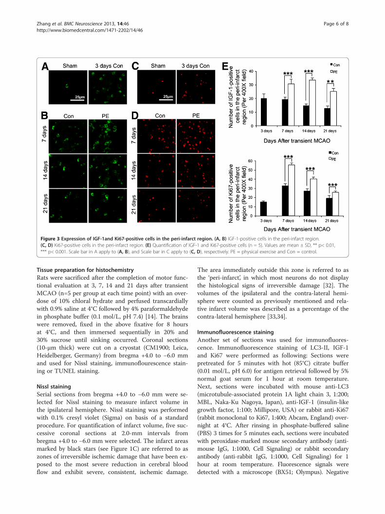
Figure 3 Expression of IGF-1and Ki67-positive cells in the peri-infarct region. (A, B) IGF-1-positive cells in the peri-infarct region.(C, D) Ki67-positive cells in the peri-infarct region. (E) Quantification of IGF-1 and Ki67-positive cells (n = 5). Values are mean ± SD, ** p< 0.01,*** p< 0.001. Scale bar in A apply to (A, B), and Scale bar in C apply to (C, D), respectively. PE = physical exercise and Con = control.
Zhang et al. BMC Neuroscience 2013, 14:46 Page 6 of 8http://www.biomedcentral.com/1471-2202/14/46
Tissue preparation for histochemistryRats were sacrificed after the completion of motor func-tional evaluation at 3, 7, 14 and 21 days after transientMCAO (n=5 per group at each time point) with an over-dose of 10% chloral hydrate and perfused transcardiallywith 0.9% saline at 4°C followed by 4% paraformaldehydein phosphate buffer (0.1 mol/L, pH 7.4) [14]. The brainswere removed, fixed in the above fixative for 8 hoursat 4°C, and then immersed sequentially in 20% and30% sucrose until sinking occurred. Coronal sections(10-μm thick) were cut on a cryostat (CM1900; Leica,Heidelberger, Germany) from bregma +4.0 to −6.0 mmand used for Nissl staining, immunoflourescence stain-ing or TUNEL staining.
Nissl stainingSerial sections from bregma +4.0 to −6.0 mm were se-lected for Nissl staining to measure infarct volume inthe ipsilateral hemisphere. Nissl staining was performedwith 0.1% cresyl violet (Sigma) on basis of a standardprocedure. For quantification of infarct volume, five suc-cessive coronal sections at 2.0-mm intervals frombregma +4.0 to −6.0 mm were selected. The infarct areasmarked by black stars (see Figure 1C) are referred to aszones of irreversible ischemic damage that have been ex-posed to the most severe reduction in cerebral bloodflow and exhibit severe, consistent, ischemic damage.
The area immediately outside this zone is referred to asthe ‘peri-infarct’, in which most neurons do not displaythe histological signs of irreversible damage [32]. Thevolumes of the ipsilateral and the contra-lateral hemi-sphere were counted as previously mentioned and rela-tive infarct volume was described as a percentage of thecontra-lateral hemisphere [33,34].
Immunofluorescence stainingAnother set of sections was used for immunofluores-cence. Immunofluorescence staining of LC3-II, IGF-1and Ki67 were performed as following: Sections werepretreated for 5 minutes with hot (85°C) citrate buffer(0.01 mol/L, pH 6.0) for antigen retrieval followed by 5%normal goat serum for 1 hour at room temperature.Next, sections were incubated with mouse anti-LC3(microtubule-associated protein 1A light chain 3, 1:200;MBL, Naka-Ku Nagoya, Japan), anti-IGF-1 (insulin-likegrowth factor, 1:100; Millipore, USA) or rabbit anti-Ki67(rabbit monoclonal to Ki67, 1:400; Abcam, England) over-night at 4°C. After rinsing in phosphate-buffered saline(PBS) 3 times for 5 minutes each, sections were incubatedwith peroxidase-marked mouse secondary antibody (anti-mouse IgG, 1:1000, Cell Signaling) or rabbit secondaryantibody (anti-rabbit IgG, 1:1000, Cell Signaling) for 1hour at room temperature. Fluorescence signals weredetected with a microscope (BX51; Olympus). Negative

Zhang et al. BMC Neuroscience 2013, 14:46 Page 7 of 8http://www.biomedcentral.com/1471-2202/14/46
control sections were incubated with PBS instead of pri-mary antibodies and showed no positive signals.
Double-immunofluorescence analysisDouble-Immunofluorescence studies were performedfor LC3 plus TUNEL (the terminal deoxynucleotidyltransferase-mediated dUTP in situ nick-end labeling).Staining steps were the same as those described above:sections were pretreated for 5 minutes with hot (85°C)citrate buffer (0.01 mol/L, pH 6.0) for antigen retrievalfollowed by 5% normal goat serum for 1 hour at roomtemperature. Then, sections were incubated with mouseanti-LC3 (microtubule-associated protein 1A light chain3, 1:200; MBL, Naka-Ku Nagoya, Japan), overnight at4°C. After rinsing in phosphate-buffered saline (PBS)3 times for 5 minutes each, sections were incubatedwith peroxidase-marked mouse secondary antibody(anti-mouse IgG, 1:1000, Cell Signaling) plus Terminaldeoxynucleotidyl transferase and digoxigenin-labeled nu-cleotides (In Situ Cell Death Detection Kit, AP, RocheCorp., Switzerland) for 1 h at 37°C. After rinsing, the co-location of LC3 and TUNEL signals were observed witha microscope (BX51; Olympus).
Image analysis and quantificationAll histological images were captured at the same expos-ure and analyzed with Image-Pro Plus image analysissoftware (Media Cybernetics, Silver Spring, MD, USA)by one author who was not aware of the assignment ofanimals’ group assignment. The regions of interest weredefined as a zone with 700 μm width and length in theperi-infarct region, which is immediately outside theinfarct zone (Figure 1C) [1]. For cell counting of LC3/TUNEL/IGF-1/Ki67-immunopositive cells, eight con-secutive sections at 240-μm intervals from bregma 0.20to −2.20 mm were analyzed. The number of LC3/TUNEL/IGF-1/Ki67-immunopositive cells in the peri-infarct region (Figure 1C, marked by black squares) wascounted by Image-Pro Plus image analysis software in 4non-overlapping fields (425 μm X 320 μm) under X 400magnification and was presented as the average cellnumber per field on each section [34]. The final cellnumber per rat was the average cell number of all thesections [34].
Statistical analysisNumerical data were presented as mean ± SD. Repeatedmeasures ANOVA was used to evaluate MNSS variables.A nonparametric test was used to evaluate MNSS andinfarct volume values. Two independent samples t-testwas used for 2-group comparisons of the number ofLC3/TUNEL/Ki67/IGF-1 positive cells variables. Pearsonbivariate correlation was used to run correlation analysis.Statistical analysis was performed using SPSS 16.0
for windows (SPSS Inc, Chicago, IL, USA). *p < 0.05,**p < 0.01, ***p < 0.001 when comparison was made.
AbbreviationsMCAO: Middle cerebral artery occlusion; IF: Immunofluorescence; IGF-1: Insulin-like growth factor-1; LC3-II: Microtubule-associated protein 1A lightchain 3-II; AVs: Autophagic vacuoles; MNSS: Modified Neurological SeverityScore; CCA: Common carotid artery; ICA: Internal carotid artery; ECA: Externalcarotid artery; TUNEL: Deoxynucleotidyl transferase-mediated dUTP in situnick-end labeling.
Competing interestsThe authors declare that they have no conflict of interest.
Authors' contributionsLZ, LL, XC carried out the experiments. JL performed the statistical analysis.LZ drafted the manuscript. XH, RH participated in the design of the studyand ZP conceived of the study and participated in its design andcoordination and helped to draft the manuscript. All authors read andapproved the final manuscript.
AcknowledgementsThis study is supported by the National Natural Science Foundation of China(81071607); Guangdong Province Science and Technology DepartmentProject (2011B060300013).
Author details1Department of Rehabilitation Medicine, the Third Affiliated Hospital, SunYat-Sen University, Guangzhou 510630, China. 2Department of Neurology,the First Affiliated Hospital, Sun Yat-Sen University, Guangzhou 510080,China.
Received: 31 May 2012 Accepted: 27 March 2013Published: 8 April 2013
References1. Zheng YQ, Liu JX, Li XZ, Xu L, Xu YG: RNA interference-mediated
downregulation of Beclin1 attenuates cerebral ischemic injury in rats.Acta Pharmacol Sin 2009, 30:919–927.
2. Wen YD, Sheng R, Zhang LS, Han R, Zhang X, Zhang XD, Han F, FukunagaK, Qin ZH: Neuronal injury in rat model of permanent focal cerebralischemia is associated with activation of autophagic and lysosomalpathways. Autophagy 2008, 4:762–769.
3. Puyal J, Vaslin A, Mottier V, Clarke PG: Postischemic treatment of neonatalcerebral ischemia should target autophagy. Ann Neurol 2009, 66:378–389.
4. Mortimore GE, Schworer CM: Induction of autophagy by amino-aciddeprivation in perfused rat liver. Nature 1977, 270:174–176.
5. Yu WH, Cuervo AM, Kumar A, Peterhoff CM, Schmidt SD, Lee JH, Mohan PS,Mercken M, Farmery MR, Tjernberg LO, Jiang Y, Duff K, Uchiyama Y, NäslundJ, Mathews PM, Cataldo AM, Nixon RA: Macroautophagy–a novel Beta-amyloid peptide-generating pathway activated in Alzheimer's disease.J Cell Biol 2005, 171:87–98.
6. Shintani T, Klionsky DJ: Autophagy in health and disease: a double-edgedsword. Science 2004, 306:990–995.
7. Mizushima N: The pleiotropic role of autophagy: from proteinmetabolism to bactericide. Cell Death Differ 2005, 12:1535–1541.
8. Koike M, Shibata M, Tadakoshi M, Gotoh K, Komatsu M, Waguri S, KawaharaN, Kuida K, Nagata S, Kominami E, Tanaka K, Uchiyama Y: Inhibition ofautophagy prevents hippocampal pyramidal neuron death afterhypoxic-ischemic injury. Am J Pathol 2008, 172:454–469.
9. Rami A, Langhagen A, Steiger S: Focal cerebral ischemia inducesupregulation of Beclin 1 and autophagy-like cell death. Neurobiol Dis2008, 29:132–141.
10. Wirawan E, Vanden Berghe T, Lippens S, Agostinis P, Vandenabeele P:Autophagy: for better or for worse. Cell Res 2012, 22:43–61.
11. Shang JW, Deguchi K, Yamashita T, Ohta Y, Zhang HZ, Morimoto N, Liu N,Zhang XM, Tian FF, Matsuura T, Funakoshi H, Nakamura T, Abe K:Antiapoptotic and antiautophagic effects of glial cell line-derivedneurotrophic factor and hepatocyte growth factor after transient middlecerebral artery occlusion in rats. J Neurosci Res 2010, 88:2197–2206.

Zhang et al. BMC Neuroscience 2013, 14:46 Page 8 of 8http://www.biomedcentral.com/1471-2202/14/46
12. Nygren J, Wieloch T, Pesic J, Brundin P, Deierborg T: Enriched environmentattenuates cell genesis in subventricular zone after focal ischemia inmice and decreases migration of newborn cells to the striatum. Stroke2006, 37:2824–2829.
13. Ziemka-Nalecz M, Zalewska T: Endogenous neurogenesis induced byischemic brain injury or neurodegenerative diseases in adults. ActaNeurobiol Exp (Wars). 2012, 72:309–324.
14. Hu XQ, Zheng HQ, Yan TB, Pan SQ, Fang J, Jiang RS, Ma SF: Physicalexercise induces expression of CD31 and facilitates neural functionrecovery in rats with focal cerebral infarction. Neurol Res 2010,32:397–402.
15. Chaudhry K, Rogers R, Guo M, Lai Q, Goel G, Liebelt B, Ji X, Curry A, CarranzaA, Jimenez DF, Ding Y: Matrix metalloproteinase-9 (MMP-9) expressionand extracellular signal-regulated kinase 1 and 2 (ERK1/2) activation inexercise-reduced neuronal apoptosis after stroke. Neurosci Lett 2010,474:109–114.
16. Eadie BD, Redila VA, Christie BR: Voluntary exercise alters thecytoarchitecture of the adult dentate gyrus by increasing cellularproliferation, dendritic complexity, and spine density. J Comp Neurol2005, 486:39–47.
17. Chen CY, Hsu HC, Lee BC, Lin HJ, Chen YH, Huang HC, Ho YL, Chen MF:Exercise training improves cardiac function in infarcted rabbits:involvement of autophagic function and fatty acid utilization. Eur J HeartFail 2010, 12:323–330.
18. Trejo JL, Carro E, Torres-Aleman I: Circulating insulin-like growth factor 1mediates exercise-induced increases in the number of new neurons inthe adult hippocampus. J Neurosci 2001, 21:1628–1634.
19. Carro E, Nuñez A, Busiguna S, Torres-Aleman I: Circulating insulin-likegrowth factor 1 mediates effects of exercise on the brain. J Neurosci2000, 20:2926–2933.
20. Carro E, Trejo JL, Busiguina S, Torres-Aleman I: Circulating insulin-likegrowth factor 1 mediates the protective effects of physical exerciseagainst brain insults of different etiology and anatomy. J Neurosci 2001,21:5678–5684.
21. Meijer AJ, Codogno P: Signalling and autophagy regulation in health,aging and disease. Mol Aspects Med 2006, 27:411–425.
22. Quindry JC, Miller L, McGinnis G, Kliszczewicz B, Irwin JM, Landram M,Urbiztondo Z, Nanayakkara G, Amin R: Ischemia reperfusion injury, KATPchannels, and exercise-induced cardioprotection against apoptosis. JAppl Physiol 2012, 113:498–506.
23. Adhami F, Liao G, Morozov YM, Schloemer A, Schmithorst VJ, Lorenz JN,Dunn RS, Vorhees CV, Wills-Karp M, Degen JL, Davis RJ, Mizushima N, RakicP, Dardzinski BJ, Holland SK, Sharp FR, Kuan CY: Cerebral ischemia-hypoxiainduces intravascular coagulation and autophagy. Am J Pathol 2006,169:566–583.
24. Degterev A, Huang Z, Boyce M, Li Y, Jagtap P, Mizushima N, Cuny GD,Mitchison TJ, Moskowitz MA, Yuan J: Chemical inhibitor of nonapoptoticcell death with therapeutic potential for ischemic brain injury. Nat ChemBiol 2005, 1:112–119.
25. Puyal J, Clarke PG: Targeting autophagy to prevent neonatal strokedamage. Autophagy 2009, 5:1060–1061.
26. Zhu C, Wang X, Xu F, Bahr BA, Shibata M, Uchiyama Y, Hagberg H,Blomgren K: The influence of age on apoptotic and other mechanismsof cell death after cerebral hypoxia-ischemia. Cell Death Differ 2005,12:162–176.
27. Klionsky DJ, Emr SD: Autophagy as a regulated pathway of cellulardegradation. Science 2000, 290:1717–1721.
28. Hoehn BD, Palmer TD, Steinberg GK: Neurogenesis in rats after focalcerebral ischemia is enhanced by indomethacin. Stroke 2005,36:2718–2724.
29. Borlongan CV, Randall TS, Cahill DW, Sanberg PR: Asymmetrical motorbehavior in rats with unilateral striatal excitotoxic lesions as revealed bythe elevated body swing test. Brain Res 1995, 676:231–234.
30. Schallert T, Kozlowski DA, Humm JL, Cocke RR: Use-dependent structuralevents in recovery of function. Adv Neurol 1997, 73:229–238.
31. Germano AF, Dixon CE, D’Avella D, Hayes RL, Tomasello F: Behavior deficitsfollowing experimental subarachnoid hemorrhage in the rat.J Neurotrauma 1994, 11:345–353.
32. Imai H, Harland J, McCulloch J, Graham DI, Brown SM, Macrae IM: Specificexpression of the cell cycle regulation proteins, GADD34 and PCNA, in
the peri-infarct zone after focal cerebral ischemia in the rat. Eur JNeurosci 2002, 15:1929–1936.
33. Swanson RA, Morton MT, Tsao-Wu G, Savalos RA, Davidson C, Sharp FR: Asemiautomated method for measuring brain infarct volume. J CerebBlood Flow Metab 1990, 10:290–293.
34. Zhang J, Zhang YS, Li JJ, Xing SH, Li C, Li YL, Dang C, Fan YH, Yu J, Pei Z,Zeng JS: Autophagosomes accumulation is associated with β-amyloiddeposits and secondary damage in the thalamus after focal corticalinfarction in hypertension rats. J Neurochem 2012, 120:564–573.
doi:10.1186/1471-2202-14-46Cite this article as: Zhang et al.: Physical exercise improves functionalrecovery through mitigation of autophagy, attenuation of apoptosisand enhancement of neurogenesis after MCAO in rats. BMC Neuroscience2013 14:46.
Submit your next manuscript to BioMed Centraland take full advantage of:
• Convenient online submission
• Thorough peer review
• No space constraints or color figure charges
• Immediate publication on acceptance
• Inclusion in PubMed, CAS, Scopus and Google Scholar
• Research which is freely available for redistribution
Submit your manuscript at www.biomedcentral.com/submit