research article open access molecular cloning and ... · peptidase genes in the fish-pathogenic...
TRANSCRIPT
Seo et al. BMC Veterinary Research 2013, 9:10http://www.biomedcentral.com/1746-6148/9/10
RESEARCH ARTICLE Open Access
Molecular cloning and expression analysis ofpeptidase genes in the fish-pathogenicscuticociliate Miamiensis avidusJung Soo Seo1, Eun Ji Jeon1, Sung Hee Jung1, Myoung Ae Park1, Jin Woo Kim2, Ki Hong Kim3, Sung Ho Woo4
and Eun Hye Lee1*
Abstract
Background: Parasite peptidases have been actively studied as vaccine candidates or drug targets for preventionor treatment of parasitic diseases because of their important roles for survival and/or invasion in the host. Like otherparasites, the facultative histophagous ciliate Miamiensis avidus would possess peptidases that are closely associatedwith the invasion into the host tissue and survival in the host.
Results: The 17 genes encoding peptidases, including seven cathepsin-like cysteine peptidases, four serinecarboxypeptidases, a eukaryotic aspartyl protease family protein, an ATP-dependent metalloprotease FtsH familyprotein, three leishmanolysin family proteins and a peptidase family M49 protein were identified from a Miamiensisavidus cDNA library by BLAST X search. Expression of genes encoding two cysteine peptidases, threeleishmanolysin-like peptidases and a peptidase family M49 protein was up-regulated in the cell-fed ciliatescompared to the starved ciliates. Especially, one cysteine peptidase (MaPro 4) and one leishmanolysin-like peptidase(MaPro 14) were transcribed more than 100-folds in the cell-fed ciliates.
Conclusions: The genetic information and transcriptional characteristics of the peptidases in the present resultswould be helpful to elucidate the role of peptidases in the invasion of scuticociliates into their hosts.
Keywords: Scuticociliates, Miamiensis avidus, Peptidases, RT-PCR
BackgroundParasite peptidases have been widely studied as potentialvaccine candidates or promising targets of anti-parasiticagents for prevention or treatment of parasitic diseases,because of their crucial roles in completing the lifecycles or diseases they produce. In many protozoanparasites that cause malaria, trypanosomiasis, leishman-iasis, amebiasis, toxoplasmosis, giardiasis, cryptosporidi-osis, and trichomoniasis, the major roles of parasitepeptidases include invasion by degradation of host cellsand tissues, degradation of mediators of the immuneresponses, and the catabolism of host proteins for para-site growth and survival [1-5].
* Correspondence: [email protected] Division, National Fisheries Research & Development Institute(NFRDI), 152-1, Haean-Lo, Gijang-Up, Gijang-Gun, Busan 619-705, South KoreaFull list of author information is available at the end of the article
© 2013 Seo et al.; licensee BioMed Central LtdCommons Attribution License (http://creativecreproduction in any medium, provided the or
In the facultative histophagous Miamiensis avidus(= synonym of Philasterides dicentrarchi), which causeshigh mortality in cultured olive flounder (Paralichthysolivaceus) in Korea [6,7], peptidases might play importantroles in the process of transforming of the ciliates fromthe free-living form into the invasive, infectious form,which might make the peptidases as candidates for vac-cine antigen or treatment drug target. It has been reportedthat peptidases secreted by Philasterides dicentrarchi candegrade type-I collagen, modulate host cellular immuneresponses, and induce apoptosis of leucocytes [8-11].Moreover, P. dicentrarchi peptidases could affect hosthumoral immune responses by degrading the host immu-noglobulins and reducing host complement activity in fishserum and ascitic fluid [12].Although there are several reports about the important
roles of peptidases in scuticociliate M. avidus, no studiescombined with genetic identification of peptidase genes
. This is an Open Access article distributed under the terms of the Creativeommons.org/licenses/by/2.0), which permits unrestricted use, distribution, andiginal work is properly cited.
Seo et al. BMC Veterinary Research 2013, 9:10 Page 2 of 10http://www.biomedcentral.com/1746-6148/9/10
and gene expression related to the function on the inva-sion have been performed. Therefore, the purpose of thisstudy was to identify peptidase genes that are expectedto have features related to infection of M. avidus bycomparison of expression level between the cell-fed andthe starved ciliates.
MethodsCiliatesCiliates were isolated from ascitic fluid of an infectedolive flounder Paralichthys olivaceus collected from alocal fish farm in Korea, and were identified as Mia-miensis avidus using species-specific oligonucleotide pri-mers [6]. Chinook salmon embryo (CHSE)-214 cells,incubated at 20°C in Eagle’s minimum essential medium(MEM, Sigma, St. Louis, Mo, USA) supplemented with10% heat-inactivated fetal bovine serum (FBS), were usedas grazing material to grow the ciliates under axenic cul-ture conditions.To obtain cell-free cultured ciliates, ciliates harvested
from routine CHSE cell-feeding cultures were transferredto filtered sea water without any nutrient components andstarved at 20°C for at least 1 month. To obtain cell-fedciliates, ciliates were inoculated in sufficiently grownCHSE-214 cells in routine MEM supplemented with 10%heat-inactivated FBS or in sufficiently grown CHSE-214cells in filtered seawater supplemented with 10% heat-inactivated FBS or were intraperitoneally injected intoolive flounder. The ciliates from different culture condi-tions were harvested using a method described previously[13]. Briefly, the ciliates were harvested by centrifugationat 200 × g for 5 min, and washed more than 3 times bycentrifugation at 150 × g for 5 min in Hanks’ balanced saltsolution (Sigma) or filtered seawater. The experimentsusing fish and treatment of dead fish were performed inaccordance with the guideline approved by Ministry forFood, Agriculture, Forestry and Fisheries.
RNA preparation, cDNA library construction andexpressed sequence tag (EST) analysisTotal RNA from CHSE-cultured M. avidus was preparedusing Trizol reagent (Invitrogen, Carlsbad, CA, USA)according to the manufacturer’s instructions. Poly A+RNA from the total RNA prepared from CHSE-culturedM. avidus was isolated using the Stratagene AbsolutelymRNA Purification Kit (Stratagene, La Jolla, CA, USA).A cDNA library was constructed using the ZAP ExpresscDNA Synthesis Kit and Gigapack III Gold packingextract (Stratagene) according to the manufacturer’sinstructions. The titer of constructed cDNA library was5.6 × 105 plaque-forming units (pfu)/ml.The expressed sequence tags (ESTs) were analyzed by
DNA sequencing of kanamycin resistant Escherichia coliclones containing cDNA fraction-harbored phagemid
(pBK-CMV) after mass excision of the lambda phagelibrary. DNA sequencing was conducted with T3/T7 pha-gemid sequencing primers using an ABI3730 automaticsequencer (96-capillary, Applied Biosystems, Foster City,CA, USA) and Applied Biosystems BigDyeW TerminatorCycle Sequencing Kits v3.1, in accordance with themanufacturer’s recommendations. A total of 1,265 ESTsequences, obtained cDNA library of M. avidus RNA, wereanalyzed by sequence comparison with previously reportedsequences in the EMBL/GenBank databases using theBLAST X search program of the National Center for Bio-technology Information (NCBI). The domain search ofdeduced amino acid sequences was analyzed using theSMART web and the NCBI protein blast program.
Real-time reverse transcriptase polymerase chain reaction(RT-PCR) of peptidase genes in Miamiensis avidusRT-PCR was performed to further verify the expressionpatterns of the isolated peptidase genes. Total RNA wasisolated from cell-free cultured ciliates and cell-fed ciliatesusing the RNeasy Mini Kit (Qiagen, Valencia, CA, USA).Total RNA was isolated several times, and the pooled totalRNA was used for cDNA synthesis. cDNA was synthesizedfrom 1 μg of total RNA using Superscript II Reverse Tran-scriptase and Oligo (dT) 20 primer (Invitrogen). RT-PCRwas performed using Fast Start SYBR Green Master Mix(Roche, Indianapolis, IN-, USA) and 100 ng of synthesizedcDNA in a 20 μl reaction volume. Quantitative PCR wasconducted using an iQ5 Multicolor Real-Time PCR instru-ment (Bio-Rad, Hercules, CA, USA), and the β-tubulin(BTU) gene was used as the internal control fornormalization. Thermal cycling conditions were one cycleof 3 min at 95°C (initial denaturation) followed by 40 cyclesof 10 s at 95°C, 10 s at 55°C, 20 s at 72°C. The specific PCRprimers for amplification ofM. avidus peptidase genes weredesigned from the unique sequences obtained by analysis ofESTs using the OLIGO 5.0 software (National Bioscience)(Table 1) and the expected sizes of PCR products are listedin Table 2. The results of RT-PCR from triplicate experi-ments were expressed as mean Ct (Cycle threshold) valuesand standard deviation. The fold change in relative geneexpression under the different culture conditions was deter-mined by the 2−ΔΔCt method [14,15]. ΔΔCt (delta delta Ct)values were calculated using an equation, where ΔΔCt =(CtMaPro - CtBTU)cell-fed - (CtMaPro - CtBTU)cell-free. Significantdifferences of Ct values were determined by Paired t-testafter normalization using those of β-tubulin gene.
ResultsIsolation and sequence analysis of peptidase genes fromthe Miamiensis avidus cDNA libraryWe isolated 32 clones harboring peptidase gene sequencesfrom 1,265 EST clones of the M. avidus cDNA library andobtained 17 different peptidase gene sequences including
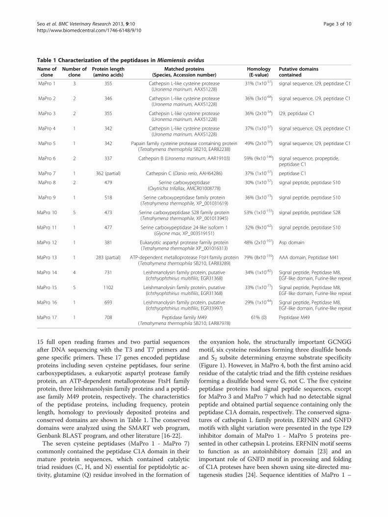
Table 1 Characterization of the peptidases in Miamiensis avidus
Name ofclone
Number ofclone
Protein length(amino acids)
Matched proteins(Species, Accession number)
Homology(E-value)
Putative domainscontained
MaPro 1 3 355 Cathepsin L-like cysteine protease(Uronema marinum, AAX51228)
31% (1x10-57) signal sequence, I29, peptidase C1
MaPro 2 2 346 Cathepsin L-like cysteine protease(Uronema marinum, AAX51228)
36% (3x10-68) signal sequence, I29, peptidase C1
MaPro 3 2 355 Cathepsin L-like cysteine protease(Uronema marinum, AAX51228)
36% (2x10-54) I29, peptidase C1
MaPro 4 1 342 Cathepsin L-like cysteine protease(Uronema marinum, AAX51228)
37% (1x10-57) signal sequence, I29, peptidase C1
MaPro 5 1 342 Papain family cysteine protease containing protein(Tetrahymena thermophila SB210, EAR82238)
49% (2x10-59) signal sequence, I29, peptidase C1
MaPro 6 2 337 Cathepsin B (Uronema marinum, AAR19103) 59% (9x10-146) signal sequence, propeptide,peptidase C1
MaPro 7 1 362 (partial) Cathepsin C (Danio rerio, AAH64286) 37% (1x10-57) peptidase C1
MaPro 8 2 479 Serine carboxypeptidase(Oxytricha trifallax, AMCR01008778)
30% (1x10-57) signal peptide, peptidase S10
MaPro 9 1 518 Serine carboxypeptidase family protein(Tetrahymena thermophile, XP_001031619)
36% (3x10-73) signal peptide, peptidase S10
MaPro 10 5 473 Serine carboxypeptidase S28 family protein(Tetrahymena thermophile, XP_001013945)
53% (1x10-155) signal peptide, peptidase S28
MaPro 11 1 477 Serine carboxypeptidase 24-like isoform 1(Glycine max, XP_003519151)
32% (9x10-62) signal peptide, peptidase S10
MaPro 12 1 381 Eukaryotic aspartyl protease family protein(Tetrahymena thermophile XP_001016313)
48% (2x10-107) Asp domain
MaPro 13 1 283 (partial) ATP-dependent metalloprotease FtsH family protein(Tetrahymena thermophila SB210, EAR83289)
79% (8x10-159) AAA domain, Peptidase M41
MaPro 14 4 731 Leishmanolysin family protein, putative(Ichthyophthirius multifiliis, EGR31368)
34% (1x10-82) Signal peptide, Peptidase M8,EGF-like domain, Furine-like repeat
MaPro 15 5 1102 Leishmanolysin family protein, putative(Ichthyophthirius multifiliis, EGR31368)
33% (1x10-73) Signal peptide, Peptidase M8,EGF-like domain, Furine-like repeat
MaPro 16 1 693 Leishmanolysin family protein, putative(Ichthyophthirius multifiliis, EGR33997)
29% (1x10-64) Signal peptide, Peptidase M8,EGF-like domain, Furine-like repeat
MaPro 17 1 708 Peptidase family M49(Tetrahymena thermophila SB210, EAR87978)
61% (0) Peptidase M49
Seo et al. BMC Veterinary Research 2013, 9:10 Page 3 of 10http://www.biomedcentral.com/1746-6148/9/10
15 full open reading frames and two partial sequencesafter DNA sequencing with the T3 and T7 primers andgene specific primers. These 17 genes encoded peptidaseproteins including seven cysteine peptidases, four serinecarboxypeptidases, a eukaryotic aspartyl protease familyprotein, an ATP-dependent metalloprotease FtsH familyprotein, three leishmanolysin family proteins and a peptid-ase family M49 protein, respectively. The characteristicsof the peptidase proteins, including frequency, proteinlength, homology to previously deposited proteins andconserved domains are shown in Table 1. The conserveddomains were analyzed using the SMART web program,Genbank BLAST program, and other literature [16-22].The seven cysteine peptidases (MaPro 1 - MaPro 7)
commonly contained the peptidase C1A domain in theirmature protein sequences, which contained catalytictriad residues (C, H, and N) essential for peptidolytic ac-tivity, glutamine (Q) residue involved in the formation of
the oxyanion hole, the structurally important GCNGGmotif, six cysteine residues forming three disulfide bondsand S2 subsite determining enzyme substrate specificity(Figure 1). However, in MaPro 4, both the first amino acidresidue of the catalytic triad and the fifth cysteine residuesforming a disulfide bond were G, not C. The five cysteinepeptidase proteins had signal peptide sequences, exceptfor MaPro 3 and MaPro 7 which had no detectable signalpeptide and obtained partial sequence containing only thepeptidase C1A domain, respectively. The conserved signa-tures of cathepsin L family protein, ERFNIN and GNFDmotifs with slight variation were presented in the type I29inhibitor domain of MaPro 1 - MaPro 5 proteins pre-sented in other cathepsin L proteins. ERFNIN motif seemsto function as an autoinhibitory domain [23] and animportant role of GNFD motif in processing and foldingof C1A proteses have been shown using site-directed mu-tagenesis studies [24]. Sequence identities of MaPro 1 –

Table 2 List of primers used in quantitative reverse-transcription PCR (RT-PCR)
Target clone/gene Primers Sequences (50- 30) Product size (bp)
MaPro 1 Forward TGCTTCCACTTCAGTTTTATCAGTCG 266
Reverse GGTTAAGTTCAACTGTGGGGATTTCTAA
MaPro 2 Forward TCTTGAGAGCTTCTGCTGCCAC 271
Reverse TCTTGGATGTTTAATTCGGTGCTGT
MaPro 3 Forward AATCCAACGAAGACATCAGAATCTTCT 246
Reverse CAGGGACTTATCTGGAAGGTCTGGA
MaPro 4 Forward TAGCTTCAATTGCTTCTGGTAGTCTTG 277
Reverse ATCCATGTTTATTCCACATAGTCCATTAC
MaPro 5 Forward ATTTCAAGCGATTGGAAGCTAAGAATC 308
Reverse AATAATCCCAAATAGAATATTACCCATCTTC
MaPro 6 Forward ACTTGGACTGCTGGATACAACAAAC 310
Reverse CGGTGGAGATTCTGGTTTGAAC
MaPro 7 Forward CGGTGGAGATTCTGGTTTGAAC 212
Reverse TTCGGCATCAACAGAATGGTAGATAC
MaPro 8 Forward ACGTTTTATTAGAAAGCCAAGGTAACC 260
Reverse GGTATTTTCGTCGGTGTATTTGTAGCT
MaPro 9 Forward GGGAAAAGGAAACTCTGCATTCG 348
Reverse CATCCATTTTCAGCAGTGTACAGTTCTAT
MaPro 10 Forward CAGATAATGGCTCTACTAATATTGCACTC 243
Reverse AGGGATCTTCACTTCCATTTGTGAATAC
MaPro 11 Forward ATTTGGCTCAATGGAGGACCTG 217
Reverse CAGCGGTATTATCATCAGTGTAAGAGT
MaPro 12 Forward CCACCTACAAACCCCAAGGAGAC 268
Reverse GCGAAAGAGTTGTCTTCCCAGACT
MaPro 13 Forward CAGCTCTTTTAACAGAAGGAGCTACAC 352
Reverse TTTCTGAACTTCGGTATCCACCATA
MaPro 14 Forward AGGGTATCTTCGAACAGCTCTTCG 319
Reverse CATTGGGACAAGAGACTGAACAGTC
MaPro 15 Forward AGCCTTGGAATGGAAATACTTTCGCTG 319
Reverse CCAACACAGTATCCGTTAGAGCTACAG
MaPro 16 Forward ACTCACGGATAGAACAATGCTCTTGC 314
Reverse TTAAAGTGCTTGCGAGCCACTTCC
MaPro 17 Forward TTGCAAGTTTCCTCTGGATTTGAATC 327
Reverse TCCGTATATAAGTTCAATTGTGGCATC
β-tubulin Forward GTATGATCATTGATAACGAAGCCCTCTACG 323
Reverse TCTGGGATCGGCGGCGCACATCATG
Seo et al. BMC Veterinary Research 2013, 9:10 Page 4 of 10http://www.biomedcentral.com/1746-6148/9/10
MaPro 5 with the cathepsin L protein of Uronema mari-num (UmCatL, AAX51228), which is one of the mostrelated scuticociliate species, were 30.7%, 35.9%, 35.6%,37% and 22.4%, respectively. The MaPro 6 protein se-quence contained a signal peptide, a propeptide C1 do-main, and a peptidase C1A domain containing twelveconserved cysteine residues forming disulfide bonds andthe “occluding loop” which is a specific feature of cathe-psin B-like peptidases distinct from other C1 superfamily
peptidases and had a 58.9% identity to the cathepsinB-like peptidase protein of U. marinum (Figure 1). Weidentified four different serine carboxypeptidase proteinsand three different leishmanolysin family proteins in M.avidus. All four serine carboxypeptidase proteins con-tained a signal peptide, and three had a peptidase S10domain, and one had a peptidase S28 domain that com-monly exist in serine carboxypeptidase proteins. Theleishmanolysin family proteins had a signal peptide, a
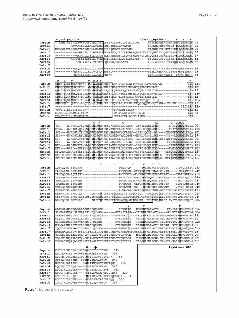
Figure 1 (See legend on next page.)
Seo et al. BMC Veterinary Research 2013, 9:10 Page 5 of 10http://www.biomedcentral.com/1746-6148/9/10
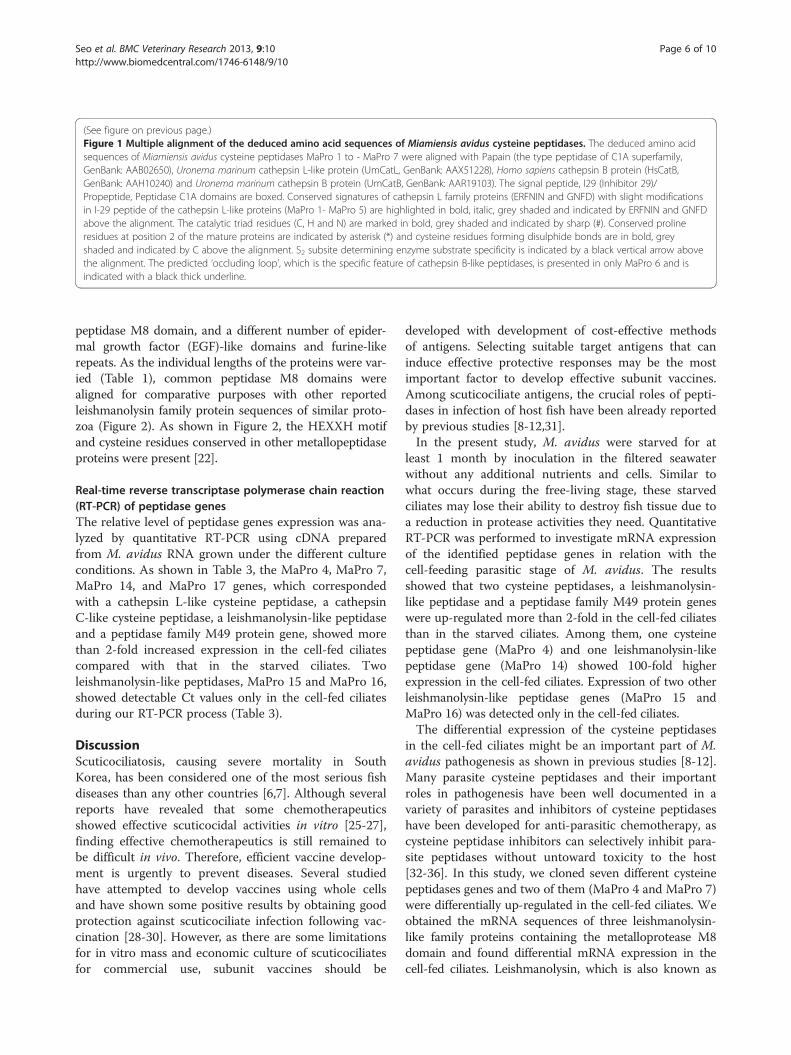
(See figure on previous page.)Figure 1 Multiple alignment of the deduced amino acid sequences of Miamiensis avidus cysteine peptidases. The deduced amino acidsequences of Miamiensis avidus cysteine peptidases MaPro 1 to - MaPro 7 were aligned with Papain (the type peptidase of C1A superfamily,GenBank: AAB02650), Uronema marinum cathepsin L-like protein (UmCatL, GenBank: AAX51228), Homo sapiens cathepsin B protein (HsCatB,GenBank: AAH10240) and Uronema marinum cathepsin B protein (UmCatB, GenBank: AAR19103). The signal peptide, I29 (Inhibitor 29)/Propeptide, Peptidase C1A domains are boxed. Conserved signatures of cathepsin L family proteins (ERFNIN and GNFD) with slight modificationsin I-29 peptide of the cathepsin L-like proteins (MaPro 1- MaPro 5) are highlighted in bold, italic, grey shaded and indicated by ERFNIN and GNFDabove the alignment. The catalytic triad residues (C, H and N) are marked in bold, grey shaded and indicated by sharp (#). Conserved prolineresidues at position 2 of the mature proteins are indicated by asterisk (*) and cysteine residues forming disulphide bonds are in bold, greyshaded and indicated by C above the alignment. S2 subsite determining enzyme substrate specificity is indicated by a black vertical arrow abovethe alignment. The predicted ‘occluding loop’, which is the specific feature of cathepsin B-like peptidases, is presented in only MaPro 6 and isindicated with a black thick underline.
Seo et al. BMC Veterinary Research 2013, 9:10 Page 6 of 10http://www.biomedcentral.com/1746-6148/9/10
peptidase M8 domain, and a different number of epider-mal growth factor (EGF)-like domains and furine-likerepeats. As the individual lengths of the proteins were var-ied (Table 1), common peptidase M8 domains werealigned for comparative purposes with other reportedleishmanolysin family protein sequences of similar proto-zoa (Figure 2). As shown in Figure 2, the HEXXH motifand cysteine residues conserved in other metallopeptidaseproteins were present [22].
Real-time reverse transcriptase polymerase chain reaction(RT-PCR) of peptidase genesThe relative level of peptidase genes expression was ana-lyzed by quantitative RT-PCR using cDNA preparedfrom M. avidus RNA grown under the different cultureconditions. As shown in Table 3, the MaPro 4, MaPro 7,MaPro 14, and MaPro 17 genes, which correspondedwith a cathepsin L-like cysteine peptidase, a cathepsinC-like cysteine peptidase, a leishmanolysin-like peptidaseand a peptidase family M49 protein gene, showed morethan 2-fold increased expression in the cell-fed ciliatescompared with that in the starved ciliates. Twoleishmanolysin-like peptidases, MaPro 15 and MaPro 16,showed detectable Ct values only in the cell-fed ciliatesduring our RT-PCR process (Table 3).
DiscussionScuticociliatosis, causing severe mortality in SouthKorea, has been considered one of the most serious fishdiseases than any other countries [6,7]. Although severalreports have revealed that some chemotherapeuticsshowed effective scuticocidal activities in vitro [25-27],finding effective chemotherapeutics is still remained tobe difficult in vivo. Therefore, efficient vaccine develop-ment is urgently to prevent diseases. Several studiedhave attempted to develop vaccines using whole cellsand have shown some positive results by obtaining goodprotection against scuticociliate infection following vac-cination [28-30]. However, as there are some limitationsfor in vitro mass and economic culture of scuticociliatesfor commercial use, subunit vaccines should be
developed with development of cost-effective methodsof antigens. Selecting suitable target antigens that caninduce effective protective responses may be the mostimportant factor to develop effective subunit vaccines.Among scuticociliate antigens, the crucial roles of pepti-dases in infection of host fish have been already reportedby previous studies [8-12,31].In the present study, M. avidus were starved for at
least 1 month by inoculation in the filtered seawaterwithout any additional nutrients and cells. Similar towhat occurs during the free-living stage, these starvedciliates may lose their ability to destroy fish tissue due toa reduction in protease activities they need. QuantitativeRT-PCR was performed to investigate mRNA expressionof the identified peptidase genes in relation with thecell-feeding parasitic stage of M. avidus. The resultsshowed that two cysteine peptidases, a leishmanolysin-like peptidase and a peptidase family M49 protein geneswere up-regulated more than 2-fold in the cell-fed ciliatesthan in the starved ciliates. Among them, one cysteinepeptidase gene (MaPro 4) and one leishmanolysin-likepeptidase gene (MaPro 14) showed 100-fold higherexpression in the cell-fed ciliates. Expression of two otherleishmanolysin-like peptidase genes (MaPro 15 andMaPro 16) was detected only in the cell-fed ciliates.The differential expression of the cysteine peptidases
in the cell-fed ciliates might be an important part of M.avidus pathogenesis as shown in previous studies [8-12].Many parasite cysteine peptidases and their importantroles in pathogenesis have been well documented in avariety of parasites and inhibitors of cysteine peptidaseshave been developed for anti-parasitic chemotherapy, ascysteine peptidase inhibitors can selectively inhibit para-site peptidases without untoward toxicity to the host[32-36]. In this study, we cloned seven different cysteinepeptidases genes and two of them (MaPro 4 and MaPro 7)were differentially up-regulated in the cell-fed ciliates. Weobtained the mRNA sequences of three leishmanolysin-like family proteins containing the metalloprotease M8domain and found differential mRNA expression in thecell-fed ciliates. Leishmanolysin, which is also known as
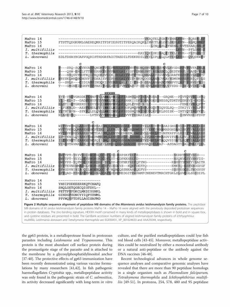
Figure 2 Multiple sequence alignment of peptidase M8 domains of the Miamiensis avidus leishmanolysin family proteins. The peptidaseM8 domains of M. avidus leishmanolysin family proteins MaPro 14 – MaPro 16 were aligned with the previously deposited protozoan sequencesin protein databases. The zinc-binding signature, HEXXH motif conserved in many kinds of metallopeptidases is shown in bold and in square box,and cysteine residues are presented in bold. The GenBank accession numbers of aligned leishmanolysin family proteins of Ichthyophtiriusmultifiliis, Leishmania donovani and Tetrahymena thermophila are EGR30431, XP_001024633 and AAA29244, respectively.
Seo et al. BMC Veterinary Research 2013, 9:10 Page 7 of 10http://www.biomedcentral.com/1746-6148/9/10
the gp63 protein, is a metalloprotease found in protozoanparasites including Leishmania and Trypanosoma. Thisprotein is the most abundant cell surface protein duringthe promastigote stage of the parasite and is attached tothe membrane by a glycosylphosphatidylinositol anchor[37-40]. The protective effects of gp63 immunization havebeen recently demonstrated using various vaccine formu-lations by many researchers [41,42]. In fish pathogenichaemoflagellates Cryptobia spp., metallopeptidase activitywas only found in the pathogenic strain of C. salmositica,its activity decreased significantly with long-term in vitro
culture, and the purified metallopeptidases could lyse fishred blood cells [43-45]. Moreover, metallopeptidase activ-ities could be neutralized by either a monoclonal antibodyor a natural anti-peptidase or the antibody against theDNA vaccines [46-48].Recent technological advances in whole genome se-
quence analyses and comparative genomic analyses haverevealed that there are more than 90 peptidase homologsin a single organism such as Plasmodium falciparum,Tetrahymena thermophila and Ichthyophthirius multifi-liis [49-51]. In protozoa, 254, 578, 480 and 95 peptidase

Table 3 Real time RT-PCR analysis of peptidase genes in Miamiensis avidus under the different culture conditions
Name of clone Mean Cta value (SDb) Fold increase in expression (2−ΔΔCt(SD))
Cell-free culture Cell-feeding culture Cell-free culture Cell-feeding culture
MaPro 1 28.48(0.51) 22.67(0.32) 1.05(0.40) 1.01(0.22)
MaPro 2** 25.38(0.24) 21.25(0.12) 1.01(0.16) 0.31(0.03)
MaPro 3 27.43(0.34) 22.11(0.10) 1.02(0.23) 0.71(0.05)
MaPro 4** 34.96(0.28) 21.95(0.24) 1.02(0.20) 147.58(24.89)
MaPro 5 33.19(0.44) 29.71(0.63) 1.03(0.30) 0.21(0.08)
MaPro 6** 23.93(0.36) 20.01(0.19) 1.02(0.27) 0.27(0.04)
MaPro 7* 33.61(0.31) 25.72(0.11) 1.01(0.22) 4.19(0.33)
MaPro 8** 26.94(0.21) 24.11(0.31) 1.01(0.14) 0.13(0.03)
MaPro 9* 29.33(0.58) 26.79(0.58) 1.05(0.42) 0.11(0.04)
MaPro 10 32.67(1.44) 26.44(0.29) 1.29(0.86) 1.34(0.27)
MaPro 11 30.26(1.30) 25.09(0.25) 1.24(0.79) 0.64(0.11)
MaPro 12** 25.57(0.08) 22.90(0.29) 1.00(0.06) 0.11(0.02)
MaPro 13* 34.37(0.34) 31.15(1.30) 1.02(0.22) 0.21(0.16)
MaPro 14** 32.51(0.41) 19.37(0.01) 1.03(0.27) 159.78(1.40)
MaPro 15 N/Ac 31.12(0.76)
MaPro 16 N/A 31.52(0.57)
MaPro 17* 34.68(0.77) 26.87(0.32) 1.10(0.54) 4.04(0.84)
β-tubulin 23.37(0.45) 17.55(0.22)aCt: Cycle threshold, bSD: standard deviation, cN/A: No detectable Ct value was obtained within 40 cycles, * : Significant differences P<0.05, ** : Significantdifferences P<0.005.
Seo et al. BMC Veterinary Research 2013, 9:10 Page 8 of 10http://www.biomedcentral.com/1746-6148/9/10
genes have been identified in Ichthyophthrius multifiliis,Paramecium tetraurelia, Tetrahymena thermophile, andPlasmodium falciparum, respectively [51]. Like otherparasites, M. avidus may express many peptidase pro-teins to undergo various biological processes includingparasite survival and pathogenesis. In this study, weobtained 17 different peptidase genes from a M. aviduscDNA library by ESTs sequence screening, and theresults of differential mRNA expression related to patho-genesis were also obtained. Based on the analysis ofstructurally conserved regions and motifs presented inthe deduced amino acid sequences of each peptidaseproteins, five cathepsin L-like cysteine peptidases, onecathepsin B-like cysteine peptidase, one cathepsin C-likecysteine peptidase, four serine carboxypeptidase, aeukaryotic aspartyl protease family protein, an ATP-dependent metalloprotease FtsH family protein, threeleishmanolysin family proteins, and a peptidase familyM49 protein were identified although there were somestructurally differences with previously reported similarproteins. Although the number of identified peptidasegenes obtained from this study was relatively lower thanexpected, this is the first report of cloning and mRNA ex-pression of peptidase gene homologs as important viru-lence factors in M. avidus. Moreover, the information ofexact protein sequence obtained from this study could helpto perform futher studies to develop specific inhibitors.
We are currently performing studies on the actual ac-tivities using recombinant proteins of cloned peptidasegenes to understand whether these proteins are biologic-ally active at the protein level. We will further analyze ofM. avidus genome using large scale-genome analysistechniques to identify more peptidase sequences, andwill perform combined research of transcriptional ana-lysis and enzymatic activities of each peptidase proteins.
ConclusionsIn conclusion, the genetic information obtained fromthis study could help to design specific vaccine formula-tions and inhibitors of peptidases to prevent and controlof fish scuticociliatosis caused by M. avidus, althoughfurther studies to elucidate the exact roles of these pepti-dases should be conducted.
Competing interestsThe authors declare that they have no competing interests.
Authors’ contributionsEHL contributed to the design of the study, performing experiments, dataanalysis and preparation of the manuscript. JSS contributed to the design ofthe study and advised on data analysis and review of the manuscript. SHWand EJJ participated in data collection and experimental procedure. SHJ,MAP, JWK, KHK involved with the review of the manuscript. All authors readand approved the final manuscript.
Seo et al. BMC Veterinary Research 2013, 9:10 Page 9 of 10http://www.biomedcentral.com/1746-6148/9/10
AcknowledgementsThis study was supported by a research fund of the National Fisheries Researchand Development Institute (NFRDI, RP-2012-AQ-062), Republic of Korea.
Author details1Pathology Division, National Fisheries Research & Development Institute(NFRDI), 152-1, Haean-Lo, Gijang-Up, Gijang-Gun, Busan 619-705, SouthKorea. 2Aquatic Life Disease Control Division, NFRDI, 152-1, Haean-Lo,Gijang-Up, Gijang-Gun, Busan 619-705, South Korea. 3Department of AquaticLife Medicine, Pukyong National University, 599-1 Daeyondong, Namgu,Busan 608-737, South Korea. 4Institute of Fisheries Sciences, PukyongNational University, 295, Dongbaek-ri, Ilgwang-myeon, Gijang-gun, Busan619-911, South Korea.
Received: 4 May 2012 Accepted: 19 December 2012Published: 11 January 2013
References1. McKerrow JH, Sun E, Rosenthal PJ, Bouvier J: The proteases and
pathogenicity of parasitic protozoa. Annu Rev Microbiol 1993, 47:821–853.2. Que X, Reed SL: The role of extracellular cysteine proteinases in
pathogenesis of Entamoeba histolytica invasion. Parasitol Today 1997,13:190–194.
3. Rosenthal PJ: Proteases of protozoan parasites. Adv Parasitol 1999, 43:105–159.4. Jime’nez JC, Uzcanga G, Zambrano A, Di Prisco MC, Lynch NR:
Identification and partial characterization of excretory/secretoryproducts with proteolytic activity in Giardia intestinalis. J Parasitol 2000,86:859–862.
5. Klenmba M, Goldberg DE: Biological roles of proteases in parasiticprotozoa. Annu Rev Biochem 2002, 71:275–305.
6. Kim SM, Cho JB, Kim SK, Nam YK, Kim KH: Occurrence of scuticociliatosis inolive flounder Paralichthys olivaceus by Philasterides dicentrarchi(Ciliophora: Scuticociitida). Dis Aquat Organ 2004, 62:233–238.
7. Jung SJ, Kitamura S, Song JY, Oh MJ: Miamiensis avidus (Ciliophora:Scuticociliatida) causes systemic infection of olive flounder Paralichthysolivaceus and is a senior synonym of Philasterides dicentrarchi. Dis AquatOrgan 2007, 73(3):227–234.
8. Paramá A, Iglesias R, Alvarez MF, Sanmartn ML, Leiro J: Chemotacticresponses of the fish-parasitic scuticociliate Philasterides dicentrarchi toblood and blood components of the turbot Scophthalmus maximus,evaluated using a new microplate multiassay. J Microbiol Methods 2004,58:361–366.
9. Paramá A, Iglesias R, Alvarez MF, Leiro J, Ubeira FM, Sanmartín ML: Cysteineproteinase activities in the fish pathogen Philasterides dicentrarchi(Ciliophora: Scuticociliatida). Parasitology 2004, 128:541–548.
10. Paramá A, Castro R, Arranz JA, Sanmartín ML, Lamas J, Leiro J: Scuticociliatecysteine proteinases modulate turbot leucocyte functions. Fish ShellfishImmunol 2007, 23(5):945–956.
11. Paramá A, Castro R, Lamas J, Sanmartín ML, Santamarina MT, Leiro J:Scuticociliate proteinases may modulate turbot immune response byinducing apoptosis in pronephric leucocytes. Int J Parasitol 2007, 37:87–95.
12. Piazzon C, Lamas J, Leiro M: Role of scuticociliate proteinases in infectionsuccess in turbot, Psetta maxima (L.). Parasite Immunol 2011, 33:535–544.
13. Lee EH, Kim KH: Identification of differentially expressed genes inparasitic phase Miamiensis avidus (Ciliophora: Scuticociliatia) usingsuppression subtractive hybridization. Dis Aquat Organ 2011, 94:135–142.
14. Giulietti A, Oververgh L, Valckx D, Decallonne B, Bouillon R, Mathieu C: Anoverview of real-time quantitative PCR: applications to quantify cytokinegene expression. Methods 2001, 25:386–401.
15. Livak KJ, Schmittgen TD: Analysis of Relative Gene Expression Data UsingReal-Time Quantitative PCR and the 2−ΔΔCT Method. Methods 2001,25:402–408.
16. Jesudhasan PR, Tan CW, Hontzeas N, Woo PT: A cathepsin L-like cysteineproteinase gene from the protozoan parasite, Cryptobia salmositica.Parasitol Res 2007, 100:881–886.
17. Herrmann L, Erkelenz M, Aldag I, Tiedtke A, Hartmann MW: Biochemicaland molecular characterisation of Tetrahymena thermophila extracellularcysteine proteases. BMC Microbiol 2006, 6:19.
18. Jousson O, Di Bello D, Donadio E, Felicioli A, Pretti C: Differential expression ofcysteine proteases in developmental stages of the parasitic ciliateIchthyophthirius multifiliis. FEMS Microbiol Lett 2007, 269:77–84.
19. Ahn SJ, Seo JS, Kim MS, Jeon SJ, Kim NY, Jang JH, Kim KH, Hong YK, ChungJK, Lee HH: Cloning, site-directed mutagenesis and expression ofcathepsin L-like cysteine protease from Uronema marinum (Ciliophora:Scuticociliatida). Mol Biochem Parasitol 2007, 156:191–198.
20. Lim SU, Seo JS, Kim MS, Ahn SJ, Jeong HD, Kim KH, Park NG, Kim JK, ChungJK, Lee HH: Molecular cloning and characterization of Cathepsin B from ascuticociliate, Uronema marinum. Comp Biochem Physiol B Biochem MolBiol 2005, 142:283–292.
21. Sajid M, McKerrow JH: Cysteine proteases of parasitic organisms. MolBiochem Parasitol 2002, 120:1–21.
22. Jiang W, Bond JS: Families of metalloendopeptidases and theirrelationships. FEBS Lett 1992, 312:110–114.
23. Karrer KM, Peiffer SL, DiTomas ME: Two distinct gene subfamilies within thefamily of cysteine protease genes. Proc Natl Acad Sci USA 1993, 90:3063–3067.
24. Vernet T, Berti PJ, DeMontigny C, Musil R, Tessier DC, Menard R, Magny MC,Storer AC, Thomas DY: Processing of the papain precursor. The ionizationstate of a conserved amino acid motif within the Pro region participatesin the regulation of intra-molecular processing. J Biol Chem 1995,270:18038–10846.
25. Iglesias R, Paramá A, Álvarez MF, Leiro J, Sanmartín ML: Antiprotozoalseffective in vitro against the scuticociliate fish pathogen Philasteridesdicentrarchi. Dis Aquat Organ 2002, 49:191–197.
26. Quintela JM, Peinador C, González L, Iglesias R, Paramá A, Álvarez F,Sanmartín ML, Riguera R: Piperazine N-substituted naphthyridines,pyridothienopyrimidines and pyridothienotriazines: new antiprotozoalsactive against Philasterides dicentrarchi. Eur J Med Chem 2003, 38:265–275.
27. Paramá A, Piazzon MC, Lamas J, Sanmartín ML, Leiro J: In vitro activity ofthe nonsteroidal anti-inflammatory drug indomethacin on ascuticociliate parasite of farmed turbot. Vet Parasitol 2007, 48:318–324.
28. Sitjà-Bobadilla A, Palenzuela O, Alvarez-Pellitero P: Immune response ofturbot, Psetta maxima (L.) (Pisces: Teleostei), to formalin-killedscuticociliates (Ciliophora) and adjuvanted formulations. Fish ShellfishImmunol 2007, 24:1–10.
29. Sanmartín ML, Paramá A, Castro R, Cabaleiro S, Leiro J, Lamas J, Barja JL:Vaccination of turbot, Psetta maxima (L.), against the protozoan parasitePhilasterides dicentrarchi: effects on antibody production and protection.J Fish Dis 2008, 31:135–140.
30. Lee EH, Kim KH: Immobilization antigen-independent protection of oliveflounder (Paralichthys olivaceus) against Philasterides dicentrarchi(Ciliophora: Scuticociliatia) infection. Aquaculture 2008, 279:211–213.
31. Lee EH, Kim CS, Cho JB, Ahn KJ, Kim KH: Measurement of protease activityof live Uronema marinun (Ciliata: Scuticociliatida) by fluorescencepolarization. Dis Aquat Organ 2003, 54:85–88.
32. Sajid M, MeKerrow JH: Cysteine proteases of parasitic organisms. MolBiochem Parasitol 2002, 120:1–21.
33. Mckerrow JH, Engel JC, Caffrey CR: Cysteine protease inhibitors aschemotherapy for parasitic infections. Bioorg Med Chem 1999, 7:639–644.
34. Engel JC, Doyle PS, Hsieh I, McKerrow JH: Cysteine protease inhibitors curean experimental Trypanosoma cruzi infection. J Exp Med 1998, 188:725–734.
35. McKerrow JH, Rosenthal PJ, Swenerton R, Doyle P: Development ofprotease inhibitors for protozoan infections. Curr Opin Infect Dis 2008,21:668–672.
36. Atkinson HJ, Babbitt PC, Sajid M: The global cysteine peptidase landscapein parasites. Trends Parasitol 2009, 25:573–581.
37. Bouvier J, Etges RJ, Bordier C: Identification and purification of membraneand soluble forms of the major surface protein of Leishmaniapromastigotes. J Biol Chem 1985, 260:15504–15509.
38. Bouvier J, Bordier C, Vogel H, Reichelt R, Etges R: Characterization of thepromastigote surface protease of Leishmania as a membrane-bound zincendopeptidase. Mol Biochem Parasitol 1989, 37:235–245.
39. Etges R, Bouvier J, Bordier C: The major surface protein of Leishmaniapromastigotes is a protease. J Biol Chem 1986, 261:9098–9101.
40. Etges R, Bouvier J, Bordier C: The major surface protein of Leishmaniapromastigotes is anchored in the membrane by a myristic acidlabeledphospholipid. EMBO J 1986, 5:597–601.
41. Mazumder S, Maji M, Das A, Ali N: Potency, efficacy and durability of DNA/DNA, DNA/protein and protein/protein based vaccination using gp63against Leishmania donovani in BALB/c mice. PLoS One 2011, 6:e14644.
42. Bhowmick S, Ravindran R, Ali N: gp63 in stable cationic liposomes conferssustained vaccine immunity to susceptible BALB/c mice infected withLeishmania donovani. Infect Immun 2008, 76:1003–1015.

Seo et al. BMC Veterinary Research 2013, 9:10 Page 10 of 10http://www.biomedcentral.com/1746-6148/9/10
43. Zuo X, Woo PTK: Proteases in pathogenic and nonpathogenichaemoflagellates, Cryptobia spp. (Sarcomastigophora: Kinetoplastida), offishes. Dis Aquat Organ 1997, 29:57–65.
44. Uo X, Woo PTK: Purified metalloprotease from the pathogenichaemoflagellate, Cryptobia salmositica, and its in vitro proteolyticactivities. Dis Aquat Organ 1997, 30:177–185.
45. Woo PT: Immunological and therapeutic strategies against salmonidcryptobiosis. J Biomed Biotechnol 2010, :Article ID 341783.
46. Zuo X, Woo PTK: In vivo neutralization of Cryptobia salmositica metallo-protease by α2-macroglobulin in the blood of rainbow troutOncorhynchus mykiss and in brook charr Salvelinus fontinalis. Dis AquatOrgan 1997, 29:67–72.
47. Zuo X, Woo PTK: Natural anti-proteases in rainbow trout, Oncorhynchusmykiss and brook charr, Salvelinus fontinalis and the in vitro neutralizationof fish α2-macroglobulin by the metalloprotease from the pathogenichaemoflagellate, Cryptobia salmositica. Parasitology 1997, 114:375–382.
48. Tan CW, Jesudhasan PRR, Woo PTK: Towards a metalloprotease-DNAvaccine against piscine cryptobiosis caused by Cryptobia salmositica.Parasitol Res 2008, 102:265–275.
49. Wu Y, Wang X, Liu X, Wang Y: Data-mining approaches reveal hiddenfamilies of proteases in the genome of malaria parasite. Genome Res2003, 13:601–616.
50. Eisen JA, Coyne RS, Wu M, Wu D, Thiagarajan M, Wortman JR, Badger JH, RenQ, Amedeo P, Jones KM, Tallon LJ, Delcher AL, Salzberg SL, Silva JC, Haas BJ,Majoros WH, Farzad M: Macronuclear genome sequence of the ciliateTetrahymena thermophila, a model eukaryote. PLoS Biol 2006, 4:e286.
51. Coyne RS, Hannick L, Shanmugam D, Hostetler JB, Brami D, Joardar VS,Johnson J, Radune D, Singh I, Badger JH, Kumar U, Saier M, Wang Y, Cai H,Gu J, Mather MW, Vaidya AB, Wilkes DE, Rajagopalan V, Asai DJ, Pearson CG,Findly RC, Dickerson HW, Wu M, Martens C, Van de Peer Y, Roos DS,Cassidy-Hanley DM, Clark TG: Comparative genomics of the pathogenicciliate Ichthyophthirius multifiliis, its free-living relatives and a hostspecies provide insights into adoption of a parasitic lifestyle andprospects for disease control. Genome Biol 2011, 12:R100.
doi:10.1186/1746-6148-9-10Cite this article as: Seo et al.: Molecular cloning and expression analysisof peptidase genes in the fish-pathogenic scuticociliate Miamiensisavidus. BMC Veterinary Research 2013 9:10.
Submit your next manuscript to BioMed Centraland take full advantage of:
• Convenient online submission
• Thorough peer review
• No space constraints or color figure charges
• Immediate publication on acceptance
• Inclusion in PubMed, CAS, Scopus and Google Scholar
• Research which is freely available for redistribution
Submit your manuscript at www.biomedcentral.com/submit