research article open access consequences of induced
TRANSCRIPT

Schröder et al. BMC Plant Biology 2014, 14:309http://www.biomedcentral.com/1471-2229/14/309
RESEARCH ARTICLE Open Access
Consequences of induced brassinosteroiddeficiency in Arabidopsis leavesFlorian Schröder1*, Janina Lisso1, Toshihiro Obata2, Alexander Erban2, Eugenia Maximova2, Patrick Giavalisco2,Joachim Kopka2, Alisdair R Fernie2, Lothar Willmitzer2 and Carsten Müssig1
Abstract
Background: The identification of brassinosteroid (BR) deficient and BR insensitive mutants provided conclusiveevidence that BR is a potent growth-promoting phytohormone. Arabidopsis mutants are characterized by acompact rosette structure, decreased plant height and reduced root system, delayed development, and reducedfertility. Cell expansion, cell division, and multiple developmental processes depend on BR. The molecular andphysiological basis of BR action is diverse. The BR signalling pathway controls the activity of transcription factors,and numerous BR responsive genes have been identified. The analysis of dwarf mutants, however, may to someextent reveal phenotypic changes that are an effect of the altered morphology and physiology. This restrictionholds particularly true for the analysis of established organs such as rosette leaves.
Results: In this study, the mode of BR action was analysed in established leaves by means of two approaches. First,an inhibitor of BR biosynthesis (brassinazole) was applied to 21-day-old wild-type plants. Secondly, BR complementationof BR deficient plants, namely CPD (constitutive photomorphogenic dwarf)-antisense and cbb1 (cabbage1) mutantplants was stopped after 21 days. BR action in established leaves is associated with stimulated cell expansion, anincrease in leaf index, starch accumulation, enhanced CO2 release by the tricarboxylic acid cycle, and increasedbiomass production. Cell number and protein content were barely affected.
Conclusion: Previous analysis of BR promoted growth focused on genomic effects. However, the link betweengrowth and changes in gene expression patterns barely provided clues to the physiological and metabolic basis ofgrowth. Our study analysed comprehensive metabolic data sets of leaves with altered BR levels. The data suggestthat BR promoted growth may depend on the increased provision and use of carbohydrates and energy. BR maystimulate both anabolic and catabolic pathways.
Keywords: Brassinosteroids, Arabidopsis, Tricarboxylic acid cycle, Biomass, Cell expansion, Growth
BackgroundBrassinolide (BL) was identified in 1979 through its abil-ity to promote internode growth [1]. Since then, thegrowth-promoting effect has been studied in hundredsof articles. Excised tissues (e.g. hypocotyl, epicotyl, coty-ledons, internodes, leaves, and roots), protoplasts, cellsuspension cultures, intact seedlings, and whole plantswere subjected to brassinosteroid (BR) treatments, andin all cases BR had the potential to stimulate growth [2].A large number of BR deficient and BR insensitive mu-tants in Arabidopsis thaliana and in crops such as rice,
* Correspondence: [email protected] of Potsdam, c/o Max Planck Institute of Molecular PlantPhysiology, Am Mühlenberg 1, 14476 Potsdam-Golm, GermanyFull list of author information is available at the end of the article
© 2014 Schröder et al.; licensee BioMed CentrCommons Attribution License (http://creativecreproduction in any medium, provided the orDedication waiver (http://creativecommons.orunless otherwise stated.
tomato, and pea were identified [3]. These mutants aregenerally dwarfed and exhibit rounded, dark green leaves,delayed flowering, reduced male fertility and seed set, anddelayed senescence. Further proven roles of BR includethe control of xylem formation [4,5], stomata develop-ment [6-8], and further developmental processes [9-12].Numerous studies analysed gene expression patterns uponBR treatment and BR deficiency. However, these studiesbarely clarified the metabolic and physiological basis ofBR dependent growth because the precise functions ofisoenzymes, cell wall proteins, and other factors oftenremains obscure.BR plays non redundant roles since it is not possible
to complement BR mutants with other phytohormones ortheir antagonists [13]. Overexpression of the major BR
al Ltd. This is an Open Access article distributed under the terms of the Creativeommons.org/licenses/by/4.0), which permits unrestricted use, distribution, andiginal work is properly credited. The Creative Commons Public Domaing/publicdomain/zero/1.0/) applies to the data made available in this article,
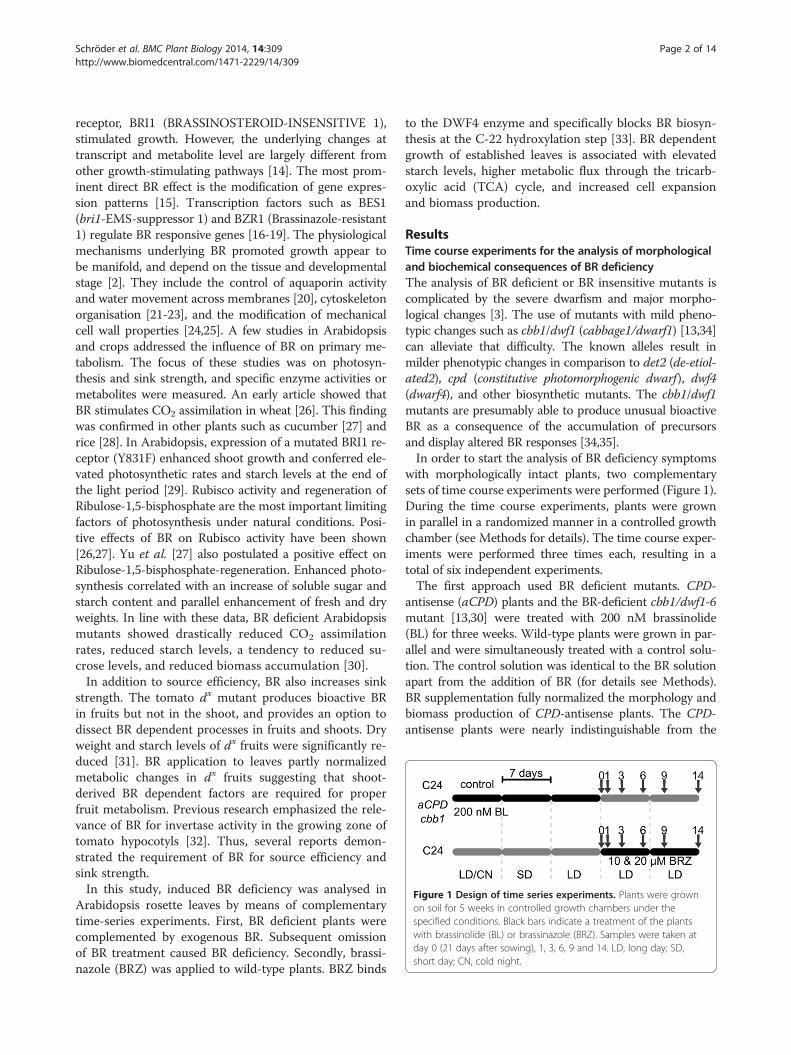
Figure 1 Design of time series experiments. Plants were grownon soil for 5 weeks in controlled growth chambers under thespecified conditions. Black bars indicate a treatment of the plantswith brassinolide (BL) or brassinazole (BRZ). Samples were taken atday 0 (21 days after sowing), 1, 3, 6, 9 and 14. LD, long day; SD,short day; CN, cold night.
Schröder et al. BMC Plant Biology 2014, 14:309 Page 2 of 14http://www.biomedcentral.com/1471-2229/14/309
receptor, BRI1 (BRASSINOSTEROID-INSENSITIVE 1),stimulated growth. However, the underlying changes attranscript and metabolite level are largely different fromother growth-stimulating pathways [14]. The most prom-inent direct BR effect is the modification of gene expres-sion patterns [15]. Transcription factors such as BES1(bri1-EMS-suppressor 1) and BZR1 (Brassinazole-resistant1) regulate BR responsive genes [16-19]. The physiologicalmechanisms underlying BR promoted growth appear tobe manifold, and depend on the tissue and developmentalstage [2]. They include the control of aquaporin activityand water movement across membranes [20], cytoskeletonorganisation [21-23], and the modification of mechanicalcell wall properties [24,25]. A few studies in Arabidopsisand crops addressed the influence of BR on primary me-tabolism. The focus of these studies was on photosyn-thesis and sink strength, and specific enzyme activities ormetabolites were measured. An early article showed thatBR stimulates CO2 assimilation in wheat [26]. This findingwas confirmed in other plants such as cucumber [27] andrice [28]. In Arabidopsis, expression of a mutated BRI1 re-ceptor (Y831F) enhanced shoot growth and conferred ele-vated photosynthetic rates and starch levels at the end ofthe light period [29]. Rubisco activity and regeneration ofRibulose-1,5-bisphosphate are the most important limitingfactors of photosynthesis under natural conditions. Posi-tive effects of BR on Rubisco activity have been shown[26,27]. Yu et al. [27] also postulated a positive effect onRibulose-1,5-bisphosphate-regeneration. Enhanced photo-synthesis correlated with an increase of soluble sugar andstarch content and parallel enhancement of fresh and dryweights. In line with these data, BR deficient Arabidopsismutants showed drastically reduced CO2 assimilationrates, reduced starch levels, a tendency to reduced su-crose levels, and reduced biomass accumulation [30].In addition to source efficiency, BR also increases sink
strength. The tomato dx mutant produces bioactive BRin fruits but not in the shoot, and provides an option todissect BR dependent processes in fruits and shoots. Dryweight and starch levels of dx fruits were significantly re-duced [31]. BR application to leaves partly normalizedmetabolic changes in dx fruits suggesting that shoot-derived BR dependent factors are required for properfruit metabolism. Previous research emphasized the rele-vance of BR for invertase activity in the growing zone oftomato hypocotyls [32]. Thus, several reports demon-strated the requirement of BR for source efficiency andsink strength.In this study, induced BR deficiency was analysed in
Arabidopsis rosette leaves by means of complementarytime-series experiments. First, BR deficient plants werecomplemented by exogenous BR. Subsequent omissionof BR treatment caused BR deficiency. Secondly, brassi-nazole (BRZ) was applied to wild-type plants. BRZ binds
to the DWF4 enzyme and specifically blocks BR biosyn-thesis at the C-22 hydroxylation step [33]. BR dependentgrowth of established leaves is associated with elevatedstarch levels, higher metabolic flux through the tricarb-oxylic acid (TCA) cycle, and increased cell expansionand biomass production.
ResultsTime course experiments for the analysis of morphologicaland biochemical consequences of BR deficiencyThe analysis of BR deficient or BR insensitive mutants iscomplicated by the severe dwarfism and major morpho-logical changes [3]. The use of mutants with mild pheno-typic changes such as cbb1/dwf1 (cabbage1/dwarf1) [13,34]can alleviate that difficulty. The known alleles result inmilder phenotypic changes in comparison to det2 (de-etiol-ated2), cpd (constitutive photomorphogenic dwarf), dwf4(dwarf4), and other biosynthetic mutants. The cbb1/dwf1mutants are presumably able to produce unusual bioactiveBR as a consequence of the accumulation of precursorsand display altered BR responses [34,35].In order to start the analysis of BR deficiency symptoms
with morphologically intact plants, two complementarysets of time course experiments were performed (Figure 1).During the time course experiments, plants were grownin parallel in a randomized manner in a controlled growthchamber (see Methods for details). The time course exper-iments were performed three times each, resulting in atotal of six independent experiments.The first approach used BR deficient mutants. CPD-
antisense (aCPD) plants and the BR-deficient cbb1/dwf1-6mutant [13,30] were treated with 200 nM brassinolide(BL) for three weeks. Wild-type plants were grown in par-allel and were simultaneously treated with a control solu-tion. The control solution was identical to the BR solutionapart from the addition of BR (for details see Methods).BR supplementation fully normalized the morphology andbiomass production of CPD-antisense plants. The CPD-antisense plants were nearly indistinguishable from the

Schröder et al. BMC Plant Biology 2014, 14:309 Page 3 of 14http://www.biomedcentral.com/1471-2229/14/309
wild type. The growth defect of cbb1 plants was partlycomplemented by exogenous BR (Figure 2, day 0). Freshweight of 21-day-old cbb1 shoots was identical to the wildtype. However, leaf length and width were diminished incomparison to the wild type, leaves were more erect andhad a slightly crinkled surface, and rosettes appeared com-pact. Thus, exogenous BR could not fully substitute for en-dogenous BR. After three weeks, BR treatment was stopped(day 0). The CPD-antisense and cbb1 plants started to runinto BR deficiency or pronounced BR deficiency, respect-ively. At this point the sampling began. Samples for bio-chemical analysis were taken at day 0, 1, 3, 6, 9 and 14 afterthe respective treatment was stopped. Under the appliedconditions, plants were in the vegetative phase during thecomplete experiments and did not start bolting.The second approach used wild-type plants (C24) that
were grown for three weeks without any treatment(Figure 3, day 0). Subsequently, plants were treated with10 μM BRZ, 20 μM BRZ, or control solution. Lowerconcentrations such as 1 to 5 μM BRZ have previouslybeen applied in synthetic growth medium (e.g. [36,37])but were inapplicable in our time course experimentssince they induced only minor growth effects in soil-grown plants. The necessity for higher BRZ concentra-tions may reflect weaker uptake by leaves through afunctional epidermis. After the onset of BRZ application
Figure 2 Growth parameters of CPD-antisense and cbb1 plants in complants were grown as described in Figure 1. A, Shoot fresh weight. B, Reprthree and four. D, Width of rosette leaves three and four. Data are given assignificantly different from the wild type (t test, P <0.05).
(day 0), samples for biochemical analysis were taken atthe same points in time as described above (i.e. day 1, 3,6, 9, and 14).The parallel analysis of CPD-antisense, cbb1, and BRZ
treated wild-type plants allows avoiding genotype- ortreatment-specific limitations during the evaluation ofBR deficiency.
Elevated CPD and DWF4 transcript levels indicateemerging BR deficiencyThe CPD [38] and DWF4 [39] genes encode enzymes in-volved in BR biosynthesis. The expression of these genesis negatively associated with endogenous BR levels. Hightranscript levels indicate low BR levels and vice versa [40].CPD and DWF4 transcript levels were analysed by meansof quantitative RT-PCR (Figure 4).Unchanged transcript levels one day after the BR sup-
plementation was stopped or after the BRZ treatmentwas started may indicate the presence of remaining BRor a time lag in the induction of BR biosynthetic genes.Stronger differences were observed from day 3 onwards.CPD transcript levels in CPD-antisense plants were previ-ously described [30]. Due to incomplete CPD gene repres-sion, the phenotypic changes of CPD-antisense plants areconsiderably milder in comparison to the cpd/cbb3 knock-out mutant and other BR deficient mutants such as cbb1/
parison to the wild type. Wild-type (C24), CPD-antisense, and cbb1esentative plants at day 0, 1, 3, 6, 9, and 14. C, Length of rosette leavesmean ± SE (n =10 plants). Values denoted with an asterisk are

Figure 3 Growth parameters of BRZ treated plants in comparison to mock treated plants. Wild-type plants were grown and treated asdescribed in Figure 1. A, Shoot fresh weight. B, Representative plants at day 0, 1, 3, 6, 9, and 14. C, Length of rosette leaves three and four.D, Width of rosette leaves three and four. Data are given as mean ± SE (n =10 plants). Values denoted with an asterisk are significantly differentfrom the wild type (t test, P <0.05).
Schröder et al. BMC Plant Biology 2014, 14:309 Page 4 of 14http://www.biomedcentral.com/1471-2229/14/309
dwf1-6 [13,38]. Stronger DWF4 expression in the cbb1mutant in comparison to the CPD-antisense plants corre-sponds to the observed growth defect (Figure 4A and B).Only minor differences were detected between plantstreated with different concentrations of BRZ. Applicationof 10 μM BRZ induced CPD and DWF4 expression nearlyas effectively as 20 μM BRZ (Figure 4C and D).
Induced BR deficiency impairs leaf expansionSignificant differences in shoot fresh weight and lengthof rosette leaves three and four developed after one dayin cbb1 plants (Figure 2A and C). Both growth parametersof CPD-antisense plants became significantly differentfrom the wild type after six days and one day, respectively(Figure 2A and C). At the end of the analysed period (day14), cbb1 and CPD-antisense plants had 36% and 68% ofthe wild-type fresh weight, respectively (Figure 2A). BRdeficiency caused a less pronounced effect on leaf width(Figure 2D). The resulting decrease in the leaf index (i.e.more roundish leaves; [41]) is a well described featureof BR deficient plants (e.g. [42]). BRZ treated plantsexhibited a small reduction in the shoot fresh weight atday 1 (Figure 3A). The biomass difference to controlplants increased over time. Leaf length and leaf widthwere reduced. Similarly to the CPD-antisense and cbb1
plants, leaf width was less affected than leaf length(Figure 3C and D).Leaf thickness depends on the BR level and genotype.
For example, BR deficient mutants such as det2 exhib-ited an increased leaf thickness. Low concentrations ofexogenous BR decreased leaf thickness of det2 and wild-type plants. Higher concentrations caused an increase ofleaf thickness in the wild type [43]. In this study, leafthickness of CPD-antisense plants and BRZ treated plantsdisplayed diminished enhancement from day 0 to day 6.In contrast, leaf thickness of cbb1 plants increased moreduring that period (Figure 5).Leaves five and six were analysed in parallel at day 3
and day 6. Similar effects on leaf length, leaf width, andleaf thickness were observed (Additional file 1: Figures S1and S2, A-C).
Reduced growth is mainly due to reduced cell sizeSmaller leaf size of BR deficient plants could be basedon impaired cell expansion, cell proliferation, or a combin-ation of both. Previous analyses of BR mutants revealed ef-fects on both cell proliferation and cell expansion. BRdeficient mutants such as dwf1, det2 [42] and cpd [44] arecharacterized by reduced cell division rates and reducedcell expansion.

Figure 4 Quantitative RT-PCR analysis of CPD and DWF4 transcript levels. Plants were grown and harvested as described in Figure 1. A,Relative CPD transcript levels in wild-type (C24) and cbb1 plants. B, Relative DWF4 transcript levels in wild-type, CPD-antisense, and cbb1 plants.C, Relative CPD transcript levels in BRZ treated plants. D, Relative DWF4 transcript levels in BRZ treated plants. eIF1a CT values were subtractedfrom respective CT values of the gene of interest resulting in dCT. Subsequently, differences were subtracted from an arbitrary value (i.e. 40).Higher numbers indicate higher transcript levels. A difference of one unit indicates a fold change of two. Data are given as mean ± SE of gene ofinterest in three technical replicates. The data shown are from one experiment representative of three independent biological replicates.
Schröder et al. BMC Plant Biology 2014, 14:309 Page 5 of 14http://www.biomedcentral.com/1471-2229/14/309
In this study, similar size of palisade and spongy paren-chyma cells in CPD-antisense, cbb1, and wild-type plantswere observed at day 0 (Figure 6A and B), suggesting thatthe previous BR application normalized cell expansion.Later on, palisade and spongy parenchyma cells of CPD-antisense and cbb1 plants became smaller in comparison
Figure 5 Leaf thickness. Plants were grown and harvested as described itransversal sections. Values are given as mean ± SE. 20 leaves were analyseddifferent from the wild type (C24) or control (t test, P <0.05). A, Leaf thicknof BRZ treated plants.
to the wild type (Figure 6A and B). Similar results wereobtained for younger leaves (Additional file 1: Figure S1,D and E). Effects on cell number were less evident. Thecbb1 leaves exhibited a tendency towards lower cellnumbers, indicating an incomplete normalization of celldivision rates by the previous BR treatment. In contrast,
n Figure 1. Thickness of the leaves three and four was measured usingper point in time. Values denoted with an asterisk are significantly
ess of wild-type (C24), CPD-antisense, and cbb1 plants. B, Leaf thickness

Figure 6 Cell sizes and cell numbers of rosette leaves of CPD-antisense and cbb1 plants. Wild-type (C24), CPD-antisense, and cbb1 plantswere grown and harvested as described in Figure 1. Cell sizes were measured using transversal sections of rosette leaves three and four. Cellnumbers were calculated from the respective other half of the same leaf. Data are given as mean ± SE. 20 leaves were analysed per point in time.Values denoted with an asterisk are significantly different from the wild type (t test, P <0.05). A, Area of palisade cells. B, Area of spongyparenchyma cells. C, Palisade cells per leaf. D, Spongy parenchyma cells per leaf.
Schröder et al. BMC Plant Biology 2014, 14:309 Page 6 of 14http://www.biomedcentral.com/1471-2229/14/309
CPD-antisense plants were identical to the wild type atday 0 and later (Figure 6C and D; Additional file 1:Figure S1, F-H; Additional file 1: Figure S3A). Applicationof BRZ to the wild type reduced cell sizes (Figure 7Aand B; Additional file 1: Figure S2, D and E), but did notsignificantly reduce cell numbers (Figure 7C and D;Additional file 1: Figure S2, F-H).
Reduced starch and unchanged protein levels in BRdeficient plantsSynthetic BR stimulates CO2 assimilation [26-29], andcbb1 and CPD-antisense plants exhibit reduced photo-synthetic rates [30]. Both biochemical and morphologicalfactors could account for reduced photosynthesis. Theconsequences of reduced carbon supply include reducedstarch levels, impaired energy balance, reduced provisionof biosynthetic precursors, and decreased growth [45].In line with previous reports, starch levels were dimin-ished in CPD-antisense, cbb1, and BRZ treated plantsfrom day 3 onwards (Figure 8A and B). The reduction ofstarch levels in CPD-antisense and cbb1 plants is relativelysmall in comparison to previously determined levels [30].This may reflect the lack of severe cellular abnormalitiesthat were avoided by the initial BR supplementation.
Hexose and sucrose levels were not significantly altered(Additional file 1: Table S1). Examination of plastid ultra-structure revealed intact chloroplasts in BR deficientplants. BRZ treated, CPD-antisense, and cbb1 chloroplaststended to develop a thylakoid network with reducedgrana stacking at day 3 and to a more minor extent atday 6 (Figures 9 and 10).Early studies on BR demonstrated that inhibitors of pro-
tein synthesis (e.g. cycloheximide and puromycin) inter-fere with BR dependent growth [46]. It was suggested thatBR induces the synthesis of a large number of specific pro-teins, but does not indiscriminately increase overall pro-tein synthesis. In agreement with that view, the overallprotein content was not significantly altered in leaves ofBR deficient plants (Figure 8C and D).
Reduced TCA cycle activity in BRZ treated plantsMitochondrial respiratory metabolism is the major sourceof ATP and associated with proper maintenance of cellu-lar metabolism as a whole [47,48]. The tricarboxylic acid(TCA) cycle is a crucial component of respiratory me-tabolism. It links the oxidation of the acetyl group ofacetyl-CoA to CO2 with the generation of NADH forthe oxidation by the mitochondrial respiratory chain. In

Figure 7 Cell sizes and cell numbers of rosette leaves of BRZ treated plants. BRZ treated plants were grown and harvested as describedin Figure 1. Cell sizes were measured using transversal sections of rosette leaves three and four, and cell numbers were calculated from therespective other half of the same leaf. Data are given as mean ± SE. 20 leaves were analysed per point in time. Values denoted with an asteriskare significantly different from the wild type (t test, P <0.05). A, Area of palisade cells. B, Area of spongy parenchyma cells. C, Palisade cells perleaf. D, Spongy parenchyma cells per leaf.
Schröder et al. BMC Plant Biology 2014, 14:309 Page 7 of 14http://www.biomedcentral.com/1471-2229/14/309
plants, acetyl-CoA is derived from the products of gly-colysis through oxidative decarboxylation of pyruvate bythe pyruvate dehydrogenase [49,50].Leaf discs were incubated in [3:4-14C]-glucose or [1-14C]-
glucose. CO2 from the C3 and C4 positions is preferentiallyreleased by the actions of pyruvate dehydrogenase or malicenzyme [51,52]. Feeding with [3:4-14C]-glucose to BRZtreated leaves resulted in a lower evolution of 14CO2 incomparison to the control (Figure 11A). C1 of glucose isreleased either by an enzyme of the oxidative pentosephosphate pathway (OPPP), namely 6-phosphogluconatedehydrogenase, or an enzyme of the TCA cycle, isocitratedehydrogenase [51,52]. Feeding of BRZ treated plants with[1-14C]-glucose tended to result in a lower 14CO2 evolu-tion in comparison to mock-treated plants (Figure 11B).The relative content of TCA cycle intermediates was
determined by mass spectrometry. Aspartate is synthe-sized by transamination of oxaloacetate and can be usedto estimate oxaloacetate levels. Levels of several TCAcycle intermediates were increased in BRZ treated plants(Figure 12). Levels of citrate, malate, and aspartate weresignificantly different from the control (Additional file 1:Table S2). A tendency to higher levels of TCA cycle inter-mediates was also observed in the cbb1 mutant (Additional
file 1: Figure S5). The ketoglutarate level was significantlyincreased at day 6 (Additional file 1: Table S3).Lower CO2 release from [3:4-14C]-glucose by the pyru-
vate decarboxylase and/or malic enzyme and increasedlevels of TCA cycle intermediates suggest a weaker TCAcycle activity in BR deficient plants. The release of CO2
from [1-14C]-glucose is furthermore consistent with a re-duced flux through the TCA cycle, but could also suggesta reduced activity of the oxidative pentose phosphatepathway (OPPP).
DiscussionExperimental approaches to study BR deficiencyBR deficient plants display dwarfism and multiple de-fects in cell elongation, cell division, cell differentiation,reproduction and senescence, and light control of develop-ment [3]. Reduced fertility and male sterility are commonfeatures of BR deficient mutants. BR appears to be largelydispensable for embryogenesis [53]. Seedling development,however, critically depends on BR. Hypocotyl length, coty-ledon growth, and responses to environmental stimuli wereimpaired in Arabidopsis mutants [2,3,12,13]. BR deficiencyimpairs plant growth at early stages, and later phenotypicchanges are inevitably modified by the early growth

Figure 8 Starch and protein levels. Plants were harvested at the middle of the light period. Data are given as mean ± SE (n =3 pools of 10plants). Values denoted with an asterisk are significantly different from the wild type or control (t test, P <0.05). A, Starch levels of wild-type (C24),CPD-antisense, and cbb1 plants. B, Starch levels of BRZ treated plants. C, Protein levels of C24, CPD-antisense, and cbb1 plants. D, Protein levels ofBRZ treated plants.
Schröder et al. BMC Plant Biology 2014, 14:309 Page 8 of 14http://www.biomedcentral.com/1471-2229/14/309
defects. Thus, mutant analyses can properly address earlyphases in plant development, but conclusions about BRfunction at later stages are fraught with uncertainty.One approach to study the mode of action of BR at
later developmental stages is the application of inhibitors
Figure 9 Transmission electron microscopy of plastids in leaf three oas described in Figure 1 at the middle of the light period. A, Wild type (C2E, CPD-antisense at day 6. F, cbb1 at day 6. Subcellular structures are exems, starch granule; bar: 1 μm.
of BR biosynthesis [33]. Previous approaches usuallysupplemented BRZ [33,37,54] and other azole deriva-tives (e.g. propiconazole, [55]; voriconazole, [56]; YCZ,[57]) to synthetic growth medium, implying that seed-lings or small plantlets were analysed. An alternative
f CPD-antisense and cbb1 plants. Plants were grown and harvested4) at day 3. B, CPD-antisense at day 3. C, cbb1 at day 3. D, C24 at day 6.plarily indicated in A; cw, cell wall; g, granum (stack of thylakoids);

Figure 10 Transmission electron microscopy of plastids in leaf three of BRZ treated plants. Plants were grown and harvested as describedin Figure 1 at the middle of the light period. A, control (0 μM BRZ) at day 3. B, 10 μM BRZ at day 3. C, 20 μM BRZ at day 3. D, control at day 6.E, 10 μM BRZ at day 6. F, 20 μM BRZ at day 6. Subcellular structures are exemplarily indicated in A; cw, cell wall; g, granum (stack of thylakoids);s, starch granule; bar: 1 μm.
Schröder et al. BMC Plant Biology 2014, 14:309 Page 9 of 14http://www.biomedcentral.com/1471-2229/14/309
approach is the complementation of BR deficient mu-tants for a limited period, and the subsequent deprivationof synthetic BR. Both approaches have pros and cons.For example, BRZ is seen as a highly specific inhibi-tor, but it presumably also affects other P450s. Al-though BR deficient mutants respond to synthetic BR,BR feeding cannot fully mimic the endogenous distri-bution of BR.For those reasons, both approaches were followed in
the current study and analysed in parallel (Figure 1).The first three weeks of the experiments presumed thepresence of wild-type BR levels or continuous supply ofsynthetic BR (mutant complementation for three weeks).At this point (day 0), transcript levels of BR biosynthesisgenes and growth were similar in the mutants and thecontrol (Figures 2, 3, 4). Increased CPD and DWF4 tran-script levels suggest that plants became impoverished forBR within one to three days (Figure 4).
Figure 11 TCA cycle flux. Evolution of 14CO2 of BRZ treated plants at dayharvested as described in Figure 1. A, Leaf discs were incubated with [3:4-1
are given as mean ± SE (n =5 pools of leaf discs from 10 plants).
BR deficiency in established leaves impairs cell expansionLeaves grow initially mainly by cell proliferation. Cellsdivide and grow simultaneously. A proliferation gradi-ent develops between cell division and expansion at thetransition zone. The transition from cell proliferation toexpansion (cell growth without cell division) is con-trolled by a network of factors. Cell division first ceasesat the tip of the leaf, and progressively ceases along thelongitudinal axis [58]. The final size of the organ isachieved by elongation growth.Mutant analyses indicated that both cell division
and cell elongation are affected by BR, because leavesof BR deficient mutant such as det2 and cpd exhibitboth decreased cell size and cell numbers [42,44]. BRcontrols the transition between cell division and ex-pansion [16]. In addition, BR controls organ boundaryformation [59,60], xylem formation [61], and stomatadevelopment [8,62].
6 when incubated with labelled glucose. Plants were grown and4C]-glucose. B, Leaf discs were incubated with [1-14C] -glucose. Data

Figure 12 Levels of TCA cycle intermediates in BRZ treated plants. Plants were grown and harvested as described in Figure 1. Relativemetabolite levels are given as mean ± SE of three biological replicates. Fold change values are given in Additional file 1: Table S2.
Schröder et al. BMC Plant Biology 2014, 14:309 Page 10 of 14http://www.biomedcentral.com/1471-2229/14/309
We focused our analysis on established leaves at laterdevelopmental stages. At this stage (21 days after sowingand later), cell expansion and cell proliferation tookplace in leaves three to six. Palisade and spongy paren-chyma were significantly smaller in CPD-antisense, cbb1,and BRZ treated plants (Figures 6, 7, Additional file 1:Figures S1, S2). The cell number was slightly reduced inthe cbb1 mutant (Figure 6, Additional file 1: Figures S1, S3).However, cell proliferation in CPD-antisense and BRZtreated plants was similar to the wild type (Figures 6, 7,Additional file 1: Figures S1-3). Thus, cell expansion inestablished leaves depends on BR, but cell division isbarely impaired.Leaf thickness is determined by the mesophyll anat-
omy and cell size. The cellular organization of mesophylltissues modifies the interception of light and CO2 diffu-sion to the sites of photosynthesis. The effects of BR orother phytohormones and stimuli on leaf thickness arenot well documented, because changes in leaf growth
have usually been assessed in two dimensions. In thisstudy, induced BR deficiency in BRZ treated and CPD-antisense plants was associated with reduced leaf thick-ness (Figure 5, Additional file 1: Figures S1, S2, S4). Incontrast, leaf thickness in cbb1 plants increased moreuntil day 6 (Figure 5, Additional file 1: Figures S1, S4).The reason for the difference between the genotypescould be the incomplete normalization of cbb1 plants.Alternatively, cbb1 plants could synthesize alternativeBR and respond in a different manner to BR as has beenreported for the rice brd2 (BR-deficient dwarf2) mutant(Hong et al. [35]).
Reduced starch accumulation may cause reduced growthReduced growth of BR deficient plants may be a conse-quence of reduced carbon availability. Starch levels in cbb1,CPD-antisense, and BRZ treated leaves were lower in com-parison to the wild type (Figure 8). This presumably is aconsequence of drastically reduced CO2 assimilation rates

Schröder et al. BMC Plant Biology 2014, 14:309 Page 11 of 14http://www.biomedcentral.com/1471-2229/14/309
[30]. Optimal starch metabolism is pivotal for the diurnalcarbon balance and growth [45]. Mutants impaired instarch synthesis such as phosphoglucomutase (pgm) or mu-tants impaired in starch degradation such as starch excess1 (sex1) show dwarfism [63]. Both too rapid and too slowmobilization of starch during the night can result in dimin-ished growth rates [64]. Thus, carbon undersupply maycause impaired growth in BR deficient mutants.Electron micrographs of BRZ treated plants and the
cbb1 mutant revealed a tendency to less grana thylakoids(Figures 9, 10). The molecular basis of reduced thylakoidstacking is unknown. It could reflect a delay in plastiddevelopment, an altered adaptation of the thylakoid archi-tecture to the light conditions, an altered protein compos-ition, or changes in other regulatory mechanisms. Granaconfer functional advantages such as enhancement of lightcapture and fine-tuning of energy distribution between thephotosystems [65,66]. Conceivably, the observed changesin chloroplast structure contribute to the reduced photo-synthetic rate and starch accumulation. Plastid structureand function were previously analysed in BR mutants.One reason for that interest is the link between BR actionand photomorphogenesis [12]. Light-grown det2 plantsdeveloped structurally altered chloroplasts. For example,eight-day-old det2 chloroplasts had a smaller, roundershape, reduced grana stacking, and an abnormally high ra-tio of chlorophyll a/b in comparison to the wild type,indicating an immature status [67]. However, Azpiroz andcoworkers [68] did not describe an altered chloroplaststructure of light-grown dwf4 plants. Given the multiplereports that describe altered properties of plastids of BRtreated plants or BR mutants ([69] and references therein),a more detailed analysis of the underlying structural andmolecular changes may be worthwhile.
Reduced growth is associated with reduced TCA cycleactivityThe TCA cycle links the oxidation of pyruvate and mal-ate with the generation of NADH. NADH is used by themitochondrial respiratory chain for ATP production. Thereduced release of 14CO2 from labelled glucose in BRZtreated plants (Figure 11) and elevated levels of TCA cycleintermediates in BRZ treated (Figure 12) and cbb1 plants(Additional file 1: Figure S5) suggest a reduced carbon fluxthrough the TCA cycle in BR deficient plants. ReducedTCA cycle activity may compromise efficient use of carbo-hydrates and impair growth especially during the darkperiod. Furthermore, the TCA cycle provides precursorsfor various biosynthetic pathways [48,50].Reduced production of ATP for sucrose synthesis and
carbon precursors for anabolism could be consequenceor cause of reduced growth. BR deficiency and reducedgrowth presumably goes along with reduced demandfor carbohydrates, amino acids, and other biosynthetic
precursors. On the other hand, reduced photosynthesisin BR deficient plants could diminish the supply of sub-strates for mitochondrial reactions and reduce the fluxthrough the TCA cycle. The situation becomes evenmore complex in view of the multifaceted links betweenphotosynthesis and TCA cycle. Altered TCA cycle enzymeactivities can result in increased, decreased, or unvariedphotosynthesis [70,71]. Thus, identification of cause andeffect of metabolic changes is complicated. Labelling stud-ies and application of network models will be necessary toprecisely determine the flux of metabolites and interplayof metabolic pathways in BR deficient plants.
ConclusionsThe morphology of BR deficient mutants was described indetail. Numerous studies addressed the consequences ofBR deficiency at the molecular and cellular level. In thatway, the current understanding of BR was developed. How-ever, the analysis of BR deficient mutants is complicated bythe dwarfism and multiple morphological changes. Themode of action of BR at later developmental stages cannotbe faultlessly determined. In this study, we used two ap-proaches for the analysis of BR action in established leaves.An inhibitor of BR biosynthesis was applied and BR com-plementation of BR deficient plants was stopped after threeweeks. For the first time the metabolic changes upon BRdeficiency were analysed comprehensively by means ofmetabolic profiling. Our analyses revealed that induced BRdeficiency impairs starch accumulation, TCA cycle activity,cell expansion, and biomass production. Further studies areneeded to determine alterations in metabolic fluxes and theprecise link between genomic BR effects and catabolic andanabolic pathways. Transgenic approaches such as the in-ducible expression of RNAi hairpins represent another ap-proach that would enable tissue-specific repression of BRbiosynthesis. This could particularly help to separate therole of BR in sink and source tissues.
MethodsGrowth conditionsC24 wild type was obtained from the NottinghamArabidopsis Stock Centre (NASC) - NASC ID: N906.The CPD-antisense line and the cbb1 mutant were de-scribed before [13,30]. Seeds for growth experimentswere derived from plants grown in parallel in a green-house. The cbb1 mutant was repeatedly treated with BRbefore and during seed set. Seeds were allowed to ger-minate and seedlings grew for two weeks in controlledgrowth chambers (7 days: 16 h light [140 μmol m−2 s−1,20°C, 75% relative humidity]/8 h night [6°C, 75% relativehumidity]; thereafter 7 days: 8 h light [140 μmol m−2 s−1,20°C, 60% relative humidity]/16 h night [16°C, 75% rela-tive humidity]). Subsequently, plants were transferred tolong-day conditions in a controlled growth chamber (16 h

Schröder et al. BMC Plant Biology 2014, 14:309 Page 12 of 14http://www.biomedcentral.com/1471-2229/14/309
light [140 μmol m−2 s−1, 20°C, 60% relative humidity]/8 hnight [16°C, 75% relative humidity]). All genotypes weregrown in the same chamber at the same time in a ran-domized manner, each replicate one after another. All ne-cessary measures were taken in order to avoid biotic andabiotic stress.Plants were sprayed at midday for three (BL) or five
(BRZ) times a week with an aqueous solution containingBL or BRZ, respectively, and 0.01% Tween 20. Methanolwas used as solvent for stock solutions. The same vol-ume of methanol was added to the control solution. BLand BRZ experiments were performed in the same growthchamber. BRZ was sprayed more often to ensure loweredBR levels.
Gene expression analysisGene expression analyses were performed as describedbefore [72]. Primer sequences for quantitative RT-PCRwere as follows: CPD_fw 5’ GGA AAC ACT CTC TGCTTC TTA TGA AAG GT 3’, CPD_rev 5’ AAG TAAAGC CAC CAA GAA GTC AAC AAT CT 3’, DWF4_fw5’AAT CCT TGG AGA TGG CAA CAG C 3’, DWF4_rev5’ TCT GAA CCA GCA CAT AGC CTT GG 3’, eIF1α_fw5’ TTG ACA GGC GTT CTG GTA AGG 3’ andeIF1α_rev 5’CAG CGT CAC CAT TCT TCA AAA A 3’(At5g60390).
MicroscopyLight microscopy was performed as described before [72].Cell size and number determination covered all parts ofthe leaf sparing cells surrounding the primary vein and atthe edge of the leaves. For transmission electron micros-copy, leaf samples were fixed in 2.5% glutaraldehyde,0.1 M cacodylate buffer (pH 7.4), 5 mM calcium chloridefor 4 h at 4°C, and post-fixed with 1% Os04 and 0.8% K3Fe(CN)6 for 2 h at 4°C. The samples were washed with waterand post-stained with 2% aqueous uranyl acetate for 2 h.Subsequently, the tissue was dehydrated in a series ofethanol and propylene oxide and embedded in Spurr’s lowviscosity epoxy resin. Ultrathin sections (60–70 nm) werecut with a Leica UC6 ultramicrotome using a diamondknife, stained with uranyl acetate and lead citrate andexamined on an energy-filtering transmission electronmicroscope (EFTEM, Zeiss) at 120 kV.
Protein and starch levelsProtein and starch levels were determined in leaves one tofour. The Quick Start™ Bradford Protein Assay (BioRad)was used as described in the manufacturer’s description.Starch levels were determined as described before [73].
Metabolite analysis50 mg of powdered plant material from leaves one tofour was used per extraction. Extraction of metabolites,
LC-MS measurements and data analysis was performedas described by Giavalisco and coworkers [74]. For GC-MS analysis, the polar phase of the same extraction wasused and carried out as described before [75].
14C-flux analysisLeaf discs from leaves three and four were incubatedwith labeled glucose in the light. Capture of the releasedCO2 and analysis of the samples were performed as de-scribed before [51].
Availability of supporting dataThe data sets supporting the results of this article are in-cluded within the article and its additional file.
Additional file
Additional file 1: Figure S1. Growth parameters of rosette leaves fiveand six of CPD-antisense and cbb1 plants. Figure S2. Growth parametersof rosette leaves five and six of BRZ treated plants. Figure S3. Epidermiscell number of leaves three and four. Figure S4. Transversal sections ofrosette leaves three and four. Figure S5. Relative levels of TCA cycleintermediates in the wild type and cbb1 mutant. Table S1. Relativehexose and sucrose levels. Table S2. Relative levels of TCA cycleintermediates in BRZ treated plants. Table S3. Relative α-ketoglutaratelevels in the cbb1 mutant.
AbbreviationsaCPD: CPD-antisense; BL: Brassinolide; BR: Brassinosteroid; BRZ: Brassinazole;GC-MS: Gas chromatography–mass spectrometry; LC-MS: Liquidchromatography-mass spectrometry; OPPP: Oxidative pentosephosphatepathway; TCA: Tricarboxylic acid.
Competing interestsThe authors declare that they have no competing interests.
Authors’ contributionsFS carried out the plant experiments, gene expression, protein andmetabolite analysis and helped to draft the manuscript. JL carried out thelight microscopy. TO contributed to the flux analysis. AE and PG did thepre-processing of the metabolite data. EM performed the electron microscopy.JK, ARF and LW participated in the design of this study. CM conceived thestudy, participated in its design and coordination and drafted the manuscript.All authors read and approved the final manuscript.
AcknowledgementsThis work was supported by a grant from the DFG (MU 1738/7-1). We thankÄnne Eckhard and Gudrun Wolter for technical assistance.
Author details1University of Potsdam, c/o Max Planck Institute of Molecular PlantPhysiology, Am Mühlenberg 1, 14476 Potsdam-Golm, Germany. 2Max PlanckInstitute of Molecular Plant Physiology, Am Mühlenberg 1, 14476Potsdam-Golm, Germany.
Received: 23 June 2014 Accepted: 27 October 2014
References1. Grove MD, Spencer GF, Rohwedder WK, Mandava NB, Worley JF, Warthen JD,
Steffens GL, Flippen-Anderson JL, Cook JC: Brassinolide, a plantgrowth-promoting steroid isolated from Brassica napus pollen. Nature1979, 281:216–217.
2. Müssig C: Brassinosteroid-promoted growth. Plant Biol 2005, 7:110–117.3. Clouse SD: Brassinosteroids. Arabidopsis Book Am Soc Plant Biol 2011, 9:e0151.

Schröder et al. BMC Plant Biology 2014, 14:309 Page 13 of 14http://www.biomedcentral.com/1471-2229/14/309
4. Caño-Delgado A, Yin Y, Yu C, Vafeados D, Mora-Garcia S, Cheng JC,Nam KH, Li J, Chory J: BRL1 and BRL3 are novel brassinosteroid receptorsthat function in vascular differentiation in Arabidopsis. Development 2004,131:5341–5351.
5. Ibañes M, Fàbregas N, Chory J, Caño-Delgado AI: Brassinosteroid signalingand auxin transport are required to establish the periodic pattern ofArabidopsis shoot vascular bundles. Proc Natl Acad Sci U S A 2009,106:13630–13635.
6. Gudesblat GE, Schneider-Pizoń J, Betti C, Mayerhofer J, Vanhoutte I,van Dongen W, Boeren S, Zhiponova M, de Vries S, Jonak C, Russinova E:SPEECHLESS integrates brassinosteroid and stomata signalling pathways.Nat Cell Biol 2012, 14:548–554.
7. Khan M, Rozhon W, Bigeard J, Pflieger D, Husar S, Pitzschke A, Teige M,Jonak C, Hirt H, Poppenberger B: Brassinosteroid-regulated GSK3/Shaggy-like kinases phosphorylate mitogen-activated protein (MAP)kinase kinases, which control stomata development in Arabidopsisthaliana. J Biol Chem 2013, 288:7519–7527.
8. Kim TW, Michniewicz M, Bergmann DC, Wang ZY: Brassinosteroidregulates stomatal development by GSK3-mediated inhibition of a MAPKpathway. Nature 2012, 482:419–422.
9. Depuydt S, Hardtke CS: Hormone signalling crosstalk in plant growthregulation. Curr Biol 2011, 21:R365–R373.
10. Vanstraelen M, Benková E: Hormonal interactions in the regulation ofplant development. Annu Rev Cell Dev Biol 2012, 28:463–487.
11. Vriet C, Russinova E, Reuzeau C: Boosting crop yields with plant steroids.Plant Cell 2012, 24:842–857.
12. Wang ZY, Bai MY, Oh E, Zhu JY: Brassinosteroid signaling network andregulation of photomorphogenesis. Annu Rev Genet 2012, 46:701–724.
13. Kauschmann A, Jessop A, Koncz C, Szekeres M, Willmitzer L, Altmann T:Genetic evidence for an essential role of brassinosteroids in plantdevelopment. Plant J 1996, 9:701–713.
14. Gonzalez N, De Bodt S, Sulpice R, Jikumaru Y, Chae E, Dhondt S,Van Daele T, De Milde L, Weigel D, Kamiya Y, Stitt M, Beemster GT, Inzé D:Increased leaf size: different means to an end. Plant Physiol 2010,153:1261–1279.
15. Vert G, Nemhauser JL, Geldner N, Hong F, Chory J: Molecular mechanismsof steroid hormone signaling in plants. Annu Rev Cell Dev Biol 2005,21:177–201.
16. Gudesblat GE, Russinova E: Plants grow on brassinosteroids. Curr OpinPlant Biol 2011, 14:530–537.
17. Sun Y, Fan XY, Cao DM, Tang W, He K, Zhu JY, He JX, Bai MY, Zhu S, Oh E,Patil S, Kim TW, Ji H, Wong WH, Rhee SY, Wang ZY: Integration ofbrassinosteroid signal transduction with the transcription network forplant growth regulation in Arabidopsis. Dev Cell 2010, 19:765–777.
18. Ye H, Li L, Yin Y: Recent advances in the regulation of brassinosteroidsignaling and biosynthesis pathways. J Integr Plant Biol 2011, 53:455–468.
19. Yu X, Li L, Zola J, Aluru M, Ye H, Foudree A, Guo H, Anderson S, Aluru S,Liu P, Rodermel S, Yin Y: A brassinosteroid transcriptional networkrevealed by genome-wide identification of BES1 target genes inArabidopsis thaliana. Plant J 2011, 65:634–646.
20. Morillon R, Catterou M, Sangwan RS, Sangwan BS, Lassalles JP: Brassinolidemay control aquaporin activities in Arabidopsis thaliana. Planta 2001,212:199–204.
21. Catterou M, Dubois F, Schaller H, Aubanelle L, Vilcot B, Sangwan-Norreel BS,Sangwan RS: Brassinosteroids, microtubules and cell elongation inArabidopsis thaliana. II. effects of brassinosteroids on microtubules andcell elongation in the bul1 mutant. Planta 2001, 212:673–683.
22. Lanza M, Garcia-Ponce B, Castrillo G, Catarecha P, Sauer M, Rodriguez-Serrano M, Paez-Garcia A, Sanchez-Bermejo E, Mohan TC, Leo del Puerto Y,Sandalio LM, Paz-Ares J, Leyva A: Role of actin cytoskeleton in brassinosteroidsignaling and in its integration with the auxin response in plants. Dev Cell2012, 22:1275–1285.
23. Wang C, Zhang J, Yuan M, Ehrhardt DW, Wang Z, Mao T: ArabidopsisMICROTUBULE DESTABILIZING PROTEIN40 is involved in brassinosteroidregulation of hypocotyl elongation. Plant Cell 2012, 24:4012–4025.
24. Wolf S, Mravec J, Greiner S, Mouille G, Höfte H: Plant cell wall homeostasisis mediated by brassinosteroid feedback signaling. Curr Biol 2012,22:1732–1737.
25. Zurek DM, Rayle DL, McMorris TC, Clouse SD: Investigation of geneexpression, growth kinetics, and wall extensibility during brassinosteroid-regulated stem elongation. Plant Physiol 1994, 104:505–513.
26. Braun P, Wild A: The influence of brassinosteroid on growth andparameters of photosynthesis of wheat and mustard plants. J PlantPhysiol 1984, 116:189–196.
27. Yu JQ, Huang LF, Hu WH, Zhou YH, Mao WH, Ye SF, Nogues S: A role forbrassinosteroids in the regulation of photosynthesis in Cucumis sativus.J Exp Bot 2004, 55:1135–1143.
28. Wu CY, Trieu A, Radhakrishnan P, Kwok SF, Harris S, Zhang K, Wang J, Wan J,Zhai H, Takatsuto S, Matsumoto S, Fujioka S, Feldmann KA, Pennell RI:Brassinosteroids regulate grain filling in rice. Plant Cell 2008, 20:2130–2145.
29. Oh MH, Sun J, Oh DH, Zielinski RE, Clouse SD, Huber SC: EnhancingArabidopsis leaf growth by engineering the BRASSINOSTEROIDINSENSITIVE1 receptor kinase. Plant Physiol 2011, 157:120–131.
30. Schlüter U, Köpke D, Altmann T, Müssig C: Analysis of carbohydratemetabolism of CPD antisense plants and the brassinosteroid‐deficientcbb1 mutant. Plant Cell Environ 2002, 25:783–791.
31. Lisso J, Altmann T, Müssig C: Metabolic changes in fruits of the tomato dx
mutant. Phytochemistry 2006, 67:2232–2238.32. Goetz M, Godt DE, Roitsch T: Tissue-specific induction of the mRNA for an
extracellular invertase isoenzyme of tomato by brassinosteroids suggests arole for steroid hormones in assimilate partitioning. Plant J 2000, 22:515–522.
33. Asami T, Mizutani M, Fujioka S, Goda H, Min YK, Shimada Y, Nakano T,Takatsuto S, Matsuyama T, Nagata N, Sakata K, Yoshida S: Selective interactionof triazole derivatives with DWF4, a cytochrome P450 monooxygenase ofthe brassinosteroid biosynthetic pathway, correlates with brassinosteroiddeficiency in planta. J Biol Chem 2001, 276:25687–25691.
34. Klahre U, Noguchi T, Fujioka S, Takatsuto S, Yokota T, Nomura T, Yoshida S,Chua NH: The Arabidopsis DIMINUTO/DWARF1 gene encodes a proteininvolved in steroid synthesis. Plant Cell 1998, 10:1677–1690.
35. Hong Z, Ueguchi-Tanaka M, Fujioka S, Takatsuto S, Yoshida S, Hasegawa Y,Ashikari M, Kitano H, Matsuoka M: The Rice brassinosteroid-deficient dwarf2mutant, defective in the rice homolog of Arabidopsis DIMINUTO/DWARF1, is rescued by the endogenously accumulated alternativebioactive brassinosteroid, dolichosterone. Plant Cell 2005, 17:2243–2254.
36. Asami T, Min YK, Nagata N, Yamagishi K, Takatsuto S, Fujioka S, Murofushi N,Yamaguchi I, Yoshida S: Characterization of brassinazole, a triazole-typebrassinosteroid biosynthesis inhibitor. Plant Physiol 2000, 123:93–100.
37. Nagata N, Asami T, Yoshida S: Brassinazole, an inhibitor of brassinosteroidbiosynthesis, inhibits development of secondary xylem in cress plants(Lepidium sativum). Plant Cell Physiol 2001, 42:1006–1011.
38. Szekeres M, Németh K, Koncz-Kálmán Z, Mathur J, Kauschmann A,Altmann T, Rédei GP, Nagy F, Schell J, Koncz C: Brassinosteroids rescue thedeficiency of CYP90, a cytochrome P450, controlling cell elongation andde-etiolation in Arabidopsis. Cell 1996, 85:171–182.
39. Choe S, Dilkes BP, Fujioka S, Takatsuto S, Sakurai A, Feldmann KA: The DWF4gene of Arabidopsis encodes a cytochrome P450 that mediates multiple22a-hydroxylation steps in brassinosteroid biosynthesis. Plant Cell 1998,10:231–243.
40. Bancos S, Nomura T, Sato T, Molnár G, Bishop GJ, Koncz C, Yokota T, Nagy F,Szekeres M: Regulation of transcript levels of the Arabidopsis cytochromeP450 genes involved in brassinosteroid biosynthesis. Plant Physiol 2002,130:504–513.
41. Tsukaya H: Mechanism of leaf-shape determination. Annu Rev Plant Biol2006, 57:477–496.
42. Nakaya M, Tsukaya H, Murakami N, Kato M: Brassinosteroids control theproliferation of leaf cells of Arabidopsis thaliana. Plant Cell Physiol 2002,43:239–244.
43. Ramonell KM, Kuang A, Porterfield DM, Crispi ML, Xiao Y, McClure G,Musgrave ME: Influence of atmospheric oxygen on leaf structure and starchdeposition in Arabidopsis thaliana. Plant Cell Environ 2001, 24:419–428.
44. Zhiponova MK, Vanhoutte I, Boudolf V, Betti C, Dhondt S, Coppens F,Mylle E, Maes S, Gonzalez-Garcia MP, Cano-Delgado AI, Inzé D, Beemster GT,De Veylder L, Russinova E: Brassinosteroid production and signalingdifferentially control cell division and expansion in the leaf. New Phytol2013, 197:490–502.
45. Smith AM, Stitt M: Coordination of carbon supply and plant growth.Plant Cell Environ 2007, 30:1126–1149.
46. Mandava NB: Plant growth-promoting brassinosteroids. Annu Rev PlantPhysiol Plant Mol Biol 1988, 39:23–52.
47. Araújo WL, Nunes-Nesi A, Nikoloski Z, Sweetlove LJ, Fernie AR: Metaboliccontrol and regulation of the tricarboxylic acid cycle in photosyntheticand heterotrophic plant tissues. Plant Cell Environ 2012, 35:1–21.

Schröder et al. BMC Plant Biology 2014, 14:309 Page 14 of 14http://www.biomedcentral.com/1471-2229/14/309
48. Nunes-Nesi A, Araújo WL, Obata T, Fernie AR: Regulation of the mitochondrialtricarboxylic acid cycle. Curr Opin Plant Biol 2013, 16:335–343.
49. Plaxton WC, Podestá FE: The functional organization and control of plantrespiration. Crit Rev Plant Sci 2006, 25:159–198.
50. Sweetlove LJ, Beard KF, Nunes-Nesi A, Fernie AR, Ratcliffe RG: Not just a circle:flux modes in the plant TCA cycle. Trends Plant Sci 2010, 15:462–470.
51. Nunes-Nesi A, Carrari F, Lytovchenko A, Smith AM, Loureiro ME, Ratcliffe RG,Sweetlove LJ, Fernie AR: Enhanced photosynthetic performance andgrowth as a consequence of decreasing mitochondrial malatedehydrogenase activity in transgenic tomato plants. Plant Physiol 2005,137:611–622.
52. Rees TA, Beevers H: Pathways of glucose dissimilation in carrot slices.Plant Physiol 1960, 35:830–838.
53. Lau S, Slane D, Herud O, Kong J, Jürgens G: Early embryogenesis inflowering plants: setting up the basic body pattern. Annu Rev Plant Biol2012, 63:483–506.
54. Nagata N, Min YK, Nakano T, Asami T, Yoshida S: Treatment of dark-grownArabidopsis thaliana with a brassinosteroid-biosynthesis inhibitor,brassinazole, induces some characteristics of light-grown plants.Planta 2000, 211:781–790.
55. Hartwig T, Corvalan C, Best NB, Budka JS, Zhu JY, Choe S, Schulz B:Propiconazole is a specific and accessible brassinosteroid (BR)biosynthesis inhibitor for Arabidopsis and maize. PLoS One 2012, 7:e36625.
56. Rozhon W, Husar S, Kalaivanan F, Khan M, Idlhammer M, Shumilina D,Lange T, Hoffmann T, Schwab W, Fujioka S, Poppenberger B: Geneticvariation in plant CYP51s confers resistance against voriconazole, anovel inhibitor of brassinosteroid-dependent sterol biosynthesis.PLoS One 2013, 8:e53650.
57. Yamada K, Yajima O, Yoshizawa Y, Oh K: Synthesis and biologicalevaluation of novel azole derivatives as selective potent inhibitors ofbrassinosteroid biosynthesis. Bioorg Med Chem 2013, 21:2451–2461.
58. Donnelly PM, Bonetta D, Tsukaya H, Dengler RE, Dengler NG: Cell cyclingand cell enlargement in developing leaves of Arabidopsis. Dev Biol 1999,215:407–419.
59. Bell EM, Lin WC, Husbands AY, Yu L, Jaganatha V, Jablonska B, Mangeon A,Neff MM, Girke T, Springer PS: Arabidopsis lateral organ boundariesnegatively regulates brassinosteroid accumulation to limit growth inorgan boundaries. Proc Natl Acad Sci U S A 2012, 109:21146–21151.
60. Gendron JM, Liu JS, Fan M, Bai MY, Wenkel S, Springer PS, Barton MK, Wang ZY:Brassinosteroids regulate organ boundary formation in the shoot apicalmeristem of Arabidopsis. Proc Natl Acad Sci U S A 2012, 109:21152–21157.
61. Fàbregas N, Ibañes M, Caño-Delgado AI: A systems biology approach todissect the contribution of brassinosteroid and auxin hormones tovascular patterning in the shoot of Arabidopsis thaliana. Plant SignalBehav 2010, 5:903–906.
62. Casson SA, Hetherington AM: GSK3-like kinases integrate brassinosteroidsignaling and stomatal development. Sci Signal 2012, 5:pe30.
63. Zeeman SC, Smith SM, Smith AM: The diurnal metabolism of leaf starch.Biochem J 2007, 401:13–28.
64. Stitt M, Zeeman SC: Starch turnover: pathways, regulation and role ingrowth. Curr Opin Plant Biol 2012, 15:282–292.
65. Chow WS, Kim EH, Horton P, Anderson JM: Granal stacking of thylakoidmembranes in higher plant chloroplasts: the physicochemical forces atwork and the functional consequences that ensue. Photochem PhotobiolSci 2005, 4:1081–1090.
66. Nevo R, Charuvi D, Tsabari O, Reich Z: Composition, architecture anddynamics of the photosynthetic apparatus in higher plants. Plant J 2012,70:157–176.
67. Chory J, Nagpal P, Peto CA: Phenotypic and genetic analysis of det2, anew mutant that affects light-regulated seedling development inArabidopsis. Plant Cell 1991, 3:445–459.
68. Azpiroz R, Wu Y, LoCascio JC, Feldmann KA: An Arabidopsisbrassinosteroid-dependent mutant is blocked in cell elongation.Plant Cell 1998, 10:219–230.
69. Holá D: Brassinosteroids and photosynthesis. In Brassinosteroids: A class of planthormone. Edited by Hayat S, Ahmad A. Dordrecht: Springer; 2011:143–192.
70. Millar AH, Whelan J, Soole KL, Day DA: Organization and regulation ofmitochondrial respiration in plants. Annu Rev Plant Biol 2011, 62:79–104.
71. Nunes-Nesi A, Araújo WL, Fernie AR: Targeting mitochondrial metabolismand machinery as a means to enhance photosynthesis. Plant Physiol2011, 155:101–107.
72. Schröder F, Lisso J, Lange P, Müssig C: The extracellular EXO proteinmediates cell expansion in Arabidopsis leaves. BMC Plant Biol 2009, 9:20.
73. Hendriks JH, Kolbe A, Gibon Y, Stitt M, Geigenberger P: ADP-glucosepyrophosphorylase is activated by posttranslational redox-modificationin response to light and to sugars in leaves of Arabidopsis and otherplant species. Plant Physiol 2003, 133:838–849.
74. Giavalisco P, Li Y, Matthes A, Eckhardt A, Hubberten HM, Hesse H, Segu S,Hummel J, Köhl K, Willmitzer L: Elemental formula annotation of polarand lipophilic metabolites using (13) C, (15) N and (34) S isotopelabelling, in combination with high-resolution mass spectrometry. Plant J2011, 68:364.376.
75. Wulff-Zottele C, Gatzke N, Kopka J, Orellana A, Hoefgen R, Fisahn J, Hesse H:Photosynthesis and metabolism interact during acclimation ofArabidopsis thaliana to high irradiance and sulphur depletion. Plant CellEnviron 2010, 33:1974–1988.
doi:10.1186/s12870-014-0309-0Cite this article as: Schröder et al.: Consequences of inducedbrassinosteroid deficiency in Arabidopsis leaves. BMC Plant Biology2014 14:309.
Submit your next manuscript to BioMed Centraland take full advantage of:
• Convenient online submission
• Thorough peer review
• No space constraints or color figure charges
• Immediate publication on acceptance
• Inclusion in PubMed, CAS, Scopus and Google Scholar
• Research which is freely available for redistribution
Submit your manuscript at www.biomedcentral.com/submit