research article open access behavioral mechanisms and
TRANSCRIPT
RESEARCH ARTICLE Open Access
Behavioral mechanisms and morphologicalsymptoms of zombie ants dying from fungalinfectionDavid P Hughes1,2*, Sandra B Andersen2, Nigel L Hywel-Jones3, Winanda Himaman4, Johan Billen5 andJacobus J Boomsma2
Abstract
Background: Parasites that manipulate host behavior can provide prominent examples of extended phenotypes:parasite genomes controlling host behavior. Here we focus on one of the most dramatic examples of behavioralmanipulation, the death grip of ants infected by Ophiocordyceps fungi. We studied the interaction between O.unilateralis s.l. and its host ant Camponotus leonardi in a Thai rainforest, where infected ants descend from theircanopy nests down to understory vegetation to bite into abaxial leaf veins before dying. Host mortality isconcentrated in patches (graveyards) where ants die on sapling leaves ca. 25 cm above the soil surface whereconditions for parasite development are optimal. Here we address whether the sequence of ant behaviors leadingto the final death grip can also be interpreted as parasite adaptations and describe some of the morphologicalchanges inside the heads of infected workers that mediate the expression of the death grip phenotype.
Results: We found that infected ants behave as zombies and display predictable stereotypical behaviors of randomrather than directional walking, and of repeated convulsions that make them fall down and thus precludesreturning to the canopy. Transitions from erratic wandering to death grips on a leaf vein were abrupt andsynchronized around solar noon. We show that the mandibles of ants penetrate deeply into vein tissue and thatthis is accompanied by extensive atrophy of the mandibular muscles. This lock-jaw means the ant will remainattached to the leaf after death. We further present histological data to show that a high density of single celledstages of the parasite within the head capsule of dying ants are likely to be responsible for this muscular atrophy.
Conclusions: Extended phenotypes in ants induced by fungal infections are a complex example of behavioralmanipulation requiring coordinated changes of host behavior and morphology. Future work should address thegenetic basis of such extended phenotypes.
Keywords: extended phenotype behavioral manipulation, ants, fungi, convergent evolution, parasites
BackgroundSome parasites can adaptively take over and completelycontrol the behavior of their hosts leading to positivefitness returns for parasite genes [1-4]. Host behavior isan extended phenotype of the parasite [5]. The degreeof behavioral manipulation varies greatly across parasitesfrom very slight alterations of pre-existing behaviors [6]to the expression of wholly novel behaviors that are
never seen in healthy hosts [7]. Extended phenotypeshave gained considerable prominence in community-[8], evolutionary- [9] and behavioral ecology [10].Early studies of extended phenotypes focused on detail-
ing behavioral changes and inferring whether they repre-sent adaptations for parasites or should rather beinterpreted as adaptive defense mechanisms of the host oras by-products of infection [11-13]. Recently, more inte-grative approaches have emerged which includes a greaterfocus on the mechanisms by which behavioral changesoccur. An important component is a fuller understandingof the biology of particular study systems and the timing
* Correspondence: [email protected] of Entomology and Biology, Penn State University, PA 16802,USAFull list of author information is available at the end of the article
Hughes et al. BMC Ecology 2011, 11:13http://www.biomedcentral.com/1472-6785/11/13
© 2011 Hughes et al; licensee BioMed Central Ltd. This is an Open Access article distributed under the terms of the Creative CommonsAttribution License (http://creativecommons.org/licenses/by/2.0), which permits unrestricted use, distribution, and reproduction inany medium, provided the original work is properly cited.
of observation or experimentation, since parasite inducedbehavioral changes are highly dynamic [14].Here we focus on a study system that is a dramatic
example of adaptive manipulation of animal behavior bya parasite. Worker ants infected by fungal parasitesbelonging to the genus Ophiocordyceps express deathgrip behavior shortly before dying for no apparent otherpurpose than to assist parasite reproduction [15,16].Worker ants are infected during foraging by spores thatattach to the cuticle. The fungus is an obligate, directlytransmitted parasite that requires ants for reproduction.Germination and subsequent penetration of the cuticlelead to rapidly progressing infections inside the hostbody [17,18], but fungal reproduction is only possibleafter the growth of a large stalk from the back of theant’s head followed by a propulsive release of sporesfrom this fruiting body [16]. The fungus inevitably killsthe ant and must do this outside the colony becauseants quickly remove dead nest-mates [19], so that dyingin the nest would not allow sufficient time for stalkdevelopment and spore release [20]. This host death asa developmental necessity implies that Ophiocordycepsinfections would also match the functional definition ofbeing a parasitoid [21].While the Ophiocordyceps clade has a global distribu-
tion local interactions tend to be highly specific withhighly stereotyped host behaviors [16,22]. Ants of thetribe Camponotinii (Camponotus, Polyrhachis and Echi-nopla) are known to leave their nest to bite into leavesbefore dying from infections with a representative of thespecies complex Ophiocordyceps unilateralis sensu lato[16]. (See taxonomic note in Methods). A recent study ina Thai rainforest showed that leaf biting behavior byinfected workers of this ant species was adaptive for thefungus because it secures a stable microclimatic niche forthe post mortem development of the stalk and the subse-quent release of spores [20]. In this intensively studiedpopulation infected worker ants leave their colony in thedry, hot canopy and descend to the humid understorywhere they appear to actively select leaves of saplings ca.25 +/- 3 cm above the soil surface [20]. These parasitemanipulated ants always bite into abaxial leaf veins andnot the laminar blade, edge or upper surface (adaxial).They also predominantly die in areas where the cadaversof previously manipulated ants are already abundantleading to graveyards where local densities of ants killedby the fungus may exceed 25/m2 [23]. Graveyards ofOphiocordyceps infected ants have also been reportedfrom other continents [24,25]. The healthy ants, thoughnesting in the high canopy, do periodically walk in theunderstory leading to new infections [23].Behavioral manipulation of worker ants by these fungi
creates zombie ants [2,4]. Once infected ants exit theircolony to die they have no further fitness gains through
their own actions (being sterile workers they only haveinclusive fitness via helping nest-mates). In fact, bydying within the foraging area of their own colony theirbehavior may still reduce inclusive fitness [26-28]. Theterm zombie ants underlines that, while the manipulatedindividual may look like an ant, it represents a fungalgenome expressing fungal behavior through the body ofan ant.Using the explicit ‘parasite’s eye view’ framework out-
lined above, we set out to test two hypotheses. First, wehypothesized that pre-biting behavior may have animportant function to help positioning dying ants indeath biting habitats that would be optimal for subse-quent fungal reproduction. Second, we hypothesizedthat the death grip requires changes in the mandibularmuscles to transform functional mandibles into deathgrip lock-jaws to secure that dead ants become perma-nently fixed to leaves against the force of gravity.
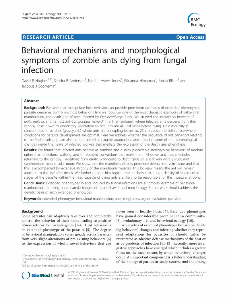
ResultsPre-biting behaviorThe ant species that is the primary host of Ophiocordy-ceps unilateralis s.l. at our field site is Camponotus leo-nardi [23]. This ant is canopy dwelling, rarelydescending to the forest floor and when it does it alwaystravels on well defined trails (Additional File 1). Trailindividuals do not forage on the forest floor and trailsnormally ascend into the canopy within 3-5 m fromwhere they descended (suggesting that workers descendonly because breaks in the canopy necessitate a descentto reach adjacent foraging crowns in the canopy). Unlikeants on trails the manipulated ants in the pre-leaf bitingstage were all discovered walking alone on low vegeta-tion, usually on saplings <50 cm above soil level andonly during the time interval 09:30-12:45 h (n = 21,Figure 1 and 2). All 21 zombie ants that we followedwere confirmed to be infected either via dissection ofthe head to reveal fungal cells or by observing the emer-gence of O. unilateralis s.l. following death on the leaf(Figure 3a). Post mortem fungal growth starts withabundant hyphae emanating from the intersegmentalmembranes within 2-3 days after host death and ulti-mately leads to stalk formation from the back of theant’s head [20].The host ant is diurnal at our field site [23] andinfected ants (n = 42) appeared even more restricted intheir activity as they were never observed in the earlymorning or late afternoon (15:00-18:00 hrs), in spite ofour searches covering these early and late periods of theday. The understory vegetation of our study site wasextensively searched during a year-long census programthat examined every leaf below 2 m height in 1360 m2
of forest habitat [23]. We therefore conclude that pre-biting infected C. leonardi ants at this site were only
Hughes et al. BMC Ecology 2011, 11:13http://www.biomedcentral.com/1472-6785/11/13
Page 2 of 10
active in the morning and that this observation was notaffected by sampling bias. The occasional trails ofhealthy ants that can be found on the forest floor(Additional File 1) were observed both during themorning hours and in the late afternoon, with activity
on trails always ceasing around sunset, i.e. between17:00-18:00 h.Since behavioral manipulation alters normal behavior we
could not a priori exclude the possibility that infectedindividuals would have become nocturnal. We thereforeconducted evening (after 18:00 h) and night (22:00-0:00 h)surveys using torches, but did not find any C. leonardiants active in the dark. Furthermore, a collected colony ofC. leonardi that likely contained some naturally infectedants was maintained under field laboratory conditions for2 days and did not show any activity in the dark, suggest-ing that behaviorally manipulated C. leonardi ants remainonly active during daylight.We also performed 20 hours and 28 minutes of focal
observations on 12 infected ants that were found walk-ing alone (infection status was later confirmed asdescribed above)(see Figure 1, individuals 10-21). Theseindividuals all expressed irregularly spaced whole bodyconvulsions (vertical bars on the periodogram in Figure1), which often made the ant fall from the vegetationonto the ground (denoted as stars on the periodogram).After falling infected ants always resumed walking andalways climbed a small sapling or comparable plant,which were abundantly present in the understory.We never observed trail ants falling from vegetation. To
document this, we removed 13 such ants from a trail on aliana approximately 1 m above the forest floor in the samearea where we observed the behaviorally manipulated ants.The liana descended from the canopy and the trailascended into the canopy via a tree trunk less than 3 mfrom where we collected the ants. The trail ants (assumedto be uninfected) were placed on the ground and they allquickly ascended into the canopy where we collectedthem again from tree trunks ca. 1.5 m above ground. Theonly exception was one trail ant that was predated uponby a spider (none of the behaviorally manipulated ants weobserved were predated upon). Trail ants did not spendextensive time walking in the understory. Their mediantime between release and reaching the trunk on whichthey ascended into the canopy was 28 minutes (range 7-51, total observation time 6 h, 2 m). After these observa-tions, the collected trail ants were maintained singly with-out food and died within a few days without signs of O.unilateralis s.l. fungal growth.Before biting a leaf, infected ants were predominantly
walking (average proportion of time walking 0.62, range0.11-1.00, total observation time 15 h, 35 m). They tra-versed an average of 99 leaves (range 52-239, 8 focalants), which was ca. twice the number traversed by trailants (average 51, range 8-140, 13 focal ants). Becausetrail ants were never observed walking on leaves exceptat the times when we removed them from trails andplaced them on the ground we conclude that traversingleaves is not a normal behavior. Therefore we did not
Figure 1 Zombie ant behavior. Focal animal observationperiodogram of ants infected by Ophiocordyceps unilateralis s.l. Theblue horizontal bars mark the observation period, the red trianglesmark moment of biting, the vertical bars mark spasm events andthe grey diamonds the falling off events. For four individuals thatbelong to focal animals 1-9 only the biting time was recorded. Thebiting time was recorded for 16 ants but only 15 triangles arevisible as two ants bite at exactly the same time (12:05). Insetpicture shows a dead ant on a leaf with the fungal stalk and sporebody that emerged from the head.
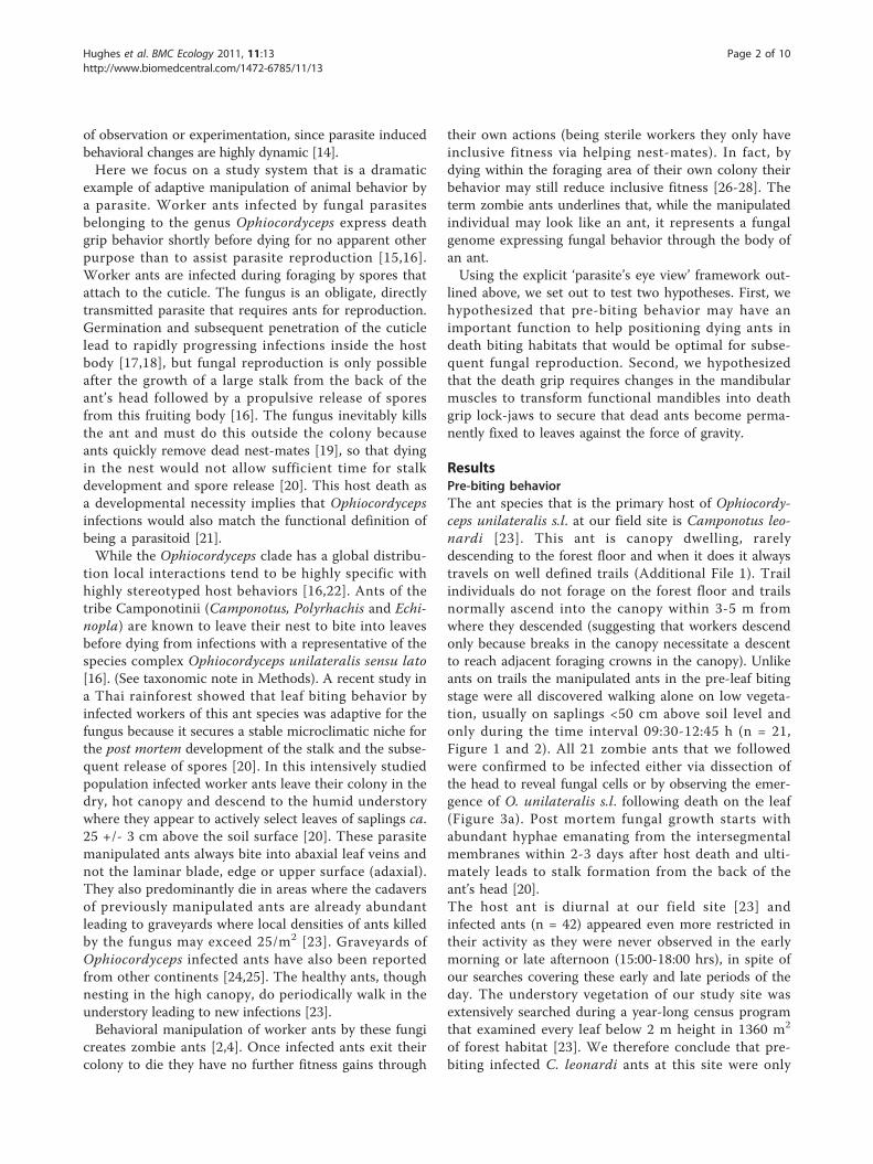
Figure 2 Synchronized manipulation of ants by fungi. A sunposition chart of the death grip. Solar altitude is represented by theyellow bars and plotted against the y-axis and the biting times arethe red circles and plotted on the x-axis in solar time (this is truelocal time accounting for longitude and different from Time inFigure 1). The red circles are stacked to prevent overlapping. At11:47 two ants bite so only 15 circles are visible though 16 antswere recorded.
Hughes et al. BMC Ecology 2011, 11:13http://www.biomedcentral.com/1472-6785/11/13
Page 3 of 10
statistically test for a difference between the numbers ofleaves traversed, as this was not biologically meaningful.During the pre-biting phase behaviorally manipulated
ants appeared to express a random “drunkard’s walk”such that an individual remained close to its startingpoint [29] but precise trajectories were not mapped sothis remains a heuristic assessment. In all cases theinfected ants finally bit into leaves <3 m from wherethey were first observed.
The timing at which infected ants bit into leaves wassynchronised around noon (Figure 1 &2; n = 16), sug-gesting either a direct solar cue or an indirect one viacorrelated temperature or humidity. The solar elevationat the moment of biting was 80.28° +/- 1.32 SE, whichwas close to the maximum solar elevation of 87.29° +/-0.39 SE during our study period. Once they had bittenleaves, ants rarely became detached and when this hap-pened it was due to disturbance (two cases, # 15 and 18
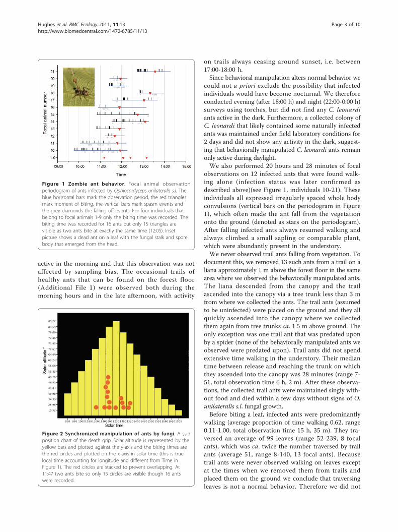
Figure 3 Heads of manipulated ants colonized by fungi. A (top panel) is a light micrograph (LM) saggital section through the head of an O.unilateralis s.l infected ant that was biting a leaf at the moment of fixation (i.e. alive). The small grey blobs are fungal hyphal bodies that fill thehead and mandibles. Note the spacing between the muscle fibers. The insect shows a close up of hyphal bodies around the post-pharyngealgland (PPG). B is the brain, Mu, Muscles and Cu is cuticle. B) is a LM of healthy muscle and C) is a LM of muscle from a behaviorally manipulatedant that was biting a leaf and alive when removed for fixation. The small blobs between the fibers are fungal cells.
Hughes et al. BMC Ecology 2011, 11:13http://www.biomedcentral.com/1472-6785/11/13
Page 4 of 10

in Figure 1, after very heavy rain). Biting leaves is notpart of the repertoire of healthy ants of this species.
Post-death grip behaviorAfter biting into leaves infected ants always died as thisis a developmental necessity for the subsequent growthof the fungus [15,20]. It was not possible to determinethe exact time of death since obvious signs such as mus-cle activity could be the result of fungal action, but itdid appear that ants could remain alive for as long assix hours after biting. Video recordings of six live antsbiting leaves revealed very little behavior of interestbesides a periodic twitching of the legs (Additional File2). The arrival of an ant of a different species close to abiting ant provoked no responses (Additional File 2), incontrast to healthy ants on trails, which were veryaggressive to other ant species they encountered at foodsources as well as to flying insects like wasps and fliesthat landed near honey baits.
Muscular atrophy accompanies behavioral manipulationAt the moment of the death grip, when the ant is underfungal control and biting into the major vein of a leafits head is filled with fungal cells (Figure 3). These cells,called hyphal bodies, were very abundant and could befound between the muscle fibers and surrounding thebrain and post pharyngeal gland (Figure 3), but notinside muscles, brains or glands.The most prominent other sign of infection, besides
the abundance of fungal cells inside the head capsule,was that the mandibular muscles were atrophied. Wesectioned the heads of 10 ants that were biting leavesand the pathology was the same across all 10. Mandibu-lar muscle fibers, which normally attach to the headcapsule, often appeared to have become detached (Fig-ure 3c) and where fibers remained attached they werestretched (compare 3b and 3c). Ant workers have bothmandibular opening and closing muscles and these canbe discriminated in healthy ants by their typical lengthof sarcomeres: 2-3 μm for opening muscles and 5-6 μmfor closing muscles (Figure 3b). However, in parasitizedants the characteristic stretching of sarcomeres made itimpossible to accurately distinguish between these twotypes of muscles. This may imply that fungal effects onthese muscles are unlikely to be cell specific at the timeof biting. Our behavioral observations revealed that themandibles worked normally in the hours preceding thedeath grip as infected ants were observed to self groom,cleaning their antennae and legs, which involves preciseopening and closing of the mandibles as these appen-dages are pulled across the maxillae to be cleaned.At the sub-cellular level (as seen with TEM) the mus-
cles of infected ants were very distinct from those ofhealthy individuals (Figure 4). Striated muscles (such as
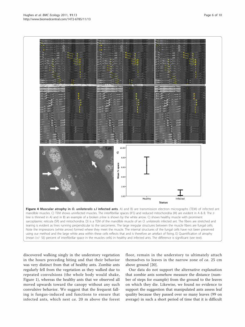
the mandible muscles) are composed of fibers that aremultinucleated cells formed as a result of cell fusion.These fibers contain thick (myosin) and thin (actin) fila-ments which attach during cross-bridge cycling leadingto muscle shortening. To achieve contraction mitochon-dria and sarcoplasmic reticulum provide energy (ATP)and ionic calcium (Ca++), respectively. At the end ofeach sarcomere unit there is a z-line (sarcomeres are infact defined as the area between z-lines), which can bethought of as the anchor points for muscle contraction.Infected ants sampled during the death grip had brokenz-lines and significantly less dense sarcoplasmic reticu-lum and mitochondria. This was determined from ameasurement of the increase of interfibrillar spaces thatappears following the loss of organelles, which in thiscase are sarcoplasmic reticulum and mitochondria(Kruskal-Wallis test, 20.25, df = 1, p < 0.0001, n = 6,Figure 4e). Similar to the light micrographs, the trans-mission electron micrographs showed a distinct atrophyin the muscles of infected ants.Despite the apparent atrophy of muscles the behavio-
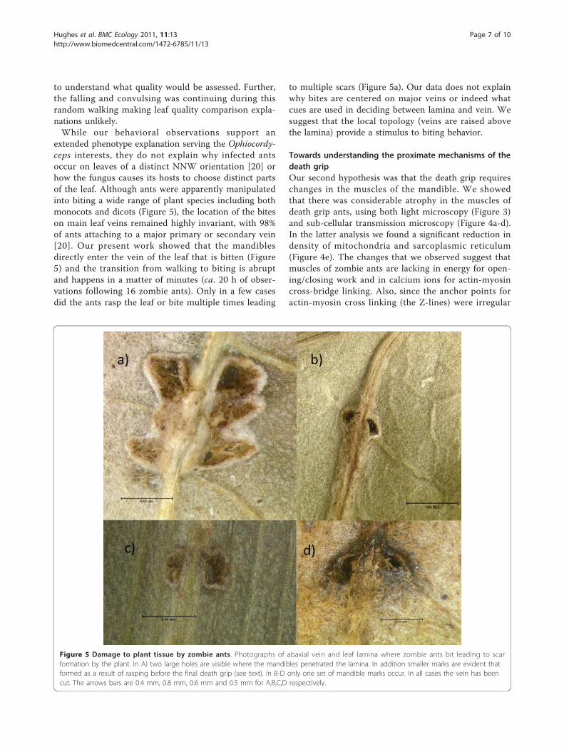
rally manipulated ants were able to exert considerableforce. We removed 29 dead ants from diverse species ofmonocotyledonous and dicotyledonous leaves collectedat our site (n = 10 and 19, respectively). On each leaflarge puncture wounds were evident where the antmandible had penetrated into the leaf (Figure 5). Onlyin two cases was the major vein on which biting wascentered not cut into.
DiscussionThe first biologist documented to have seen Ophiocordy-ceps-induced body snatching extended phenotypes wasAlfred Russell Wallace in 1859, as this features in histravelling notes from Sulawesi [30]. Yet despite the pres-tige of this original collector and the long period of timesince that discovery, we have learned remarkably littleabout how these parasitic fungi might control the beha-vior of their insect hosts [1]. The present study and itsrecent predecessors [20,23] thus merely reveal fragmentsof a fascinating parasite adaptation to the host whichcoming decades may be able to resolve because of therevolutionary developments in sequenced based technol-ogy. We believe, however, that such studies will be par-ticularly rewarding when they are based on fieldobservations, the implications of which we summarizein the sections below.
Multiple behavioral mechanisms explain niche choice bythe fungusOur first hypothesis was that the pre-biting behaviorwas important for positioning zombie ants in the pre-cise niche observed during earlier studies at our fieldsite [20,23]. We observed that infected ants could be
Hughes et al. BMC Ecology 2011, 11:13http://www.biomedcentral.com/1472-6785/11/13
Page 5 of 10
discovered walking singly in the understory vegetationin the hours preceding biting and that their behaviorwas very distinct from that of healthy ants. Zombie antsregularly fell from the vegetation as they walked due torepeated convulsions (the whole body would shake,Figure 1), whereas the healthy ants that we observed allmoved upwards toward the canopy without any suchconvulsive behavior. We suggest that the frequent fall-ing is fungus-induced and functions to ensure thatinfected ants, which nest ca. 20 m above the forest
floor, remain in the understory to ultimately attachthemselves to leaves in the narrow zone of ca. 25 cmabove ground [20].Our data do not support the alternative explanation
that zombie ants somehow measure the distance (num-ber of steps for example) from the ground to the leaveson which they die. Likewise, we found no evidence tosupport the suggestion that manipulated ants assess leafquality because they passed over so many leaves (99 onaverage) in such a short period of time that it is difficult
Figure 4 Muscular atrophy in O. unilateralis s.l infected ants. A) and B) are transmission electron micrographs (TEM) of infected antmandible muscles. C) TEM shows uninfected muscles. The interfibrillar spaces (IFS) and reduced mitochondria (M) are evident in A & B. The z-line is thinned in A) and in B) an example of a broken z-line is shown by the white arrow. C) shows healthy muscle with prominentsarcoplasmic reticula (SR) and mitochondria. D) is a TEM of the mandible muscle of an O. unilateralis infected ant. The fibers are stretched andtearing is evident as lines running perpendicular to the sarcomeres. The large irregular structures between the muscle fibers are fungal cells.Note the impressions (white arrow) formed where they meet the muscle. The internal structures of the fungal cells have not been preservedusing our method and the large white area within these cells reflects that and is therefore an artefact of fixing. E) Quantification of atrophy(mean (+/- SE) percent of interfibrillar space in the muscles cells) in healthy and infected ants. The difference is significant (see text).
Hughes et al. BMC Ecology 2011, 11:13http://www.biomedcentral.com/1472-6785/11/13
Page 6 of 10
to understand what quality would be assessed. Further,the falling and convulsing was continuing during thisrandom walking making leaf quality comparison expla-nations unlikely.While our behavioral observations support an
extended phenotype explanation serving the Ophiocordy-ceps interests, they do not explain why infected antsoccur on leaves of a distinct NNW orientation [20] orhow the fungus causes its hosts to choose distinct partsof the leaf. Although ants were apparently manipulatedinto biting a wide range of plant species including bothmonocots and dicots (Figure 5), the location of the biteson main leaf veins remained highly invariant, with 98%of ants attaching to a major primary or secondary vein[20]. Our present work showed that the mandiblesdirectly enter the vein of the leaf that is bitten (Figure5) and the transition from walking to biting is abruptand happens in a matter of minutes (ca. 20 h of obser-vations following 16 zombie ants). Only in a few casesdid the ants rasp the leaf or bite multiple times leading
to multiple scars (Figure 5a). Our data does not explainwhy bites are centered on major veins or indeed whatcues are used in deciding between lamina and vein. Wesuggest that the local topology (veins are raised abovethe lamina) provide a stimulus to biting behavior.
Towards understanding the proximate mechanisms of thedeath gripOur second hypothesis was that the death grip requireschanges in the muscles of the mandible. We showedthat there was considerable atrophy in the muscles ofdeath grip ants, using both light microscopy (Figure 3)and sub-cellular transmission microscopy (Figure 4a-d).In the latter analysis we found a significant reduction indensity of mitochondria and sarcoplasmic reticulum(Figure 4e). The changes that we observed suggest thatmuscles of zombie ants are lacking in energy for open-ing/closing work and in calcium ions for actin-myosincross-bridge linking. Also, since the anchor points foractin-myosin cross linking (the Z-lines) were irregular
Figure 5 Damage to plant tissue by zombie ants. Photographs of abaxial vein and leaf lamina where zombie ants bit leading to scarformation by the plant. In A) two large holes are visible where the mandibles penetrated the lamina. In addition smaller marks are evident thatformed as a result of rasping before the final death grip (see text). In B-D only one set of mandible marks occur. In all cases the vein has beencut. The arrows bars are 0.4 mm, 0.8 mm, 0.6 mm and 0.5 mm for A,B,C,D respectively.
Hughes et al. BMC Ecology 2011, 11:13http://www.biomedcentral.com/1472-6785/11/13
Page 7 of 10
or broken (Figure 4b) normal opening and closingwould probably have become impossible. Once the anthas become attached, the muscles apparently atrophyrapidly inducing the characteristic lock-jaw followed bythe death of the host ant some six hours later (Video 1).Thus while energy and ions for normal functioning arenot present the atrophy likely functions to imbed themandibles deep into the plant tissue due to this atrophy.Whatever the time course of muscular degradation
and its precise relationship to the extended phenotype(zombie behavior and biting) and unknown metaboliteproduction, it is difficult not to expect that the enor-mous population of fungal cells in the head of zombieants plays a decisive role (Figure 1). But much of thisawaits clarification in future studies.
Convergent evolution of zombie ant phenotypesThe synchronization of biting around solar noon that wefound adds an intriguing angle to our study suggestingthat future studies addressing the neurobiological andmolecular mechanisms behind such possible adaptationswill be highly rewarding. The sequences of behaviors weobserved leading from walking to convulsions and finallyto biting would make timed transcriptional profiles inter-esting and feasible. Fungi are well known to have clockgenes useful in synchronizing activity. Further insightscould be gained by extending such work to cover theother known cases where parasites have realized similarmanipulative syndromes to create zombie ants.Where Ophiocordyceps belongs to the fungal sub-phy-
lum Ascomycota, there is also an entomopthoraleanfungus Pandora that is known to turn European For-mica wood ants into zombies at distinct times of theday [31]. While the details differ in that Pandora zom-bies do not produce fungal fruiting bodies, but sporulatedirectly from mycelium on the surface of the dead ants,it is striking that this convergent evolution happened inspite of the last common ancestor of these fungal cladeshaving lived more than 500 mya [32]. Even more taxo-nomically distinct from both these fungal parasites isthe wood ant brain worm, the trematode Dicrocoeliumdendriticum, that also causes worker ants to leave theirnest and bite into grass blades at distinct times of theday [33] to facilitate transmission to sheep as additionalhosts (the ants acquire these trematode infections froma third host, a snail). With transcriptomic studiesbecoming more feasible, it would be highly intriguing tofind out whether these extended phenotypes rely onsimilar or different secondary metabolites to expressconvergent behavior of infected hosts.
ConclusionExtended phenotypes in ants induced by fungal infec-tions are a complex example of behavioral manipulation
requiring coordinated changes of host behavior andmorphology. Here we demonstrated some of themechanisms by which such changes can be induced dueto the effect of the fungi on ant walking behavior andmuscular activity, leading to the pronounced biting thatis a hallmark of this system. The insights from beha-vioral and histological studies point the way to moredetailed work which can, ideally, lead to a detailed pic-ture of the mechanisms by which organisms haveevolved to control the behavior of other groups, evenwhen they are in different kingdoms.
MethodsFieldwork took place in September 2007, ca. 20 km eastof Trang in southern Thailand (7°32’49.50"N, 99°47’14.73"E). Here a 24 ha Forest Dynamics Plot (FDP)was established on the North-North-eastern slopes ofthe hills in the peninsular Khao Chong Botanic Gardenas part of the Center for Tropical Forest Science (CTFS)pan-global FDP initiative http://www.ctfs.org. The site isexceptionally sandy with coarse and fine-grained sandsand covered by a primary mixed evergreen forest, withan understory dominated by saplings (<1 m). The cli-mate is tropical with seasonal monsoons and a meanmonthly maximum temperature range from 29.0°C to33.4°C, peaking in March-April. Rainfall is heaviest fromMay to September while the driest season is fromNovember to February.Live Camponotus leonardi ants were discovered by
searching the understory vegetation for lone individuals.In previous studies the main host for O. unilateralis wasC. leonardi and workers of this ant were encounteredforaging in trails in the high canopy (>20 m) but thesetrails were rare in the understory [20,23]. Therefore,individual C. leonardi worker ants walking alone in theunderstory were all considered to be potentially infectedjustifying detailed observation as focal individuals (seealso below). Because our initial indicator of infectionwas behavior, infection status of each ant was laterchecked. This was done in two ways: The first was byremoving the ant from the leaf and dissecting it to allowa microscopic determination of fungal cells (hyphalbodies) inside the head capsule [20]. This is a reliabletechnique as hyphal bodies are clearly visible upon dis-section [18]. The second technique was to return thefollowing day to the marked ant that had fixed itself toa leaf to determine whether O. unilateralis had startedgrowing from the body of the dead ant. This parasitefungus has a characteristic growth pattern followinghost death and a distinct colour (brownish hyphae) thatfacilitate a clear and unambiguous identification. Allfocal ants turned out to be infected.During the focal individual observations, ants were
monitored continuously from when they were discovered
Hughes et al. BMC Ecology 2011, 11:13http://www.biomedcentral.com/1472-6785/11/13
Page 8 of 10
until 15 minutes after they had bitten a leaf. At each 5-minute interval the state of behavior was recorded (e.g.walking, grooming, resting or feeding) and non-statebehaviors (events: falling, convulsing, rasping leaves andbiting leaves) were recorded as they occurred. The num-ber of leaves that ants traversed was recorded as well.This only happened for 8 individuals because accuratelycounting the number of leaves that an ant traversed overthe entire monitoring period (around 2 hours) was diffi-cult because leaves of small understory plants often over-lap which hampered accurate counting.Ants from a foraging trail on a liana where assumed
to be uninfected since they displayed normal behavior(foraging, communication with other ants on the trailand a display of aggression when disturbed). Individualsto be used for focal observations of healthy ants wereremoved from the trail and placed directly onto the for-est floor. Also these ants were continuously monitoredby one of three observers in the same way as describedabove for the infected ants. The observation periodstopped when the ant ascended a tree to a height of ca.1.5 m (where they were recollected) or if the ant waslost or predated upon. None of the infected ants werelost or predated upon, so this category was not men-tioned above.Searches for infected ants at other times of the day
besides 09:00-14:00 hrs occurred regularly through thecourse of fieldwork in 2006 (September/October 35 days;November, 3 days) and 2007 (January, 27 days; Septem-ber, 25 days). In September 2006 video recording wasused on ants that were discovered after they had bittenleaves. The leaf on which an ant was biting was recordedfor 10 minutes and behavior subsequently scored.We used the freely available Solar Position Calculator
to obtain the solar time and solar altitude for each antthat bit into a leaf. Ant observations took place on 10,11, 12 13, 14, 15, 16 and 19 September 2007. As anexample, sunrise was at 6:15 on the 10th and sunset wasat 18:21 on the 10th of September. We used Latitude7.54347 and Longitude 99.798 to calculate the solartime and elevation.
HistologyThe heads of healthy ants and ants that had bittenleaves were removed and were fixed in 2% glutaralde-hyde (buffered at pH 7.3 with 50 mM sodium cacodylateand 150 mM saccharose) and post-fixed in 2% osmiumtetroxide in the same buffer. Dehydration was carriedout in a graded acetone series and preceded embeddingin Araldite and sectioning with a Reichert Ultracut Emicrotome. Thin sections of 1 μm for light microscopywere stained with methylene blue and thionin. Ultrathinsections of 70 nm were double stained (lead citrate and
uranyl acetate) and examined with a Zeiss EM900 trans-mission electron microscope.
Taxonomic noteWe use the designation sensu lato throughout toemphasize that based on an on-going taxonomic revi-sion of the globally dispersed fungal species Ophiocordy-ceps unilateralis, a new name will be required for thespecies at our study site [34].
Additional material
Additional file 1: Trails of the ant Camponotus leonardi. The videoshows ants running in a trail on a branch above the forest floor in atropical forest in Southern Thailand.
Additional file 2: A zombie ant biting a leaf vein. An ant attached byits mandibles to the main vein of a leaf in a tropical forest in SouthernThailand. The ant remains attached until its death and does not respondto external factors such as another ant approaching as in the video.
Acknowledgments and FundingWe thank Maria Moltesen, Sylvia Gerritsma and Kanoksri Tasanathai forassistance in the field and Prof Morakot Tanticharoen and Dr KanyawimKirtikara and the staff of BIOTEC for continued help and support. We aregrateful to Dr Sarayudh Bunyavejchewin and the Department of NationalParks, Wildlife and Plant Conservation, Thailand for support and Dr DavidLohman at Khao Chong Research Station for much help. We are grateful toWulfila Gronenberg and Robin Maytum for providing us with insights intomuscular physiology. This work was supported by a grant from the DanishNational Research Foundation (JJB) and a Marie Curie Intra-EuropeanFellowship and a Novozymes/WWF grant (DPH).
Author details1Departments of Entomology and Biology, Penn State University, PA 16802,USA. 2Centre for Social Evolution, Department of Biology, University ofCopenhagen, Universitetsparken 15, 2100 Copenhagen, Denmark. 3MycologyLaboratory, National Center for Genetic Engineering and Biotechnology,Science Park, Pathum Thani 12120, Thailand. 4Forest Entomology andMicrobiology Group, National Park, Department of National Parks, Wildlifeand Plant Conservation, Bangkok 10900, Thailand. 5Zoological Institute,University of Leuven, Naamsestraat 59, 3000 Leuven, Belgium.
Authors’ contributionsConceived and designed the experiments: DPH. Performed the experiments:DPH, SBA, JB, WH Analyzed the data: DPH, SBA, JB Wrote the paper: DPH,JJB, JB, SBA, NLH-J, MBP, JJK. All authors read and approved the finalmanuscript.
Received: 17 March 2011 Accepted: 9 May 2011 Published: 9 May 2011
References1. J Moore, Parasites and the behavior of animals. (Oxford: Oxford University
Press, 2002)2. K Lafferty, AM Kuris, Parasitic castration: the evolution and ecology of body
snatchers. Trends in Parasitology. 25(12):564–572 (2009). doi:10.1016/j.pt.2009.09.003
3. R Poulin, Progenesis and reduced virulence as an alternative transmissionstrategy in a parasitic trematode. Parasitology. 123, 623–630 (2001)
4. T Lefevre, SA Adamo, DG Biron, D Misse, D Hughes, F Thomas, Invasion ofthe Body Snatchers: the diversity and evolution of manipulative strategiesin host-parasite interactions. Advances in Parasitology. 68, 45–83 (2009)
5. R Dawkins, The extended phenotype. (Oxford: W.H. Freeman, 1982)
Hughes et al. BMC Ecology 2011, 11:13http://www.biomedcentral.com/1472-6785/11/13
Page 9 of 10
6. ME Rogers, PE Bates, Leishmania manipulation of sand fly feeding behaviorresults in enhanced transmission. Plos Pathogens. 3(6):e91 (2007).doi:10.1371/journal.ppat.0030091
7. WG Eberhard, Spider manipulation by a wasp larva. Nature.406(6793):255–256 (2000). doi:10.1038/35018636
8. AM Kuris, RF Hechinger, JC Shaw, KL Whitney, L Aguirre-Macedo, CA Boch,AP Dobson, EJ Dunham, BL Fredensborg, TC Huspeni., et al, Ecosystemenergetic implications of parasite and free-living biomass in three estuaries.Nature. 454(7203):515–518 (2008). doi:10.1038/nature06970
9. F Thomas, S Adamo, J Moore, Parasitic manipulation: where are we andwhere should we go? Behavioural Processes. 68(3):185–199 (2005).doi:10.1016/j.beproc.2004.06.010
10. DP Hughes, DJC Kronauer, JJ Boomsma, Extended Phenotype: Nematodesturn ants into bird-dispersed fruits. Current Biology. R294–295 (2008)
11. R Poulin, The evolution of parasite manipulation of host behavior: atheoretical analysis. Parasitology. 109, S109–S118 (1994). doi:10.1017/S0031182000085127
12. R Poulin, Manipulation of host behaviour by parasites: a weakeningparadigm? Proceedings of the Royal Society of London Series B.267(1445):787–792 (2000). doi:10.1098/rspb.2000.1072
13. R Dawkins, Parasites, desiderata lists and the paradox of the organism.Parasitology. 100, S63–S73 (1990). doi:10.1017/S0031182000073029
14. MI Sanchez, F Ponton, A Schmidt-Rhaesa, DP Hughes, D Misse, F Thomas,Two steps to suicide in crickets harbouring hairworms. Animal Behaviour.76, 1621–1624 (2008). doi:10.1016/j.anbehav.2008.07.018
15. HC Evans, Entomogenous fungi in the tropical forest ecosystems: anappraisal. Ecological Entomology. 7, 47–60 (1982). doi:10.1111/j.1365-2311.1982.tb00643.x
16. HC Evans, RA Samson, Cordyceps species and their anamorphs pathogenicon ants (Formicidae) in tropical forest ecosystems. II. The Camponotus(Formicinae) complex. Transactions of the British Mycolocical Society. 82,127–150 (1984). doi:10.1016/S0007-1536(84)80219-3
17. A Van Pelt, The occurrence of a Cordyceps on the ant Camponotuspennsylvanicus (De Geer) in the Highlands, N.C. region. Journal of theTennesee Academy of Sciences. 33(120-122) (1958)
18. DP Hughes, HC Evans, NL Hywel-Jones, JJ Boomsma, SAO Armitage, Novelfungal disease in complex leaf-cutting ant societies. Ecological Entomology.34(2):214–220 (2009). doi:10.1111/j.1365-2311.2008.01066.x
19. B Hölldobler, EO Wilson, The ants. (Cambridge, Mass.: Harvard UniversityPress, 1990)
20. SB Andersen, S Gerritsma, KM Yusah, D Mayntz, NL Hywel-Jones, J Billen, JJBoomsma, DP Hughes, The life of a dead ant: the expression of an adaptiveextended phenotype. American Naturalist. 174(3):424–433 (2009).doi:10.1086/603640
21. AM Kuris, Trophic interactions: similarity of parasitic castrators to parasitoids.Quarterly Review of Biology. 49, 129–148 (1974). doi:10.1086/408018
22. HC Evans, RA Samson, Cordyceps species and their anamorph pathogenicon ants (Formicidae) in tropical forest ecosystems. I. The Cephalotes(Myrmicinae) complex. Transactions of the British Mycolocical Society. 79,431–453 (1982). doi:10.1016/S0007-1536(82)80037-5
23. M-B Pontoppidan, W Himaman, NL Hywel-Jones, JJ Boomsma, DP Hughes,Graveyards on the move: the spatio-temporal distribution of deadOphiocordyceps-infected ants. PLoS ONE. 4(3):e4835 (2009). doi:10.1371/journal.pone.0004835
24. T Sanjuán, L Guillermo Henao, G Amat, Distribución espacial de Cordycepsspp. (Ascomycotina: Clavicipitaceae) y su impacto sobre las hormigas enselvas del piedemonte amazónico de Colombia. Revista de BiologiaTropical. 49(3-4):945–955 (2001)
25. HC Evans, Natural control of Arthropods with special reference to ants(Formicidae) by fungi in the tropical high forest of Ghana. The Journal ofApplied Ecology. 11(1):37–49 (1974). doi:10.2307/2402003
26. DP Hughes, Parasitic manipulation: a social context. Behavioural Processes. ,3: 263–266 (2005)
27. DP Hughes, The extended phenotype within the colony and how itobscures social communication. in Sociobiology of Communication: aninterdisciplinary perspective, ed. by d?’?Ettorre P, Hughes DP (Oxford OxfordUniversity Press, 2008)
28. DP Hughes, NE Pierce, JJ Boomsma, Social insect symbionts: evolution inhomeostatic fortresses. Trends in Ecology & Evolution. 23(12):672–677(2008). doi:10.1016/j.tree.2008.07.011
29. K Pearson, The problem of the random walk. Nature. 72, 342–342 (1905)
30. W Fawcett, Description of Cordyceps llyodii in ants. Annals and Magazine ofNatural History. 5(XVIII):317 (1886)
31. PI Marikovsky, On some features of behaviour of the ants Formica rufa L.infected with fungous disease. Insectes Sociaux. 9(173-179) (1962)
32. JE Blair, Fungi. (Oxford Oxford University Press, 2009)33. MY Manga-Gonzalez, C Gonzalez-Lanza, E Cabanas, R Campo, Contributions
to and review of dicrocoeliosis, with special reference to the intermediatehosts of Dicrocoelium dendriticum. Parasitology. 123, S91–S114 (2001)
34. HC Evans, SL Elliot, DP Hughes, Hidden diversity behind the Zombie-antfungus Ophiocordyceps unilateralis: Four new species described fromCarpenter ants in Minas Gerais, Brazil. PLoS ONE. 6(3):e17024 (2011).doi:10.1371/journal.pone.0017024
doi:10.1186/1472-6785-11-13Cite this article as: Hughes et al.: Behavioral mechanisms andmorphological symptoms of zombie ants dying from fungal infection.BMC Ecology 2011 11:13.
Submit your next manuscript to BioMed Centraland take full advantage of:
• Convenient online submission
• Thorough peer review
• No space constraints or color figure charges
• Immediate publication on acceptance
• Inclusion in PubMed, CAS, Scopus and Google Scholar
• Research which is freely available for redistribution
Submit your manuscript at www.biomedcentral.com/submit
Hughes et al. BMC Ecology 2011, 11:13http://www.biomedcentral.com/1472-6785/11/13
Page 10 of 10