research article open access a proteomic study of cmyc
TRANSCRIPT
RESEARCH ARTICLE Open Access
A proteomic study of cMyc improvementof CHO cultureDarrin Kuystermans1,2, Michael J Dunn2, Mohamed Al-Rubeai1,2*
Abstract
Background: The biopharmaceutical industry requires cell lines to have an optimal proliferation rate and a highintegral viable cell number resulting in a maximum volumetric recombinant protein product titre. Nutrient feedinghas been shown to boost cell number and productivity in fed-batch culture, but cell line engineering is anotherroute one may take to increase these parameters in the bioreactor. The use of CHO-K1 cells with a c-myc plasmidallowing for over-expressing c-Myc (designated cMycCHO) gives a higher integral viable cell number. In this studythe differential protein expression in cMycCHO is investigated using two-dimensional gel electrophoresis (2-DE)followed by image analysis to determine the extent of the effect c-Myc has on the cell and the proteins involvedto give the new phenotype.
Results: Over 100 proteins that were differentially expressed in cMycCHO cells were detected with high statisticalconfidence, of which 41 were subsequently identified by tandem mass spectrometry (LC-MS/MS). Further analysisrevealed proteins involved in a variety of pathways. Some examples of changes in protein expression include: anincrease in nucleolin, involved in proliferation and known to aid in stabilising anti-apoptotic protein mRNA levels,the cytoskeleton and mitochondrial morphology (vimentin), protein biosysnthesis (eIF6) and energy metabolism(ATP synthetase), and a decreased regulation of all proteins, indentified, involved in matrix and cell to celladhesion.
Conclusion: These results indicate several proteins involved in proliferation and adhesion that could be useful forfuture approaches to improve proliferation and decrease adhesion of CHO cell lines which are difficult to adapt tosuspension culture.
BackgroundChinese hamster ovary cells (CHO) are the most popu-lar commercial platform for the production of therapeu-tic proteins with much development going into the useof such cell lines for increasing product yields. Hencethe productivity of cell cultures has improved more than100-fold in the last two decades mainly due to develop-ments of fed-batch culture systems, media and processoptimisations in conjunction with expression technolo-gies [1,2]. As much as improvements in specific produc-tivity (Qp) are important in cell lines, growthcharacteristics also have a significant impact on the pro-cess. A good cell line proliferative capacity and a highintegral viable cell number (IVC) can result in highvolumetric recombinant protein production rates. Thus
the mammalian biopharmaceutical industry has researchinterests directed towards the development of cell lineswith high proliferation rate that can be grown to highdensities and have high production capabilities. Induc-tion of the transcription factor Myc promotes cell prolif-eration and transformation by activating growthpromoting genes or by repressing the expression ofgrowth arrest genes [3-11]. The gene, c-myc, is a primecandidate that regulates cell proliferation in such a waythat its introduction into cell lines may be advantageous.Research has shown that transfection of adherent
CHO cell line with the c-myc gene resulted in increasedproliferation rate and cell number [11,12]. To under-stand the cellular activity that results from the overex-pression of c-Myc (via the transfection with c-mycplasmid) in CHO cells, the techniques of two-dimen-sional polyacrylamide gel electrophoresis (2-DE) and sta-tistically viable image analysis combined with mass
* Correspondence: [email protected] of Chemical and Bioprocess Engineering, University College Dublin,Belfield, Dublin 4, Ireland
Kuystermans et al. BMC Biotechnology 2010, 10:25http://www.biomedcentral.com/1472-6750/10/25
© 2010 Kuystermans et al; licensee BioMed Central Ltd. This is an Open Access article distributed under the terms of the CreativeCommons Attribution License (http://creativecommons.org/licenses/by/2.0), which permits unrestricted use, distribution, andreproduction in any medium, provided the original work is properly cited.
spectrometry were employed to help identify the pro-teins involved. This technique allows for the separationof complex protein mixtures with a relative high resolu-tion involving a two-step separation of the proteins, firstby isoelectric point and then by size to generate proteinmaps of the investigated proteome.Currently, the database of the proteomes of CHO cell
is not complete, but due to similarities of mammalianproteins between species successful identification of pro-teins can be done across species [13]. This has made itpossible to carry out several proteomic studies on theCHO cells including a general proteome map [14,15].Further analysis of the protein regulation under con-trolled conditions has led to the 2D proteome analysisof CHO cells in response to hyperosmotic conditions[16], increased production levels[17,18], low temperatureshift [19], and growth factor stimulation [20]. Also theproteomic work carried out on c-Myc is limited, not inCHO cells, and none has been done using the 2DEapproach[21,22] In this study, it is the objective to iden-tify potential proteins involved in producing the pheno-type seen with a c-myc plasmid in CHO cells in thehope of increasing our understanding of the intracellularand physiological changes and providing further insightsinto possible CHO cell manipulation for improved cellline development. In this work the cell line containingc-myc plasmid is compared to the parental cell line todetermine if any bioprocessing benefit would have beenachieved. It may be useful to state that a comparison ofthe cells containing the c-myc plasmid with cells con-taining blank plasmid would provide further specificinformation on the c-myc effects.
MethodsCell Transfection and MaintenanceThe transfection and selection protocol has been pre-viously reported [12]. Briefly, the cMycCHO (c-mycplasmid containing CHO-K1) cell line resulted by cal-cium-mediated stable transfection of CHO-K1, with theDORclaG123 (c-Myc plasmid). The plasmid was kindlydonated by Dr. T. Littlewood (then at Imperial CancerResearch Fund, UK). Surviving cells were pooled sepa-rately from each transfection and maintained in Ham’sF12 supplemented with 5% FCS. Dilution cloning wasused to select a stable cMycCHO clone with high overexpression of the c-myc gene for subsequent experi-ments. Selection pressure was maintained by incubationin DMEM/F12 with 5% FBS (Lonza Biologics) and 1mg/mL geneticin G418 (Sigma Aldrich, UK) every 10th
generation. Samples were taken before and after incuba-tion to ensure stable overexpression of the c-Myc pro-tein. To negate the effects of the antibiotic selectionpressure was removed 2 passages before an experimentalrun.
Static Batch Culture of CHO-K1 and cMycCHOIn subsequent experiments CHO-K1 and cMycCHOwere maintained in DMEM/F12 with 5% FBS (LonzaBiologics, UK) at 37°C vented in 5% CO2 incubator. Sta-tic cultures from each cell line were plated in 25 cm2
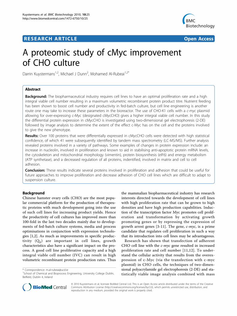
(75 cm2 for proteome isolation) vented T-flasks with aninitial density of 2 × 10⁵ cells/ml allowing triplicateflasks to be sacrificed at regular intervals for each cellline. Viable cell concentration and viability were moni-tored using the trypan blue exclusion method [23]. Thesample for proteomic analysis was taken at late expo-nential phase of the culture. This point also put empha-sis on that in a limited nutrient environment thecMycCHO can maintain a higher cell density. To verifythe inoculum had c-Myc over expression a separateduplicate culture was run for Western analysis.
Determination of Extracellular Glucose and LactateGlucose was measured with an Ascencia contour (BayerDiagnostics, Ireland) glucose meter using pre-calibratedMircofill test strips (Bayer Diagnostics, Ireland). Lactatewas measured using the Accutrend Lactate Metertogether with BM-Lactate Strips (Roche DiagnosticsGmbH, Germany). Briefly, The BM-Lactate calibrationstrip was used to calibrate the instrument to the accom-panied BM-Lactate strips. Lactate was determined byreflectance photometry at a wave length of 657 nm via acolorimetric-oxidase mediator reaction.
Proteome IsolationThe experimental setup is illustrated in Figure 1. Aminimum of 1 × 10⁷ cells of each replicate was used toobtain total cellular protein which was washed in iso-tonic (0.35 M) sucrose to reduce salt contamination andthen resuspended in 400 μl of lysis buffer (Ultrapurereagents from GE Healthcare, Uppsala, Sweden): 2% (w/v) CHAPS, 9.5 M urea, 0.8% (w/v) Pharmalyte pH 3-10,1% (w/v) DTT (GE Healthcare) and 1 protease inhibitortablet per 10 ml (Roche Molecular, Mannheim, Ger-many) for 30 minutes. The cells where then centrifugedat 4°C and 17,000 g for 30 minutes. Samples were storedat -80°C for minimum of 24 hours before concentrationdetermination using a modified Bradford assay [24].
Two-Dimensional Sodium Polyacrylamide GelElectrophoresis (2-DE)An amount of 100 ug protein/gel was used for analyticalgels and 400 ug protein/gel for preparative gels. Lysedsamples were diluted in rehydration buffer (8 M urea,0.5% [w/v] CHAPS, 0.2% [w/v] DTT, 0.2% [w/v] Phar-malyte pH 3-10); and the appropriate protein loadapplied to each 24 cm, linear pH 4-7, immobilized pHgradient (IPG) strip (GE Healthcare, Uppsala, Sweden)by in-gel rehydration [25-27]. IPG strips were focused at
Kuystermans et al. BMC Biotechnology 2010, 10:25http://www.biomedcentral.com/1472-6750/10/25
Page 2 of 13
50 uA/strip at 20°C for 4 voltage changes, these were;(1) 75,000 Volt-hours (Vh) at 3500 V, followed by (2) agradient increase to 8000 V for 10 min, which was thenleft at (3) 8000 V for 1 hour, followed by, (4) 100 V fora maximum of 3 hours using an IPGphor System (GEHeathcare). Subsequently, the IPG strips were onceequilibrated in a buffer containing 6 M Urea, 50 mMTrisCl pH 8.8, 30% (v/v) glycerol, 2% (w/v) SDS and 1%(w/v) DTT for 15 min and then in the same buffer butwith the addition of 4.8% (w/v) iodacetamide for afurther 15 min. Proteins were separated in the seconddimension by SDS-PAGE (gel format 24 × 20 × 0.15 cm).After gel fixation in methanol/acetic acid solution, theanalytical gels were stained with 2D-Silver Stain IIKit (Daiichi Pure Chemicals, Tokyo, Japan) while thepreparative gels were stained with the PlusOne™ Silver
Staining Kit (GE Healthcare) using a modified, massspectrometry compatible protocol [28].
Image and Data Analysis2-D gel images were captured using a Molecular ImagerGS-800 scanner (Bio-Rad), warped with TT900 S2S soft-ware (Nonlinear Dynamics, Newcastle, UK) and quanti-tatively analysed using Progenesis PG240 SameSpot™software (Nonlinear Dynamics, Newcastle upon Tyne,UK). Protein spot detection and normalization (an inte-gration of the area and optical density, as a percentageof the total volume of all detected spots) was performedautomatically, followed by manual checking of detectedspots; reassigning a numerical spot designation andremoving background noise before renormalization andapplying the SameSpot™ analysis algorithm (SSAA). This
Figure 1 Experimental setup and inverted microscopic image of adherent culture of CHO-K1 and cMycCHO cell lines. The Morphologyof cell lines shows adherent c-Myc over-expression causing morphological changes to the cell with the formation of foci. The experimentaldesign above demonstrates that from the 3 biological replica culture samples, 3 technical sample replicates from each of these were used inproteomic image analysis setup.
Kuystermans et al. BMC Biotechnology 2010, 10:25http://www.biomedcentral.com/1472-6750/10/25
Page 3 of 13

procedure, addresses measurement variations from gelto gel while pixel level alignment corrects for positionalvariation from gel to gel. The fold change value is calcu-lated from the mean normalised volumes betweencMycCHO and CHO-K1. All spots were compared tocontrol reference gel and for eliminating experimentaldiscrepancies between spots. SSAA applies the samespot outline to corresponding spots for all gels beforethe application of ANOVA (p < 0.05) for statistically sig-nificant changes in their protein expression based on thenormalized volume of each spot. Further ontology analy-sis was carried out by transferring the Progenesis data toGenePilot™ V1.95b (TG Services, Inc. CA USA).
LC-MS/MS and Data AnalysisManual excision of the protein spots of interest andtryptic digestion were carried out using an Ettan Diges-ter (GE Healthcare, Uppsala, Sweden) and peptide analy-sis was carried out using a linear ion trap massspectrometer (LTQ Mass Spectrometer; Thermo-Finni-gan Ltd, Hemel Hempstead, UK). The mass spectraparameters were set to a full scan MS mass range of400-1600 m/z. The mass spectrometer was equippedwith a nanospray ion source (Thermo Electron Corpora-tion, Waltham, MA) with a spray voltage of 1.9 kV. Bio-Works 3.2 software (Thermo Electron Corporation),featuring the TurboSEQUEST search algorithm, wasused to query the Swiss-Prot database for protein identi-fication. Two filters were used to consider significanthits, peptide Xcorr (statistical criteria correlation value)versus charge state (charge state: +1, +2, +3; set-points:1.9, 2.0, 2.5) with a peptide probability-based scoringalgorithm cut-off of < 10-3.
Western BlotA protein load of 50 μg/well underwent electrophoresison a 12% sodium dodecyl sulfate-polyacrylamide (SDS-PAGE) mini gel (Pierce, Rockford, IL) subsequent todilution in 1× Laemli buffer with prior denaturation at95°C for 10 minutes. The gel was stained with Instant-blue™ (Novexin Ltd, UK) for visual verification of load-ing before de-staining for electrotransfer. The proteinwas then transferred to an Immobilon™-P membrane(0.45 μm) (Millipore, Cork, Ireland) followed by block-ing of the membranes with 5% Marvel-PBS for 1 hourat room temperature and probed overnight at 4°C byincubation with the primary antibody (Santa Cruz Bio-technology, CA), either anti-myc 9E10, rabbit anti-RKIP,rabbit anti-NUCL, rabbit anti-eIF6, or rabbit anti-GRP78. The blot was washed and then incubated with aHRP conjugated anti-mouse (only for anti-myc) or anti-rabbit whole IgG as the secondary antibody. Bands weredeveloped by chemiluminescent detection (SuperSignalWest Pico System, Pierce) according to the
manufacturer’s instructions. Any required blot strippingwas carried out by incubating in NaOH followed by anovernight blocking in 10% Marvel-PBS at 4°C. Themembranes were stained with Coomassie Brilliant BlueR-250 (Sigma Aldrich, UK) to re-verify loading and uni-formity of the electrotransfer.
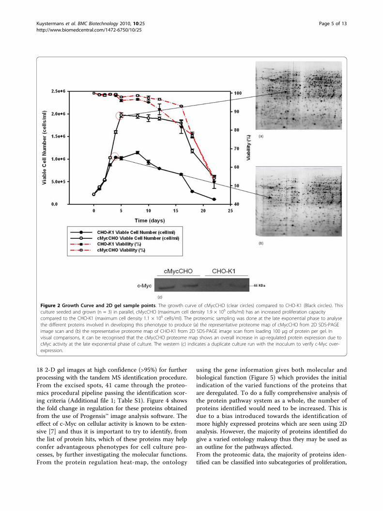
Results and DiscussionGrowth of Cell LinesFor proteomic profiling, a batch culture, in triplicate wasgrown (Figure 2) with adherent cMycCHO alongside itscontrol CHO-K1. The cMycCHO culture had anincrease in overall proliferative capacity being able toproliferate to higher cell densities of 1.94 × 10⁶ cells/mlcompared to 1.14 × 10⁶ cells/ml of CHO-K1. The viabi-lity remained above 90% for both cell lines until day 10after which CHO-K1 started a steady decline in viabilitywhile cMycCHO culture continued to survive at anaverage of 93% on day 16 compared to CHO-K1 whichhad a viability of 82%. Under these growth conditionscMycCHO had an increased growth rate of 0.54 day-1
compared to 0.48 day-1 for CHO-K1. The increase inmaximal cell density combined with the increasedgrowth rate, for an 8 day culture period, resulted in anIVC increase of 59% for the cMycCHO cell line reach-ing 10.54 × 10⁹ cell day L-1. Proteomic samples weretaken from where the biggest variation in cell numberwas seen between the CHO-K1 and cMycCHO whilestill at the highest viability for both cultures and thiswas seen to be the late exponential phase.The increased growth rate had an effect on specific
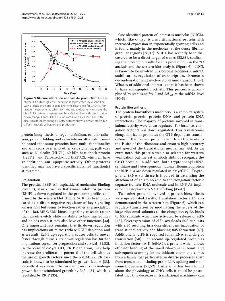
glucose utilisation and lactate production (Figure 3). InFigure 3 it can be observed that the glucose and lactateconsumption and production profiles of cMycCHO areonly slightly greater than those of the control. Thuswhen calculating these as rates per cell there was adecrease in the specific glucose rate of 1.72 μmole 10-6
cells day-1, for cMycCHO, compared to a rate of 2.15μmole 10-6 cells day-1 for CHO-K1. This shows that thecMycCHO cell line could be considered more efficientat glucose utilisation. Similarly, specific Lactate produc-tion by cMycCHO had decreased by 49.5% in compari-son to the CHO-K1 value of 4.32 μmole 10-6 cells day-1.These results are in agreement with earlier batch cultureexperiments where both glucose utilisation and lactateproduction have decreased in the c-myc engineered cellline [12].
Proteome ProfileThe effect of constitutive expression of c-myc plasmidon the CHO proteome was fairly extensive with initiallymore then a 300 proteins deregulated by visual analysis.Using the Progensis™ statistical package, there were 122spots detected as being differentially expressed from the
Kuystermans et al. BMC Biotechnology 2010, 10:25http://www.biomedcentral.com/1472-6750/10/25
Page 4 of 13
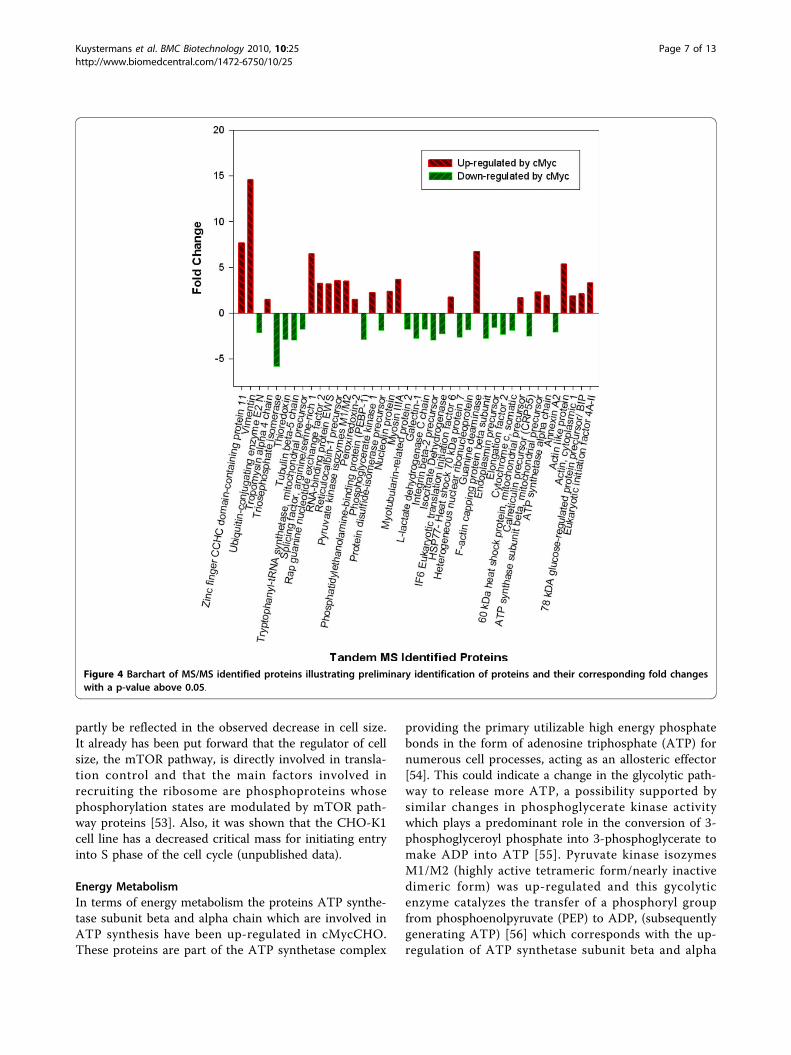
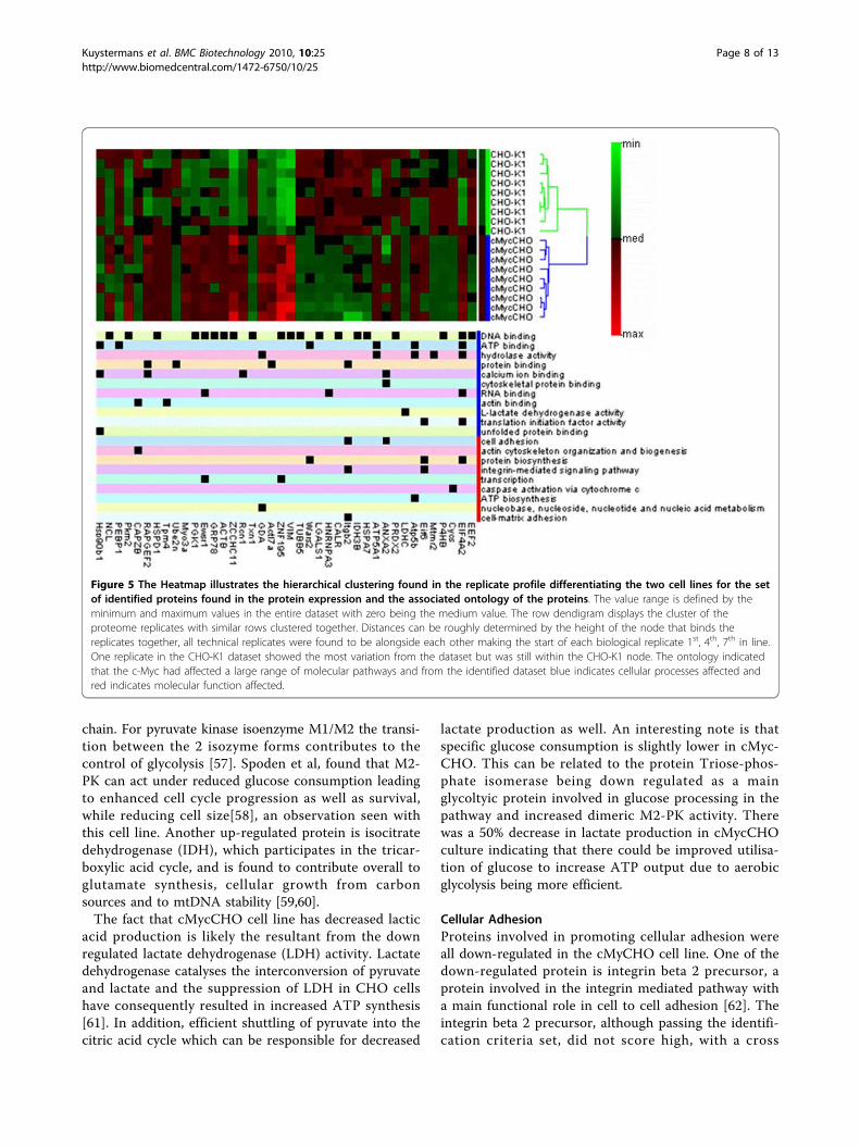
18 2-D gel images at high confidence (>95%) for furtherprocessing with the tandem MS identification procedure.From the excised spots, 41 came through the proteo-mics procedural pipeline passing the identification scor-ing criteria (Additional file 1; Table S1). Figure 4 showsthe fold change in regulation for these proteins obtainedfrom the use of Progensis™ image analysis software. Theeffect of c-Myc on cellular activity is known to be exten-sive [7] and thus it is important to try to identify, fromthe list of protein hits, which of these proteins may helpconfer advantageous phenotypes for cell culture pro-cesses, by further investigating the molecular functions.From the protein regulation heat-map, the ontology
using the gene information gives both molecular andbiological function (Figure 5) which provides the initialindication of the varied functions of the proteins thatare deregulated. To do a fully comprehensive analysis ofthe protein pathway system as a whole, the number ofproteins identified would need to be increased. This isdue to a bias introduced towards the identification ofmore highly expressed proteins which are seen using 2Danalysis. However, the majority of proteins identified dogive a varied ontology makeup thus they may be used asan outline for the pathways affected.From the proteomic data, the majority of proteins iden-tified can be classified into subcategories of proliferation,
Figure 2 Growth Curve and 2D gel sample points. The growth curve of cMycCHO (clear circles) compared to CHO-K1 (Black circles). Thisculture seeded and grown (n = 3) in parallel, cMycCHO (maximum cell density 1.9 × 10⁶ cells/ml) has an increased proliferation capacitycompared to the CHO-K1 (maximum cell density 1.1 × 10⁶ cells/ml). The proteomic sampling was done at the late exponential phase to analysethe different proteins involved in developing this phenotype to produce (a) the representative proteome map of cMycCHO from 2D SDS-PAGEimage scan and (b) the representative proteome map of CHO-K1 from 2D SDS-PAGE image scan from loading 100 μg of protein per gel. Invisual comparisons, it can be recognised that the cMycCHO proteome map shows an overall increase in up-regulated protein expression due tocMyc activity at the late exponential phase of culture. The western (c) indicates a duplicate culture run with the inoculum to verify c-Myc over-expression.
Kuystermans et al. BMC Biotechnology 2010, 10:25http://www.biomedcentral.com/1472-6750/10/25
Page 5 of 13
protein biosynthesis, energy metabolism, cellular adhe-sion, protein folding and cytoskeleton although it mustbe noted that some proteins have multi-functionalityand will cross over into other cell signaling pathwayssuch as Nucleolin (NUCL), 60 kDa heat shock protein(HSPD1), and Peroxiredoxin 2 (PRDX2), which all havean additional anti-apoptotic activity. Other proteinsidentified may not have a specific classified function(s)at this time.
ProliferationThe protein, PEBP-1(Phosphatidylethanolamine BindingProtein), also known as Raf kinase inhibitor protein(RKIP) is down regulated in the proteome profile, con-firmed by the western blot (Figure 6). It has been impli-cated as a direct negative regulator of key signalingkinases [29] but seems to function rather as a modulatorof the Raf/MEK/ERK kinase signaling cascade ratherthan an off-switch while its ability to bind nucleotidesand opiods mean it may also have other functions [30].One important fact remains, that its down regulationhas implications on mitosis where RKIP depletion andas a result, Raf-1 up-regulation, causes cells to moverfaster through mitosis. Its down-regulation has furtherimplications on cancer progression and survival [31,32].In the case of cMycCHO, RKIP depletion, may helpincrease the proliferation potential of the cell withoutthe use of growth factors since the Raf/MEK/ERK cas-cade is known to be stimulated by growth factors [33].Recently it was shown that ovarian cancer cells undergogrowth factor stimulated growth by Raf-1 [34] which isregulated by RKIP [35].
One identified protein of interest is nucleolin (NUCL),which, like c-myc, is a multifunctional protein withincreased expression in exponentially growing cells andis found mainly in the nucleolus, at the dense fibrillargranular regions [36,37]. NUCL has recently been dis-covered to be a direct target of c-myc [22,38], confirm-ing the proteomic results for this protein both in the 2Danalysis and the western blot analysis (Figure 6). NUCLis known to be involved in ribosome biogenesis, mRNAstabilisation, regulation of transcription, chromatindecondensation and nucleocytoplasmic transport [39].What is of additional interest is that it has been shownto have anti-apoptotic activity. This process is accom-plished by stabilising bcl-2 and bcl-XL at the mRNA level[40-43].
Protein BiosynthesisThe protein biosynthesis machinery is a complex systemof protein-protein, protein-DNA, and protein-RNAinteractions. The majority of proteins involved in trans-lational activity were down regulated. For instance, elon-gation factor 2 was down regulated. This translationalelongation factor promotes the GTP-dependent translo-cation of the nascent protein chain from the A-site tothe P-site of the ribosome and ensures high accuracyand speed of the translational mechanism [44]. As anextra note, this protein was also selected for westernverification but the rat antibody did not recognize theCHO protein. In addition, both tryptophanyl tRNAsynthtase and heterogeneous nuclear ribonucleoprotein(hnRNP A3) are down regulated in cMycCHO. Trypto-phanyl tRNA synthtase is involved in catalysing theattachment of an amino acid to the elongation factor 2cognate transfer RNA molecule and hnRNP A3 impli-cated in cytoplasmic RNA trafficking [45-47].Two other proteins involved in protein biosynthesis
were up-regulated. Firstly, Translation Factor eIF6, alsodemonstrated in the western blot (Figure 6), which canregulate translation by modulating the access of thelarge ribosomal subunits to the elongation cycle, bindsto 60S subunits which are activated by release of eIF6[48]. Overexpression of eIF6 overloads 60S subunitswith eIF6 resulting in a dose-dependent inactivation oftranslational activity and blocking 80S formation [49].Additionally, eIF6 is required for miRNA silencing oftranslation [50]. The second up-regulated protein isinitiation factor 4A-II (eif4A2), a protein which allowsefficient binding of the small ribosomal subunit, andsubsequent scanning for the initiator codon and comesfrom a family that participates in diverse processes apartfrom translation, including pre-mRNA splicing and ribo-some biogenesis [51,52]. Using what has been foundabout the physiology of CHO cells it could be postu-lated that this decrease in translational machinery can
Figure 3 Glucose utilization and lactate production. For thecMycCHO culture, glucose utilisation is represented by a solid linewith a black circle and a solid line with clear circle for CHO-K1. Forlactate measurements, taken from the extracellular environment, thecMycCHO culture is represented by a dashed line with black upsidedown triangles and CHO-K1 is indicated with a dashed line withclear upside down triangles. Both cultures show a similar profile butdiffer in specific utilisation and production.
Kuystermans et al. BMC Biotechnology 2010, 10:25http://www.biomedcentral.com/1472-6750/10/25
Page 6 of 13
partly be reflected in the observed decrease in cell size.It already has been put forward that the regulator of cellsize, the mTOR pathway, is directly involved in transla-tion control and that the main factors involved inrecruiting the ribosome are phosphoproteins whosephosphorylation states are modulated by mTOR path-way proteins [53]. Also, it was shown that the CHO-K1cell line has a decreased critical mass for initiating entryinto S phase of the cell cycle (unpublished data).
Energy MetabolismIn terms of energy metabolism the proteins ATP synthe-tase subunit beta and alpha chain which are involved inATP synthesis have been up-regulated in cMycCHO.These proteins are part of the ATP synthetase complex
providing the primary utilizable high energy phosphatebonds in the form of adenosine triphosphate (ATP) fornumerous cell processes, acting as an allosteric effector[54]. This could indicate a change in the glycolytic path-way to release more ATP, a possibility supported bysimilar changes in phosphoglycerate kinase activitywhich plays a predominant role in the conversion of 3-phosphoglyceroyl phosphate into 3-phosphoglycerate tomake ADP into ATP [55]. Pyruvate kinase isozymesM1/M2 (highly active tetrameric form/nearly inactivedimeric form) was up-regulated and this gycolyticenzyme catalyzes the transfer of a phosphoryl groupfrom phosphoenolpyruvate (PEP) to ADP, (subsequentlygenerating ATP) [56] which corresponds with the up-regulation of ATP synthetase subunit beta and alpha
Figure 4 Barchart of MS/MS identified proteins illustrating preliminary identification of proteins and their corresponding fold changeswith a p-value above 0.05.
Kuystermans et al. BMC Biotechnology 2010, 10:25http://www.biomedcentral.com/1472-6750/10/25
Page 7 of 13
chain. For pyruvate kinase isoenzyme M1/M2 the transi-tion between the 2 isozyme forms contributes to thecontrol of glycolysis [57]. Spoden et al, found that M2-PK can act under reduced glucose consumption leadingto enhanced cell cycle progression as well as survival,while reducing cell size[58], an observation seen withthis cell line. Another up-regulated protein is isocitratedehydrogenase (IDH), which participates in the tricar-boxylic acid cycle, and is found to contribute overall toglutamate synthesis, cellular growth from carbonsources and to mtDNA stability [59,60].The fact that cMycCHO cell line has decreased lactic
acid production is likely the resultant from the downregulated lactate dehydrogenase (LDH) activity. Lactatedehydrogenase catalyses the interconversion of pyruvateand lactate and the suppression of LDH in CHO cellshave consequently resulted in increased ATP synthesis[61]. In addition, efficient shuttling of pyruvate into thecitric acid cycle which can be responsible for decreased
lactate production as well. An interesting note is thatspecific glucose consumption is slightly lower in cMyc-CHO. This can be related to the protein Triose-phos-phate isomerase being down regulated as a mainglycoltyic protein involved in glucose processing in thepathway and increased dimeric M2-PK activity. Therewas a 50% decrease in lactate production in cMycCHOculture indicating that there could be improved utilisa-tion of glucose to increase ATP output due to aerobicglycolysis being more efficient.
Cellular AdhesionProteins involved in promoting cellular adhesion wereall down-regulated in the cMyCHO cell line. One of thedown-regulated protein is integrin beta 2 precursor, aprotein involved in the integrin mediated pathway witha main functional role in cell to cell adhesion [62]. Theintegrin beta 2 precursor, although passing the identifi-cation criteria set, did not score high, with a cross
Figure 5 The Heatmap illustrates the hierarchical clustering found in the replicate profile differentiating the two cell lines for the setof identified proteins found in the protein expression and the associated ontology of the proteins. The value range is defined by theminimum and maximum values in the entire dataset with zero being the medium value. The row dendigram displays the cluster of theproteome replicates with similar rows clustered together. Distances can be roughly determined by the height of the node that binds thereplicates together, all technical replicates were found to be alongside each other making the start of each biological replicate 1st, 4th, 7th in line.One replicate in the CHO-K1 dataset showed the most variation from the dataset but was still within the CHO-K1 node. The ontology indicatedthat the c-Myc had affected a large range of molecular pathways and from the identified dataset blue indicates cellular processes affected andred indicates molecular function affected.
Kuystermans et al. BMC Biotechnology 2010, 10:25http://www.biomedcentral.com/1472-6750/10/25
Page 8 of 13

correlation score of 10.1 but the presence of this proteinwas supported by the presence of other down regulatedproteins involved in adhesion which was seen to be con-sistent throughout the analysis. Another protein whichis down-regulated and known to promote cell adhesionis Galectin-1 (Gal-1). This protein has been shown toincrease the adhesion of various cell lines to the extracellular matrix (ECM) through crosslinking the integrinswith ECM components such as laminin and fibronectin[63-65]. Gal-1 also causes the biphasic modulation ofcell growth [66,67]. The down-regulated annexin-A2(ANX2, formerly called annexin II), is a protein encodedby some 20 different genes and promotes enhanced cellto cell adhesion when expressed on the surface of meta-static cells [68]. This protein has multi-functionalitysince it is shown to be associated with cell-matrix inter-action and been suggested to have a role in exocytosisand endocytosis [69]. Early work has briefly looked atthe attachment and detachment properties of cMycCHOshowing that the cell line was only able to attach 85% of
cells onto a substratum surface as a monolayer whilethe control attached 100% of cells after 200 minutes,while cell detachment rate was shown to be faster thanthe control [12].Another protein that has been shown to affect the adhe-
sion of the cell is Calreticulin (CRT). This multifunctionalprotein is also known to be involved in endoplasmic reti-culum (ER) associated pathways but when expressed onthe cell surface it can modulate the adhesion propertieswhere it has been reported to complex with integrins[70,71]. Furthermore it is able to bind to laminin [72] andfibrinogen [73], which would explain why CRT down reg-ulation is associated with weaker attachment of the cMyc-CHO cell line to both of these surface proteins[72,73].CRT has also been shown to regulate vinculin (a cytoske-letal protein involved in adhesion) in fibroblast cells whereits overexpression increased cell to cell adhesion and cellsubstratum adhesion [74].Earlier [12] and current work on CHO cell adhesion
(data not shown),) together with this proteomic
Figure 6 Immunoblot validation of identified protein spots. A western analysis of biological triplicate samples with c-Myc over-expressionobserved in the cMycCHO cell lines validated the observed proteomic results. The membranes were stained with Coomassie brilliant blue tore-verify loading and uniformity of the electro-transfer after band detection, due to c-myc de-regulating both cytoskeleton and metabolic proteins.
Kuystermans et al. BMC Biotechnology 2010, 10:25http://www.biomedcentral.com/1472-6750/10/25
Page 9 of 13
investigation discovering these resultant identified pro-teins were found to impart the less adhesive phenotypemay be further studied to reengineer cell lines with evengreater suspension culture adaptation efficiency or tostop the reversion of cell lines to adherent culture asthis is the case with many CHO cell lines.
CytoskeletonSeveral proteins related to cytoskeletal activity werede-regulated in the cMycCHO cell line, such as tropo-myosin which is involved in binding actin affecting theassembly and disassembly of actin filaments but itsexact mechanism still remains to be determined [75].Tropomyosin has several isoforms that are cell cycledependant [76,77]. This relationship with the cell cyclecould explain the up-regulation of the protein withinthe cMycCHO cell line. A cytoskeletal associated pro-tein that recently has been implicated in cell and mito-chondrial morphology plus organisation is vimentin.This protein has been suggested to be the intermediatebetween the mitochondria and microtubules and itsdepletion results in mitochondrial reorganization anddefragmentation [78]. The up-regulation of the dynami-cally structured vimentin could be related to the up-reg-ulation of proteins involved in ATP and glucoseprocessing discussed earlier, but further studies into therole of this protein and its interaction with the mito-chondria will need to be investigated in order to cometo a firm conclusion. In previous proteomic studies ofCHO cells, vimentin was shown to be up-regulatedwhen the cells where temperature shifted to a lowertemperature to induce increased protein secretion ratesbut decrease cell cycle kinetics [79,80], although thecells seemed to be at a resting state arrested in G0/G1of the cell cycle, in both cases the proteins related tometabolic energy production pathways were up-regu-lated and studies involving glucose consumption mea-surements at lower temperatures confirm these resultsin CHO cells [81]. This further supports a possible roleof vimentin in determining the metabolic state of thecell.Another cytoskeletal protein, which has been investi-
gated for its role in secretory vesicle transport is F-actincapping protein [82]. F-actin capping protein regulatesgrowth of the actin filament by capping the ends ofgrowing filaments. It is involved in exocytosis of the cellby negatively regulating exocytosis via binding andblocking Syntaxin 4 accessibility [82]. The over-expres-sion of F-actin capping protein has been shown toincrease protein secretion [18]. Another proteomicstudy found that this protein is down regulated 1.5 foldat 37°C after 144 hours but not at 31°C [80], but sincethese results where in a non-productive cell line theaffect this has on productivity could not be concluded.
In cMycCHO, F-actin capping protein is also down-regulated giving the possibility that c-Myc overexpres-sion may divert secretory cellular machinery to decreaseprotein secretion.
Protein foldingAs mentioned before CRT is also involved in the ER pro-cessing pathways in addition to its role in cell adhesionprocesses. CRT is known to function as a molecular Ca2+
binding/storage chaperone involved in the folding of pro-teins and glycoproteins [83]. It normally resides in the ERlumen where it binds to mis-folded proteins and preventsthem from being exported from the ER to the Golgiapparatus. It has been shown, using fluorescently labeledproteins, that calreticulin interacts with protein disulfideisomerase (PDI) in a Ca2+-dependent manner [84]. As ithappens PDI is also down regulated in cMycCHO cellsalong with another ER protein; endoplasmin precursor(GRP94). The other identified ER protein was GRP78,which was found to be up-regulated, both in the 2D ana-lysis (Figures 4 and 5) and western detection assay (Fig-ure 6). The ER lumenal molecular chaperones known tobe involved in monoclonal antibody synthesis and assem-bly are PDI, GRP94, and GRP78 [85]. The effect this hason protein secretion has been shown to be cell type andprotein specific [86-89]. This might indicate that cMychas an effect on the productivity, but it must not be ruledout that it may be dependent on the type of proteinsecreted as well, and thus further investigation would beneeded to address this particular question. Thioredoxinwas found to be down regulated, and since this protein isknown to reduce disulfide bonds in target protein it mayaffect protein production/secretion rates. Thioredoxin isalso involved in DNA synthesis as a hydrogen donor forribonucleotide reductase [90]. Since cell division hasincreased for the cMycCHO cell line, it is likely that areduction in protein folding could, partly, be due todown-regulated thioredoxin in the cytoplasm, meaningless thioredoxin is available for this particular, processrather then the thioredoxin associated with DNA synth-esis. A future study using cellular fractionation mightgive further insight into the function of this protein inthe folding pathway.Another protein involved in protein folding is the 60
kDA heat shock protein (HSP60) which was found to beup-regulated. This is a mitochondrial chaperone respon-sible for the transportation and refolding of proteinsfrom the cytoplasm into the mitochondrial matrix [91].Since mitochondrial associated proteins that have beenidentified related to increases in energy production areup-regulated it might explain the increase in HSP60 tohelp the mitochondrial function optimally. HSP60 mayalso play a key role in anti-apoptotic activity by bindingto BAX and Bak [92]. Another anti-apoptotic
Kuystermans et al. BMC Biotechnology 2010, 10:25http://www.biomedcentral.com/1472-6750/10/25
Page 10 of 13
contributor with multi-functionality identified in theproteome profile is PRDX2. This is an antioxidantenzyme that uses its cysteine residues to breakdownreactive oxygen species (ROS) and found to have cha-perone activity as well, when the cell is under stressfulconditions [93]. A reduced PRDX2 has limited capacityto eliminate ROS but a chaperone activity becomesmore prominent at this stage [94]. To survive at highercell densities than the control, cMycCHO must be ableto augment its internal machinery to prevent apoptosisoccurring while growth takes place beyond the controlcell line. The increase in PRDX2 and HSP60 activityboth relate to cell survival and HSP60, additionally sup-ports the mitochondrial proteins to work efficiently bybeing a mitochondrial chaperone.A protein also known to affect apoptotic pathways is
Cytochrome C, which was down-regulated in cMycCHOculture. This protein is known for sensitizing the celltowards the mitochondrial activated pathway for apop-tosis and could help explain why the cMycCHO cellappeared to have a longer stationary phase culture (withviability above 90%).
ConclusionThe present study has identified several proteins in anenhanced-proliferation cell line with increased maximalcell density that can relate to the phenotype that may beused as future targets in CHO cell engineering strate-gies. Changes in proteins involved in energy metabolismappear to be related to the increased cell density. Also,the decreased lactate production can be explained bythe down regulation of LDH. Moreover, this study givesfurther insights into the mechanism related to the downregulation of adhesion proteins by c-Myc that may formthe bases for developing an anchorage independentCHO cell lines.
Additional file 1: Table S1 displaying protein species with achanged regulation for cMycCHO. A list of protein species with achanged regulation level for cMycCHO versus the CHO-K1 control cellline. XCorr Score is an abbreviation for the raw cross-correlation score ofthe top candidate peptide or protein for a given input data file. Thehigher the XCorr Score, the better the match to the searched sequenceat a probability cut off (P > 0.001). Note that Xcorr values in combinationwith the search parameters rather than the matched peptides should beconsidered as significant hits.
AcknowledgementsThis study was funded by Science Foundation Ireland (SFI). Access toinstrumentation of the UCD Conway Institute Mass Spectrometry Resource isgratefully acknowledged.
Author details1School of Chemical and Bioprocess Engineering, University College Dublin,Belfield, Dublin 4, Ireland. 2UCD Conway Institute of Biomolecular andBiomedical Research, University College Dublin, Belfield, Dublin 4, Ireland.
Authors’ contributionsDK, MJD and MAR designed the research. DK performed all the experimentsand analyzed the data. MAR conceived the study. DK and MAR wrote themanuscript. All authors read and approved the final manuscript.
Competing interestsThe authors declare that they have no competing interests.
Received: 14 May 2009 Accepted: 22 March 2010Published: 22 March 2010
References1. Birch JR, Racher AJ: Antibody production. Adv Drug Deliv Rev 2006,
58:671-685.2. Wurm FM, Gwinn KA, Kingston RE: Inducible overproduction of the
mouse c-myc protein in mammalian cells. Proc Natl Acad Sci USA 1986,83:5414-5418.
3. Amati B, Littlewood TD, Evan GI, Land H: The c-Myc protein induces cellcycle progression and apoptosis through dimerization with Max. EMBO J1993, 12:5083-5087.
4. Berns K, Hijmans EM, Bernards R: Repression of c-Myc responsive genes incycling cells causes G1 arrest through reduction of cyclin E/CDK2 kinaseactivity. Oncogene 1997, 15:1347-1356.
5. Amati B, Alevizopoulos K, Vlach J: Myc and the cell cycle. Front Biosci 1998,3:d250-d268.
6. Eilers M: Control of cell proliferation by Myc family genes. Mol Cells 1999,9:1-6.
7. Dang CV: c-Myc target genes involved in cell growth, apoptosis, andmetabolism. Mol Cell Biol 1999, 19:1-11.
8. Gartel AL, Ye X, Goufman E, Shianov P, Hay N, Najmabadi F, Tyner AL: Mycrepresses the p21(WAF1/CIP1) promoter and interacts with Sp1/Sp3.Proc Natl Acad Sci USA 2001, 98:4510-4515.
9. Pelengaris S, Khan M, Evan G: c-MYC: more than just a matter of life anddeath. Nat Rev Cancer 2002, 2:764-776.
10. Patel JH, Loboda AP, Showe MK, Showe LC, McMahon SB: Analysis ofgenomic targets reveals complex functions of MYC. Nat Rev Cancer 2004,4:562-568.
11. Ifandi V, Al-Rubeai M: Regulation of cell proliferation and apoptosis inCHO-K1 cells by the coexpression of c-Myc and Bcl-2. Biotechnol Prog2005, 21:671-677.
12. Ifandi V, Al-Rubeai M: Stable transfection of CHO cells with the c-mycgene results in increased proliferation rates, reduces serum dependency,and induces anchorage independence. Cytotechnology 2003, 41:1-10.
13. Cordwell SJ, Wilkins MR, Cerpa-Poljak A, Gooley AA, Duncan M, Williams KL,Humphery-Smith I: Cross-species identification of proteins separated bytwo-dimensional gel electrophoresis using matrix-assisted laserdesorption ionisation/time-of-flight mass spectrometry and amino acidcomposition. Electrophoresis 1995, 16:438-443.
14. Champion KM, Arnott D, Henzel WJ, Hermes S, Weikert S, Stults J,Vanderlaan M, Krummen L: A two-dimensional protein map of Chinesehamster ovary cells. Electrophoresis 1999, 20:994-1000.
15. Hayduk EJ, Choe LH, Lee KH: A two-dimensional electrophoresis map ofChinese hamster ovary cell proteins based on fluorescence staining.Electrophoresis 2004, 25:2545-2556.
16. Lee MS, Kim KW, Kim YH, Lee GM: Proteome analysis of antibody-expressing CHO cells in response to hyperosmotic pressure. BiotechnolProg 2003, 19:1734-1741.
17. Van Dyk DD, Misztal DR, Wilkins MR, Mackintosh JA, Poljak A, Varnai JC,Teber E, Walsh BJ, Gray PP: Identification of cellular changes associatedwith increased production of human growth hormone in a recombinantChinese hamster ovary cell line. Proteomics 2003, 3:147-156.
18. Hayduk EJ, Lee KH: Cytochalasin D can improve heterologous proteinproductivity in adherent Chinese hamster ovary cells. Biotechnol Bioeng2005, 90:354-364.
19. Kaufmann H, Mazur X, Fussenegger M, Bailey JE: Influence of lowtemperature on productivity, proteome and protein phosphorylation ofCHO cells. Biotechnol Bioeng 1999, 63:573-582.
20. Lee KH, Harrington MG, Bailey JE: Two-dimensional electrophoresis ofproteins as a tool in the metabolic engineering of cell cycle regulation.Biotechnology and Bioengineering 1996, 50:336-340.
Kuystermans et al. BMC Biotechnology 2010, 10:25http://www.biomedcentral.com/1472-6750/10/25
Page 11 of 13
21. Shiio Y, Donohoe S, Yi EC, Goodlett DR, Aebersold R, Eisenman RN:Quantitative proteomic analysis of Myc oncoprotein function. EMBO J2002, 21:5088-5096.
22. Koch HB, Zhang R, Verdoodt B, Bailey A, Zhang CD, Yates JR III, Menssen A,Hermeking H: Large-scale identification of c-MYC-associated proteinsusing a combined TAP/MudPIT approach. Cell Cycle 2007, 6:205-217.
23. Simpson NH, Milner AE, AlRubeai M: Prevention of hybridoma cell deathby bcl-2 during suboptimal culture conditions. Biotechnology andBioengineering 1997, 54:1-16.
24. Ramagli LS, Rodriguez LV: Quantitation of Microgram Amounts of Proteinin Two-Dimensional Polyacrylamide-Gel Electrophoresis Sample Buffer.Electrophoresis 1985, 6:559-563.
25. Gorg A, Obermaier C, Boguth G, Harder A, Scheibe B, Wildgruber R,Weiss W: The current state of two-dimensional electrophoresis withimmobilized pH gradients. Electrophoresis 2000, 21:1037-1053.
26. Gorg A, Weiss W, Dunn MJ: Current two-dimensional electrophoresistechnology for proteomics. Proteomics 2004, 4:3665-3685.
27. Pennington K, McGregor E, Beasley CL, Everall I, Cotter D, Dunn MJ:Optimization of the first dimension for separation by two-dimensionalgel electrophoresis of basic proteins from human brain tissue. Proteomics2004, 4:27-30.
28. Yan JX, Wait R, Berkelman T, Harry RA, Westbrook JA, Wheeler CH, Dunn MJ:A modified silver staining protocol for visualization of proteinscompatible with matrix-assisted laser desorption/ionization andelectrospray ionization-mass spectrometry. Electrophoresis 2000,21:3666-3672.
29. Trakul N, Rosner MR: Modulation of the MAP kinase signaling cascade byRaf kinase inhibitory protein. Cell Res 2005, 15:19-23.
30. Granovsky AE, Rosner MR: Raf kinase inhibitory protein: a signaltransduction modulator and metastasis suppressor. Cell Res 2008,18:452-457.
31. Beach S, Tang H, Park S, Dhillon AS, Keller ET, Kolch W, Yeung KC: Snail is arepressor of RKIP transcription in metastatic prostate cancer cells.Oncogene 2008, 27:2243-2248.
32. Lee HC, Tian B, Sedivy JM, Wands JR, Kim M: Loss of Raf kinase inhibitorprotein promotes cell proliferation and migration of human hepatomacells. Gastroenterology 2006, 131:1208-1217.
33. von Kriegsheim A, Pitt A, Grindlay GJ, Kolch W, Dhillon AS: Regulation ofthe Raf-MEK-ERK pathway by protein phosphatase 5. Nat Cell Biol 2006,8:1011-1016.
34. McPhillips F, Mullen P, Macleod KG, Sewell JM, Monia BP, Cameron DA,Smyth JF, Langdon SP: Raf-1 is the predominant Raf isoform thatmediates growth factor-stimulated growth in ovarian cancer cells.Carcinogenesis 2006, 27:729-739.
35. Trakul N, Menard RE, Schade GR, Qian Z, Rosner MR: Raf kinase inhibitoryprotein regulates Raf-1 but not B-Raf kinase activation. J Biol Chem 2005,280:24931-24940.
36. Bugler B, Caizergues-Ferrer M, Bouche G, Bourbon H, Amalric F: Detectionand localization of a class of proteins immunologically related to a 100-kDa nucleolar protein. Eur J Biochem 1982, 128:475-480.
37. Shaw PJ, Jordan EG: The nucleolus. Annu Rev Cell Dev Biol 1995, 11:93-121.38. Greasley PJ, Bonnard C, Amati B: Myc induces the nucleolin and BN51
genes: possible implications in ribosome biogenesis. Nucleic Acids Res2000, 28:446-453.
39. Dambara A, Morinaga T, Fukuda N, Yamakawa Y, Kato T, Enomoto A, Asai N,Murakumo Y, Matsuo S, Takahashi M: Nucleolin modulates the subcellularlocalization of GDNF-inducible zinc finger protein 1 and its roles intranscription and cell proliferation. Exp Cell Res 2007, 313:3755-3766.
40. Zhang J, Tsaprailis G, Bowden GT: Nucleolin stabilizes Bcl-X L messengerRNA in response to UVA irradiation. Cancer Res 2008, 68:1046-1054.
41. Otake Y, Soundararajan S, Sengupta TK, Kio EA, Smith JC, Pineda-Roman M,Stuart RK, Spicer EK, Fernandes DJ: Overexpression of nucleolin in chroniclymphocytic leukemia cells induces stabilization of bcl2 mRNA. Blood2007, 109:3069-3075.
42. Sengupta TK, Bandyopadhyay S, Fernandes DJ, Spicer EK: Identification ofnucleolin as an AU-rich element binding protein involved in bcl-2 mRNAstabilization. J Biol Chem 2004, 279:10855-10863.
43. Soundararajan S, Chen W, Spicer EK, Courtenay-Luck N, Fernandes DJ: Thenucleolin targeting aptamer AS1411 destabilizes Bcl-2 messenger RNAin human breast cancer cells. Cancer Res 2008, 68:2358-2365.
44. Redpath NT, Price NT, Severinov KV, Proud CG: Regulation of elongationfactor-2 by multisite phosphorylation. Eur J Biochem 1993, 213:689-699.
45. Ma AS, Moran-Jones K, Shan J, Munro TP, Snee MJ, Hoek KS, Smith R:Heterogeneous nuclear ribonucleoprotein A3, a novel RNA traffickingresponse element-binding protein. J Biol Chem 2002, 277:18010-18020.
46. Li T, Evdokimov E, Shen RF, Chao CC, Tekle E, Wang T, Stadtman ER,Yang DC, Chock PB: Sumoylation of heterogeneous nuclearribonucleoproteins, zinc finger proteins, and nuclear pore complexproteins: a proteomic analysis. Proc Natl Acad Sci USA 2004,101:8551-8556.
47. Dreyfuss G, Kim VN, Kataoka N: Messenger-RNA-binding proteins and themessages they carry. Nat Rev Mol Cell Biol 2002, 3:195-205.
48. Holcik M, Pestova TV: Translation mechanism and regulation: old players,new concepts. Meeting on translational control and non-coding RNA.EMBO Rep 2007, 8:639-643.
49. Ceci M, Gaviraghi C, Gorrini C, Sala LA, Offenhauser N, Marchisio PC, Biffo S:Release of eIF6 (p27BBP) from the 60S subunit allows 80S ribosomeassembly. Nature 2003, 426:579-584.
50. Chendrimada TP, Finn KJ, Ji X, Baillat D, Gregory RI, Liebhaber SA,Pasquinelli AE, Shiekhattar R: MicroRNA silencing through RISCrecruitment of eIF6. Nature 2007, 447:823-828.
51. Sudo K, Takahashi E, Nakamura Y: Isolation and mapping of the humanEIF4A2 gene homologous to the murine protein synthesis initiationfactor 4A-II gene Eif4a2. Cytogenet Cell Genet 1995, 71:385-388.
52. Gingras AC, Raught B, Sonenberg N: eIF4 initiation factors: effectors ofmRNA recruitment to ribosomes and regulators of translation. Annu RevBiochem 1999, 68:913-963.
53. Hay N, Sonenberg N: Upstream and downstream of mTOR. Genes Dev2004, 18:1926-1945.
54. Jouaville LS, Pinton P, Bastianutto C, Rutter GA, Rizzuto R: Regulation ofmitochondrial ATP synthesis by calcium: evidence for a long-termmetabolic priming. Proc Natl Acad Sci USA 1999, 96:13807-13812.
55. Pollack JD, Li Q, Pearl DK: Taxonomic utility of a phylogenetic analysis ofphosphoglycerate kinase proteins of Archaea, Bacteria, and Eukaryota:insights by Bayesian analyses. Mol Phylogenet Evol 2005, 35:420-430.
56. Puricelli L, Iori E, Millioni R, Arrigoni G, James P, Vedovato M, Tessari P:Proteome analysis of cultured fibroblasts from type 1 diabetic patientsand normal subjects. J Clin Endocrinol Metab 2006, 91:3507-3514.
57. Stetak A, Veress R, Ovadi J, Csermely P, Keri G, Ullrich A: Nucleartranslocation of the tumor marker pyruvate kinase M2 inducesprogrammed cell death. Cancer Res 2007, 67:1602-1608.
58. Spoden GA, Rostek U, Lechner S, Mitterberger M, Mazurek S, Zwerschke W:Pyruvate kinase isoenzyme M2 is a glycolytic sensor differentiallyregulating cell proliferation, cell size and apoptotic cell deathdependent on glucose supply. Exp Cell Res 2009, 315:2765-2774.
59. Zhao WN, ister-Henn L: Expression and gene disruption analysis of theisocitrate dehydrogenase family in yeast. Biochemistry 1996, 35:7873-7878.
60. McCammon MT, ister-Henn L: Multiple cellular consequences of isocitratedehydrogenase isozyme dysfunction. Arch Biochem Biophys 2003,419:222-233.
61. Jeong DW, Cho IT, Kim TS, Bae GW, Kim IH, Kim IY: Effects of lactatedehydrogenase suppression and glycerol-3-phosphate dehydrogenaseoverexpression on cellular metabolism. Mol Cell Biochem 2006, 284:1-8.
62. Lukas Z, Dvorak K: Adhesion molecules in biology and oncology. ActaVeterinaria Brno 2004, 73:93-104.
63. Ellerhorst J, Nguyen T, Cooper DN, Lotan D, Lotan R: Differentialexpression of endogenous galectin-1 and galectin-3 in human prostatecancer cell lines and effects of overexpressing galectin-1 on cellphenotype. Int J Oncol 1999, 14:217-224.
64. Moiseeva EP, Spring EL, Baron JH, de Bono DP: Galectin 1 modulatesattachment, spreading and migration of cultured vascular smoothmuscle cells via interactions with cellular receptors and components ofextracellular matrix. J Vasc Res 1999, 36:47-58.
65. van den Brule BF, Califice S, Garnier F, Fernandez PL, Berchuck A,Castronovo V: Galectin-1 accumulation in the ovary carcinomaperitumoral stroma is induced by ovary carcinoma cells and affects bothcancer cell proliferation and adhesion to laminin-1 and fibronectin. LabInvest 2003, 83:377-386.
66. Adams L, Scott GK, Weinberg CS: Biphasic modulation of cell growth byrecombinant human galectin-1. Biochim Biophys Acta 1996, 1312:137-144.
Kuystermans et al. BMC Biotechnology 2010, 10:25http://www.biomedcentral.com/1472-6750/10/25
Page 12 of 13

67. Vas V, Fajka-Boja R, Ion G, Dudics V, Monostori E, Uher F: Biphasic effect ofrecombinant galectin-1 on the growth and death of early hematopoieticcells. Stem Cells 2005, 23:279-287.
68. Siever DA, Erickson HP: Extracellular annexin II. Int J Biochem Cell Biol 1997,29:1219-1223.
69. Mai J, Waisman DM, Sloane BF: Cell surface complex of cathepsin B/annexin II tetramer in malignant progression. Biochim Biophys Acta 2000,1477:215-230.
70. Zhu Q, Zelinka P, White T, Tanzer ML: Calreticulin-integrin bidirectionalsignaling complex. Biochem Biophys Res Commun 1997, 232:354-358.
71. Kwon MS, Park CS, Choi K, Ahnn J, Kim JI, Eom SH, Kaufman SJ, Song WK:Calreticulin couples calcium release and calcium influx in integrin-mediated calcium signaling. Mol Biol Cell 2000, 11:1433-1443.
72. White TK, Zhu Q, Tanzer ML: Cell surface calreticulin is a putativemannoside lectin which triggers mouse melanoma cell spreading. J BiolChem 1995, 270:15926-15929.
73. Gray AJ, Park PW, Broekelmann TJ, Laurent GJ, Reeves JT, Stenmark KR,Mecham RP: The mitogenic effects of the B beta chain of fibrinogen aremediated through cell surface calreticulin. J Biol Chem 1995,270:26602-26606.
74. Opas M, Szewczenko-Pawlikowski M, Jass GK, Mesaeli N, Michalak M:Calreticulin modulates cell adhesiveness via regulation of vinculinexpression. J Cell Biol 1996, 135:1913-1923.
75. Helfman DM, Berthier C, Grossman J, Leu M, Ehler E, Perriard E, Perriard JC:Nonmuscle tropomyosin-4 requires coexpression with other lowmolecular weight isoforms for binding to thin filaments incardiomyocytes. J Cell Sci 1999, 112(Pt 3):371-380.
76. Percival JM, Thomas G, Cock TA, Gardiner EM, Jeffrey PL, Lin JJ,Weinberger RP, Gunning P: Sorting of tropomyosin isoforms insynchronised NIH 3T3 fibroblasts: evidence for distinct microfilamentpopulations. Cell Motil Cytoskeleton 2000, 47:189-208.
77. Pawlak G, Helfman DM: Cytoskeletal changes in cell transformation andtumorigenesis. Curr Opin Genet Dev 2001, 11:41-47.
78. Tang HL, Lung HL, Wu KC, Le AH, Tang HM, Fung MC: Vimentin supportsmitochondrial morphology and organization. Biochem J 2008,410:141-146.
79. Baik JY, Lee MS, An SR, Yoon SK, Joo EJ, Kim YH, Park HW, Lee GM: Initialtranscriptome and proteome analyses of low culture temperature-induced expression in CHO cells producing erythropoietin. BiotechnolBioeng 2006, 93:361-371.
80. Kumar N, Gammell P, Meleady P, Henry M, Clynes M: Differential proteinexpression following low temperature culture of suspension CHO-K1cells. BMC Biotechnol 2008, 8:42.
81. Yoon SK, Hwang SO, Lee GM: Enhancing effect of low culturetemperature on specific antibody productivity of recombinant Chinesehamster ovary cells: clonal variation. Biotechnol Prog 2004, 20:1683-1688.
82. Jewell JL, Luo W, Oh E, Wang Z, Thurmond DC: Filamentous actinregulates insulin exocytosis through direct interaction with Syntaxin 4. JBiol Chem 2008, 283:10716-10726.
83. Gelebart P, Opas M, Michalak M: Calreticulin, a Ca2+-binding chaperoneof the endoplasmic reticulum. Int J Biochem Cell Biol 2005, 37:260-266.
84. Krause KH, Michalak M: Calreticulin. Cell 1997, 88:439-443.85. Smales CM, Dinnis DM, Stansfield SH, Alete D, Sage EA, Birch JR, Racher AJ,
Marshall CT, James DC: Comparative proteomic analysis of GS-NS0murine myeloma cell lines with varying recombinant monoclonalantibody production rate. Biotechnol Bioeng 2004, 88:474-488.
86. Dorner AJ, Krane MG, Kaufman RJ: Reduction of endogenous GRP78 levelsimproves secretion of a heterologous protein in CHO cells. Mol Cell Biol1988, 8:4063-4070.
87. Dorner AJ, Wasley LC, Kaufman RJ: Overexpression of GRP78 mitigatesstress induction of glucose regulated proteins and blocks secretion ofselective proteins in Chinese hamster ovary cells. EMBO J 1992,11:1563-1571.
88. Dorner AJ, Kaufman RJ: The levels of endoplasmic reticulum proteins andATP affect folding and secretion of selective proteins. Biologicals 1994,22:103-112.
89. Davis R, Schooley K, Rasmussen B, Thomas J, Reddy P: Effect of PDIoverexpression on recombinant protein secretion in CHO cells.Biotechnol Prog 2000, 16:736-743.
90. Arner ES, Holmgren A: Physiological functions of thioredoxin andthioredoxin reductase. Eur J Biochem 2000, 267:6102-6109.
91. Itoh H, Komatsuda A, Ohtani H, Wakui H, Imai H, Sawada K, Otaka M,Ogura M, Suzuki A, Hamada F: Mammalian HSP60 is quickly sorted intothe mitochondria under conditions of dehydration. Eur J Biochem 2002,269:5931-5938.
92. Kirchhoff SR, Gupta S, Knowlton AA: Cytosolic heat shock protein 60,apoptosis, and myocardial injury. Circulation 2002, 105:2899-2904.
93. Low FM, Hampton MB, Winterbourn CC: Peroxiredoxin 2 and peroxidemetabolism in the erythrocyte. Antioxid Redox Signal 2008, 10:1621-1630.
94. Lee W, Choi KS, Riddell J, Ip C, Ghosh D, Park JH, Park YM: Humanperoxiredoxin 1 and 2 are not duplicate proteins: the unique presenceof CYS83 in Prx1 underscores the structural and functional differencesbetween Prx1 and Prx2. J Biol Chem 2007, 282:22011-22022.
doi:10.1186/1472-6750-10-25Cite this article as: Kuystermans et al.: A proteomic study of cMycimprovement of CHO culture. BMC Biotechnology 2010 10:25.
Submit your next manuscript to BioMed Centraland take full advantage of:
• Convenient online submission
• Thorough peer review
• No space constraints or color figure charges
• Immediate publication on acceptance
• Inclusion in PubMed, CAS, Scopus and Google Scholar
• Research which is freely available for redistribution
Submit your manuscript at www.biomedcentral.com/submit
Kuystermans et al. BMC Biotechnology 2010, 10:25http://www.biomedcentral.com/1472-6750/10/25
Page 13 of 13