research article of oxidative stress and apoptosis in...
TRANSCRIPT
Int. J. Pharm. Sci. Rev. Res., 47(2), November - December 2017; Article No. 20, Pages: 111-118 ISSN 0976 – 044X
International Journal of Pharmaceutical Sciences Review and Research . International Journal of Pharmaceutical Sciences Review and Research Available online at www.globalresearchonline.net
© Copyright protected. Unauthorised republication, reproduction, distribution, dissemination and copying of this document in whole or in part is strictly prohibited.
.
. Available online at www.globalresearchonline.net
111
Aml Salem Saleh Ahmed* Zoology Department, Women’s College for Arts, Science And Education –Ain Shams University, Egypt.
*Corresponding author’s E-mail: [email protected]
Received: 13-11-2017; Revised: 30-11-2017; Accepted: 15-12-2017.
ABSTRACT
Deca- Durabolin (Nandrolone decanoate, ND) treatment causes damage in the male reproductive system. Rutin (RT) is a naturally occurring flavonoid glycoside that has antioxidant and anti-inflammatory properties. This study aimed to investigate effects of rutin against ND-induced reproductive toxicity in male rats. Forty adult male rats were used. The control group received olive oil with oral gavage during 6 weeks. ND Groups received different doses (25, 50, 100 mg Kg-1, i.m.). RT + ND groups received RT (50 mg kg -1, P.o.) during 6 weeks, associated with (25,50,100 mg Kg-1, i.m.). Biochemical (total protein, testosterone, MDA, SOD and CAT), histological, histochemical and immunohistochemical studies of testicles were evaluated. ND treatment increased the oxidative stress and induced testicular degeneration and apoptosis when compared to the control group. However, RT treatment relieves these side effects when compared to the ND group alone. It is concluded that RT treatment may reduce ND-induced reproductive toxicity as a potential antioxidant compound.
Keywords: Nandrolone decanoate- Testes- rutin- apoptosis- oxidative stress.
INTRODUCTION
nabolic androgenic steroids (AASs) are synthetic testosterone analogues. Thus, they have the same structure and function of it.1 Testosterone is the
main androgen hormone for regulating the male secondary sexual characteristics. It also stimulates sperm production. Also, AASs have the primary function to develop and maintain male sexual features. They are imperative pharmacologically for several medical disorders treatment, as growth deficiency, osteoporosis, certain blood disorders, breast cancer and the delayed puberty instigation in men and growth promotion.2
AASs have more anabolic than androgenic activity. They amplify protein synthesis within the cells and result in the cellular tissue buildup, especially in muscles. Therefore, it becomes one of the most used AASs in the world.3 After the administration of ND; it is metabolized rapidly in the body to a number of metabolites that are excreted in the urine. Serious health problems produced via long-term use or excessive doses of anabolic steroids.4 There are many adverse effects caused by using them including liver dysfunction
5, kidney disease, cardiovascular disorders,
testicular problems6 and psychiatric and behavioral
abnormalities in both sexes.7
Rutin is a flavonol, found in plants as passion flower, buckwheat, tea, apples, tomato leaves, onion and spinach. It is also called vitamin P with antioxidant
8,
cytoprotective9, anticarcinogenic10, hepatoprotective11, and anti-inflammatory
12 and immunomodulatory
properties.13
Consequently, the present study aims to identify the adverse effect of nandrolone decanoate and rutin to
ameliorate those changes induced by AASs on the testes of the adult male rats.
MATERIALS AND METHODS
Experimental animals
The current investigation was conducted on 40 adult male albino rats (Rattus norvegicus) weighing 150±10g. They were kept under observation for 2 weeks before the onset of the experiment to exclude any intercurrent infection. All the animals were kept in standardized cages at Medical Research Center, Ain Shams University. Rats were kept at normal atmospheric temperature (25± 5ᵒC), humidity (55± 5%) and with an alternating 12-h light and dark cycle. During, the entire period of the study, the rats had access to food and water ad libitum. The animals were used according to the guidelines of the committee on Care and use of Experimental Animal Resources, Medical Research Center, Ain Shams University.
Chemicals and drugs
Deca- Durabolin [nandrolone decanoate (ND)] is an oily solution that is manufactured by the Nile Co. for Pharmaceuticals and Chemical Industries- Egypt, under license of N.V. Organon. Oss- Holland. Rutin (RT) (3,3,4,5,7- pentahydroxyflavone-3 rhamnoglucoside) was purchased from Sigma Chemicals (Sigma Aldrich Louis, MO, USA). MDA, SOD, CAT, testosterone and total protein kits were purchased from Biodiagnostic Co., Egypt.
Experimental design
Group (1): Control group were treated with 0.5ml of olive oil.
Group (2): Rats were treated with RT (50mg/ kg b.w.).
Modulatory Effect of Rutin on Deca-Durabolin- Induced Reproductive Damage Via Suppression of Oxidative Stress and Apoptosis in Adult Male Rats.
A
Research Article
Int. J. Pharm. Sci. Rev. Res., 47(2), November - December 2017; Article No. 20, Pages: 111-118 ISSN 0976 – 044X
International Journal of Pharmaceutical Sciences Review and Research . International Journal of Pharmaceutical Sciences Review and Research Available online at www.globalresearchonline.net
© Copyright protected. Unauthorised republication, reproduction, distribution, dissemination and copying of this document in whole or in part is strictly prohibited.
.
. Available online at www.globalresearchonline.net
112
Group (3): Animals were injected with low dose of ND (25mg/ kg b.w.).
Group (4): Animals were injected with mid dose of ND (50mg/ kg b.w.).
Group (5): Animals were injected with high dose of ND (100mg/ kg b.w.).
Group (6): Rats were treated with RT+ ND low dose.
Group (7): Rats were treated with RT+ ND mid dose.
Group (8): Animals were treated with RT+ ND high dose.
RT was injected orally daily while, ND was injected intramuscularly once weekly for 6 weeks.
Determination of body and absolute testis weights:
The body weight of all groups was assessed once weekly during the experiment and once on the day of sacrifice. After scarification, the testes were rapidly removed, washed in saline, blotted with a piece of filter paper and weighed.
Biochemical estimations
The blood samples were collected from the heart puncture by plastic syringes and left for coagulation. The sera were separated by centrifuging at 3000 r.p.m for 15 min. Quantitative colorimetric total protein determination was performed.14 Testosterone was accomplished.15 Testes were fixed in 10% phosphate- buffered, homogenized and the supernatant was
collected and stored at 80ᵒC for MDA, SOD and CAT levels were recorded, respectively.16-18
Histological and histochemical investigations
After scarification, testes were removed, placed into Bouin’s fluid immediately for 24 hour. The specimens were then embedded in paraffin and cut into sections with thickness of 5 µm. Subsequently, these sections stained with hematoxylin and eosin and bromophenol blue for proteins.
19,20
Immunohistochemical investigations
Caspase-3 is one of the proteins activated during cell apoptosis and necrosis. This done by incubation of testes sections with monoclonal antibodies against caspase-3.21
Statistical analysis
Data were expressed as mean values± SE and statistical analysis was performed and subjected to one-way analysis of variance (ANOVA) to assess significant differences among treatment groups. P values less than 0.05 were considered significant. All statistical analyses were performed using SPSS statistical version 17 software package (SPSS® Inc., USA).
RESULTS
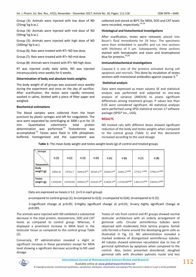
ND treated rats with different doses showed significant reduction of the body and testes weights when compared to the control group (Table 1) and this decrement increased according to the used dosage.
Table 1: The mean body weight and testes weight levels (g) of control and treated groups
Data are expressed as means ± S.E. (n=5 in each group).
a=compared to control group (1); b=compared to G(3); c=compared to G(4); d=compared to G (5).
1=significant change at p<0.05; 2=highly significant change at p<0.01; 3=very highly significant change at p<0.001.
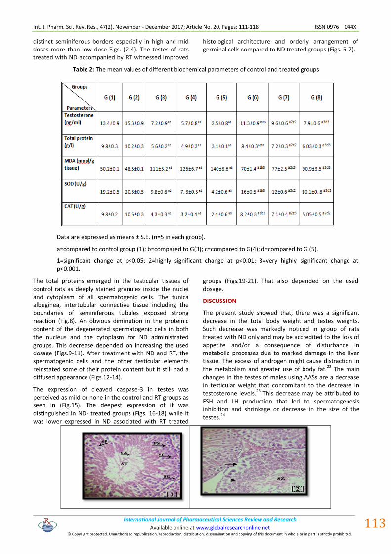
The animals were injected with ND exhibited a substantial decrease in the total protein, testosterone, SOD and CAT levels as compared to control group. Whereas, it displayed a prominent increase in MDA level in the testicular tissue as compared to the control group Table (2).
Conversely, RT administration revealed a slight or significant increase in these parameters except for MDA level showing a significant decrease according to ND used dosage.
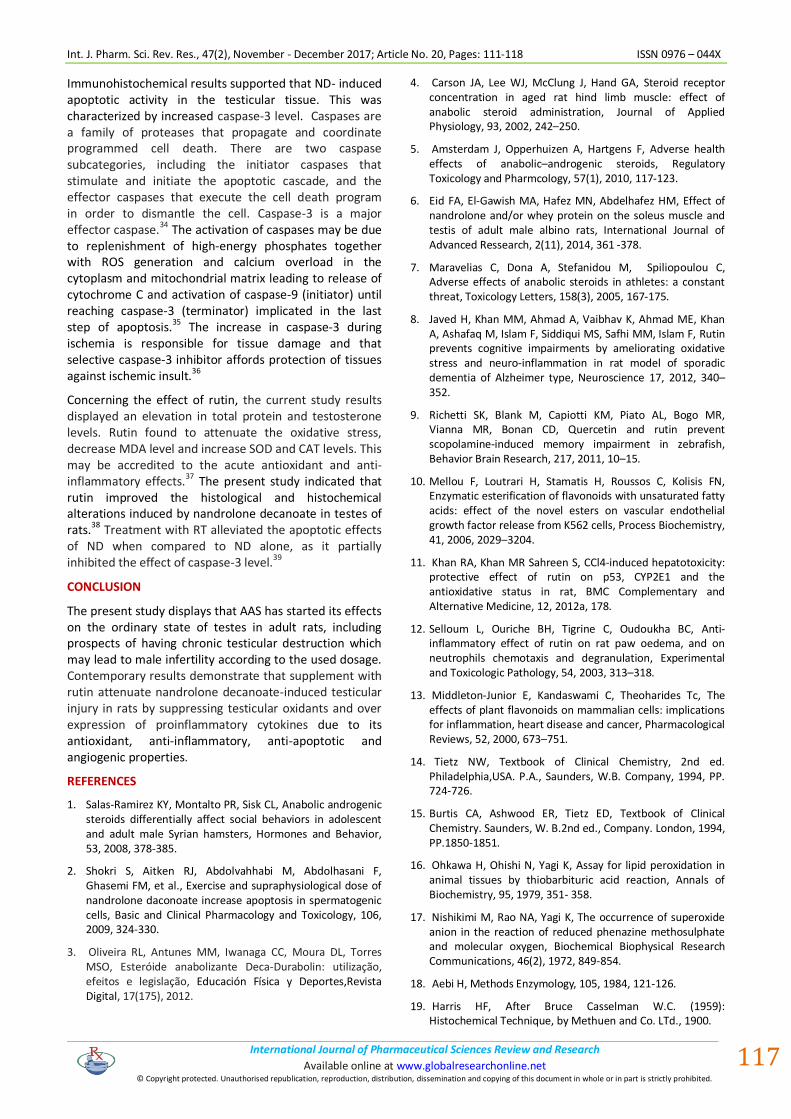
Testes of rats from control and RT groups showed normal testicular architecture with an orderly arrangement of germinal cells. Circular seminiferous tubules were observed with moderately thick lamina propria. Sertoli cells formed a frame around the developing germ cells as illustrated in Fig. (1). ND administration revealed a marked evidence of disorganized seminiferous tubules. All tubules showed extensive vacuolation due to loss of germinal epithelium by apoptosis when compared to the control. Also, testes presented disordered, sloughed germinal cells with shrunken pyknotic nuclei and less
Int. J. Pharm. Sci. Rev. Res., 47(2), November - December 2017; Article No. 20, Pages: 111-118 ISSN 0976 – 044X
International Journal of Pharmaceutical Sciences Review and Research . International Journal of Pharmaceutical Sciences Review and Research Available online at www.globalresearchonline.net
© Copyright protected. Unauthorised republication, reproduction, distribution, dissemination and copying of this document in whole or in part is strictly prohibited.
.
. Available online at www.globalresearchonline.net
113
distinct seminiferous borders especially in high and mid doses more than low dose Figs. (2-4). The testes of rats treated with ND accompanied by RT witnessed improved
histological architecture and orderly arrangement of germinal cells compared to ND treated groups (Figs. 5-7).
Table 2: The mean values of different biochemical parameters of control and treated groups
Data are expressed as means ± S.E. (n=5 in each group).
a=compared to control group (1); b=compared to G(3); c=compared to G(4); d=compared to G (5).
1=significant change at p<0.05; 2=highly significant change at p<0.01; 3=very highly significant change at p<0.001.
The total proteins emerged in the testicular tissues of control rats as deeply stained granules inside the nuclei and cytoplasm of all spermatogenic cells. The tunica albuginea, intertubular connective tissue including the boundaries of seminiferous tubules exposed strong reaction (Fig.8). An obvious diminution in the proteinic content of the degenerated spermatogenic cells in both the nucleus and the cytoplasm for ND administrated groups. This decrease depended on increasing the used dosage (Figs.9-11). After treatment with ND and RT, the spermatogenic cells and the other testicular elements reinstated some of their protein content but it still had a diffused appearance (Figs.12-14).
The expression of cleaved caspase-3 in testes was perceived as mild or none in the control and RT groups as seen in (Fig.15). The deepest expression of it was distinguished in ND- treated groups (Figs. 16-18) while it was lower expressed in ND associated with RT treated
groups (Figs.19-21). That also depended on the used dosage.
DISCUSSION
The present study showed that, there was a significant decrease in the total body weight and testes weights. Such decrease was markedly noticed in group of rats treated with ND only and may be accredited to the loss of appetite and/or a consequence of disturbance in metabolic processes due to marked damage in the liver tissue. The excess of androgen might cause distraction in the metabolism and greater use of body fat.
22 The main
changes in the testes of males using AASs are a decrease in testicular weight that concomitant to the decrease in testosterone levels.
23 This decrease may be attributed to
FSH and LH production that led to spermatogenesis inhibition and shrinkage or decrease in the size of the testes.24

Int. J. Pharm. Sci. Rev. Res., 47(2), November - December 2017; Article No. 20, Pages: 111-118 ISSN 0976 – 044X
International Journal of Pharmaceutical Sciences Review and Research . International Journal of Pharmaceutical Sciences Review and Research Available online at www.globalresearchonline.net
© Copyright protected. Unauthorised republication, reproduction, distribution, dissemination and copying of this document in whole or in part is strictly prohibited.
.
. Available online at www.globalresearchonline.net
114
Figures (1-7): Photomicrographs of rat testes sections stained with H&E (x 400). (1) control group showing seminiferous tubule (ST) including successive stages of spermatogenesis that involved spermatogonia (SG), primary spermatocytes (PS), secondary spermatocytes (SP), spermatozoa (SZ) and Sertoli cells (SC) are attached by their bases to the basal lamina.(2) testes section in a group treated with ND (25mg) showing slight degenerated spermatocytes (arrow) and karyolysed nuclei (arrow head). (3) Testes section in a group treated with ND (50mg) showing dilated congested blood vessel (*), mildly degenerated vacuolated spermatocytes (arrow heads) and partially loss of sperms (arrow) and marked portal inflammatory (arrow head). (4) Testes section in a group treated ND (100mg) showing congested blood vessel (arrow head) with vacuolar degeneration (curved arrows), highly loss of sperms (*) and pyknotic nuclei (arrows). (5) Testes section in a group treated with ND (25mg) + RT showing virtually common testicular architecture (6) Testes section in a group treated with ND (50mg) + RT showing moderate restoration of testicular pattern. However, slightly vacuolar degeneration (arrows) and karyolytic nuclei (curved arrow) could be observed. (7) Testes section in a group treated with ND (100mg) + RT showing faint restoration of testicular pattern. As, moderate vacuolar degeneration (arrow heads) and pyknotic nuclei (curved arrows), partially loss of sperms (*) and damaged spermatocytes (arrows) could be detected.

Int. J. Pharm. Sci. Rev. Res., 47(2), November - December 2017; Article No. 20, Pages: 111-118 ISSN 0976 – 044X
International Journal of Pharmaceutical Sciences Review and Research . International Journal of Pharmaceutical Sciences Review and Research Available online at www.globalresearchonline.net
© Copyright protected. Unauthorised republication, reproduction, distribution, dissemination and copying of this document in whole or in part is strictly prohibited.
.
. Available online at www.globalresearchonline.net
115
Figures (8-14): Photomicrographs of rat testes sections stained with bromophenol blue (x400). (8) Control group showing normal distribution of proteins in the seminiferous tubules. (9&10) Testes sections in groups treated with ND (25&50mg) showing slight and mild reduction in protein content predominantly in injured area (arrows), respectively. (11) Testes section in a group treated with ND (100mg) showing highly diminution in protein material (arrows). (12) Testes section in a group treated with ND (25mg)+ RT showing strong reaction in protein content of testicular tissue. (13) Testes section in a group treated with ND (50mg)+ RT showing weak to moderate reaction of the protein content in the impacted cells (arrow heads). (14) Testes section in a group treated with ND (100mg) + RT showing inequitable decline in the protein inclusions in degenerated area (arrow heads).

Int. J. Pharm. Sci. Rev. Res., 47(2), November - December 2017; Article No. 20, Pages: 111-118 ISSN 0976 – 044X
International Journal of Pharmaceutical Sciences Review and Research . International Journal of Pharmaceutical Sciences Review and Research Available online at www.globalresearchonline.net
© Copyright protected. Unauthorised republication, reproduction, distribution, dissemination and copying of this document in whole or in part is strictly prohibited.
.
. Available online at www.globalresearchonline.net
116
Figures (15-21): Photomicrographs of rat testes sections stained for caspase-3 expression (x400). (15) Control group showing none or minimal immune reactivity for caspase-3 positive stain. (16&17) Testes sections in groups treated with ND (25&50mg) showing slight and moderate increase of caspase-3 immuno reactivity in the most damaged spermatocytes few nuclei appeared stain in dark brown (arrows), respectively. (18) Testes sections in a group treated with ND (100mg) showing marked intensification of caspase-3 immuno reactivity mainly in damaged spermatocytes, many nuclei appeared stain in dark brown.(arrows). (19&20) Testes sections in groups treated with ND (25&50mg)+RT showing delicate and reasonable decrease of caspase-3 immuno reactivity in the most damaged spermatocytes (arrows), respectively. (21) Testes section in a group treated with ND (100) + RT showing more improvement, moderate decrease in caspase-3 expression immune reactivity in spermatocytes, respectively.
In this study, the total protein content was markedly decreased especially in high dose of ND administration. Total protein decrease may be attributed to the oxidative damage via testosterone administration.25,6 The current study displayed a decrease in testosterone level after ND administration especially mid and high doses. High doses of ND may influence the hypothalamic pituitary- gonadal axis, which in turn inhibited the normal level of testosterone via decrease FSH and LH hormones.
24
The present data also showed an increase in MDA level and a decrease in SOD and CAT levels in groups treated with ND alone. This showed testicular ischemia reperfusion injury. Administration of testosterone propionate directed to a significant elevation of oxidative stress.26 The pathological roles of free radicals include lipid peroxidation, macromolecules damage and apoptosis.27 Free radicals have the ability to damage sperm DNA via attacking the purine, pyrimidine bases and deoxyribose backbone as well as they can damage the sperm membrane.28 In confirmation with the previous results, there was an increase of MDA level after sustanon administration was dose dependent.29 Testosterone administration was found to cause oxidative stress through increasing the level of MDA and decreasing SOD and CAT levels, thereby enhancing lipid peroxidation dose dependently.
30
The current work showed that ND administration induced disorganization of the seminiferous tubules, some tubules showed partial separation of the germ cells and other exhibited spermatids and sperms loss. There was spermatogenic cells disintegration that in turn showed necrosis. Histopathological examination was in accordance with the elevated testicular MDA level. Also, the detached germ cells and spermatids and sperms missed location assigned to the Sertoli cells disruption and germ cells interaction.
31 Similarly, ND administration
induced marked degenerative changes of germ, Sertoli and leydig cells. These were accompanied by changes in semen parameters and testes atrophy which may be assigned to oxidative damage.32, 29
Histochemical results revealed that ND- induced reduction of total proteins in testicular tissue of rats. Reduction in protein content in testes of ND-treated animals might be due to either arrested metabolism in the testes or to usage of proteins to build up new cells or enzymes to reduce the stress. It has been speculated that the decrease in proteins could be attributed to disruption of lysosomal membranes under the effect of various toxicants leading to liberating their hydrolytic enzymes in the cytoplasm and resulted in marked lysis and dissolution of the target materials. Treatment with mancozeb caused significant decrease in the levels of proteins in testes of mice.
33
Int. J. Pharm. Sci. Rev. Res., 47(2), November - December 2017; Article No. 20, Pages: 111-118 ISSN 0976 – 044X
International Journal of Pharmaceutical Sciences Review and Research . International Journal of Pharmaceutical Sciences Review and Research Available online at www.globalresearchonline.net
© Copyright protected. Unauthorised republication, reproduction, distribution, dissemination and copying of this document in whole or in part is strictly prohibited.
.
. Available online at www.globalresearchonline.net
117
Immunohistochemical results supported that ND- induced apoptotic activity in the testicular tissue. This was characterized by increased caspase-3 level. Caspases are a family of proteases that propagate and coordinate programmed cell death. There are two caspase subcategories, including the initiator caspases that stimulate and initiate the apoptotic cascade, and the effector caspases that execute the cell death program in order to dismantle the cell. Caspase-3 is a major effector caspase.34 The activation of caspases may be due to replenishment of high-energy phosphates together with ROS generation and calcium overload in the cytoplasm and mitochondrial matrix leading to release of cytochrome C and activation of caspase-9 (initiator) until reaching caspase-3 (terminator) implicated in the last step of apoptosis.35 The increase in caspase-3 during ischemia is responsible for tissue damage and that selective caspase-3 inhibitor affords protection of tissues against ischemic insult.36
Concerning the effect of rutin, the current study results displayed an elevation in total protein and testosterone levels. Rutin found to attenuate the oxidative stress, decrease MDA level and increase SOD and CAT levels. This may be accredited to the acute antioxidant and anti- inflammatory effects.37 The present study indicated that rutin improved the histological and histochemical alterations induced by nandrolone decanoate in testes of rats.38 Treatment with RT alleviated the apoptotic effects of ND when compared to ND alone, as it partially inhibited the effect of caspase-3 level.39
CONCLUSION
The present study displays that AAS has started its effects on the ordinary state of testes in adult rats, including prospects of having chronic testicular destruction which may lead to male infertility according to the used dosage. Contemporary results demonstrate that supplement with rutin attenuate nandrolone decanoate-induced testicular injury in rats by suppressing testicular oxidants and over expression of proinflammatory cytokines due to its antioxidant, anti-inflammatory, anti-apoptotic and angiogenic properties.
REFERENCES
1. Salas-Ramirez KY, Montalto PR, Sisk CL, Anabolic androgenic steroids differentially affect social behaviors in adolescent and adult male Syrian hamsters, Hormones and Behavior, 53, 2008, 378-385.
2. Shokri S, Aitken RJ, Abdolvahhabi M, Abdolhasani F, Ghasemi FM, et al., Exercise and supraphysiological dose of nandrolone daconoate increase apoptosis in spermatogenic cells, Basic and Clinical Pharmacology and Toxicology, 106, 2009, 324-330.
3. Oliveira RL, Antunes MM, Iwanaga CC, Moura DL, Torres MSO, Esteróide anabolizante Deca-Durabolin: utilização, efeitos e legislação, Educación Física y Deportes,Revista Digital, 17(175), 2012.
4. Carson JA, Lee WJ, McClung J, Hand GA, Steroid receptor concentration in aged rat hind limb muscle: effect of anabolic steroid administration, Journal of Applied Physiology, 93, 2002, 242–250.
5. Amsterdam J, Opperhuizen A, Hartgens F, Adverse health effects of anabolic–androgenic steroids, Regulatory Toxicology and Pharmcology, 57(1), 2010, 117-123.
6. Eid FA, El-Gawish MA, Hafez MN, Abdelhafez HM, Effect of nandrolone and/or whey protein on the soleus muscle and testis of adult male albino rats, International Journal of Advanced Ressearch, 2(11), 2014, 361 -378.
7. Maravelias C, Dona A, Stefanidou M, Spiliopoulou C, Adverse effects of anabolic steroids in athletes: a constant threat, Toxicology Letters, 158(3), 2005, 167-175.
8. Javed H, Khan MM, Ahmad A, Vaibhav K, Ahmad ME, Khan A, Ashafaq M, Islam F, Siddiqui MS, Safhi MM, Islam F, Rutin prevents cognitive impairments by ameliorating oxidative stress and neuro-inflammation in rat model of sporadic dementia of Alzheimer type, Neuroscience 17, 2012, 340–352.
9. Richetti SK, Blank M, Capiotti KM, Piato AL, Bogo MR, Vianna MR, Bonan CD, Quercetin and rutin prevent scopolamine-induced memory impairment in zebrafish, Behavior Brain Research, 217, 2011, 10–15.
10. Mellou F, Loutrari H, Stamatis H, Roussos C, Kolisis FN, Enzymatic esterification of flavonoids with unsaturated fatty acids: effect of the novel esters on vascular endothelial growth factor release from K562 cells, Process Biochemistry, 41, 2006, 2029–3204.
11. Khan RA, Khan MR Sahreen S, CCl4-induced hepatotoxicity: protective effect of rutin on p53, CYP2E1 and the antioxidative status in rat, BMC Complementary and Alternative Medicine, 12, 2012a, 178.
12. Selloum L, Ouriche BH, Tigrine C, Oudoukha BC, Anti-inflammatory effect of rutin on rat paw oedema, and on neutrophils chemotaxis and degranulation, Experimental and Toxicologic Pathology, 54, 2003, 313–318.
13. Middleton-Junior E, Kandaswami C, Theoharides Tc, The effects of plant flavonoids on mammalian cells: implications for inflammation, heart disease and cancer, Pharmacological Reviews, 52, 2000, 673–751.
14. Tietz NW, Textbook of Clinical Chemistry, 2nd ed. Philadelphia,USA. P.A., Saunders, W.B. Company, 1994, PP. 724-726.
15. Burtis CA, Ashwood ER, Tietz ED, Textbook of Clinical Chemistry. Saunders, W. B.2nd ed., Company. London, 1994, PP.1850-1851.
16. Ohkawa H, Ohishi N, Yagi K, Assay for lipid peroxidation in animal tissues by thiobarbituric acid reaction, Annals of Biochemistry, 95, 1979, 351- 358.
17. Nishikimi M, Rao NA, Yagi K, The occurrence of superoxide anion in the reaction of reduced phenazine methosulphate and molecular oxygen, Biochemical Biophysical Research Communications, 46(2), 1972, 849-854.
18. Aebi H, Methods Enzymology, 105, 1984, 121-126.
19. Harris HF, After Bruce Casselman W.C. (1959): Histochemical Technique, by Methuen and Co. LTd., 1900.

Int. J. Pharm. Sci. Rev. Res., 47(2), November - December 2017; Article No. 20, Pages: 111-118 ISSN 0976 – 044X
International Journal of Pharmaceutical Sciences Review and Research . International Journal of Pharmaceutical Sciences Review and Research Available online at www.globalresearchonline.net
© Copyright protected. Unauthorised republication, reproduction, distribution, dissemination and copying of this document in whole or in part is strictly prohibited.
.
. Available online at www.globalresearchonline.net
118
20. Bonhag PF, Histochemical studies of the ovarian nurse cells, tissues and oocytes of the milkweed bug, Oncopltus fasciatus (Dallas) I. Cytology, nucleic acid and carbohydrates, Journal Morphology, 96, 1955, 381- 439.
21. Kim SK, Yoon YD, Park YS, Seo JT, Kim JH, Involvement of the Fas–Fas ligand system and active caspase-3 in abnormal apoptosis in human testes with maturation arrest and Sertoli cell-only syndrome, Fertility Sterility, 87 (3),2007, 547–553.
22. Takahashi M, Tatsugi Y, Kohno T, Endocrinological and pathological effect of anabolic androgenic steroid in male rats. Endocrine Journal, 51, 2004, 425-434.
23. Torres-Calleja J, Gonzalez-Unzaga M, De celis-Carrillo R, Calzada-Sachez L, Pedron N, Effect of androgenic-anabolic steroids on sperm quality and serum hormonal levels in adult male body builders, Life Sciences, 68, 2001, 1769-1774.
24. Thabet NS, Abdelrazek EM, Ghazy EW, Elballal SS, Effect of the anabolic steroid, boldenone undecylenate on reproductive performance of male rabbits, Journal Reproduction Infertility, 1(1), 2010, 8-17.
25. Lok S, ErdalbT, Nagehan D, Mehmet O, Long term used testosterone may cause heart and liver damage, Journal of Animal and Veterinary Advances, 9(18), 2010, 2343-2345.
26. Aydilek N, Aksakal M, Karakılçık AZ, Effects of testosterone and vitamin E on antioxidant system in rabbit testis, Andrologia, 36(5), 2004, 277-281.
27. Kothari S, Thompson A, Agarwal A, Plessis SS, Free radicals: their beneficial and detrimental effects on sperm function, Indian Journal of Experimental Biology, 48, 2010, 425-435.
28. Tremellen K, Oxidative stress and male infertility-a clinical perspective, Human Reproduction, 14(3), 2008, 243-258.
29. Rasul, KH, Aziz FM, The effect of sustanon (testosterone derivatives) taken by athletes on the testis of rat, Jordan Journal of Biological Sciences, 5(2), 2012, 113-119.
30. Sadowska-Krepa E, Kłapcinska B, Jagsz S, Sobczak A, Chrapusta SJ, Chalimoniuk M, Grieb P, Poprzecki S, Langfort J, High-dose testosterone propionate treatment reverses the
effects of endurance training on myocardial antioxidant defenses in adolescent male rats, Cardiovascular Toxicology, 11, 2011,118–127.
31. Richburg JH, The relevance of spontaneous and chemically-induced alteration in testicular germ cell apoptosis to toxicology, Toxicology Letters, 112-113, 2000, 79-86.
32. Mesbah SF, Shokri S, Karbalay-Doust S, Mirkhani H, The Effect of nandrolone decanoate on the body, testis and epididymis weight and semen parameters in adult male rats, Iranian Journal of basic Medical Sciences, 32, 2007, 93 -99.
33. Ksheerasagar RA, Kaliwal BB, Effect of mancozeb on thyroid, testis, accessory reproductive organs and biochemical constituents in albino mice, Recent Research in Science and Technology, 2(8), 2010, 7-17.
34. Parrish AB, Freel CD, Kornbluth S, Cellular mechanisms controlling caspase activation and function, Cold Spring Harb Perspect Biology, 5, 2013: pii: a008672.
35. Kirkland RA, Windelborn JA, Kasprzak JM, Franklin JL, Bax-induced pro-oxidant state is critical for cytochrome C release during programmed neuronal death, Journal of Neuroscience, 22 (15), 2002, 6480-6490.
36. Hyman BT, Yuan J, Apoptotic and non- apoptotic roles of caspases in neuronal physiology & pathophysiology, Nature Reviews Neuroscience, 13(6), 2012: 395-406.
37. Akondi BR, Challa SR, Akula A, Protective effects of rutin and naringin in testicular ischemia-reperfusion induced oxidative stress in rats, Journal of Reproduction Infertility, 12 (3), 2011, 209–214.
38. Aksu EH, Kandemir FM, Ozkaraca M, Omur AD, Kucukler S, Comakli S, Rutin ameliorates cisplatin- induced reproductive damage via suppression of oxidative stress and apoptosis in adult male rats, Andrologia, 49 2016, 1-8.
39. Arjumand W, Seth A, Sultana S, Rutin attenuates cisplatin induced renal inflammation and apoptosis by reducing NFjB, TNF-a and caspase-3 expression in wistar rats, Food Chemistry and Toxicology, 49 (9), 2011, 2013–2021.
Source of Support: Nil, Conflict of Interest: None.