research article mucocutaneous manifestations of...
TRANSCRIPT

Research ArticleMucocutaneous Manifestations of HIV and the Correlation withWHO Clinical Staging in a Tertiary Hospital in Nigeria
Olumayowa Abimbola Oninla
Department of Dermatology and Venereology, Faculty of Clinical Sciences, College of Health Sciences,Obafemi Awolowo University, P.O. Box 2545, Ile-Ife 250005, Osun State, Nigeria
Correspondence should be addressed to Olumayowa Abimbola Oninla; [email protected]
Received 31 July 2014; Revised 15 November 2014; Accepted 25 November 2014; Published 21 December 2014
Academic Editor: P. K. Nicholas
Copyright © 2014 Olumayowa Abimbola Oninla. This is an open access article distributed under the Creative CommonsAttribution License, which permits unrestricted use, distribution, and reproduction in any medium, provided the original work isproperly cited.
Skin diseases are indicators of HIV/AIDS which correlates with WHO clinical stages. In resource limited environment whereCD4 count is not readily available, they can be used in assessing HIV patients. The study aims to determine the mucocutaneousmanifestations in HIV positive patients and their correlation with WHO clinical stages. A prospective cross-sectional study ofmucocutaneous conditions was done among 215 newly diagnosed HIV patients from June 2008 to May 2012 at adult ART clinic,Wesley Guild Hospital Unit, OAU Teaching Hospitals Complex, Ilesha, Osun State, Nigeria. There were 156 dermatoses withoral/oesophageal/vaginal candidiasis (41.1%), PPE (24.4%), dermatophytic infections (8.9%), and herpes zoster (3.8%) as the mostcommon dermatoses. The proportions of dermatoses were 4.5%, 21.8%, 53.2%, and 20.5% in stages 1–4, respectively. A significantrelationship (using Pearson’s Chi square with 𝑃 value < 0.05) was obtained between dermatoses andWHO clinical stages. Pearson’scorrelation coefficient showed a positive correlation between the number of dermatoses and the WHO clinical stages. Dermatosescan therefore serve as diagnostic and prognostic markers in resource limited settings to initiate HAART in clinical stages 3 and 4.
1. Introduction
The burden of skin disease in developing countries partic-ularly the sub-Saharan Africa is high with a serious impacton the quality of life and resulting loss of productivityat work and school and disfigurement [1–4]. Infectiousdermatoses particularly superficial fungal infections, scabies,and impetigo are the most common skin problems dueto overcrowding with a hot and humid environment, poorsanitary conditions, sharing of personal effects or fomites,and poor access to medical supplies and treatment [5, 6].
The skin problems here are further compounded by thehigh prevalence of HIV which commonly causes skin lesions[7]. It was reported that approximately 90% of people livingwithHIV have skin changes and symptoms during the courseof their disease [8]. Skin diseases are significantly higheramong HIV positive than HIV negative individuals [9]. Dif-ferences in skin pigmentation, climate, hygiene, and genetic,environmental, demographic, and behavioral factors cause
different clinical presentations and epidemiologic patterns ofHIV-associated skin disease in Africa [10, 11].
Skin findings are regarded byWHO as useful in assessingseverity of HIV infection in patients in resource limitedenvironment [12].
Knowledge of the skin and mucosal signs of HIV/AIDSis important, as mucocutaneous lesions are usually the firstmanifestation of HIV, ensures early diagnosis and prompttreatment, and reveals complications as HIV causes atypicaland severe presentations of these conditions [13, 14]. Thoseinvolved in health care of HIV patients must thereforeknow the type, pattern, and prevalence of skin diseases intheir locality [15–17]. Mucocutaneous diseases have beencorrelated with CD4 counts in many studies [18], whilefew studies (and none in this country to the knowledge ofthis researcher) documented the clinical correlation of thesediseases to WHO clinical stages [19–21].
Although HIV dermatoses have been widely docu-mented, reports of the type of dermatoses in HIV patients
Hindawi Publishing CorporationAIDS Research and TreatmentVolume 2014, Article ID 360970, 6 pageshttp://dx.doi.org/10.1155/2014/360970

2 AIDS Research and Treatment
in this country are few and nonexistent in this area of study[21, 22]. This center was one of the recently established adultHIV care center with facility for diagnosis and care for HIVpatients and in collaboration with the Institute of HumanVirology center in OAUTHC, Ile-Ife. The aim of this study isto determine the cutaneous skin markers in HIV in an adultHIV clinic inNigeria.Theobjective is to identify and correlatethese mucocutaneous disorders to the clinical stages at timeof presentation for HIV care.
2. Methodology
A preliminary study of skin diseases was conducted ina newly created adult ART clinic as a prospective cross-sectional study among 215 newly diagnosed adult HIVpatients. Consecutive patients presenting at the adult ARTclinic of Wesley Guild Hospital Unit of Obafemi AwolowoUniversity TeachingHospitals Complex (OAUTHC) in Ileshafrom June 2008 to May 2012 were studied.
Patients, who reported for care after testing HIV-positiveat our screening centre, were included in the study. Theskin symptoms were documented, and detailed clinicalexamination of the skin in broad daylight was carried out.Skin findings were recorded, and patients were categorizedaccording to the WHO clinical stages at presentation usingskin and findings in other body systems.
Excludedwere old patients of the unit prior to onset of thestudy who are still receiving HIV care; those who came fromother HIV centres to continue their HIV care at this centredue to proximity or other reasons; referred patients whohad started HAART; patients on any primary or secondaryprophylaxis for opportunistic infections; and patients on anyother medication for systemic diseases. This is to preventpossible alteration in skin pathologies reported.
Diagnoses were mainly clinical and when necessarymycological, histopathological, and hematological tests werecarried out. Ethical approval for the study was obtained fromOAUTHC Research and Ethics Committee.
3. Results
There were 71 males and 144 females with a ratio of 1 : 2. Theage ranges between 18 and 77 with a mean of 35 years. A totalof 215 new cases of HIV positive patients were seen, and 113had symptoms and signs of mucocutaneous disease on theirfirst day of presentation at the adult HIV clinic. Patients withone, two, or three skin diseases were 76, 31, and 6, respectively.
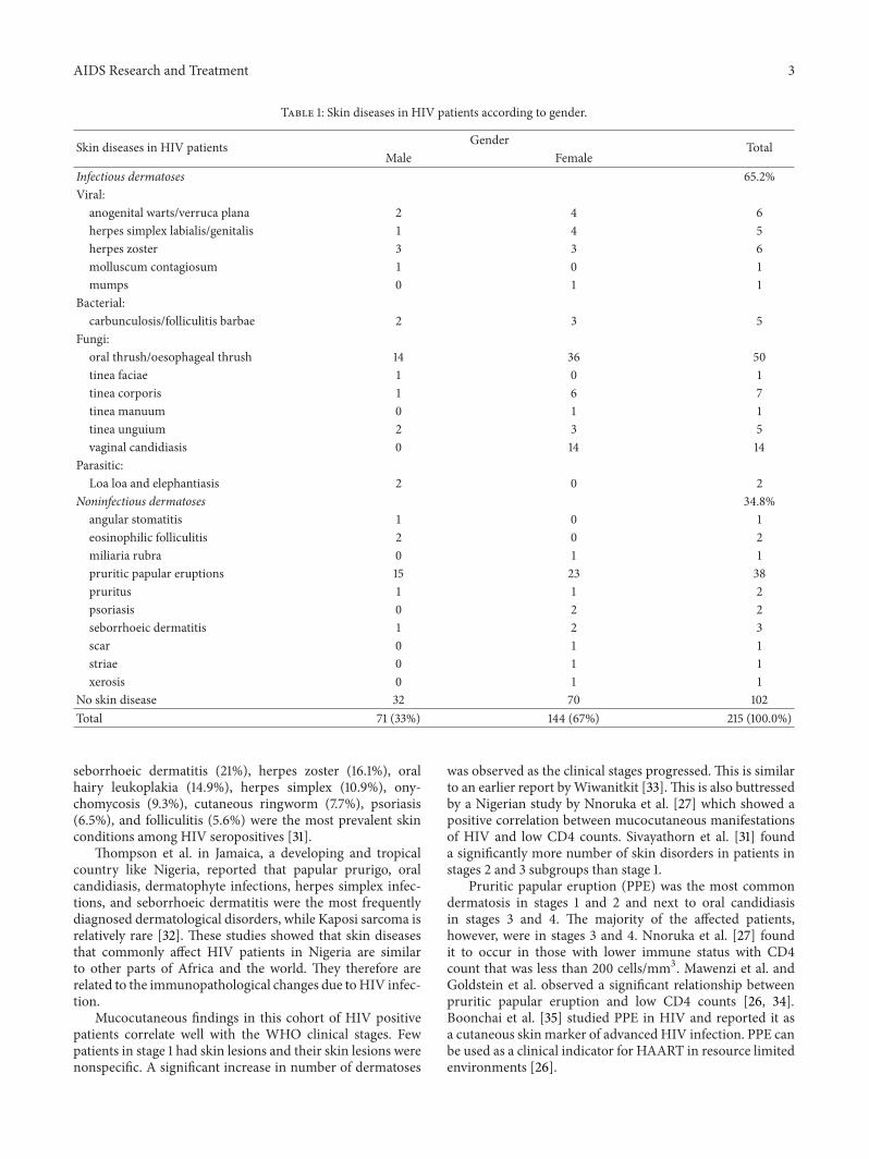
The total skin conditions found were 156 with oral andoesophageal candidiasis, pruritic papular eruption, vaginalcandidiasis, dermatophytic infections, herpes zoster, viralwarts, herpes simplex, carbunculosis/folliculitis barbae, andseborrhoeic dermatitis as the most common dermatoses.Eosinophilic folliculitis and psoriasis were uncommon. Nocase of Kaposi sarcoma was recorded. Infectious dermatosesconstituted 65.2% (fungal 50%, viral 12%, and bacterial 3.2%)of all dermatoses and noninfectious skin conditions 34.8%(see Table 1).
There were 68 patients inWHO clinical stage 1, 31 in stage2, 87 in stage 3, and 29 in stage 4.The percentage of patients in
each stage who had skin diseases was 8.8%, 77.4%, 71.3%, and72.4%, respectively. Pruritic papular eruptions were the mostcommon condition in stages 1 and 2, while oral/oesophagealthrush was more in stages 3 and 4.The different skin diseasesand the clinical stages at diagnosis of the disease are shownin Table 2. The presence of skin disease was significantlyassociated with the clinical stage of the patient (see Table 2).
The proportion of the total number (156) of cutaneousdiseases occurring in stage 1 was 4.5% and 21.8% in stage 2,53.2% in stage 3, and 20.5% in stage 4 (see Table 2). Therewas a significant relationship using Pearson’s Chi square with𝑃 value < 0.05 and Pearson’s correlation coefficient betweenthe number of skin diseases that a patient has and the clinicalstage at presentation (see Table 3).
4. Discussion
Skin diseases act as indicators of HIV and AIDS. The clinicaldiagnoses of skin diseases have been found to correlate withhistopathological findings even in HIV patients [23]. Hence,good clinical acumen is essential to make correct diagnosesof skin problems in these patients.
Themajority of the patientswere females. A higher femalepreponderance was also reported by Salami et al. in Nigeriaand Glynn et al. in Kenya [24, 25]. The greater susceptibilityof females to HIV may explain the gender differences inHIV prevalence [25]. More than half (52.6%) of the newlydiagnosed HIV patients had dermatoses. A similar study byMawenzi et al. [26] among newly diagnosed HIV patients inKenya reported a prevalence of 42.1%. Nnoruka et al. [27]obtained a prevalent rate of 93.5% in southeast Nigeria. InSanandaj city, Pakistan, it was as high as 94.3% [28].
Infectious dermatoses made up 65.2% of the dermatoses(50% fungi, 12% viral, and 3.2% bacterial) and are comparableto 64.3% reported by Salami et al. in which viral skinconditions accounted for 37.1% and fungal infections, 24.3%[24]. Another Nigerian study by Nnoruka [21] found skininfections as the most common dermatoses, while they werethe second in occurrence in a report by Yahya in northernNigeria [22]. In some developed countries, infections stillrank prominent as one of the most prevalent dermatoses inHIV [29]. Most infectious dermatoses start in early stages(stage 1) increasing in number, becoming more diffuse andmore resistant to treatment as disease progresses [18, 27]. Inthis study, infectious diseases were found mostly in stages 3and 4. A similar report was obtained in Nigerian children byOkechukwu et al. [30]. This implies that skin infections arecommonly associated with HIV infection.
The most common skin disorders in this study wereoropharyngeal candidiasis (32.1%), pruritic papular eruptions(25.3%), vaginal candidiasis (9.0%), dermatophytic infections(8.9%), herpes zoster (3.9%), carbunculosis (2.6%), herpessimplex (2.6%), and seborrhoeic dermatitis (1.9%). Papu-lopruritic eruption, seborrheic dermatitis (35.8%), herpeszoster, dermatophytosis (24.3%), oropharyngeal candidiasis,staphylococcal infection, and vaginal candidiasis were themost common in southeast Nigeria [27].
Sivayathorn et al. in Bangkok found that oral can-didiasis (34.3%), pruritic papular eruption (PPE) (32.7%),
AIDS Research and Treatment 3
Table 1: Skin diseases in HIV patients according to gender.
Skin diseases in HIV patients Gender TotalMale Female
Infectious dermatoses 65.2%Viral:
anogenital warts/verruca plana 2 4 6herpes simplex labialis/genitalis 1 4 5herpes zoster 3 3 6molluscum contagiosum 1 0 1mumps 0 1 1
Bacterial:carbunculosis/folliculitis barbae 2 3 5
Fungi:oral thrush/oesophageal thrush 14 36 50tinea faciae 1 0 1tinea corporis 1 6 7tinea manuum 0 1 1tinea unguium 2 3 5vaginal candidiasis 0 14 14
Parasitic:Loa loa and elephantiasis 2 0 2
Noninfectious dermatoses 34.8%angular stomatitis 1 0 1eosinophilic folliculitis 2 0 2miliaria rubra 0 1 1pruritic papular eruptions 15 23 38pruritus 1 1 2psoriasis 0 2 2seborrhoeic dermatitis 1 2 3scar 0 1 1striae 0 1 1xerosis 0 1 1
No skin disease 32 70 102Total 71 (33%) 144 (67%) 215 (100.0%)
seborrhoeic dermatitis (21%), herpes zoster (16.1%), oralhairy leukoplakia (14.9%), herpes simplex (10.9%), ony-chomycosis (9.3%), cutaneous ringworm (7.7%), psoriasis(6.5%), and folliculitis (5.6%) were the most prevalent skinconditions among HIV seropositives [31].
Thompson et al. in Jamaica, a developing and tropicalcountry like Nigeria, reported that papular prurigo, oralcandidiasis, dermatophyte infections, herpes simplex infec-tions, and seborrhoeic dermatitis were the most frequentlydiagnosed dermatological disorders, while Kaposi sarcoma isrelatively rare [32]. These studies showed that skin diseasesthat commonly affect HIV patients in Nigeria are similarto other parts of Africa and the world. They therefore arerelated to the immunopathological changes due toHIV infec-tion.
Mucocutaneous findings in this cohort of HIV positivepatients correlate well with the WHO clinical stages. Fewpatients in stage 1 had skin lesions and their skin lesions werenonspecific. A significant increase in number of dermatoses
was observed as the clinical stages progressed. This is similarto an earlier report byWiwanitkit [33].This is also buttressedby a Nigerian study by Nnoruka et al. [27] which showed apositive correlation between mucocutaneous manifestationsof HIV and low CD4 counts. Sivayathorn et al. [31] founda significantly more number of skin disorders in patients instages 2 and 3 subgroups than stage 1.
Pruritic papular eruption (PPE) was the most commondermatosis in stages 1 and 2 and next to oral candidiasisin stages 3 and 4. The majority of the affected patients,however, were in stages 3 and 4. Nnoruka et al. [27] foundit to occur in those with lower immune status with CD4count that was less than 200 cells/mm3. Mawenzi et al. andGoldstein et al. observed a significant relationship betweenpruritic papular eruption and low CD4 counts [26, 34].Boonchai et al. [35] studied PPE in HIV and reported it asa cutaneous skin marker of advanced HIV infection. PPE canbe used as a clinical indicator for HAART in resource limitedenvironments [26].

4 AIDS Research and Treatment
Table 2: Dermatoses occurring in each WHO clinical stage.
Dermatoses WHO clinical staging at diagnosis of skin disease Total Percentage (%)1 2 3 4
Carbunculosis/folliculitis barbae 1 2 2 0 5 3.2Eosinophilic folliculitis 0 0 1 1 2 1.3Herpes simplex 0 2 2 1 5 3.2Herpes zoster 0 4 2 0 6 3.8Loa loa and elephantiasis 0 1 1 0 2 1.3Molluscum contagiosum 0 0 1 0 1 0.6Miliaria rubra 0 1 0 0 1 0.6Mumps 0 0 0 1 1 0.6Pruritic papular eruptions 2 11 16 9 38 24.4Pruritus 0 2 0 0 2 1.3Psoriasis 0 1 1 0 2 1.3Scar 1 0 0 0 1 0.6Seborrhoeic dermatitis 0 1 2 0 3 1.9Angular stomatitis 0 0 0 1 1 0.6Striae 1 0 0 0 1 0.6Oral/oesophageal thrush 0 4 34 12 50 32.1Tinea corporis 0 0 4 3 7 4.5Tinea faciae 0 0 0 1 1 0.6Tinea manuum 0 0 1 0 1 0.6Tinea unguium 0 1 2 2 5 3.2Vaginal candidiasis 1 2 11 0 14 9.0Verruca plana/anogenital warts 1 2 2 1 6 3.8Xerosis 0 0 1 0 1 0.6Number of all skin lesions 7 34 83 32 156 100%
(4.5%) (21.8%) (53.2%) (20.5%) (100%)Presence of skin disease
Yes 6 24 62 21 113 52.6No 62 7 25 8 102 47.4
68 31 87 29 215 100%(31.6%) (14.4%) (40.5%) (14.5%) (100%)
𝜒2= 76.641; df = 3; 𝑃 = 0.000.
Table 3: Number of dermatoses in relation to WHO clinical stages.
Number of dermatoses WHO staging at the time of diagnosis of skin disease Total (%)1 2 3 4
0 62 7 25 8 102 (47.4)1 5 15 45 11 76 (35.3)2 1 7 14 9 31 (14.4)3 0 2 3 1 6 (2.8)Total 68 31 87 29 215 (100)𝜒2= 82.131; df = 9; 𝑃 = 0.000.
∗∗Pearson’s correlation coefficient = 0.449; 𝑃 = 0.000.∗∗Correlation is significant at the 0.01 level (2-tailed).
AIDS Research and Treatment 5
Oral thrush was the predominant dermatoses in stage3 and stage 4. Oral thrush particularly if extending to theesophagus has been widely reported to be synonymous withsevere immunosuppression [36]. In Nigeria, the prevalenceof oral candidiasis ranges between 36 and 80% in HIVpatients [37, 38], and the condition is predictive of severeimmunosuppression with approximately 50% of patientsdeveloping AIDS within 5 years [39]. Goh et al. [11], Sharmaet al. [40], and Puttaiah et al. [41] reported lower CD4counts less than 200 cells/mm3 in patients with oral thrush.Vaginal candidiasis also occurred in stage 3. It was foundto occur concomitantly with oral candidiasis and AIDSdefiningmedical conditions [26]. It can therefore be a clinicalindicator of advanced HIV infection.
Dermatophytic infections were found mostly in stages 3and 4. Extensive dermatophytosis was found in 26% of HIVpatients seen by Nnoruka in southeast Nigeria though theclinical stages were not stated [21]. They are often among thetop ten dermatoses in HIV [24, 33].
Herpes zoster was seen mostly in stage 2. The WHOclassified it as a stage 2 disease [19].Herpes zoster is associatedwith early immunosuppression [27], and a significant changeoccurs in the clinical stage with disease progression [16].
Viral warts occurred more in stages 2 and 3. About 13%of HIV patients seen at Irua [24] in Nigeria had plane andanogenital warts though the relationship to clinical stageswas not known. They were found to occur at CD4 counts of200–300 cells/mm3. Mawenzi et al. [26] report showed thattheywere associatedwith higher immune status inHIV (CD4> 300 cells/mm3). They have been listed as a stage 2 clinicaldisease by WHO.
Bacterial skin infections occurred in those in stages 2 and3. They have been reported to be common in HIV patientsbut correlation with clinical stages is not known thoughNnoruka et al. report them to occur in those with CD4 countsbetween 200 and 500 cells/mm3 [27]. Study by Smith et al.[20] showed a peak occurrence in early and midstage diseasewith a decreased occurrence in the late stage.
Seborrhoeic dermatitis has been listed as a stage 2 diseaseby WHO. In this study, the prevalence was low and it wasfound in those in stages 2 and 3. Nnoruka et al. [27] found apositive correlation between seborrhoeic dermatitis and CD4counts of 200–500 cells/mm3. Salami et al. [24], however,showed the occurrence to be in patients with lower immunestatus (CD4 counts < 100 cells/mm3). The differences wereadduced to be as a result of variable level of severity ofseborrhoeic dermatitis [26].
5. Conclusion
HIV-related mucocutaneous manifestations are very com-mon and with good clinical acumen are easily diagnosed.Oropharyngeal candidiasis, pruritic papular eruption, vagi-nal candidiasis, dermatophytic infections, herpes zoster,carbunculosis, herpes simplex, and seborrhoeic dermatitissignificantly correlated with WHO clinical stages.
Dermatoses can therefore serve as diagnostic and prog-nostic markers in a resource limited setting as obtained inmany HIV clinics in Nigeria to stage the severity of HIV
infection and to initiate HAART for those in clinical stages 3and 4 where laboratory tests for CD4 count are not available.Planning for HIV care in this environment should alsoinculcate plans for skin diseases which affect themmore thanthe general populace.
Conflict of Interests
The author declares that there is no conflict of interestsregarding the publication of this paper.
References
[1] S. Kingman, “Growing awareness of skin disease starts flurry ofinitiatives,” Bulletin of the World Health Organization, vol. 83,pp. 891–892, 2005.
[2] F. T. Satimia, S. R. McBride, and B. Leppard, “Prevalence of skindisease in rural Tanzania and factors influencing the choice ofhealth care, modern or traditional,” Archives of Dermatology,vol. 134, no. 11, pp. 1363–1366, 1998.
[3] M. J. Porter, “Problems and priorities for dermatology indeveloping countries,” International Journal of Dermatology,vol. 12, pp. 233–236, 1978.
[4] R. J. Hay, A. C. Steer, D. Engelman et al., “Scabies in the devel-oping world—its prevalence, complications, and management,”Clinical Microbiology and Infection, vol. 18, no. 4, pp. 131–323,2012.
[5] O. A. Oninla and O. Onayemi, “Skin infections and infestationsin prison inmates,” International Journal of Dermatology, vol. 51,no. 2, pp. 178–181, 2012.
[6] M. J. Porter, “An epidemiological approach to skin disease in thetropics,” Tropical Doctor, vol. 7, no. 2, pp. 59–66, 1977.
[7] J. Hu, K. McKoy, A. Papier et al., “Dermatology and HIV/AIDSin Africa,” Journal of Global Infectious Diseases, vol. 3, no. 3, pp.275–280, 2011, http://www.jgid.org/text.asp?2011/3/3/275/83535.
[8] N. Dlova and A. Mosam, “Cutaneous manifestations ofHIV/AIDS: part 1,” Southern African Journal of HIV Medicine,vol. 5, pp. 12–17, 2004, http://www.ajol.info/index.php/sajhivm/article/view/34816/24879.
[9] M. Bakari, E. Lyamuya, F. Mugusi et al., “The prevalence andpattern of skin diseases in relation to CD4 counts among HIV-infected police officers in Dar es Salaam,” Tropical Doctor, vol.33, no. 1, pp. 44–48, 2003.
[10] E. H. Amerson and T. A.Maurer, “Dermatologicmanifestationsof HIV in Africa,” Topics in HIV Medicine, vol. 18, no. 1, pp. 16–22, 2010.
[11] B.-K. Goh, R. K. W. Chan, P. Sen et al., “Spectrum of skindisorders in human immunodeficiency virus-infected patientsin Singapore and the relationship to CD4 lymphocyte counts,”International Journal of Dermatology, vol. 46, no. 7, pp. 695–699,2007.
[12] WHO, “WHO case definitions of HIV for surveillance andrevised clinical staging and immunological classification ofHIV-related disease in adults and children,” August 2006,http://www.who.int/hiv/pub/vct/hivstaging/en/.
[13] H. F. Jordaan, “Common skin and mucosal disorders inHIV/AIDS,” South African Family Practice, vol. 50, no. 6, pp.14–23, 2008.
[14] C. L. Kovarik, A. Kekitiinwa, and H. Schwarzwald, “Cutaneousmanifestations of HIV infection,” HIV Curriculum for theHealth Professional, http://www.bipai.org/HIV-curriculum/.

6 AIDS Research and Treatment
[15] M. A. Munoz-Perez, A. Rodriguez-Pichardo, F. Camacho, andM. A. Colmenero, “Dermatological findings correlated withCD4 lymphocyte counts in a prospective 3 year study of 1161patients with human immunodeficiency virus disease pre-dominantly acquired through intravenous drug abuse,” BritishJournal of Dermatology, vol. 139, no. 1, pp. 33–39, 1998.
[16] K. J. Smith, H. G. Skelton, J. Yeager et al., “Cutaneous findings inhiv-1-positive patients: a 42-month prospective study. militarymedical consortium for the advancement of retroviral research(MMCARR),” Journal of the AmericanAcademy ofDermatology,vol. 31, no. 5, pp. 746–754, 1994.
[17] A. D. Grant, G. Djomand, and K. M. De Cock, “Natural historyand spectrum of disease in adults with HIV/AIDS in Africa,”AIDS, vol. 11, supplement B, pp. S43–S54, 1997.
[18] P. V. Raju, G. R. Rao, T. V. Ramini, and S. Vandana, “Skindisease: clinical indicator of immune status in human immun-odeficiency virus (HIV) infection,” International Journal ofDermatology, vol. 44, no. 8, pp. 646–649, 2005.
[19] World Health Organization, WHO Case Definitions of HIV forSurveillance and Revised Clinical Staging and ImmunologicalClassification of HIV-Related Disease in Adults and Children,World Health Organization, Geneva, Switzerland, 2006.
[20] K. J. Smith, H. G. Skelton, J. Yeager et al., “Cutaneous findingsin HIV-1-positive patients: a 42-month prospective study. Mil-itary Medical Consortium for the Advancement of RetroviralResearch (MMCARR),” Journal of the American Academy ofDermatology, vol. 31, pp. 746–754, 1994.
[21] E. N. Nnoruka, “Skin diseases in south-east Nigeria: A currentperspective,” International Journal of Dermatology, vol. 44, no.1, pp. 29–33, 2005.
[22] H. Yahya, “Change in pattern of skin disease in Kaduna, north-central Nigeria,” International Journal of Dermatology, vol. 46,no. 9, pp. 936–943, 2007.
[23] S. Halder, S. Banerjee, A. Halder, and P. R. Pal, “Skin diseases inHIV-infected patients: impact of immune status and histologi-cal correlation,” Indian Journal of Sexually Transmitted Diseases,vol. 33, no. 1, pp. 65–67, 2012.
[24] T. A. T. Salami, G. M. Adewuyi, P. Echekwube, and C. Affusim,“Pattern of cutaneous pathology among a cohort of HIV/AIDSpatients accessing care in a rural/suburban adult ART clinic inNigeria,” The British Journal of Medicine & Medical Research,vol. 3, pp. 1199–1207, 2013.
[25] J. R. Glynn, M. Carael, B. Auvert et al., “Why do young womenhave a much higher prevalence of HIV than young men? Astudy in Kisumu, Kenya and Ndola, Zambia,” AIDS, vol. 15, no.4, pp. S51–S60, 2001.
[26] R. L. Mawenzi, O. R. Oguttu, H. C. Williams, and A. Joash,“Epidemiology and clinical spectrum of cutaneous diseasesmanifesting among newly diagnosed HIV seropositive adultsin Nakuru county-Kenya,” Continental Journal of MedicalResearch, vol. 7, pp. 1–9, 2013.
[27] E. N. Nnoruka, J. C. Chukwuka, and B. Anisuiba, “Correlationof mucocutaneous manifestations of HIV/AIDS infection withCD4 counts and disease progression,” International Journal ofDermatology, vol. 46, no. 2, pp. 14–18, 2007.
[28] F. Rad, E. Ghaderi, G. Moradi, and L. Mafakheri, “The relation-ship between skin manifestations and CD4 counts among HIVpositive patients,” Pakistan Journal of Medical Sciences, vol. 24,no. 1, pp. 114–117, 2008.
[29] C. Garbe, R. Husak, and C. E. Orfanos, “HIV-associateddermatoses and their prevalence in 456 HIV-infected patients.
Relation to immune status and its importance as a diagnosticmarker,” Der Hautarzt, vol. 45, pp. 623–629, 1994.
[30] A. A. Okechukwu, O. I. Okechukwu, and P. Jibril, “Patternsof skin disorder and its relationship with CD4+ cell count ina cohort of HIV-infected children,” Journal of Medicine andMedical Sciences, vol. 2, no. 10, pp. 1131–1138, 2014.
[31] A. Sivayathorn, B. Srihra, andW. Leesanguankul, “Prevalence ofskin disease in patients infectedwith human immunodeficiencyvirus in Bangkok,Thailand,”Annals of the Academy ofMedicine,Singapore, vol. 24, no. 4, pp. 528–533, 1995.
[32] D. S. Thompson, B. Bain, and A. East-Innis, “The prevalence ofmucocutaneous disorders among HIV-positive patients attend-ing an out-patient clinic in Kingston, Jamaica,” West IndianMedical Journal, vol. 57, no. 1, pp. 54–57, 2008.
[33] V. Wiwanitkit, “Prevalence of dermatological disorders in ThaiHIV-infected patients correlated with different CD4 lympho-cyte count statuses: a note on 120 cases,” International Journal ofDermatology, vol. 43, no. 4, pp. 265–268, 2004.
[34] B. Goldstein, B. Berman, E. Sukenik, and S. J. Frankel, “Cor-relation of skin disorders with CD4 lymphocyte counts inpatients with HIV/AIDS,” Journal of the American Academy ofDermatology, vol. 36, no. 2, pp. 262–264, 1997.
[35] W. Boonchai, R. Laohasrisakul, J. Manonukul, and K. Kultha-nan, “Pruritic papular eruption in HIV seropositive patients:a cutaneous marker for immunosuppression,” InternationalJournal of Dermatology, vol. 38, no. 5, pp. 348–350, 1999.
[36] O. N. Obuekwe and A. N. Onunu, “Gender and oral manifesta-tions of HIV infection among adult Nigerians,” African Journalof Reproductive Health, vol. 10, no. 2, pp. 81–89, 2006.
[37] A. N. Onunu and N. Obuekwe, “HIV-related oral diseases inBenin City, Nigeria.,” West African Journal of Medicine, vol. 21,no. 1, pp. 9–11, 2002.
[38] R. O. Shittu, M. F. Adeyemi, L. O. Odeigah, A. O. Mahmoud,S. A. Biliaminu, and A. A. Nyamngee, “Prevalence and patternof cutaneous lesions in relationship to CD4 cell counts amongnewly diagnosed HIV patients in University of Ilorin TeachingHospital (UITH), Ilorin, Nigeria,” Oral Health and DentalManagement, vol. 12, no. 4, pp. 248–254, 2013.
[39] M.Ukpebor andO.B. Braimoh, “HIV/AIDS; oral complicationsand challenges, the Nigerian experience,” Benin Journal ofPostgraduate Medicine, vol. 9, pp. 44–54, 2007.
[40] Y. K. Sharma, M. P. S. Sawhney, D. S. Bhakuni, and V. Gera,“Orocutaneous manifestations as markers of disease progres-sion in HIV infection in Indian setting,”Medical Journal ArmedForces India, vol. 60, no. 3, pp. 239–243, 2004.
[41] K. S. Puttaiah, V. Sunith, and A. Mallikarjun, “A hospital basedcross sectional study of mucocutaneous manifestations in theHIV infected,” International Journal of Collaborative Researchon Internal Medicine and Public Health, vol. 2, no. 3, pp. 50–78,2010.

Submit your manuscripts athttp://www.hindawi.com
Stem CellsInternational
Hindawi Publishing Corporationhttp://www.hindawi.com Volume 2014
Hindawi Publishing Corporationhttp://www.hindawi.com Volume 2014
MEDIATORSINFLAMMATION
of
Hindawi Publishing Corporationhttp://www.hindawi.com Volume 2014
Behavioural Neurology
EndocrinologyInternational Journal of
Hindawi Publishing Corporationhttp://www.hindawi.com Volume 2014
Hindawi Publishing Corporationhttp://www.hindawi.com Volume 2014
Disease Markers
Hindawi Publishing Corporationhttp://www.hindawi.com Volume 2014
BioMed Research International
OncologyJournal of
Hindawi Publishing Corporationhttp://www.hindawi.com Volume 2014
Hindawi Publishing Corporationhttp://www.hindawi.com Volume 2014
Oxidative Medicine and Cellular Longevity
Hindawi Publishing Corporationhttp://www.hindawi.com Volume 2014
PPAR Research
The Scientific World JournalHindawi Publishing Corporation http://www.hindawi.com Volume 2014
Immunology ResearchHindawi Publishing Corporationhttp://www.hindawi.com Volume 2014
Journal of
ObesityJournal of
Hindawi Publishing Corporationhttp://www.hindawi.com Volume 2014
Hindawi Publishing Corporationhttp://www.hindawi.com Volume 2014
Computational and Mathematical Methods in Medicine
OphthalmologyJournal of
Hindawi Publishing Corporationhttp://www.hindawi.com Volume 2014
Diabetes ResearchJournal of
Hindawi Publishing Corporationhttp://www.hindawi.com Volume 2014
Hindawi Publishing Corporationhttp://www.hindawi.com Volume 2014
Research and TreatmentAIDS
Hindawi Publishing Corporationhttp://www.hindawi.com Volume 2014
Gastroenterology Research and Practice
Hindawi Publishing Corporationhttp://www.hindawi.com Volume 2014
Parkinson’s Disease
Evidence-Based Complementary and Alternative Medicine
Volume 2014Hindawi Publishing Corporationhttp://www.hindawi.com