research article moving with ease: feldenkrais method classes...
TRANSCRIPT
Hindawi Publishing CorporationEvidence-Based Complementary and Alternative MedicineVolume 2013, Article ID 479142, 12 pageshttp://dx.doi.org/10.1155/2013/479142
Research ArticleMoving with Ease: Feldenkrais Method Classes forPeople with Osteoarthritis
Robert Webb,1 Luis Eduardo Cofré Lizama,2 and Mary P. Galea3
1 Merri Community Health Services, Coburg, VIC 3058, Australia2MOVE Research Institute Amsterdam, 1081 BT Amsterdam, The Netherlands3 Department of Medicine (Royal Melbourne Hospital), The University of Melbourne, Parkville, VIC 3052, Australia
Correspondence should be addressed to Mary P. Galea; [email protected]
Received 25 April 2013; Accepted 1 August 2013
Academic Editor: Andre-Michael Beer
Copyright © 2013 Robert Webb et al. This is an open access article distributed under the Creative Commons Attribution License,which permits unrestricted use, distribution, and reproduction in any medium, provided the original work is properly cited.
Objective. To investigate the effects of Feldenkrais Method classes on gait, balance, function, and pain in people with osteoarthritis.Design. Prospective study with pre-/postmeasures. Setting. Community. Participants. Convenience sample of 15 community-dwelling adults with osteoarthritis (mean age 67 years) attending Feldenkrais Method classes. Intervention. Series of FeldenkraisMethod classes, two classes/week for 30 weeks.Main outcomemeasures:WesternOntario andMcMaster Universities osteoarthritisscale, Human Activity Profile, stair climbing test, 6-minute walk test, timed up-and-go test, Four Square Step Test (4SST), gaitanalysis, and assessment of quality of life (AQoL). Results. Participants improved on the 4SST and on some gait parameters. Theyalso reported a greater ease of movement. Conclusions. A 30-week series of Feldenkrais classes held twice per week was feasible inthe community setting. The lessons led to improvements in performance of the four square step test and changes in gait.
1. Introduction
Osteoarthritis is a disabling and costly disease in Australia,with almost 50% of people aged over 65 years havingarthritis [1]. It is characterized by progressive degenerationof articular cartilage, resulting in a reduction of joint space,with accompanying pain, stiffness, and increased difficulty inmoving. The knee joint is most frequently affected, followedby the hip, resulting in impaired walking and difficulty inperforming daily tasks [2]. There is no cure for OA or inter-ventions that delay disease progression. Currentmanagementstrategies include education, exercise, and pharmaceuticalinterventions. However, medications result in only short-term benefits and have significant adverse effects [3]. Anondrug intervention such as exercise is desirable as it is safeand effective and improves muscle strength and joint stability[4].
Atrophy of muscles controlling the hip and knee andreduced ankle strength have been observed in those withOA, thus altering the biomechanics of movements such as
walking [5]. Ankle plantar flexion power is important forproper walking function and is a strong predictor of walkingspeed [6, 7]. However, significant reductions of ankle powerhave been observed with aging when walking at same speedsthan young subjects, which increases the reliance upon hipand knee muscles [8, 9]. These walking patterns are moreexacerbated in older adults with poor physical performance[10] and may contribute to further joint deterioration [11].Many older people with OA have more than one jointaffected [12]; therefore treatment needs to address the entiremovement pattern.
The Feldenkrais Method has the potential to help olderpeople with OA. Developed by Dr. Moshe Feldenkrais,the method is a gentle form of exercise which has beenshown to be acceptable for older people who have limitedmovement [13]. The Feldenkrais Method is taught in twoparallel forms, AwarenessThroughMovement (conducted asa group exercise) and Functional Integration (one-on oneapproach).This study explores the effectiveness of AwarenessThroughMovement lessons in helping older people with OA.

2 Evidence-Based Complementary and Alternative Medicine
Awareness Through Movement lessons are verballyguided explorations of movement that are about 30–60minutes long. Each lesson explores movement related to aparticular function (e.g., walking) to enhance awareness ofhow movements are performed and invite the participant toinvestigate how they might expand their action and abilityto function. The lessons address habitual patterns of move-ment and expand a person’s self-image. By exploring novelmovement sequences, attention is drawn to parts of the selfwhich the personmay not be aware of andmay have excludedfrom their functioning. The method aims for a heightenedself-awareness, an expansion of a person’s repertoire ofmovement, and improved functioning where the whole bodycooperates in movement and where maximum efficiency isachieved with minimum effort. Dr. Feldenkrais described theaim of the method as “a person who is organised to movewith minimum effort and maximum efficiency, not throughmuscular effort, but through increased consciousness of howmovement works” [13].
The Moving With Ease program is a selection ofAwareness Through Movement lessons from the FeldenkraisMethod. Because the lessons are gentle and enjoyable, theymay enable people with OA to move more easily and bettermanage their pain. The self-exploratory nature of the classesprovides an opportunity for participants to become aware ofhow they move, thus learning to minimize their functionallimitations. Therefore the lessons become a form of self-management that addresses a significant aspect of the processof disablement in people with osteoarthritis [11].
Several recent studies have demonstrated the effective-ness of the Feldenkrais Method in improving balance relatedoutcomes for older people [14–16]. To date, no studies haveinvestigated the feasibility or effectiveness of AwarenessThrough Movement lessons for people with OA.
The purpose of this study was to investigate whethercommunity-dwelling adults with osteoarthritis undertaking aseries of FeldenkraisMethod classes improved onmeasures ofmobility, function, balance, quality of life, andpain.Thiswas apragmatic study of a group of older adults with osteoarthritisalready enrolled in Feldenkrais Method classes.
2. Methods
The project was approved by the Human Research EthicsCommittee at the University of Melbourne.
2.1. Participants. A sample of convenience was recruited,drawn from community-dwelling older adults with osteoar-thritis responding to an advertisement of FeldenkraisMethodclasses to be conducted in a community health setting.
Inclusion criteria were aged between 55 and 75 years, OAdiagnosed by a medical practitioner using the clinical criteriafor diagnosis of OA of the hip and/or by radiographs [17], andable to rise from the floor, walk for 6 minutes and managetheir pain.Those with previous joint replacements, who werewait-listed for lower limb joint replacements, or who hadrheumatoid or other inflammatory arthritis, or major neuro-logical conditions, were excluded. All participants provided
informed consent. Those currently receiving any additionalintervention related tomobilitywere excluded from the study.
2.2. Procedures. Participants were assessed on outcomemea-sures prior to starting the classes and at completion of theprogram. Assessments included the timed up-and-go test(physical function) [18], the Four Square Step Test (dynamicbalance) [19], stair climbing test (leg power) [20], 6-minutewalk test (endurance), assessment of quality of life (AQoL)(illness, independence, social relationships, physical sensesand psychological well being) [21], Western Ontario andMcMaster Universities osteoarthritis index (WOMAC), adisease-specific measure of health status (pain, stiffness andfunction) in OA sufferers [22], and the Human Activity Pro-file (HAP) (type and extent of physical activity) [23]. Halfwaythrough and on completion of the program, participantswere requested to complete questionnaires for the WOMAC,AQoL, and HAP. Participants were also asked to complete aquestionnaire about their experience of the classes at the endof the intervention period.
Physical assessments were performed in the MovementLaboratory at the Rehabilitation Sciences Research Centre byindependent assessors. For gait analysis, reflective markerswere attached according to the Vicon Plug-in Gait model.Participants completed several walking trials on a levelwalkway at self-selected speed and at 1.2m⋅s−1 and 1.4m⋅s−1.An eight-camera motion measurement system (VICON)and 3 AMTI force plates (Watertown), were used to collectkinematic and kinetic data at the ankle, knee, and hip.
2.3. Intervention. TheMovingWith Ease program compriseda series of 60 Awareness Through Movement lessons drawnfrom the vast catalogue of lessons which comprise theFeldenkrais Method (see Supplementary Material availableonline at http://dx.doi.org/10.1155/2013/479142 for a briefdescription of each lesson). The lessons were delivered bythe Feldenkrais practitioner who devised the program (RW).Classes were conducted for one hour, twice weekly, in threesegments of ten weeks each, with a short break betweensegments. Each lesson was recorded and made available tothose who had missed a lesson, with a complete set of thelessons provided to participants at the end of the program.
The lessons were selected with the aim of improving hip,knee, and ankle function in the context of improving overallfunction. Each 10-week segment had an overall theme. Thefirst segment focussed on helping participants to learn topay attention and develop awareness, learning self-care, andimproving fundamental range of motion (flexion, extension,and rotation). Segment 2 focussed on the function of thepelvis and lower limbs. The themes included gaining controlof the pelvis, freeing the hip joints, and improving ankle,knee, and hip function. Segment 3 focussed on improvingbalance, improving walking, and integrating ankle, knee, andhip function with walking. Each individual lesson had afunctional theme. Lessons would often return to previousfunctional themes building upon them as the programprogressed. An example of this thematic development can beseen on video on Youtube (http://youtu.be/V9tf21itKuE).

Evidence-Based Complementary and Alternative Medicine 3
Reasons
Extended time away for travel
Couldn’t commit to length of
Assessed for eligibility for intervention (n = 44)
Excluded (n = 21)
Not meeting inclusion criteria (n = 13)[rheumatoid arthritis (n = 10), jointreplacement (n = 3)]
Not available at class times (n = 5)
program (n = 3)
Allocated to intervention (n = 23):
Commenced intervention (n = 22)
Did not commence classes (n = 1)
Lost to follow-up (n = 7):
Found lessons too difficult (n = 3)Illness (n = 2)Gained employment (n = 1)
(n = 1)
Analysed (n = 15)
Excluded from analysis (n = 0)
Figure 1: Flow chart of participant recruitment and retention.
3. Results
Figure 1 shows recruitment and retention of participantsin the study, and Table 1 shows participant characteristics.Of the original sample of 23 participants, 15 participantsreturned for retesting after 12 months. Some questionnaireswere incomplete.
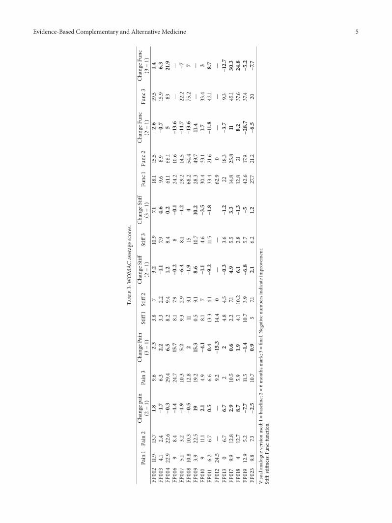
3.1. Clinical Tests and Questionnaires. Given the small samplesize, statistical analyses were not conducted on the resultsof the clinical tests and questionnaires, therefore the rawscores have been provided in Tables 2, 3, 4, and 5. On theHAP and the WOMAC, which were completed on threeoccasions, there appeared to be an overall decline in functionat the 6 month time point, with an improvement at the finalassessment. There was no observable trend in changes on theclinical tests except for a uniformly positive improvement onthe Four Square Step Test (Table 5).
3.2. Gait. All participants were able to walk without externalaids; however, two of them were not able to walk at the
Table 1: Participant characteristics.
Participant Gender Age ConditionFP002 M 69 R hip, lumbar spineFP003 F 73 Lumbar spineFP004 F 61 R kneeFP006 F 70 Both knees, lumbar spineFP007 F 72 R knee, lumbar spineFP008 F 63 Both kneesFP009 F 71 L ankle, L knee, R toeFP010 F 75 R hipFP011 F 59 L knee
FP012 F 62 Multiple joint arthritis; fusion oflarge toes of both feet July 2011
FP013 F 63 L knee
FP017 M 68Knees, lumbar spine; at
reassessment: Morton’s neuromaand plantar fasciitis on L; injured
cartilage L kneeFP018 F 61 R knee and hipFP019 F 67 Neck, lumbar spine, kneesFP023 F 72 Both knees
highest speed (1.4m⋅s−1) and were therefore excluded fromall analyses of this condition. Both subjects reported left kneeOA and one of them also had OA in the left ankle and righttoes. Descriptive statistics and repeated measures ANOVAsfor the spatiotemporal and kinetic measures are summarizedin Tables 6 and 7. Figure 2 shows bar graphs depicting averagepeak joint angles (±SD) and peak joint powers (±SD). Anasterisk highlights significant differences after intervention.
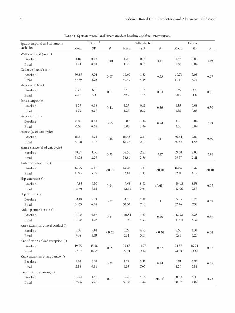
3.2.1. Spatiotemporal Measures (Table 6). Spatiotemporalmeasures of cadence, step length, stride length, step width,percentage of gait cycle in stance, and percentage of gait cyclein single stance were compared before and after intervention.When comparing the effect of intervention for all subjects atall speeds conditions, no significant differences were foundfor any measures (𝑃 > 0.01). No differences were foundfor the same measures when participants were groupedaccording to joints affected.
3.2.2. Kinematic Measures (Table 6). When comparing pre-and post-interventions peak joint angles for all subjects atall speeds tested, no significant differences were found formost of the measures (𝑃 > 0.01). However, significantincreases were found for hip extension during late stance (2.8∘increase), maximal knee flexion during swing (1.6∘ increase),and knee extension at heel contact (2.1∘ increase) duringwalking at self-selected speed (𝑃 < 0.01). Knee extensionat heel contact was also found to be significantly higher(2.0∘ increase) after intervention when participants walked at1.2m⋅s−1. Interestingly, maximal anterior pelvic tilt was found

4 Evidence-Based Complementary and Alternative Medicine
Table 2: Human activity profile.
Participant MAS 1 MAS 2 Change MAS(2 − 1) MAS 3 Change MAS
(3 − 1) AAS 1 AAS 2 Change AAS(2 − 1) AAS 3 Change AAS
(3 − 1)FP002 72 64
−8 73 1 63 41−22 64 1
FP003 78 69−9 78 0 65 58
−7 62−3
FP004 82 65−17 82 0 70 40
−30 62−8
FP006 82 63−19 82 0 68 34
−34 64−4
FP007 77 73−4 78 1 73 63
−10 75 2FP008 70 66
−4 66−4 61 48
−13 56−5
FP009 57 32−25 53
−4 47 4−43 38
−9FP010 75 55
−20 70−5 63 30
−33 61−2
FP011 82 62−20 82 0 71 53
−18 — —FP012 — — — 58 — — — 26 —FP013 70 60
−10 70 0 61 44−17 63 2
FP017 82 76−6 82 0 75 59
−16 74−1
FP018 80 46−34 61
−19 61 8−53 52
−9FP019 82 56
−26 78−4 70 47
−23 74 4FP023 77 75
−2 82 5 74 66−8 77 4
1 = baseline; 2 = 6 month mark; 3 = final.MAS (maximum activity score): highest oxygen-demanding activity still being performed; best estimate of highest level of energy expenditure in comparisonwith peers of same age and gender.AAS (adjusted activity score): a measure of usual daily activities; best estimate of average level of energy expenditure in comparison with peers of same age andgender.
to be significantly reduced after intervention at all speeds(𝑃 < 0.01), with an average reduction of 2.6∘.
3.2.3. Kinetic Measures (Table 7). No significant increaseswere found for ankle peak joint power generation (A2) at allspeed conditions after intervention. Except at self-selectedspeed, where subjects walked faster than prior to interven-tion, ankle absorption power (A1) remained similar afterintervention.
At the knee, most of the intervention effects wereobserved when subjects walked at self-selected and 1.4m⋅s−1conditions, but not at 1.2m⋅s−1. At the two highest speed con-ditions, no significant increases were found for knee powergeneration at the beginning of stance (K0) and decreases forknee power absorption (K1). Most importantly, a significantincrease (𝑃 < 0.01) in knee power absorption at the end ofthe gait cycle (K5) was found at the same conditions with anaverage increase of 0.2W/kg after intervention.
At the hip, no significant changes were observed at allspeed conditions for all peak joint powers analysed. Smallincreases, however, were observed for hip power generationafter toe-off (H3) in the post-intervention gait pattern,especially at self-selected speed (0.06W/kg increase).
3.3. Participant Comments. Class attendance was high(76.5%), and feedback from the satisfaction survey waspositive. All 15 participants said they enjoyed the program“very much.” Eleven of the fifteen participants reportedimprovements in their ability to do everyday things since
the beginning of the program, including going up anddown stairs, ability to stay longer in the garden, betterdeportment, improved walking, and more flexibility. Whenasked to describe what they had learnt by participating inthe program, comments included “how exercise/movementis crucial to managing pain,” “to exercise where it iscomfortable, not to force it,” “to walk with a more fluid,gentle motion,” and “learnt to incorporate some of theexercises into my daily life.” Participants were asked tocomment on their experience of pain and, in particular, thepain associated with their osteoarthritis after participating inthe program. Ten of the fifteen participants said their painlevel had improved, three were unsure and two said they hadnot noticed any difference. Comments included “the pain iscontinual, but I manage it better,” “at the end of the sessionI was free from pain and felt energized,” “I can experienceless pain in the knees, which is where the osteo appears formy body,” “the lessons. . .eased the pain in my lower back,”“no pain in the knees when going up stairs,” and “it is nota cure! however, it is the best “exercise” I have experiencedfor managing my osteoarthritis”. Participants were asked tocomment on their experience of the program in relationto balance, confidence, or walking. Eight of the fifteenparticipants reported an improvement in one or more ofthese areas. Comments included “my balance and confidencein my walking have all improved,” “feel more confident ofwalking/climbing up/down,” “less pressure on the kneeswhen walking,” “getting up and down from the floor is mucheasier” and “the program has helped me in every way. Thebest thing about it is that I know I can do this exercise”.
Evidence-Based Complementary and Alternative Medicine 5
Table3:WOMAC
averages
cores.
Pain
1Pain
2Ch
ange
pain
(2−1)
Pain
3Ch
ange
Pain
(3−1)
Stiff
1Stiff
2Ch
ange
Stiff
(2−1)
Stiff
3Ch
ange
Stiff
(3−1)
Func
1Fu
nc2
Change
Func
(2−1)
Func
3Ch
ange
Func
(3−1)
FP002
11.9
13.7
1.89.6
−2.3
3.8
73.2
10.9
7.118.1
15.5
−2.6
19.5
1.4FP
003
4.1
2.4
−1.7
6.3
2.2
3.3
2.2
−1.1
7.94.6
9.68.9
−0.7
15.9
6.3
FP00
422.9
22.6
−0.3
29.4
6.5
8.2
9.41.2
8.4
0.2
61.1
66.1
583
21.9
FP00
69
8.4
−1.4
24.7
15.7
8.1
7.9−0.2
8−0.1
24.2
10.6
−13.6
——
FP007
5.1
3.2
−1.9
10.3
5.2
9.32.9
−6.4
8.1
−1.2
29.2
14.5
−14.7
22.2
−7
FP008
10.8
10.3
−0.5
12.8
211
9.1−1.9
154
68.2
54.4
−13.6
75.2
7FP
009
3.9
22.5
1919.2
15.3
0.5
9.18.6
10.7
10.2
28.3
49.7
11.4
——
FP010
911.1
2.1
4.9
−4.1
8.1
7−1.1
4.6
−3.5
30.4
33.1
1.733.4
3FP
011
6.2
6.7
0.5
6.6
0.4
13.3
4.1
−9.2
11.5
−1.8
33.4
21.6
−11.8
42.1
8.7
FP012
24.5
——
9.2−15.3
14.4
0—
—62.9
0—
——
FP013
06.7
6.7
22
4.8
4.5
−0.3
3.6
−1.2
2218.3
−3.7
9.3−12.7
FP017
9.912.8
2.9
10.5
0.6
2.2
7.14.9
5.5
3.3
14.8
25.8
1145.1
30.3
FP018
412.7
8.7
5.9
1.94.1
10.2
6.1
2.8
−1.3
12.8
218.2
37.6
24.8
FP019
12.9
5.2
−7.7
11.5
−1.4
10.7
3.9
−6.8
5.7
−5
42.6
17.9
−28
.737.4
−5.2
FP023
9.87.3
−2.5
10.7
0.9
57.1
2.1
6.2
1.227.7
21.2
−6.5
20−7.7
Visualanalogue
versionused;1
=baselin
e;2=6mon
thsm
ark;3=fin
al.N
egativen
umbersindicateim
provem
ent.
Stiff:stiff
ness;Fun
c:functio
n.

6 Evidence-Based Complementary and Alternative Medicine
Table 4: Assessment of quality of life (AQoL).
Participant AQoL 1 AQoL 2 Change(2 − 1) AQoL 3 Change
(3 − 1)FP002 3 4 1 4 1FP003 7 7 0 5
−2FP004 8 7
−1 8 0FP006 9 8
−1 6−3
FP007 4 6 2 9 5FP008 10 8
−2 16 6FP009 22 20
−2 10−12
FP010 11 11 0 6−5
FP011 7 7 0 7 0FP012 11 0
−11 — —FP013 3 4 1 — —FP017 7 0
−7 1−6
FP018 2 0−2 2 0
FP019 6 5−1 6 0
FP023 4 3−1 4 0
Higher scores mean lower quality of life.1 = baseline; 2 = 6 month mark; 3 = final.
Participants were asked whether they experienced any otherbenefits from attending the program. Comments included,“It has made me move in time with my body,” “I feel moreenergetic, brighter, sleeping better,” “I am more positive eventhough pain is still prevalent,” and “enabling, empowering. . . . I feel so confident and grateful that I have found anexercise that suits me.” When asked whether participantswould undertake Feldenkrais classes in the future, eightresponded “definitely,” three responded “probably,” threeresponded “maybe, depending on cost,” and one personresponded “no”.
4. Discussion
Given the participants’ comments on the final questionnaire,it is clear that the physical assessments and questionnairesdid not adequately capture the types of functional changesresulting from undertaking the Feldenkrais classes. Theseincluded a change in the quality of their movement (i.e.,moving with ease), ability to manage their pain, the abilityto get up and down from the floor and climb stairs, betterbalance, and improvement in walking. The small sample sizemakes it difficult to conclude that there were positive changesin function; however, the uniformly positive improvementon the four square step test, coupled with the changes ingait detailed below indicating a more upright posture, issuggestive of an improvement in balance. Since people withOA have balance deficits [24], an improvement in balance forthis group would be an important outcome.
We undertook gait analysis in order to identify anychanges in gait patterns associated with the Feldenkraisclasses. To our knowledge, this is the first study to doso. The findings of decreased anterior pelvic tilt across all
speed conditions may indicate an effect of the Feldenkraisintervention in correcting upright posture. This kinematicchange may have reduced the forward inclination of thetrunk and reduced loads at the low back when walking[25]. At least 15 of the Feldenkrais lessons were focussed onactivation of the abdominal muscles in combination withothermovements. It is possible that this focus led to increasedmuscle activity or strength in the abdominal muscles which,in turn, contributed to the decrease in anterior pelvic tilt.This reduction may be beneficial especially for those subjectssuffering from OA in the lumbar spine (6 participants).Increases in anterior pelvic tilt have been also reported tobe significant in patients with severe OA [26]. Therefore, itmay be possible that reductions in this kinematic measureafter interventionmay be, in itself, a reflection of gait changesthat may contribute to decelerate OA severity progression. Inaddition, reductions in anterior pelvic tilt may contribute toreducing the probability of falls and reduce energy cost whenwalking [27, 28].
Anterior pelvic tilt reductions coupled to an increased hipextension may allow increases in hip extension (absorption)power (H2) leading to higher elastic energy storage mainlyin the iliopsoas muscle [29, 30]. This energy is releasedlater in period between maximal hip extension in the stancephase and maximal hip flexion in the swing phase duringthe second propulsive hip flexion power (H3). It is possiblethat stretching of the rectus femoris, due to increased hipextension and reduced anterior pelvic tilt, may have alsocontributed toH3 through the release of elastic energy duringlate stance [31]. Nonetheless, a reduction in H3 associatedwith hip flexion reduction was found at the two controlledspeed conditions.
DeVita and Hortobagyi [32] proposed that normal agingproduces a shift in the locus of function when walking,with an increase in proximal muscle activity and a reduc-tion in distal muscle activity to propel the body forward.Considering that participants in this study increased theirself-selected speed and were able to walk at the controlledspeed conditions, it was expected that there would be arestoration of the distribution of muscle activity across joints.However, no significant increases in A2 power and ankleplantar flexion were found. Alternatively it is possible thatstrategies in the frontal and transverse planes, not measuredin this gait analysis, may have increased their contributionto propulsion. A large proportion of the Feldenkrais lessonsinvolved rotational movements of the spine. Also, much ofthe Feldenkrais program was aimed at improving walking bybringing into awareness the role of pelvic rotation, the roleof the counter rotation of the thorax and shoulders, and therole of the head in walking. Improvement in spinal rotationand the emphasis on the involvement of the upper body inwalking, coupled with a straighter posture, may have led toa better ability to rotate the pelvis and hip when the leg isin stance [25]. This may also partly explain why there arereductions in H3 and K5 and greater flexion of knee duringswing without affecting step/stride lengths and gait speed.
A significantly lower K5 (higher absorption) at the endof the gait cycle indicates an increased eccentric activityof the hamstring muscles after intervention. This may have

Evidence-Based Complementary and Alternative Medicine 7
Table 5: Leg Power, TUG, 6MWT, and 4SST.
Participant Leg power 1 Leg power 2 Change TUG 1 TUG 2 Change 6MWT 1 6MWT 2 Change 4SST 1 4SST2 ChangeFP002 17.09 18.60 1.51 9.21 8.72 −0.49 438.5 430.3 −8.2 6.85 5.94 −0.91FP003 9.50 9.73 0.23 10.15 7.93 −2.22 339 400.2 61.2 5.41 4.65 −0.76FP004 18.00 18.71 0.71 8.91 8.37 −0.54 436 394.2 −41.8 4.29 4.08 −0.22FP006 13.19 11.86 −1.33 8.61 8.29 −0.32 372 372 0 6.02 5.14 −0.88FP007 18.89 16.37 −2.52 7.39 7.99 0.60 420 462.5 42.5 4.27 4.23 −0.05FP008 13.56 13.05 −0.51 9.33 9.83 0.51 428 417 −11 5.02 4.92 −0.09FP009 8.97 8.09 −0.88 11.20 10.88 −0.32 279 264.8 −14.2 6.83 5.37 −1.46FP010 14.78 14.97 0.19 8.28 8.20 −0.09 420 434 14 5.26 5.19 −0.07FP011 16.24 19.56 3.32 8.08 8.19 0.11 366 363.1 −2.9 5.91 4.42 −1.49FP012 12.95 17.10 4.15 9.82 10.08 0.26 372 413.5 41.5 7.49 4.46 −3.04FP013 9.82 17.22 7.40 8.50 6.02 −2.48 394 439 45 6.22 3.96 −2.27FP017 26.15 20.04 −6.11 8.07 7.36 −0.71 432.3 442.4 10.1 6.20 5.21 −0.99FP018 9.47 10.37 0.90 7.16 7.52 0.36 396 466.25 70.25 7.10 4.25 −2.86FP019 18.61 19.68 1.07 7.54 6.19 −1.35 412 442 30 3.44 3.06 −0.38FP023 13.77 14.66 0.89 8.50 8.61 0.11 420 438 18 5.24 4.66 −0.59Leg power = [(weight ∗ height of stairs (m))/time (sec)] (positive values indicate improvement).TUG: timed up-and-Go test (negative values indicate improvement).6MWT: 6-minute walk test (positive values indicate improvement).4SST: four square step test (negative values indicate improvement).1 = baseline; 2 = final.
influenced the higher knee flexion observed at heel contact atthe beginning of a new gait cycle. An increase in K5 powerabsorption is associated with a reduced step length; however,it may also increase stability of the knee and reduce forwardfoot speed in order to prepare for landing, leading to reducedslip-induced falls [33, 34]. Nevertheless, our findings showedthat step and stride length were bothmaintained. An increasein hip extension is also used as a mechanism to increase steplength [35]. We found increases in this measure which mayindicate a change in strategy to maintain step/stride lengthand make initiation of the new gait cycle more stable.
Increases in the second peak of knee flexion, significantat the self-selected speed, may contribute to a better footclearance, decreasing the probability of tripping and fallingwhen the limb is in the swing phase [36]. This knee flexionincreasemay arise from an increased activity of the hamstringmuscles during swing, which is also responsible for anincreased K5 at the end of this phase. The Feldenkraisprogram did include specific lessons targeting the hamstringmuscles which may have contributed to increased activity inthese muscles. It is possible that an overall increase in muscleactivity in the hamstrings after intervention may have notonly contributed to reduction in anterior pelvic tilt but alsoincreased their absorptive function at the knee during lateswing.
Only sagittal plane gait analysis was performed in thisstudy. Despite controlling over-ground gait speed usingtiming gates, significant differences for speed were foundwhen participants were asked to walk at 1.2m/s. This maybe explained by the fact that participants walked closer tothe lower speed limit allowed during the baseline assessmentand closer to the higher limit during the final assessment.
This is also reflected in the higher self-selected walking speedexhibited after intervention for this condition.
It is a tenet of the Feldenkrais Method that efficientmovement occurs when the work is spread throughout thebody. Although our analyses have focused on changes ingait, the intention of the lessons was not to focus solelyon the lower limbs, but to teach a comprehensive programthat would improve overall movement organisation. Futurestudies could evaluate this aspect further.
5. Study Limitations
Amajor limitation of this study was the lack of a comparisongroup due to the pragmatic nature of this study. The par-ticipant group was a sample of convenience, recruited frompeople who responded to an advertisement of FeldenkraisMethod classes. It is acknowledged that the group was nothomogeneous with respect to gender and the joints affectedby osteoarthritis. The greater proportion of women in ourgroup reflects the fact that more women are affected byarthritis than men [1] and that, in our experience, women aremore likely to volunteer for such projects than men.
However there were no adverse effects such as falls orreports of injuries during the classes and participants whocontinued with the program reported meaningful changes intheir function.
6. Conclusion
The results, high class attendance (76.5%), and survey feed-back indicate that a 30-week series of Feldenkrais classes held
8 Evidence-Based Complementary and Alternative Medicine
Table 6: Spatiotemporal and kinematic data baseline and final intervention.
Spatiotemporal and kinematicvariables
1.2m⋅s−1 Self-selected 1.4m⋅s−1
Mean SD 𝑃 Mean SD 𝑃 Mean SD 𝑃
Walking speed (m⋅s−1)Baseline 1.18 0.04 0.00 1.27 0.18 0.14 1.37 0.05 0.19Final 1.20 0.04 1.30 0.18 1.38 0.04
Cadence (steps/min)Baseline 56.99 3.74 0.07 60.00 4.85 0.33 60.71 3.09 0.07Final 57.79 3.75 60.47 3.49 61.47 3.74
Step length (cm)Baseline 63.2 6.9 0.01 62.5 3.7 0.53 67.9 3.5 0.05Final 64.6 7.3 62.7 3.7 68.2 4.0
Stride length (m)Baseline 1.25 0.08 0.42 1.27 0.13 0.36 1.35 0.08 0.59Final 1.26 0.08 1.28 0.17 1.35 0.08
Step width (m)Baseline 0.08 0.04 0.65 0.09 0.04 0.54 0.09 0.04 0.13Final 0.08 0.04 0.08 0.04 0.08 0.04
Stance (% of gait cycle)Baseline 61.91 2.81 0.46 61.45 2.41 0.11 60.54 2.07 0.89Final 61.70 2.17 61.02 2.19 60.58 1.86
Single stance (% of gait cycle)Baseline 38.27 3.76 0.39 38.53 2.81 0.17 39.30 2.83 0.81Final 38.58 2.29 38.96 2.56 39.37 2.21
Anterior pelvic tilt (∘)Baseline 14.25 6.05
<0.01 14.78 5.83<0.01 14.84 6.42
<0.01Final 11.95 5.79 12.01 5.97 12.18 6.17
Hip extension (∘)Baseline −9.93 8.30 0.04 −9.68 8.02
<0.01∗ −10.42 8.58 0.02Final −11.98 8.81 −12.46 9.04 −12.96 9.58
Hip flexion (∘)Baseline 33.18 7.83 0.07 33.50 7.81 0.11 35.05 8.76 0.02Final 31.63 6.94 32.10 7.10 32.76 7.31
Ankle plantar flexion (∘)Baseline −11.24 4.86 0.24 −10.84 4.87 0.20 −12.92 5.28 0.86Final −11.89 4.76 −11.57 4.93 −13.04 5.39
Knee extension at heel contact (∘)Baseline 5.05 5.01
<0.01 5.29 4.53<0.01 6.63 4.34 0.04
Final 7.06 5.19 7.34 5.01 7.81 5.20Knee flexion at load reception (∘)
Baseline 19.75 15.08 0.18 20.68 14.72 0.22 24.57 16.24 0.92Final 22.07 14.59 22.71 13.49 24.39 13.61
Knee extension at late stance (∘)Baseline 1.20 6.31 0.08 1.27 6.38 0.94 0.81 6.87 0.09Final 2.56 6.94 1.33 7.07 2.29 7.34
Knee flexion at swing (∘)Baseline 56.21 4.52 0.01 56.26 4.65
<0.01∗ 58.68 4.45 0.73Final 57.66 5.46 57.90 5.44 58.87 4.82

Evidence-Based Complementary and Alternative Medicine 9
05
101520253035404550
Pre Post Pre Post Pre Post
Pelvis, ankle and hip peak joint angles
MaxHipExtMaxHipFlex
MaxAPTMaxAnklePF
Self-selected1.2m/s 1.4m/s †
∗∗
∗
∗
Peak
join
t ang
le (∘
)
(a)
0.0
1.0
2.0
3.0
4.0
5.0
6.0
Pre Post Pre Post Pre Post
Self-selected
Join
t pow
er (W
/kg)
Ankle joint powers
A1A2
−2.0
−1.0
1.2m/s 1.4m/s †
(b)
0
10
20
30
40
50
60
70
Pre Post Pre Post Pre Post
Knee peak joint angles
MaxKFaMaxKE
MaxKFbMaxKEhc
Peak
join
t ang
le (∘
)
Self-selected1.2m/s 1.4m/s †
∗
∗∗
(c)
0.0
0.5
1.0
1.5
2.0
Pre Post Pre Post Pre PostSelf-selected
Join
t pow
er (W
/kg)
Knee joint powers
K0K1K2
K3K4K5
−2.5
−2.0
−1.5
−1.0
−0.5
1.2m/s 1.4m/s †
∗∗
(d)
0.00.51.01.52.02.5
Pre Post Pre Post Pre Post
Join
t pow
er (W
/kg)
Hip joint powers
H1H2
H3
−2.5−2.0−1.5−1.0−0.5
Self-selected1.2m/s 1.4m/s †
(e)
Figure 2: Peak joint angles and joint powers.

10 Evidence-Based Complementary and Alternative Medicine
Table 7: Kinetic data baseline and final intervention.
Joint powers (W/kg) 1.2m⋅s−1 Self-selected 1.4m⋅s−1
Mean SD 𝑃 Mean SD 𝑃 Mean SD 𝑃
Ankle power absorption at heeloff (A1)
Baseline −0.83 0.22 0.41 −0.87 0.24 0.27 −0.86 0.26 0.60Final −0.81 0.26 −0.91 0.32 −0.84 0.25
Ankle power generation at latestance (A2)
Baseline 3.69 0.58 0.50 3.95 1.00 0.13 4.22 0.73 0.36Final 3.73 0.57 4.12 0.97 4.30 0.76
Hip extensor power generation atstance (H1)
Baseline 0.46 0.27 0.73 0.51 0.32 0.05 0.60 0.34 0.50Final 0.44 0.28 0.58 0.34 0.57 0.33
Hip flexor power absorption atlate stance (H2)
Baseline −0.84 0.42 0.61 −0.94 0.52 0.40 −0.99 0.51 0.43Final −0.86 0.41 −0.99 0.45 −1.04 0.45
Hip flexor power generation attoe-off (H3)
Baseline 1.60 0.47 0.42 1.77 0.51 0.32 1.95 0.43 0.97Final 1.56 0.42 1.83 0.55 1.95 0.40
Knee flexor power generation atheel contact (K0)
Baseline 0.78 0.51 0.99 0.93 0.58 0.10 1.01 0.60 0.16Final 0.78 0.44 1.04 0.63 1.12 0.66
Knee extensor absorption powerat initial stance (K1)
Baseline −0.67 0.42 0.02 −0.88 0.56 0.04 −1.12 0.72 0.17Final −0.80 0.46 −1.01 0.52 −1.23 0.61
Knee extensor power generationat mid stance (K2)
Baseline 0.44 0.27 0.24 0.57 0.38 0.58 0.69 0.43 0.94Final 0.48 0.31 0.59 0.33 0.69 0.34
Knee extensor power generationat late stance (K3)
Baseline 0.32 0.24 0.22 0.38 0.33 0.37 0.39 0.32 0.12Final 0.28 0.25 0.35 0.31 0.33 0.25
Knee extensors power absorptionat initial swing (K4)
Baseline −1.25 0.42 0.22 −1.42 0.46 0.77 −1.52 0.37 0.76Final −1.20 0.35 −1.44 0.49 −1.51 0.40
Knee flexors power absorption atfinal swing (K5)
Baseline −1.36 0.35 0.01 −1.57 0.55<0.01∗ −1.67 0.45
<0.01Final −1.47 0.35 −1.74 0.59 −1.86 0.46
twice perweekwas feasible in the community setting andmaybe acceptable for other people with OA. The lessons led toimprovements in performance of the Four Square Step Testand changes in gait. Further investigation of the FeldenkraisMethod for people with OA is warranted.
Acknowledgments
The authors wish to thank all the participants involved in thisstudy. This work was supported by a grant from the MerriCommunity Health Services Ltd.

Evidence-Based Complementary and Alternative Medicine 11
References
[1] National Health Survey 2007-2008, 4364.0.55.001, AustralianBureau of Statistics.
[2] A. Abhishek and M. Doherty, “Diagnosis and clinical presen-tation of osteoarthritis,” Rheumatic Disease Clinics of NorthAmerica, vol. 39, pp. 45–66, 2013.
[3] J. M. Bjordal, A. Klovning, A. E. Ljunggren, and L. Slørdal,“Short-term efficacy of pharmacotherapeutic interventionsin osteoarthritic knee pain: a meta-analysis of randomisedplacebo-controlled trials,” European Journal of Pain, vol. 11, no.2, pp. 125–138, 2007.
[4] E. Roddy, W. Zhang, M. Doherty et al., “Evidence-basedrecommendations for the role of exercise in the managementof osteoarthritis of the hip or knee—the MOVE concensus,”Rheumatology, vol. 44, no. 1, pp. 67–73, 2005.
[5] K. Reardon, M. Galea, X. Dennett, P. Choong, and E. Byrne,“Quadriceps muscle wasting persists 5 months after total hiparthroplasty for osteoarthritis of the hip: a pilot study,” InternalMedicine Journal, vol. 31, no. 1, pp. 7–14, 2001.
[6] I. S. Raj, S. R. Bird, andA. J. Shield, “Aging and the force-velocityrelationship of muscles,” Experimental Gerontology, vol. 45, no.2, pp. 81–90, 2010.
[7] R. R. Neptune, S. A. Kautz, and F. E. Zajac, “Contributions of theindividual ankle plantar flexors to support, forward progressionand swing initiation during walking,” Journal of Biomechanics,vol. 34, no. 11, pp. 1387–1398, 2001.
[8] J. O. Judge, R. B. Davis III, and S. Ounpuu, “Step lengthreductions in advanced age: the role of ankle and hip kinetics,”Journals of Gerontology A, vol. 51, no. 6, pp. M303–M312, 1996.
[9] L. E. Cofre, N. Lythgo, D. Morgan, and M. P. Galea, “Agingmodifies joint power and work when gait speeds are matched,”Gait and Posture, vol. 33, no. 3, pp. 484–489, 2011.
[10] A. Graf, J. O. Judge, S. Ounpuu, and D. G. Thelen, “Theeffect of walking speed on lower-extremity joint powers amongelderly adults who exhibit low physical performance,” Archivesof Physical Medicine and Rehabilitation, vol. 86, no. 11, pp. 2177–2183, 2005.
[11] A. A. Guccione, “Arthritis and the process of disablement,”Physical Therapy, vol. 74, no. 5, pp. 408–414, 1994.
[12] G. Peat, E.Thomas, R. Wilkie, and P. Croft, “Multiple joint painand lower extremity disability in middle and old age,”Disabilityand Rehabilitation, vol. 28, no. 24, pp. 1543–1549, 2006.
[13] M. Feldenkrais, Awareness through Movement; Health Exercisesfor Personal Growth, Harper & Row, New York, NY, USA, 1stedition, 1972.
[14] I. Lundblad, J. Elert, and B. Gerdle, “Randomized controlledtrial of physiotherapy and Feldenkrais interventions in femaleworkers with neck-shoulder complaints,” Journal of Occupa-tional Rehabilitation, vol. 9, no. 3, pp. 179–194, 1999.
[15] K. A. Connors, M. P. Galea, and C. M. Said, “Feldenkraismethod balance classes improve balance in older adults: a con-trolled trial,” Evidence-Based Complementary and AlternativeMedicine, vol. 2011, Article ID 873672, 9 pages, 2011.
[16] F. Vrantsidis, K. D. Hill, K. Moore, R. Webb, S. Hunt, andL. Dowson, “Getting grounded gracefully: effectiveness andacceptability of feldenkrais in improving balance,” Journal ofAging and Physical Activity, vol. 17, no. 1, pp. 57–76, 2009.
[17] R. Altman, G. Alarcon, D. Appelrouth et al., “The AmericanCollege of Rheumatology criteria for the classification andreporting of osteoarthritis of the hip,” Arthritis and Rheuma-tism, vol. 34, no. 5, pp. 505–514, 1991.
[18] D. Podsiadlo and S. Richardson, “The timed “Up & Go” A testof basic functional mobility for frail elderly persons,” Journal ofthe American Geriatrics Society, vol. 39, no. 2, pp. 142–148, 1991.
[19] W. Dite and V. A. Temple, “A clinical test of stepping and changeof direction to identify multiple falling older adults,”Archives ofPhysical Medicine and Rehabilitation, vol. 83, no. 11, pp. 1566–1571, 2002.
[20] R. Margaria, P. Aghemo, and E. Rovelli, “Measurement of mus-cular power (anaerobic) in man,” Journal of Applied Physiology,vol. 21, no. 5, pp. 1662–1664, 1966.
[21] G. Hawthorne, J. Richardson, and R. Osborne, “TheAssessmentof Quality of Life (AQoL) instrument: a psychometric measureof health-related quality of life,” Quality of Life Research, vol. 8,no. 3, pp. 209–224, 1999.
[22] N. Bellamy, W. W. Buchanan, C. H. Goldsmith, J. Campbell,and L. W. Stitt, “Validation study of WOMAC: a health statusinstrument for measuring clinically important patient relevantoutcomes to antirheumatic drug therapy in patients withosteoarthritis of the hip or knee,” Journal of Rheumatology, vol.15, no. 12, pp. 1833–1840, 1988.
[23] J. A. Fix and D. M. Daughton, Human Activity Profile: Profes-sional Manual, Edited by F. L. Lutz, Psychological AssessmentResources, 1988.
[24] R. S. Hinman, K. L. Bennell, B. R. Metcalf, and K. M. Crossley,“Balance impairments in individuals with symptomatic kneeosteoarthritis: a comparison with matched controls using clini-cal tests,” Rheumatology, vol. 41, no. 12, pp. 1388–1394, 2002.
[25] D. Saha, S. Gard, and S. Fatone, “The effect of trunk flexion onable-bodied gait,” Gait and Posture, vol. 27, no. 4, pp. 653–660,2008.
[26] D. J. Jacofsky, J. D. McCamley, M. Bhowmik-Stoker, M. C.Jacofsky, and M. W. Shrader, “Advanced osteoarthritic gaitkinemtaics and kinetics,” Journal of Bone & Joint Surgery, vol.93, article 399, supplement 4, 2011.
[27] C. A. McGibbon, D. E. Krebs, and M. S. Puniello, “Mechanicalenergy analysis identifies compensatory strategies in disabledelders’ gait,” Journal of Biomechanics, vol. 34, no. 4, pp. 481–490,2001.
[28] C. A. McGibbon, M. S. Puniello, and D. E. Krebs, “Mechanicalenergy transfer during gait in relation to strength impairmentand pathology in elderly women,”Clinical Biomechanics, vol. 16,no. 4, pp. 324–333, 2001.
[29] C. A. McGibbon, “Toward a better understanding of gaitchanges with age and disablement: neuromuscular adaptation,”Exercise and Sport Sciences Reviews, vol. 31, no. 2, pp. 102–108,2003.
[30] C. A. McGibbon and D. E. Krebs, “Discriminating age anddisability effects in locomotion: neuromuscular adaptations inmusculoskeletal pathology,” Journal of Applied Physiology, vol.96, no. 1, pp. 149–160, 2004.
[31] R. R. Neptune, K. Sasaki, and S. A. Kautz, “The effect of walkingspeed on muscle function and mechanical energetics,”Gait andPosture, vol. 28, no. 1, pp. 135–143, 2008.
[32] P.DeVita andT.Hortobagyi, “Age causes a redistribution of jointtorques and powers during gait,” Journal of Applied Physiology,vol. 88, no. 5, pp. 1804–1811, 2000.
[33] D. D. Espy, F. Yang, T. Bhatt, and Y.-C. Pai, “Independentinfluence of gait speed and step length on stability and fall risk,”Gait and Posture, vol. 32, no. 3, pp. 378–382, 2010.
[34] E. Watelain, F. Barbier, P. Allard, A. Thevenon, and J.-C.Angue, “Gait pattern classification of healthy elderly men based

12 Evidence-Based Complementary and Alternative Medicine
on biomechanical data,” Archives of Physical Medicine andRehabilitation, vol. 81, no. 5, pp. 579–586, 2000.
[35] B. W. Schulz, J. A. Ashton-Miller, and N. B. Alexander, “Max-imum Step Length: relationships to age and knee and hipextensor capacities,” Clinical Biomechanics, vol. 22, no. 6, pp.689–696, 2007.
[36] D. A. Winter, “Foot trajectory in human gait: a precise andmultifactorial motor control task,” Physical Therapy, vol. 72, no.1, pp. 45–56, 1992.

Submit your manuscripts athttp://www.hindawi.com
Stem CellsInternational
Hindawi Publishing Corporationhttp://www.hindawi.com Volume 2014
Hindawi Publishing Corporationhttp://www.hindawi.com Volume 2014
MEDIATORSINFLAMMATION
of
Hindawi Publishing Corporationhttp://www.hindawi.com Volume 2014
Behavioural Neurology
EndocrinologyInternational Journal of
Hindawi Publishing Corporationhttp://www.hindawi.com Volume 2014
Hindawi Publishing Corporationhttp://www.hindawi.com Volume 2014
Disease Markers
Hindawi Publishing Corporationhttp://www.hindawi.com Volume 2014
BioMed Research International
OncologyJournal of
Hindawi Publishing Corporationhttp://www.hindawi.com Volume 2014
Hindawi Publishing Corporationhttp://www.hindawi.com Volume 2014
Oxidative Medicine and Cellular Longevity
Hindawi Publishing Corporationhttp://www.hindawi.com Volume 2014
PPAR Research
The Scientific World JournalHindawi Publishing Corporation http://www.hindawi.com Volume 2014
Immunology ResearchHindawi Publishing Corporationhttp://www.hindawi.com Volume 2014
Journal of
ObesityJournal of
Hindawi Publishing Corporationhttp://www.hindawi.com Volume 2014
Hindawi Publishing Corporationhttp://www.hindawi.com Volume 2014
Computational and Mathematical Methods in Medicine
OphthalmologyJournal of
Hindawi Publishing Corporationhttp://www.hindawi.com Volume 2014
Diabetes ResearchJournal of
Hindawi Publishing Corporationhttp://www.hindawi.com Volume 2014
Hindawi Publishing Corporationhttp://www.hindawi.com Volume 2014
Research and TreatmentAIDS
Hindawi Publishing Corporationhttp://www.hindawi.com Volume 2014
Gastroenterology Research and Practice
Hindawi Publishing Corporationhttp://www.hindawi.com Volume 2014
Parkinson’s Disease
Evidence-Based Complementary and Alternative Medicine
Volume 2014Hindawi Publishing Corporationhttp://www.hindawi.com