research article molecular detection of torque teno sus...
TRANSCRIPT

Research ArticleMolecular Detection of Torque Teno Sus Virus andCoinfection with African Swine Fever Virus in Blood Samples ofPigs from Some Slaughterhouses in Nigeria
Pam D. Luka,1,2 Joseph Erume,1 Bitrus Yakubu,2 Olajide A. Owolodun,2
David Shamaki,3 and Frank N. Mwiine1
1Department of Biomolecular Resources and Biolab Sciences, College of Veterinary Medicine,Animal Resources and Biosecurity, Makerere University, Kampala, Uganda2Biotechnology Division, National Veterinary Research Institute, Vom, Plateau State, Nigeria3Virology Division, National Veterinary Research Institute, Vom, Plateau State, Nigeria
Correspondence should be addressed to Frank N. Mwiine; [email protected]
Received 1 June 2016; Revised 15 August 2016; Accepted 19 September 2016
Academic Editor: Alessandra Lo Presti
Copyright © 2016 Pam D. Luka et al. This is an open access article distributed under the Creative Commons Attribution License,which permits unrestricted use, distribution, and reproduction in any medium, provided the original work is properly cited.
Torque teno sus virus 1 (TTSuV1a/TTSuV1b) infection is present in pig herds worldwide. This study investigated the prevalenceof TTSuV1a/TTSuV1b infections in domestic pigs from some slaughterhouses in Nigeria as well as coinfection with African swinefever virus (ASFV) and described the phylogeny in relation to global strains. One hundred and eighty-one (181) blood samplesfrom four slaughterhouses were used for the study and viral nucleic acid detection was carried out by PCR. Comparative sequenceanalysis was carried out to infer phylogeny. The overall prevalence of TTSuV1a/b was 17.7%. Prevalence of individual genotypeswas 10.5% and 7.2% for TTSuV1a and TTSuV1b, respectively. Coinfection of ASFV/TTSuV1a/b was 7.7% while that of TTSuV1aand TTSuV1b was 1.7%. ASFV alone was detected in 11.91% of the total samples. The Nigerian TTSuV1a and TTSuV1b shared asequence identity of 91–100% and 95–100%, respectively, among each other.TheASFV sequences were 100% identical tomembers ofgenotype 1. This is the first report on the presence of TTSuV1a/b in domestic pigs in Nigeria and coinfection with ASFV. Althoughthe prevalence of TTSuV1a/b in Nigeria was low, we recommend further studies to establish the trend and possible role in thepathogenesis of ASFV.
1. Introduction
Torque teno virus (TTV) is a small icosahedral and nonen-veloped, single-stranded DNA (ssDNA) virus. It is circularwith a negative genome that was first reported in a humanwith posttransfusion hepatitis in Japan [1]. The virus has alsobeen reported to infect domestic animals such as pigs andboars [2, 3]. TTV are classified into the family Anelloviridaeincluding 9 different genera among which is the genus Iota-torquevirus.Genetic analysis has shown that two genotypes ofthe genus Iotatorquevirus [torque teno sus virus 1 (TTSuV1)and torque teno sus virus 2 (TTSuV2)] and the newly groupedgenotype TTSuVk2 of the genus Kappatorquevirus exist inpigs [4].
Torque teno viruses have been reported to be distributedglobally with human TTV being ubiquitous while severalother reports of swine TTSuV infection have been reportedin Spain, Italy, Russia, China, and very recently Uganda inAfrica [5–8]. TTSuV has been reported in coinfection withother pathogens but its evidence as a pathogen of pigs and itsinvolvement in causality is yet to be elucidated [6].
The disease caused by TTSuV has not yet been definedeven though it is widely spread and species specific. However,TTSuV2 (now TTSuV1b) has been reported in domesticreared pigs with other pathogens such as porcine circovirus2 (PCV-2), hepatitis E virus (HEV), postweaning multi-systemic wasting syndrome (PMWS), porcine endogenousretrovirus, and Ndumu virus [9–13]. On the other hand,
Hindawi Publishing CorporationAdvances in VirologyVolume 2016, Article ID 6341015, 6 pageshttp://dx.doi.org/10.1155/2016/6341015

2 Advances in Virology
TTSuV1a has been suggested to trigger PMWS developmentin gnotobiotic pigs coinfected with PCV-2 [14]. Furthermore,coinfection of TTSuV1a and porcine reproductive and respi-ratory syndrome virus (PRRSV) [15] has also been reported;Blomstrom et al. [12] have also reported a novel variant ofporcine parvovirus 4 (PPV4) from bushpig (Potamochoeruslarvatus) coinfectingwith TTSuV1a andTTSuV1b.Therefore,the potential association of swine TTSuV with other diseaseoccurrence in pigs is of scientific interest.
The rising demand for livestock products in Nigeria hasresulted in government agricultural intervention leading toincreased pig production [16]. However, with the adventof African swine fever in 1997, the prospect of the pigindustry has continued to dwindle [17, 18]. Some regions inNigeria preferred pig to other food animals due to its relativerapid growth rate, short cycle, and large litter size. Giventhe extensive/semi-intensive farming system common inNigeria and the rising contact between pigs and humansin addition to poor farm practices, any potential risk fromTTSuV or coinfection with other pathogens could lead topublic health consequences in terms of the pig’s capacity toserve as reservoir and/or transmitter of several emerging andreemerging diseases. As part of ASF surveillance in Nigeria,blood samples from selected slaughterhouses were analyzedto determine the presence of TTSuV1. The main objective ofthis study was to determine the presence of swine TTSuV1aand TTSuV1b genotypes in association with ASFVs in Nige-ria.
2. Materials and Methods
2.1. Study Area and Sample Collection. Blood samples from181 domestic pigs were collected from four slaughterhousesfrom four localities in Nigeria: Jos, Makurdi and Ibadanabattoirs, and Kafanchan pig market slaughter slab (Figure 1).Sampling was carried out between January and March 2014as part of a research project on the epidemiology of Africanswine fever in some pig producing states of Nigeria. Thisprojectwaswith themandate ofNationalVeterinaryResearchInstitute (NVRI), Vom, and the Federal Department of Vet-erinary and Pest Control Services of the Federal Ministry ofAgriculture, Abuja. Apparently healthy pigs presented to theslaughterhouses were sampled.
2.2. Sample Processing and DNA Extraction. Blood samples(𝑛 = 181) were collected in sterile sample bottles withethylenediaminetetraacetic acid (EDTA) anticoagulant andkept at +4∘C to +8∘Cuntil used forDNAextraction.TheDNAextraction was carried out using a DNeasy blood and tissuekit (Qiagen, Hilden Germany) following the manufacturer’sguidelines. Extracted DNA was kept at −20∘C pending PCR.
2.3. Confirmation of ASFV by PCR. ASFV was confirmedusing the primer pair ASF-1 and ASF-2 according to theManual of Diagnostic Tests and Vaccines [19]. ASF specificprimers targeting the major capsid protein (VP72 gene) 278-bp fragment within the conserved region were amplified asdescribed by the OIE manual. A 478-bp C-terminus of the
Presence of virusTTSuV1TTSuVASFV
150km
100mi
YobeJigawa
Borno
EdoLagos
Ogun
Osun
Oyo
Ekiti KogiBenue
Taraba
Adamawa
GombeGombe
Plateau
Anambra
EbonyiEnugu
Kwara
Ondo
ImoDelta AbiaCrossRiver
AkwaIbom
RiversBayelsa
Jos
Makurdi
Kafanchan
Ibadan
Cameroon
Benin
ChadNiger
Niger
Kano
Nasarawa
Abuja
BauchiKaduna
Kebbi
Sokoto
Zamfara
Katsina
Figure 1:Map of Nigeria showing the 4 locations of slaughterhouseswhere samples were collected.
p72 gene was also amplified for genotyping as described byBastos et al. [20].
2.4. TTSuV1 and TTSuV2 Detection and Partial Sequencing.The sample extracted DNA was used for the detection ofTTSuV1a and TTSuV1b. Assessment of TTSuV genotypes 1aand 1b from the collected samples was analyzed by amplifyingan untranslated region (UTR) of the TTSuV1 viral genomeusing species specific primers as reported by Segales et al.[5]. The amplification was performed on a GeneAmp� PCRSystem 9700 machine (Applied BioSystems, USA).
The PCR amplicons were resolved on 1.8% agarose inTris-borate-EDTA- (TBE-) buffered gels stained with ethid-ium bromide. Ten microlitres of the PCR product fromeach of the tubes was mixed with 1 𝜇L of 6x buffer andelectrophoresed along with a 50-bp DNA molecular weightmarker (GeneRuler, MBI Fermentas) at a constant voltageof 100V for 45min in 1x TBE buffer. Amplified productswere viewed using a Bio-Rad Gel Doc� XR system.The PCRpositive products were purified using Wizard� SV Gel andPCR clean-up system (Promega Corporation, Madison, WI,USA) according to themanufacturers’ instructions and elutedin 30 𝜇L EB.The purified products were sequenced by InqabaBiotec�, South Africa.
2.5. Statistical Analyses of TTSuV Genotype Prevalence. Theprevalence data generated with the screening of TTSuV andASFV DNA in apparently healthy population was analyzedwith statistical tests carried out in SPSS, version 17.0. Student’s𝑡-test, Pearson’s correlation, and chi-square/Fisher’s exact testwere appliedwherever relevant.𝑃 value<0.05was consideredsignificant.
Advances in Virology 3
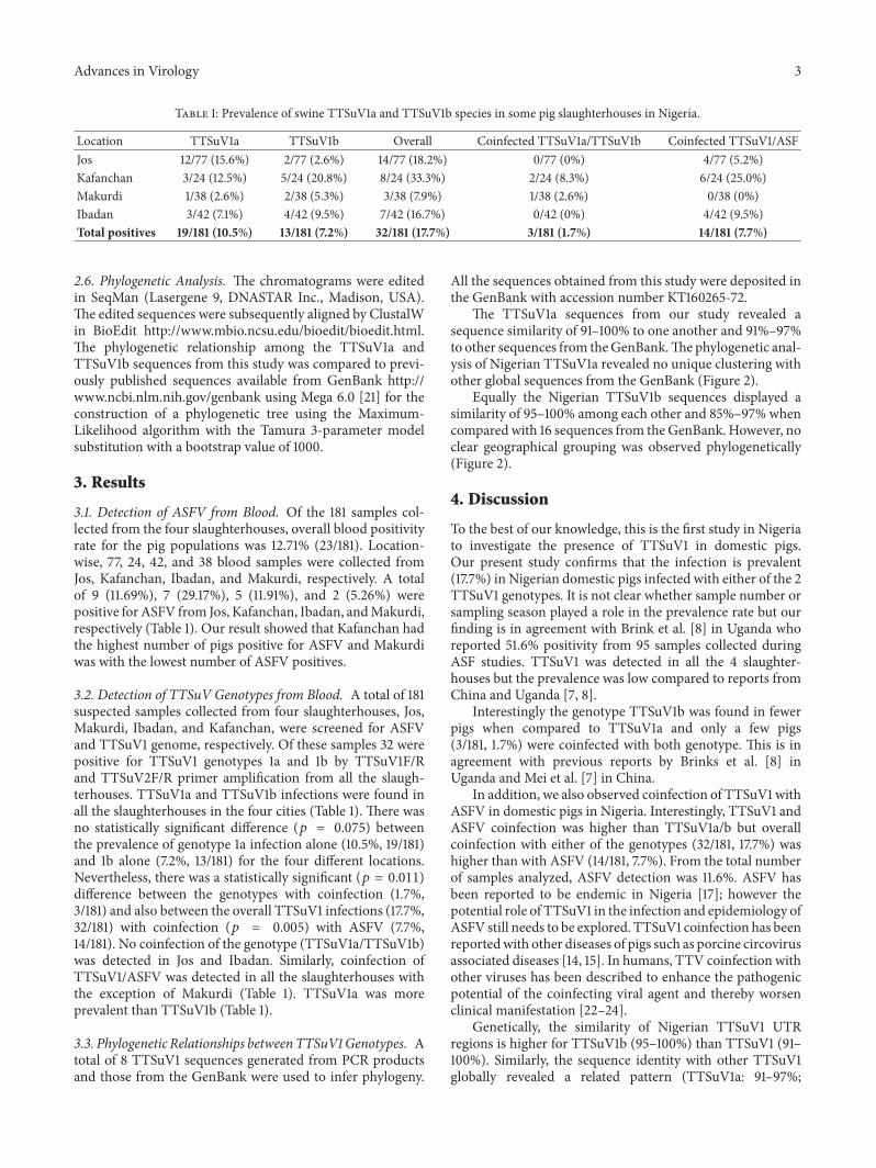
Table 1: Prevalence of swine TTSuV1a and TTSuV1b species in some pig slaughterhouses in Nigeria.
Location TTSuV1a TTSuV1b Overall Coinfected TTSuV1a/TTSuV1b Coinfected TTSuV1/ASFJos 12/77 (15.6%) 2/77 (2.6%) 14/77 (18.2%) 0/77 (0%) 4/77 (5.2%)Kafanchan 3/24 (12.5%) 5/24 (20.8%) 8/24 (33.3%) 2/24 (8.3%) 6/24 (25.0%)Makurdi 1/38 (2.6%) 2/38 (5.3%) 3/38 (7.9%) 1/38 (2.6%) 0/38 (0%)Ibadan 3/42 (7.1%) 4/42 (9.5%) 7/42 (16.7%) 0/42 (0%) 4/42 (9.5%)Total positives 19/181 (10.5%) 13/181 (7.2%) 32/181 (17.7%) 3/181 (1.7%) 14/181 (7.7%)
2.6. Phylogenetic Analysis. The chromatograms were editedin SeqMan (Lasergene 9, DNASTAR Inc., Madison, USA).The edited sequences were subsequently aligned by ClustalWin BioEdit http://www.mbio.ncsu.edu/bioedit/bioedit.html.The phylogenetic relationship among the TTSuV1a andTTSuV1b sequences from this study was compared to previ-ously published sequences available from GenBank http://www.ncbi.nlm.nih.gov/genbank using Mega 6.0 [21] for theconstruction of a phylogenetic tree using the Maximum-Likelihood algorithm with the Tamura 3-parameter modelsubstitution with a bootstrap value of 1000.
3. Results
3.1. Detection of ASFV from Blood. Of the 181 samples col-lected from the four slaughterhouses, overall blood positivityrate for the pig populations was 12.71% (23/181). Location-wise, 77, 24, 42, and 38 blood samples were collected fromJos, Kafanchan, Ibadan, and Makurdi, respectively. A totalof 9 (11.69%), 7 (29.17%), 5 (11.91%), and 2 (5.26%) werepositive forASFV from Jos, Kafanchan, Ibadan, andMakurdi,respectively (Table 1). Our result showed that Kafanchan hadthe highest number of pigs positive for ASFV and Makurdiwas with the lowest number of ASFV positives.
3.2. Detection of TTSuV Genotypes from Blood. A total of 181suspected samples collected from four slaughterhouses, Jos,Makurdi, Ibadan, and Kafanchan, were screened for ASFVand TTSuV1 genome, respectively. Of these samples 32 werepositive for TTSuV1 genotypes 1a and 1b by TTSuV1F/Rand TTSuV2F/R primer amplification from all the slaugh-terhouses. TTSuV1a and TTSuV1b infections were found inall the slaughterhouses in the four cities (Table 1). There wasno statistically significant difference (𝑝 = 0.075) betweenthe prevalence of genotype 1a infection alone (10.5%, 19/181)and 1b alone (7.2%, 13/181) for the four different locations.Nevertheless, there was a statistically significant (𝑝 = 0.011)difference between the genotypes with coinfection (1.7%,3/181) and also between the overall TTSuV1 infections (17.7%,32/181) with coinfection (𝑝 = 0.005) with ASFV (7.7%,14/181). No coinfection of the genotype (TTSuV1a/TTSuV1b)was detected in Jos and Ibadan. Similarly, coinfection ofTTSuV1/ASFV was detected in all the slaughterhouses withthe exception of Makurdi (Table 1). TTSuV1a was moreprevalent than TTSuV1b (Table 1).
3.3. Phylogenetic Relationships betweenTTSuV1Genotypes. Atotal of 8 TTSuV1 sequences generated from PCR productsand those from the GenBank were used to infer phylogeny.
All the sequences obtained from this study were deposited inthe GenBank with accession number KT160265-72.
The TTSuV1a sequences from our study revealed asequence similarity of 91–100% to one another and 91%–97%to other sequences from theGenBank.Thephylogenetic anal-ysis of Nigerian TTSuV1a revealed no unique clustering withother global sequences from the GenBank (Figure 2).
Equally the Nigerian TTSuV1b sequences displayed asimilarity of 95–100% among each other and 85%–97% whencomparedwith 16 sequences from theGenBank. However, noclear geographical grouping was observed phylogenetically(Figure 2).
4. Discussion
To the best of our knowledge, this is the first study in Nigeriato investigate the presence of TTSuV1 in domestic pigs.Our present study confirms that the infection is prevalent(17.7%) in Nigerian domestic pigs infected with either of the 2TTSuV1 genotypes. It is not clear whether sample number orsampling season played a role in the prevalence rate but ourfinding is in agreement with Brink et al. [8] in Uganda whoreported 51.6% positivity from 95 samples collected duringASF studies. TTSuV1 was detected in all the 4 slaughter-houses but the prevalence was low compared to reports fromChina and Uganda [7, 8].
Interestingly the genotype TTSuV1b was found in fewerpigs when compared to TTSuV1a and only a few pigs(3/181, 1.7%) were coinfected with both genotype. This is inagreement with previous reports by Brinks et al. [8] inUganda and Mei et al. [7] in China.
In addition, we also observed coinfection of TTSuV1 withASFV in domestic pigs in Nigeria. Interestingly, TTSuV1 andASFV coinfection was higher than TTSuV1a/b but overallcoinfection with either of the genotypes (32/181, 17.7%) washigher than with ASFV (14/181, 7.7%). From the total numberof samples analyzed, ASFV detection was 11.6%. ASFV hasbeen reported to be endemic in Nigeria [17]; however thepotential role of TTSuV1 in the infection and epidemiology ofASFV still needs to be explored. TTSuV1 coinfection has beenreportedwith other diseases of pigs such as porcine circovirusassociated diseases [14, 15]. In humans, TTV coinfection withother viruses has been described to enhance the pathogenicpotential of the coinfecting viral agent and thereby worsenclinical manifestation [22–24].
Genetically, the similarity of Nigerian TTSuV1 UTRregions is higher for TTSuV1b (95–100%) than TTSuV1 (91–100%). Similarly, the sequence identity with other TTSuV1globally revealed a related pattern (TTSuV1a: 91–97%;

4 Advances in Virology
TTSuV1b_1NIG
GQ120664 Canada
GU456384 USA
AY823990 Brazil
HM633244 China
HM633250 China
HM633257 China
TTSuV1b_4.1NIG
TTSuV1b_6.1NIG
HM633258 China
HM633247 China
HM633255 China
GU188045 Germany
HM633253 China
HM633248 China
HM633246| China
GU450331 China
HM633254 China
TTSuV1b_7.1NIG
HM633242 China
HM633252 China
TTSuV1a_1NIG
AB076001 Japan
HM633249 China
GU570202 Spain
GU570201 Spain
HM633243 Spain
TTSuV1a_7NIG
TTSuV1a_6NIG
TTSuV1a_4NIG
GU570200 Spain
GU570198 Spain
GU570199 Spain
100
99
100
100
100
79
92
98100
100
100
89
100
81
81
81
74
0.02
TTSuV1b
TTSuV1a
Genus Iotatorquevirus
Figure 2: Molecular phylogenetic analysis of TTSuV1a and TTSuV1b sequences of the UTR region using Maximum-Likelihood methodbased on the Tamura 3-parameter model. A discrete gamma distribution was used to model evolutionary rate differences among sites (5categories (+G, parameter = 0.3029)). Only bootstrap values above 70% are shown and sequences from this study are bold and highlighted(red).

Advances in Virology 5
TTSuV1b: 85–97%). However, previous studies using theUTR region did not reveal distinct geographical clustering.The phylogenetic analysis of the Nigerian sequences showedTTSuV1a and TTSuV1b distribution among other sequencesglobally as described by ICVT (2011) new classification.However, sequences fromour study revealed a similar patternto TTSuV1 reports from Spain and Uganda [5, 8].
In contrast, other studies had suggested the use of ORF1capsid gene as a marker for better resolution and molecularepidemiology because the gene is under selection pressure[5]. A complete genome sequencing has also been advocatedfor a better understanding of the virus dynamics [4, 25, 26].
ASFV sequences obtained from our study showed 100%sequence identity of the p72 gene to all other members ofgenotype 1 included in the analysis and phylogeneticallyclustered together (data not shown).
The capacity of TTSuV1 on its own to cause diseaseor influence ASF is yet to be fully elucidated, but it hasbeen reported to be involved directly or indirectly in thedevelopment of other diseases such as PMWS and PCV-2.Its association with PCV-2 has been reported especially indiseased animals [12, 14]. From the total number of samplesanalyzed, 14 were positive for ASFV from apparently healthypigs suggesting state of nonclinical manifestation of disease.Phylogenetically, the viruses were similar to those previouslyreported to be circulating in Nigeria. It is however possiblethat the pigs positive for ASFV brought to the slaughterhousecould be ASFV carriers without developing the disease orcould have been brought into the slaughterhouse prior tovisible signs or part of emergency sale.
5. Conclusion
We reported the circulation of TTSuV1 inNigeria coinfectingwithASFVand like other parts of theworldTTSuV1a/b infec-tion is not very common among domestic pigs in Nigeria.However, having detected TTSuV1 coinfecting with ASFVand given the lethal nature of ASFV, we hypothesize thatTTSuV1 may play an exacerbating role in the pathogenesis ofASF in domestic pigs in Nigeria. Further investigation needsto be carried out to establish if the trend is rising. The role ofTTSuV in the infection and epidemiology of ASFV needs tobe explored.
Competing Interests
The authors declare that there is no conflict of interests.
References
[1] T. Nishizawa, H. Okamoto, K. Konishi, H. Yoshizawa, Y.Miyakawa, and M. Mayumi, “A novel DNA virus (TTV)associated with elevated transaminase levels in posttransfusionhepatitis of unknown etiology,” Biochemical and BiophysicalResearch Communications, vol. 241, no. 1, pp. 92–97, 1997.
[2] L. Martınez, T. Kekarainen, M. Sibila et al., “Torque teno virus(TTV) is highly prevalent in the European wild boar (Susscrofa),” Veterinary Microbiology, vol. 118, no. 3-4, pp. 223–229,2006.
[3] H. Okamoto, M. Takahashi, T. Nishizawa et al., “Genomiccharacterization of TT viruses (TTVs) in pigs, cats and dogs andtheir relatedness with species-specific TTVs in primates andtupaias,” Journal of General Virology, vol. 83, no. 6, pp. 1291–1297,2002.
[4] ICTV, Virus Taxonomy, Ninth Report of the International Com-mittee on Taxonomy of Viruses, ICTV, 2012.
[5] J. Segales, L. Martınez-Guino, M. Cortey et al., “Retrospectivestudy on swine Torque teno virus genogroups 1 and 2 infectionfrom 1985 to 2005 in Spain,” Veterinary Microbiology, vol. 134,no. 3-4, pp. 199–207, 2009.
[6] S. Hino and H. Miyata, “Torque teno virus (TTV): currentstatus,”Reviews inMedical Virology, vol. 17, no. 1, pp. 45–57, 2007.
[7] M. Mei, L. Zhu, Z. Xu et al., “Molecular investigation of Torqueteno sus virus in geographically distinct porcine breeding herdsof Sichuan, China,” Virology Journal, vol. 10, article 161, 2013.
[8] M. Brink, K. Stahl, C. Masembe, A. Okurut, M. Berg, and A.-L.Blomstrom, “First time molecular detection and phylogeneticrelationships of torque teno sus virus 1 and 2 in domestic pigsin Uganda: further evidence for a global distribution,” VirologyJournal, vol. 9, article 39, 2012.
[9] T. Kekarainen, M. Sibila, and J. Segales, “Prevalence of swineTorque teno virus in post-weaning multisystemic wastingsyndrome (PMWS)-affected and non-PMWS-affected pigs inSpain,” Journal of General Virology, vol. 87, no. 4, pp. 833–837,2006.
[10] T. Kekarainen and J. Segales, “Torque teno Sus virus in pigs:an emerging pathogen?”Transboundary and EmergingDiseases,vol. 59, no. 1, pp. 103–108, 2012.
[11] B. Savic, V. Milicevic, J. Bojkovski, B. Kureljusic, V. Ivetic, andI. Pavlovic, “Detection rates of the swine torque teno viruses(TTVs), porcine circovirus type 2 (PCV2) and hepatitis E virus(HEV) in the livers of pigs with hepatitis,” Veterinary ResearchCommunications, vol. 34, no. 7, pp. 641–648, 2010.
[12] A.-L. Blomstrom, S. Belak, C. Fossum, L. Fuxler, P. Wallgren,and M. Berg, “Studies of porcine circovirus type 2, porcineboca-like virus and torque teno virus indicate the presence ofmultiple viral infections in postweaning multisystemic wastingsyndrome pigs,”Virus Research, vol. 152, no. 1-2, pp. 59–64, 2010.
[13] C. Masembe, G. Michuki, M. Onyango et al., “Viral metage-nomics demonstrates that domestic pigs are a potential reser-voir for Ndumu virus,”Virology Journal , vol. 9, article 218, 2012.
[14] J. A. Ellis, G. Allan, and S. Krakowka, “Effect of coinfectionwith genogroup 1 porcine torque teno virus on porcine cir-covirus type 2-associated postweaning multisystemic wastingsyndrome in gnotobiotic pigs,” American Journal of VeterinaryResearch, vol. 69, no. 12, pp. 1608–1614, 2008.
[15] S. Krakowka and J. A. Ellis, “Evaluation of the effects of porcinegenogroup 1 torque teno virus in gnotobiotic swine,” AmericanJournal of Veterinary Research, vol. 69, no. 12, pp. 1623–1629,2008.
[16] K. El-Hicheri, “Emergency assistance on control and eradica-tion of an outbreak of African swine fever in Western Nigeria,”Report of the FAO Consultancy Mission to Nigeria TCP/NIR/7822(E), FAO, Rome, Italy, 1998.
[17] O. A. Owolodun, B. Yakubu, J. F. Antiabong et al., “Temporaldynamics of African swine fever outbreaks in Nigeria, 2002–2007,” Transboundary and Emerging Diseases, vol. 57, no. 5, pp.330–339, 2010.
[18] O. O. Babalobi, B. O. Olugasa, D. O. Oluwayelu, I. F. Ijagbone,G. O. Ayoade, and S. A. Agbede, “Analysis and evaluation

6 Advances in Virology
of mortality losses of the 2001 African swine fever outbreak,Ibadan, Nigeria,” Tropical Animal Health and Production, vol.39, no. 7, pp. 533–542, 2007.
[19] OIEOffice International des Epizooties, “African swine fever,” inManual of Diagnostic Tests and Vaccines for Terrestrial Animals,OIE, Paris, France, 6th edition, 2008.
[20] A. D. S. Bastos, M.-L. Penrith, C. Cruciere et al., “Genotypingfield strains of African swine fever virus by partial p72 genecharacterisation,” Archives of Virology, vol. 148, no. 4, pp. 693–706, 2003.
[21] K. Tamura, G. Stecher, D. Peterson, A. Filipski, and S. Kumar,“MEGA6:molecular evolutionary genetics analysis version 6.0,”Molecular Biology and Evolution, vol. 30, no. 12, pp. 2725–2729,2013.
[22] D. Bhavnani, J. E. Goldstick,W. Cevallos, G. Trueba, and J. N. S.Eisenberg, “Synergistic effects between rotavirus and coin-fecting pathogens on diarrheal disease: evidence from acommunity-based study in northwestern Ecuador,” AmericanJournal of Epidemiology, vol. 176, no. 5, pp. 387–395, 2012.
[23] M.-B. Tang, C.-P. Yu, S.-C. Chen, and C.-H. Chen, “Co-infection of adenovirus, norovirus and torque teno virus instools of patients with acute gastroenteritis,” Southeast AsianJournal of Tropical Medicine and Public Health, vol. 45, no. 6,pp. 1326–1336, 2014.
[24] C. Liu, L. Grillner, K. Jonsson et al., “Identification of viralagents associated with diarrhea in young children during awinter season in Beijing, China,” Journal of Clinical Virology,vol. 35, no. 1, pp. 69–72, 2006.
[25] M. Cortey, L. Macera, J. Segales, and T. Kekarainen, “Geneticvariability and phylogeny of Torque teno sus virus 1 (TTSuV1)and 2 (TTSuV2) based on complete genomes,” VeterinaryMicrobiology, vol. 148, no. 2–4, pp. 125–131, 2011.
[26] Y. W. Huang, Y. Y. Ni, B. A. Dryman, and X. J. Meng, “Multipleinfection of porcine Torque teno virus in a single pig and char-acterization of the full-length genomic sequences of four U.S.prototype PTTV strains: implication for genotyping of PTTV,”Virology, vol. 396, no. 2, pp. 289–297, 2010.

Submit your manuscripts athttp://www.hindawi.com
Hindawi Publishing Corporationhttp://www.hindawi.com Volume 2014
Anatomy Research International
PeptidesInternational Journal of
Hindawi Publishing Corporationhttp://www.hindawi.com Volume 2014
Hindawi Publishing Corporation http://www.hindawi.com
International Journal of
Volume 2014
Zoology
Hindawi Publishing Corporationhttp://www.hindawi.com Volume 2014
Molecular Biology International
GenomicsInternational Journal of
Hindawi Publishing Corporationhttp://www.hindawi.com Volume 2014
The Scientific World JournalHindawi Publishing Corporation http://www.hindawi.com Volume 2014
Hindawi Publishing Corporationhttp://www.hindawi.com Volume 2014
BioinformaticsAdvances in
Marine BiologyJournal of
Hindawi Publishing Corporationhttp://www.hindawi.com Volume 2014
Hindawi Publishing Corporationhttp://www.hindawi.com Volume 2014
Signal TransductionJournal of
Hindawi Publishing Corporationhttp://www.hindawi.com Volume 2014
BioMed Research International
Evolutionary BiologyInternational Journal of
Hindawi Publishing Corporationhttp://www.hindawi.com Volume 2014
Hindawi Publishing Corporationhttp://www.hindawi.com Volume 2014
Biochemistry Research International
ArchaeaHindawi Publishing Corporationhttp://www.hindawi.com Volume 2014
Hindawi Publishing Corporationhttp://www.hindawi.com Volume 2014
Genetics Research International
Hindawi Publishing Corporationhttp://www.hindawi.com Volume 2014
Advances in
Virolog y
Hindawi Publishing Corporationhttp://www.hindawi.com
Nucleic AcidsJournal of
Volume 2014
Stem CellsInternational
Hindawi Publishing Corporationhttp://www.hindawi.com Volume 2014
Hindawi Publishing Corporationhttp://www.hindawi.com Volume 2014
Enzyme Research
Hindawi Publishing Corporationhttp://www.hindawi.com Volume 2014
International Journal of
Microbiology