research article - hindawi publishing corporationdownloads.hindawi.com/archive/2011/860109.pdf ·...
TRANSCRIPT
International Scholarly Research NetworkISRN PharmaceuticsVolume 2011, Article ID 860109, 6 pagesdoi:10.5402/2011/860109
Research Article
Ex Vivo Evaluation of Insulin Nanoparticles Using Chitosan andArabic Gum
M. R. Avadi,1, 2 A. M. M. Sadeghi,2 Naser Mohamadpour Dounighi,3 R. Dinarvand,4 F. Atyabi,4
and M. Rafiee-Tehrani4
1 Faculty of Pharmacy, Azad University of Medical Sciences, Tehran, Iran2 Hakim Pharmaceutical Company, P.O. Box 11365-5465, Tehran, Iran3 Razi Vaccine and Serum Research Institute, P.O. Box 3197619751 Karaj, Iran4 Faculty of Pharmacy, Tehran University of Medical Sciences, Tehran, Iran
Correspondence should be addressed to M. R. Avadi, [email protected]
Received 4 April 2011; Accepted 5 May 2011
Academic Editors: A. A. Abdel-Aziz, Y. Murata, and A. S. Zidan
Copyright © 2011 M. R. Avadi et al. This is an open access article distributed under the Creative Commons Attribution License,which permits unrestricted use, distribution, and reproduction in any medium, provided the original work is properly cited.
Polymeric delivery systems based on nanoparticles have emerged as a promising approach for peroral insulin delivery. The aimof the present study was to investigate the release of insulin nanoparticulate systems and ex vivo studies. The nanoparticles wereprepared by the ion gelation method. Particle size distribution, zeta potential, and polydispersity index of the nanoparticles weredetermined. It was found that the nanoparticles carried positive charges and showed a size distribution in the range of 170–200 nm.The electrostatic interactions between the positively charged group of chitosan and negatively charged groups of Arabic gum playan important role in the association efficiency of insulin in nanoparticles. In vitro insulin release studies showed an initial burstfollowed by a slow release of insulin. The mucoadhesion of the nanosystem was evaluated using excised rat jejunum. Ex vivostudies have shown a significant increase in absorption of insulin in the presence of chitosan nanoparticles in comparison withfree insulin.
1. Introduction
Since the parenteral administration is the only route ofinsulin delivery, alternative routes of administration (oral,nasal, rectal, pulmonary, and ocular) have been exten-sively investigated [1]. Insulin is a protein composed oftwo polypeptide chains which are covalently bound bydisulfide bonds between cysteine residues. Although peroralapplication is considered as the most convenient route ofdrug administration especially in long-term treatment, itis well known that the bioavailability of insulin after oralapplication is very low due to its instability in the gas-trointestinal (GI) tract and its low permeability throughthe intestinal mucosa, requiring nonoral routes of delivery[2]. Moreover, oral administration of insulin would bedirectly channeled from the intestine or colon to the liver toavoid peripheral hyperinsulinemic effects [3]. For successfulprotein absorption from GI route, both the proteins andpeptides must overcome several barriers. Initially, they have
to overcome the destructive acidic pH in stomach. Secondly,they have to be protected from the intensive proteolyticenzyme activity of the intestine, and, finally, they have to passthrough the intestinal epithelial cells which prevent transportof macromolecules due to their structural properties [4].Consequently, researchers have used different strategies formanufacturing tablets or developing drug carrier systemscapable of controlling drug delivery after oral administra-tion.
Recent researches have determined that polymeric com-pounds are useful carriers for high molecular weight drugs[5]. Biodegradable polymers such as chitosan have been usedextensively in biomedical fields in the form of sutures, woundcovering and as artificial skin. Deacetylation of chitin, thesecond most abundant biopolymer isolated from insects,crustacea such as crab and shrimps, as well as fungi, leadsto poly(β-1,4-D-glucosamine) or so called chitosan [6].Chitosan with excellent biocompatible and biodegradableproperties has been used extensively in the pharmaceutical
2 ISRN Pharmaceutics
industry as drug delivery system [7]. Moreover, chitosan hasbeen extensively investigated for its potential as a permeationenhancer across intestinal epithelium for peptides and pro-teins [8]. The mucoadhesive property of chitosan is mediatedby its ability to spread over the mucus layer and additionallyits positive ionic interactions with the negative charges of thecell surface membranes [9].
Arabic gum (Acacia), a biocompatible and biodegradablepolymer, is mainly used in oral and topical pharmaceuticalformulation as a suspending and emulsifying agent [10]. It isalso used in the preparation of lozenges and as a tablet binder.Arabic gum has also been evaluated as a bioadhesive in noveltablet formulations and modified release tablets [10].
In the last decades, many strategies have been devel-oped to enhance oral protein delivery [11]. Among theseapproaches, nanoparticulate systems have attracted especialinterests for the following reasons. Firstly, nanoparticles areable to protect active agents from degradation [12]. Secondly,they can improve the drug transmucosal transport [13] andtranscytosis by M cells, and thirdly, the particulate systemscan provide controlled release properties for encapsulateddrugs [14]. In recent years, ion gelation or polyelectrolytecomplex formation (PEC) has drawn increasing attentionfor producing nanoparticles containing peptides [15]. Thenanoparticles prepared by this method have several charac-teristics favorable for cellular uptake and colloidal stability,including suitable diameter and surface charge, sphericalmorphology, and a low polydispersity index indicative of arelatively homogeneous size distribution [16]. In addition,this method has the advantage of not necessitating aggressiveconditions such as the presence of organic solvents and/orsonication during preparation; therefore, minimizing pos-sible damage to proteins and peptides during ion gelationformation [17].
This study deals with in vitro insulin release and exvivo studies on intestinal section of sheep to investigatethe permeation of insulin in free form and in nanoparticleform.
2. Experimental
2.1. Materials. Chitosan (95% deacetylated with viscosity of1% W/V solution, 30 mPa.s) was purchased from Primex(Siglufjordur, Iceland). Crystalline recombinant humaninsulin (28.3 IU/mg) and Arabic gum were purchased fromEli-Lilly (Suresnes, France) and Arthur Branwell (Braintree,Essex, England), respectively. The other materials of pharma-ceutical and analytical grades were used as received.
2.2. Preparation of Insulin Nanoparticles. The preparation ofinsulin nanoparticles was performed by a method previouslyset up in our laboratory [18]. Briefly, Known amountsof chitosan were dissolved in 0.1% of acetic acid solutionto obtain define concentration under stirring at roomtemperature. In the second step, Arabic gum was dissolvedin water to obtain a known concentration under magneticstirring at room temperature. Nanoparticles were preparedby adding Arabic gum solution dropwise to chitosan solution
Table 1: Parameters used in the factorial design experimental.
Factor Low level High level
(x1) Chitosan concentration (mg/mL) 1 10
(x2) Arabic gum concentration (mg/mL) 1 5
(x3) Insulin amount (mg) 5 10
Table 2: Three types of selected insulin nanoparticles based on23 factorial design experiment. x1, x2, and x3 are chitosan con-centration (mg/mL), Arabic gum concentration (mg/mL) andinsulin amount (mg) respectively, and association efficiency (run intriplicate) was selected as the dependent variable (y).
Formulation code x1 x2 x3 y% (mean ± sd) (n = 3)
F1 10 1 5 31.2± 2.83
F2 10 5 5 35.8± 4.31
F3 10 5 10 37.5± 2.75
containing insulin under gentle magnetic stirring (200–300 rpm) at room temperature. Nanoparticles were recov-ered by centrifugation at 4◦C and 14,000 rpm for 15 min, andthe supernatant was used for measurement of free Insulinby HPLC chromatography (HPLC, youngling, SDV SOS,Anyang, South Korea).
The best formulation was selected using a 23 factorialdesign experiment (Table 2) and was further used for in vitrorelease studies as well as ex vivo permeation studies.
2.3. Characterization of Insulin Nanoparticles. Mean diam-eter, polydispersity index (PDI), and Z-potential of insulinnanoparticles were measured using zeta sizer apparatus(Malvern, Z-S, Worcestershire, UK), and the range of poly-dispersity index between 0-1 was determined. Nanoparticleswere analyzed with transmission electron microscopy (TEM,Phillips 400, KW 80, Eindhoven, and Holland).
2.4. Insulin Association Efficiency. Briefly, the insulin associa-tion efficiency was measured by HPLC method. The amountof insulin encapsulated in the nanoparticles was calculated bymeasuring the difference between the total amounts of theinsulin added in the nanoparticle preparation solution andthe amount of nonentrapped insulin remaining in the clearsupernatant after the centrifugation.
The insulin loading level in nanoparticles was calculatedbased on the amount of insulin in the nanoparticles that wasdetermined by measuring insulin in the HCl medium afterfiltering through a 0.1 μm syringe filter by HPLC method[18].
2.5. In Vitro Release Study. Insulin release from nanopar-ticles was done in three different pH values of 0.1 N HCl,phosphate buffer solution (PBS) pH 6.5 and 7.2 in a 25 mLtube at 37◦C and 75 rpm. Samples were withdrawn atpredetermined time intervals and filtered through a 0.1 μmfilter. The filtrates were analyzed for the amount of insulinusing HPLC method.
ISRN Pharmaceutics 3
Table 3: Characteristic of the nanoparticles containing insulin for different formulations F1–F3.
Formulation code Mean diameter (nm, n = 3) Polydispersity Zeta potential (mv) AE (%) Loading capacity
F1 191± 17 0.48 42.6± 3.4 31.2± 2.83 11.8± 0.56
F2 172± 10 0.26 41.7± 2.9 35.8± 4.31 13.73± 0.43
F3 177± 10 0.25 40.5± 3.3 37.5± 2.75 16.28± 0.78
O2-CO2
(i)
(j)(a)
(b)(c)
(d)
(e)
(f)(g)
(h)
Mucosal fluid (75 mL)
Serosal fluid (7.5 mL)
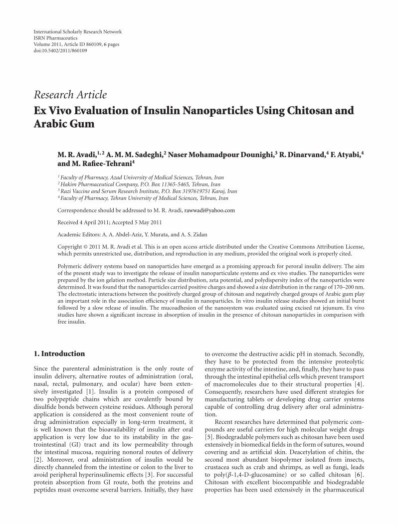
Figure 1: Apparatus used in the ex vivo studies in intestine: (a) disposable syringe for collection of serosal fluid, (b) hypodermic needle,(c) rubber stopper, (d) conical flask, (e) polyethylene centrifuge tube, (e) water bath (37◦C), (g) tape used to fasten intestine to tube, (h)intestine, (i) mixture of gas inlet (O2 95% and CO2 5%), and (j) disposable plastic syringe used to collect mucosal fluid.
2.6. Ex Vivo Study. The procedure used is a modification ofBarr and Riegelman method with some adjustments [19]. Atfirst a section of intestine (about 7 cm) was removed froma male sheep under Phenobarbital anesthesia and washedwith Krebs-Ringer bicarbonate solution, pH 7.4. The lumenwas inverted with a glass rod and a tube was inserted inone side of the intestine and tied securely with tape. Theother side of the intestine was tied and 1mL of Krebs-Ringerbicarbonate solution was poured through the hypodermicneedle in the tube. The lumen of intestine was placed ina medium standard with 95% O2, 5% CO2 in phosphatebuffer solution pH 6.5 at 37◦C. In absorption studies, O2
and CO2 mixture was bubbled into the intestinal to obtainintestinal peristaltic movement (Figure 1). In certain periodsof time samples with known volume were collected from themedium and were assayed by HPLC method, and the amountof insulin was calculated.
3. Results and Discussion
The insulin nanoparticles were prepared by ion gelationmethod using the electrostatic interactions between pos-itively charged chitosan and negatively charged Arabicgum. In our previous study, various formulations (F1–F8)were prepared using 23 factorial design experiments [18].According to the results of factorial design experiments,three formulations (Table 2) were selected. The associationefficiency (AE) of the nanoparticles was investigated in thisstudy.
Moreover, the electrostatic interactions between thepositively charged group of chitosan and negatively charged
Table 4: Kinetic constants (k), diffusional (n) and determinationcoefficient (r2) determined by the linear regression of Ln(Mt/M∞)against Ln t.
Formulation n (X ± SD, n = 3) k (X ± SD, n = 3) r2
F2 0.823± 0.0341 0.0008± 0.0001 0.956
F3 0.494± 0.0268 0.0019± 0.0003 0.958
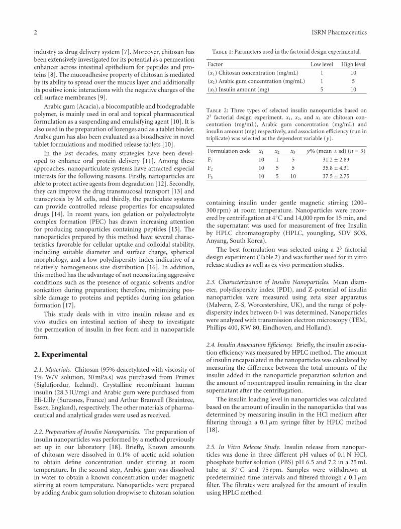
groups of Arabic gum plays an important role in theassociation efficiency of insulin in nanoparticles. Thesestudies have shown that increasing the concentrations ofchitosan and Arabic gum or the amount of insulin usedin nanoparticle preparation are important factors in AE(Table 2) [18]. Figure 2 shows the TEM photograph of thenanoparticles. The nanoparticles showed spherical or ovalshapes with relatively smooth surface. According to thesefigures and the results obtained from the zeta potentialstudies, the positive charges on the surface of nanoparticlescould prevent the aggregation process.
The particle size of nanoparticles is presented in Table 3for the three selected formulations. The mean diameter ofparticles is 191, 172, and 177 nanometers for formulationsF1, F2, and F3, respectively, and exhibited relatively narrowparticle size distribution, as indicated by a relatively lowpolydispersity index values (Table 3). Zeta potential isused to determine the surface charge of nanoparticles [20].Chitosan nanoparticles are all positively charged as shownin Table 3. This could be related to the type of particleformation mechanism between positively charged aminegroups of chitosan that are neutralized by their interactionwith negative charge of Arabic gum polymer. The residual
4 ISRN Pharmaceutics
(a) (b)
Figure 2: TEM micrographs of insulin nanoparticles for formulation (a) F2 and (b) F3.
0
20
40
60
80
100
0 60 120 180 240 300
Rel
ease
(%)
Time (min)
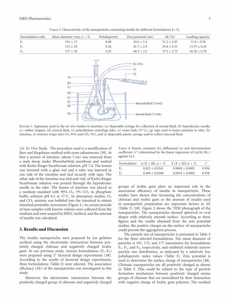
Figure 3: In vitro release profile of insulin from chitosan nanopar-ticles in HCl, pH 1.2.
amine groups would be responsible for the positive zetapotential. This effect may be related to the absorption ofanionic groups by the long amino groups of chitosan to keepthe high value of the electrical double layer thickness whichin turn prevents the aggregation [21].
The insulin release of nanoparticles was studied as afunction of time for formulation F3. Figures 3 and 4 haveshown the release profile of insulin in three different pHmedium HCl 0.1 N, PBS pH 6.5, and pH 7.2 [18]. Aburst effect of insulin release in acidic medium is relatedto high solubility of both chitosan and insulin (Figure 3).Furthermore, it is fair to say that a lot of insulin moleculesare loosely attached on the surface of nanoparticles, andtherefore insulin tends to easily move out and diffuse to theexternal medium.
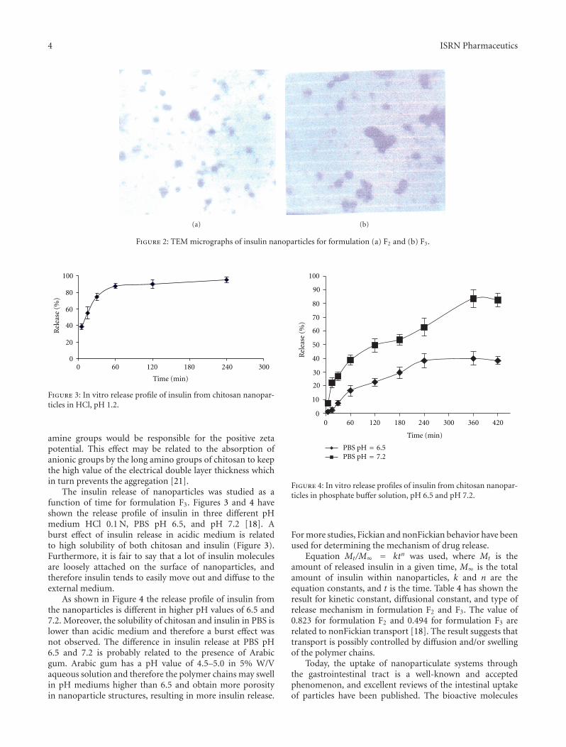
As shown in Figure 4 the release profile of insulin fromthe nanoparticles is different in higher pH values of 6.5 and7.2. Moreover, the solubility of chitosan and insulin in PBS islower than acidic medium and therefore a burst effect wasnot observed. The difference in insulin release at PBS pH6.5 and 7.2 is probably related to the presence of Arabicgum. Arabic gum has a pH value of 4.5–5.0 in 5% W/Vaqueous solution and therefore the polymer chains may swellin pH mediums higher than 6.5 and obtain more porosityin nanoparticle structures, resulting in more insulin release.
0
10
20
30
40
50
60
70
80
90
100
0 60 120 180 240 300 360 420
Time (min)
PBS pH =
7.2PBS pH =
6.5
Rel
ease
(%)
Figure 4: In vitro release profiles of insulin from chitosan nanopar-ticles in phosphate buffer solution, pH 6.5 and pH 7.2.
For more studies, Fickian and nonFickian behavior have beenused for determining the mechanism of drug release.
Equation Mt/M∞ = ktn was used, where Mt is theamount of released insulin in a given time, M∞ is the totalamount of insulin within nanoparticles, k and n are theequation constants, and t is the time. Table 4 has shown theresult for kinetic constant, diffusional constant, and type ofrelease mechanism in formulation F2 and F3. The value of0.823 for formulation F2 and 0.494 for formulation F3 arerelated to nonFickian transport [18]. The result suggests thattransport is possibly controlled by diffusion and/or swellingof the polymer chains.
Today, the uptake of nanoparticulate systems throughthe gastrointestinal tract is a well-known and acceptedphenomenon, and excellent reviews of the intestinal uptakeof particles have been published. The bioactive molecules
ISRN Pharmaceutics 5
Preabsorption
phenomenaAbsorption Translocation Destination
Aggregation
Adsorption
Adhesion
Uptake by Mcells
Transcellularprocesses
Uptake by tightjunctions
Passage
through
lymphatics
Transfer to
blood
Uptake by
liver, spleen,
Interactions in
lymph and blood
Extravastion
and so on
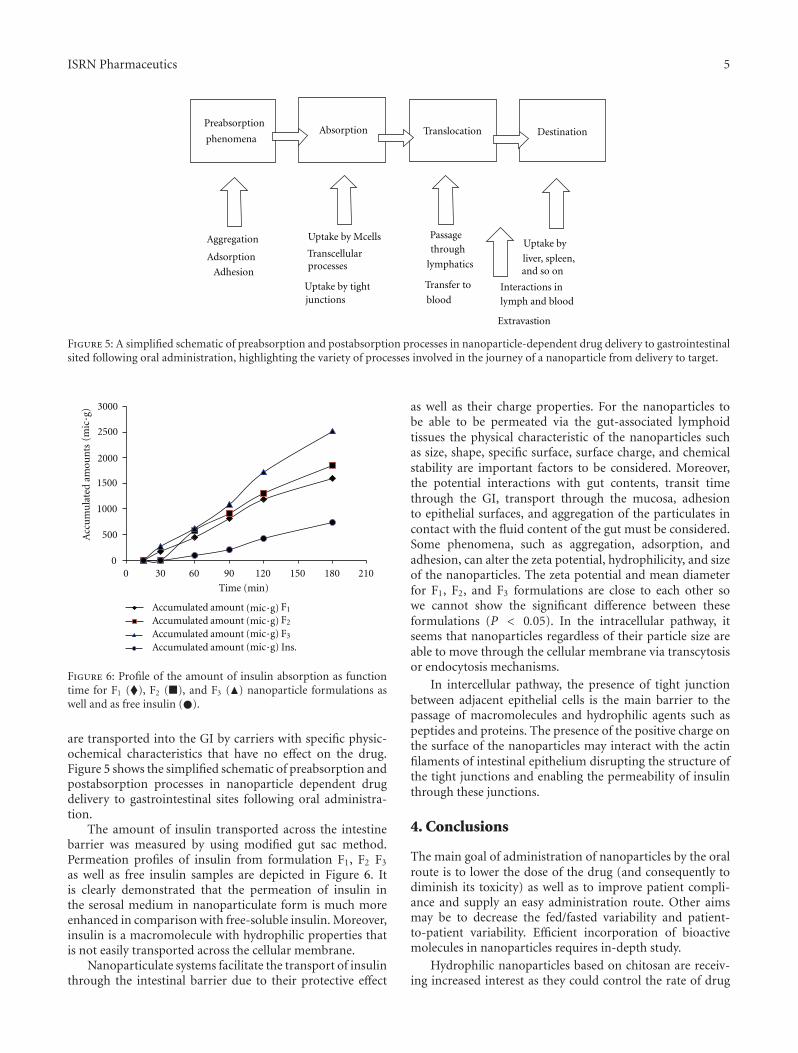
Figure 5: A simplified schematic of preabsorption and postabsorption processes in nanoparticle-dependent drug delivery to gastrointestinalsited following oral administration, highlighting the variety of processes involved in the journey of a nanoparticle from delivery to target.
0
500
1000
1500
2000
2500
3000
030 60 90 120 150 180 210
Acc
um
ula
ted
Accumulated
amou
nts
Time (min)
amountAccumulated amountAccumulated amountAccumulated amount Ins.
F1
F2
F3
(mic·g
)
(mic·g)(mic·g)(mic·g)(mic·g)
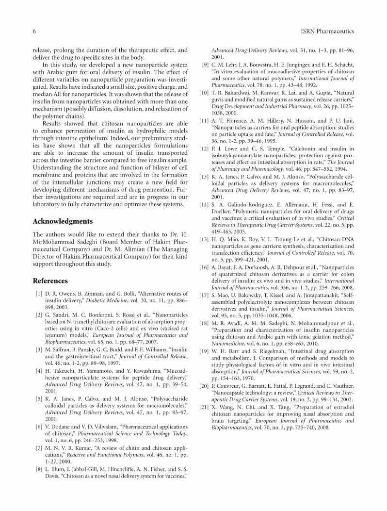
Figure 6: Profile of the amount of insulin absorption as functiontime for F1 (�), F2 (�), and F3 (�) nanoparticle formulations aswell and as free insulin (�).
are transported into the GI by carriers with specific physic-ochemical characteristics that have no effect on the drug.Figure 5 shows the simplified schematic of preabsorption andpostabsorption processes in nanoparticle dependent drugdelivery to gastrointestinal sites following oral administra-tion.
The amount of insulin transported across the intestinebarrier was measured by using modified gut sac method.Permeation profiles of insulin from formulation F1, F2 F3
as well as free insulin samples are depicted in Figure 6. Itis clearly demonstrated that the permeation of insulin inthe serosal medium in nanoparticulate form is much moreenhanced in comparison with free-soluble insulin. Moreover,insulin is a macromolecule with hydrophilic properties thatis not easily transported across the cellular membrane.
Nanoparticulate systems facilitate the transport of insulinthrough the intestinal barrier due to their protective effect
as well as their charge properties. For the nanoparticles tobe able to be permeated via the gut-associated lymphoidtissues the physical characteristic of the nanoparticles suchas size, shape, specific surface, surface charge, and chemicalstability are important factors to be considered. Moreover,the potential interactions with gut contents, transit timethrough the GI, transport through the mucosa, adhesionto epithelial surfaces, and aggregation of the particulates incontact with the fluid content of the gut must be considered.Some phenomena, such as aggregation, adsorption, andadhesion, can alter the zeta potential, hydrophilicity, and sizeof the nanoparticles. The zeta potential and mean diameterfor F1, F2, and F3 formulations are close to each other sowe cannot show the significant difference between theseformulations (P < 0.05). In the intracellular pathway, itseems that nanoparticles regardless of their particle size areable to move through the cellular membrane via transcytosisor endocytosis mechanisms.
In intercellular pathway, the presence of tight junctionbetween adjacent epithelial cells is the main barrier to thepassage of macromolecules and hydrophilic agents such aspeptides and proteins. The presence of the positive charge onthe surface of the nanoparticles may interact with the actinfilaments of intestinal epithelium disrupting the structure ofthe tight junctions and enabling the permeability of insulinthrough these junctions.
4. Conclusions
The main goal of administration of nanoparticles by the oralroute is to lower the dose of the drug (and consequently todiminish its toxicity) as well as to improve patient compli-ance and supply an easy administration route. Other aimsmay be to decrease the fed/fasted variability and patient-to-patient variability. Efficient incorporation of bioactivemolecules in nanoparticles requires in-depth study.
Hydrophilic nanoparticles based on chitosan are receiv-ing increased interest as they could control the rate of drug
6 ISRN Pharmaceutics
release, prolong the duration of the therapeutic effect, anddeliver the drug to specific sites in the body.
In this study, we developed a new nanoparticle systemwith Arabic gum for oral delivery of insulin. The effect ofdifferent variables on nanoparticle preparation was investi-gated. Results have indicated a small size, positive charge, andmedian AE for nanoparticles. It was shown that the release ofinsulin from nanoparticles was obtained with more than onemechanism (possibly diffusion, dissolution, and relaxation ofthe polymer chains).
Results showed that chitosan nanoparticles are ableto enhance permeation of insulin as hydrophilic modelsthrough intestine epithelium. Indeed, our preliminary stud-ies have shown that all the nanoparticles formulationsare able to increase the amount of insulin transportedacross the intestine barrier compared to free insulin sample.Understanding the structure and function of bilayer of cellmembrane and proteins that are involved in the formationof the intercellular junctions may create a new field fordeveloping different mechanisms of drug permeation. Fur-ther investigations are required and are in progress in ourlaboratory to fully characterize and optimize these systems.
Acknowledgments
The authors would like to extend their thanks to Dr. H.MirMohammad Sadeghi (Board Member of Hakim Phar-maceutical Company) and Dr. M. Alimian (The ManagingDirector of Hakim Pharmaceutical Company) for their kindsupport throughout this study.
References
[1] D. R. Owens, B. Zinman, and G. Bolli, “Alternative routes ofinsulin delivery,” Diabetic Medicine, vol. 20, no. 11, pp. 886–898, 2003.
[2] G. Sandri, M. C. Bonferoni, S. Rossi et al., “Nanoparticlesbased on N-trimethylchitosan: evaluation of absorption prop-erties using in vitro (Caco-2 cells) and ex vivo (excised ratjejunum) models,” European Journal of Pharmaceutics andBiopharmaceutics, vol. 65, no. 1, pp. 68–77, 2007.
[3] M. Saffran, B. Pansky, G. C. Budd, and F. E. Williams, “Insulinand the gastrointestinal tract,” Journal of Controlled Release,vol. 46, no. 1-2, pp. 89–98, 1997.
[4] H. Takeuchi, H. Yamamoto, and Y. Kawashima, “Mucoad-hesive nanoparticulate systems for peptide drug delivery,”Advanced Drug Delivery Reviews, vol. 47, no. 1, pp. 39–54,2001.
[5] K. A. Janes, P. Calvo, and M. J. Alonso, “Polysaccharidecolloidal particles as delivery systems for macromolecules,”Advanced Drug Delivery Reviews, vol. 47, no. 1, pp. 83–97,2001.
[6] V. Dodane and V. D. Vilivalam, “Pharmaceutical applicationsof chitosan,” Pharmaceutical Science and Technology Today,vol. 1, no. 6, pp. 246–253, 1998.
[7] M. N. V. R. Kumar, “A review of chitin and chitosan appli-cations,” Reactive and Functional Polymers, vol. 46, no. 1, pp.1–27, 2000.
[8] L. Illum, I. Jabbal-Gill, M. Hinchcliffe, A. N. Fisher, and S. S.Davis, “Chitosan as a novel nasal delivery system for vaccines,”
Advanced Drug Delivery Reviews, vol. 51, no. 1–3, pp. 81–96,2001.
[9] C. M. Lehr, J. A. Bouwstra, H. E. Junginger, and E. H. Schacht,“In vitro evaluation of mucoadhesive properties of chitosanand some other natural polymers,” International Journal ofPharmaceutics, vol. 78, no. 1, pp. 43–48, 1992.
[10] T. R. Bahardwaj, M. Kanwar, R. Lai, and A. Gupta, “Naturalguvis and modified natural gums as sustained release carriers,”Drug Development and Industrial Pharmacy, vol. 26, pp. 1025–1038, 2000.
[11] A. T. Florence, A. M. Hillery, N. Hussain, and P. U. Jani,“Nanoparticles as carriers for oral peptide absorption: studieson particle uptake and fate,” Journal of Controlled Release, vol.36, no. 1-2, pp. 39–46, 1995.
[12] P. J. Lowe and C. S. Temple, “Calcitonin and insulin inisobutylcyanoacrylate nanoparticles: protection against pro-teases and effect on intestinal absorption in rats,” The Journalof Pharmacy and Pharmacology, vol. 46, pp. 547–552, 1994.
[13] K. A. Janes, P. Calvo, and M. J. Alonso, “Polysaccharide col-loidal particles as delivery systems for macromolecules,”Advanced Drug Delivery Reviews, vol. 47, no. 1, pp. 83–97,2001.
[14] S. A. Galindo-Rodrıguez, E. Allemann, H. Fessi, and E.Doelker, “Polymeric nanoparticles for oral delivery of drugsand vaccines: a critical evaluation of in vivo studies,” CriticalReviews in Therapeutic Drug Carrier Systems, vol. 22, no. 5, pp.419–463, 2005.
[15] H. Q. Mao, K. Roy, V. L. Troung-Le et al., “Chitosan-DNAnanoparticles as gene carriers: synthesis, characterization andtransfection efficiency,” Journal of Controlled Release, vol. 70,no. 3, pp. 399–421, 2001.
[16] A. Bayat, F. A. Dorkoosh, A. R. Dehpour et al., “Nanoparticlesof quaternized chitosan derivatives as a carrier for colondelivery of insulin: ex vivo and in vivo studies,” InternationalJournal of Pharmaceutics, vol. 356, no. 1-2, pp. 259–266, 2008.
[17] S. Mao, U. Bakowsky, T. Kissel, and A. Jintapattanakit, “Self-assembled polyelectrolyte nanocomplexes between chitosanderivatives and insulin,” Journal of Pharmaceutical Sciences,vol. 95, no. 5, pp. 1035–1048, 2006.
[18] M. R. Avadi, A. M. M. Sadeghi, N. Mohammadpour et al.,“Preparation and characterization of insulin nanoparticlesusing chitosan and Arabic gum with ionic gelation method,”Nanomedicine, vol. 6, no. 1, pp. e58–e63, 2010.
[19] W. H. Barr and S. Riegelman, “Intestinal drug absorptionand metabolism. I. Comparison of methods and models tostudy physiological factors of in vitro and in vivo intestinalabsorption,” Journal of Pharmaceutical Sciences, vol. 59, no. 2,pp. 154–163, 1970.
[20] P. Couvreur, G. Barratt, E. Fattal, P. Legrand, and C. Vauthier,“Nanocapsule technology: a review,” Critical Reviews in Ther-apeutic Drug Carrier Systems, vol. 19, no. 2, pp. 99–134, 2002.
[21] X. Wang, N. Chi, and X. Tang, “Preparation of estradiolchitosan nanoparticles for improving nasal absorption andbrain targeting,” European Journal of Pharmaceutics andBiopharmaceutics, vol. 70, no. 3, pp. 735–740, 2008.
Submit your manuscripts athttp://www.hindawi.com
PainResearch and TreatmentHindawi Publishing Corporationhttp://www.hindawi.com Volume 2014
The Scientific World JournalHindawi Publishing Corporation http://www.hindawi.com Volume 2014
Hindawi Publishing Corporationhttp://www.hindawi.com
Volume 2014
ToxinsJournal of
VaccinesJournal of
Hindawi Publishing Corporation http://www.hindawi.com Volume 2014
Hindawi Publishing Corporationhttp://www.hindawi.com Volume 2014
AntibioticsInternational Journal of
ToxicologyJournal of
Hindawi Publishing Corporationhttp://www.hindawi.com Volume 2014
StrokeResearch and TreatmentHindawi Publishing Corporationhttp://www.hindawi.com Volume 2014
Drug DeliveryJournal of
Hindawi Publishing Corporationhttp://www.hindawi.com Volume 2014
Hindawi Publishing Corporationhttp://www.hindawi.com Volume 2014
Advances in Pharmacological Sciences
Tropical MedicineJournal of
Hindawi Publishing Corporationhttp://www.hindawi.com Volume 2014
Medicinal ChemistryInternational Journal of
Hindawi Publishing Corporationhttp://www.hindawi.com Volume 2014
AddictionJournal of
Hindawi Publishing Corporationhttp://www.hindawi.com Volume 2014
Hindawi Publishing Corporationhttp://www.hindawi.com Volume 2014
BioMed Research International
Emergency Medicine InternationalHindawi Publishing Corporationhttp://www.hindawi.com Volume 2014
Hindawi Publishing Corporationhttp://www.hindawi.com Volume 2014
Autoimmune Diseases
Hindawi Publishing Corporationhttp://www.hindawi.com Volume 2014
Anesthesiology Research and Practice
ScientificaHindawi Publishing Corporationhttp://www.hindawi.com Volume 2014
Journal of
Hindawi Publishing Corporationhttp://www.hindawi.com Volume 2014
Pharmaceutics
Hindawi Publishing Corporationhttp://www.hindawi.com Volume 2014
MEDIATORSINFLAMMATION
of