research article dermatoglyphics: a noninvasive diagnostic
TRANSCRIPT
RESEARCH ARTICLE
Dermatoglyphics: A Noninvasive Diagnostic Tool in Predicting Class III Skeletal Malocclusion in ChildrenAshwitha C Belludi1, Arvind Sridhara2, Narayana Chandra Kumar3, Sapna Konde4, Sunil Raj Noojadi5
Ab s t r Ac t Background: Dermatoglyphics is a scientific study that deals with the epidermal ridges and their configurations on certain body parts such as fingers, palms, and soles. In humans, during the intrauterine life (IUL) the primary palate, lip, and dermal ridges are formed during the same period, the genetic code engineered in the genome normal or abnormal is mirrored on these developing structures. Thus making dermatoglyphic a preceding tool in dental diagnosis.Aims and objectives: The study aimed at evaluating dermatoglyphics as a tool in diagnosing malocclusion by comparing qualitative and quantitative dermal patterns in class I and class III skeletal malocclusion.Materials and methods: Sixty subjects fulfilling inclusion-criteria were segregated into two groups, group I (class I skeletal malocclusion) and group II ( class III skeletal malocclusion) with 30 subjects in each group. Dermatoglyphic patterns were recorded using ink method following rolling impression technique on recording sheets. The dermatoglyphic data were assessed for different finger ridge patterns, total finger ridge count, a–b ridge count, and atd angle.Results: The data were analyzed using Chi-square and paired t tests. In skeletal class III malocclusion, there was an increase in loop count and a decrease in the count of whorls and arches as compared to class I malocclusion (p = 0.037). However, in relation to total finger ridge count, a–b ridge count, and atd angle, there was no statistically significant difference found between the groups.Conclusion: The end of the study derived that the fingerprint patterns are valuable and ineradicable markers of malocclusion. Thus, the dermatoglyphics can be utilized as a screening tool for early prediction of skeletal class III malocclusion at a younger age-group. Further studies are suggested with the inclusion of other parameters using the inkless biometric method in different populations.Keywords: atd angle, Dermatoglyphic patterns, Dermatoglyphics, Fingerprints, Skeletal class III malocclusion, Total ridge count.International Journal of Clinical Pediatric Dentistry (2021): 10.5005/jp-journals-10005-1934
In t r o d u c t I o n Fingerprints is an incredibly complex and intricate system, the study of which has been a great source of people’s fascination. Along the palm side and soles of every individual are characteristic attributes that are viewed as an un-repeatable distinctive feature to every individual.
Cummins and Midlo in the year 1926 coined the term “Dermatoglyphics”–referring to the study of complex dermal ridge configuration on the skin enveloping the palmar and plantar facets of hand and feet.1 The term dermatoglyphic is derived from Greek literature “Derma” meaning Skin; and “Glyphe” referring to Carve, implying that something has been carved on the skin facets. Through decades of scientific research, dermatoglyphics has turned out to be an excellent alternative to other diagnostic methods in identifying specific genetic origin syndromes.2
Dermatoglyphics has been studied in various malformations caused by autosomal aberrations such as Patau’s syndrome, Down’s syndrome, and Edward syndrome, sex chromosomal aberrations including Turner’s syndrome and Klinefelter’s syndrome, inherited or genetic malformations (hypohidrotic ectodermal dysplasia, schizophrenia, psychosis, autism, carcinoma of breast, leukemia, congenital heart disease, and psoriasis), systemic disorders (diabetes mellitus, hypertension, and rheumatoid arthritis), and others.3–10 In dentistry, dermatoglyphics has been studied in cleft lip and palate, hereditary gingival fibromatosis, periodontal diseases, dental caries, dental and skeletal malocclusions, and potentially malignant and malignant disorders (oral submucous fibrosis, leukoplakia, oral cancer) of the oral cavity.11–16
By the 7th week of intrauterine life (IUL), the development of the primary palate and the lip is complete, toward the 12th week the secondary palate is completely developed. However, the dermal ridges begin to develop at the 6th week of IUL, reach the maximum between the 12th and 13th weeks, implying that genetic code engineered in the genome containing either normal or abnormal code is mirrored on these developing structures.12
1,2Department of Pedodontics and Preventive Dentistry, Subbaiah Dental College, Purle, Shimoga, Karnataka, India3Department of Pedodontics and Preventive Dentistry, Vydehi Institute of Dental Science, Whitefield, Bengaluru, Karnataka, India4Department of Pedodontics and Preventive Dentistry, AECS Maaruti College of Dental Sciences and Research Centre, Bengaluru, Karnataka, India5Department of Pedodontics and Preventive Dentistry, Bangalore Institute of Dental Sciences, Bengaluru, Karnataka, IndiaCorresponding Author: Ashwitha C Belludi, Department of Pedodontics and Preventive Dentistry, Subbaiah Dental College, Purle, Shimoga, Karnataka, India, Phone: +98892093846, e-mail: [email protected] to cite this article: Belludi AC, Sridhara A, Kumar NC, et al. Dermatoglyphics: A Noninvasive Diagnostic Tool in Predicting Class III Skeletal Malocclusion in Children. Int J Clin Pediatr Dent 2021;14(1): 63–69.Source of support: NilConflict of interest: None
© Jaypee Brothers Medical Publishers. 2021 Open Access This article is distributed under the terms of the Creative Commons Attribution 4.0 International License (https://creativecommons.org/licenses/by-nc/4.0/), which permits unrestricted use, distribution, and non-commercial reproduction in any medium, provided you give appropriate credit to the original author(s) and the source, provide a link to the Creative Commons license, and indicate if changes were made. The Creative Commons Public Domain Dedication waiver (http://creativecommons.org/publicdomain/zero/1.0/) applies to the data made available in this article, unless otherwise stated.

Evaluation of Dermatoglyphics in Children with Class III Skeletal Malocclusion
International Journal of Clinical Pediatric Dentistry, Volume 14 Issue 1 (January–February 2021)64
Subsequently, the extraneous factors, at the time of development causing deflection from normal occlusion will correspondingly reflect in dermal patterns.
Among skeletal malocclusions, on treatment perspective, skeletal Class III malocclusion is the most difficult to treat, having an incidence of 1–5% amid the Caucasian population.17–19 The pathognomic features in the majority of subjects with class III malocclusion comprises maxillary retrognathism or hypoplasia, in association with a normal or minimally protruded/prognathic mandible.20 Hallmark studies in class III malocclusion by Guyer et al. delineated a simple retrusion of maxilla in 25%, protrusion of mandible in 18.7% and a combination of both in 22.2% of their study sample.21
Preliminary detection and rectification of growth pattern deviation have been among the principal objectives of orthodontics for a long time. Given that late identification of skeletal malocclusion drive patients to orthognathic surgery, this study was taken forward to investigate the different dermatoglyphic patterns and to compare them with skeletal malocclusions which in turn can be applied in preventive and early interceptive orthodontics in high-risk groups and also for parent education and counseling. We additionally sought to ascertain the practicality of dermatoglyphics in the prediction of skeletal discrepancies.
MAt e r I A l s A n d Me t h o d s The study was carried out on 60 apparently healthy children aged between 9 years and 15 years including diagnosed cases of skeletal malocclusion. The study included both genders in equal numbers, segregated to form two groups comprising of 30 children each based on their craniofacial skeleton and dental status. The study design, objectives, and methodology were explained to the selected children and their parents. Formal consent was acquired antecedently from the parents.
The study sample contained:Group I: Class I Craniofacial Types, comprised of 30 children with
class I molar relationship having an acceptable overjet and overbite.Group II: Class III Craniofacial Types, comprised of 30 children
with clinically apparent mandibular prognathism.The detailed case history was recorded and the complete
intraoral examination was carried out under artificial illumination. The subjects were then subjected to lateral cephalogram using the KODAK-8000C Digital Panoramic and Cephalometric system following radiation protection protocol. The exposure parameters used were 78 kVp, 12 mA, 1 second. Skeletal malocclusion was assessed by taking lateral cephalogram following radiation protection protocol. The captured images were traced for measurements of cephalometric landmarks and were evaluated using Steiner’s analysis (both skeletal and dental analysis).
Pr o c e d u r e f o r obtA I n I n g de r M Ato g lyP h I c Pr I n ts The ink technique depicted by Cummins and Midlo1 was used to record the finger and palm prints. The hands of kids incorporated in the study were washed with a cleanser and water to eliminate earth and oil from the furrowed skin and blot dried to improve the nature of prints. Two A4-sized white bond sheets which were manually designed for finger and palm printing per individual (one for the left hand and the other for the right) were used (Fig. 1).
FingerprintsThe right and the left fingerprints of all the study subjects were recorded using duplicating ink by applying it on their fingers. To obtain the entire fingerprint pattern, the fingers were rolled on the ink pad to ensure the ink was evenly covered. The impression was made by rolling the side of the finger bulb placing the finger upon the white A4 size bond sheet clipped on to a board. The finger was then rolled to the antagonist side until it faced the opposite side. To facilitate convenient natural movement of the forearm, the hand had to be turned from a more strenous position to an effortless one. This required thumb to be moved toward and fingers away from the center of the person’s body. To prevent smudging, each finger was rolled from nail to another in suitable space on bond sheet, taking consideration to lift each finger up after rolling.
Palm PrintsFor the sense of evenness palm prints of all the subjects were recorded using black duplicating ink which was thoroughly applied using an ink roller until the entire palmar area was covered with a thin and even layer of ink. The technique of recording palmar prints included gently pressing the completely inked palmar surface on a customized white bond paper placed over a tubular object and later the heel or base of the subject’s palm was placed on the tubular object and rolled in a pulling motion from the heel of the hand to the fingertips. The same technique was followed on the other hand.
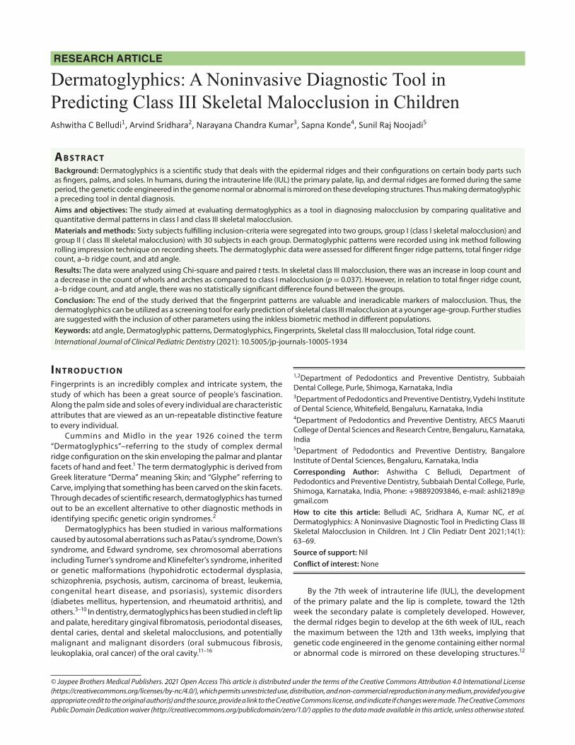
A 2× magnification glass was used to check the clarity of obtained handprints and were coded. The existence of core (the innermost turning point where the fingerprint ridges form a loop), and triradii/delta (the point where these ridges form a triangulating shape) of the dermatoglyphic pattern were checked all together to incorporate the handprint in the study (Fig. 2). In total, 120 palmar prints were obtained.
Method of Reading HandprintsUnder a magnifying glass 2× power, the handprints were seen consecutively from the left-hand little finger until the thumb followed by the thumb finger of the right hand until the little finger.
Fig. 1: The palm and fingerprint patterns of the participants recorded with a rolling impression technique using duplicating ink on customized executive bond paper
Evaluation of Dermatoglyphics in Children with Class III Skeletal Malocclusion
International Journal of Clinical Pediatric Dentistry, Volume 14 Issue 1 (January–February 2021) 65
The dermatoglyphic analysis comprises the following; qualitative analysis which includes patterns of fingertips, i.e., loops, whorls, and arches. Quantitative analysis that encompasses finger ridge count, total finger ridge count, ab ridge count, and atd angle.
QuA l I tAt I v e de r M Ato g lyP h I c An A lys I s The true patterns of loops, whorls, arches, and their frequencies were identified and counted on the fingertips of all the 10 digits in subject of both groups, which was later assessed for increase or decrease in mean frequencies.
Type of dermatoglyphic pattern:
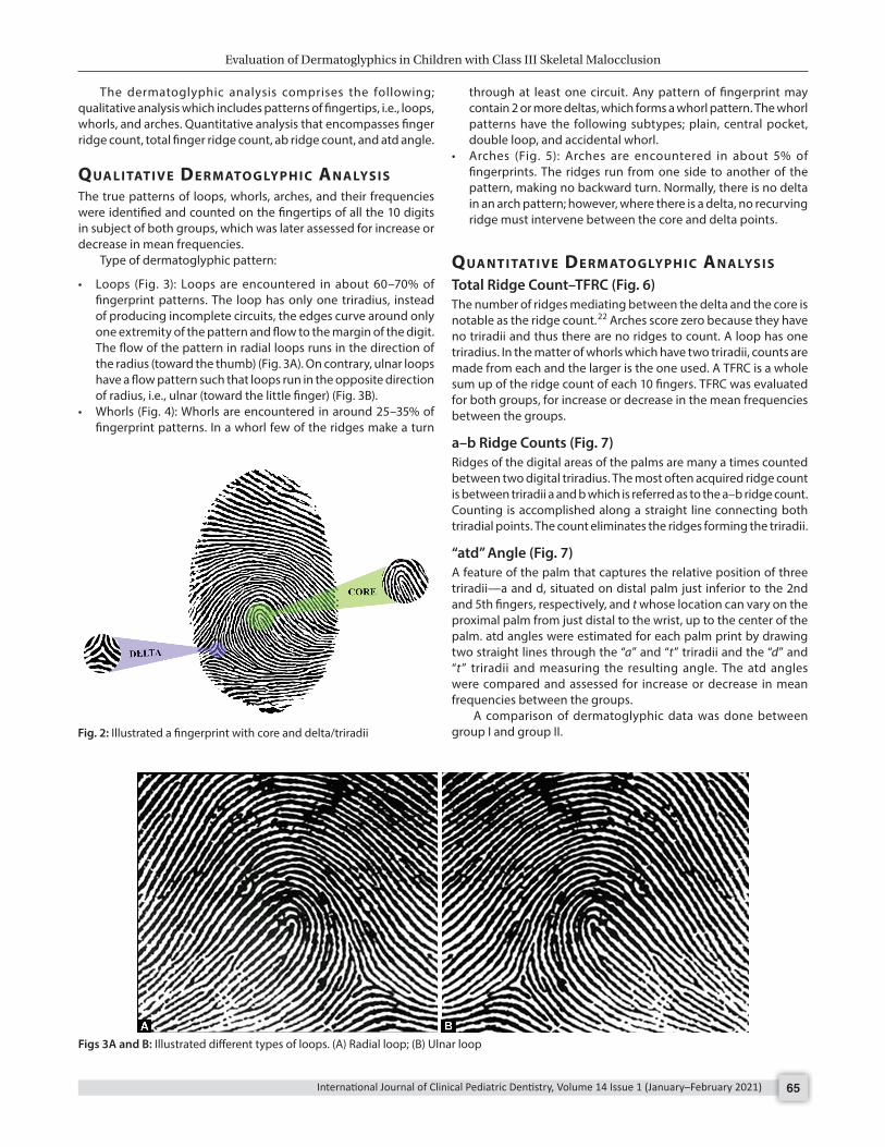
• Loops (Fig. 3): Loops are encountered in about 60–70% of fingerprint patterns. The loop has only one triradius, instead of producing incomplete circuits, the edges curve around only one extremity of the pattern and flow to the margin of the digit. The flow of the pattern in radial loops runs in the direction of the radius (toward the thumb) (Fig. 3A). On contrary, ulnar loops have a flow pattern such that loops run in the opposite direction of radius, i.e., ulnar (toward the little finger) (Fig. 3B).
• Whorls (Fig. 4): Whorls are encountered in around 25–35% of fingerprint patterns. In a whorl few of the ridges make a turn
through at least one circuit. Any pattern of fingerprint may contain 2 or more deltas, which forms a whorl pattern. The whorl patterns have the following subtypes; plain, central pocket, double loop, and accidental whorl.
• Arches (Fig. 5): Arches are encountered in about 5% of fingerprints. The ridges run from one side to another of the pattern, making no backward turn. Normally, there is no delta in an arch pattern; however, where there is a delta, no recurving ridge must intervene between the core and delta points.
QuA n t I tAt I v e de r M Ato g lyP h I c An A lys I s Total Ridge Count–TFRC (Fig. 6)The number of ridges mediating between the delta and the core is notable as the ridge count.22 Arches score zero because they have no triradii and thus there are no ridges to count. A loop has one triradius. In the matter of whorls which have two triradii, counts are made from each and the larger is the one used. A TFRC is a whole sum up of the ridge count of each 10 fingers. TFRC was evaluated for both groups, for increase or decrease in the mean frequencies between the groups.
a–b Ridge Counts (Fig. 7)Ridges of the digital areas of the palms are many a times counted between two digital triradius. The most often acquired ridge count is between triradii a and b which is referred as to the a–b ridge count. Counting is accomplished along a straight line connecting both triradial points. The count eliminates the ridges forming the triradii.
“atd” Angle (Fig. 7)A feature of the palm that captures the relative position of three triradii—a and d, situated on distal palm just inferior to the 2nd and 5th fingers, respectively, and t whose location can vary on the proximal palm from just distal to the wrist, up to the center of the palm. atd angles were estimated for each palm print by drawing two straight lines through the “a” and “t” triradii and the “d” and “t” triradii and measuring the resulting angle. The atd angles were compared and assessed for increase or decrease in mean frequencies between the groups.
A comparison of dermatoglyphic data was done between group I and group II.Fig. 2: Illustrated a fingerprint with core and delta/triradii
Figs 3A and B: Illustrated different types of loops. (A) Radial loop; (B) Ulnar loop

Evaluation of Dermatoglyphics in Children with Class III Skeletal Malocclusion
International Journal of Clinical Pediatric Dentistry, Volume 14 Issue 1 (January–February 2021)66
dAtA An A lys I s Relevant statistical analyses were applied, i.e., Chi-square test and paired t test with a significance level set at α = 0.05. The decision criterion was to compare the p value with the level of significance. The null hypothesis was rejected and the alternate hypothesis was accepted if p < 0.05 and the null hypothesis would be accepted if p ≥ 0.05.
re s u lts An increase in the number of loops was detected in group II (class III), while increase in whorls and arches was identified in group I (class I),which was statistically significant (p = 0.037) (Table 1).
The mean TFRC in group I (class I) was 150.57 ± 44.78 and in group II (class III) was 143.87 ± 36.28. Though there was an increase in the mean TFRC in class I, the difference between TFRC
Figs 5A and B: Illustrated different types of arch. (A) Plain arch; (B) Tented arch
Figs 4A to D: Illustrated different types of whorls. (A) Plain; (B) Central pocket; (C) Double loop; (D) Accidental

Evaluation of Dermatoglyphics in Children with Class III Skeletal Malocclusion
International Journal of Clinical Pediatric Dentistry, Volume 14 Issue 1 (January–February 2021) 67
in class I and class III was not statistically significant (p = 0. 527) (Table 2).
The difference between a–b ridge count in right (p = 0.613) and left (p = 0.982) hand of group I and group II was not statistically significant (Table 3).
Similarly, the difference between the atd angle in the right (p = 0.647) and left (p = 0.936) hands of group I and group II were not statistically significant (Table 4).
dI s c u s s I o n Preliminary detection and rectification of growth pattern deviation have been among the principal objectives of orthodontics for a
long time. Given that late analysis of skeletal malocclusions drives patients to orthognathic surgery, this study was taken forward to investigate the different dermatoglyphic patterns and to compare them with skeletal malocclusions. In the present study, the age group of 9–15 years was chosen, as this is the mixed dentition period when permanent maxillary incisors are present in the oral cavity, for recording the overjet and overbite.
Finger Ridge PatternsIn the present study, the percentage frequency of digital pattern whorls and arches was found to be increased in class I when compared to class III, which was found to be statistically significant
Table 1: Evaluation and comparison of the percentage frequency of digital patterns among children with skeletal class I and skeletal class III malocclusion on both right and left hand combined
Class I Class III Total Chi-square testLoops 158 (46.06) 185 (53.94) 343 (100) p = 0.037 (significant)Whorls 127 (54.04) 108 (45.96) 235 (100)Arches 15 (68.18) 7 (31.82) 22 (100)Total 300 (50) 300 (50) 600 (100)
Table 2: Comparison of mean total finger ridge count in class I and class III malocclusion
Total finger ridge count
Class N Mean SD Mean difference T T testClass I 30 150.57 44.78 6.700 0.637 p = 0.527Class III 30 143.87 36.28
Table 3: Evaluation and comparison of the mean “a–b” ridge count between children with skeletal class I and skeletal class III malocclusion on both right and left hand combined
Class N Mean SD Mean difference T T testRight ab ridge count Class I 30 36.63 5.70 −0.733 −0.509 p = 0.613 Class III 30 37.37 5.46Left ab ridge count Class I 30 38.07 5.39 0.033 0.023 p = 0.982 Class III 30 38.03 5.69
Fig. 6: Sketch reflecting the various types of ridges that the classifier will encounter when engaging in counting loop patterns Fig. 7: Illustrated ab ridge and atd angle

Evaluation of Dermatoglyphics in Children with Class III Skeletal Malocclusion
International Journal of Clinical Pediatric Dentistry, Volume 14 Issue 1 (January–February 2021)68
(p = 0. 039). This is in accordance with the study conducted by Rajput et al.,23 who observed that loops were more frequent in class II and class III malocclusion subjects, whereas whorls were frequent in class I malocclusion subjects. On contrary, Trehan et al.24 found that the frequency of whorls was more in number in class I and class III and the frequency of radial loops and arches was more in class I and class II division I cases. However, the results of the present study were in accordance with Kharbanda et al.25 who concluded that skeletal class III subjects showed significant increase in arches and ulnar loops at the expense of whorls on all fingers except digit II along with increase in occurrence of whorls and radial loops which was based on the explanation given by Uchida and Solton26 who stated that among Mongoloids certain number of genes carried by autosome (trisomy 21) are responsible for the deviated dermatoglyphics. Similar dermal patterns in class III and mongolism, the class III craniofacial skeletal pattern is autosomally inherited and not sex linked, which was further supported by the autosomal inheritance of mandibular prognathism theory proposed by Bookman.25
An increased number of loops in the experimental group could be because the variable ridge configuration is determined partly by genetic factors compounded by other factors such as stress and tension in the growth of the part during fetal life. Individual genes may affect the whole hand or only one or more of the fingers.3,27 Slatis et al.28 concluded that ulnar loops is the most common fingerprint pattern. Deviations from this basic pattern may be in three directions: toward whorls, arches, or radial loops. The increased number of arches in skeletal class II and class I groups may be due to X-linked inheritance.11,25 There is wide agreement that the mechanism of inheritance of many dermatoglyphic features conforms to a polygenic system with each gene contributing a small additive effect. Chromosomal loci of genes influencing dermatoglyphics include the X chromosome, chromosome 18 and 21 which could play a role in the formation of patterns.9
Total Finger Ridge CountIn the present study, mean TFRC was found to be slightly increased in skeletal class I but the difference between TFR count in skeletal class III and class I group was not statistically significant (p = 0.527). This finding is in accordance with Kharbanda et al.25 and Reddy et al.29 Eslami et al.30 who observed different skeletal malocclusion patients and found that there was no statistical significant difference between the groups in terms of their TFRC. However in contrary to our study, Trehan et al.24 observed that TFRC was increased in dental class III malocclusion and class I control group.
The mean number of total finger ridge count decreased in skeletal class III because it is entirely determined by additive or co-dominant genes and also the total finger ridge count follows
a polygenic mode of inheritance. Similar findings were reported by Penrose and Losch31 and Holt,32 which is in accordance with our study.
a–b Ridge CountIn the present study, the mean a–b ridge count on the right hand and left hand of children with skeletal class I and class III malocclusion group did not show any significant variation (p = 0.613, p = 0.982), which is in accordance with the study conducted by Kharbanda et al.,25 Reddy et al.,29 and Rajput et al.,23 who observed that a–b ridge count on both right and left hand did not show any significant variation among the experimental groups.
“atd” AngleEvaluation and comparison of “atd” angle in both right and left hand of children with skeletal class I and class III malocclusion did not show any significant variation among the two groups (p = 0.647, p = 0.936), respectively, which was in accordance with the study conducted by Reddy et al.,29 who observed that all the experimental groups (class I, class II div. 1, div.2 and class III) exhibited higher values of atd angle as compared to ideal control group and there was no significant difference between right and left hands groups. Eslami et al.30 and Rajput et al.,23 observed that atd angle in both right and left hands present no significant difference between three study groups.
The result of our study is contrary to study by Reddy et al.,11 who observed that the mean value of atd angle decreased in all experimental groups (class II div. 1, div.2 and class III) when compared to the class I control group on both right and left hands.
co n c lu s I o n In the present dermatoglyphic study, data were assessed for finger ridge patterns, total finger ridge count (TFRC), a–b ridge count, and atd angle. Our study concluded that finger patterns, i.e., loop patterns showed an increase count in class III malocclusion whereas the whorl and arch pattern was increased in class I malocclusion, which was significant statistically. The other parameters such as total finger ridge count (TFRC), a–b ridge count, and atd angle showed no statistical significance. Further studies using inkless biometric method in different population would be a futuristic approach in diagnosis of dental diseases using dermatoglyphics.
re f e r e n c e s 1. Cummins H, Midlo C. Palmar and planter epidermal configuration
(dermatoglyphics) in European. Am J Phys-Anthropol 1926;9(4):471–502. DOI: 10.1002/ajpa.1330090422.
2. Gibbs RC. Fundamentals of dermatoglyphics. Arch Derm 1967;96(6):721–725. DOI: 10.1001/archderm.1967.01610060115023.
Table 4: Evaluation and comparison of the mean “atd” angle between children with skeletal class I and skeletal class III malocclusion on both right and left hand combined
Class N Mean SD Mean difference T T testRight atd angle Class I 30 45.83 5.70 0.667 0.461 p = 0.647 Class III 30 45.17 5.51Left atd angle Class I 30 45.67 6.12 0.117 0.08 p = 0.936 Class III 30 45.55 5.06

Evaluation of Dermatoglyphics in Children with Class III Skeletal Malocclusion
International Journal of Clinical Pediatric Dentistry, Volume 14 Issue 1 (January–February 2021) 69
17. Massler M, Frankel J. Prevalence of malocclusion in children aged 14 to 18 years. Am J Orthod 1951;37(10):751–768. DOI: 10.1016/0002-9416(51)90047-4.
18. Haynes S. The prevalence of malocclusion in English school children aged 11–12 years. Transact Eur Orthod Soc 1970. 89–98.
19. Thilander Bmyrberg N. The prevalence of malocclusion in Swedish schoolchildren. Eur J Oral Sci 1973;81(1):12–20. DOI: 10.1111/j.1600-0722.1973.tb01489.x.
20. Mayor P, El-Badrawy H. Maxillary protraction for early orthopaedic correction of class III malocclusion. Pediat Dentis 1993;15:203–207.
21. Guyer EC, Ellis EE, McNamara JA, et al. Components of class III malocclusion in juveniles and adolescents. Angle Orthod 1986;56(1):7–30. DOI: 10.1043/0003-3219(1986)0562.0.CO;2.
22. The science of fingerprints. [Washington]: U.S. Dept. of Justice, Federal Bureau of Investigation; 1984.
23. Rajput S, Shenoy S, Thoke B. Palmar dermatoglyphics versus malocclusion: a pilot study. IJRID 2014;4(6):48–56.
24. Trehan M, Kapoor DN, Tandon P, et al. Dermatoglyphic study of normal occlusion and malocclusion. J Ind OrthodSoc 2000;33:11–16.
25. Kharbanda OP, Sharma VP, Gupta DS. Dermatoglyphic evaluation of mandibular prognathism. J Ind Dent Assoc 1982;54(5):179–186.
26. Uchida JA, Solton HC. Evaluation of dermatoglyphics in medical genetics. Pediatr Clin North Am 1963;10(2):409–422. DOI: 10.1016/S0031-3955(16)31409-2.
27. Boeck EM, Lunardi N, Pinto AS, et al. Occurrence of skeletal malocclusions in Brazilian patients with dentofacial deformities. Braz Dent J 2011;22(4):340–345. DOI: 10.1590/s0103-644020110004 00014.
28. Slatis HM, Katznelson MBM, Bonne-Tamir B. The inheritance of fingerprint patterns. Am J Hum Genet 1976;28(3):280–289.
29. Reddy BRM, Sankar SG, Roy ET, et al. A comparative study of dermatoglyphic in individuals with normal occlusion and malocclusions. J Clin Diagnos Res 2013;7(12):3060–3065. DOI: 10.7860/JCDR/2013/7663.3853.
30. Eslami N, Jahanbin A, Ezzati A, et al. Can dermatoglyphics be used as a marker for predicting future malocclusions? Elect Phys 2016;8(2):1927–1932. DOI: 10.19082/1927.
31. Penrose LS, Losch D. The effect of sex chromosome on some characteristics of dermal ridges on palm and finger tips. Genet Pol 1969;10:328.
32. Holt SB. The hypothenar radial arch, a genetically determined epidermal ridge configuration. Am J Phys Anthropol 1975;42(2):211–214. DOI: 10.1002/ajpa.1330420206.
3. Verbov J. Clinical significance and genetics of epidermal ridges-a review of deramtoglyphics. J Investigat Dermatol 1970;54(4):261–271. DOI: 10.1111/1523-1747.ep12258550.
4. Bramon E, Walshea M, McDonald C. Dermatoglyphics and schizophrenia: a meta-analysis and investigation of the impact of obstetric complications upon a–b ridge count. Schizophr Res 2005;75(2-3):399–404. DOI: 10.1016/j.schres.2004.08.022.
5. Saha S, Mc Grath J, Chant D, et al. Directional and fluctuating asymmetry in finger and a-b ridge counts in psychosis: a case-control study. BMC Psychiatry 2003;3(1):1–9. DOI: 10.1186/1471-244X-3-1.
6. Milicic J, Petkovic B, Bozikov J. Dermatoglyphs of digito-palmar complex in autistic disorder: family analysis. Croat Med J 2003;44(4):469–476.
7. Natekar PE, DeSouza FM. Fluctuating asymmetry in dermatoglyphics of carcinoma of breast. Indian J Hum Genet 2006;12(2):76–81. DOI: 10.4103/0971-6866.42320.
8. Edelstein J. Dermatoglyphics and acute lymphocytic leukemia in chi ldren. J Pediatr Oncol Nurs 1991;8(1):30 –38. DOI : 10.1177/104345429100800106.
9. Alter M, Schulenberg R. Dermatoglyphics in congenital heart disease. Circulation 1970;XLI(1):49–54. DOI: 10.1161/01.cir.41.1.49.
10. Ziegler AG, Mathies R, Ziegelmayer G, et al. Dermatoglyphics in type 1 diabetes mellitus. Diabet Med 1993;10(8):720–724. DOI: 10.1111/j.1464-5491.1993.tb00154.x.
11. Reddy S, Prabhakar AR, Reddy VVS. A dermatoglyphic predictive and comparative study of class I, class II div.1, div.2 and class III malocclusions. J Indian Soc Pedod Prev Dent 1997;15(1):13–19.
12. Mathew L, Hegde AM, Rai K. Dermatoglyphic peculiarities in children with oral clefts. J Indian Soc Pedod Prev Dent 2005;23(4):179–182. DOI: 10.4103/0970-4388.19005.
13. Škrinjarić IJ, Bačić M. Hereditary gingival fibromatosis: report on three families and dermatoglyphic analysis. J Periodontal Res 1989;24(5):303–309. DOI: 10.1111/j.1600-0765.1989.tb00874.x.
14. Atasu M. Dermatoglyphic findings in periodontal diseases. Int J Anthropol 2005;20(12):63–75. DOI: 10.1007/BF02445214.
15. Sharma A, Somani R. Dermatoglyphic interpretation of dental caries and its correlation to salivary bacteria interactions: an in-vivo study. J Indian Soc Pedod Prev Dent 2009;27(1):17–21. DOI: 10.4103/0970-4388.50811.
16. Venkatesh E, Bagewadi A, Keluskar V, et al. Palmar dermatoglyphics in oral leukoplakia and oral squamous cell carcinoma patients. J Indian Acad Oral Med Radiol 2008;20(3):94–99. DOI: 10.4103/0972-1363.52774.