research article dentin moisture conditions strongly influence its … · 2020-05-08 · root canal...
TRANSCRIPT

1/9https://rde.ac
ABSTRACT
Objectives: It is known that bioactive materials interact with the dentin to undergo biomineralization. The exact role of moisture in this interaction is unknown. Here, we investigate the effects of dentin moisture conditions on the dislocation resistance of two bioactive root canal sealers (MTA Fillapex [Angelus Solucoes Odontologicas] and GuttaFlow BioSeal [Colténe/Whaledent AG]) at 3 weeks and 3 months after obturation.Materials and Methods: Mandibular premolars (n = 120) were prepared and randomly divided into 3 groups based on the dentin condition: group 1, dry dentin; group 2, moist dentin; group 3, wet dentin. Each group was divided into 2 subgroups for root canal filling: MTA Fillapex and GuttaFlow BioSeal. Dislocation resistance was evaluated by measuring the push-out bond strength at 3 weeks and 3 months. Failure modes were examined under a stereomicroscope. Data were statistically analyzed by Kruskal-Wallis test with a significance level of 5%.Results: Moist dentin resulted in higher bond strength values for both materials at both time points. This was significantly higher than wet and dry dentin for both the sealers at the 3 months (p < 0.05), while at 3 weeks it was significant only for GuttaFlow Bioseal. The different moisture conditions demonstrated similar trends in their effects on the dislocation resistance of the 2 root canal sealers.Conclusions: The dentin moisture conditions had a significant impact on its interaction with the bioactive materials tested. Maintaining moist dentin, but not dry or wet dentin, may be advantageous before the filling root canals with bioactive sealers.
Keywords: Bioactive; Bond strength; Dentin; Dislocation resistance; GuttaFlow BioSeal; MTA Fillapex
INTRODUCTION
Root canal debridement is considered the cornerstone of endodontic treatment. Therefore, root canals are prepared with instruments to mechanically debride and shape them so that they can be cleaned with irrigating solutions and then filled [1]. Gutta-percha, as the core material, used with a variety of root canal sealers is the standard obturating material in
Restor Dent Endod. 2020 May;45(2):e24https://doi.org/10.5395/rde.2020.45.e24pISSN 2234-7658·eISSN 2234-7666
Research Article
Received: Feb 2, 2020Revised: Feb 8, 2020Accepted: Feb 11, 2020
Ozlek E, Gündüz H, Akkol E, Neelakantan P
*Correspondence toPrasanna Neelakantan, MDS, PhDClinical Assistant Professor, Discipline of Endodontology, Division of Restorative Dental Sciences, Faculty of Dentistry, The University of Hong Kong, The Prince Philip Dental Hospital, 34, Hospital Road, Sai Ying Pun, Hong Kong SAR.E-mail: [email protected]
Copyright © 2020. The Korean Academy of Conservative DentistryThis is an Open Access article distributed under the terms of the Creative Commons Attribution Non-Commercial License (https://creativecommons.org/licenses/by-nc/4.0/) which permits unrestricted non-commercial use, distribution, and reproduction in any medium, provided the original work is properly cited.
Conflict of InterestNo potential conflict of interest relevant to this article was reported.
Author ContributionsConceptualization: Ozlek E. Data curation: Ozlek E, Neelakantan P. Formal analysis: Neelakantan P. Investigation: Ozlek E, Gündüz H, Akkol E. Methodology: Gündüz H, Akkol E. Project administration: Ozlek E. Supervision: Neelakantan P. Validation: Ozlek E, Neelakantan P. Writing - original draft: Ozlek E. Writing - review & editing: Neelakantan P.
Esin Ozlek ,1 Hüseyin Gündüz ,1 Elif Akkol ,1 Prasanna Neelakantan 2*
1Department of Endodontics, Faculty of Dentistry, The University of Van Yuzuncu Yil, Van, Turkey2 Discipline of Endodontology, Department of Restorative Dental Sciences, Faculty of Dentistry, The University of Hong Kong, Hong Kong SAR
Dentin moisture conditions strongly influence its interactions with bioactive root canal sealers

ORCID iDsEsin Ozlek https://orcid.org/0000-0003-1446-284XHüseyin Gündüz https://orcid.org/0000-0003-1580-3159Elif Akkol https://orcid.org/0000-0002-9362-612XPrasanna Neelakantan https://orcid.org/0000-0003-3025-7598
clinical practice [2]. New obturation materials have been introduced into the endodontic market over the last decade. Root canal sealers used in the light of endodontic developments have gained diversity, and a variety of new sealers are now available. [3,4].
One of the most dramatic developments in this regard has been the introduction of the so-called “bioactive” materials, which can form a biomineralized interface with host tissue, under optimal conditions. Among these, mineral trioxide aggregate (MTA), the most studied bioactive material in endodontics, has undergone numerous transformations, some of which permit its use as a root canal sealer. For instance, one commercial formulation, MTA Fillapex (Angelus Solucoes Odontologicas, Londrina, PR, Brazil) contains salicylate resin, diluting resin, natural resin, bismuth oxide, nanoparticulated silica, and MTA [3]. Guttaflow 2 (Colténe/Whaledent AG, Langenau, Germany) is a formulation that combines sealer and gutta-percha in powder form with a particle size < 30 μm [5,6]. A more recent introduction by the same manufacturer, Guttaflow Bioseal contains a mixture of polydimethylsiloxane and gutta-percha powder with calcium silicate particles.
The moisture condition of the root canal dentin may influence the adhesion of sealers to dentin by influencing dentin wettability and, thereby, the sealer penetration [3,7]. While the recommendation to maintain a moist (but not dry or wet) dentin for methacrylate resin-based materials are rather clear [3,8], the effect of dentin moisture on bioactive sealers remains unknown. Therefore, the aim of this in vitro study was to determine the effects of dentin moisture on the dislocation resistance of different root canal sealers at 3 weeks and 3 months after obturation. The null hypothesis was that the moisture condition of root dentin has no significant influence on the bond strength of the 2 root canal sealers at 3 weeks and 3 months.
MATERIALS AND METHODS
The study was approved by the Institutional Review Board and Ethics committee of Van Yuzuncu Yil University (03/08/2018-03). Freshly extracted, single-rooted, non-carious mandibular premolar teeth (n = 120) were collected. Only single-rooted teeth with a single canal and a closed apex were included in the study. Teeth with curved roots, cracks, fractures, dental anomalies, dental caries, or root resorption were excluded. A radiograph was taken to ensure the presence of a single root canal. The teeth were cleaned using periodontal curettes to remove the calculus and periodontal ligaments and stored in distilled water with 0.5% thymol until use.
Root canal preparationThe roots were sectioned 12 mm from the apex to achieve standardized root lengths under water cooling with a low-speed saw (Isomet 1000; Buehler, Lake Bluff, IL, USA). The working length was determined by placing a size 15 K-file (Dentsply Sirona, York, PA, USA) into the root canal until the tip of the file was visible at the apical foramen and then subtracting 1 mm from this measurement. The root canals were instrumented with ProTaper Universal rotary instruments (Dentsply Sirona) up to size F3. During instrumentation, root canals were irrigated with 2 mL of 5.25% sodium hypochlorite (NaOCl) (Imicrly, Konya, Turkey) using a 31-gauge side-vented needle. Final irrigation was performed using 5 mL of 17% ethylenediaminetetraacetic acid for 2 minutes, and 5 mL 5.25% NaOCl for 2 minutes. The root canals were then rinsed with 5 mL of distilled water.
2/9https://rde.ac https://doi.org/10.5395/rde.2020.45.e24
Dentin moisture and root canal sealers

The specimens were randomly divided into 3 groups (n = 40) according to different intracanal moisture conditions before obturation, in a similar manner to that previously described by Zmener et al. [9]:
Group 1 (Dry dentin): The root canals were dried with paper points until the last paper points came out totally dry.
Group 2 (Moist dentin): The microcannula of the negative-pressure irrigation system (EndoVac, SybronEndo, Orange, CA, USA) was placed into the root canal at working length and moved in up-down motion for 5 seconds followed by 1 single paper point for 1 second.
Group 3 (Wet dentin): The root canals were not dried before the obturation of root canals. They were left wet with distilled water.
Root canal fillingTeeth in each group were divided randomly into 2 subgroups (n = 20) according to the sealer used; MTA Fillapex and GuttaFlow BioSeal. The sealers were prepared according to the manufacturer's recommendations and placed into the root canal with a lentulo spiral. The root canals were sealed with a temporary filling material. The specimens were further divided into two groups (n = 10), wherein one group was subject to push-out testing at 3 weeks and the other group at 3 months. All groups were stored in an incubator at 37°C and 100% humidity.
Dislocation resistance test by push-out bond strengthEach sample was sectioned horizontally using an Isomet saw under water cooling, to obtain 10 horizontal root sections of 1 ± 0.2 mm thickness. The thickness of the sections was checked using a digital caliper (Mitutoyo Corp., Kanagawa, Japan). The bond strength was determined using the push-out test. The push-out test was performed using stainless steel plungers in a universal testing machine (Shimadzu Corporation, Kyoto, Japan) at a crosshead speed of 1 mm/min to apply push-out force in the apico-coronal direction. Custom-made plungers were fabricated for the push-out test as described previously [10]: 1.10, 0.8, and 0.3 mm for the coronal, middle, and apical regions, respectively.
The push-out force was applied in an apico-coronal direction until bond failure occurred. This force was recorded as newton with Trapezium X Software (Shimadzu Corporation, Kyoto, Japan). The maximum failure load was recorded in Newtons. It was then converted to MPa by applying the following formula.
Push-out bond strength (MPa) = N/A; N = maximum failure load, A = adhesion area (mm2). The bonding surface area of each slices was calculated as: [π (r1 + r2)] x [(r1 − r2)2 + h2 ]1/2; π is the constant 3.14, r1, and r2 are the smaller and larger radii, and h is the thickness of the section in mm3 [11].
Failure mode analysisThe failure modes were analyzed under a stereomicroscope (Olympus, SZ X7; Leica Microsystems GmBH, Wetzlar, Germany) at 40× magnification. All samples were classified into i) adhesive failure between dentin and sealer (no sealer visible on dentin walls), ii) cohesive failure in sealer (dentin walls totally covered with sealer) and iii) mixed failure when both adhesive and cohesive failures could be observed (in both the sealer and dentin).
3/9https://rde.ac https://doi.org/10.5395/rde.2020.45.e24
Dentin moisture and root canal sealers
Two samples representative of the failure modes from each group were evaluated under a scanning electron microscope by random selection.
Data analysisThe normality of the data was checked by Shapiro-Wilk test. If the data did not follow a normal distribution, non-parametric tests were performed. Kruskal-Wallis test with pairwise comparison was performed to compare the effects of moisture conditions on the push-out bond strength of different root canal sealers with radicular dentin at 3 weeks and 3 months after obturation separately. Similarly, Mann-Whitney U test was also performed to compare the subgroups and time effects separately. Bonferroni adjustment was applied due to multiple testing. All of the tests were performed as 2-sided and the significance level was set at 0.05. All statistical analyses were performed using IBM SPSS Statistics for Windows, Version 25 (IBM Corp; Armonk, NY, USA).
RESULTS
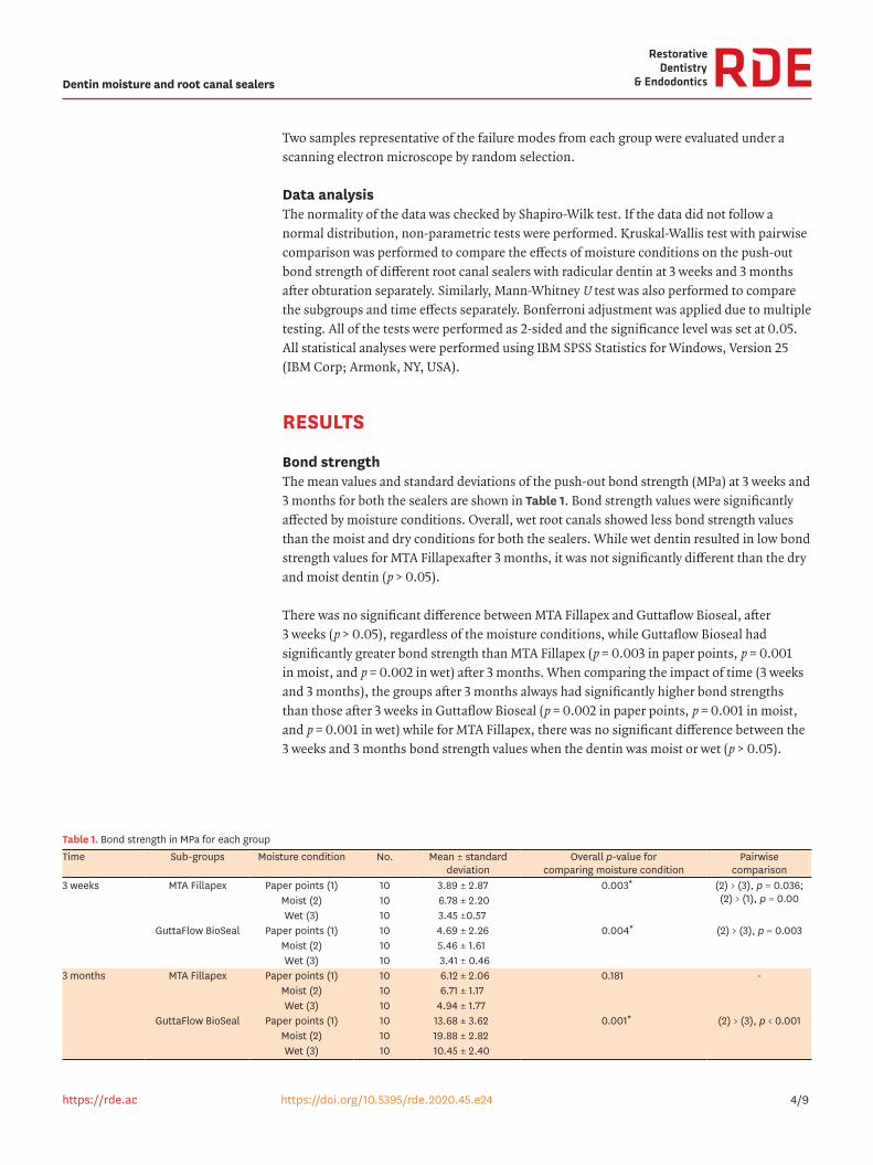
Bond strengthThe mean values and standard deviations of the push-out bond strength (MPa) at 3 weeks and 3 months for both the sealers are shown in Table 1. Bond strength values were significantly affected by moisture conditions. Overall, wet root canals showed less bond strength values than the moist and dry conditions for both the sealers. While wet dentin resulted in low bond strength values for MTA Fillapexafter 3 months, it was not significantly different than the dry and moist dentin (p > 0.05).
There was no significant difference between MTA Fillapex and Guttaflow Bioseal, after 3 weeks (p > 0.05), regardless of the moisture conditions, while Guttaflow Bioseal had significantly greater bond strength than MTA Fillapex (p = 0.003 in paper points, p = 0.001 in moist, and p = 0.002 in wet) after 3 months. When comparing the impact of time (3 weeks and 3 months), the groups after 3 months always had significantly higher bond strengths than those after 3 weeks in Guttaflow Bioseal (p = 0.002 in paper points, p = 0.001 in moist, and p = 0.001 in wet) while for MTA Fillapex, there was no significant difference between the 3 weeks and 3 months bond strength values when the dentin was moist or wet (p > 0.05).
4/9https://rde.ac https://doi.org/10.5395/rde.2020.45.e24
Dentin moisture and root canal sealers
Table 1. Bond strength in MPa for each groupTime Sub-groups Moisture condition No. Mean ± standard
deviationOverall p-value for
comparing moisture conditionPairwise
comparison3 weeks MTA Fillapex Paper points (1) 10 3.89 ± 2.87 0.003* (2) > (3), p = 0.036;
(2) > (1), p = 0.00Moist (2) 10 6.78 ± 2.20Wet (3) 10 3.45 ±0.57
GuttaFlow BioSeal Paper points (1) 10 4.69 ± 2.26 0.004* (2) > (3), p = 0.003Moist (2) 10 5.46 ± 1.61Wet (3) 10 3.41 ± 0.46
3 months MTA Fillapex Paper points (1) 10 6.12 ± 2.06 0.181 -Moist (2) 10 6.71 ± 1.17Wet (3) 10 4.94 ± 1.77
GuttaFlow BioSeal Paper points (1) 10 13.68 ± 3.62 0.001* (2) > (3), p < 0.001Moist (2) 10 19.88 ± 2.82Wet (3) 10 10.45 ± 2.40

Failure modesThe failure analysis results of the groups are summarized in Table 2. Under wet conditions, a majority of the specimens had adhesive failures. Under dry and moist conditions, the most common type of failure mode was cohesive within the sealer. Representative scanning electron microscope images of the cohesive failure mode from each group are shown in Figure 1.
DISCUSSION
This study investigated the effect of dentin moisture on the dislocation resistance of 2 bioactive sealers. It showed that the moisture conditions and the type of sealer had a significant effect on its dentin interactions. Therefore, the null hypothesis was rejected. Our results revealed that the magnitude of difference between the 3 moisture conditions was higher for Guttaflow Bioseal than for MTA Fillapex. Nagas et al. [3] reported that MTA Fillapex exhibited a higher bond strength in moist dentin compared to dry and normal moisture conditions, and bonding did not occur in over-wet moisture conditions. Except for the study mentioned above [3], no other study has focused on the effects of the different moisture levels on the bond strength of MTA Fillapex. In our study, moist dentin had better bond values compared to wet and normal moisture conditions. In addition, a weak bond was observed in “wet” moisture conditions. The conflicting results between these two studies are likely related attributed to the different storage periods. Furthermore, it has been reported that MTA “cured” (completely set) in 21 days in the presence of moisture [11]. Therefore, we tested 3 weeks and 3 months storage period.
Specifically, for sealers that bond to dentin, over-drying may remove the water in the dentinal tubules, which may, in turn, deter the effective penetration of hydrophilic sealers, which likely reflects as a reduction in bond strength [9,12]. Indeed, this may not be true for sealers such as zinc oxide eugenol that do not demonstrate any bonding to the dentin substrate [9]. Studies have shown that the moisture in the root canal might affect the adhesion
5/9https://rde.ac https://doi.org/10.5395/rde.2020.45.e24
Dentin moisture and root canal sealers
Table 2. Failure modes (%) for each groupTime Moisture condition MTA Fillapex GuttaFlow Bioseal3 weeks
Adhesive Paper point 10.0 5.0Moist 6.7 5.0Wet 15.0 11.7
Cohesive Paper point 53.3 65.0Moist 63.3 70.0Wet 71.7 63.3
Mixed Paper point 36.7 30.0Moist 30.0 25.0Wet 13.3 25.0
3 monthsAdhesive Paper point 3.3 1.7
Moist 3.3 0.0Wet 5.0 5.0
Cohesive Paper point 73.3 78.3Moist 76.7 85.0Wet 85.0 80.0
Mixed Paper Point 23.3 20.0Moist 20.0 15.0Wet 10.0 15.0

quality between the dentin and sealer [7,13,14]. The results of this study also showed that MTA Fillapex and GuttaFlow Bioseal root canal sealers exhibited weaker bond strength in both 3 weeks and 3 months periods if the root canals were completely dried with a paper point, compared to moist dentin. Guttaflow BioSeal is a relatively new material, containing gutta-percha and calcium silicate, and is marketed as a bioactive sealer [15]. The impact of moist conditions on the adhesion of this material is not known, and our data could not be compared with any other study.
In this study, the highest bond values were observed in moist conditions. We used the EndoVac device to achieve moisture [13]. The apical negative pressure of this system [16,17] appears to effective in drying the root canals as shown by our results. Although different sealers were compared by Zmener et al. [9], moisture was found to influence the adhesion of methacrylate-resin based sealers. Indeed, this is not surprising as the need for moist dentin for this class of resins is well-known from studies in coronal dentin. However, our results contrasted those of another study [13], which tested the effect of moisture on dentin bond strength of an epoxy resin-based sealer (AH Plus; Dentsply DeTrey, Konstanz, Germany), a calcium hydroxide-based sealer (Sealapex; Kerr Endodontics, Orange, CA, USA) and MTA Fillapex. The afore-mentioned study reported lower bond strength values in the groups
6/9https://rde.ac https://doi.org/10.5395/rde.2020.45.e24
Dentin moisture and root canal sealers
MTA
Fill
apex
Gut
taFl
ow B
ioSe
al
Paper points
3 w
eeks
3 m
onth
s3
wee
ks3
mon
ths
Moist Wet
D
DD
D
D D D
D
D
D
D
D
Figure 1. Representative scanning electron microscope images showing the cohesive failure modes after bond failure. Arrows indicate sealer. D, dentin.

where root canals were dried with EndoVac. One possible reason could have been the use of the macrocannula by the afore-mentioned work [13], while we used the microcannula. The fact that the macro-cannula cannot be inserted until the end of the root canal [16] could be a potential reason for such results.
With regards to the storage time, our results showed that GuttaFlow BioSeal exhibited higher bond values at 3 months than at 3 weeks, regardless of the dentin moisture conditions. MTA Fillapex showed no significant difference in the bond strength values between 3 weeks and 3 months when the dentin was moist or wet. Rather surprisingly, the bond strength values for MTA Fillapex increased at 3 months, when the dentin was dry, albeit the fact that this was not significantly higher than the value at 3 weeks. In general, the magnitude of time-dependent increase in bond strength values was higher Guttaflow Bioseal than for MTA Fillapex. While the deeper penetration of the sealers into dentinal tubules may be one a plausible explanation for this phenomenon, the more appropriate reason may be the biomineralization of the sealers in question. It is known that bioactive materials undergo biomineralization when in contact with biological tissues. It has been suggested that the biomineralization of bioactive materials (such as MTA) improves its bond strength [18,19]. The low percentage of MTA (13.2%) in this material may be a reason for this effect [20]. More studies are needed to investigate the biomineralization by Guttaflow Bioseal.
We used the push-out bond strength method in this study. Although the push-out test provides beneficial information about the interaction between the root canal filling materials and dentin, it does not reliably represent the adhesion between the root filling materials and dentin [10]. Hence, the term dislocation resistance is more favorable than using the term bond strength [19]. In the present study, under wet and dry moisture conditions, the most common failure mode was adhesive. Therefore, it appears that an insufficient level of adhesion occurs between the sealer and dentin in terms of bond strength, regardless of the time of the test application (3 weeks or 3 months) and the sealer used (MTA Fillapex or GuttaFlow BioSeal). On the other hand, the high cohesive failures observed in normal moisture conditions indicates that the root canal filling material showed good adhesion to the dentin walls.
CONCLUSION
The different moisture levels (wet, moist, and dry) has a similar trend in its effects on both the bioactive sealers tested. This indicates that slight moisture in dentin may be advantageous before the filling process of the root canals. Further research is needed to characterize the biomineralization at the material-dentin interface with different moisture levels, to make clinical recommendations when bioactive cements are used as root canal sealers.
REFERENCES
1. Ng YL, Mann V, Rahbaran S, Lewsey J, Gulabivala K. Outcome of primary root canal treatment: systematic review of the literature - part 1. Effects of study characteristics on probability of success. Int Endod J 2007;40:921-939. PUBMED | CROSSREF
7/9https://rde.ac https://doi.org/10.5395/rde.2020.45.e24
Dentin moisture and root canal sealers

2. Hülsmann M, Peters OA, Dummer PM. Mechanical preparation of root canals: shaping goals, techniques and means. Endod Topics 2005;10:30-76. CROSSREF
3. Nagas E, Uyanik MO, Eymirli A, Cehreli ZC, Vallittu PK, Lassila LV, Durmaz V. Dentin moisture conditions affect the adhesion of root canal sealers. J Endod 2012;38:240-244. PUBMED | CROSSREF
4. Parirokh M, Torabinejad M. Mineral trioxide aggregate: a comprehensive literature review--Part I: chemical, physical, and antibacterial properties. J Endod 2010;36:16-27. PUBMED | CROSSREF
5. Mandal P, Zhao J, Sah SK, Huang Y, Liu J. In vitro cytotoxicity of GuttaFlow 2 on human gingival fibroblasts. J Endod 2014;40:1156-1159. PUBMED | CROSSREF
6. Akcay M, Arslan H, Durmus N, Mese M, Capar ID. Dentinal tubule penetration of AH Plus, iRoot SP, MTA fillapex, and GuttaFlow BioSeal root canal sealers after different final irrigation procedures: a confocal microscopic study. Lasers Surg Med 2016;48:70-76. PUBMED | CROSSREF
7. Razmi H, Bolhari B, Karamzadeh Dashti N, Fazlyab M. The effect of canal dryness on bond strength of bioceramic and epoxy-resin sealers after irrigation with sodium hypochlorite or chlorhexidine. Iran Endod J 2016;11:129-133. PUBMED | CROSSREF
8. Ayad MF, Farag AM, Garcia-Godoy F. Effect of lactic acid irrigant on shear bond strength of Epiphany adhesive sealer to human dentin surface. Oral Surg Oral Med Oral Pathol Oral Radiol Endod 2010;109:e100-e106. PUBMED | CROSSREF
9. Zmener O, Pameijer CH, Serrano SA, Vidueira M, Macchi RL. Significance of moist root canal dentin with the use of methacrylate-based endodontic sealers: an in vitro coronal dye leakage study. J Endod 2008;34:76-79. PUBMED | CROSSREF
10. Reddy P, Neelakantan P, Sanjeev K, Matinlinna JP. Effect of irrigant neutralizing reducing agents on the compromised dislocation resistance of an epoxy resin and a methacrylate resin-based root canal sealer in vitro. Int J Adhes Adhes 2018;82:206-210. CROSSREF
11. Gancedo-Caravia L, Garcia-Barbero E. Influence of humidity and setting time on the push-out strength of mineral trioxide aggregate obturations. J Endod 2006;32:894-896. PUBMED | CROSSREF
12. Dias KC, Soares CJ, Steier L, Versiani MA, Rached-Júnior FJ, Pécora JD, Silva-Sousa YT, de Sousa-Neto MD. Influence of drying protocol with isopropyl alcohol on the bond strength of resin-based sealers to the root dentin. J Endod 2014;40:1454-1458. PUBMED | CROSSREF
13. Paula AC, Brito-Júnior M, Araújo CC, Sousa-Neto MD, Cruz-Filho AM. Drying protocol influence on the bond strength and apical sealing of three different endodontic sealers. Braz Oral Res 2016;30:e50. PUBMED | CROSSREF
14. Zhong X, Shen Y, Ma J, Chen WX, Haapasalo M. Quality of root filling after obturation with gutta-percha and 3 different sealers of minimally instrumented root canals of the maxillary first molar. J Endod 2019;45:1030-1035. PUBMED | CROSSREF
15. Barbosa-Ribeiro M, Arruda-Vasconcelos R, Fabretti FL, Silva EJNL, De-Deus G, Gomes BPFA. Evaluation of apically extruded debris using positive and negative pressure irrigation systems in association with different irrigants. Braz Dent J 2018;29:184-188. PUBMED | CROSSREF
16. Silva EJNL, Carvalho CR, Belladonna FG, Prado MC, Lopes RT, De-Deus G, Moreira EJL. Micro-CT evaluation of different final irrigation protocols on the removal of hard-tissue debris from isthmus-containing mesial root of mandibular molars. Clin Oral Investig 2019;23:681-687. PUBMED | CROSSREF
17. Khalil MM, Abdelrahman MH, El-Mallah S. Bond strength and solubility of a novel polydimethylsiloxane-gutta-percha calcium silicate-containing root canal sealer. Dent Med Probl 2019;56:161-165. PUBMED | CROSSREF
18. Reyes-Carmona JF, Felippe MS, Felippe WT. The biomineralization ability of mineral trioxide aggregate and Portland cement on dentin enhances the push-out strength. J Endod 2010;36:286-291. PUBMED | CROSSREF
8/9https://rde.ac https://doi.org/10.5395/rde.2020.45.e24
Dentin moisture and root canal sealers

19. Neelakantan P, Ahmed HM, Wong MC, Matinlinna JP, Cheung GS. Effect of root canal irrigation protocols on the dislocation resistance of mineral trioxide aggregate-based materials: a systematic review of laboratory studies. Int Endod J 2018;51:847-861. PUBMED | CROSSREF
20. Neelakantan P, Grotra D, Sharma S. Retreatability of 2 mineral trioxide aggregate-based root canal sealers: a cone-beam computed tomography analysis. J Endod 2013;39:893-896. PUBMED | CROSSREF
9/9https://rde.ac https://doi.org/10.5395/rde.2020.45.e24
Dentin moisture and root canal sealers