research article comparative evaluation of...
TRANSCRIPT

Research ArticleComparative Evaluation of Anthelmintic Activity ofEdible and Ornamental Pomegranate Ethanolic Extracts againstSchistosoma mansoni
Doaa A. Yones,1 Dalia M. Badary,2 Hesham M. B. Sayed,3 Soad A. H. Bayoumi,3
Azza A. Khalifa,3 and Ahmed M. El-Moghazy3
1Parasitology Department, Faculty of Medicine, Assiut University, Assiut 71526, Egypt2Pathology Department, Faculty of Medicine, Assiut University, Assiut 71526, Egypt3Pharmacognosy Department, Faculty of Pharmacy, Assiut University, Assiut 71526, Egypt
Correspondence should be addressed to Doaa A. Yones; [email protected]
Received 29 June 2016; Revised 14 August 2016; Accepted 30 August 2016
Academic Editor: Charles Spencer
Copyright © 2016 Doaa A. Yones et al. This is an open access article distributed under the Creative Commons Attribution License,which permits unrestricted use, distribution, and reproduction in any medium, provided the original work is properly cited.
Due to the development of praziquantel (PZQ) schistosomes resistant strains, the discovery of new antischistosomal agents is ofhigh priority in research. This work reported the in vitro and in vivo effects of the edible and ornamental pomegranate extractsagainst Schistosoma mansoni. Leaves and stem bark ethanolic extracts of both dried pomegranates were prepared at 100, 300, and500 𝜇g/mL for in vitro and 600 and 800mg/kg for in vivo. Adult worms Schistosomamansoni in RPMI-1640medium for in vitro andS. mansoni infected mice for in vivo tests were obtained fromTheodor Bilharz Research Institute, Cairo, Egypt. In vitro activity wasmanifested by significant coupledworms separation, reduction ofmotor activity, lethality, andultrastructural tegumental alterationsin adult worms. In vivo activity was manifested revealed by significant reduction of hepatic granulomas number and diameter,decreased number of bilharzial eggs in liver tissues, lowered liver inflammatory infiltration, decreased hepatic fibrosis, and induciblenitric oxide synthase (iNOS) expression. Ethanolic stem bark extract of edible pomegranate exhibited highest antischistosomalactivities both in vitro and in vivo. Therefore, pomegranate showed a good potential to be used as a promising new candidate forthe development of new schistosomicidal agents.
1. Introduction
Schistosomiasis is one of the world major public healthproblems, caused by the blood-dwelling fluke of the genusSchistosoma. The clinically worldwide relevant species are S.mansoni, S. haematobium, and S. japonicum [1]. In additionto the previously mentioned species, S. mekongi and S. inter-calatum represent the most important pathogenic species forhuman beings. Depending on the species, the schistosomeworms persist in the liver and hepatic portal system or theurinary tract system of humans. Mature schistosomes layeggs within their host, which often get trapped in the host’stissues, resulting in inflammatory and obstructive diseases ofthe affected organs [2, 3].
Schistosomiasis is one of the most widely occurringneglected tropical diseases with high levels of incidence inAsia, Africa, and Latin America. Studies have shown approx-imately 779 million people living at higher risk of infection,and 239 million people are infected with schistosomes [4, 5].
To cure morbidity and prevent the development of severechronic stage hepatosplenomegaly, praziquantel (PZQ) isthe only choice of chemotherapy against all species ofSchistosoma. Presenting good efficacy and low toxicity, thedrug has been widely used for more than three decadesand is therefore susceptible to the emergence of praziquantelresistant schistosomes [6].
The study of medicinal plants as a new approach forschistosomiasis treatment is feasible and promising one [7,
Hindawi Publishing CorporationBioMed Research InternationalVolume 2016, Article ID 2872708, 15 pageshttp://dx.doi.org/10.1155/2016/2872708

2 BioMed Research International
8]. The research on medicinal plants is encouraged by theWHO, considering that certain traditional knowledge oncurative plants could add up to the development of newpharmaceutical products as well as to the combat againstdiseases that affects the populations of developing countries[9, 10].
The search for new antischistosomal treatments has ledto the study of natural substances such as artemisinin and itsderivatives, curcumin, phytol, and pomegranate [11–13].
Pomegranate (Punica granatum L.) is a favorite table fruitin tropical countries, belonging to Punicaceae family. Thisfamily is unusual in having the sole genus Punica, a genusof large shrubs or small trees with two species. One is P.protopunica Balf. S., which is found wild in Socotra island,and the other is P. granatum L. (edible pomegranate), whichis cultivated in tropical and subtropical parts of the world forits edible fruits [14].
Punica granatum L. var. nana is a dwarf variety of P.granatum L. popularly planted as an ornamental plant ingardens. It could well be a wild form with distinct origin. Itdoes not usually produce edible fruits [15].
The peel and seeds of P. granatum L. showed varioustherapeutic applications such as antibacterial, antifungal,antioxidant, antitumor, antiviral, antimalarial, and antimuta-genic effects [16, 17]. Edible pomegranate extracts have beenreported to have promising results against S. mansoni eitherin vitro or in vivo [12, 18, 19].
The root, stem bark, and, to lesser extent, the fruit rindof pomegranate have been commonly used as vermifugal ortaenicidal agents [20]. Pomegranate has also antiprotozoalactivity and it is used in folk medicine for treatment ofdysentery [21].
The methanolic extract of P. granatum L. var. nana leavesexhibited antioxidant activity, nematicidal activity againstthree root-knot nematode species, and hepatoprotectiveactivity against carbon tetrachloride induced hepatotoxicity[22].
In our locality, many factors, including the high preva-lence, wide distribution of schistosomiasis, and drug resis-tance for the already used treatment, necessitate the needfor control of this helminthiasis and impulse the studiesfor new and more comprehensive alternative therapeuticswithout adverse effects. Hence, the current study aimed toinvestigate the in vitro and in vivo activity of leaves and stembark ethanolic extracts of both dried edible and ornamentalpomegranate against Schistosoma mansoni (Egyptian strain).
2. Materials and Methods
2.1. Plant Materials. 200 g of each of the leaves of ediblepomegranate (LEP) and stem bark of edible pomegranate(SEP) (P. granatum L.) and similar leaves of ornamen-tal pomegranate (LOP) and stem bark of ornamentalpomegranate (SOP) of P. granatum L. var. nana was locallycollected from Faculty of Pharmacy garden of medicinalplants and Experimental Station of Agriculture, Faculty ofAgriculture Assiut University, Assiut, Egypt. Test plants were
authenticated by Dr. Naeem E. Keltawy, Professor of Orna-mental Horiculture and Floriculture, Faculty of Agriculture,AssiutUniversity, Assiut, Egypt. Voucher specimens (number45)were kept in the herbarium, PharmacognosyDepartment,Faculty of Pharmacy, Assiut University.
2.2. Preparation of Extracts. Plant samples were allowed todry at room temperature before being ground to fine powder.Powdered plant materials were extracted with 70% ethanolat room temperature by maceration and then filtered andthe filtrates were concentrated under vacuum using rotaryevaporator. The obtained solvent-free residue of each plantextract was stored at 4∘C for subsequent preparation of therequired doses.
2.3. Dose Preparations of Extracts. Plant extracts were freshlyprepared before usage by suspending 1 g of each extract in50mL 3% tween 80 dissolved in 0.9% saline. For in vitroantischistosomal testing, 100, 300, and 500 𝜇g/mL were used,while 600 and 800mg/kg for in vivo assay were prepared [12].
2.4. Cytotoxicity Assays (CTAs) on Tissue Culture Cells. CTAswere performed on mouse fibroblast cell BALB/c 3T3 (VAC-SERA, Egypt) supplemented with 10% bovine calf serum,4mML-glutamine, 100 IU penicillin, and 100 𝜇g/mL strepto-mycin (Bioanalyse, Turkey) using the neutral red uptake assayfor all pomegranate extracts at the higher concentration [23].
2.5. Evaluation of Microbial Contamination and EndotoxinProduction. Total aerobic microbial count and total com-bined yeasts/moulds count were used for quantitative enu-meration ofmesophilic bacteria or fungi thatmay growunderaerobic conditions in all pomegranate extracts at the higherconcentration and for PZQ using the pour plating technique(EDQM Council of Europe, 2014). The bacterial endotoxintest was performed by the limulus amoebocyte lysate assay(gel-clot technique) as reported by Hussaini and Hassanali[24].
2.6. Standard Antischistosomal Treatment. Praziquantel sus-pension, a product of Egyptian International PharmaceuticalIndustries Company (EIPI Co.), was purchased locally. PZQwas used as positive control at concentration of 10 𝜇g/mL forin vitro and 200mg/kg for in vivo experiments [25, 26].
2.7. Schistosome Parasites and Experimental Infected Hosts.S. mansoni (Egyptian strain) adults were purchased fromthe experimental animal research unit of the SchistosomeBiological Supply Center (SBSC), Theodor Bilharz ResearchInstitute (TBRI), Cairo, Egypt. Swiss albino female miceCD strain, weighing 20–25 g and aged 4 weeks, were indi-vidually infected using the tail immersion technique byexposure to a suspension containing 100 S. mansoni cercariae(100 cercariae/mouse) from naturally infected Biomphalariaalexandrina for 2 h according to the method described byLiang et al. [27]. Mice were bred under environmentallycontrolled conditions (temperature ∼25∘C and 12 h light and

BioMed Research International 3
dark cycle) and fed with a standard stock commercial pelletdiet (containing 24% protein) and water ad libitum.
3. Experimental Design
3.1. Experiment (1)
3.1.1. In Vitro Assessment of the Antischistosomal Effects of thePrepared Extracts. For in vitro bioassay, Schistosomamansoniadult worm pairs of Egyptian strain were retrieved asepticallyfrom sacrificed infected mice and collected by perfusion ofthe hepatic portal system and mesenteric veins using citratedsaline according to the technique of Stirewalt andDorsey [28]from mice livers 8 weeks postinfection.
Adult worms were washed three times with the RPMI1640 (Roswell ParkMemorial Institute 1640) culturemedium(Invitrogen, Carlsbad, California, USA), which was used forculturing the parasite. The medium was supplemented withL-glutamine, 20% fetal calf serum, and antibiotics (300 𝜇gstreptomycin, 300 IU penicillin, and 160 𝜇g gentamycin permL) [12]. After washing, 5 couples of worms were transferredto each well of a 24-well culture plate (TPP, St. Louis, MO)containing the same medium.
TwomL of the tested doses (100, 300, and 500𝜇g/mL)from leaves and stem bark extracts was added to each well.The final volume in each well was 2mL. The plate wasincubated at 37∘C in a humid atmosphere containing 5%CO
2
[29]. The parasites were kept for 12 h and monitored every2 h. A pure medium and medium with 3% tween 80 in 0.9%saline were used as negative controls, while PZQ (10 𝜇g/mL)was used as a positive control. All the steps were performedunder a sterilized laminar flow chamber.The experiment wascarried out in triplicate and repeated three times.
Treated worms weremonitored for their mating (pairing)of the worms, motility (worm’s motor activity changes),and mortality rate using an inverted optical microscope(Olympus CK2). Worms which did not show motility forone minute were considered dead. Changes in worm’s motoractivity (motility) of schistosomes were assessed qualitativelyand their motor activity reduction was defined as “slight” or“significant” [30].
The effect of the treatment was also assessed with anemphasis on morphological alterations in the tegumentwhich were observed using scanning electron microscopy(SEM) [31]. Observation of adult schistosomes in the in vitroexperiment was performed at 2 h intervals throughout the12 h experimental incubation period and the results werereported at 2, 4, 6, and 12 h (the end point of the experimentfor the negative control groups).
3.1.2. Preparation of Adult S. mansoni Worms for SEM. Toobserve morphological changes in the tegument of adultparasites, schistosome worms, when they died, and controlworms at 12 h, the end point of the experiments, were washedthoroughly with distilled water. The parasites were fixed for2 h in 4% glutaraldehyde (pH 7.4) and 5% paraformalde-hyde in 0.1M cacodylate buffer (pH 7.2). They were rinsedovernight in cacodylate buffer, dehydrated, dried in a critical
point dryer according to Hayat [32], mounted on stubs, andsputter-coated with gold particles in the sputter coating appa-ratus for 6 minutes. Specimens were processed, examined,and photographed using Jeol-JSM-5400 LV at the ScanningElectron Microscope Unit, Assiut University, Assiut, Egypt.
3.2. Experiment (2)
3.2.1. In Vivo Assessment of the Antischistosomal Effects of thePrepared Extracts. Fifty-five S. mansoni infected female micewere obtained from TBRI, Cairo, Egypt, 8 weeks postinfec-tion. Infected mice were randomly allocated into 11 groupswith 5 animals each, at the time of the experiment:
(G1) Infected untreated control mice (negative control 1)(G2) Infected mice given 3% tween 80 in saline (negative
control 2)(G3) Infected mice treated with 200mg/kg PZQ (positive
control)(G4) Infected mice treated with 600mg/kg LEP(G5) Infected mice treated with 600mg/kg SEP(G6) Infected mice treated with 800mg/kg LEP(G7) Infected mice treated with 800mg/kg SEP(G8) Infected mice treated with 600mg/kg LOP(G9) Infected mice treated with 600mg/kg SOP(G10) Infected mice treated with 800mg/kg LOP(G11) Infected mice treated with 800mg/kg SOP
Each mouse was given a single oral dose daily for 7consecutive days using stainless-steel esophageal tube. Allmice were sacrificed by cervical dislocation after 7 daysof treatment. Assessment of the treatment in vivo wasperformed through histopathological examination of livertissue for detection of hepatic inflammation, hepatic fibrosis,and schistosomal granulomas formation.The assessment wasalso done through immunohistochemical analysis of iNOSreactivity in liver tissue. The experiment was repeated threetimes.
3.2.2. Histopathological Assessment. Liver samples of the leftlobe of each sacrificed mouse were rinsed with phosphate-buffered saline and fixed in 10% formalin for 24 h. Liver sam-ples were dehydrated in increasing concentrations of ethanol,diaphonized in xylol, and embedded in paraffin wax blocks.Sections of 4 𝜇m thickness were stained with hematoxylinand eosin [33]. The sections were evaluated using the brightfield microscopy to evaluate the degree of inflammation,fibrosis, and granuloma formation followed by image cap-ture and processing using Camidia image manager. All thegranulomas found in 10 histologic sections of random fieldswere counted. Measurement of mean granuloma diameterwas performed using an ocular micrometer at magnificationof 100x. Only nonconfluent, lobular granulomas containingeggs in their centers were measured (periocular granulomas)[34].

4 BioMed Research International
3.2.3. Immunohistochemistry for Determination of iNOS Reac-tivity. Left lobe liver sections from the previously preparedparaffin blocks, 4 𝜇mthickness,mounted on glass slides, werekept overnight at 56∘C.They were deparaffinized with xyleneand rehydrated with decreasing percentages of ethanol andfinally with water. For antigen retrieval, slides were heatedby microwaving in 10mM citrate buffer (pH 6.0) for 12min.Slides were left to cool for 20min at room temperature andrinsed with distilled water. Surroundings of the sections weremarked with a PAP pen. The endogenous peroxidase activitywas blocked with H
2O2for 10min at room temperature
and later rinsed with distilled water and PBS (phosphate-buffered saline). Liver sections were then incubated for 1 hat room temperature with the following antibody: iNOSrabbit Pab (Neomarker, RB-1605-P) antibodies. Antibodieswere diluted at 1 : 100. The sections were washed and rinsedwith PBS three times for 5min each. Slides were incubatedfor 30min at room temperature with biotinylated goat anti-rabbit antibodies. The streptavidin peroxidase label reagentwas applied to the slides after being washed in PBS, for30min at room temperature in a humid chamber. Afterblotting off excess buffer, a universal staining colored prod-uct was developed by incubation with AEC (3-Amino-9-Ethylcarbazole) Chromogen (Lab Vision, TA-004-HAC) for5min according tomanufacturer’s instructions. Finally, slideswere dehydrated and cleared. The slides were counterstainedwith Mayer’s hematoxylin and mounted in glycerol gelatinafter washing in distilled water and mounted with cover slips[35].
3.3. Ethical Considerations. A standard protocol was drawnup in accordance with the Good Laboratory Practice (GLP)regulations of the World Health Organization (WHO). Theprinciples of laboratory animal care were duly followed inthis study [36]. Ethical animal practices were followed understandard regulations dictated by the animal care committee ofFaculty of Medicine, Assiut University. Ethical approval wasgranted by the Research and Ethic Committee of Faculty ofMedicine, Assiut University.
3.4. Statistical Data Analysis. The results were analyzed usingthe SPSS (Statistical Package for the Social Sciences, version16 for Windows) software (SPSS Inc., Chicago, Illinois,USA). Significant differences were determined by one-wayanalysis of variance (ANOVA). The values were presented asmean ± standard deviation (SD). Data were analyzed usingStudent’s Tukey’s test (𝑡-test) which was used to calculatethe significance of differences observed betweenmean valuesof experimental and control groups in each experiment. 𝑃values of less than 0.05, 0.01, or 0.001 were used to indicatestatistical significance.
4. Results
The preliminary phytochemical screening of both plantextracts showed the presence of volatile constituents, poly-phenols glycosides, triterpenes, sterols, flavonoids, antho-cyanins, triglycerides, tannins, and alkaloids.
4.1. Cytotoxicity Assays (CTAs). The optical density (OD)540
of each of the tested extracts was compared with the meanvalue OD
540for the negative control (distilled water). Tested
pomegranate extracts fulfilled the mentioned acceptancecriteria through absence of cytotoxic effects of the studiedextracts. Cell viabilities were more than 70% relative to thenegative control for tested extracts at their highest concen-trations. Thus, the concentrations in which pomegranatepresented the schistosomicidal activity were not associatedwith cytotoxic effects on fibroblast cell.
4.2. Evaluation of Microbial Contamination and EndotoxinProduction. Total aerobic microbial count and total com-bined yeasts/moulds count were negative for the testedpomegranate extracts. Tested pomegranate extracts wereendotoxin free.
4.3. In Vitro Treatment Efficacy of LEP, SEP, LOP, and SOP Ex-tracts on Adults S. mansoni at Different Concentrations. Allthe tested extracts influenced the process of natural mating,causing separation of couple schistosomes depending on theconcentration used and exposure time. Nearly 95% of theworms had been separated within the first 2 h with the useof 500𝜇g/mL SEP, compared to the negative control groups(Table 1). PZQ (10 𝜇g/mL) caused couple worm separationafter the first 2 h of incubation. Negative control groupsshowed couple separation nearly at 10 h after incubation.Moreover, concentrations which were not 100% lethal to theworms were proven as efficient mating inhibitors, once all ofthem separated the couples in all samples.
Concerning the motility, a significant reduction in theparasites movements was observed in all concentrations.The percentage of worms that had their motility reducedwas directly proportional to the concentration and to theperiod of incubation. A slight decrease in motor activitywas observed after 2 h of incubation for all adult wormsexposed to 500, 300, and 100 𝜇g/mL concentration of SEP.Total motility loss occurred at 4, 6, and 12 h, respectively.No change in motor activity was observed at 4 h interval,while it decreased at 6 h interval and complete loss of motilityoccurred at 10 h interval in the negative control groups. Onthe other hand, PZQ (10𝜇g/mL) resulted in decrease inmotoractivity starting from the first 2 h of incubation and completeloss of motor activity in all worms occurred at 4 h interval.
The survival of S. mansoni adults exposed to ethanolicextracts of LEP, SEP, LOP, and SOP depended directlyon both concentration and incubation period. The 500,300, and 100 𝜇g/mL concentrations of SEP caused deathof 100% of parasites within 4, 6, and 12 h of incubation,respectively (Table 1). Ethanolic extracts of LEP at 500, 300,and 100 𝜇g/mL concentrations caused death of 100% wormsafter 6 and 12 h of incubation, respectively (Table 1). LOPand SOP (500 𝜇g/mL) caused significant mortality (𝑃 <0.01) among schistosome parasites after 6 h of incubation,while 100 and 300 𝜇g/mL concentrations of the same extractsexpressed their mortality effect on adults S. mansoni after12 h of incubation (Table 1). No difference was observedbetweenmale and female adult worms in response to different

BioMed Research International 5
Table1:In
vitro
effectsof
leaves
andste
mbark
ethano
licextractsof
edibleandornamentalp
omegranateon
adultw
ormso
fS.m
ansoni
after
12hincubatio
nperio
d.
Group
Con
c(𝜇g/mL)
Incubatio
nperio
d(h)
Num
bero
fseparated
worm
s(%)
%of
worm
mortality
%of
wormsw
ithtegu
mentalalteratio
nPartial
Extensive
SEP
500
295
90—
—4
99100
1090
6100
100
——
12100
100
——
300
287
80—
—4
9999
——
699
100
3070
12100
100
——
100
271
60—
—4
8065
——
695
90—
—12
100
100
5050
LEP
500
280
0—
—4
8545
——
699
100
2080
12100
100
——
300
275
0—
—4
8570
——
699
90—
—12
100
100
1585
100
265
0—
—4
800
——
695
90—
—12
100
100
6040
LOP
500
240
0—
—4
7525
——
685
100
2080
12100
100
——
300
230
0—
—4
6512
——
675
34—
—12
100
100
5050
100
220
0—
—4
550
——
670
0—
—12
100
9870
30

6 BioMed Research International
Table1:Con
tinued.
Group
Con
c(𝜇g/mL)
Incubatio
nperio
d(h)
Num
bero
fseparated
worms(%)
%of
worm
mortality
%of
wormsw
ithtegumentalalteratio
nPartial
Extensive
SOP
500
220
10—
—4
6045
——
676
100
3070
12100
100
——
300
218
0—
—4
600
——
670
90—
—12
100
100
8515
100
215
0—
—4
550
——
670
90—
—12
100
100
9010
PZQ
102
9595
——
4100
100
2575
Con
trol−
ve
20
0—
—4
600
——
670
10—
—12
100
100
00
Incubatio
nperio
d:12h;control–ve
inRP
MI-1640
andmedium
with
3%tween80
in0.9%
salin
e.
BioMed Research International 7
(a) (b) (c)
gc
(d) (e) (f)
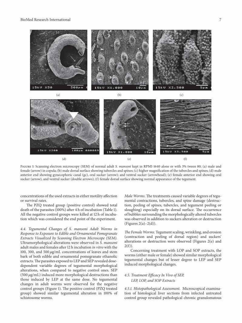
Figure 1: Scanning electron microscopy (SEM) of normal adult S. mansoni kept in RPMI-1640 alone or with 3% tween 80; (a) male andfemale (arrow) in copula; (b) male dorsal surface showing tubercles and spines; (c) higher magnification of the tubercles and spines; (d) maleanterior end showing gynecophoric canal (gc), oral sucker (arrow); and ventral sucker (arrowhead); (e) female anterior end showing oralsucker (arrow), and ventral sucker (double arrows); (f) female dorsal surface showing normal appearance of the tegument.
concentrations of the used extracts in eithermotility affectionor survival rates.
The PZQ treated group (positive control) showed totaldeath of the parasites (100%) after 4 h of incubation (Table 1).All the negative control groups were killed at 12 h of incuba-tion which was considered the end point of the experiment.
4.4. Tegumental Changes of S. mansoni Adult Worms inResponse to Exposure to Edible and Ornamental PomegranateExtracts Visualized by Scanning Electron Microscope (SEM).Ultramorphological alterations were observed in S. mansoniadult males and females after 12 h incubation in vitrowith the100, 300, and 500 𝜇g/mL concentrations of leaves and stembark of both edible and ornamental pomegranate ethanolicextracts.Theparasites exposed to LEP and SEP revealed dose-dependent variable degrees of tegumental morphologicalalterations, when compared to negative control ones. SEP(500𝜇g/mL) induced more morphological destructions thanthose induced by LEP at the same dose. No tegumentalchanges in adult worms were observed for the negativecontrol groups (Figure 1). The positive control (PZQ treatedgroup) showed similar tegumental alteration in 100% ofschistosome worms.
MaleWorms. The treatments caused variable degrees of tegu-mental contractions, tubercles, and spine damage (destruc-tion, peeling of spines, tubercles, and tegument peeling orsloughing) especially on its dorsal surface. The occurrenceof bubbles surrounding themorphologically altered tubercleswas observed in addition to suckers alteration or destruction(Figures 2(a)–2(d)).
TheFemaleWorms. Tegument scaling, wrinkling, and erosion(contraction and peeling of dorsal region) and suckers’alterations or destruction were observed (Figures 2(e) and2(f)).
Concerning treatment with LOP and SOP extracts, theworms (eithermale or female) showed similarmorphologicaltegumental changes but of lesser degree to LEP and SEPinduced morphological changes.
4.5. Treatment Efficacy In Vivo of SEP,LEP, LOP, and SOP Extracts
4.5.1. Histopathological Assessment. Microscopical examina-tion of histological liver sections from infected untreatedcontrol group revealed pathological chronic granulomatous

8 BioMed Research International
(a) (b) (c)
osvs
(d) (e)
os
vs
(f)
Figure 2: Scanning electron microscopy (SEM) of adult S. mansoniworms after their exposure to 100, 300, and 500 𝜇g/mL ethanolic extractsof leaves and stem bark of edible and ornamental pomegranate. (a) Separated male and female (arrow); (b) male dorsal surface showingtegumental peeling with destruction and peeling of tubercles and spines; (c) male showing bubbles surrounding the morphologically alteredtubercles on its dorsal surface; (d)male suckers’ alterations or destruction, os: oral sucker and vs: ventral sucker; (e) female showing tegumentalscaling, wrinkling, and erosion; (f) female suckers’ alterations or destruction, os: oral sucker and vs: ventral sucker.
lesions in the hepatic parenchyma. These lesions formed ofnumerous bilharzial eggs containing miracidia, surroundedby numerous chronic inflammatory cells in form of epithe-lioid cells, lymphocytes, plasma cells, macrophages, andeosinophils forming granuloma with severe areas of fibrosis(Figure 3(a)).
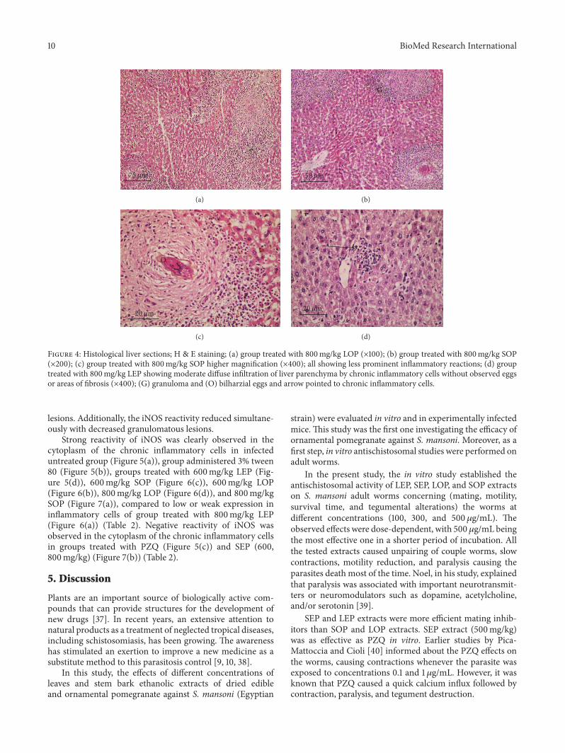
Nearly similar observations were detected in the hepatichistological sections from groups treated with 600mg/kgSOP, LOP, and LEP (Figures 3(b)–3(d)). These inflamma-tory reactions were less prominent in groups treated with800mg/kg LOP and SOP in the formof granulomawith fewereggs, less fibrosis, and moderate chronic inflammatory cellinfiltration (Figures 4(a)–4(c)).
Histological liver sections from groups treated with LEP(800mg/kg) showed moderate diffuse infiltration of liverparenchyma by chronic inflammatory cells without observedeggs or areas of fibrosis (Figure 4(d)). Similar observationswere reported in histological liver sections from groupstreated with 600 and 800mg/kg SEP and PZQ (200mg/kg).They showed absence of bilharzial eggs and fibrosis withsignificant reduction of liver parenchyma infiltration by thechronic inflammatory cells. For hepatic granulomas num-ber and diameter, histological liver sections from infecteduntreated control groups revealed about 121.3 granulomas ofaverage diameter 235.7 ± 16.1 𝜇m. Livers of different treated
groups showed decrease in granuloma size and number withminimal degenerative changes in liver tissues as shown inTable 2.
Oral administration of 800mg/kg LEP decreased hepaticgranulomas number and size to 54.1 and 37.2%, respectively,while administration of 600mg/kg of the same extractdecreased them to 51.3 and 34.5%, respectively. Followingoral administration of 600mg/kg SEP, there was decreasein the mean granuloma diameter to 40.1%, while 800mg/kgof the same extract showed about 40.9% reduction. Otherextracts and PZQ treatments showed variable effects onhepatic granuloma number and diameter (Table 2). Thus,the hepatic granuloma average diameter was significantlysmaller (𝑃 < 0.01) in groups treated with SEP and LEPin comparison to groups treated with SOP and LOP (600and 800mg/kg) and control groups.These hepatic granulomaaverage diameters were nearly similar to positive controlgroup (PZQ 200mg/kg) with insignificant difference (𝑃 >0.01) (Table 2).
4.5.2. Expression of iNOS Detected by Immunohistochem-istry. In comparison to immunohistochemical expressionof iNOS between different groups, iNOS reactivity (cyto-plasmic expression) was stronger in the hepatocytes of

BioMed Research International 9
G
50 𝜇m
(a)
O
50 𝜇m
(b)
d
20 𝜇m
(c)
F
20 𝜇m
(d)
Figure 3: Histological liver sections; H&E staining; (a) infected untreated group showing numerous bilharzial eggs surrounded by numerouschronic inflammatory cells (×200); (b) group treated with 600mg/kg SOP (×200); (c) group treated with 600mg/kg LOP (×400); (d) grouptreated with 600mg/kg LEP (×400). All showed similar structures: (G) granuloma, (O) bilharzial eggs, and (F) fibrosis and arrow pointed tochronic inflammatory cells.
Table 2: Effect of oral administration of different doses of SEP, LEP, LOP, and SOP extracts in vivo.
Group Dose(mg/kg)
Granuloma number Granuloma diameter (GD)Eggs in liver
tissue
Immunohistochemicalfindings
Mean ± SE Reduction% Mean (𝜇m) ±SE Reduction% iNos
hepatocytes
iNosinflammatory
cellsSEP(G5&G7)
600 51.2∗∗ 57.8 141.2 ± 22.1∗∗ 40.1 Absent Weakest Negative800 45.3∗∗ 62.7 139.2 ± 24.5∗∗ 40.9 Absent Weakest Negative
LEP(G4&G6)
600 59.1∗ 51.3 154.3 ± 11.2∗∗ 34.5 Numerous Strong Strong800 55.7∗∗ 54.1 148.1 ± 22.1∗∗ 37.2 Absent Weak Weak
LOP(G8&G10)
600 63.2∗ 47.9 168.1 ± 22.1∗ 28.7 Numerous Strong Strong800 60.8∗ 49.9 164.5 ± 21.2 30.2 Few Weak Strong
SOP(9&G11)
600 68.2∗ 43.8 189.4 ± 24.5∗ 19.6 Numerous Strong Strong800 57.3∗∗ 52.8 176.2 ± 25.1∗ 25.2 Few Strong Strong
PZQ(G3) 200 39.4∗ 67.6 135.4 ± 20.3 42.5 Absent Weakest NegativeInfected untreated controls
(G1&G2) 121.3 — 235.7 ± 16.1 — Numerous Strong Strong
The difference was significant at ∗𝑃 < 0.01 and ∗∗𝑃 < 0.001 compared to infected untreated control group.
infected untreated group, group administered 3% tween80 (Figure 5(a)), groups treated with 600mg/kg LEP (Fig-ure 5(d)), 600mg/kg SOP (Figure 6(a)), 600mg/kg LOP(Figure 6(b)), and 800mg/kg SOP (Figure 7(a)) comparedto low or weak expression in the hepatocytes of groups
treated with 800mg/kg LEP (Figure 6(c)) and 800mg/kgLOP (Figure 6(d)) (Table 2). Groups treated with PZQ (Fig-ure 5(c)) and 600, 800mg/kg SEP (Figure 7(b)) revealedthe lowest iNOS expression (reactivity) in the hepatocytes.The iNOS reactivity was stronger around the granulomatous
10 BioMed Research International
75 𝜇m
(a)
G
50 𝜇m
(b)
O
20 𝜇m
(c)
20 𝜇m
(d)
Figure 4: Histological liver sections; H & E staining; (a) group treated with 800mg/kg LOP (×100); (b) group treated with 800mg/kg SOP(×200); (c) group treated with 800mg/kg SOP higher magnification (×400); all showing less prominent inflammatory reactions; (d) grouptreated with 800mg/kg LEP showing moderate diffuse infiltration of liver parenchyma by chronic inflammatory cells without observed eggsor areas of fibrosis (×400); (G) granuloma and (O) bilharzial eggs and arrow pointed to chronic inflammatory cells.
lesions. Additionally, the iNOS reactivity reduced simultane-ously with decreased granulomatous lesions.
Strong reactivity of iNOS was clearly observed in thecytoplasm of the chronic inflammatory cells in infecteduntreated group (Figure 5(a)), group administered 3% tween80 (Figure 5(b)), groups treated with 600mg/kg LEP (Fig-ure 5(d)), 600mg/kg SOP (Figure 6(c)), 600mg/kg LOP(Figure 6(b)), 800mg/kg LOP (Figure 6(d)), and 800mg/kgSOP (Figure 7(a)), compared to low or weak expression ininflammatory cells of group treated with 800mg/kg LEP(Figure 6(a)) (Table 2). Negative reactivity of iNOS wasobserved in the cytoplasm of the chronic inflammatory cellsin groups treated with PZQ (Figure 5(c)) and SEP (600,800mg/kg) (Figure 7(b)) (Table 2).
5. Discussion
Plants are an important source of biologically active com-pounds that can provide structures for the development ofnew drugs [37]. In recent years, an extensive attention tonatural products as a treatment of neglected tropical diseases,including schistosomiasis, has been growing. The awarenesshas stimulated an exertion to improve a new medicine as asubstitute method to this parasitosis control [9, 10, 38].
In this study, the effects of different concentrations ofleaves and stem bark ethanolic extracts of dried edibleand ornamental pomegranate against S. mansoni (Egyptian
strain) were evaluated in vitro and in experimentally infectedmice. This study was the first one investigating the efficacy ofornamental pomegranate against S. mansoni. Moreover, as afirst step, in vitro antischistosomal studies were performed onadult worms.
In the present study, the in vitro study established theantischistosomal activity of LEP, SEP, LOP, and SOP extractson S. mansoni adult worms concerning (mating, motility,survival time, and tegumental alterations) the worms atdifferent concentrations (100, 300, and 500 𝜇g/mL). Theobserved effects were dose-dependent, with 500𝜇g/mL beingthe most effective one in a shorter period of incubation. Allthe tested extracts caused unpairing of couple worms, slowcontractions, motility reduction, and paralysis causing theparasites death most of the time. Noel, in his study, explainedthat paralysis was associated with important neurotransmit-ters or neuromodulators such as dopamine, acetylcholine,and/or serotonin [39].
SEP and LEP extracts were more efficient mating inhib-itors than SOP and LOP extracts. SEP extract (500mg/kg)was as effective as PZQ in vitro. Earlier studies by Pica-Mattoccia and Cioli [40] informed about the PZQ effects onthe worms, causing contractions whenever the parasite wasexposed to concentrations 0.1 and 1𝜇g/mL. However, it wasknown that PZQ caused a quick calcium influx followed bycontraction, paralysis, and tegument destruction.

BioMed Research International 11
S
S
(a)
S
S
(b)
W
N
(c)
S
S
(d)
Figure 5: The distribution and intensity of iNOS in liver sections analyzed by immunohistochemistry; (a) infected untreated group; (b)group administered tween 80 showing strong iNOS reactivity (cytoplasmic expression) in the hepatocytes (×400); (c) group treated with PZQshowing lowest iNOS expression (reactivity) in the hepatocytes (×400); (d) groups treated with 600mg/kg LEP showing similar observationto (a) and (b) (×400); (S) strong, (W) weak, and (N) negative intensity.
The use of inverted optical microscopy did not allowdetailing the tegumental changes presented in the parasite;a qualitative analysis to evaluate the tegumental damageof specimens after treatment in vitro through the scanningelectron microscopy (SEM) was used in this study. SEM hadbeen employed by several authors in order to elucidate themechanisms of action of drugs/compounds used in the exper-imental treatment of schistosomiasis [9, 31, 41].
The changes induced by treatments with ethanolicextracts of dried edible and ornamental pomegranate wererelated to damage in suckers, oral and acetabular in bothmaleand female schistosomes. SEM examinations of adult schisto-somes showed that the treatments caused an extensive peelingof the integument especially in the dorsal region, resultingin the exposure of the antigens of this surface. Furthermore,blebs were visible on the tegument of male worms exposedto treatment with pomegranate extracts. Similar results wereobserved by de Oliveira et al. [9] who evaluated the invitro effect of crude dichloromethane and aqueous fractionextracts of Baccharis trimera on S. mansoni. It is believedthat the morphological changes caused by a drug/compoundwith schistosomicidal activity over sarcoplasmic membraneand the tegument of the parasite may be accompanied byan increasing of the exposure to antigens on the surfaceof the worm. These changes were identified and connectedwith the host immune-response, required to complement
the activity of the drug. For this reason, the tegument ofschistosomes had been investigated in the development ofnew antischistosomal drugs since the late 40s to the presentdays [9, 42].
In the present study, the in vivo antischistosomal activityof LEP, SEP, LOP, and SOP extracts on S. mansoni infectedmice was evaluated concerning histopathological changes(hepatic inflammation, schistosomal granulomas affection,and number of eggs in liver tissues) and immunologicalresponses through determination of iNOS reactivity. Thetested extracts showed dose-dependent reduction in bothgranuloma diameter and number, number of eggs in livertissues, liver inflammatory infiltration, and fibrosis comparedto infected untreated control groups. SEP and LEP extractswere more effective than SOP in reducing granuloma num-ber and diameter. SEP extract showed significant reduc-tion in inflammatory liver infiltration and hepatic fibrosissimilar to PZQ. LEP showed moderate effect, while SOPand LOP extracts showed less prominent effects. The stembark extracts (SEP and SOP) were almost better than theircorresponding leaf extracts (LEP and LOP). SEP extract atthe higher dose showed significant results compared to PZQin reducing granuloma number and diameter.This reductionin the size of granulomatous inflammation indicated ananti-inflammatory effect of the used extracts. Concerningbilharzial eggs in tissues of infected animals, there was

12 BioMed Research International
S
W
(a)
S
S
(b)
S
S
(c)
S
S
(d)
Figure 6:The distribution and intensity of iNOS in liver sections analyzed by immunohistochemistry; (a) group treated with 600mg/kg SOP(×400); (b) group treated with 600mg/kg LOP (×200) showing strong iNOS reactivity (cytoplasmic expression) in the hepatocytes; (c) grouptreated with 800mg/kg LEP (×200); (d) group treated with 800mg/kg LOP (×400) showing low or weak expression in the hepatocytes; (S)strong and (W) weak iNOS expression.
S
S
(a)
W
N
(b)
Figure 7:The distribution and intensity of iNOS in liver sections analyzed by immunohistochemistry; (a) group treated with 800mg/kg SOPshowing strong iNOS reactivity (cytoplasmic expression) in the hepatocytes (×200); (b) groups treated with 600 and 800mg/kg SEP showinglowest iNOS expression (reactivity) in the hepatocytes (×400); (S) strong, (W) weak, and (N) negative iNOS expression.
variable response with different extracts, where LOP and SOPextracts reduced the number of eggs in the liver tissues atthe higher dose, while no eggs were found in liver tissueswith LEP at the same dose and SEP extracts at the lowerdose compared to PZQ. Comparable results were obtainedby previous trials in vitro and in vivo conducted by the otherauthors, using compounds isolated from Piper tuberculatum,
8-hydroxyquinoline derivatives from Artemisia annua, andBaccharis trimera that have demonstrated activity against S.mansoni [8, 9, 43].
Nitric oxide (NO) is an endogenously secreted freeradical, formed as a byproduct of conversion of arginine andoxygen into citrulline in an enzymatic reaction mediatedby NO synthase (NOS). Three NOS isoforms have been

BioMed Research International 13
described to date, inducible NOS (iNOS), which is expressedin response to proinflammatory cytokines. NO productionis upregulated in response to parasitic infection. Duringinfection with S. mansoni, there is prolonged production oflarge amounts of NO, so hepatic iNOS is upregulated inSchistosoma infected mice, indicating that NO production isa part of an innate immune-response [44, 45]. In our study,iNOS expression was measured by immunohistochemicalmethods either in hepatocytes or in inflammatory cells. Forcytoplasmic iNOS expression in hepatocytes, it decreasedwith SEP, LEP, and LOP extracts compared to high expressionrate in infected untreated control group, while SOP extractfailed to reduce iNOS expression. SEP extract produced thehighest decrease in iNOS expression at both doses usedcompared to PZQ group, while LEP and LOP extracts causedweak expression of iNOs at only the higher dose, so theinhibitory effect of the extracts on iNOs expression was dose-dependent. Regarding iNOs expression in inflammatory cells,only SEP and LEP extracts showed inhibitory effect, whereSEP abolished iNOS expression at both doses used whichwas comparable to PZQ group, while LEP at the higher dosecaused weak expression of iNOs.
Pomegranate is of a great interest to research in phar-maceutical and new drug development fields because ofits distinctive bioactivities, such as hypolipidemic, antiviral,antifungal, antineoplastic, anti-inflammatory, antimutagenic,antioxidant, antibacterial, and antidiarrheal [46–49]. Fewstudies investigated the effects of edible pomegranate asantischistosomal alternative and reported similar changes inthemotility and in the survival rates of the parasite [12, 18, 19]
By reviewing all the available literature, no previousworkscame across on the use of any ornamental pomegranateextract against S. mansoni. Hence, the present work was thefirst one to prove its antischistosomal activities.
The pharmacological properties of various different partsof this plant have been attributed to its high content of bioac-tive secondary metabolites, such as polyphenols glycosides,triterpenes, sterols, flavonoids, anthocyanins, triglycerides,tannins, and alkaloids [20, 50].
Pomegranate and its constituents have safely been con-sumed for centuries without adverse effects. Studies ofpomegranate constituents in animals at concentrations andlevels commonly used in folk and traditional medicine didnot report any toxic effects [51].
6. Conclusion
Ornamental and edible pomegranate extracts have in vitroand in vivo antischistosomal activity against S. mansoni. Thein vitro activity was manifested in couple worm’s separationand reduction or complete loss of motor activity and lethalityand ultramorphological changes in adult worms. The in vivoactivity was manifested in reduction of hepatic granulomasnumber and diameter, decrease of number of bilharzial eggsin liver tissues, less liver inflammatory infiltration, less hep-atic fibrosis, and decreased iNOS expression, thus indicatinganti-inflammatory effect. Extracts of edible pomegranatewere more effective than those of ornamental pomegranate.The highest antischistosomal activity was observed for the
ethanolic stem bark extract of edible pomegranate, whichgave comparable results to PZQ both in vitro and in vivo.More studies are needed in order to isolate and identifypomegranate active compounds against the worm and tounderstand pomegranate mechanism of action on the tegu-ment.
Competing Interests
The authors declare that there are no competing interestsregarding the publication of this paper.
Acknowledgments
The authors are grateful to Dr. Mohamed A.Mokhtar, Micro-biology and Immunology Department, Faculty of Medicine,Assiut University, Assiut, Egypt, for performing cytotoxic-ity assays and evaluation of microbial contamination andendotoxin production. They also thank Mr. Ahmed Ibrahim(Scanning Electron Microscope Unit, Assiut University) forhis expert help with the SEM studies.
References
[1] N. Cowan and J. Keiser, “Repurposing of anticancer drugs:in vitro and in vivo activities against Schistosoma mansoni,”Parasites & Vectors, vol. 8, article 417, 2015.
[2] B. Gryseels, K. Polman, J. Clerinx, and L. Kestens, “Humanschistosomiasis,” The Lancet, vol. 368, no. 9541, pp. 1106–1118,2006.
[3] L.-J. Song, H. Luo, W.-H. Fan et al., “Oxadiazole-2-oxides mayhave other functional targets, in addition to SjTGR, throughwhich they causemortality in Schistosoma japonicum,” Parasites& Vectors, vol. 9, article 26, 2016.
[4] WHO, “Accelerating work to overcome the global impact ofneglected tropical diseases: a roadmap for implementation,”2012, http://www.who.int/neglected diseases/NTD RoadMap2012 Fullversion.pdf.
[5] D. G. Colley, A. L. Bustinduy, W. E. Secor et al., “Humanschistosomiasis,”The Lancet, vol. 383, pp. 2253–2264, 2014.
[6] W.Wang, L. Wang, and Y. S. Liang, “Susceptibility or resistanceof praziquantel in human schistosomiasis: a review,” Parasitol-ogy Research, vol. 111, no. 5, pp. 1871–1877, 2012.
[7] S. M. Allegretti, C. N. F. de Oliveira, R. N. de Oliveira, T. F.Frezza, and V. L. G. Rehder, “The use of Brazilian medicinalplants to combat Schistosoma mansoni,” in Schistosomiasis, M.B. Rokni, Ed., chapter 3, pp. 27–70, InTech, Rijeka, Croatia, 2012.
[8] J. de Moraes, C. Nascimento, L. F. Yamaguchi, M. J. Kato, andE. Nakano, “Schistosoma mansoni: in vitro schistosomicidalactivity and tegumental alterations induced by piplartine onschistosomula,” Experimental Parasitology, vol. 132, no. 2, pp.222–227, 2012.
[9] R. N. de Oliveira, V. L. G. Rehder, A. S. S. Oliveira, V. D. L.S. Jeraldo, A. X. Linhares, and S. M. Allegretti, “Anthelminticactivity in vitro and in vivo of Baccharis trimera (Less) DCagainst immature and adult worms of Schistosoma mansoni,”Experimental Parasitology, vol. 139, no. 1, pp. 63–72, 2014.
[10] J. de Moraes, “Natural products with antischistosomal activity,”Future Medicinal Chemistry, vol. 7, no. 6, pp. 801–820, 2015.
14 BioMed Research International
[11] L. Shaohong, T. Kumagai, A. Qinghua et al., “Evaluation of theanthelmintic effects of artesunate against experimental Schis-tosoma mansoni infection in mice using different treatmentprotocols,” Parasitology International, vol. 55, no. 1, pp. 63–68,2006.
[12] Z. H. Fahmy, A. M. El-Shennawy, W. El-Komy et al., “Potentialantiparasitic activity of pomegranate extracts against shistoso-mules and mature worms of Schistosoma Mansoni: in vitro andin vivo study,” Australian Journal of Basic and Applied Sciences,vol. 3, pp. 4634–4643, 2009.
[13] J. de Moraes, R. N. de Oliveira, J. P. Costa et al., “Phytol, aditerpene alcohol from chlorophyll, as a drug against neglectedtropical disease schistosomiasismansoni,” PLoS Neglected Trop-ical Diseases, vol. 8, no. 1, Article ID e2617, 2014.
[14] E. Stover and E. W. Mercure, “The pomegranate: a new look atthe fruit of paradise,” HortScience, vol. 42, no. 5, pp. 1088–1092,2007.
[15] M.M.Mir, I. Umar, S. A.Mir,M. U. Rehma, G. H. Rather, and S.A. Banday, “Quality evaluation of pomegranate crop—a review,”International Journal of Agriculture and Biology, vol. 14, no. 4,pp. 658–667, 2012.
[16] G. Dipak, P. Axay, C. Manodeep et al., “Phytochemical andpharmacological profile of Punica granatum: an overview,”International Research Journal of Pharmacy, vol. 3, pp. 65–68,2012.
[17] M. Hajoori, M. Naik, K. Naik et al., “Evaluation of antimicrobialactivity of Punica granatum peel extracts using different solventsystem,” International Journal of Pharmacological ScreeningMethods, vol. 4, pp. 26–31, 2014.
[18] K. Abozeid, M. Shohayeb, and A. Ismael, “In vitro tests for effi-cacy of tannins extracted from pomegranate (Punica granatum)against Schistosoma mansoni miracidia,” Journal of Science andTechnology, vol. 13, no. 1, pp. 55–65, 2012.
[19] G. Y. Osman, A. H. Mohamed, T. A. Salem et al., “Immunopar-asitological effect of Punica granatum in Schistosoma man-soni infected mice,” in Proceedings of the 10th InternationalConference on Future Horizon of Environmental SustainableDevelopment in Arab Countries and Facing the Challenges,Sharm El-Sheikh, Egypt, December 2013.
[20] C. Prakash and I. Prakash, “Bioactive chemical constituentsfrom pomegranate (Punica granatum) juice, seed and peel—a review,” International Journal of Research in Chemistry andEnvironment, vol. 1, no. 1, pp. 1–18, 2011.
[21] R. Wang, Y. Ding, R. Liu et al., “Pomegranate: constituents,bioactivities and pharmacokinetics,” Fruit, Vegetable and CerealScience and Biotechnology, vol. 4, pp. 77–78, 2010.
[22] A. M. Emam, M. A. Ahmed, M. A. Tammam et al., “Isolationand structural identification of compounds with antioxidant,nematicidal and fungicidal activities from Punica granatumL. var. nana,” International Journal of Scientific & EngineeringResearch, vol. 6, no. 11, pp. 1023–1040, 2015.
[23] G. Repetto, A. Del-Peso, and J. L. Zurita, “Neutral red uptakeassay for the estimation of cell viability/cytotoxicity,” NatureProtocols, vol. 3, no. 7, pp. 1125–1131, 2008.
[24] S. N. Hussaini and H. T. Hassanali, “Limulus amoebocyte lysateassay of endotoxin: a method for visual detection of the positivegel reaction,” Journal of Medical Microbiology, vol. 24, no. 1, pp.89–90, 1987.
[25] G. Allam and A. S. A. Abuelsaad, “In vitro and in vivo effects ofhesperidin treatment on adult worms of Schistosoma mansoni,”Journal of Helminthology, vol. 88, no. 3, pp. 362–370, 2014.
[26] L. Dong, W. Duan, J. Chen, H. Sun, C. Qiao, and C.-M. Xia,“An artemisinin derivative of praziquantel as an orally activeantischistosomal agent,” PLoS ONE, vol. 9, no. 11, Article IDe112163, 2014.
[27] Y. S. Liang, I. John, J. I. Bruce et al., “Laboratory cultivation ofschistosome vector snails and maintenance of schistosome lifecycles,” in Proceedings of the 1st Sino-American Symposium, vol.1, pp. 34–45, 1987.
[28] M. A. Stirewalt and C. H. Dorsey, “Schistosoma manonsi:cercarial penetration of host epidermis at the ultrastructurallevel,” Experimental Parasitology, vol. 35, no. 1, pp. 1–15, 1974.
[29] S.-H. Xiao, J. Keiser, J. Chollet et al., “In vitro and in vivo activ-ities of synthetic trioxolanes against major human schistosomespecies,” Antimicrobial Agents and Chemotherapy, vol. 51, no. 4,pp. 1440–1445, 2007.
[30] J. de Moraes, A. A. C. Almeida, M. R. M. Brito et al.,“Anthelmintic activity of the natural compound (+)-limoneneepoxide against Schistosoma mansoni,” Planta Medica, vol. 79,no. 3-4, pp. 253–258, 2013.
[31] R. N. de Oliveira, V. L. G. Rehder, A. S. S. Oliveira et al.,“Schistosoma mansoni: in vitro schistosomicidal activity ofessential oil of Baccharis trimera (less) DC,” ExperimentalParasitology, vol. 132, no. 2, pp. 135–143, 2012.
[32] M. A. Hayat, Principles and Techniques of Electron Microscopy:Biological Applications, vol. 1, UnionNew Jersey University ParkPress, 2nd edition, 1981.
[33] C. Hirsch, C. S. Zouain, J. B. Alves, and A. M. Goes, “Inductionof protective immunity and modulation of granulomatoushypersensitivity in mice using PIII, an anionic fraction ofSchistosoma mansoni adult worm,” Parasitology, vol. 115, no. 1,pp. 21–28, 1997.
[34] I. R. B. Aly, M. A. Hendawy, E. A. Ali, E. Hassan, and M.M. F. Nosseir, “Immunological and parasitological parametersafter treatment with dexamethasone in murine Schistosomamansoni,” Memorias do Instituto Oswaldo Cruz, vol. 105, no. 6,pp. 729–735, 2010.
[35] M. R. Hussein, A. K. Haemel, and G. S. Wood, “p53-relatedpathways and the molecular pathogenesis of melanoma,” Euro-pean Journal of Cancer Prevention, vol. 12, no. 2, pp. 93–100,2003.
[36] G. D. Ruxton and N. Colegrave, Experimental Design for theLife Sciences, Oxford University Press, New York, NY, USA, 2ndedition, 2006.
[37] L. R. S. Tonuci, N. I. Melo, H. J. Dias et al., “In vitro schisto-somicidal effects of the essential oil of Tagetes erecta,” RevistaBrasileira de Farmacognosia, vol. 22, no. 1, pp. 88–93, 2012.
[38] J. de Moraes, “Antischistosomal natural compounds: presentchallenges for new drug screens,” in Current Topics in TropicalMedicine, A. J. Rodriguez-Morales, Ed., pp. 333–358, InTech,Rijeka, Croatia, 2012.
[39] F. Noel, “Sistema neuromuscular e controle da motilidade doverme adulto,” in Schistosomamansoni & Esquistossomose: UmaVisao Multidisciplinar, O. S. Carvalho, P. M. Z. Coelho, and H.L. Lenzi, Eds., pp. 207–244, FIOCRUZ, Rio de Janeiro, Brazil,2008.
[40] L. Pica-Mattoccia and D. Cioli, “Sex- and stage-related sensitiv-ity of Schistosoma mansoni to in vivo and in vitro praziquanteltreatment,” International Journal for Parasitology, vol. 34, no. 4,pp. 527–533, 2004.
[41] R. El Ridi, H. Tallima, M. Salah et al., “Efficacy and mechanismof action of arachidonic acid in the treatment of hamsters

BioMed Research International 15
infected with Schistosoma mansoni or Schistosoma haemato-bium,” International Journal of Antimicrobial Agents, vol. 39, no.3, pp. 232–239, 2012.
[42] T. Manneck, Y. Haggenmuller, and J. Keiser, “Morphologicaleffects and tegumental alterations induced by mefloquine onschistosomula and adult flukes of Schistosoma mansoni,” Par-asitology, vol. 137, no. 1, pp. 85–98, 2010.
[43] G. Allam, A. F. Eweas, and A. S. A. Abuelsaad, “In vivoschistosomicidal activity of three novels 8-hydroxyquinolinederivatives against adult and immature worms of Schistosomamansoni,” Parasitology Research, vol. 112, no. 9, pp. 3137–3149,2013.
[44] L. R. Brunet, M. Beall, D. W. Dunne, and E. J. Pearce, “Nitricoxide and the Th2 response combine to prevent severe hepaticdamage during Schistosoma mansoni infection,” The Journal ofImmunology, vol. 163, no. 9, pp. 4976–4984, 1999.
[45] E. M. Ali, S. M. Hamdy, and T. M. Mohamed, Nitric Oxide Syn-thase and Oxidative Stress: Regulation of Nitric Oxide Synthase,InTechOpen, Rijeka, Croatia, 2012.
[46] S. H. Abdollahzadeh, R. Y. Mashouf, and H. Mortazavi,“Antibacterial and antifungal activities of Punica granatum peelextracts against oral pathogens,” Journal of Dentistry, vol. 8, pp.1–6, 2011.
[47] S. Das and S. Barman, “Antidiabetic and antihyperlipidemiceffects of ethanolic extract of leaves of Punica granatumin alloxan-induced non-insulin-dependent diabetes mellitusalbino rats,” Indian Journal of Pharmacology, vol. 44, no. 2, pp.219–224, 2012.
[48] M. A. Dkhil, “Anti-coccidial, anthelmintic and antioxidantactivities of pomegranate (Punica granatum) peel extract,”Parasitology Research, vol. 112, no. 7, pp. 2639–2646, 2013.
[49] J. G. El Diasty, M. M. Hassan, and O. M. Kamal, “Evaluation ofsome agricultural waste extracts against mosquito larvae, andsome types of microorganisms as insecticidal and antibioticagents,” Egyptian Academic Journal of Biological Sciences, vol. 6,pp. 1–16, 2014.
[50] M. A. Tantray, S. Akbar, R. Khan, K. A. Tariq, and A. S. Shawl,“Humarain: a new dimeric gallic acid glycoside from Punicagranatum L. bark,” Fitoterapia, vol. 80, no. 4, pp. 223–225, 2009.
[51] A. Vidal, A. Fallarero, B. R. Pena et al., “Studies on the toxicity ofPunica granatum L. (Punicaceae) whole fruit extracts,” Journalof Ethnopharmacology, vol. 89, no. 2-3, pp. 295–300, 2003.

Submit your manuscripts athttp://www.hindawi.com
Stem CellsInternational
Hindawi Publishing Corporationhttp://www.hindawi.com Volume 2014
Hindawi Publishing Corporationhttp://www.hindawi.com Volume 2014
MEDIATORSINFLAMMATION
of
Hindawi Publishing Corporationhttp://www.hindawi.com Volume 2014
Behavioural Neurology
EndocrinologyInternational Journal of
Hindawi Publishing Corporationhttp://www.hindawi.com Volume 2014
Hindawi Publishing Corporationhttp://www.hindawi.com Volume 2014
Disease Markers
Hindawi Publishing Corporationhttp://www.hindawi.com Volume 2014
BioMed Research International
OncologyJournal of
Hindawi Publishing Corporationhttp://www.hindawi.com Volume 2014
Hindawi Publishing Corporationhttp://www.hindawi.com Volume 2014
Oxidative Medicine and Cellular Longevity
Hindawi Publishing Corporationhttp://www.hindawi.com Volume 2014
PPAR Research
The Scientific World JournalHindawi Publishing Corporation http://www.hindawi.com Volume 2014
Immunology ResearchHindawi Publishing Corporationhttp://www.hindawi.com Volume 2014
Journal of
ObesityJournal of
Hindawi Publishing Corporationhttp://www.hindawi.com Volume 2014
Hindawi Publishing Corporationhttp://www.hindawi.com Volume 2014
Computational and Mathematical Methods in Medicine
OphthalmologyJournal of
Hindawi Publishing Corporationhttp://www.hindawi.com Volume 2014
Diabetes ResearchJournal of
Hindawi Publishing Corporationhttp://www.hindawi.com Volume 2014
Hindawi Publishing Corporationhttp://www.hindawi.com Volume 2014
Research and TreatmentAIDS
Hindawi Publishing Corporationhttp://www.hindawi.com Volume 2014
Gastroenterology Research and Practice
Hindawi Publishing Corporationhttp://www.hindawi.com Volume 2014
Parkinson’s Disease
Evidence-Based Complementary and Alternative Medicine
Volume 2014Hindawi Publishing Corporationhttp://www.hindawi.com