repub.eur.nl · pdf filecardiovascular pharmacology of vasodilating drugs in the pig a study...
TRANSCRIPT

CARDIOVASCULAR PHARMACOLOGY OF VASODILATING DRUGS IN THE PIG
A STUDY ON DIHYDROPYRIDINE CALCIUM-CHANNEL BLOCKERS, PYRIDAZINONE-DERIV ATIVES
AND NICORANDIL
CARDIOVASCULAIRE PHARMACOLOGIE VAN VAATVERWIJDERS IN HET VARKEN
EEN STUDIE BETREFFENDE DIHYDROPYRIDINE CALCIUM-KANAAL BLOKKEERDERS,
PYRIDAZINONE-DERIVATEN EN NICORANDIL
PROEFSCHRIFT
TER VERKRUGING VAN DE GRAAD VAN DOCTOR AAN DE ERASMUS UNIVERSITEIT ROTTERDAM
OP GEZAG VAN DE RECTOR MAGNIFICUS PROE DR. A.H.G. RINNOOY KAN
EN VOLGENS BESLUIT VAN HET COLLEGE VAN DEKANEN. DE OPENBARE VERDEDIGING ZAL PLAATSVINDEN OP
WOENSDAG 24 FEBRUARI 1988 DES NAMIDDAGS TE 15.45 UUR
DOOR
DIRK-JAN GERARDUS MARIA DUNCKER
GEBOREN TE ROTTERDAM
1988
Offsetdrukk:erij Kanters B.V., Alblasserdam

PROMOTIECOMMISSIE:
PROMOTOR; PROF. DR. P.R. SAXENA OVERIGE LEDEN: PROF. DR. J.R.T.C. ROELANDT
PROF. DR. H.A.J. STRUYKER BOUDIER PROF. DR. P.A. VAN ZWIETEN
CO-PROMOTOR: DR. P.O. VERDOUW
Financial support by the Netherlands Heart Foundation for the publication of this thesis is gratefully acknowledged.

Aan mijn ouders

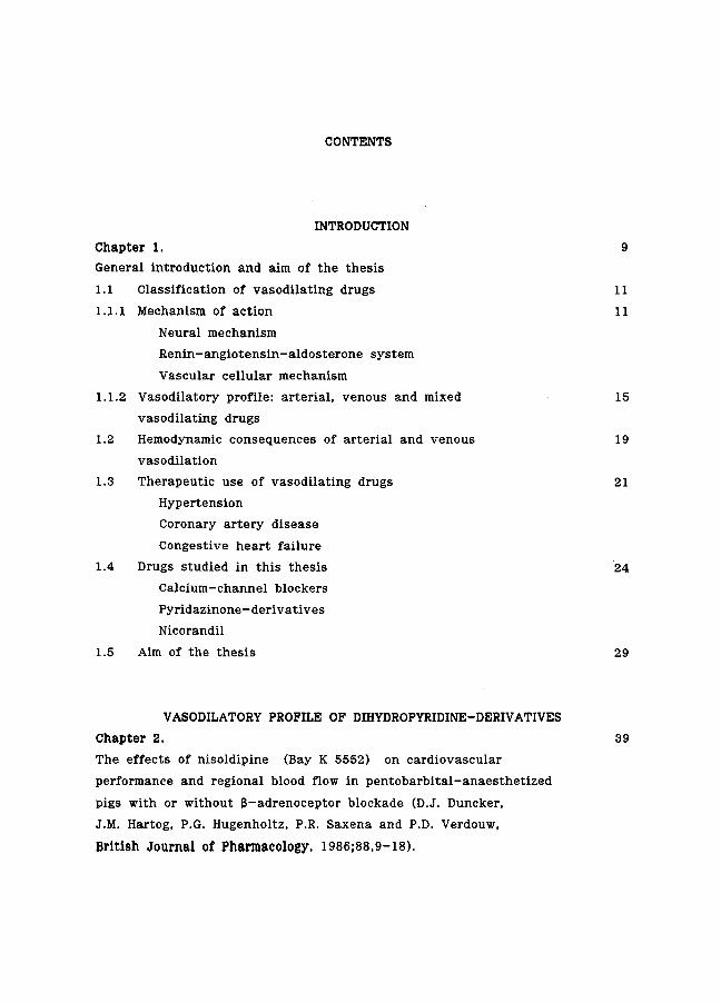
CONTENTS
INTRODUCTION
Chapter 1.
General introduction and aim of the thesis
1.1 Classification of vasodilating drugs
1.1.1 Mechanism of action
Neural mechanism
Renin -angiotensin -aldosterone system
Vascular cellular mechanism
1.1.2 Vasodilatory profile: arterial, venous and mixed
vasodilating drugs
1.2
1.3
1.4
1.5
Hemodynamic consequences of arterial and venous
vasodilation
Therapeutic use of vasodilating drugs
Hypertension
Coronary artery disease
Congestive heart failure
Drugs studied in this thesis
Calcium-channel blockers
Pyridazinone-derivatives
Nicorandil
Aim of the thesis
V ASODILATORY PROFILE OF DIHYDROPYRIDINE-DERIVATIVES
Chapter 2.
The effects of nisoldipine (Bay K 5552) on cardiovascular
performance and regional blood flow in pentobarbital-anaesthetized
pigs with or without 13-adrenoceptor blockade (D.J. Duncker,
J.M. Hartog, P.G. Hugenholtz, P.R. Saxena and P.D. Verdouw,
British Journal of Pharmacology, 1986;88,9-18).
9
11
11
15
19
21
24
29
39

Chapter 3.
The effects of nisoldipine alone and in combination with
beta-adrenoceptor blockade on systemic haemodynamics and
myocardial performance in conscious pigs (D.J. Duncker,
P.R. Saxena and P.D. Verdouw, European Heart Journal,
1987; in press).
Chapter 4.
Nimodipine-i;nduced changes in the distribution of carotid
blood flow and cardiac output in pentobarbitone-anaesthetized
pigs (D.J. Duncker, J. Heiligers, E.J. Mylecharane,
P.R. Saxena and P.D. Verdouw, British Journal of
Pharmacoiogy, 1986;89,35-46).
Chapter 5.
Enhancement of vasoconstrictor and attenuation of vasodilator
effects of 5-hydroxytryptamine by the calcium ~hannel blockers
nimodipine and nifedipine in the pig (D.J. Duncker, M.J. Yland,
L.P. Van der Wey, P.R. Saxena and P.D. Verdouw,
European Journal of Pharmacology, 1987;136,11-21).
VASODILATORY PROFILE OF PYRIDAZINONE-DERIVATIVES
Chapter 6.
Cardiovascular profile of pimobendan, a benzimidazole-pyridazinone
derivative with vasodilating and inotropic properties
(P.D. Verdouw, J.M. Hartog, D.J. Duncker, W. Roth and
P.R. Saxena, European Journal of Pharmacology. 1986;126,21-30).
Chapter 7.
Usefulness of pimobendan in the treatment of heart failure
(D.J. Duncker, F.J. van Dalen, J.M. Hartog, J.M.J. Lamers,
R.J. Rensen, P.R. Saxena and P.D. Verdouw,
Arzneimittel Forschung/Drug Research, 1986;36(II),l740-1744).
51
61
75
91
105

Chapter 8.
Cardiovascular effects of UD-CG 212 CL, a metabolite of
pimobendan, in anaesthetized pigs (P.O. Verdouw, L. Levinsky,
D.J. Duncker, A.M. Rutteman and P.R. Saxena,
European Journal of Pharmacology, 1987;137,219-226).
VASODILATORY PROFILE OF NICORANDIL
Chapter 9.
Nicorandil-lnduced changes in the distribution of cardiac output
and coronary blood flow in pigs (P.O. Verdouw, L.M.A. Sassen,
D.J. Duncker, I.O.L. Schmeets, R.J. Rensen and P.R. Saxena,
Naunyn-Schmiedeberg's Archives of Pharmacology, 1987;336,352-358).
SYSTEMIC HEMODYNAMIC ACTIONS OF VASODILATING DRUGS IN THE
ABSENCE OR PRESENCE OF ~-ADRENOCEPTOR BLOCKADE
Chapter 10.
Comparison of the systemic hemodynamic actions of
dihydropyridine calcium-channel blockers in conscious pigs
with or without 13-adrenoceptor blockade.
Chapter 11.
Systemic haemodynamic actions of pimobendan (UD-CG 115 BS) and
its 0-demethylmetabolite UD-CG 212 Cl in the conscious pig
(D.J. Duncker, J.M Hartog, L. Levinsky and P.O. Verdouw,
British Journal of Pharmacology, 1987;91,609-615).
VASODILATOR THERAPY AND MYOCARDIAL ISCHEMIA
Chapter 12.
Nisoldipine and perfusion of post-stenotic myocardium in
conscious pigs with different degrees of concentric stenosis
(D.J. Duncker, J.P.C. Heiligers, P.R. Saxena and P.O. Verdouw,
British Journal of Pharmacology; in press).
111
123
135
147
159

Chapter 13.
Exercise-induced ischemia in pigs: Effects of nisoldipine
with or without propranolol (D.J. Duncker, J.P.C. Heiligers,
P.R. Saxena and P.D. Verdouw, submitted).
DISCUSSION
Chapter 14.
General discussion and conclusions.
14.1 Methodological considerations
The experimental animal
Model for the study of myocardial ischemia
14.2 Systemic hemodynamic actions of vasodilating drugs in
conscious pigs
177
193
195
198
14.3 Influence of anesthesia on the systemic hemodynamic profile 203
of vasodilating drugs
14.4 Vasodilatory profile of vasodilating drugs 204
14.5 Conclusions and perspectives 208
SUMMARY 215
SAMENV ATTING 225
LIST OF PUBLICATIONS 235
CURRICULUM VITAE 241
DANKWOORD 245

INTRODUCTION
CHAPTER 1
GENERAL INTRODUCTION AND AIM OF THE THESIS


11
INTRODUCTION
Chapter 1. General introduction and aim of the thesis
1.1 Classification of vasodilating drugs
Vasodilating drugs can be classified on basis of the mechanism of action
but also on the ultimate vasodilator profile, i.e. venodilation, arteriodilation
or a combination of both. In the following sections a brief overview has
been presented of the classification of vasodilating drugs according to these
two criteria.
1.1.1 Mechanism of action
Vasodilation can be induced basically by three different mechanisms of
action: Interference with the neural or humoral (the renin-angiotensin
aldosterone system) control of vasomotor tone and an action on the vascular
wall.
Neural mechanism
The cardiovascular control centers of the medulla oblongata receive their
input from higher brain centers and from the afferent nerve endings of
mechanoreceptors (Fig. 1 ). From these medullary centers signals are mediated
to the heart via both sympathetic and parasympathetic nerve fibers and to
the vasculature via predominantly sympathetic nerve fibers. Drugs may
interact with the neural regulation of vasomotor tone at different levels. For
example, veratrum alkaloids decrease the sympathetic activity by an increase
in sensitivity of the baroreceptors. Examples of drugs affecting the higher
brain centers are the sedatives, hypnotics, tranquilizers, rauwolfia alkaloids
and a-methyldopa. The latter, as well as the a2-adrenoceptor agonist
clonidine, also directly affects the medullary centers. On the level of the
ganglia, ganglion-blockers like hexamethonium impair neural transmission. At
the adrenergic nerve endings interference with noradrenaline synthesis
(a-methyldopa), storage (rauwolfia alkaloids), or release into the synaptic cleft
(guanethidine, bretylium) can reduce the noradrenaline-mediated vasomotor
tone. Finally, interference with neural control can be accomplished by
blockade of post-junctional eXt- (present on veins and arterioles) and

Fig. 1
Afferent fibers of baroreceptors
Arterioles
®
Veins
Interference with the neural regulation of vasomotor tone by drugs at different levels of the nervous system. 1 = Veratrum alkaloids; 2 = sedatives, hypnotics, tranquillizers and a-methyldopa; 3 = a-methyldopa and clonidine; 4 = ganglion-blocking drugs; 5 = a-methyldopa; 6 = rauwolfia alkaloids; 7 = adrenergic blocking drugs; 8 = at-adrenoceptor blockers; 9 = a2-adrenoceptor blocker. Presynaptic receptors have been omitted from this figure. For a description see text.

13
az- {present principally on arterioles) adrenoceptors with prazosin {a1),
yohimbine (az) and phentolamine {a1 and az). The role of pre-junctional
receptors will not be discussed here; for a review on this subject see Langer
and Armstrong (1986).
Renin -angiotensin -aldosterone system
In addition to the neural control, vasomotor tone can be influenced
humorally via the renin-angiotensin-aldosterone system {for an extensive
review see Dzau and Pratt, 1986). Several factors including an increase in
sympathetic nerve activity, a reduction in renal perfusion pressure and the
anti-diuretic hormone concentration, a decreased sodium and/or chloride load
and other changes in plasma electrolyte concentrations (K+ , Mg2+ ) enhance
the release of the proteolytic enzyme renin (for a review see Derkx, 1987)
from the juxtaglomerular cells in the afferent arterioles of the kidneys
(Fig. 2). By cleaving the leucyl-leucine bond, renin converts angiotensinogen
into angiotensin-! which in turn is converted into angiotensin-11 via the
angiotensin converting enzyme. Angiotensin-11 exerts a number of
pharmacological actions, such as an aldosterone-mediated sodium retention,
arteria- and venoconstriction and .cardiostimulation. Drugs may interfere with
the actions of the renin-angiotensin-aldosterone system by ~-adrenoceptor
blockade at the level of the juxtaglomerular cells or by inhibition of renin or
the angiotensin converting enzyme. Furthermore, the action of angiotensin-11
can be blocked by the angiotensin-II antagonists and the angiotensin-11-
induced increase in the aldosterone activity can be offset by aldosterone
inhibitors like spironolactone (Fig. 2).
Vascular cellular mechanism
Vasodilating drugs which induce vasodilation at the level of the vascular
wall are often considered to be "true vasodilators". Some of the events
involved in the vascular contraction and possible sites of interference with
contractile processes by vasodilating drugs have been schematically depicted
in Fig. 3; for details, one may refer to review reports {Bolton, 1979; Cauvin
et al., 1983; Van Zwieten, 1984). The free calcium concentration in the
cytosol can be increased by a calcium release from intracellular stores and
transmembrane influx through receptor- and potential operated channels. The
increase in the free calcium concentration in the cytosol leads via

14
Fig. 2
Renal perfusion pressure
Heart rate i myocardial contractility
Sympathetic nervous system
CD p
Juxtaglomerular cells
Angiotensinogen
Angiotensin I
.. II®
I Angiotensin II I
=t® Ne""'' aod l
adrenal catecholamine
release
=I=® j_ Arterial· l and
venous tone
Other influences 14-- (~. ADH, ANF)
ACE
II I Aldosterone II L
@) = ~®
Plasma volume
il
i
Interference with the humoral regulation of vasomotor tone by drugs at different levels of the renin-angiotension-aldosterone system. 1 = 13-adrenoceptor blockers; 2 = renin inhibitors; 3 = angiotensin converting enzyme inhibitors; 4 = angiotensin-II antagonists; 5 = aldosterone antagonists. For a description see text.

15
calcium-calmodulin binding to kinase activation and, finally, to actin-myosin
activation resulting in smooth muscle contraction. Interference with the
influx of extracellular calcium can be induced by blockade of the receptors
regulating the receptor operated channels (az-adrenoceptor and probably the
angiotensin-II receptor and 5-hydroxytryptaminez receptor), by
hyperpolarization which inhibits influx through the potential operated
channels or by blocking the potential- and probably also receptor operated
channels with .calcium-channel blockers. Calcium-channel blockers do not
seem to antagonize the a1-adrenoceptor-mediated vasoconstriction since these
receptors induce a release of calcium from intracellular stores rather than
mediate an influx of calcium through calcium-channels (Van Zwieten and
Timmermans, 1983). Drugs that increase the intracellular cAMP- or cGMP
content cause relaxation probably due to inhibition of kinase activity (cAMP
and cGMP), a reduction in the free calcium concentration (cAMP) or
inhibition of calcium influx (cGMP). An increase in these two second
messengers can be accomplished by 13z-adrenoceptor stimulation (cAMP), an
inhibition of cAMP breakdown by phosphodiesterase-inhibitors, an
enhancement of the cyclase-activity either "directly", for example by nitrates,
or via the release of endothelium derived compounds such as the endothelium
derived relaxing factor(s) or prostacyclin. Vasodilation can thus also be
induced via the endothelium by the "endothelium-dependent" vasodilators of
which acetylcholine, ATP, ADP, substance P, bradykinin, arachidonic acid and
the phospholipase Az inhibitor quinacrine are representatives. These
substances need, in contrast to the "endothelium-independent" vasodilators,
like the calcium-channel blockers and nitrates, an intact endothelium for their
vasodilator actions and a lack thereof may lead to vasoconstriction rather
than vasodilation (see Furchgott, 1983). The (or one of the) endothelium
derived relaxing factor(s) has recently been claimed to be nitric oxide
(Moncada et al, 1987).
1.1.2 Vasodilator profile: arterial, venous and mixed vasodilation.
Vasodilator drugs often act preferentially on the arterial or venous
vasculature (Table 1), but the mechanism underlying this preference is not
always fully understood. Phentolamine, which blocks both the a1-and
az-adrenoceptors, dilates the arterioles more so than the veins (Miller et al.,
19 7 6), whereas the 0:1-blocker prazosin dilates the venous and arterial

16
Fig. 3
VASCULAR SMOOTH MUSCLE CELL
Adrenaline Noradrenaline
cGMP GTP
Nitrates
Cell membrane
Adrenaline
/
cAM\ Phospho-Calmoduli ~ diesterase 1 S'AMP
Actin-Myosin activation Kinase
cAMP ATP
ATP Adenosine
~
Contraction
Phosphodiesterase inhibitors
Schematic representation of some of the pathways 1 involved in the contraction of a vascular smooth muscle cell and possible sites of interefence with vasoconstriction by vasodilating substances. ROC = receptor operated channels; POC = potential operated channel; AC = adenylate cyclase; GC = guanylate cyclase; EDRF = endothelium derived relaxing factor; a1 = a1- adrenoceptor; 15z = 15z-adrenoceptor; Pz = purinergicz-receptor. Az = adenosinez-receptor. For a description see text.

17
vasculature to a similar extent (see Scriabine and Taylor, 1986). It can be
speculated that these differences are due to the distribution of a-adrenergic
receptors. While both post-synaptic a1 and az-adrenoceptors have been
clearly shown in 'in-vivo' studies to be present in arterioles, the
a1-adrenoceptor seems to be the dominant receptor in the venous capacitance
vessels in several species (see Langer and Hicks, 1984). The calcium-channel
blockers act predominantly on the arterial side which might be due to the
distribution of az-adrenoceptors as suggested above. The az-adrenoceptor
mediated vasoconstriction has namely been associated with an influx of
extracellular calcium and been demonstrated to be susceptible to
calcium-channel blockade (Van Zwieten and Timmermans, 1983). However, a
great variety exists between different tissues and species with respect to
a-adrenoceptor distribution and the susceptibility of the a-adrenoceptors to
calcium-channel blockers (Timmermans and van Meel, 1983; Cauvin et al.,
1983; Vanhoutte, 1985). This indicates that no firm conclusions can be drawn
when trying to explain the profile of the above mentioned drugs.
Hydralazine, minoxidil and to a lesser extent, angiotensin converting enzyme
inhibitors also have a preference for arterial vessels. Nitrates, like
nitroglycerine and isosorbide-dinitrate, act predominantly on the venous
vasculature whereas nitroprusside and, as mentioned above, prazosin act
equally on both the arterial and venous vessels.
Table 1. Classification of vasodilating drugs based on their preference for
arterial, venous or both types of blood vessels.
Arterial
Hydralazine
Minoxidil
Calcium-channel blockers
Angiotensin converting
enzyme inhibitors
Phentolamine
Venous
Nitroglycerin
Isosorbide-dinitrate
Arterial + Venous
Prazosin
Nitroprusside

18
Fig. 4
tone
TPR
e
co
Preload
Venous j capacitance
e
e
Renal blood flow
i
Renin-.__ Angiotensin j
system activity
Heart rate j Contractility 1+--------Jf'-----'
Circulating i 1+------1 blood volume
Aldosterone j
Schematic representation of the changes in systemic hemodynamic variables and in tissue blood flow and some of the consequences of counterregulatory mechanisms invoked during treatment with vasodilating drugs. TPR = total peripheral resistance; CO = cardiac output; BP = blood pressure. The interrupted (with a minus sign) and uninterrupted lines indicate, respectively, the reduction and facilitation of the variable involved.

19
1.2 Hemodynamic consequences of arterial and venous vasodilation The systemic and regional hemodynamic profiles of vasodilators have been
recently reviewed (Saxena and Bolt, 1986). Briefly, arterial vasodilation
results in a decrease in total peripheral vascular resistance and consequently
in a decrease in arterial blood pressure, whereas venous vasodilation leads to
an increase in venous capacitance and, therefore, a reduction in ventricular
filling pressure, stroke volume and cardiac output (Fig. 4). Thus vasodilation
on either side results in a drop in arterial blood pressure. In an attempt to
maintain arterial blood pressure, several counterregulatory mechanisms (neural
as well as humoral) become operative; these counterregulatory responses may
modify the direct vasodilator actions of vasodilating drugs on organs and
tissues (Fig. 4).
The effects of counterregulation are unique for the left ventricle since
this part of the heart compromises its own blood supply during part of the
cardiac cycle. Only 15% of the coronary blood flow, nourishing almost
exclusively the subepicardium, occurs during systole because of the
extravascular compression and wall tension developed during this part of the
cardiac cycle (see Berne and Rubio, 1979). The remaining 85% of coronary
flow occurs during diastole and supplies the subendocardial and, to a lesser
extent, subepicardial layers. In the subendocardial layers, which depend thus
entirely on diastole for their perfusion, vasodilator reserve is exhausted at a
higher perfusion pressure (70 mmHg in dogs) than in the subepicardial layers
(40 mmHg; Winbury and Howe, 1979). In the physiological state, like during
exercise, vasodilator reserve in the subendocardial layers is sufficient to meet
the increased oxygen-demand of the myocardium. The increase in heart rate,
which causes a reduction in duration of diastole, does not exhaust vasodilator
reserve because even during heavy exercise arterial blood pressure is
maintained or elevated. On the other hand, the reduction in blood pressure
(by peripheral vasodilation) and the baroreceptor reflex-mediated tachycardia
(especially during acute administration of vasodilators) decrease both the
perfusion pressure of the subendocardial layers and the perfusion time
(Fig. 5). As a result the increase in subendocardial blood flow is. less than
the increase in subepicardial flow. This holds true especially for arterial
vasodilation since with venodilation preload, and thus intramyocardial pressure
in the subendocardial layers, is reduced which favorably influences the
perfusion pressure of the subendocardial layers. For myocardium supplied by

20
Fig. 5
Subendocardial blood flow i
Total l t-------------.! peripheral
Subepicardial blood flow i
e
resistance
el I le
L--------Blood 1 pressure
Diastolic perfusion time
Heart rate i
Diastolic perfusion pressure
Preload l / 8/
/
Vasodilation and its consequences for the perfusion of the different myocardial layers. V = vasodilator; The interrupted lines (with a minus sign) and interrupted lines indicate, respectively, the reduction and facilitation of the variable involved. For a description see text.

21
non -diseased coronary arteries the reduction in the subendocardial
subepicardial blood flow ratio induced by the vasodilating drugs has no
clinical implications as subendocardial blood flow is still commensurate with
the needs of the subendocardial layers. However, when a coronary artery is
obstructed and vasodilator reserve is already reduced, the combination of a
decrease in arterial blood pressure and a reflex-tachycardia may have
deleterious effects on the perfusion of especially the subendocardial layers
(see section 1.3 coronary artery disease).
1.3 Therapeutic use of vasodllatlng drugs Hypertension
The use of vasodilators in hypertension is a rational approach since these
drugs antagonize the major hemodynamic disturbance in most forms of
hypertension namely the increase in total peripheral resistance. To prevent
counterregulatory mechanisms from abolishing the beneficial actions of
vasodilators, a combination therapy with 13-adrenoceptor antagonists and/or
diuretic agents is often needed in the clinical situation. Furthermore, during
chronic treatment 13-adrenoceptor antagonists have an additive hypotensive
action which may allow reduction of the doses of the drugs used.
Coronary artery disease
Nitrates, which have a more marked action on the venous side, have been
used for decades in the treatment of angina pectoris due to different causes.
The beneficial actions of nitrates is primarily ascribed to the reduction of
cardiac preload and, to some extent, afterload (Williams et al., 1965; Burggraff
and Parker, 1974) which decrease ventricular dimension and wall tension and
therefore lower myocardial oxygen demand. The decrease in wall tension also
increases the effective diastolic perfusion pressure of the subendocardial
layers. Another more recently appreciated factor contributing to the anti
ischemic actions of nitrates is that these drugs dilate the large epicardial
vessels and thereby reduce the severity of the coronary artery stenosis
(Brown et al., 1981).
In the last decade calcium-channel blockers have been under investigation
in the treatment of coronary artery disease. Although their efficacy has been
proven in coronary artery spasm, the usefulness of calcium-channel blockers
in stable angina pectoris remains a matter of debate as in the clinical setting

22
arterial vasodilators not always favorably influence the myocardial oxygen
balance. By reducing the afterload the arterial vasodilators often cause an
increase in heart rate, a major determinant of myocardial oxygen-demand.
The increase in heart rate also reduces the duration of the diastolic perfusion
time. This, together with the reduction in perfusion pressure by the
hypotensive action of these drugs may be harmful for, in particular, flow to
the subendocardial layers. An increase in blood flow can still occur via
dilatiop at the ,site of the coronary artery stenosis {which is not always
possible), via dilation of coronary collaterals (which may not always be
present) or via dilation in the terminal arteriolar bed (which is generally
believed to be maximal during myocardial ischemia). But vasodilators have
also been reported to induce "coronary steal" either from subendocardium to
subepicardium distal to a fixed corQnary artery stenosis (Gross and Warltier,
1981; Weintraub et al., 1981; Gewirtz et al., 1984) or from ischemic
myocardium distal to a completely occluded coronary artery to normally
perfused myocardium (Gross and Warltier, 1981). These observations have led
to the concept that in ischemic myocardium arterial vasodilators reduce the
distal coronary perfusion pressure , by vasodilation irt non-ischemic
myocardium,
myocardium.
thereby "stealing" blood from perfusion pressure dependent
Recently, however, several studies have suggested that
vasodilation is not maximal in ischemic myocardium in which case vasodilator
therapy may still be useful (Gorman et al., 1984; Heusch and Deussen, 1984;
Aversano and Becker, 1985; Canty and Klocke, 1985; Pantely et al., 1985).
Furthermore, combination therapy with ~-adrenoceptor antagonists has proven
superior to monotherapy with 1 either class of drugs. This has revived
interest in vasodilators as a therapeutic in (stable) angina pectoris.
Congestive Heart Failure
A role for vasodilators in heart failure (a situation in which the heart is
unable to pump blood at a rate commensurate with the metabolic needs of
body tissues), seems at first less obvious than in the case of hypertension
since the primary cause for heart failure is often the myocardium. However,
the peripheral vasculature responds to cardiac pump failure with constriction
of the arterioles and veins causing a further deteriorating of myocardial
performance. The aim of vasodilator therapy is therefore to reduce pre- and
afterload of the heart, thereby normalizing the ventricular dimension and

23
pump function (Cohn and Franciosa, 1977; Chatterjee and Parmley, 1977).
Furthermore, in the failing circulation the arteriolar constriction is
particularly pronounced in the renal, splanchnic, dermal and muscular regions
(see Drexler et al., 1985, 1986a) and vasodilators should therefore preferably
dilate these vascular beds in order to normalize perfusion thereof.
Migraine
The pathogenesis of migraine and related headaches remains controversial.
Several hypotheses, like the neuronal, vascular, or ischemia hypothesis (Blau,
1987), have not been able to completely explain the clinical signs of
migrainous headaches. The reason for these different theories regarding the
pathophysiology of migraine may be that migraine, like hypertension, is a
syndrome where the underlying causative factors may vary in different
patients.
The introduction of vasodilators (calcium-channel blockers) in the
treatment of migraine is based on two separate reasonings. The first
involves the hypothesis by which advocates initial, mainly intracranial,
vasoconstriction during the prodromal phase, followed by extracranial
vasodilation causing the head pain (Wolff, 1963). Although this concept of a
pure vascular basis of migraine headache may be an oversimplification (Olesen
et al., 1981; Bruyn, 1984), a number of studies have found a reduced cerebral
blood flow during the initial phase of migraine (O'Brien, 1971; Skinhoj, 1973;
Simard and Paulson, 1973; Norris et al., 1975; Henry et al., 1978; Sakai and
Meyer, 1978). Yamamoto and Meyer (1980) considered that calcium-channel
blockers would prevent the initial cerebral vasospasm and, therefore, also
mitigate the subsequent painful vasodilation during the headache phase.
Indeed, some studies indicate that nimodipine (Gelmers, 1983; Meyer and
Hardenberg, 1983) and nifedipine (Kahan et al., 1983), may be of value in
migraine prophylaxis. The second reasoning, based on which another calcium
channel blocker flunarizine was introduced assumes that migraine is due to a
focal cerebral ischaemia (Amery, 1982). The resulting accumulation of calcium
and the consequent cell damage and migraine headache would then be
suppressed by calcium-channel blockers.
Another observation which appears to be of interest is the high potency
of nimodipine in antagonizing rabbit basilar artery contractions induced by
5-hydroxytryptamine (Towart, 1981); the latter is released (and then depleted)

24
from blood platelets during migraine headaches (Lance, 1982).
5-Hydroxytryptamine and several antimigraine drugs (Johnson and Saxena,
1978; Saxena and Verdouw, 1982; Saxena et al.,l983; Saxena, 1987) are
extremely effective in constricting cranial extracerebral arteriovenous
anastomoses, which may open up during the headache phase of migraine
(Heyck, 1969; Saxena, 1978}. The relationship between 5-hydroxytryptamine,
arteriovenous shunting and calcium-channel blockers is, however, not known.
·gl ~ N02
CHs~o-q C·O-CH o I o s CH3 ~ CHs
H
Nifedipine
Fig. 6
Verapamil
~OCH3
V"ocH3
(:(»_:::· Nl-0-~ I o CH2-CH2 - N (CH3)2
Diltiazem
Chemical structure of the three 'first generation' calcium-channel blockers verapamil, nifedipine and diltiazem. Note the great differences in structure.
1.4 Drugs used in this thesis
Calcium-channel blockers
In the early sixties German scientists (Lindner, 1960; Haas and Hartfelder,
1962} observed that prenylamine and verapamil exerted, besides coronary
vasodilator actions, negative inotropic effects on isolated cat and rabbit

25
myocardium. At first these drugs were believed to be 13-adrenoceptor
blocking agents as they opposed the catecholamine-induced effects on the
heart (Melville and Benfey, 1965; Haas and Busch, 1967). However,
Fleckenstein et al. (1967) reported that the effects of prenylamine and
TABLE 2 RELATIVE EFFECTS OF CALCIUM ANTAGONISTS IN EXPERIMENTAL PREPARATIONS COMPARED WITH THERAPEUTIC LEVELS IN MAN*
Verapamil Nifedipine Diltiazem
Therapeutic level in man ng/ml 80-400 25-100 50-300 molecular weight 455 346 415 molar weight 2-8 X 10·7 M 0.5-2 X 1 o-7 M 1-7 X 10·7 M protein binding about 90% about 95% about 85% molar value, corrected for protein 2-8 X 10·8 M 0.3-1 X 10·8 M 1-5 X 10·8 M binding
Isolated coronary artery contraction 10·7 M 10·8 M 10·7 M 50% inhibition
Myocardial depression sx10-aM 5X1Q·7 M 10·4 M 40% depression of contractile force
Fast sodium current depression 10·4 M no effect 10·4 M
Slowing of heart rate by 20% 1o-a M 10·5 M 10·8 M
Relative effect on AV node vs 6.5:1 1:1 20:1 contractile force
* From Singh and Opie (1984)
verapamil were the result of inhibition of the influx of calcium into the
I'll:yocardial cells. Consequently, these agents were called calcium-antagonists
and they were ascribed two main properties: 1) suppression of the slow
calcium current in ventricular muscle and 2) the reversal of such suppression
by an increase in external calcium concentration (Fleckenstein, 1968, 1977).
Later, other properties were discovered such as the protection against
isoprenaline-induced myocardial calcium overload and inhibition of

26
calcium-induced tension development in vascular smooth muscle. With the
gradual elucidation of their main mechanism of action these agents are now
more generally called calcium-entry blockers or calcium-channel blockers.
During the last decade the "first generation" calcium-channel blockers
verapamil, nifedipine and diltiazem, which are structurally unrelated to each
other (Fig. 6), have become widely used in cardiovascular disorders. With
the differences in chemical structure also the affinity for the myocardium and
the vasculature varies widely (Table 1). In recent years the so called "second
generation" agents, analogues of the first generation drugs, have been
developed in an attempt to obtain substances with greater specificity for
either cardiac (sinus-node, A V -node, myocytes) or vascular (specific vascular
bed) tissues. In this respect especially the dihydropyridine-derivatives
Nifedipine
Nimodipine Nisoldipine
Fig. 7 Chemical structure of the dihydropyridine calcium-channel blockers nifedipine, nisoldipine and nimodipine.

27
(nifedipine, nisoldipine, nimodipine; Fig. 7) have grown in number. Compared
to the parent compound nifedipine, some of these agents have been claimed to
have a greater selectivity for vascular tissue over cardiac tissue (Kazda et al.,
1980) or for cerebral or coronary arterial beds over other regional beds
(1'akenaka et al., 1976; Kazda et al., 1980, 1982).
Current indications for the clinical use of calcium-channel blockers vary
from stable angina pectoris (Scheidt, 1982), angina at rest and coronary
vasospasm (Schroeder, 1982), hypertension, hypertrophic cardiomyopathy and
myocardial infarction (Toggart and Zelis, 1982) and cardiac arrhythmias (Singh
et al., 1982) to prophylaxis of migraine (Gelmers, 1983) and cerebral ischemia
due to post-hemorrhagic cerebral vasospasm (Allen et al., 1983).
Xanthine
0 CH3
H3C ~x· N N~ O~N I N
I CH3
Caffe"ine
N
~~0-CH3 Pimobendan
Fig. 8
0 H
H3C, ~):N N I )
0)-__N N I
CH3
Theophylline
H
O~*N,IN '7 I N
CH3 ~ ~~OH
UD·CG 212 Cl
Chemical structure of xanthine, the methylxanthines caffeine and theophylline and the pyridazinone-derivatives pimobendan and its 0-demethylmetabolite UD-CG 212 Cl. *All substances contain the "active" part of the molecule which is thought to be responsible for the phosphodiesterase-inhibiting actions of these drugs.

28
Pyridazinone-derivatives
This class of drugs, to which pimobendan and its 0-demethylmetabolite
UD-CG 212 Cl (2-(4-hydroxy-phenyl)-5-(5-methyl-3-oxo-4,5-dihydro-2H-6-
pyridazinyl)benzimidazole HCl) belong (Fig. 8), exert vasodilator actions on
the arterial as well as on the venous side (Diederen et al., 1982). Besides
vasodilator effects the pyridazinone-derivatives possess cardiostimulatory
actions (Diederen et al., 1982; Van Meel, 1985). The mechanism through
which the pyridazinone-derivatives exert their actions was at first thought to
be via phosphodiesterase-inhibition (see Honerjager et al., 1984; Berger et al.,
1985); they share with the phosphodiesterase inhibiting (methyl) xanthines the
so called "active site" of the molecule which is thought to be responsible for
phosphodiesterase-inhibition (Fig. 8). However, in addition, the cardiotonic
actions may also be due to an increased sensitivity of myocardial contractile
proteins to calcium (Ruegg et al., 1984; Van Meel et al., 1986) or a
prolongation of the action potential, allowing more calcium to enter the
myocyte (Honerjager et al., 1984).
The cardiovascular profile, i.e. vasodilation of venous and arterial beds
and positive inotropic actions, appears to be particularly favorable for the
therapy of congestive heart failure (Cohn and Franciosa, 1978). Indeed,
pimobendan has recently been reported to improve cardiac pump function and
normalize preload in patients with congestive heart failure at rest (Brand and
Hagemeyer, 1987) and during exercise (Hagemeyer and Brand, 1987).
NITROGLYCERIN NICORANDIL
Fig. 9 Chemical structure of the classical nitrate nitroglycerin and the nitrate-like substance nicorandil.

29
Nicorandil
Nicorandil (SG-75, N-(2-hydroxyethyl) nicotinamide nitrate; Fig. 9) is a
new anti-anginal drug with coronary vasodilator properties (Uchida et al.,
1978). In vitro studies have shown that besides the nitrate-like action, an
increase in intracellular cGMP (Fig. 3), nicorandil also enhances the
conductance of potassium-channels thereby hyperpolarizing the cell membrane
(Taira, 1987). As a result nicorandil, in addition to the classical nitrate
venodilatory action, has a potent effect on the arterial smooth muscle
vasculature and dilates epicardial conductance as well as intramyocardial
resistance vessels (Taira, 1987; Suryapranata et al., 1988). At clinical
dose-range the drug possesses negligible negative inotropic actions (Belz et
al., 1984).
The profile of venous and arterial dilation, together with the reported
anti -ischemic actions (Uchida, 1978; Thorman et al., 1982, 1983) suggest that
nicorandil might be useful in the treatment of heart failure, coronary artery
disease and hypertension.
1.6 Aim of the thesis
In this thesis the cardiovascular pharmacological actions of a number of
vasodilating drugs have been studied and compared in pigs. Firstly, we
compared the vasodilator profile of the dihydropyridine-derivatives
nisoldipine, nimodipine and nifedipine and also studied the claimed preference
of nisoldipine for the coronary circulation (Kazda et al., 1980; Serruys et al.,
1985; Drexler et al., 1986b) and of nimodipine for the cerebral circulation
(Kazda et al., 1982) (chapters 2-5). Secondly, since the perfusion of organs
and tissues may be compromised in several pathological disorders for which
vasodilating drugs might be employed (ischemic heart disease, heart failure,
hypertension), we compared the vasodilator profiles of the dihydropyridine
derivatives nisoldipine and nimodipine with those of pimobendan, UD-CG 212
CL and nicorandil after intravenous administration in anesthetized pigs
(chapters 6-9). Thirdly, in view of the potentially useful combination of
vasodilators and 13-adrenoceptor antagonists to reduce h;,rpotension -induced
baroreceptor reflex-mediated tachycardia, we compared the systemic
hemodynamic actions of the different vasodilating drugs in conscious pigs in
the absence or presence of 13-adrenoceptor blockade (chapters 9-11). Finally,
the long-held view that vasodilation in ischemic myocardium is maximal (see

30
Berne and Rubio, 1979) and, therefore, vasodilators may induce coronary
"steal" (Weintraub et al., 1981; Gross and Warltier, 1981; Gewirtz et al., 1984),
may not hold true. Recently, vasodilator therapy has been shown to induce
an improvement of perfusion and function of ischemic myocardial areas by
recruiting vasodilator reserve (Gorman. et al., 1984; Heusch and Deussen, 1984;
Aversano and Becker, 1985; Canty and Klocke, 1985; Pantely et al., 1985). In
view of these new findings, lending support to the possible usefulness of
vasodilator drugs in myocardial ischemia, we investigated the effects of
nisoldipine on perfusion of myocardium distal to a fixed concentric coronary
artery stenosis in conscious pigs (chapter 12). In. addition, the possible
anti-ischemic action of this drug was studied during myocardial ischemia
induced in animals subjected to treadmill-exercise (chapter 13).
References
Allen, G.S., Ahn, H.S., Preziosi, T.J., Battye, R., Boone, S.C., Boone, S.C., Chou, S.N. Kelly, D.L., Weir, B.K., Crabbe, R.A., Lavik, P.J., Rosenbloom, S.B., Dorsey, F.C., Insram, C.R., Mellits, D.D., Bertsch, L.A., Bo~svert, D.P., Hundley, M.B., Johnson, R.K., Strom, J.A. and Transou, C.R. Cerebral arterial spasm - a controlled trial of nimodipine in patients with subarachnoid hemorrhage. N. Engl. J. Med .. 308 (11), 619-624, 1983.
Amery, W.H. Brain hypoxia: the turning-point in the genesis of the migraine attack? Cephalalgia 2, 83-109, 1982.
Aversano, T. and Becker, L.C. l'ersistence of coronary vasodilator reserve despite functionally significant flow reduction. Am. J. Physiol. 248, H403-H411, 1985.
Belz, G.G., Mathews, J., Heinrich, J. and Wagner, G. Controlled comparison of the pharmacodynamic effects of nicorandil (SG-75) and isosorbide dinitrate in man. Eur. J. Clin. Pharmacol. 26, 681-685, 1984.
Berger, C., Meyer, W., Scholz, H. and Starbatty, J. Effects of the benzimidazole deri va ti ves pimobendan and 2- ( 4- Hydroxyphenyl)-5-(5-methyl-3-oxo-4,5-dihydro-2H-6-pyridazinyl) benzimidazole HCl on phosphodiesterase activity and force of contraction in guinea-pig hearts. Arzneim. Forsch. I Drug Res. 35(1I), 1668-1673, 1985.
Berne, R.M. and Rubio, R. Coronary circulation. In: Handbook of physiology, Section 2: The Cardiovascular System, Vol. I: The heart, Eds: Berne, R.M., Sperelakis, N., Geiger, S.R., Waverly Press, Inc., Baltimore, 1979, pp. 873-952.
Blau, J.N. (Ed.). Migraine: Clinical, Therapeutic, Conceptual and Research Aspects, Chapman and Hall, London, 1987.
Bolton, T.B. Mechanisms of action of transmitters and other substances on smooth muscle. Physiol. Rev. 3, 606-718, 1979.
Brand, H.J. and Hagemeijer, F. Hemodynamic effects of pimobendan given orally to patients with severe heart failure. Eur. Heart J. 8 (Abstr. Suppl. 2), 276, 1987.

31
Brown, B.G., Bolson, E., Peterson, R.B., Pierce, C.D. and Dodge, H.T. The mechanism of nitroglycerin action: stenosis vasodilatation as a major component of the drug response. Circulation 64, 1098-1097, 1981.
Bruyn, G.W. The pathomechanism of migraine as a basis for pharmacotherapy: a clinician's epilogue. In: The pharmacological basis of migraine therapy. Eds: Amery, W.K., Van Nueten, J.M. and Wauquier, A. Pitman, London, 1984, pp. 267-278.
Burggraf!, G.W. and Parker, J.O. Left ventricular volume changes after amyl nitrate and nitroglycerin in man, as measured by ultrasound. Circulation
49, 136-143, 1974. Canty, J.M. and Klocke, F.J. Reduced regional myocardial perfusion in the
presence of pharmacologic vasodilator reserve. Circulation 71 (2), 370-377. 1985.
Cauvin, C., Loutzenhiser, R. and Van Breemen, C. Mechanisms of calcium antagonist-induced vasodilation. Ann. Rev. Pharmacol. To xi col. 23, 373-396, 1983.
Chatterjee, K. and Parmley, W.W. The role of vasodilator therapy in heart failure. Prog. Cardiovasc. Dis. 19, 301-325, 1977.
Cohn, J.N. and Franciosa, J.A. Vasodilator therapy of cardiac failure. N. Engl. J. Med. 297, 27-31, 254-258, 1977.
Cohn, J.N. and Franciosa, J.A. Selection of vasodilator inotropic or combined therapy for the management of heart failure. Am. J. Med. 65, 181, 1978.
Derkx, F.H.M. Human prorenin. Ph.D. thesis, Erasmus University Rotterdam, Rotterdam, the Netherlands, 1987.
Diederen, W., Dammgen, J. and Kadatz, R. Cardiovascular profile of UD-CG 115, a new orally and long acting cardiotonic compound, not related to 13-mimetics or cardiac glycosides. Naunyn -Schmiedeberg's Arch.· Pharmac. 321, R36, 141, 1982.
Drexler, H., Depenbusch, J.W., Truog, A.G., Zelis, R. and Flaim, S.F. Effects of diltiazem on cardiac function and regional blood flow at rest and during exercise in a conscious rat preparation of chronic heart failure (myocardial infarction). Circulation 71, 1262-1270, 1985.
Drexler, H., Toggart, E.J., Glick, M.R., Heald, J., Flaim, S.F. and ZeUs, R. Regional vascular adjustments during recovery from myocardial infarction in rats. J. Am. Coll. Cardiol. 8, 134-142, 1986a.
Drexler, H., Truog, A.G., Zelis, R. and Flaim, S.F. Regional vascular and hemodynamic effects of orally administered nisoldipine in conscious rats. J. Cardiovasc. Pharmacol. 8, 151-155, 1986b.
Dzau, V.J. and Pratt, R.E. Renin-angiotensin system: Biology, physiology, and pharmacology. In: The Heart and Cardiovascular System, Eds: Fozzard, H.A. Haber E., Jennings R.B., Katz A.M., Morgan H.E., Raven Press, New York, 1986, pp. 1631-1662. I
Fleckenstein, A., Kammermeier,· H., Doring, H. and Freund, H.J. Zum Wirkungs-mechanismus neuartiger Koronardilatatoren mit gleichzeitig Sauerstoff-einsparenden Myokard-Effekten, Prenylamin und Iproveratril. Z. Kreislaufforsch. 56, 716-744, 839-853, 1967.
Fleckenstein, A. Experimental heart failure due to disturbances in high-energy phosphate metabolism. In: Proceedings of the Fifth European Congress of Cardiology, Athens, 1968, pp. 255-269.
Fleckenstein, A. Specific pharmacology of calcium in myocardium, cardiac pacemakers and vascular smooth muscle. Annu. Rev. Pharmacol. Toxicol. 17, 149-166, 1977.

32
Furchgott, R.F. The role of endothelium in the responses of vascular smooth muscle to drugs. Ann. Rev. Pharmacol. Toxicol. 24, 175-197, 1984.
Gelmers, H.J. Nimodipine, a new calcium antagonist, in the prophylactic treatment of migraine. Headache 23, 106-109, 1983.
Gewirtz, H., Gross, S.L., Williams, D.O. and Most, A.S. Contrasting effects of nifedipine and adenosine on regional myocardial flow distribution and metabolism distal to a severe coronary arterial stenosis: observations in sedated, closed-chest, domestic swine. Circulation 5, 1048-1057, 1984.
Gorman, M.W., Wangler, R.D., Dewit, D.F. and Sparks, H.V. jr. Progressive vasomotor changes in ischaemic myocardium. Acta Med. Scand. 694 (Suppl), 38-44, 1984.
Gross, G.J. and Warltier, D.C. Coronary steal in four models of single or multivessel obstruction in dogs. Am. J. Cardiol. 48, 84-92, 1981.
Haas, H., and Busch, E. Vergleichende Untersuchungen der wirkung von a-isopropyl-a(n -methyl-N- homoveratryl)-T-aminopropyl)-3,4-dimethoxyphenylacetonitril, seiner Derivate sowie einiger andere Coronardilatatoren und ~-receptor-affiner Substanzen. Arzneim. Forsch. I Drug Res. 17, 257-271, 1967.
Haas, H. and Hartfelder, G. a-isopropyl-a-(n-methylhomoveratryl)T- aminopropyl)- 3, 4- dimethoxy-phenylacetonilril, eine Substanz mit coronargefaberweiternden Eigenschaften. Arzneim. Forsch. I Drug Res. 12, 549-558, 1962.
Hagemeyer, F. and Brand, H.J. Bicycle ergometry in heart failure: decreasing stroke volume with increasing pulmonary wedge pressure. Improvement after oral pimobendan. Eur. Heart J. 8 (Abstr. Suppl. 2), 352, 1987.
Henry, P.Y., Vernheit, J., Orgogozo, J.M., Caille, J.M. Cerebral bloodflow in migraine and cluster headache. Res. Clin. Stud. Headache 6, 81-88, 1978.
Heyck, H. Pathogenesis of migraine. Res. Clin. Stud. Headache 2, 1-28, 1969. Reusch, G. and Deussen, A. Nifedipine prevents sympathetic vasoconstriction
distal to severe coronary stenoses. J. Cardiovasc. Pharmacol. 6, 378, 1984. Honerjager, P., Heiss, A., Schafer-Korting, M., Schonsteiner, G. and
Reiter, M. UD-CG 115 a cardiotonic pyridazinone which elevates cyclic AMP and prolongs the action potential in guinea-pig papillary muscle. Naunyn-Schmiedeberg's Arch. Pharmac. 325, 259-269, 1984.
Johnson, B.M. and Saxena, P.R. The effect of ergotamine on tissue blood flow and the arteriovenous shunting of radioactive microspheres in the head. Br. J. Pharmacol. 63, 541-549, 1978.
Kahan, A, Weber, S., Amor, B., Guerin, F. and Degeorges, M. Nifedipine in the treatment of migraine in patients with Raynaud's phenomenon. N. Engl. J. Med. 308, 1102-1103, 1983.
Kazda, s., Garthoff, B., Meyer, H., Schlossmann, K., Stoepel, K., Towart, R., Vater, W. and Wehinger, E. Pharmacology of a new calcium antagonistic compound, isobutyl methyl 1,4-dihydro-2,6-dimethyl-4-(2-nitrophyenyl)-3,5-pyridinedicarboxylate (Nisoldipine, Bay K 5552). Arzneim. Forsch. I Drug Res. 30, 2144-2162, 1980.
Kazda, S., Garthoff, B., Krause, H.P. and Schlossmann, K. Cerebrovascular effects of the calcium antagonistic dihydropyridine derivative nimodipine in animal experiments. Arzneim. Forsch. I Drug Res. 32, 331-337, 1982.
Lance, J.W. Mechanism and Management of Migraine, 4th edn., Butterworths, London, 1982.

33
Langer, S.Z. and Hicks, P.E. Alpha-Adrenoceptor Subtypes in Blood Vessels: Physiology and Pharmacology. J. Cardiovasc. Pharmacol. 6, S547-S558, 1984.
Langer, S.Z. and Armstrong, J.M. Prejunctional receptors and the cardiovascular system: pharmacological and therapeutic relevance. In: Cardiovascular Pharmacology, Ed: Antonaccio, M., Raven Press, New York, 1984, pp. 197-213.
Lindner, E. Phenyl-propyl-diphenyl-propylamin, eine neue Substanz mit coronargefaber-weiternder Wirkung. Arzneim. Forsch. I Drug Res. 10, 569-573, 1960.
Melville, K.I. and Benfey, B.C. Coronary vasodilatory and cardiac adrenergic blocking effects of iproveratril. Can. J. Physiol. Pharmacol. 43, 339-342, 1965.
Meyer, J.S. and Hardenberg, J. Clinical effectiveness of calcium entry blockers in prophylactic treatment of migraine and cluster headaches. Headache 23, 266-277. 1983.
Meyer, J.S., Hata, T., Imai, A. Evidence supporting a vascular pathogenesis of migraine and cluster headache. In: Migraine - clinical, therapeutical, conceptual and research aspects, Ed: Blau, J.N., Chapman and Hall, London, 1987, pp. 265-302.
Miller, R.R., Vismara, L.A., Williams, D.O., Amsterdam, E.A. and Mason, D.T. Pharmacological mechanisms for left ventricular unloading in clinical congestive heart failure: differential effects of nitroprusside, phentolamine, and nitroglycerin on cardiac function and peripheral circulation. Circ. Res. 39, 127-133, 1976.
Moncada, s., Herman, A.G. and Vanhoutte, P. Endothelium-derived relaxing factor is identified as nitric oxide. Trends in Pharmacological Sciences, 8, 365-368, 1987.
Norris, J.W., Hachinsky, V.C., Cooper, P.W. Changes in cerebral bloodflow during a migraine attack. Br. Med. J. 3, 676-684, 1975.
O'Brien, M.D. Cerebral blood changes in migraine. Headache 10, 139-143, 1971.
Olesen, J., Tfelt-Hansen, P., Henriksen, L. and Larsen, B. The common migraine attack may not be initiated by cerebral ischaemia. Lancet ii, 438-440, 1981.
Pantely, G.A., Bristow, J.D., Swenson, L.J, Ladley, H.D., Johnson, W.B. and Anselone, e.G. Incomplete coronary vasodilation during myocardial ischemia in swine. Am. J. Physiol. 249, H638-H647, 1985.
Ruegg, J.C., Pfitzer, G., Eubler, D. and Zeugner, C. Effect on contractility of skinned fibres from mammalian heart and smooth muscle by a new benzimidazole deri va ti ve, 4, 5- dihydro- 6- [ 2- (4-methoxyphenyl)-1 H- benzimidazol-5-yl) -5-methytl-3(2H) -pyridazinone. Arzneim. Forsch. I Drug Res. 34(1I), 1736-1738, 1984.
Sakai, F and Meyer, J.S. Regional cerebral hemodynamics during migraine and cluster headaches measured by the 133Xe inhalation method. Headache 18, 122-132, 1978.
Saxena, P.R. Arteriovenous shunting and migraine. Res. Clin. Stud. Headache 6, 89-112, 1978.
Saxena, P.R. Arteriovenous anastomoses and veins in migraine research. In: Migraine - Clinical, Therapeutical, Conceptual and Research Aspects, Ed: Blau J.N., Chapman and Hall, London, 1987.

34
Saxena, P.R. and De Vlaam-Schluter, G.M. Role of some biogenic substances in migraine and relevant mechanism in antimigraine action of ergotaminestudies in an experimental model for migraine. Headache 13, 142-163, 1982.
Saxena, P.R. and Verdouw, P.D. Redistribution by 5-hydroxytryptamine of carotid arterial blood at the expense of arteriovenous anastomotic blood flow. J. Physiol. 332, 501-520, 1982.
Saxena, P.R. and Bolt, G.R. Haemodynamic profiles of vasodilators in experimental hypertension. Tips 7, 501-506, 1986.
Saxena, P.R., Koedam, N.A., Heiligers, J., and Hof, R.P. Ergotamine-induced constriction of cranial arteriovenous anastomoses in dogs pretreated with phentolamine and pizotifen. Cephalalgia 3, 71-78, 1983.
Scheidt, S.S. The Role of the Calcium Blockers in the Treatment of Chronic Stable Angina. In: Calcium Blockers. Mechanisms of Action and Clinical Applications, Eds: Flaim, S.F. and Zelis, R., Urban and Schwarzenberg Baltimore-Munich, 1982, pp. 231-244.
Schroeder, J.S. Treatment of Coronary Artery Spasm with Calcium Blockers -Variant Angina and Unstable Angina. In: Calcium Blockers. Mechanisms of Action and Clinical Applications, Eds: Flaim, S.F. and Zelis, R., Urban and Schwarzenberg Baltimore-Munich, 1982, pp. 219-230.
Scriabine, A. and Taylor, D.G. Antihypertensive drugs. In: Cardiovascular Pharmacology, Ed: Antonaccio, M., Raven Press, New York, 1984, pp. 257-294.
Serruys, P.W., Suryapranata, H., Planellas, J., Wijns, W., Vanhaleweyk, G.L.J., Soward, A., Jaski, B.E. and Hugenholtz, P.G. Acute effects of intravenous nisoldipine on left ventricular function and coronary hemodynamics. Am. J. Cardiol. 56, 140-146, 1985.
Simard, D., Paulson, O.B. Cerebral vasomotor paralysis during migraine attack. Arch. Neurol. 29, 207-209, 1973.
Singh, B.N., Koonlawee, N. and Feld, G. Calcium Blockers in the Treatment of Cardiac Arrhythmias. In: Calcium Blockers, Mechanisms of Action and Clinical Applications. Eds: Flaim, S.F. and Zelis, R. Urban and Schwarzenb,erg, Baltimore-Munich, 1982, pp. 245-264.
Singh, B.N. and Opie, L.H. Drugs for the Heart. III Calcium Antagonists. Grune and Stratton, Orlando, 1984, pp. 39-64.
Skinhoj, E. Hemodynamic studies within the brain during migraine. Arch. Neurol. 29, 95-98, 1973.
Suryapranata, H., Serruys, P.W., De Feyter, P.J., Verdouw, P.D. and Hugenholtz, P.G. Coronary vasodilatory action following a single dose of nicorandil. Am. J. Cardiol. 1988 (in press).
Taira, N. Similarity and dissimilarity in the mode and mechanism of action between nicorandil and classical nitrates: An overview. J. Cardiovasc. Pharmacal. 10 (Suppl. 8), S1-S9, 1987.
Takenaka, T., Usuda, S., Nomura, T., Maeno, H. and Sado, T. Vasodilator profile of a new 1,4-dihydropyridine derivative YC-93. Arzneim. Forsch. I Drug Res. 26, 2172-2178, 1976.
Thormann, J ., Schlepper, M., Kramer, W. and Gottwik, M. Effectiveness of nicorandil (SG-75), a new long-acting drug with nitroproperties, in patients with coronary artery disease: improvement in left ventricular function and regional wall motion without pacing-induced angina. Z. Kardiol. 71, 747-753, 1982.

35
Thormann, J., Schlepper, M., Kramer, W., Gottwik, M. and Kindler, M. Effectiveness of nicorandil (SG-75), a new long-acting drug with nitroglycerin effects, in patients with coronary artery disease: improved left ventricular function and regional wall motion and abolition of pacing-induced angina. J. Cardiovasc. Pharmacal. 5, 371-377, 1983.
Timmermans, P.B.M.W.M. and Van Meel, J.C.A. Differential inhibition of a1-adrenoceptor mediated pressor effects by calcium entry blockers in pithed normotensive rats. Naunyn-Schmiedeberg's Arch. Pharmacal. 322 (Suppl), R71, 1983.
Toggart Jr., J.T. and Zelis, R. The Role of Calcium Blockers in the Treatment of Other Cardiovascular Disorders. In: Calcium Blockers. Mechanisms of Action and Clinical Applications, Eds: Flaim, S.F. and Zelis, R., Urban and Schwarzenberg, Baltimore-Munich, 1982, pp. 265-283.
Towart, R. The selective inhibition of serotonin-induced contractions of rabbit cerebral smooth muscle by calcium-antagonistic dihydropyridines. Circ. Res. 48, 650-657, 1981.
Uchida, Y. Antianginal action of 2-nicotinamidoethyl nitrate (SG-75). Saishin Igaku 33, 1629-1635, 1978.
Vanhoutte, P.M. Calcium-entry blockers, vascular smooth muscle and systemic hypertension. Am. J. Cardiol. 55, 1 7B-23B, 1985.
Van Meel, J .C.A. Cardiovascular effects of the positive inotropic agents pimobendan and sulmazole in vivo. Arzneim. Forsch. I Drug ~Res. 35, 284-288, 1985.
Van Meel, J.C.A., Gerstenberg, W., Boss, H. and Mrwa, U. Effects of some cardiotonics on calcium sensitivity of skinned myocardial fibres. Br. J. Pharmacal. 87 (Suppl), 102P, 1986.
Van Zwieten, P.A. Handboo·k of Hypertension: Pharmacology of Antihypertensive Drugs. Ed: Van Zwieten, P.A. Elsevier Science Publishers BV, Amsterdam, 1984, vol. 3, pp. 1-6.
Van Zwieten, P.A. and Timmermans, P.B.M.W.M. Review. Cardiovascular az-receptors. J. Mol. Cell. Cardiol. 15, 717-733, 1983.
Weintraub, W.S., Hattori, S., Agarwal, J., Bodenheimer, M.M., Banka, V.S. and Helfant, R.H. Variable effect of nifedipine on myocardial blood flow at three grades of coronary occlusion in the dog. Circ. Res. 48, 937-942, 1981.
Williams, J.F. jr, Glick, G. and Braunwald, E. Studies on cardiac dimensions in intact unanesthetized man. V. Effects of nitroglycerin. Circulation 32, 767-771, 1965.
Winbury, M.M. and Howe, B.B. Stenosis: Regional myocardial ischemia and reserve. In: Ischemic Myocardium and Antianginal Drugs. Eds: Winbury, M.M. and Abiko, Y. Raven Press, New York, 1979, pp. 55-76.
Wolff, H.G. Headache and other head pain. Second edition. Oxford University Press, New York, 1963.
Yamamoto, M. and Meyer, J .s. Hemicrania! disorder of vasomotor adrenoceptors in migraine and cluster headache. Headache 20, 321-335, 1980.


37
V ASODILATORY PROFILE OF DIHYDROPYRIDINE-DERIVATIVES


CHAPTER 2
THE EFFECTS OF NISOLDIPINE (BAY K 5552) ON
CARDIOVASCULAR PERFORMANCE AND REGIONAL BLOOD FLOW
IN PENTOBARBITAL-ANAESTHETIZED PIGS
WITH OR WITHOUT 13-ADRENOCEPTOR BLOCKADE
39
D.J. Duncker, J.M. Hartog, P.G. Hugenholtz, P.R. Saxenat and P.D. Verdouw.
Laboratory for Experimental Cardiology (Thoraxcenter)
and 1Department of Pharmacology,
Erasmus University Rotterdam, Rotterdam, The Netherlands.
Br. J. Pharmacal. 88, 9-18, 1986.


Br. J. Pharmac. (1986), 88, 9-18
The effects of nisoldipine (Bay K 5552) on cardiovascular performance and regional blood flow in pentobarbital - anaesthetized pigs with or without J:J-adrenoceptor blockade D.J. Duncker, J.M. Hartog, P.G. Hugenholtz, P.R. Saxena* & P.D. Verdouw1
Laboratory for Experimental Cardiology (Thoraxcenter) and Department of Pharmacology*, Erasmus University Rotterdam, Rotterdam, The Netherlands
1 The effects of the 1,4-dihydropyridine derivative nisoldipine, infused intravenously (i.v.) at 3 different rates (0.25, 0.5 and 1.0 j.lg kg- 1 min - 1), were studied in anaesthetized pigs on cardiovascular performance with or without jl-adrenoceptor blockade produced by propranolol. 2 Nisoldipine caused dose-dependent decreases in arterial blood pressure (30% ), systemic vascular resistance (30%) and left ventricular filling pressure ( 15% ), but raised heart rate (25%) and LV dP f dt max (20% ). Cardiac output was not significantly affected. 3 Transmural myocardial blood flow and vascular conductances increased dose-dependently after nisoldipine. The elevation in blood flow to the left ventricle favoured epicardial layers. Endocardial blood flow showed small increases as the changes in conductance of the endocardial layer more than compensated for the loss in perfusion pressure. The endo-epi blood flow ratio decreased from 1.16 ± 0.05 to 0.70 ± 0.01. Myocardial Orconsumption was unaltered as the decrease in arterialc:;oronary venous Orcontent difference (30%) was balanced by the increase in transmural blood flow. 4 Nisoldipine increased blood flow to skeletal muscle (500%), stomach (50%) and adrenals (25%), but decreased that to the liver (50%), spleen (25%) and kidneys (25% ). No changes were noticed in the small intestine, skin and brain. In spite of differential effects on blood flow, vascular conductance in all organs and tissues, with the exception of the liver, increased. 5 After jl-adrenoceptor blockade the responses of mean arterial blood pressure, cardiac output and systemic vascular resistance to nisoldipine remained virtually unchanged, but the elevations in heart rate and LV dP(dt max were abolished, as was the decrease in left ventricular filling pressure. 6 A higher dose of nisoldipine was required after jl-adrenoceptor blockade to elicit significant vasodilatation in the epi- and endocardial layers. However, the reduction in endo-epi blood flow ratio by nisoldipine was not affected by propranolol. Myocardial Orconsumption tended to decrease as the diminution in the arterial-coronary venous 0 2-content difference (30%) slightly exceeded the increase of left ventricular blood flow (30% ). 7 Except for the brain and liver, effects of nisoldipine on regional vascular conductances were attenuated after jl-adrenoceptor blockade.
Introduction
41
jl-Adrenoceptor antagonists and calcium channel blockers are widely used in the treatment of hypertension and ischaemic heart disease. Since these drugs act through different mechanisms, their combined use might be attractive. Some of the I ,4 dihydropyridines (nisoldipine, felodipine), a subgroup of the calcium channel blocking agents, exert a strong vasodilator effect at concentrations that only slightly affect myocardial contractile behaviour. jl-Adrenoceptor
1 Author for Correspondence.
antagonists usually lower cardiac output and thereby decrease perfusion of most organs and tissues (van Boom & Saxena, 1983). Since significant lowering of blood pressure can be expected with nisoldipine, combined use of these drugs could be detrimental for some of these organs particularly when their perfusion depends on perfusion pressure. The effects of nisoldipine on the distribution of cardiac output have been studied during rest and exercise by Drexler et a/. (1985), but regional blood flow data on nisoldipine after jl-adrenoceptor blockade have not been docu-
_© The Macmillan Press Ltd 191\6

42
D.J. DUNCKER ez a/.
men ted. Furthermore, only limited experimental data are available on the effects of the combination of 11-adrenoceptor antagonists and calcium channel blockers (Wolffenbuttel & Verdouw, 1983; Warltier et al., 1984a). We therefore evaluated the cardiovascular effects, in particular the distribution of cardiac output, of varying doses of nisoldipine with and without 11-adrenoceptor blockade in the domestic swine.
Methods
General
After an overnight fast Yorkshire pigs (20-30kg) were anaesthetized with 120 mg azaperone i.m. and 150mg metomidate i.v. (both compounds: Janssen Pharmaceutica, Beerse, Belgium), intubated and ventilated with a mixture of 0 2 and N20 (1 :2)." Respiratory rate and tidal volume were adjusted in order to keep arterial blood gases within normal limits. A double lumen 8 French (F) catheter was placed in the
-superior caval vein for administration of sodium pentobarbitone (20 mg kg- 1 h -•), and pancuronium bromide (4mg), while two 7F catheters were positioned in the inferior caval vein, for infusions of Haemaccel (to replace blood loss), propranolol and nisoldipine. Left ventricular and aortic pressures were obtained with SF Millar micro-tipped catheters. Ascending aortic blood flow was measured by placing an electromagnetic flow probe around the vessel after thoracotomy. Cardiac output was derived by adding _ myocardial blood flow (measured with radioactive microspheres; see below) to ascending aorta blood flow. Oxygen (00 saturation and haemoglobin were determined in blood samples withdrawn from the abdominal aorta and the great cardiac vein. Myocardial 0 2-conslimption was calculated by multiplying the difference between the aortic 0 2 content and that of the great cardiac vein, by myocardial blood flow. A stabilization period of at least 30 min was allowed before baseline data were collected.
Regional blood flow
Distribution of cardiac output was determined by the radioactive microsphere method (for details, see Saxena & Verdouw, 1985). Microspheres of 15 ± lp.m (mean± s.d.) diameter, labelled with 5 different isotopes e03Ru; 113Sn; ~c; 95Nb and 141Ce), were injected in random order via a cannula inserted into the left atrial appendage. To calibrate flow measurements, an arterial reference blood sample was withdrawn (10 ml min -I) starting 10 s before and continuing until 1 min after completion of each microsphere injection. At the end of each experiment the animal was killed and various organs and tissues
(see later) were dissected out, weighed, and placed in plastic vials for counting radioactivity. Data were processed by use of a set of computer progammes described elsewhere (Saxena et al., 1980).
Experimental protocol
Fifteen animals received three continuous lOmin infusions of nisoldipine (0.25, 0.5 and 1.0 1-1g kg- 1
min- 1), seven without and eight after 11-adrenoceptor blockade with propranolol (0.5mgkg- 1 ±0.5mg kg- 1 h -•). Microspheres were injected and haemodynamic data obtained at baseline and at the end of each infusion rate. An additional batch of microspheres was injected 15 min after administration of propranolol in the animals that received the 11-adrenoceptor antagonist. The adequacy of the dose of propranolol to provide fl-adrenoceptor blockade and the stability of the preparation have been described in an' earlier communication (Wolffenbuttel & Verdouw, 1983).
Statistical analysis
Statistical analysis was performed by use of a two-way analysis of variance followed by the Duncan new multiple range test (Steel & Torrie, 1980). P values less than 0.05 were considered to be statistically significant.
Drugs
Apart from the anaesthetics, the only drugs used were propranolol hydrochloride (ICI-Farma, Rotterdam, The Netherlands) and nisoldipine (Bay K 5552, Bayer AG, Wuppertal, West-Germany), dissolved in a mixture of polyethylene glycol 400, glycerol and water. The nisoldipine solution (0.1 mgml- 1) was diluted with 0.9% wfv NaCl immediately before use. The effects of the solvent on haemodynamics were negligible (unpublished data" from this laboratory).
Results
Baseline values of the two groups of animals and the effects of propranolol are presented in Tables 1 and 2.
Systemic haemodynamics
Nisoldipine caused dose-dependent increases in heart rate (up to 25%), while mean arterial blood pressure decreased dose-dependently up to 30% (Figure 1). The decline in blood pressure was mainly due to vasodilatation in peripheral vascular beds since cardiac output was virtually unchanged. Myocardial contractility (assessed as LV dP/dt max), was not compromised by this calcium channel blocker. Left

NISOLDIPINE AND II-BLOCKADE
Table 1 Baseline values of cardiovascular parameters for tbe animals !bat received nisoldipine witbout (group I, n = 7) and after j!-adrenoceptor blockade (group 2, n = 8)
Group I Group 2 Before After
propranolol propranolol
Systemic circulation Heart rate (beats min - 1) 92±4 96±3 84± 3• Mean arterial blood pressure (mm Hg) 84± 3 81 ±2 77±2 LV dp/dtmax (mmHgs- 1) 2450± 250 2330 ± 270 1660 ± 170• Cardiac output 0 min- 1) 2.8 ±0.2 3.1 ± 0.3 2.6 ± 0.2• LV end-diastolic pressure (mm Hg) 10.1 ± 0.9 12.1 ± 0.8 11.4 ± 1.0 Systemic vascular resistance (mmHgl- 1 min) 31 ±4 27± 2 32± 3•
Coronary circulation LV transmural blood flow (mlmin- 1 g- 1) 1.41 ± 0.18 1.45 ± 0.10 1.15 ± 0.12° Endo-epi blood flow ratio 1.16 ± 0.05 1.10 ± 0.03 1.09 ± 0.04 Arterial-coronary venous oxygen content difference 3.4 ± 0.2 3.7 ± 0.3 3.4±0.2 (mmoW 1)
Myocardial 0 2 consumption (llmol min -I g- 1) 4.6 ± 0.3 5.4± 0.5 3.8 ± 0.4•
LV dP/dt max= maximal rate of rise ofleft ventricular pressure; LV= left ventricular. Endo-epi blood flow ratio = ratio of tbe endocardial and epicardial blood flows. Data are presented as mean ± s.e.mean; • P < 0.05 vs before propranolol.
Table 2 Baseline values of organ blood flows and vascular conductances for tbe animals which received nisoldipine witbout (group I, n = 7) and after j!-adrenoceptor blockade (group 2, n = 8)
43
Flow (mlmin- 1100g- 1) Conductance (I0- 2 mlmin~ 1 mm Hg- 1 100g- 1)
Group I Group 2 Group I Group 2
Before After Before After propranolol propranolol propranolol propranolol
LA 115 ± 31 123±9 108±4 140± 42 153 ± 12 139 ± 19 LVT 141 ± 18 145 ± 10 115 ± 12° 172± 28 178 ±II 147 ± 13° LV-endo 149± 15 150± II 117 ± 12• 180± 23 185 ±II 150 ± 13° LV-epi 132 ± 20 137 ± 10 IIO ± 12• 161 ± 29 168 ±II 141 ± 14 RA 133 ± 32 155 ± 22 146 ± 26 163 ±44 191 ±26 185 ± 31 RV 110 ± 21 120 ±II 106± 14 134± 29 148 ± 13 135 ± 16 Liver 46± 7 54± 10 43 ± 10• 56±9 65± 12 55± 12 Spleen 131 ± 17 96± 12 73 ± 9° 131 ± 17 118± 14 94± 10• Stomach 15.2 ± 1.6 21 ± 3 17.4 ± 2.5 18±2 26±4 23±3 Small intest. 30±4 32±6 27 ± 4° 36± 5 40± 8 35 ± 5 Kidneys 277±47 304± 28 265 ± 24 339 ± 66 375 ± 34 341 ± 29 Adrenals 214± 39 237±49 188 ± 39• 260± 54 293 ± 60 238 ±47 Skel. muscle 4.6± 0.9 4.0± 0.4 3.0 ± 0.5 5.6 ± 1.1 4.9± 0.5 3.8 ± 0.6 Skin 0.65 ± 0.13 1.10 ± 0.39 0.68 ± 0.13 0.82 ± 0.20 1.41 ± 0.52 0.88 ± 0.18 Brain 29± 5 26± 2 24±2 35±7 32±2 30±2
LA=left atrium; LVT=Ieft ventricular transmural; LV-endo=left ventricular endocardium; LV-epi=left ventricular epicardium; RA=right atrium; RV=right ventricle; small intest.=small intestine; Skel. muscle=skeletal muscle. Data are presented as mean ± s.e.mean; • P < 0.05 vs before propranolol.

44
D.J. DUNCKER eta/.
140 140 HR
*_-0 MAP
_-a ~ 100 o-o i 100 ~=::::::::i~* ·--,-. -· *~i~*
* * i 60 60
LV dP/dtm/2__..-~ co o-6 100 0-2 * 100 o--o---
i ;g 0
i-·
,_, __ , ___ 60
-· * 60 --i
140 140 LVEDP SVR
i-i~i ~ 100 o-o ' • #100 0-2~ ~i ,_ :::::::::- - -
! --Q ~----2
60 60
1.] I I I I 1-:t I I I I Nisoldipine (f.Lg kg-1 min-1) Nisoldipine (f.Lg kg_, min-1 )
Figure 1 Effects of continuous 10 min infusions of nisoldipine without ( 0) or after (e) ~-adrenoceptor blockade with propranolol on heart rate (HR), mean arterial blood pressure (MAP), myocardial contractility (LV dPjdt max), cardiac output (CO), left ventricular end-diastolic pressure (L VEDP) and systemic vascular resistance (SVR). Data are expressed as percentage of baseline values (pre-propranolol values in the ~-blocked animals). •p<0.05 vs prenisoldipine values.
ventricular end-diastolic pressure (L VEDP) declined by 15% after the highest dose.
After propranolol the effects of nisoldipine on systemic haemodynarnics were only slightly modified. Instead of an increase, we now observed either no changes (first 2 doses) or slight decreases (highest dose) in heart rate and LV dPfdtmax during increasing nisoldipine infusion rates. These decreases, however, were not statistically different from those observed at the same time period in animals that received propranolol only; compare data reported earlier by Wolffenbuttel & Verdouw (1983). Mean arterial blood pressure and cardiac output responses to nisoldipine were similar to those without p-adrenoceptor blockade, while L VEDP did not change.
Coronary haemodynamics and myocardial Or consumption
Nisoldipine caused a considerable elevation of left ventricular blood flow (up to 55% at the end of the highest infusion rate, Figure 2). The microsphere data revealed that the epicardial layers especially benefited from the increase in flow and, as a result, the endo-epi blood flow ratio decreased dose-dependently by up to 40%. The combined effects of the changes in the determinants of myocardial 0 2-demand resulted in unaltered Orconsumption as the decrease in arterialcoronary venous 0 2-content difference was balanced by the increase in blood flow.
The nisoldipine-induced increases in blood flow

45
NISOLDIPINE AND II-BLOCKADE
160 LV-flow --1 ~a-cv) 0 2-content
120 I 11 : ~ !~j * i 120 I 80 ~i~*
o-2 / *! 80 ·-i-·
endo/epi M0 2-cons 6
100 i~* 100 0~6_...-/)3--• *"i ~ *~~ t !--.
60 60 ------!-!
1.:f~1]~ Nisoldipine (j.Lg kg-1 min-') Nisoldipine (j.Lg kg-1 min-1)
Figure 2 Effects of continuous 10 min infusions of nisoldipine without (0) or after (e) jl-adrenoceptor on left ventricular (LV) blood flow, myocardial Orconsumption (M02-cons), the ratio of blood flows in the endo- and epicardium (endofepi) and the arterial-coronary venous ~-content difference ((a-cv) 0 2-content). Data are expressed as percentage of baseline values (pre-propranolol values in the Jl-blocked animals). • P < 0.05 vs pre-nisoldipine values.
were considerably less after j!-adrenoceptor blockade (up to 30% after the highest dose). Inspection of Figure 2 reveals that, after propranolol, a higher infusion rate of nisoldipine was required ·to enhance transmural myocardial blood flow. Although transmural flow was reduced by propranolol, the latter had no effect on the nisoldipine-induced decrements in endo-epi blood flow ratio. The arterial-coronary venous 0 2-difference again decreased, causing slight accentuation on nisoldipine-induced decreases in myocardial 0 2-consumption. After j!-adrenoceptor blockade, nisoldipine caused lesser increments in transmural conductance (flow/pressure), more so in the right ventricle than in the left ventricle (Figure 3). Right and left atria showed responses similar to those of the ventricles. Although the endo-epi blood flow ratio decreased, endocardial blood flow was maintained under both experimental conditions and was even augmented after the second dose of nisoldipine in untreated animals (Figure 4). Vascular conductances in the endo- and epicardial layers of the left ventricle increased during nisoldipine infusions, but the responses weakened in the j!-blocked animals.
Cardiac output distribution
Nisoldipine infusions did not exert a uniform effect on the various regional vascular beds (Figure 5). Perfusion of some organs and tissues increased (skeletal muscles, stomach and adrenals), decreased (liver, spleen and kidneys), or was maintained (small intestine, brain and skin). Decreases in flow were, with the exception to the liver, always less than the drop in mean arterial blood pressure. Therefore, vascular conductance in all organs and tissues, except the liver, increased (Figure 6). The greatest vasodilator response was elicited in the skeletal muscles (up to 700% increase), followed by the skin (140% with the highest dose), stomach (120%), adrenals (60%) and brain (50%). The increases in vascular conductance in the spleen and kidneys were significant only at the second dose.
After j!-adrenoceptor blockade the changes in conductances were less pronounced at the higher doses of nisoldipine, except for the brain and liver. The
· vasodilator response remained most marked in the skeletal muscle as conductance still increased by

46
D.J. DUNCKER eta/.
a b
/I r 260 260
x/. ;!_i l 180 ~ 180 ;~~I g/ /~ ~iji~!
100 i~i~l---' 100 i::::::::=i--, --·--1.0 E ,_~ E
0 • Nisoldipine (!J.g kg 1 min-1
) Nisoldipine (f.Lg kg-1 min-1)
Figure 3 Effects of continuous 10 min infusions of nisoldipine without (open symbols) or after (closed symbols) /ladrenoceptor blockade on vascular conductance in the myocardium. In (a) are shown the conductances in the left (D.•) and right (O,e) atrium. In (b) are shown the conductances in the left (D.•) and right (O,e) ventricle. Data are expressed as percentage of baseline values (pre-propranolol values in the jl-blocked animals). *P<O.OS vs prenisoldipinc values.
600%, followed by the skin (70%), brain (60%) and stomach, small intestine and adrenals ( 40% ). Conductance in the liver again decreased.
Discussion
Effects of nisoldipine without /l-adrenoceptor blockade
As reported by many investigators (Kazda et al., 1980; Vogtetal., 1980; Warltieretal., 198l;Vogt&Kreuzer, 1983; Verdouw et al., 1984; Warltier et al., 1984a,b; Drexler et al., 1985) the major haemodynamic effect of nisoldipine was a reduction of the systemic vascular resistance leading to a decline in mean arterial blood pressure. Presumably due to the baroceptor reflex, heart rate increased, which is consistent with the findings of some investigators (Kazda et al., 1980; Vogt et al., 1980; Warltier et al., 1984a), but at variance with those of others (Vogt & Kreuzer, 1983; Verdouw et al., 1984). The absence of an increase in heart rate after oral administration of nisoldipine reported by Vogt & Kreuzer (1983) might be the result of the moderate decrease in mean arterial blood pressure reported in that study. Also the already enhanced sympathetic drive might have played a role,
as the patients in their study suffered from chronic congestive heart failure. An explanation for the discrepancy in heart rate responses with an earlier study performed in our laboratory (Verdouw et al., 1984), might be the higher infusion regimen (2 and 4 11g kg- 1
min- 1) used in those experiments. Warltier et al. (1981) also reported in anaesthetized dogs dissimilar effects on heart rate as a 15% increase was observed after l~tgkg- 1 min~ 1 whereas there was virtually no change after 3 11g kg-• min_,_ Higher doses might lead to a greater direct negative chronotropic effect (Kazda et al., 1980, Hof & Scholtysik, 1983) and a stronger suppression of the baroceptor reflex (Warltier et al., 1984b). That the experimental conditions are important is illustrated by Warltier et al. (l984a) who found an increase in heart rate in conscious dogs after intravenous nisoldipine in doses up to 25 11g kg- 1
min- 1, while we observed similar changes after oral administration up to 500 11g kg- 1 in the conscious pig (unpublished data).
Nisoldipine did not affect myocardial 0 2-consumption, as the elevation of heart rate was balanced by decreases in arterial blood pressure and preload. Rousseau et al. (1984) also described no effect on myocardial Orconsumption in angina pectoris patients in spite of a decline in pressure-rate product.

a
180
140
100
60
*
! l 1/
---g • i =::::::::: i-- ~ ..
·-- _./. 1 ! I
47
NISOLDIPINE AND ~BLOCKADE
b
260
180
100
20
Nisoldipine (j.L.g kg-' min-'l Nisoldipine (IJ.g kg-' min-')
Figure 4 Effects of continuous 10 min infusiohs of nisoldipine without (open symbols) or after (closed symbols) ~ adrenoceptor blockade on blood flows to (a) land conductances in (b) the left ventricular epicardial (O,e) and endocardial (D ,•) layers. Data are expressed as percentage ofbaseline values (pre-propranolol values in the jl-blocked animals. • P < 0.05 vs pre-nisoldipine values. I
I
I Kazda eta/. (1980), however, found that nisoldipine lowered myocardial 0 2-consumption in the abaesthetized dog, which might have been due to the decrease in heart rate in their experiments. I
Augmented transmural myocardial blood flow, also demonstrated by other investigators (Kazda et a/., 1980; Warltier et a/., 1981; 1984a; Roussead et a/., 1984) was completely accounted for by the inctease in epicardial blood flow. Although the endo-epi blood flow ratio declined during infusions of higher toncentrations of nisoldipine, no deleterious effect was exerted on the endocardium as endocardial bl9od flow was maintained or even enhanced. Warltier et a/. (1981) also documented similar changes in endo-epi blood flow ratio, after 1 p.g kg- 1 min -I, while subendocardial perfusion was augmented in spite of tachycardia. Serruys eta/. (1985) found in man that a 30% decrease in total systemic vascular resistance was accompanied by a 50% decrease in coronary vascular resistance. From these observations they prematurely concluded that nisoldipine is primarily a coronary vasodilator. In our study a dose of I p.gkg- 1 min- 1
produced a 30% and 50% decrease in systemic and
coronary vascular resistances, respectively. However, the various regions contributed very differently, as vascular conductance in skeletal muscle increased 7 fold, while that in the kidneys, spleen and liver was hardly affected, or even diminished.
The data on organ and tissue perfusion demonstrate that the vasodilator action of nisoldipine is most marked in skeletal muscle, as reported with other dihydropyridines (Hof, 1983; Bolt & Saxena, 1984a). Only in the liver was a vasoconstrictor response observed. Drexler et a/. (1985) reported in rats a general vasodilatation, although at the dose used (1.6 p.g kg- 1 min - 1) this was not always statistically significant. Higher organ and tissue conductances were in most cases sufficient to compensate for the loss of perfusion pressure. Blood flow was therefore maintained in most regions. The kidneys are known to possess an autoregulatory mechanism for maintaining stable blood flow, which is primarily myogenic in nature (Thurau & Kramer, 1959; Hashimoto et a/., 1980). Hashimoto et al. (1980) also reported the capacity of calcium channel blockers to interfere with this autoregulation. Our data show that only with the

48
D.J. DUNCKER et al.
50 t ~ of
-50
25 ~ Small
- 0 --eft -r-<1 --50
Stomach 100
~ 50 ~
0 ---. 'Till -25
c
200 *
~ ~100 c-. -5~
2 3
* Skin Sk•l~ol m"~'• ~
~6..~ 400
* * 200
LL .;. ~
0 --. ~ -100
c 2 3 c 2 3
Figure 5 Effects of continuous !Omin infusions ofnisoldipine (0.25; 0.5 and 1.0 l'gkg- 1 min- 1, depicted as I, 2 and 3 respectively) without (D) or after (•) Jl-adrenoceptor blockade on tissue blood flows. C denotes control state or, in the Jl-blocked group, the effect of propranolol. Data are expressed as percentage of baseline values (pre-prcipranolol values in the Jl-blocked animals). • P < 0.05 vs pre-nisoldipine values.
second dose was there an increase in conductance, while after the highest dose renal blood flow decreased significantly as conductance remained constant.
Effects of nisoldipine after P-adrenoceptor blockade
Consistent with the findings of Kazda eta/. (1980), we observed that pretreatment of the animals with the Padrenoceptor antagonist propranolol did not attenuate nisoldipine-induced decreases in arterial blood pressure and systemic vascular resistance, but abolished the increments in heart rate and LV dP/dt max. It therefore appears that the latter effects are mediated primarily via a reflex augmentation of the sympathetic nervous system (Grosset a/., 1979; Spedding, 1982; Bolt & Saxena, 1984a). However, in certain circumstances, an additional mechanism resistant to P-adrenoceptor blockade, namely a withdrawal of parasympathetic tone, may also be involved (Nakaya et al., 1983; Warltier eta/., 1984a).
After propranolol, nisoldipine caused a slight decrease in myocardial 0,-consumption and a less marked increase in coronary blood flow. This can partly be explained by a decreased metabolic demand, as shown by Vatner & Hintze (1982). In contrast,
Warltier eta/. (1984a) found no difference in coronary blood flow responses to nisoldipine under the two experimental conditions but, in their experiments, the tachycardia following nisoldipine was not completely eliminated by P-adrenoceptor blockade. The endo-epi blood flow ratio showed similar responses to nisoldipine irrespective of the presence or absence of propranolol. However, individual vasodilator responses of epi- and endocardium were attenuated by propranolol. Except for the liver and brain, the same was the case in other organs and tissues. It is interesting to recall that P-adrenoceptor blockade interferes with vasodilator responses to another arteriolar vasodilator, hydralazine (Bolt & Saxena, 1984b).
Finally, we would like to compare the effects of nisoldipine in the present series of experiments with those of another dihydropyridine analogue, nifedipine, which was infused directly into the left anterior descending coronary artery (W olffenbuttel & Verdouw, 1983). As can be expected, intracoronary infusions ofnifedipine (up to 0.5 l'gkg- 1 min- 1) seem to produce a greater cardiodepressant effect (arterial pressure, cardiac output and LV dP/dtmax decreased up to 23%, 18% and 35%, respectively), which was either unchanged or attenuated in propranolol-treated animals. In spite of a slight additional cardiodepres-
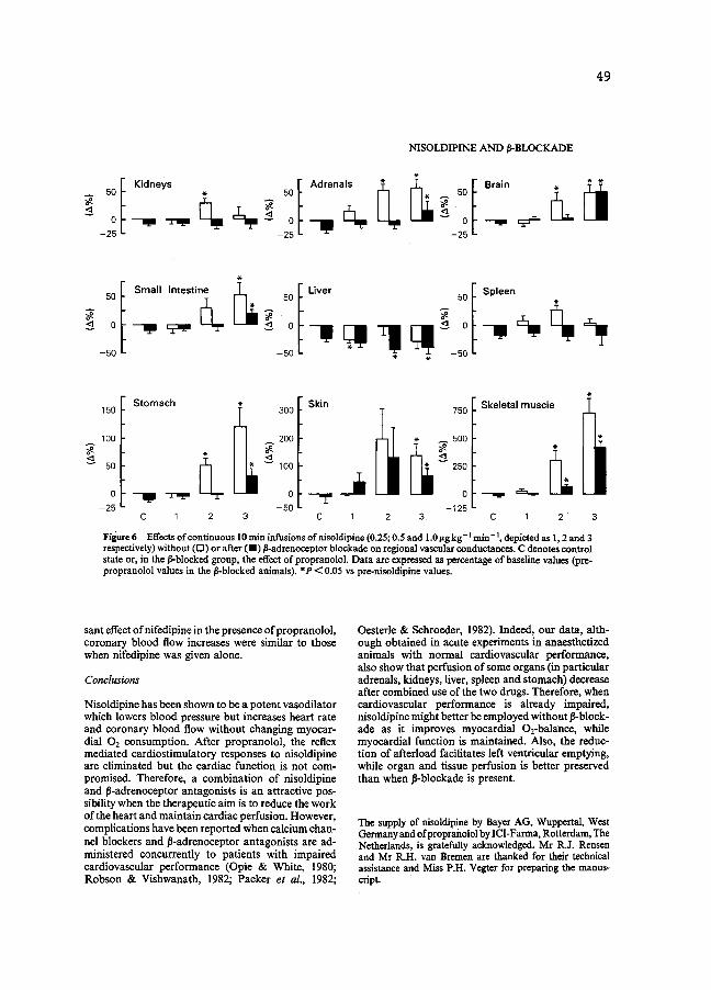
49
NISOLDIPINE AND /}-BLOCKADE
* Kidneys Adrenals
Q ~~-~~ Brain * *
w~ ~~ ~LI * #.
o.~ 1ft.
---~ ~ <l
0 ~ "'!"'111!!11 - 0 ---err"'" -25 -25
*
'"! Small intestine l.i rw· Spleen
50
'"t *
#. --. '?'J!I ~ * #.
~.,~~0--- ~o.~ ~
-5:
~ 0-.
-50 * * -50
* Stomach * Skin Skeletal muscle 150
Q~ 300
~~ 750
100 200
~ <'oo *
#. ?f. * ~ <l
"'~ ~ 50 - 100
0 -. TJII!IIII 0 ---r -- .::.._.
-25 -50 -125 c 3 c 2 3. c 2 3
Figure 6 Effects of continuous 10 min infusions of nisoldipine (0.25; 0.5 and 1.0 l'g kg- 1 min -I, depicted as I, 2 and 3 respectively) without (0) or after <•) /J-adrenoceptor blockade on regional vascular conductances. C denotes control state or, in the /}-blocked group, the effect of propranolol. Data are expressed as percentage of baseline values (prepropranolol values in the /}-blocked animals). • P < 0.05 vs pre-nisoldipine values.
sant effect of nifedipine in the presence of propranolol, coronary blood flow increases were similar to those when nifedipine was given alone.
Conclusions
Nisoldipine has been shown to be a potent vasodilator which lowers blood pressure but increases heart rate and coronary blood flow without changing myocardial 0 2 consumption. After propranolol, the reflex mediated cardiostimulatory responses to nisoldipine are eliminated but the cardiac function is not compromised. Therefore, a combination of nisoldipine and P-adrenoceptor antagonists is an attractive possibility when the therapeutic aim is to reduce the work of the heart and maintain cardiac perfusion. However, complications have been reported when calcium channel blockers and P-adrenoceptor antagonists are administered concurrently to patients with impaired cardiovascular performance (Opie & White, 1980; Robson & Vishwanath, 1982; Packer et a/., 1982;
Oesterle & Schroeder, 1982). Indeed, our data, although obtained in acute experiments in anaestheti2ed animals with normal cardiovascular performance, also show that perfusion of some organs (in particular adrenals, kidneys, liver, spleen and stomach) decrease after combined use of the two drugs. Therefore, when cardiovascular performance is already impaired, nisoldipine might better be employed without P-blockade as it improves myocardial 0 2-balance, while myocardial function is maintained. Also, the reduction of afterload facilitates left ventricular emptying, while organ and tissue perfusion is better preserved than when P-blockade is present.
The supply of nisoldipine by Bayer AG, Wuppertal, West Germany and of propranolol by ICI-Farma, Rotterdam, The Netherlands, is gratefully acknowledged. Mr R.J. Rensen and Mr R.H. van Bremen are thanked for their technical assistance and Miss P.H. Vegter for preparing the manuscript.

50
D.J. DUNCKER et al.
References
BOLT, G.R. & SAXENA, P.R. (1984a). Acute systemic and regional haemodynamic effects of felodipine, a new calcium antagonist, in conscious renal hypertensive rabbits. J. cardiovasc. Pharmac., 6, 707-712.
BOLT, G.R. & SAXENA, P.R. (1984b). Interaction of atenolol with the systemic and regional hemodynamic effects of hydralazine in conscious renal hypertensive rabbits. J. Pharmac. exp. Ther., 230, 205-213.
DREXLER, H., FLAIM, S.F., FIELDS, R.H. & ZEUS, R. (1985). Effects of nisoldipine on cardiocirculatory dynamics and cardiac output distribution in eonscious rats at rest and during treadmill exercise. J. Pharmac. exp. Ther., 232, 376-381.
GROSS, R., KIRCHHEIM, H. & voN OLSHAUSEN, K. (1979). Effects of nifedipine on coronary and systemic hemodynamics in the conscious dog. Arzneim. Forsch., 29, 1361-1368.
HASHIMOTO, K., ONO, H. &O'HARA, N. (1980). Blockade of renal autoregulatory vasoconstriction by calcium antagonists. In Calcium-antagonismus, ed. Fleckenstein, A. & Roskamm, H. pp.221-229. Berlin, Heidelherg, New York: Springer.
HOF, R.P. (1983). Calcium antagonists and the peripheral circulation: differences and sintilarities between PY 108-068, nicardipine, verapamil and diltiazem. Br. J. Pharmac., 78, 375-394.
HOF, RP. & SCHOLTYSIK, G. (1983). Effects of the calcium antagonist PY 108-068 on myocardial tissues in vitro and on reflex tachycardia in vivo. J. cardiovasc. Pharmac., S, 176-183.
KAZDA, S., GARTHOFF, B., MEYER, H., SCHLOSSMANN, K., STOEPEL, K., TOW ART, R, VATER, W. & WEHINGER, E. (1980). Pharmacology of a new calcium antagonistic compound, isobutyl methyl 1,4-dihydro-2,6-dimethyl-4-(2-nitrophyenyl}-3,5-pyridinedicarboxylate (Nisoldipine, Bay K 5552). Arzneim. Forsch., 30, 2144-2162.
NAKAYA, H., SCHWARTZ, A. & MILLARD, R.W. (1983). Reflex chronotropic and inotropic effects of calcium channel-blocking agents in conscious dogs. Diltiazem, verapamil and nifedipine compared. Circulation Res., 52, 302-311.
OESTERLE, S.N. & SCHROEDER, J.S. (1982). Editorial: Calcium-entry blockade, heta-adrenergic blockade and the reflex control of circulation. Circulation, 65, 669-670.
OPIE, L.H. & WHITE, D.A. (1980). Adverse interaction hetween nifedipine and heta-blockade. Br. med. J., 281, 1462.
PACKER, M., MELLER, J., MEDINA, N., YSHNAK, M., SMITH, H., HOT, J., GUERERRA, J., TODD, G.D., McALLISTER, R.G. & GORLIN, R. (1982). Hemodynamic consequences of combined beta-adrenergic and slow channel blockade in man. Circulation, 65, 660-668.
ROBSON, RH. & VISHW ANA TH, M.C. (1982). Nifedipine and heta-blockade as a cause of cardiac failure. Br. med. J., 284, 104.
ROUSSEAU, M.F., VINCENT, M.F., VAN HOOF, F., VAN DEN
BERGHE, G., CHARLIER, A.A. & POULEUR, H. (1984). Effects of nicardipine and nisoldipine on myocardial metabolism, coronary blood flow and oxygen supply in angina pectoris.Am. J. Cardiol., 54, 1189-1194.
SAXENA, P.R., SCHAMHARDT, H.C., FORSYTH, R.P. & LOEVE, J. (1980). Computer programs for the radioactive microsphere technique. Determination of regional blood
flows and other haemodynamic variables in different experimental circumstances. Comp. Progr. Biomed., 12, 63-84.
SAXENA, P.R. & VERDOUW, P.O. (1985). 5-Carboxamide tryptamine, a compound with high affinity for 5-hydroxytryptamine1 binding sites, dilates arterioles and constricts arteriovenous anastomoses. Br. J. Pharmac., 84, 533-544.
SERRUYS, P.W., SURYAPRANATA, H., PLANELLAS, J., WIJNS, W., VANHALEWEYK,G.L.J.,SOWARD,A.,JASKI, B.E. & HUGENHOLTZ, P.G. (1985). Acute effects of intravenous nisoldipine on left ventricular function and coronary hemodynamics. Am. J. Cardiol., (in press).
SPEDDING, M. (1982). Differences hetween the effects of calcium antagonists in the pithed rat preparation. J. cardiovasc. Pharmac., 4, 973-979.
STEEL, R.G.D. & TORRIE, J.H. (1980). Principles and Procedures of Statistics, A Biomedical Approach. 2nd edn. Tokyo: McGraw-Hill Kogakusha Ltd.
THURAU, K. & KRAMER, K. (1959). Weitere Untersuchung zur myogenen Natur des Autoregulation des Nierenkreislaufes. Pfleugers Arch., 269, 77-93.
VAN BOOM, M.P. & SAXENA, P.R. (1983). Tissue blood flow changes induced by propranolol infusion in conscious normotensive and renal hypertensive rabbits. Archs int. Pharmacodyn. Ther., 264, 96-109.
VATNER, S.F. & HINTZE, T.H. (1982). Effects of a calciumchannel antagonist on large and small coronary arteries in conscious dogs. Circulation, 66, 579-588.
VERDOUW, P.O., SLAGER, C.J., VAN BREMEN, R.H. & VERKESTE, C.M. (1984). Is nisoldipine capable of reducing left ventricular preload? Eur. J. Pharmac., 98, 137-140.
VOGT, A.&. KREUZER, H. (1983). Hemodynamic effects of nisoldipine in chronic congestive heart failure. Arnzneim. Forsch., 33, 877-879.
VOGT, A., NEUHAUS, K.L. & KREUZER, H. (1980). Hemodynamic effects ofthe new vasodilator drug Bay K 5552 in man. Arzneim. Forsch., 30, 2162-2164.
WARLTIER, D.C., ME1LS, C.M., GROSS, G.J. & BROOKS, H.L. (1981 ). Blood flow in normal and acutely ischemic myocardium after 'verapamil, diltiazem and nisoldipine (Bay K 5552}, a new dihydropyridine calcium antagonist. J. Pharmac. exp. Ther., 218,296-302.
WARLTIER, D.C., Z¥VOLOSKI, M.G., GROSS, G.J. & BROOKS, H.L. (1984a). Comparative actions of dihydropyridine slow channel calcium blocking agents in conscious dogs: systemic and coronary hemodynamics with and without combined heta-adrenergic blockade. J. Pharmac. exp. Ther., 230, 367-375.
WARLTIER, D.C. & ZYVOLOSKI, M.G., GROSS, GJ. & BROOKS, H.L. (1984b). Comparative actions of dihydropyridine slow channel calcium blocking agents in conscious dogs: alterations in baroreflex sensitivity, J. Pharmac. exp. Ther., 230,367-382.
WOLFFENBUTTEL, B.H.R. & VERDOUW, P.O. (1983). Nifedipine and myocardial performance in the presence and absence of heta-blockade with propranolol. Archs int. Pharmacodyn. Ther., 266, 83-92.
(Received July 11, 1985. Revised November I, 1985.
Accepted December 6. 1985.)

CHAPTER 3
THE EFFECTS OF NISOLDIPINE ALONE AND IN COMBINATION
WITH BETA-ADRENOCEPTOR BLOCKADE ON SYSTEMIC
HAEMODYNAMICS AND MYOCARDIAL PERFORMANCE IN CONSCIOUS PIGS.
D.J. Duncker, P.R. Saxena1 and P.D. Verdouw.
Laboratory for Experimental Cardiology (Thoraxcenter)
and 1Department of Pharmacology,
Erasmus University Rotterdam, Rotterdam, The Netherlands.
Eur. Heart J. 1987 (in press).
51


I.J~GS 7IJ.IIOEIIJ 28.09.87 KIKI ]00] L02
European Heart Journal ( 1987) 8, 000--000
53
The effects of nisoldipine alone and in combination with beta-adrenoceptor blockade on systemic haemodynamics
and myocardial performance in conscious pigs
D. J. DuNCKER*, P.R. SAXENAt and P. D. VERDOUW*
• Luhoratory.f(Jr Expaim('ll/a/ Cardiology (Tiwraxcentre) andtDepartment of Pharmacology, Erasmus Unh•ersity Rouerdam, Rotterdam, The Netherlands
KEY WORDS: nisoldipine, propranolol, regional blood flow, beta-adrenoceptor blockade, calciumantagonist, vasodilatation.
The peuk e.!Ji•cts <!( /0 mg nisoldipine p.o. ll'ith or ll'ithout 80 mg propranolol p.o. on systemic and regional haemc)((l'llmnics in conscious pigs ll'ere investigated. Niso/dipine increased heart rate (70%), cardiac output f(J7%) and maxLVdP/dt (75%), but decreased mean arterial pressure (21%) as systemic vascular conductance increased by 120%. Left 1•entricular systolic and end-diastolic pressures were not affected. I 'asodilatation occurred in most organs. The increase in left ventricular blood flow ( 150%) favoured the epicardial ( 195%) overt he endocardial (I 10%) layers. As a result the end~ pi blood flow ratio decreased by 30%.
Whennisoldipine 1\'as administered simultaneously with propranolol, heart rate (29%), cardiac output I 35%) allll systemic vascular conductance (65%) increased, but max LV dPfdt did not change. Mean arterial 1 /8%) and left ventricular systo'tic ( 10%) pressure decreased; left l'entricular end-diastolic pressure was again unq!Ji•c1ed. In mosT organs vasodilatation 1ras al/enuated, but still present, compared to the changes qfier nisoldipine alone. The increase in epicardial blood flow (70%) again exceeded that in endocardial h/oodflmr (35%J.Izowev£'r, the endo-epi ratio decreased by only 15%. In the presence of propranolol, ni.wlclipine did not exert a 11£'gative inotropic action while the reflex-tachycardia was attenuated. In addition, no detrimemal c:ffects on pe~fusion of regionalrascu/ar beds ll'ere obserl'ed.
Introduction
Calcium-channel blockers and beta-adrenoceptor antagonists are widely used in the treatment of: coronary artery disease and hypertension. As they act through different mechanisms, the combined use of these drugs might be a useful. therapeutic tooJI'-11. In this respect a combination of beta-· adrenoceptor antagonists and the I A-dihydropyridine derivatives seems especially attractive. The potent vasodilatory effect of this subgroup of calcium-channel blockers often induces a reflexmediated cardio-stimulatory response that may completely override their direct cardioinhibitory action, which is less marked than that of other calcium-channel blockers such as verapamil and
Submitted for publication on 24 February 1987 and in revised form ~9 May 1987.
A<Mrt•.r ... for c·orrt•sptmdl'na: P. D. Verdouw. Ph.D .. laboratorj, Cor Experimental Cardiol('l~)'. Era:-;mu:-; Uni\'ersity Rotterdam. P.O. Box 173X. 3000 DR Roll<rdam. The Nethcrlan~s.
11l'l5 M~X X7 OOtX10tH 00 SO~.OO,O
diltiazem. Although beneficial effects of a combination of nifedipine and beta-adrenoceptor antagonists have been reportedi1.,2,41, adverse interactions like severe cardiodepression, which are more frequently observed with verapamil'4•
51 or diltiazem161, ha\'e also been documentedi4-Kl.
Recently nisoldipine, another dihydropyridine derivative, has been shown in vitro to exert a 4-10 times stronger vasodilatory effect and an equal or weaker cardiodepressant action compared to nifedipine at equimolar dosesi91.Jn 1•ivo, nisoldipine causes pronounced vasodilatation of the systemic and coronary bed and elicits marked reflexmediated increases in heart rate and myocardial contractility parameters19- 11 1. Although some reports on systemic haemodynamic effects following a combination ofnisoldipine with beta-blockers are a\'ailab]el 11• 131, regional haemodynam1c mvestigation is restricted to one report in anaesthetized pigs The following study was performed to define whether oral administration of nisoldipine,
@ 1987 The European Society of Cardiology

54
D. J. Dunckeret a!.
when combined with that of propranolol, might have detrimental effects on myocardial performance and on perfusion of regional vascular beds in conscious pigs.
Materials and methods
GESERAL
After an overnight fast, Yorkshire pigs (18-20 kg, N = 7) pretreated with a mixture of pro• caine penicillin-G and benzathine-penicillin-G (Duplocillin. Gist-Brocades N.V., Delft, The Netherlands), both 300 000 units i.m., were sedated w.ith 30 mg kg- 1 ketamine HCI i,m. (Aescoket, Aesculaap B.V .. Boxtel, The Netherlands). The animals were intubated and connected to a respirator for artificial ventilation with a mixture of02 and N20 (I :2) to which 1% halothane was added. A jugular vein and common carotid artery were cannulated for administration of drugs and measurement of mean arterial blood pressure, respectively. The chest was opened via the left fifth intercostal space to expose the heart. A transducer (P 4 .5 ,
Konigsberg Instruments Inc. Pasedena, California, U.S.A.) was implanted into the apex of the left ventricle of the heart for recording left ventricular pressure. The left atrium was cannulated for recording left atrial pressure which, together with the aortic blood pressure, was used for calibration of the Konigsberg transducer signals. The aorta was approached through the third intercostal space and an electromagnetic flowprobe (Skalar, Delft, The Netherlands) was positioned around the ascending aorta. Cardiac output was calculated by adding myocardial blood flow (see later) to ascending aorta blood flow. Catheters and wires were tunnelled subcutaneously to the back, the chest was closed and the animals allowed to recover. During the next 14 days the animals received daily intravenous bolus injections of 500 mg amoxicilline (Clamoxil, Beecham Farma B.V., Amstelveen, The Netherlands) and in addition, during the first week, 500 mg kanamycin (Kamynex, Gist-Brocades N.V., Delft, The Netherlands) to prevent infection. The catheters were flushed daily with an isotonic saline solution containing heparin (500 I.U. ml- 1
) to avoid clotting of blood in the lumen. After a I week postoperative recovery period, at least four sessions were·held to adapt the animals to the experimental and laboratory facilities. The experimental protocol was executed 2-3 weeks after the operation. All tracings were written on a Graphtec Linearcorder
. (F WR 3701, Ankersmit, Breda, The Netherlands).
Arterial acid-base balance and oxygenation during the experiments were similar to those reported for young Yorkshire pigs by Lagerweyf141: pH= 7 ·45 ± 0·02, PC02 = 37 ± 3 mmHg, P02 = 81 ± 5 mmHg, Hb02:saturation = 92± 3%.
MEASUREMENT OF REGIONAL BLOOD FLOWS
Regional blood flows were measured by use of the radioactive microsphere technique. Although the microsphere technique offers the advantage of simultaneously measuring blood flow to a large number of tissues in conscious animals with relatively little surgical trauma, an inherent drawback of this method is that only a limited number of isotopes can be employed. Therefore only one dose of nisoldipine with and without propranolol could be used in each individual animal; the dose selected was one that elicited a reasonably marked pharmacological response (see later).
Microspheres(l5± I (SO) 11m diameter),labelled with either 141 Ce, 113Sn, 103Ru or 95Nb (NEN Chemicals GmbH, Dreieich, West Germany) and suspended in saline containing a drop of Tween 80, were injected in random order into the left atrium over a period of30-45 sf151.
At the end of each experiment the animal was killed with an overdose of pentobarbitone sodium. The various tissues, as specified later, were dissected out, weighed and placed in plastic vials. The radioactivity in the vials containing the tissues was counted for 5-10 min in a y-scintillation counter (Packard, model 5986) equipped with a multichannel pulse height analyser (Conrac) using suitable windows for discriminating the different isotopes usedl 161. The microsphere and other data were processed by a PDP-11(70 computer using a set of programmes especially developed for the microsphere techniquel 161. The amount of blood flow distributed to the various tissues (Q.;,) was calculated as follows:
Q,;,(ml min- 1) = (I,;JI,.,) x CO
where 1,;,, I,., and CO are, respectively, radioactivities (cpm) in the individual tissues and the total number of microspheres injected (calculated as the difference in the radioactivity present in the glass vial before and after injection), and the cardiac output (in ml min- 1 ). Tissue vascular conductance (flow/pressure) was calculated by dividing respective tissue blood flow values by mean arterial blood pressure.

s.o
160
"' i 80 E
60
..:-"' " E E 30
";' 0
e l
co
~ lU
MBP
.· .
svc
200
. c ·;; ~ 100
.c
6000
.• 0.. 3000
" ~
160
g, i 80 E
HR
maxLVdP/dt
LVSP
so
E 2s
20
g, i 10 E
55
NistJidipine and beta-adrenoceptor blockade
[J] SV
rll .
.
. LVEDP
~baseline
~:::~~:ne nisoldipine
propranolol
Fi!!""' I Enects of nisoldipine (!Omg p.o.) without and with propranolol (80 mg p.o.) on systemic haemodynamics in seven conscious pigs. CO, cardiac nut put: HR. heart rate; SV, stroke volume; M BP, mean arterial blood pressure; muxl VdP.'dt. maximal rate of rise of left ventricular pressure; LVEDP, left ventricular end-diastolic pressure; SVC, systemic Vdscular conductance; LVSP, loft ventricular systolic blood pressure. Data are expressed as mean±SEM; ?,.;0·05 rs. baseline or recovery. ••change induced by nisoldipine with propranolol si~nificantly different (P,;;0·05) from that by nisoldipine without propranolol.
EXPERIMENTAL PROTOCOLS In pigs fasted for 24 h, 10 mg nisoldipine (equiv
alent to 0·40 ± 0·02 mg kg- 1) was administered
orally (p.o.) after baseline data had been recorded and the first batch of radioactive microspheres had been injected for the determination of regional blood flows. All measurements were repeated when nisoldipine-induced changes in heart rate had reached a stable peak level (50± 6 min after administration). As a pilot-study in eight pigs had revealed that, 24 h after ingestion of 10 mg of the drug. plasma-levels of nisoldipine could no longer
be detected and that haemodynamic parameters had returned to baseline, the next day the same protocol was repeated, but now 80 mg p.o. propranolol (equivalent to 2·88 ± 0-44 mg kg- 1
) was given along with the 10 ing nisoldipine tablet. This second part of the protocol was performed in the same animals to avoid inter-animal differences. Data were again obtained during baseline and at the peak heart rate effect ( 48 ± 4 min after administration). In the pilot study (N = 8) we had observed that peak changes in heart rate (from 108±5 to 160±12 beats min- 1
)
corresponded with peak plasma concentrations

56
D. J. Dun("k£'r et al.
(3·14±0·64 ng mg- 1) ofnisoldipine reached about I h after the drug administration. When in three of these animals nisoldipine and propranolol were administered simultaneously' the peak concen" tration of nisoldipine was 2·81 ±0·70 ng ml- 1
which was not statistically significantly different from that obtained after nisoldipine alone.
STATISTIC"AL ANALYSIS
Unless otherwise stated, all data are presented as mean±standard error of mean. Statistical analysis was performed by use of Duncan's new multiplerange test once a parametric two-way analysis of variance (randomized block design) had revealed that the samples represented different populationsP71. A P value of0·05 or Jess (two-tailed) was considered to be statisti~ally significant.
DRUGS
The only drugs used in this study were tablets of nisoldipine (Bay k SSS2, Bayer AG, Wuppertal, F.R.G.) and propranolol hydrochloride (ICIFarma. Rotterdam, The Netherlands).
Results
SYSTEMIC HAEMODYNAMICS
Peak effects of nisoldipine (10 mg p.o.) on systemic haemodynamics are presented in Fig. I. Nisoldipine caused marked increases in cardiac output (67%), heart rate (70%), and maxLVdP/dt (7S%). Despite the increase in cardiac output, mean arterial blood pressure dropped by 21%. Consequently, the calculated systemic vascular conductance increased considerably (120%). No changes were observed in left ventricular systolic or enddiastolic pressure and stroke volume. After simulta~ neous administration of nisoldipine (I 0 mg p.o.) and propranolol(80 mg p.o.), the increases in cardiac output (3S%), heart rate (29%) and systemic vascular conductance (65%) were much Jess, and that in maxLVdP/dt even abolished, when compared to those after nisoldipine alone. Mean arterial blood pressure, however, decreased by a similar magnitude (18%), while left ventricular systolic pressure was now slightly reduced (I 0% ). Left ventricular end-diastolic pressure and stroke volume were again unaffected.
CORONARY HAEMODYNAMICS
Administration of nisoldipine alone increased both the transmural left ventricular blood flow (I SO%) and vascular conductance (22S%) but. as
qoo
200
LVBF
1.0
endo/epl
~~"
' "' % E E
' c i
!
LVC
0 2-demand
Figure 2 Effects ofnisoldipine (10 mg p.o.) without and w.ith propranolol (80 mg p.o.) on coronary haemodynamics in seven conscious pigs. LVBF, left ventricular blood flow; LVC, left ventricular conductance; endo/epi, ratio between endocardial and epicardial normalized blood flows; 0 2-
demand, heart rate x left ventricular systolic pressure. The four different columns represent, respectively, baseline, nisoldipiite. recovery and nisoldipine with propranolol. Data are expressed as mean± SEM. • P.;;O·OS ••s. baseline or recovery. ••changes induced by nisoldipine with propranolol significantly different (P.;;O·OS) from that by nisoldipine without propranolol.
the elevations in blood flow and conductance favoured the epicardial (195% and 280%, respectively) over the endocardial (110% and 170%, respectively) layers, the endocardial-epicardial (endo-epi) blood flow ratio decreased by 30% (Fig. 2). As the double product (left ventricular systolic pressure x heart rate), an index of myocardial oxygen demand, increased only by 60%, it would appear that the enhancement in coronary blood flow, i.e. oxygen supply, exceeded the increased oxygen demand. When nisoldipine was combined with propranolol, the elevations in transmural, epicardial and endocardial blood flows (SO, 70 and 34%, respectively) and conductances (95, 120 and 70%, respectively) were only moderate, but still statistically significant (P .s;; O·OS). Furthermore, the reduction in endo-epi blood flow ratio (IS%) did not reach the level induced by treatment with the

dihydrophyridine derivative alone,while the double product was only slightly ( < 10%) enhanced.
l.liSTRIBUTIOl' OF CARDIAC OUTPUT
The effects of nisoldipine on regional haemodynamics have been presented in Figs 3 and 4. Nisoldipine caused the largest increase in blood flow to the right atrium (700%), followed by the right-ventricle (280%). stomach (190%),skeletal muscle (150%) left ventricle (150%. Fig. 2), small intestine (120%). adrenals (110%) and brain (30% ). Renal and hepatic artery blood flow as well as flow to the skin and spleen were not significantly affected. When nisoldipine was administered simultaneously with propranolol, increases in blood flow were again observed, though generally (except skeletal muscles) of a lesser magnitude, in the right ;ttrium (260%), skeletal muscles (250%), stomach ( 110%). right ventricle (105%). small iniestine
,.
10
--·~ 200
'< i
'! • £ 100
~ j ~ a:
•••
, ..
liver
.
rU .. · >· . ."
. ..
skeletal muscle
·: .. rill. akin
:- eli" . cU. ': '• ~ ~:
.. · '•
stomach sm•ll Intestine brain
1ddneys right ventricle
57
Nisoldipine and beta-adrenoceplor blockade
(70%) and left ventricle (50%). Blood flow to the adrenals, brain, liver and kidneys was not signifi.cantly affected while that to the skin (50%) and spleen (35%) decreased. As nisoldipine induced a moderate decrease in blood pressure, vascular conductances increased in most organs, except the liver, skin and spleen (Fig. 4). The most pronounced increase was observed in the right atrium (900%), followed by the right ventricle (400%), stomach (280% ), skeletal muscles (230% ), left ventriCle (225%), small intestine (180%), adrenals (170%), brain (60%) and kidneys (50%). With propranolol, nisoldipine induced the greatest vasodilatory response in the right atrium (360%) followed by skeletal muscle (300%). stomach (160%), right ventricle (160%), small intestine (110%), left ventricle (95%) and brain (.50%). No statistically significant changes were noted in the liver, skin, spleen, adrenals and kidneys.
~ buellne
nlsoldiplne
recovery
nlsoldiplne . propr•noJol
spleen
L.
. .
·right 1trlum
• Figur,· 3 Region"! blood now in seven conscious pigs after nisoldlpine (10 mg p.o.) without and with pmpnuiolol (80 mg p.o.). Data arc expressed as mean± SfM. •p,.0-05 "-'· b"$eline or recovery. ••change induced by nisoldipine with rr•'l'r""''lol signific.,ntly dill'crent (P<0·05) from that by nisoldipine wilhout prtlpr:mol<ll.

58
D. J. Duncker et at.
O.l
0.15
£ E 1.5
! ~ ~ •• 75
1 . . 1 > j 5.0
~ a:
2.5
rt~tll Uvtir lkelet.l
muscle
kidneys
r-tt:. skin
right ventricle
~ b .. ollne
~laoldlplne
rtCDYel")
nlaoldlplne . propranolol
Spleen
1 right •trlum
Figur<•4 Regional vascular conductance in seven conscious pigsafiernisoldipine ( I 0 mg p.o.) without and with propranolol (80 mg p.o.). Data are expressed as mc-dn ± SEM. • P,. 0·05 •·s. baseline or recovery. **Change induced by nisoldipine wilh propranolol significan1ly different (P,.0·05) from that by nisoldipine wi1hou1 propranolol.
Discussion
EFFECTS OF NISOLDIPINE ALONE
As has been reported by many investigators19·
IJ.IM-~~1. we also observed that nisoldipine induced a pronounced systemic vasodilatation. This resulted in only a moderate fall in mean arterial blood pressure since cardiac output was markedly elevated. Due to the baroceptor-reflex, contractility and heart rate were augmented but stroke volume remained unchanged. These observations are in general agreement with data obtained after i.v. infusions of nisoldipine in anaesthetized pigsl121. The major difference is that cardiac output in the anaesthetized animals was unchanged as the reflextachycardia was accompanied by a concomitant reduction in stroke volume. It appears that this
reduction resulted from the use of pentobarbitone anaesthesia, since equal infusion rates in conscious pigs caused a significant increase in cardiac output solely due to a tachycardia as stroke volume was maintained (unpublished data from this laboratory). In most studies the elevation of cardiac output results primarily from an increase in heart rate, with stroke volume being either unchangedl10•181 or increasedl10
•191. In rats with high baseline heart rates
(up to 400 beats min- 1), the elevation in cardiac
output is primarily caused by an augmentation of stroke volume120•211.
Due to the reflex-tachycardia, the oxygen demand (rate-pressure product) was moderately enhanced. The increase in coronary blood flow, however, exceeded this enhancement, thereby

increasing the oxygen supply- oxygen demand ratio. Serruys et a/P~I reported after nisoldipine in humans an unchanged double product and oxygen consumption, while coronary blood flow was increased by 30%. Similar improvements of myocardial oxygen balance have also been demonstrated by other investigators19• 1 ~·' 91. The decrease in endocardial-epicardial blood flow ratio is probably due to the combination of an increased heart rate and hypotension'~;!. Nevertheless, an increase in endocardial blood flow was present, which far exceeded the increase in oxygen demand.
I!FFECTS OF NISOLDIPINE AND BETA-ADRENOCEPTOR
IILOCKADE
Although the elevation of systemic vascular conductance was reduced after combined nisoldipine and propranolol, the drop in blood press.ui'e was not different from that observed after nisoldipine aione. This was due to the attenuated increase in cardiac output, which again depended entirely on the increase in heart rate. An increase in heart rate by nisoldipine in the presence ofbeta-adrenoceptor blockade has also been reported by others'"·"l and this probably results from withdrawal of parasympathetic tonel~•l, as the atria and the conductance tissue of the left ventricle are richly innervated by the parasympathetic nervous systemflSJ. The left ventricular myocardium is only scarcely innervated by the parasympathetic system and this might explain why maxLVdP/dt was unaffected by the combination. In contrast, in anaesthetized pigs1 '~1 both heart rate and maxLVdP/dt were significantly decreased by the combination suggesting attenuation of cardiovascular reflexes during pentobartital anaesihesia. .
·Due to the beta-adrenoceptor blockade the oxygen demand remained essentially unchanged, as the attenuated increase in heart rate was accompanied by a decrease in left ventriCular systolic pressure. Furthermore, as heart rate was only moderately increased, the duration of diastole, i.e. the perfusion time of the endocardium, was not reduced as much as after nisoldipine alone. As a result, the endo-epi blood flow ratio decreased less. As in anaesthetized pigsl111, vasodilatation was inhibited in almost all organs studied (except skeletal muscles), when nisoldipine was administered together with propranolol. It is known that· beta-adrenoceptor antagonists may generally reduce responses to vasodilator drugs because beta-adrenoceptor blockade leads to a reflex increase in sympathetic tone, which
59
Niso/dipine and beta-adrenoceptor blockade
interferes with vasodilatation, and activation of vasoconstrictor alpha-adrenoceptors.
We conclude that nisoldipine is a potent systemic and coronary vasodilator in the conscious pig. The combination ofnisoldipine and beta-adrenoceptor blockade exerted no cardiodepressant action, while the reflex-tachycardia was attenuated. Our data, obtained in conscious pigs with a normal coronary circulation, support the recent findings of Silke et a/.1'31 in patients with stable angina pectoris. In addition, we observed no detrimental effects of the combination on perfusion of regional vascular beds.
Nisoldipine was generously supplied by Bayer A.G. (Wuppertal), West Germany), while propranolol was pro· vided by ICI Farma (Rotterdam, The Netherlands). We would like to thank the Laboratory for Experimental Surgery for their skilful assistance during surgery and the postsurgical period and Mr J.P. C. Heiligers for technical assistance during the experiments. Our special thanks go· to Ms P. H. Vegter for her assistance in the preparation of this manuscript.
References [I) Daly K, Bergman G, Rothman M, Atkinson L, Jackson
G, Jewitt DE. Beneficial effect of adding nifedipine to beta-adrenergic blocking therapy in angina pectoris. Eur Heart J 1982; 3: 42-6.
[2) Harris L, Dargie HJ, ·Lynch PG, Bulpitt CJ, Krikler OM. Blood pressure and heart rate in patients with ischaemic heart disease receiving nifedipine and propranolol. Br Med J 1982; 284: 1148-51.
[3] Allen LL, Young PM, Reichman MJ, Bough EW, Boden WE, Hemberger JA; McGraw BF. Effects of diltiazem in combination with propranolol/nitrate in anginal patients. Clin Pharmacol Ther 1984; 3.5: 226.
(4] Brouwer RM, Follath F, Buehler FR. Review of the cardiovascular adversity of the calcium antagonist betablocker combination: implications for antihypertensive therapy. J Cardiovasc Pharmacol 1985; 7 (Suppl 4): S38-44.
[5] Singh BN, Ellrodt.G, Peter CT. Verapamil: a review of its pharmacological properties and therapeutic use. Drugs 1978; 15: 169-97.
(6] Hung J, Lamb IH, Connolly SJ, Jutzy KR, Goris ML, Schroeder JS. The effect of diltiazem and propranolol alone and in combination on exercise performance and left ventricular function in patients with stable effort angina. A double blind, randomized, and placebo controlled study. Circulation 1983; 68: 56(}-7.
[7] Opie LH, White DA. Adverse interaction between nifedipine and beta-blockade. Br Med J 1980; 281: 1462.
[8] Robson RH, Vishwanath MC. Nifedipine and betablockade as a cause of cardiac failure. Br Med J 1982; 284: 104.
[9] Verdouw PO, Duncker OJ, Saxena PR, Serruys PW, Hugenholtz PG. Cardiovascular actions of nisoldipine in health and coronary artery disease. Am J Cardiol 1987: in press.

60
D. J. Dunckeret al.
II OJ Kazda S. GarthoiT B. Meyer H. <'I a/. Pharmacology of a new calcium antagonistic compound, isobutyl methyl 1.4-dihydro-~.6-dimethyl-4-(2-nitrophyenyl)-3. 5-pyridinedicarboxylate (Nisoldipine. ·Bay K 5552). Arzneimittelforsch 1980; 30: 2144-62.
Ill] Warltier DC. Z\'\'oloski MG. Gross GJ, Brooks HL. Comparative aciions of dihydropyridine slow channel calcium blocking agents in conscious dogs: systemic and coronary hemodynamics with and without combined bcta-adrenc•rgic blockade. J Pharmacal Exp Ther 1984; 230: 367-75.
112] Duncker DJ. HartogJM. Hugenholtz PG, Saxena PR, Verdouw PD. The effects of nisoldipine (Bay k 5552) on cardiovascular performance and regional blood ftow in pentobarbital- anaesthe.tized pigs with or without heta-:<drenoccptor blockade. Br J Ph:<rmacol 1986; 88: 9 18. .
IDJ Silke B. Verma SP. Midtbo KA. Muller P. Frais MA. Reynolds G. Taylor SH. A hacmodynamic study of the dTects of combined slow-channel blockade (nisoldipine) and beta-blockade (metoprolol) in coronary heart disease.lntJCardiol1986; 13:231-41.
il-l] Lagerwey E. Anaesthesia in swine for experimental purpose. Ph.D. dissertation. University of Utrecht. The Netherlands.
I 1'5] Saxena PR. Vcrdouw PD. 5-Carboxamide tryptamine. a compound with high affinity for 5-hydroxytryptamine hinding sit~s. dihatcs arterioles and constricts ancrio~ venous anastomoses. Br J Pharmacal 1985; 84: 533-44.
!In] Saxena PR. Schamhardt HC. Forsyth RP. Loeve J. Computer programs for the radioactive microsphere technique. Determination of regional blood ftows and other haemodynamic variables in different experimental circumstances. Comp Progr Biomed 1980: 12:63-84.
117] Steel RGD. Torrie JH. Principles and ·procedures of st;llistics. a biomedical approach, 2nd edn. Tokyo: McGraw-Hill Kogakusha Ltd. 1980.
118] Vogt A, Neuhaus KL, Kreuzer H. Haemodynamic effects of the new vasodilator drug Bay K 5552 in man. Ar2neimittelforsch 1980; 30:2162-4.
119] Maxwell GM. Crompton S, Rencis V. Effect ofnisoldipine upon the general and coronary hemodynamics of the anesthetized dog. J Cardiovasc Pharmacal 1982; 4: 393-7.
120] Drexler H, Flaim SF, Fields RH, Zelis R. Effects of nisoldipine on cardiocirculatory dynamics and cardiac output distribution in conscious rats at rest and during treadmill exercise. J Pharmacol Exp Ther 1985; 232: 376-81.
1211 Drexler H. Truog AG, Zelis R. Flaim SF. Regional vasculu and hemodynamic effects of orally administered nisoldipine in conscious rats. J Cardiovasc Pharmacol 1986:8: 151-5.
122] Serruys PW, Suryapranata H, Planellas J et a/. Acute effects of intravenous nisoldipine · on left ventricular function and coronary hemodynamics. Am J Cardiol 1985; 56: 140-0.
123] Feigl EO. Coronary physiology. Physiol Rev 1983: 63: 1-205. .
124] Nakaya H, Schwartz A. Millard RW. Reflex chronotropic and inotropic effects of calcium channel-blocking agents in conscious dogs. Diltiazem, verapamil and nifedipine compared. Circulation Res 1983; 52: 302-11.
[25] Higgins CB. Vatner SF, Braunwald E. Parasympathetic control of the heart. Pharmacol Rev 1973; 25: 119-48.
CHAPTER 4
NIMODIPINE-INDUCED CHANGES IN THE
DISTRIBUTION OF CAROTID BLOOD FLOW AND CARDIAC OUTPUT
IN PENTOBARBITONE-ANAESTHETIZED PIGS.
D.J. Duncker, J. Heiligers1, E.J. Mylecharane1,2,
P.R. Saxena and P.D. Verdouw.
Laboratory for Experimental Cardiology {Thoraxcenter)
and 1Department of Pharmacology,
Erasmus University Rotterdam, Rotterdam, The Netherlands.
2Present address: Department of Pharmacology,
University of Sydney, Sydney, NSW 2006, Australia.
Br. J. Pharmacal. 89, 35-46, 1986.
61


Br. J. Pharmac. (1986), 89, 35-46
Nimodipine-induced changes in the distribution of carotid blood flow and cardiac output in pentobarbitone-anaesthetized pigs D.J. Duncker*·**, J. Heiligers**, E.J. Mylechl:!-rane**2
, P.R. Saxena**1 & P.D. Verdouw*
Departments of Experimental Cardiology (Thorax Center)* and Pharmacology**, Erasmus University Rotterdam, P.O. Box 1738, 3000 DR Rotterdam, The Netherlands
1 In .view of the claimed effectiveness of nimodipine in migraine and its possible selectivity for cerebral vessels, we investigated the effects of nimodipine in anaesthetized pigs on the fractionation of carotid arterial blood flow into non-nutrient (arteriovenous anastomoses; AVAs) and nutrient (capillary) parts, and on regional tissue blood flows and vascular conductances. 2 Intracarotid infusions of nimodipine (0.05-1.25 j.lg kg- 1 min-') redistributed carotid blood flow in favour of its nutrient compartment, particularly to the skeletal muscles and tongue. Vascular conductance in the non-nutrient (A VAs) compartment decreased (40%), most likely, as a result of 'steal' following profound (5.5 fold) arteriolar dilatation. 3 Intravenous infusions of nimodipine (0.05-6.25 j.lg kg- 1 min- 1) caused hypotension, bradycardia, a decrease in conduction in the non-nutrient fraction, and an increase in conduction in the nutrient fraction (mostly in the skeletal muscles, but also in the gastrointestinal tract, cerebral hemispheres, heart and adrenals). 4 Probably due to the hypotensive effect, only skeletal muscle blood flow increased. The nimodipineinduced increase in vascular conductance in the skeletal muscles showed regional variation; the effect wasmost pronounced in the cheek muscles, followed by the muscles of the chest, abdominal, trunk and gluteal regions. 5 We conclude that: (i) A VA flow seems to represent a 'reserve' perfusion which can be readily diverted to tissues in the case of increased metabolism and/or vasodilatation, (ii) though the overall response to nimodipine of carotid blood flow distribution qualitatively resembles that to some antimigraine drugs, the relevance of such acute effects in the prophylactic usefulness of nimodipine in migraine remains to be ascertained, and (iii) nirnodipine lacks a selective cerebral vasodilator action in the anaesthetized pig.
Introduction
63
One of the most significant recent advances in drug development has been the discovery of agents interfering with calcium (Ca2+) channels. These drugs are now being used for the treatment of cardiovascular disorders such as angina pectoris, cardiac arrhythmias and hypertension (see Fleckenstein, 1983). The Ca2+ channel antagonists exhibit considerable heterogeneity with respect to their effects on the heart and different vascular smooth muscle preparations (Cauvin et a/., 1983; Fleckenstein, 1983; Nayler, 1983; Peroutka, 1 Author for correspondence at Department of Pharmacology. 2Present Address: Department of Pharmacology, University of Sydney, Sydney, NSW 2006, Australia.
1983). The dihydropyridine derivative nimodipine appears to have a preferential action on cerebral vessels; it antagonizes vasoconstrictor effects of 5-hydroxytryptamine · (5-HT), blood and carboxylic thromboxane A2 on the rabbit basilar artery more effectively than on the rabbit saphenous artery (Towart, 1981; Towart & Perzbom, 1981; Towart et a/., 1982). Clinically, nimodipine bas shown potential in the therapy of cerebral vascular spasm following subarachnoid haemorrhage (Aueret a/., 1982; Grotenhuis et a/., 1984; Kostron et a/., 1984) and in the treatment of migrainous headaches (Gelmers, 1983; Meyer & Hardenberg, 1983).
In the present investigation we have addressed
©The Macmillan Press Ltd 1986

64
D.J. DUNCKER et a/.
ourselves to two questions. Firstly, in view of the possible involvement of cephalic arteriovenous anastomoses (A VAs) in the pathophysiology of migraine (Heyck, 1969; Saxena, 1978; 1984), we have studied the effects of local infusions of nimodipine on the fractionation of carotid blood flow into nutrient (capillary) and non-nutrient (AVA) parts in the anaesthetized pig. The second part of this study deals with regional haemodynamics. Though several studies have shown that basal cerebral blood flow may increase in some species (Harper et al., 1981; Kazda et al., 1982; McCalden et al., 1984; Mohamed et al., 1984), only Haws et al. (1983), using rabbits, have directly compared the cerebrovascular effects of nimodipine with those on some other tissues. Therefore, the effects of intravenous (i.v.) infusions of nimodipine have been studied on regional tissue blood flows and vascular conductances to determine whether the drug causes a selective vasodilatation in the cerebral vascular bed of another species (pig).
Methods
Three series of experiments were performed. In the first series, intracarotid administration of the drug solvent was used as a control for evaluation of the stability of the preparation. In a second group of animals, the effects oflocal infusions of nimodipine on the total common carotid artery blood flow and its distribution were determined. Finally, i.v. infusions were used to evaluate the effects of nimodipine on cardiac output and its distribution. A wide range of doses of nimodipine was selected; the difference between the first and last infusion rate was 25 and 125 fold in the intracarotid and i.v. experiments, respectively.
Experimental preparation
After an overnight fast, 25 Yorkshire pigs (mean body weight± s.e.mean, 25.7 ± 0.8kg; age 12-16 weeks) were initially sedated with 120mg (i.m.) azaperone (Stresnil) and 150mg (i.v.) metomidate (Hypnodil). After the animals had been intubated, they were connected to a respirator for intermittent positive pressure ventilation with oxygen and nitrous oxide (1 :2). Respiratory rate and tidal volume were adjusted to keep arterial blood gases, measured with an ABL-3 (Radiometer, Copenhagen), within normal limits (pH, 7.35-7.45; Po2, 90-150 mmHg, Pco2, 35-45 mmHg); sodium bicarbonate (8.4%, wfv) was infused, if necessary, until base excess was near zero. An electric blanket was used to maintain the animal's temperature around 37•c. A continuous i.v. infusion of pentobarbitone sodium(15-25mgkg- 1 h- 1) and an i.v. bolus of 4 mg pancuronium bromide (Pavulon) were used to
maintain anaesthesia. Arterial blood pressure (obtained via a 7F catheter placed in the left femoral artery and connected to a Statham pressure transducer) and heart rate (counted from ECG signals) were monitored on a Gould Brush recorder. Catheters in both femoral veins and the other femoral artery were used for i.v. administration of drugs and fluids, and for monitoring arterial blood gases.
In the animals which received intracarotid infusions of nimodipine, both common carotid arteries were dissected free in the neck and bilateral cervical vagosympathectomy was performed to avoid reflex influences on the carotid circulation. Two 0.5 mm (external diameter) needles, connected to suitable polyethylene tubings, were inserted directly into one of the common carotid arteries for the infusion of nimodipine and the injection ofmicrospheres, respectively. Blood flow in this artery was measured with a 2.5 or 3 mm (i.d.) calibrated flow probe connected to a sine wave electromagnetic blood flowmeter (Skalar, Delft). In the animals used to study the effects ofi.v. administration of nimodipine on the distribution of cardiac output, a mid-sternal thoracotomy was performed. A cannula was inserted into the left atrial appendage for injection of the microspheres and a catheter in the femoral artery was · employed to withdraw a reference blood sample during microsphere injection (Saxena et al., 1980). Ascending aorta blood flow was measured with a suitable electromagnetic flow probe placed around the vessel. Cardiac output was derived by adding myocardial blood flow (measured with radioactive microspheres; see below) to the ascending aorta blood flow.
Distribution of common carotid blood flow and cardiac output
Injection of radioactive microspheres Radioactive microspheres (15 ± 1 rs.d.]jtm diameter), labelled with 141Ce, 113Sn, 103Ru, 9'"sNb or 46Sc (NEN Chemicals GmbH, Dreieich, West Germany) and suspended in saline containing a drop of Tween 80, were used (for details, see Saxena & Verdouw, 1982, 1984). Prior to use, the spheres were deaggregated by mechanical agitation. The distribution of common carotid arterial blood flow was determined by injecting 1-2 x 10⁵
microspheres into the artery against the direction of blood flow over a period of 15-20 s. The distribution of cardiac output was determined similarly except that 1-2 x 10⁶ microspheres were injected into the left atrium. Starting about 5 s before the injection of microspheres, blood was withdrawn (rate: 12mlmin-1) from a femoral artery for a total period of 60-65 s.
Counting of radioactivity At the end of each experiment the animal was killed with an overdose of

65
REGIONAL HAEMODYNAMIC EFFECTS OF NIMODIPINE
pentobarbitone sodium and the various tissues, as specified later, were dissected out, weighed and placed in vials. The radioactivity in the vials containing the tissues and blood samples was counted for 5-10 min in a y-scintillation counter (Packard, model 5986) equipped with a multichannel pulse height analyser (Conrae) using suitable windows for discriminating the different isotopes used (Saxena et a/., 1980).
Calculations The microsphere and other data were processed by a PDP-11/70 computer using a set of programmes especially developed for the microsphere technique (Saxena et al., 1980). The amount of carotid blood distribut~d to the individual tissues (Qu'!fa'J) was calculated by: Qus[carJ (ml min- 1
) = (IuJI.oJ x ~.lear and Ous[carJ (%) = (IuJI,0 ,) X 100, where 1,;, and 1,0 , are, respectively, the radioactivity (c. p.m.) in a particular tissue and that detected in all tissues (i.e. tissues of the head, including the compl~te brain, and the neck and lungs) collectively, and Qcar is carotid blood flow (ml min- 1) (see Saxena & Verdouw, 1982). The amount pf cardiac output distriputed to the various tissues !Q,;,) was. calculated ~s: Q,;, (mlmin- 1) = (1,;,/ I,.,) x Qan and Q,;, (%) = (Q,;JCO) x 100, where 1,;, and Ian are, respectively, the radioactivity (c. p.m.) in a particqlar tissue and that of the arterial blood sample, while Q." is the rate of withdrawal of blood samples and CO is cardiac output in mlmin-•. The various tissues selected were: heart, kidneys, ·adrenal glands, skeletal muscles from several regions, skin, spleen, liver, small and large intestine, including caecum and rectum (gastrointestinal tract), eyes and a large part of the brain (cerebral hemispheres). Tissue vascular conductance was calculated by dividing respective blood flow values by mean arterial blood pressure.
The values determined for lungs, when microspheres were injected into the carotid artery, represent the AVA part of the carotid circulation (see Johnston & Saxena, 1978; Saxena & Verdouw, 1982). In the case of left atrial injection, the lungs receive microspheres
via both peripheral A VAs and bronchial arteries; however, the contribution via the latter route appears to be only about l% (Baile et al., 1982). Thus, even in this case, the values for 'lung blood flow' can be used as an index of peripheral AVA flow (i.e. the nonnutrient part of cardiac output). The nutrient part of cardiac output was calculated by subtracting 'lung blood flow' from cardiac output.
Experimental protocol
In all experiments, the baseline values were obtained following a stabilization period of 60 min after completion of the surgical procedures. The measurements consisted of recordings of the heart rate, mean arterial blood pressure and common carotid artery blood flow (or cardiac output), while a batch of microspheres was· injected into the carotid artery (or into the left atrium). In the common carotid blood flow distribution experiments, three successively increasing intracarotid infusions of nimodipine (0.05, 0.25 and 1.25 11g kg- 1
min- 1) were then administered for lOmin each. Microspheres were injected at the end (l 0 min) of the 0.05 and 0.25 11g kg- 1 min- 1 infusions, and 2 and lOmin after the last infusion step (l.25~tgkg- 1 min- 1 ). In the cardiac output distribution experiments, successively increasing i. v. doses of 0.05, 0.25, 1.25 and 6.25 Jtgkg- 1 min-• of nimodipine were infused for a period of 10 min each; microspheres were injected at the end of each i.v. infusion period. Control experiments were performed with the drug solvent which was injected into the common carotid artery in amounts (0.5, 2.5 and 12.5 ~tl kg- 1 min- 1) which corresponded to the three intracarotid doses of nimodipine; l 0 min after each infusion, microspheres were injected into the carotid artery.
Data presentation and statistical analysis
Except as mentioned otherwise, all data in the text and
Table 1 Effects ofintracarotid administration of nimodipine (n = 9) and eqnivalent amounts of its solvent (n = 6) on mean arterial blood pressure and heart rate in pigs
Solvent Nimodipine
Solvent Nimodipine
Baseline values
104±7 105±4
100±4 114± 8
% change by nimodipine (JJgkg- 1 min- 1) or solvent'
1.25 1.25 0.05 0.25 (2 min) (10 min)
Mean arterial pressure (mmHg) -2±1 -3±1
-14± 3•'••
-8± 1• -7±2
-24±4•' .. -34±4•
Heart rate (beats min-t) -11 ± t• -17±5• -18±4•
-4± 1° -47± 5••••
-12±2• -23±4•' ..
'The corresponding doses of solvent were 0.5, 2.5 and 12.5 ,.I kg-• min-•; •Significantly different from the respective baseline value; .. Change significantly more than the respective change caused by the drug solvent.

66
D.J. DUNCKER eta/.
illustrations are presented as means ± s.e.mean. In general absolute values have been given but in order to facilitate comparison between the effects of nimodipine and its solvent (carotid blood flow distribution experiments), we have presented data in Tables 1 and 2 as % changes from the respective baseline values. These values are means of the % changes in each animal, as are the other %. changes included in the text. The significance of the effects of the solvent or nimodipine on the different variables was evaluated by Duncan's new multiple-range test once an analysis of variance (randomized block design) had . revealed that the samples represented different populations. The baseline values and the effects of the solvent were compared similarly but, in this case, one-way analysis of variance was used (Saxena, 1985). A P value of 0.05 or less (two-tailed) was considered statistically significant.
Drugs
No drugs other than the anaesthetics and nimodipine (Bay e 9736; Bayer, Wuppertal) were used in this study. The nimodipine solvent used was a mixture of polyethylene glycol 400, glycerol and water. The nimodipine solution (0.1 mgml- 1
) and the solvent were diluted with 0.9% w /v NaCl solution immediately before use.
Results
Effects of intracarotid infusions of solvent and nimodipine
Arterial blood pressure and heart rate Baseline mean arterial blood pressure and heart rate did not differ
Table 2 Effects of intracarotid administration of nimodipine (n = 9) and equivalent amount of its solvent (n = 6) on total carotid blood liow and its distribution in pigs
A VA-fraction Solvent Nimodipine
Nutrient-fractionb Solvent Nimodipine
Total carotid Solvent Nimodipine
AVA-fraction Solvent Nimodipine
Nutrient-fraction Solvent Nimodipine
Total carotid Solvent Nimodipine
AVA-fraction Solvent Nimodipine
Nutrient-fraction Solvent Nimodipine
Baseline values
82± I 82± 2
18± I 18± 2
166± 17 232± 20
137 ±IS 190 ± 18
29± 3 40± 5
1.6 ±0.2 2.2 ±0.2
1.3 ±0.2 1.8 ±0.2
0.28±0.02 0.38 ± 0.04
% change by nimodipine ( p.g kg -I min- 1) or solvent"
/.25 1.25 0.05 0.25 (2min) (/Omin)
Blood flow(% carotid liow)
3± 2 -31 ± 6*'**
-13± 9 i22 ± 18*'*0
2± I 4± 8
6± 2 -31± 4*'**
-II± 10 135± 34*'**
2± 3 -48 ± s•·••
-11±13 232 ± 23*'**
Blood flow (ml min- 1)
3± 2 10± 7
S± 3 -44± 5*'**
~8± 13 264± 29*'**
-63 ± s•
323 ± 32*
9± 7
-60± 6*
323 ± 32*
Conductance (mlmin- 1 mmHg- 1)
5± I 7± 3* 21± 10 47 ± 10*'** 67± 8*
8± 2* 8± 4* -20± 4*'** -27± 4*'** -40± 7*
-9± 10 -5± 13 175 ± 42*'** 385 ± 48*'** 551 ±53*
I± 3 -65 ± s•·•• -I± 16 268 ± 29*'*0
2± 2 -14± 7*
3± 3 -70± 4*'**
I± 18 208± 14*'"*
7± 3* 68 ± 16*'••
7± 3* -44± S*"**
6±20 512 ± 68*'**
"The corresponding doses of solvent were 0.5, 2.5 and 12.5 p.l kg- 1 min -I; blncludes botb extracerebral and cerebral (2%) components. *Significantly different from tbe respective baseline value; **Change significantly more tban tbe respective change caused by tbe drug solvent.

67
REGIONAL HAEMODYNAMIC EFFECTS OF NIMODIPINE
significantly in the animals subsequently treated with the solvent or nimodipine (Table 1). Intracarotid administration of the solvent had little or no effect on mean arterial blood pressure but caused slight decreases in heart rate. Nimodipine produced substantial decreases in arterial blood pressure at all three infusion rates, but heart rate decreased significantly more than that with the equivalent amount of solvent only at the highest rate (1.25 !Lg kg- 1 min - 1) of infusion (Table 1).
Fractionation of carotid blood flow into non-nutrient and nutrient parts The effects of nimodipine and its solvent on the distribution of carotid blood flow are shown in Table 2. Though the baseline values of total carotid blood flows in the two series differed significantly, none of the other baseline values was significantly different. Infusions of the solvent caused little or no change in either the blood flow to or the
Brain
100 40
7 c: 75 30 .E
l ;: 50 20 0
;;:: 1J 0 0 25 10 a;
0 Head muscles
0
15
Head skin
conductance in the carotid vascular bed (both in nonnutrient and nutrient parts). On the other hand, there was a marked redistribution of blood flow with nimodipine. AVA blood flow and conductance decreased dose-dependently by up to 70 and 44%, respectively, but these decreases were associated with marked increases in nutrient blood flow and conductance (Table 2). Therefore, total carotid artery blood flow was not affected until! 0 min after the infusion of the highest dose when flow had significantly decreased by 14%. Since mean arterial blood pressure had decreased with nimodipine, there were increases in the calculated total carotid conductance, but these were much smaller than the increases observed in the nutrient part.
Tissue distribution of the nutrient part of carotid blood flow Figures l and 2 show that the various tissues in the head were not equally affected by nimodipine. The
*
~* -::. • " Eyes Fat
20
15
10
5
Tongue 0
Salivary glands
Bones
Figure 1 Effects of intracarotid administration of nimodipine on tissue distribution of carotid nutrient blood flow in pigs (n = 9). The five columns represent values at baseline (open columns), and after nimodipine {l&gkg-1 min- 1)
infusions of 0.05 (stippled columns), 0.25 (hatched, dark lines on white background, columns) and 1.25 at 2 min (hatched, white lines on dark background columns) and at 10 min (solid columns). *Significantly different from the respective baseline value.

68
D.J. DUNCKER et al.
contribution via the carotid artery to nutrient blood supply of the eyes and bones decreased, but that to the fat, skeletal muscles, tongue, salivary glands and skin increased. The increases in the blood flow to the skeletal muscles (6-14 fold) and tongue (2-12 fold) were particularly marked. No significant change was noticed in the brain and ears (Figure 1).
Vascular conductance increased in all tissues except the eyes and bones (Figure 2). The magnitude of the vasodilator response varied greatly. The largest increase in conductance (20 fold) was observed in skeletal muscles and tongue, followed by fat (9 fold), skin (4 fold) and salivary glands (3 fold). Conductance of the cerebral vascular bed showed the least increase (1.6 fold) and that, too, only after 10 min of the highest infusion rate.
Effects of i. v. infusions of nimodipine
Systemic haemodynamics The effects of nimodipine
'i * "'"'
]Lifll u:I: r::: E ~E ::>--g ~5 8~
.S Brain
125 50
f" 100 40
"'"' u:I: r:::e ~E 75 30 ::>-, 'c "·-8~ 50 20
.S 25 10
Head muscles
'i 15
"C). u:I: r:::e 20 ~E ::>-
"C 'r::: 5 "·-8~ .S
Head skin
on systemic haemodynamic variables are shown in Table 3. The drug decreased mean arterial blood pressure and heart rate dose-dependently (by up to 60 ± 4 and 36 ± 5%, respectively). During baseline condition,·· 55± 2% of cardiac output (2.9lmin- 1)
was used for the nutrition of tissues, while 45 ± 2% bypassed tissues via A VAs. Nunodipine decreased cardiac output but, except for the highest dose (38 ± 7%), these effects were relatively minor (<15%). The decrease in cardiac output was entirely in the AVA-part which was reduced by 12 ± 3, 41 ± 5, 70 ± 4 and 87 ± 3%, respectively, by the four infusion rates (0.05, 0.25, 1.25 and 6.25 flg kg- 1 min -I) used. The nutrient part of cardiac output remained unchanged, or even increased (after 1.25 Jlgkg- 1 min- 1 of nimodipine). Systemic vascular conductance increased (up to 65 ± 13%) after the two highest doses of the drug. This increase in the systemic conductance (Table 3) was due to the increase in conductance of the nutrient part of the vascular beds of the various tissues
~ Fat
25
20
15
10
5
0 Tongue Salivary glands
Ears Bones
Figure 2 Effects of intracarotid administration of nimodipine on tissue conductance in the nutrient part of carotid vascular bed of pigs (n= 9). The five columns represent values at baseline (open columns), and after nimodipine (f.lgkg- 1 min- 1
) infusions of0.05 (stippled columns), 0.25 (hatched, dark on white background, columns) and 1.25 at 2 min (hatched, white lines on dark background, columns) and at 10 min (solid columns). *Significantly different from the respective baseline value.

REGIONAL HAEMODYNAMIC EFFECTS OF NIMODIPINE
Table 3 Systemic haemodynamic effects ofi.v. administration ofnimodipine in pigs (n = 10)
Baseline values 0.05 0.25 1.25 6.25
MAP(mmHg) 84±4 78±4 63±4• 45±2• 34± 3• HR (beatsmin- 1) 98± 3 95±3 92±4 87± s• 63± s• CO(Imin- 1) 2.9 ±0.2 2.5 ±0.1 2.4 ± 0.1• 2.4 ± 0.1• 1.7 ± 0.2• AVA ftow (I min- 1) 1.3 ± 0.1 1.1 ±0.1 ... o.1 ±o.I• 0.3 ± 0.04• 0.2 ± 0.04• NCO (I min- 1) , 1.6 ± 0.1 1.4 ± 0.1 1.7 ± 0.1 2.1 ±o.1• 1.5 ± 0.2 SVC (mlmin- 1mmHg- 1) 35± 3 34± 3 41 ±3 54±3' s1 ±s• AVAC (mlmin- 1mmHg- 1) 16± I 15± 2 12± •• 8± •• 5± •• NYC (mlmin- 1mmHg- 1) 19±2 19±2 29±3• 46±3• 46± s•
MAP, mean arterial blood pressure; HR, heart rate; CO, cardiac output; AVA flow, peripheral arteriovenous anastomoses blood ftow; NCO, nutrient part of cardiac output. SVC, AVAC and NYC are conductances of, respectively, total systemic, peripheral AVA and nutrient channels. •Significantly different from the respective baseline value.
69
(2.4 fold with the highest dose), as the conductance of A VAs decreased dose-dependently to 30% of baseline values.
Tzssue blood flow and conductance The changes in nutrient cardiac output were not equally distributed (Figure 3). The perfusion of the skeletal muscles
300 15
I 200 10 5:"' .g~ -c-g,t5 mE
]. 100 5
Liw 0 0 Heart Kidneys Adrenals Skeletal muscles Skin
7 80 40
5:0) oo -0 -~ -c-Ol o.E 40 20 iDE
].
0 0 Spleen liver Gastrointestinal Eyes Cerebral
tract hemispheres
Figore3 Effects ofi.v. administration ofnimodipine on tissue distribution of nutrient cardiac output in pigs (n = 10). The five columns represent values at baseline (open columns), and after infusions of 0.05 (stippled columns), 0.25 (hatched, dark lines on white background, columns), 1.25 (hatched, white lines on dark background, columns) and 6.25 (solid columns) 11g kg- 1 min-t of nimodipine. •significantly different from the respective baseline value.

70
D.J. DUNCKER et al.
increased, while that of other organs either did not change (heart, adrenal glands, skin and gastrointestinal tract) or even decreased (kidneys, spleen, liver, eyes and cerebral hemispheres). Consequently, nutrient vascular conductance (Figure 4) of some organs increased (heart, adrenal glands, skeletal muscles, gastrointestinal tract and cerebral hemispheres) and of others either remained unchanged (skin. and liver) or decreased (kidneys, spleen and eyes). The most pronounced increase was in the conductance of skeletal muscles (271 ±57%) which was followed by gastrointestinal tract ( 124 ± 19%) and cerebral hemispheres(98 ± 16%).0n theotherhand,conductanceof the vascular bed of the eye was reduced by 49 ± 7%, which was followed by the renal vascular bed (41 ± 6%).
Skeletal muscles of different regions As shown in Table 4, the effects of nimodipine varied in the skeletal
4
Heart Kidneys
"i 1.2 0.6 "' 0
"'~ ~~
"' '"' E::t: 0.8 0.4 => E 'gE 87 c .E 0.4 0.2
:g
0 Spleen Liver
muscles obtained from different regions. With the highest dose the average increase in vascular conductance was 9 fold in the cheek muscles, 4 fold in the chest muscles, 3 fold in the abdominal muscles and only 2 fold in the trunk and gluteal muscles.
Diseussion
Systemic haemodynamics
Both intracarotid and i.v. administration of nimodipine caused a fall in arterial blood pressure. Since even the local intracarotid infusions of the drug substantially lowered blood pressure, it would appear that the drug quickly appeated in the systemic circulation. Whether th.:: cervical vagosympathetic nerves were sectioned (intracarotid infusions) or not (i.v. infusions), the hypotensive effect of nimodipine was
0.20
0.15
0.10
0.05
Adrenals
Gastrointestinal tract
LM Skeletal muscles Skin
Eyes Cerebral hemispheres
Figure 4 Effects ofi.v. administration of nimodipine on nutrient regional vascular conductance in pigs (n = 10). The five columns represent values at baseline (open columns), and after infusions of0.05 (stippled columns), 0.25 (hatched, dark lines on white background, columns), 1.25 (hatched, white lines on dark background, columns) and 6.25 (solid columns) 1'8 kg- 1 min- 1 of nimodipine. •Significantly different from the respective baseline value.

71
REGIONAL HAEMODYNAMIC EFFECTS OF NIMODIPINE
Table 4 Effect of i.v. administration of nimodipine on skeletal muscles ~f different regions of pigs (n = 10)
Baseline values 0.05 0.25 1.25 6.25
Blood flow (mlmin- 1 100g- 1)
Cheek 3.5 ± 0.6 2.6 ± 0.4 2.0 ± 0.2 5.5 ± 0.5 5.9 ± 0.8 4.4 ± 0.5 5.3 ± 0.4
3.1 ± 0.4 4.5 ± 1.2 14 ± 2* II± 2* 4.3 ± 1.1* 2.7 ± 0.8 6.6 ± 1.8 6.1 ± 1.8 5.4 ± 1.6 6.6 ± 1.4
Chest Abdomen Right trunk Left trunk Right gluteus Left gluteus
2.1 ± 0.4 2.9 ± 0.5 4.8 ± 0.7* 1.7 ± 0.2 1.7 ± 0.3 3.2 ± 0.4* 4.6 ± 0.9 5.3 ± 1.0 9.0 ± 1.3* 4.4 ± 0.8 5.4 ± 1.0 8.9 ± 1.9* 3.7 ± 0.5 5.0 ± 0.7 6.9 ± 1.2* 4.5 ± 0.6 6.7 ± 0.8 9.6 ± 1.3*
Conductance (mlmin- 1 mmHg- 1 Ioog- 1)
Cheek 0.04± 0.01 0.03 ± 0.01 0.02± 0.004 O.o7 ± O.oi O.o7 ± 0.01 0.05 ± 0.01 0.06± 0.01
0.04 ± O.ot O.o7 ± O.o2 0.31 ± 0.05* 0.32± 0.04* 0.12± 0.02* O.o7 ± 0.02* 0.19 ± 0.03* 0.17 ± 0.03* 0.15 ± 0.04* 0.19 ± 0.03*
Chest Abdomen Right trunk Left trunk Right gluteus Left gluteus
0.03 ± 0.01 0.05 ± 0.01 O.ll ± 0.01* 0.02 ± 0.004 0.03 ± 0.005 0.07 ± 0.1* 0.06 ± O.ot 0.09 ± 0.01 0.19 ± 0.02* 0.06 ± 0.01 0.08 ± 0.01 0.19 ± 0.03* 0.05 ± 0.01 0.08 ± 0.01 0.15 ± 0.03* 0.06 ± 0.01 0.11 ± 0.01* 0.21 ± 0.02*
*Significantly different from tbe respective baseline value.
not accompanied by any evidence of reflex activation of the sympathetic nervous system via the baroreceptors; both heart rate and cardiac output decreased in the present experiments (Tables 1 and 3). The decrease in cardiac output Oargely, if not entirely, confined to the non-nutrient part) was moderate and, therefore, the hypotensive effect of nimodipine was mainly due to an increase in systemic vascular conductance,
The lack of reflex effects during nimodipine-induced hypotension in anaesthetized pigs is in contrast to the effects of the structurally related Ca2+ channel antagonists nifedipine (Gross et a/., 1979) and felodipine (Bolt & Saxena, 1984) in conscious animals, where both heart rate and cardiac output increase prominently. This discrepancy appears to be, at least partly, due to the anaesthetic agents used in the present experiments; in conscious or anaesthetized (fentanyl plus etomidate) patients undergoing cardiac bypass surgery, nimodipine increases cardiac output (Boldt et a/., 1985), and therefore causes less hypotension. However, heart rate is not or only slightly elevated by nimodipine, even in conscious rabbits (Haws et a/., 1983) or man (Boldt eta/., 1985), but is increased by nisoldipine, another related Ca2+ channel antagonist, in anaesthetized pigs (Duncker et a/., 1986).
Carotid haemodynamics
The results obtained in the present experiments confirm our previous observations that a large (about 80%) fraction of common carotid blood flow in the pig is shunted via A VAs (Saxena & Verdouw, 1982; 1984;
Verdouw eta/., 1984a,b), which are mainly located in the skin and ears (Saxena & Verdouw, 1985). Local infusions of nimodipine, but not of its solvent, redistributed carotid arterial blood flow in favour of the nutrient (tissue, arteriolar) component at the expense of the non-nutrient (A VA) component. Unlike 5-HT where the increased nutrient flow is mainly distributed to the skin and ears (Saxena and Verdouw, 1982; 1984), nimodipine enhanced primarily that to the skeletal muscles and tongue.
The dilatation of arterioles by nimodipine is apparently due to Ca2+ channel blockade, but the mechanism responsible for the decrease in AVA flow and conductance needs further examination. A baroreceptor reflex-mediated stimulation of the sympathetic nervous system seems unlikely for the reasons discussed above and, moreover, in young pigs as used in the present experiments, A VAs are only poorly constricted via noradrenergic mechanisms (Verdouw et a/., 1984b). Another possibility may be that nimodipine in some way interferes with the release of an endogenous substance responsible for opening up A VAs in the pig. Though the formation of endothelium-derived relaxing factor (EDRF) in some arterial preparations in vitro is Ca2+ -dependent (Singer & Peach, 1982; Rubanyi et at., 1985), and can be inhibited by Ca2+ channel antagonists (Singer & Peach, 1982), it is not yet known if such an effect of Ca2+ channel antagonists is observed in vivo or if EDRF can indeed relax A VAs. Thus, it follows that the reduction in AVA flow and conductance most probably results from 'steal' as a consequence of

72
D.J. DUNCK.ER eta/.
profound dilatation in the nutrient vascular channels . caused by nimodipine. This suggestion is indirectly supported by comparing similar data found with 5-IIT,.which.elicits..an 'active' constriction of A VAs. In the face of a 3 fold increase in nutrient vascular conductance, 5-HT reduces AVA conductance by over 80% (Saxena & Verdouw, 1982), whereas in association with an even greater increase (5.5 fold) in nutrient conductance, nimodipine decreased AVA conductance by only about 40% (Table 2). This haemodynamic redistribution as observed with nimodipine may be of physiological significance; the data in this study reinforce the idea ·that, in situations .of increased
-metabolism and/or vasodilatation, nutrient blood supply to tissues can be readily made available from arteriovenous shunting which apparently represents 'reserve' perfusion.
Arteriovenous shunting has been implicated in the pathophysiology of migraine where profound vasodilatation in these large shunt vessels may lead to a reverse 'steal' (Heyck, 1969; Saxena, 1978; 1984). Indeed anti-migraine drugs, particularly the ergot alkaloids effective in the treatinent of individual attacks (Johnston & Saxena, 1978; Schamhardt eta/., 1979; Spierings & Saxena, 1980), as well as 5-HT (Saxena & Verdouw, 1982) which may also alleviate migraine attacks (Kimball eta/., 1960; Lance, 1982), cause an .'active' constriction of A VAs. Though the overall effect of nimodipine on the distribution of carotid artery blood flow into nutrient and nonnutrient fractions qualitatively resembles that of the above drugs, nimodipine is less efficacious and, as discussed above, has a 'passive' effect on A VAs. Probably because of this passive and moderate effect of A VAs, in contrast to the ergot alkaloids, nimodipine has recently been found to be ineffective in the treatment of individual attacks of classic migraine (Jensen et a/., 1985). Whether the acute effects of nimodipine on carotid haemodynamics, as observed in this study, are related to the beneficial properties of the drug reported after long term prophylactic use in migraine (Gelmers, 1983; Meyer & Hardenberg, 1983) remains to be ascertained.
Regional haemodynamics
The regional haemodynamic effects ofnimodipine on the nutrient fraction were not uniform. Only skeletal
References
AUER, L.M., ITO, Z., SUZUKI, A. & OHTA, H. (1982). Prevention of symptomatic vasospasm: a controlled trial of nimodipine in subarachnoid hemorrhage patients. Acta Neurochir., 63, 297-302.
BAILE; E.M., NELEMS, J.M.B., SCHULZER, M. & PARE, P.O.
muscle blood flow increased. Since arterial blood pressure decreased, conductance increases (i.e. vasodilatation) were observed (in decreasing order of magnitude) in the skeletal muscles, the gastrointestinal· tract, the cerebral hemispheres, the heart and the adrenals. Cerebral vasodilatation has been observed in all studies, but nimodipine, used in doses similar to those in the present experiments, causes variable effects on basal cerebral blood flow. In anaesthetized animals, the drug elicits no (Haws & Heistad, 1984) or a moderate (18-50%) increase in cerebral blood flow (Harper eta/., 1981; Kazda eta/., 1982; McCalden et a/., 1984; Mohamed eta/., 1984), probably due to a fall in systemic perfusion pressure. In unanaesthetized rabbits, however, the drug produced a 2 fold increase in cerebral blood flow despite the usual fall in blood pressure (Haws eta/., 1983). The other differences with respect to tissue blood flow changes were that Haws et a/. (1983) observed~ increase in the flow to the heart, no change in the flow to the kidneys and only a moderate increase in the flow to the skeletal muscles (masseter). In our experiments, renal blood flow decreased substantially and the increase in blood flow to the skeletal muscles, which showed a regional variation, was very pronounced in the cheek muscles. Skeletal muscle blood flow is also greatly increased by other dihydropyridine Ca2+ channel antagonists such as nifedipine, darodipine, felodipine and nisoldipine (Hof, 1983; 1984; 1985; Bolt & Saxena, 1984; Duncker eta/., 1986), but not by verapamil or diltiazem (Hof, 1983).
Lastly, though this study did not demonstrate a selective vasodilatation in the brain with nimodipine, it may be that when cerebral (and other) vessels are in spasm or have a higher basal tone (e.g. in animals without anaesthesia, or in clinical situations) nimodipine may show a different spectrum of activity. Hof (1984, 1985) has convincingly shown that the pattern of anti-vasoconstrictor effects ofCa2+ channel antagonists ( darodipine and verapamil) differs considerably from their pattern of vasodilatation, indicating that the selectivity of action of these drugs, and the influx of extracellular Ca2+ (Deth & Van Bremen, 1977; Bolton, 1979), depend not only on the vascular bed but also on the presence of vasoconstrictor influences. The authors are grateful to Bayer A.G., Wuppertal for financial support.
(1982). Measurement of regional bronchial arterial flow and bronchovascular resistance in dogs. J. app/. Physiol.: Respir. Environ. Exer. Physiol., 53, 1044-1049.
BOLDT, J., BORMANN, B.V., KLING, D., GORLACH, G. & HEMPELMANN, G. (1985). Hiimodynamische Veran-
73
REGIONAL HAEMODYNAMIC EFFECTS OF NIMODIPINE
derungen unter Infusion von Nimodipin. Anaesthesist, 34, 229-235.
BOLT, G.R. & SAXENA, P.R. (1984). Acute systemic and regional hemodynamic effects of felodipine, a new calcium antagonist, in conscious renal hypertensive rabbits. J. cardiovasc. Pharmac., 6, 707-712.
BOLTON, T.B. (1979). Mechanisms of action of transmitters and other substances on smooth muscle. Physiol. Re~ .• 59, 606-718.
CAUVIN. C., LOUTZENHISER, R. & VAN BREMEN, C. (1983). Mechanisms of calcium antagonist-induced vasodilatation. Rev. Pharmac. Tox., 23,373-396.
DETH,R.&VANBREMEN,C.(l977).Agonistinducedrelease of intracellular Ca2+ in the rabbit aorta. J. Membrane Bioi., 30, 363-380.
DUNCKER, OJ., HARTOG, J.M., HUGENHOLTZ, P.O., SAXENA, P.R. & VERDOUW, P.O. (1986). The effects of nisoldipine (Bay k 5552) on cardiovascular performance and regional blood flows in pentobarbital-anaesthetized pigs with or without beta-adrenoceptor blockade. Br. J. Pharmac., 88, 9-18.
FLECKENSTEIN, A. (1983). Calcium antagonism in heart and smooth muscles. Experimental facts and therapeutic prospects. New York: John Wiley & Sons.
GELMERS, H.J. (1983). Nimodipine, a new calcium antagonist, in the prophylactic treatment of migraine. Headache, 23, 106-109.
GROSS, R., KIRCHHEIM, H. & VON OLSHAUSEN, K. (1979). Effects of nifedipine on coronary and systemic hemodynamics in the conscious dog. Arzneim. Forsch., 29, 1361-1368.
GROTENHUIS, J.A., BETTAG, W., FIEBACH, B.J. & DABIR, K. (1984). Intracarotid slow bolus injection of nimodipine during angiography for treatment of cerebral vasospasm after SAH. A preliminary report. J. Neurosurg., 61, 231-240.
HARPER, A.M., CRAIGEN, L. & KAZDA, S. (1981). Effect of calcium antagonist, nimodipine, on cerebral blood flow and metabolism in the primate. J. Cereb. Blood Flow Metab., 1, 349-356.
HAWS, C.W., GOURLEY, J.K. & HEISTAD, D.O. (1983). Effects ofnimodipine on cerebral blood flow. J. Pharmac. exp. Ther., 225, 24-28.
HAWS, C.W. & HEIST AD, D.O. (1984). Effects ofnimodipine on cerebral vasoconstrictor responses., Am. J. Physiol., 247, Hl70-Hl76.
HEYCK, H. (1969). Pathogenesis of migraine. Res. Clin. Stud. Headache, 2, l-28.
HOF, R.P. (1983). Calcium antagonist and the peripheral circulation: differences and similarities between PY 108-068, nicardipine, verapamil and diltiazem. Br. J. Pharmac., 78, 375-394.
HOF, R.P. (1984). The calcium antagonists PY 108-068, and verapamil diminish the effects of angiotensin II: sites of interaction in the peripheral circulation of anaesthetized cats. Br. J. Pharmac., 82, 51-60.
HOF, R.P. (1985). Modification of vasopressin- and angiotensin 11-induced changes by calcium antagonists in the peripheral circulation of anaesthetized rabbits. Br. J. Pharmac., 85, 75-87.
JENSEN, K., TFELT-HANSEN, P., LAURITZEN, M. & OLESEN, J. (1985). Clinical trial ofnimodipine for single attacks of classic migraine. Cephalalgia, 5, 125-132.
JOHNSTON, B.M. & SAXENA, P.R. (1978). The effect of ergotamine on tissue blood flow and the arteriovenous shunting of radioactive microspheres in the head. Br. J. Pharmac., 63, 541-549.
KAZDA, S., GARTHOFF, B., KRAUSE, H.P. & SCHLOBMANN, K. (1982). Cerebrovascular effects of the calcium antagonistic dihydropyridine derivative nimodipine in animal experiments. Arzneim. Forsch., 32, 331-337.
KIMBALL, R.W., FRIEDMAN, A.P. & VALLEJO, E. (1960). Effect of serotonin in migraine patients. Neurology, 10, 107-lll.
KOSTRON, H., TWERDY, K., STAMPFL, G., MOHSENIPOUR, 1., FISCHER, J. & GRUNERT, V. (1984). Treatment of the traumatic cerebral vasospasm with the calcium channel blocker nimodipine: a preliminary report. Neurol. Res., 6, 29-32.
LANCE, J.W. (1982). Mechanism and management of headache. London: Butterworth Scientific.
McCALDEN, T.A., NATH, R.G. & THIELE, K. (1984). The effects of calcium antagonist (nimodipine) on basal cerebral blood flow and reactivity to various agonists. Stroke, 15, 527-530.
MEYER, J.S. & HARDENBERG, J. (1983). Clinical effectiveness of calcium entry blockers in prophylactic treatment of migraine and cluster headaches. Headache, 23, 266-277.
MOHAMED, A.A., McCULLOCH, J., MENDELOW, A.D., REASDALE, G.M. & HARPER, A.M. (1984). Effect of the calcium antagonist nimodipine on local cerebral blood flow: relationship to arterial blood pressure. J. Cereb. Blood Flow Metab., 4, 206-211.
NAYLER, W.O. (1983). The heterogeneity of slow channel blockers (calcium antagonists). Int. J. Cardiol., 3, 391-400.
PEROUTKA, SJ. (1983). The pharmacology of calcium channel antagonists: a novel class of anti-migraine agents? Headache, 23, 278-283.
RUBANYI, G.M., SCHWARTZ, A. & VANHOUTTE, P.M. (1985). The calcium agonists Bay k 8644 and ( + )202,791 stimulate the release of endothelial relaxing factor from canine femoral arteries. Eur. J. Pharmac., 117, 143-144.
SAXENA, P.R. (1978). Arteriovenous shunting and migraine. Res. Clin. Stud. Headache, 6, 89-102.
SAXENA, P.R. (1984). The effects of antimigraine drugs on arterio-venous anastomoses. In The Pharmacological Basis of Migraine Therapy. ed. Amery, W.K., Van Nueten, J.M. & Wauquier, A., pp. 50-68, Bath: Pitman Press.
SAXENA, P.R. (1985). An interactive computer programme for data management and parametric and nonparametric statistical analysis. Br. J. Pharmac. Proc. Suppl.,86, 818P.
SAXENA, P.R., SCHAMHARDT, H.C., FORSYTH, R.P. & LOEVE, J. (1980). Computer programs for the radioactive microsphere technique. Determination of regional blood flows and other haemodynamic variable in different experimental circumstances. Comp. Progr. Biomed., 12, 63-84.
SAXENA, P.R. & VERDOUW, P.O. (1982). Redistribution by 5-hydroxytryptamine of carotid arterial blood at the expense of arteriovenous blood flow. J. Physiol., 332, 501-520.
SAXENA, P.R. & VERDOUW, P.O. (1984). Effects ofmethysergide and 5-hydroxytryptamine (5-HT) on carotid blood

74
D.J. DUNCKER eta/.
flow distribution 1n pigs: further evidence for the presence ofatypical5-HT receptors. Br. J. Pharmac., 82, 817-826.
SAXENA, P.R. & VERDOUW, P.O. (1985). Tissue blood flow and localization of arteriovenous anastomoses in pigs with microspheres of four different sizes. Pjliigers Arch., 403, 128-135.
SCHAMHARDT, H.C., VERDOUW, P.O., VAN DER HOEK, T.M. & SAXENA, P.R. (1979). Regional myocardial perfusion and wall thickness and arteriovenous shunting after ergotamine administration to pigs with a fixed coronary stenosis. J. cardiovasc. Pharmac., I, 673-686.
SINGER, H.A. & PEACH, MJ. (1982). Calcium- and endothelial-mediated vascular smooth muscle relaxation in rabbit aorta. Hypertension, 4, Suppl. II, 11-19-II-25.
SPIERINGS, E.L.H. & SAXENA, P.R. (1980). Antimigraine drugs and cranial arteriovenous shunting in the cat. Neurology, 30, 676-701.
TOW ART, R. (1981), The selective inhibition of serotonininduced contractions of rabbit cerebral vascular smooth
muscle by calcium-antagonistic dihydropyridines. Circulation Res., 48, 650-657.
TOW ART, R. & PERZBORN, E. (1981). Nimodipine inhibits carboxylic thromboxane-induoed contractions of cerebral arteries. Eur. J. Pharmac., 69, 213-215.
TOWART, R., WEHINGER, E., MEYER, H. & KAZDA, S. (1982). The effects ofnimodipine, its optical isomers and metabolites on isolated vascular smooth muscle. Arzneim. Forsch., 32, 337-346.
VERDOUW, P.O., JENNEWEIN, H.M., HEILIGERS, J., OUNCKER, OJ. & SAXENA, P.R. (1984a). Redistribution of carotid artery blood flow by 5-HT: effects of the 5-HT2 receptor antagonists ketanserin and WAL 1307. Eur. J. Pharmac., 102, 499-509.
VERDOUW, P.O., OUNCKER, O.J. & SAXENA, P.R. (1984b). Poor vasoconstrictor response to adrenergic stimulation in the arteriovenous anastomoses present in the carotid vascular bed of young Yorkshire pigs. Arch. int. Pharmacodyn. Ther., 272, 56-70.
(Received October 29, 1985. Revised April 29, 1986.
Accepted May 6, 1986.)

CHAPTER 6
ENHANCEMENT OF VASOCONSTRICTOR AND ATTENUATION
OF VASODILATOR EFFECTS OF 6-HYDROXYTRYPTAMINE BY THE
CALCIUM CHANNEL BLOCKERS NIMODIPINE AND NIFEDIPINE IN THE PIG.
75
D.J. Duncker, M.J. Yland, L.P. van der Weij, P.R. Saxenal and P.D. Verdouw.
Laboratory for Experimental Cardiology (Thoraxcenter)
and 1Department of Pharmacology,
Erasmus University Rotterdam, Rotterdam, The Netherlands.
Eur. J. Pharmacol. 136, 11-21, 1987.


European Journal of Pharmacology, 136 (1987) 11-21 Elsevier
EJP 00697
77
Enhancement of vasoconstrictor and attenuation of vasodilator effects of 5-hydroxytryptamine by the calcium channel blockers nimodipine
and nifedipine in the pig
Dirk J. Duncker 1.2, Marc J. Yland 2, Leo P. Vander Weij 2
, Pramod R. Saxena 1·* and Pieter D. Verdouw 2
1 Departments of Pharmacology and 2 Experimental Card;ology (Thorax Center), Erasmus University Rotterdam, P.O. Box 1738, 3000 DR Rotterdam, The Netherlands
Received-26 May 1986, revised MS received 9 December 1986, accepted 18 January 1987
As calcium (Ca2+) channel blockers are effective against the vasoconstrictor responses to 5-hydroxytryptamine (5-HT) in vitro, and a favourable response is claimed for these drugs in migraine prophylaxis, we studied the interaction between ni.modipine or nifedipine, and 5-HT for effects on carotid haemodynamics in the anaesthetized pig. Intracarotid infusions of ni.modipine (0.25 p.g · kg-1 • min- 1), nifedipine (0.75 p.g · kg- 1 • min-1) or 5-HT (2.0 p.g · kg-1 · min-1) caused a redistribution of carotid blood flow in favour of the nutrient (capillary) fraction at the expense of the non-nutrient (arteriovenous anastomoses; AVA) fraction. Compared to those of 5-HT, the effects of the Ca 2+ channel blockers on cranial A VAs were much weaker and the increase in the capillary fraction was observed mainly in the skeletal muscles, rather than in the skin. and ears as with 5-HT. When 5-HT was infused in the presence of ni.modipine or nifedipine, the amine-induced vasoconstrictor responses in the total carotid vascular bed and its AVA fraction were either not attenuated or were increased while the vasodilator responses were reduced. We conclude that: (i) in contrast to what was found in vitro, the 5-HT-induced vasoconstriction in vivo, involving either ''5-HT1-like' {AV As) or 5-HT2 (arterioles) receptors, was not antagoni2ed by ni.modipine or nifedipine; (ii) the attenuation of the 5-HT-induced dermal vasodilatation by the two Ca2+ channel blockers is most likely to.be the result of a 'steal' due to the profound vasodilatation in the skeletal muscle region; and (iii) the comparatively mild reduction in A VA conductance caused by the Ca2 + channel blockers may be one of the reasons for their inability to abort acute attacks of migraine. The increase in nutrient blood flow is of. potential benefit, but whether this property of the Ca 2+ channel blockers is linked to their usefulness in migraine prophylaxis remains to be ascertained.
Arteriovenous anastomosis; Calcium channel blockers; Carotid artery; 5-Hydroxytryptamine; Migraine; Nifedipine; Ninlodipine; Radioactive microspheres; Blood flow (regional)
1. Introduction
Though the magnitude of the effect varies with individual drugs, calcium (Ca2+) channel blockers inhibit the influx of extracellular Ca2+ into cardiac and vascular smooth muscle cells. These drugs are
• To whom all correspondence should be addressed: Erasmus University Rotterdam, P.O. Box 1738, 3000 DR Rotterdam, The Netherlands. '
primarily effective in the treatment of cardiovascular disorders such as hypertension, cardiac arrhythmias md angina pectoris.
Recently, some Ca2+ channel blockers were reported to show therapeutic promise in cerebral vascular spasm following subarachnoid haemorrhage and in migraine prophylaxis (Louis, 1981; Auer et al., 1982; Gelmers, 1983; Meyer and Hardenberg, 1983). These beneficial.effects clainied for Ca2+ channel blockers in migraine, and
0014-2999/87/$03.50 © 1987 Elsevier Science Publishers B.V. (Biomedical Division)

78
perhaps also in subarachnoid haemorrhage, may be related to 5-hydroxytryptarnine (5-HT). It is known that, after an initial increase in the preheadache period, the concentration of 5-HT in blood decreases (together with increased urinary excretion of 5-hydroxyindole acetic acid) during migraine headaches (see Lance, 1978; Fozard, 1982). Therefore, while headache itself seems to be associated with vasodilatation in the non-nutrient (arteriovenous anastomoses; AVAs) part of the cranial non-cerebral circulation, possibly linked to a low blood 5-HT concentration (Heyck, 1969; Saxena, 1978), an enhanced release of 5-HT could be involved in the initial cerebral vasoconstriction. Indeed, in vitro studies have shown convincingly that the drugs which block Ca2+ channels can also attenuate the vascular smooth muscle contractions elicited by 5-HT (Towart, 1981; Miiller-Schweinitzer and Neuman, 1983; Van Nueten, 1984). The present series of experiments were designed to study the interaction between 5-HT and two dihydropyridine Ca2 + channel blockers, nimodipine and nifedipine, on the distribution of common carotid artery blood flow in the anaesthetized pig.
2. Materials and methods
2.1. General
Three series of experiments were performed in young Yorkshire pigs (22-28 kg). In the first series (n = 6), infusions into the co=on carotid artery were used to evaluate the effects of 5-HT on the distribution of total co=on carotid artery blood flow, before and during local infusions of nirnodipine. A similar protocol was followed in the second series (n = 5) but now we used nifedipine instead of nirnodipine. A third group of animals (n = 6), served to study the carotid hemodynamic effects of intracarotid infusion of nifedipine alone.
2.2. Experimental set-up
After an overnight fast the pigs were initially sedated with 120 mg azaperone (Stresnil) i.m., and 120-150 mg metomidate (Hypnodil) i.v., intubated
and connected to a respirator for intermittent positive pressure ventilation with a mixture (1 : 2) of oxygen and nitrous oxide. A continuous i.v. infusion of pentobarbitone sodium (20 mg · kg- 1 •
h - 1) was administered throughout the experiment
to mai:il.tain anesthesia. Respiratory rate and tidal volume were adjusted or sodium bicarbonate (8.4% wjv) was infused (until base excess was near zero) to keep arterial blood gases within normal limits (pH, 7.35-7.45; P0 ,, 90~150 rnrnHg; Pc0 ,, 35-45 =Hg). Body temperature was kept around 37 o C with an electric blanket. Aortic blood pressure was monitored with a microtipped Millar catheter inserted into the left femoral artery. Both carotid arteries were dissected free in all animals and bilateral vagosyrnpathectomy was performed to avoid reflex effects on the carotid circulation. Blood flow in one of the co=on carotid arteries was measured with a precalibrated flow probe connected to a sine wave electromagnetic blood flowmeter (Skalar, Delft, The Netherlands). Three 0.5 = (external diameter) hubless needles, connected to suitable polyethylene tubing, were inserted directly into the main artery for intracarotid infusions of 5-HT, nirnodipine or nifedipine and injection of radioactive microspheres.
2.3. Distribution of common carotid artery blood flow
2.3.1. Injection of radioactive microspheres The distribution of carotid blood flow into
nutrient (tissue; capillary) and non-nutrient (A VA) fractions was determined by injecting into the co=on carotid artery a batch of 1-2 X 10⁵ (15 ± 1 (s.d.) pm) microspheres, labeled with 141 Ce, 113Sn, 103 Ru, 95 NB, or 46 Sc (NEN Chemicals GmbH, Dreieich, West Germany) over a 15-20 s period, against the direction of blood flow (Saxena and Verdouw, 1982).
2.3.2. Counting of the radioactivity The animals were killed at the end of each
experiment with an overdose of pentobarbitone sodium. The various tissues of the head, and the lungs, heart and kidneys were dissected out, weighed and placed in vials. The radioactivity in these vials was counted for 5-10 min in a y-scintil-

lation counter (Packard, model 5986) equipped with a multichannel analyser (Contrac) using suitable windows for discriminating the different isotopes (Saxena et a!., 1980).
2.3.3. Calculations The microsphere and other data were processed
by a PDP-11/70 computer with a set of specially developed programmes (Saxena et a!., 1980). The fraction of common carotid blood flow distributed to the various organs (Qtis{carJ) was calculated as:
Q,*.,1(ml·min- 1)- (Iti,/!,0 ,) XQ~, and
Q,;*"'i(%) = (I,;,/1 10,) XlOO
where I tis and Ito, are, respectively, the radioactivity (c.p.m.) in a particular tissue and that detected in all tissues collectively, and Qcar is total carotid blood flow (ml · min -I). Vascular conductance was calculated by dividing the respective blood flow values by the mean arterial blood pressure. The values obtained for lungs when microspheres were injected into the carotid artery represent the AVA part of the carotid circulation (see Saxena and Verdouw, 1982).
2.4. Experimental protocols
Baseline values were obtained for all experiments after the preparation had been in a stable hemodynamic condition for at least 30 min after completion of the surgical procedures. The measurements consisted of the recording of heart rate, mean arterial blood pressure and common carotid artery blood flow, while a batch of microspheres was injected for the determination of nutrient (tissue; capillary) and non-nutrient (AVA) blood flow. In the first and second series, the measurements were repeated following a 10 min intracarotid infusion of 5-HT (2.0 p.g · kg- 1 · min- 1)
and again, in the first series, after a recovery period of 20 min. An intracarotid infusion of 0.25 p. g · kg- 1
• min - 1 of nimodipine (first series) or 0.75 p.g · kg- 1
• min- 1 of nifedipine (second series) was then started and was continued for 20 min; the doses of the Ca2 + channel blockers were chosen after preliminary experiments and approximated those used in patients (for example, see Gelmers, 1983; Hugenholtz eta!., 1984; Jensen et
79
a!., 1985). All measurements were repeated 10 min after the start of infusion of either Ca2+ channel blocker, both before and 10 min after another infusion of 5-HT (2.0 p.g·kg- 1 -min- 1). In the third series data were recorded before and 10 and 20 min after an infusion of nifedipine (0.75 p.g · kg -I · min - 1
) had been started and once again 30 min after the infusion had been stopped.
2.5. Data presentation and statistical evaluation
Unless stated otherwise, all data in the text and illustrations are presented as means ± S.E.M. The significance of the differences between the variables was evaluated by Duncan's new multiplerange test after an analysis of variance (randomized block design) had revealed that the samples represented different populations (Steel and Torrie, 1980). The paired t-test was used to compare the 5-HT-induced changes during the infusion of nimodipine or nifedipine to the 5-HT-induced changes observed before the Ca2+ channel blockers. A P value of less than 0.05 (two-tailed) was considered to be statistically significant.
2.6. Drugs
The drugs, other than anaesthetics, used in this study were 5-hydroxytryptarnine creatinine sulphate (Janssen Chimica, Beerse, Belgium), nimodipine and nifedipine (Bayer A.G., Wuppertal, F.R.G.). The doses refer to the base of the substances. The Ca2 + channel blockers were dissolved in a mixture of glycerol (60 g), H 20 (100 g) and polyethylene glycol (ad 1129 g), and the stock solution (nimodipine: 0.2 mg · ml-\ nifedipine: 0.1 mg · mi -I) was diluted with 0.9% N aCl to the concentrations used (nimodipine: 0.25 p.g · kg- 1
•
min- 1; nifedipine: 0.75 p.g·kg- 1 -min- 1). The
above solvent mixture has no effect on systemic or carotid haemodynarnics (Duncker eta!., 1986b).
3. Results
3.1. Systemic hemodynamics
Neither heart rate nor mean arterial blood pressure was affected by intracarotid infusions of 2.0
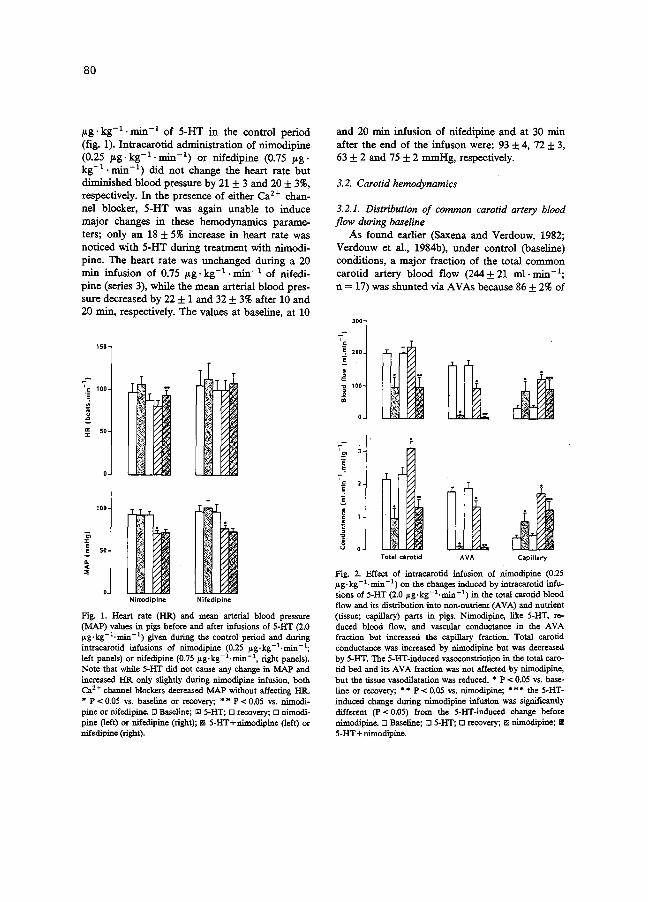
80
J.!g·kg- 1 ·min- 1 of 5-HT in the control period (fig. 1). Intracarotid administration of nimodipine (0.25 J.!g·kg- 1 ·min-1) or nifedipine (0.75 J.!g· kg- 1
· min- 1) did not change the heart rate but
diminished blood pressure by 21 ± 3 and 20 ± 3%, respectively. In the presence of either Ca2+ channel blocker, 5-HT was again unable to induce major changes in these hemodynamics parameters; only an 18 ± 5% increase in heart rate was noticed with 5-HT during treatment with nimodipine. The heart rate was unchanged during a 20 min infusion of 0.75 J.!g · kg- 1 • min-1 of nifedipine (series 3), while the mean arterial blood pressure decreased by 22 ± 1 and 32 ± 3% after 10 and 20 min, respectively. The values at baseline, at 10
1SO
--' 0 100 -~ . ! 0: so "'
100
c, "' E E SO
0. < ::e
~h~ ;~~~ v.
r, ~· %.
~ ~:i Nimodipine
Fig. 1. Heart rate (HR) and mean arterial blood pressure (:MAP) values in pigs before and after infusions of 5-HT (2.0 f'g·kg- 1·min-1 ) given during the control period and during intracarotid infusions of nimodipine (0.25 f'g·kg- 1·min- 1;
left panels) or nifedipine (0.75 f'g·kg- 1·min-l, right panels). Note that while 5-HT did not cause any change in MAP and increased HR only slightly during nimodipine infusion, both Ca 2+ channel blockers decreased MAP without affecting HR. • P < 0.05 vs. baseline or recovery; • • P < 0.05 vs. nimodipine or nifedipine. D Baseline; liD 5-HT; D recovery; D nimodipine (left) or nifedipine (right); 1!11 5-HT + nimodipine (left) or nifedipine (right).
and 20 min infusion of nifedipine and at 30 min after the end of the infuson were: 93 ± 4, 72 ± 3, 63 ± 2 and 75 ± 2 =Hg, respectively.
3.2. Carotid hemodynamics
3.2.1. Distribution of common carotid artery blood flow during baseline
As found earlier (Saxena and Verdouw, 1982; Verdouw et al., 1984b), under control (baseline) conCiitions, a major fraction of the total co=on carotid artery blood flow (244 ± 21 ml · min-\ n = 17) was shunted via A VAs because 86 ± 2% of
' 0
300
~ 200
! ~ 'C 100 8 ii
' "' "' E ~
rul d' 'ili . w~ ~~-.~·
V.:" ~.· ... v.:.,
.. ~.
Total carotid AVA Capillary
Fig. · 2. Effect of intracarotid infusion of nimodipine (0.25 f<g·kg- 1·min- 1) on the changes induced by intracarotid infusions of 5-HT (2.0 ,..g·kg- 1·min-1) in the total carotid blood flow and its distribution into non-nutrient (A VA) and nutrient (tissue; capillary) parts in pigs. Nimodipine, like 5-HT, reduced blood flow, and vascular conductance in the A VA fraction but increased the capillary fraction. Total carotid conductance was increased by nimodipine but was decreased by 5-HT. The 5-HT-induced vasoconstriction in the total carotid bed and its AVA fraction was not affected by nimodipine, but the tissue vasodilatation was reduced. • P < 0.05 vs. baseline or recovery; •• P < 0.05 vs. nimodipine; ••• the 5-HTinduced change during nimodipine infusion was significantly different cP < 0.05) from the 5-HT -induced change before nimodipine. D Baseline; D 5-HT; D recovery; !l3 nimodipine; Ill 5-HT + nimodipine.

the injected microspheres was detected in the lungs whereas only a very minor fraction (1.6 ± 0.4%) was used to supply the brain. The blood flow to the extracerebral tissues was: skin, 1.5 ± 0.2 ml · min- 1 (0.6 ± 0.1%); skeletal muscles, 6.1 ± 0.6 ml · min - 1 (2.6 ± 0.2%) ears, 0.9 ± 0.2 ml· min - 1 (0.4 ± 0.1%); tongue, 2.0 ± 0.5 ml· min- 1 (0.9 ± 0.2%); salivary glands, 5.8 ± 1.1 ml· min - 1 (2.6 ± 0.5%); eyes, 2.0 ± 0.3 ml· min- 1 (0.9 ± 0.1%) and bone, 9.2 ± 1.3 ml· min- 1 (4.2 ± 0.6%). Finally, less than 2% of the carotid blood flow was distributed to the contralateral half of the head and less than 0.1% of the microspheres was detected in the heart and kidneys, signifying an efficient entrapment of the spheres in the lung vasculature after their escape via the cranial A VAs.
90 •• 0 ..
60 30 e • L 0 ;;:: ., 30 8 iii
15
1.2
' 0> :r E
0.9 ~ 7
0 .. ! 0.6 . u 0
1J . ~ 0.3 'g 0 u
Skin Skeletal Ears muscles
81
3.2.2. Effects of ~imodipine on the 5-HT-induced changes in the distribution of common carotid artery blood flow and vascular conductance
The effects of intracarotid infusions of 5-HT, before and during nimodipine infusion, on the distribution of carotid blood flow into nutrient (tissue; capillary) and non-nutrient (AVA) fractions are shown in fig. 2. 5-HT caused a decrease in total common carotid artery blood flow (54± 14%), which was entirely due to a reduction (75 ± 14%) in AVA blood flow; nutrient flow more than doubled. Similar changes were observed in vascular conductances, as the blood pressure was not much affected by 5-HT. In spite of a reduction in blood pressure by nimodipine, the total carotid blood flow was maintained· as the total carotid
Tongue Salivary glands
Brain
Fig. 3. Effect of intracarotid infusion of nimodipine (0.25 JLg·kg" 1·min" 1) on the vasodilatation induced in various tissues by intracarotid infusions of 5-HT (2.0 JLg·kg" 1·min" 1). While 5-HT produced its most prominent vasodilator effect in the skin and ears, nimodipine selectively and potently increased blood flow to and vascular conductance in the skeletal muscles. During nimodipine infusion, 5-HT caused vasoconstriction in the skeletal muscles, and its vasodilator response in the tissues was reduced. • P < 0.05 vs. baseline or recovery; • • P < 0.05 vs. nimodipine; • • • the 5-HT-induced change during nimodipine infusion was significantly different (P < 0.05) from the 5-HT-induced change before nimodipine. 0 Baseline; 0 5-HT; 0 recovery; !ll nimodipine: !II 5-HT + nimodipine.

82
conductance increased (41 ± 9%). 1bis was accompanied by a large elevation (300 ± 25%) in vascular conductance in the nutrient part, which outweighed the 19 ± 7% reduction in vascular conductance in A VAs. Although nimodipine markedly affected vascular conductance in both the total carotid vascular bed and its AVA fraction, it had no influence on 5-HT-induced vasoconstrictor responses. In contrast, nimodipine converted the amine-evoked vasodilatation into vasoconstriction in the nutrient part.
Of all tissues and organs, the skin and ears showed the largest vasodilator response to 5-HT (18- and 12-fold increase in vascular conductance, respectively). Skeletal muscles exhibited a marked vasodilatation (15-fold increase in vascular conductance) during nimodipine infusion. Vascular conductance was also increased, but only moderately so in the salivary glands (fig. 3). In the presence of nimodipine, the 5-HT-induced vasodilatation in the skin and ears tended to be less marked. However, in the skeletal muscles ~-HT now elicited a marked vasoconstriction which reduced the nimodipine-induced vasodilatation by more than 50%. Although the carotid artery contribution to the blood supply of the brain decreased slightly in response to both 5-HT and nimodipine, vascular conductance in the brain did not change significantly. Tongue vessels were not affected by either drug.
3.2.3. Effects of nifedipine on the 5-HT-induced changes in the distribution of common carotid artery blood flow and vascular conductance
5-HT induced vasodilatation in the nutrient fraction of the carotid vascular bed (conductance increased by 350 ± 85%). 1bis vasodilatation was unable to compensate for the decline (by 80 ± 5%) in A VA conductance, resulting in a 24 ± 3% reduction in vascular conductance of the total carotid bed (fig. 4). The decrease in mean arterial blood pressure during intracarotid nifedipine infusion was compensated for by a 4-fold elevation in vascular conductance of the capillary fraction which, by compensating for the slight decrease in AVA conductance, enhanced total carotid conductance. As a result, total carotid blood flow was maintained during nifedipine infusion. The 5-HT-
400
_ ...... 300
' c ·~
! 200
~ ;::
~ ID 100
' "' :r E ~
' c -~
! . u c . u , ~
0-
Jj.
. . .
Capillary
Fig. 4. Effect of intracarotid infusion of nifedipine (0.75 l'g· kg" 1·min-1) on the changes induced by intracarotid infusions of 5-HT (2.0 l'g·kg- 1·min-1) in the total carotid blood flow and its distribution into non-nutrient (AVA) and nutrient (tissue; capillary) parts in pigs. Nifedipine, like 5-HT, reduced blood flow to and vascular conductance in the AVA fraction but increased the capillary fraction. Total carotid conductance was increased by nifedipine but was decreased by 5-HT. The 5-HT-induced vasoconstriction was either not affected (AVA fraction) or was even enhanced (total carotid vascular bed) by nifedipine, but the 5-HT-induced tissue vasodilatation was reduced. • P < 0.05 vs. baseline; • • P < 0.05 vs. nifedipine; • • • the 5-HT-induced change during nifedipine infusion was significantly different (P < 0.05) from the 5-HT-induced change before nifedipine. D Baseline; lill 5-HT; D recovery; Ell nifedipine; Ell 5-HT + nifedipine.
induced vasoconstriction in the total carotid vascular bed was enhanced in the presence of nifedipine but that in the AVA part was unchanged. On the contrary, the vasodilator response to 5-HT in the nutrient (tissue; capillary) fraction was completely abolished (fig. 4).
5-HT caused a 30-fold increase in vascular con-
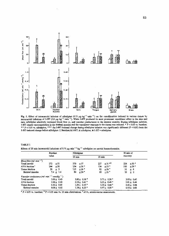
90
' 0
"E 60
"E
• 0 ;;: '0
30
8 iii
1.2
--"' "' E 0.9 ~
7 0
"E "E 0.6 . u
~ 0.3 '0 g u
45
30
15
0.60
0.&15
0.30
0.15
Ear-s Tongue Salivary glands
83
Br-ain
Fig. 5. Effect of intracarotid infusion of nifedipine (0.75 l'g·kg-1·min- 1) on the vasodilatation induced in various tissues by intracarotid infusions of 5-HT (2.0 I'S·kg- 1·min- 1). While 5-HT produced its most prominent vasodilator effect in the skin and ears, nifedipine selectively increased blood flow to, and vascular conductance in the skeletal muscles. During rtifedipine infusion, 5-HT caused vasoconstriction in the skeletal muscles and the vasodilator response in the tissues was reduced. • P < 0.05 vs. baseline; • • P < 0.05 vs. nifedipine; • • • the 5-HT-induced change during nifedipine infusion was significantly different (P < 0.05) from the 5-HT-induced change before nifedipine. 0 Baseline; iilJ 5-HT; 12 nlfedipine; !II 5-HT+ rtifedipine.
TABLE 1
Effects of 20 min intracarotid infusions of 0.751'g·min- 1·kg-1 rtifedipine on carotid haemodynamics.
Blood flow (ml· min 1)
Total carotid AVA fraction c
Tissue fraction Skeletal muscles
Baseline value
272 ±31 244 ±28 28 ± 5 7.4 ± 1.4
Vascular conductance (ml· min- 1 • mmHg- 1)
Total carotid 3.00 ± 0.43 AVA fraction 2.69 ± 0.40 Tissue fraction 0.30± 0.05
Skeletal muscles 0.08 ± 0.02
Nifedipine
10min 20min
270 ±37 227 ±31a.b 154 ±26 a 134 ±25 a
115 ±26 a 92 ±26 a 84 ±24 a 60 ±21a
3.80± 0.56 a 3.71± 0.58 a 2.19± 0.41a 2.21 ± 0.49 a 1.59± 0.35 a 1.50± 0.44 a 1.18± 0.33 a 0.97± 0.35.
a P < 0.05 vs. baseline; b P < 0.05 min vs. 10 min observations. cAVA, arteriovenous anastomotic.
30 min of recovery
224 ±30 a 193 ±29 a
31 ± 6 10 ± 2
3.03± 0.47 2.61± 0.46 0.41± 0.08 0.13± 0.03

84
ductance in the skin (fig. 5), which was significantly attenuated when the animals were treated with nifedipine. Vasodilator responses to 5-HT (12- and 4-fold increase in vascular conductance, respectively) in the ears and tongue were also attenuated. 5-HT reduced the nifedipine-induced elevation (almost 16-fold) in vascular conductance in the skeletal muscles by approximately 50%.
3.2.4. Effects of nifedipine infusions alone Table 1 shows the changes in the distribution of
common carotid artery blood flow observed during a 20 min intracarotid infusion of nifedipine (0.75 j.tg·kg-1 ·min- 1) without concomitant administration of 5-HT. As described above, nifedipine decreased AVA flow and vascular conductance but increased capillary blood flow and vascular conductance, particularly to the skeletal muscles. There were no differences between the effects of nifedipine at the 10 and the 20 min period, except that the total carotid blood flow was decreased significantly after 20 min. The carotid hemodynamic variables returned towards baseline values (except total carotid blood flow) 30 min after the nifedipine infusion was stopped.
4. Discussion
4.1. Carotid vascular responses to 5-HT
As reported previously (Saxena and Verdouw, 1982; Verdouw et al., 1984a,b), a large fraction (about 80%) of the total common carotid blood flow in the anesthetized pig was shunted via A VAs located in the skin and ears (Saxena and Verdouw, 1985a). Infusion of 5-HT drastically changed the distribution of carotid blood flow so that, with or without a concomitant decrease in total carotid blood flow, the non-nutrient (AVA) fraction decreased and the nutrient (tissue; capillary) fraction, particularly that to the skin and ears, increased; recovery from the effects of 5-HT was observed within 20 min (Saxena and Verdouw, 1982; Verdouw et al., 1984b). These hemodynamic effects are due to activation of vascular 5-HT2 receptors causing constriction, mainly of the 'large' conductance vessels (Heistad et al., 1976; Saxena
and Verdouw, 1982), and vascular '5-HTclike' receptors mediating constriction of A VAs and relaxation of arterioles (Saxena and Verdouw, 1985b; Saxena et al., 1986). Besides confirming the above findings with 5-HT, we now report the carotid vascular effects of two Ca2 + channel blockers of the dihydropyridine type (nimodipine and nifedipine) and their influence on the responses to 5-HT.
4.2. Carotid vascular effects of nimodipine and nifedipine
Local intracarotid infusions of both nimodipine and nifedipine resulted in a redistribution of carotid blood flow in favour of the nutrient fraction and at the expense of the non-nutrient fraction. While in the case of 5-HT the increased nutrient flow was distributed mainly to the skin and ears (Saxena and Verdouw, 1982; Verdouw et al., 1984b; present results), nimodipine and nifedipine selectively affected the skeletal muscles. It is of interest to recall that vascular conductance in, and blood flow to the skeletal muscles is also increased by other dihydropyridine-type Ca2+ channeJ blockers ( darodipine, felodipine and nisoldipine), but not by veraparnil or diltiazem (Hof, 1983; Bolt and Saxena, 1984; Duncker et al., 1986a,b). Low affinity but high capacity dihydropyridine binding sites, as yet unassociated with any specific functional response, have recently been described in the skeletal muscles (Miller and Freedman, 1984). It is tempting to suggest that the vascular effects in the skeletal muscles may be related in some way to the above binding sites.
The relaxation of arterioles is apparently due to Ca2+ channel blockade but the mechanism responsible for the decrease in AVA flow and conductance is less obvious. It is unlikely to be a baroreceptor reflex-mediated stimulation of the sympathetic nervous system since the animals were bilaterally vagosympathectomized. Moreover, in young pigs A VAs are only poorly constricted via noradrenergic mechanisms (Verdouw et al., 1984a). Thus, the reduction in AVA flow most likely results from 'steal' due to the profound vasodilatation in the nutrient vascular channels. This impression is enhanced when the data for nimodipine and nifedipine are compared to those for

5-HT. While 5-HT reduced the vascular conductance of A VAs by approximately 85% in the face of a 3-fold increase in conductance of the nutrient fraction, nimodipine and nifedipine reduced AVA conductance by, respectively, only 25 and 18%. These latter effects were accompanied by even greater increases in nutrient conductance (4.6- and 5.3-fold, respectively). It therefore seems that, in contrast to 5-HT which actively constricts A VAs, the Ca2 + channel blockers cause a 'passive' reduction in AVA flow and conductance.
4.3. Modification of carotid vascular responses to 5-HT by nimodipine and nifedipine
When nifedipine was infused alone for 20 min (series 3 experiments), the effects of the drug were not much different whether measured at 10 or 20 min. This suggests that the changes (from values after 10 min nifedipine infusion) obtained after simultaneous infusions of nifedipine and 5-HT (series 2 experiments) were in fact due to 5-HT and not to a major change in the effects of nifedipine with a longer period of infusion. The same may also hold true for nimodipine, though this drug was not infused alone for the full 20 miit period.
A comparison of the responses to 5-HT before and after the infusions of the two Ca2+ channel blockers revealed three interesting facts: (i) the vasoconstrictor response in the total carotid vascular bed not only remained unattenuated but seemed to be enhanced; (ii) the increase in the tissue fraction due to 5-HT was completely eliminated (total capillary flow) or reduced (skin and ears); and (iii) blood vessels in the skeletal muscles - strongly dilated under the influence of the Ca2+ channel blockers - responded to 5-HT with a marked vasoconstriction.
The lack of attenuation by the Ca2+ channel blockers, nimodipine and nifedipine, of the 5-HTinduced constrictions in vivo of A VAs (mediated by '5-HT1-like' receptors) or of 'small' resistance vessel (arterioles; mediated by 5-HT2 receptors) suggests that, in contrast to the 5-HT-induced constrictions of 'large' conducting arteries in vitro (Towart, 1981; Miiller-Schweinitzer and Neuman, 1983; Van Nueten, 1984), these responses are not
85
dependent on influx of extracellular Ca2+. A similar conclusion has recently been reached by others. Except for verapamil which has some affinity for 5-HT2 binding sites (see Kalkman et a!., 1984), a number of Ca2+ channel blockers (diltiazem, flunarizine, nimodipine or darodipine) failed to antagonize the 5-HT-induced elevations of perfusion and arterial blood pressures in the rat (Cavero and Lefevre-Borg, 1981; Kalkman et a!., 1984) and the decrease in total peripheral AVA flow in the cat (Hof et a!., 1985). Hof et a!. (1985) also observed that 5-HT, which did not significantly alter coronary haemodynamics, caused an unmistakable coronary vasoconstriction when given after darodipine.
Unlike the vasoconstrictor responses, the 5-HT-induced vasodilatation, particularly that in the skin vasculature, was reduced by the two Ca2+ channel blockers. It appears to us that the attenuated increase in flow to, and the vascular conductance in the skin is secondary to the strong vasodilatation elicited by the two Ca2+ channel blockers in other parts of the nutrient bed mainly of the skeletal muscles. When the blood vessels of skeletal muscles were strongly relaxed under the influence of the Ca2+ channel blockers, it is logical to expect that 5-HT elicited no further vasodilatation but caused arterial vasoconstriction which is mediated by 5-HT2 receptors (Verdouw et a!., 1984b; Saxena and Lawang, 1985; Meschig eta!., 1985). Unlike the vasoconstriction in 'large' vessels in vitro, this vasoconstriction was not inhibited by nifedipine and nimodipine in vivo (see above).
4.4. Possible implications in migraine
Cerebral blood flow has been reported to decrease, probably as a result of 'spreading depression of Leao', in 'classical' but not in 'non-classical' migraine patients (Lauritzen and Olesen, 1984). If the release of 5-HT is involved, Ca2+
channel blockers probably do not act via this mechanism as shown by their ineffectiveness against the vasoconstrictor responses to 5-HT in vivo.
As mentioned at the outset, sudden opening of cranial A VAs in the headache phase (Heyclc, 1969;

86
Saxena, 1978), probably associated with the decrease in blood 5-HT level (Lance, 1978; Fozard, 1982), has also been pathophysiologically implicated in migraine. Compatible with this suggestion, antimigraine drugs - particularly the ergot alkaloids - effective in the treatment of individual attacks (Johnson and Saxena, 1978; Spierings and Saxena, 1980) as well as 5-HT (Saxena and Verdouw, 1982; Verdouw et al., 1984b) which has been reported to alleviate acute attacks (Kimball et al., 1960) decrease AVA shunting by eliciting an 'active' constriction of· cranial A VAs. The 'passive' reduction of AVA flow induced by the two Ca2+ channel blockers may be one of the reasons for their inability to abort acute attacks of migraine (Jensen et al., 1985). The redistribution of carotid blood flow towards the nutrient compartment, as observed in the present study, should obviously be considered as potentially beneficial. Whether this. property of Ca2+ channel blockers is linked to the usefulness of these drugs in the prophylactic therapy of migraine (Gelmers, 1983; Meyer and Hardenberg, 1983) remains to be ascertained.
Acknowledgements
The authors are grateful to Bayer A.G., Wuppertal for fmancial support and to Mr. J. Heiligers and Ms. AM Rutte· man for technical assistance.
References'
Auer, L.M., Z. Ito, A Suzuki and H. Ohta, 1982, Prevention of symptomatic vasospasm; a controlled trial of nimodipine in subarachnoid haemorrhage patients, Acta Neurochir. 63, 297.
Bolt, G.R. and P.R. Saxena, 1984, Acute systemic and regional hemodynamic effects of felodipine, a new calcium antagonist, in conscious renal hypertensive rabbits, J. Cardiovasc. Pharrnacol. 6, 707.
Cavero, I. and F. Lefevre-Borg, 1981, Effects of diltiazem and verapamil on pressor responses mediated by stimulation of alpha, alpha2-adrenoceptors, angiotensin and 5-hydroxytryptarnine receptors in pithed rats, Br. J. Pharrnacol. 74, 839P.
Duncker, DJ., J.M. Hartog, P.O. Hugenholtz, P.R. Saxena and P.D. Verdouw, 1986a, The effects of nisoldipine (Bay K 5552) on cardiovascular performance and regional blood flows in pentobarbital-anaesthetized pigs with or without beta-adrenoceptor blockade, Br. J. Pharrnacol 88, 9.
Duncker, D.J., J. Heiligers, E.J. Mylecharane, P.R. Saxena, and P.D. Verdouw, 1986b, Nimodipine-induced changes in the distribution of carotid blood flow and cardiac output in pentobarbital-anaestetized pigs, Br. J. Pharmacol. 89, 35.
Fozard, J.R., 1982, Serotonin, migraine and platelets. In Progress in Pharmacology, eds. P.A. Van Zwieten and E. SchOnbaum (Gustav Fischer Verlag, Stuttgart) p. 135.
Gelme..S, HJ., 1983, Nimodipine, a new calcium antagonist, in the prophylactic treatment of migraine, Headache 23, 106.
Heistad, D.D., M.L. Marcus, S. Sandberg and F.M. Abboud, 1976, Effect of sympathetic nerve stimulation on cerebral bloodflow and on large cerebral of dogs, Circ. Res. 41, 342.
Heyck, H., 1969, Pathogenesis of migraine, Res. Clin. Stud. Headache 2, 1.
Hof, R.P., 1983, Calcium antagonist and the peripheral circulation: differences and similarities between PY 108.068, nicardipine, verapamil and diltiazem, Br. J. Pharmacol. 78, 375.
Hof, R.P., A Hof and H.O. Kalkman, 1985, Modification of the vasoconstrictor effects of noradrenaline and serotonin by the selective calcium antagonist PY 108.068 in the peripheral circulation of anesthetized cats, J. Cardiovasc. Pharrnacol. 7 (Suppl. 6), S61.
Hugenholtz, P.G., P.D. Verdouw, J.W. De Jong and P.W. Serruys, 1984, Nifedipine for angina and acute myocardial infarction. In Calcium Antagonists and Cardiovascular Diseases, Perspectives in Cardiovascular Research, Vol. 9, Ed. Opie, L.H., pp. 237-255, New York: Raven Press.
Jensen, K., P. Tfelt-Hansen, M. Lauritzen and J. Olesen, 1985, Clinical trial of nimodipine for single attacks of classic migraine, Cephalalgia 5, 125.
Johnson, B.M. and P.R. Saxena, 1978, The effect of ergotamine on tissue blood flow and the arteriovenous shunting of radioactive microspheres in the head, Br. J. Pharrnacol 63, 541.
Kalkman, H.O., P.B.M.W.M. Timmerrnans and P.A. Van Zwieten, 1984, The vasopr.;.sor response to serotonin (5-IIT) in rats; its dependency upon extracellular calcium, Arch. Int. Pharrnacodyn. 268, 232.
Kimball, R. W., A.P. Friedman and E. Vallejo, 1960, Effect of serotonin in migraine patients, Neurology 10, 107.
Lance, J.W., 1982, Mechanism and Management of Migraine, London: Butterworth Scientific, p. 162.
Lauritzen, M. and J. Olesen, 1984, Regional cerebral blood flow during migraine attacks by Xenon-133 inhalation and emission tomography, Brain 107, 447:
Louis, P., 1981, A double-blind placebo-controlled prophylactic study of flunarizine (Sibelium) in migraine, Headache 21, 235.
Meschig, R., J. Breuer and G. Arnold, 1985, Serotonin-induced vasoconstriction in the perfused canine femoral artery can be blocked in vivo by ketanserin, J. Cardiovasc. Pharmacol. 7 (Suppl. 7), S56.
Meyer, J.S. and J. Hardenberg, 1983, Clinical effectiveness of calcium entry blockers in prophylactic treatment of migraine and cluster headaches, Headache 23, 266.
Miller, RJ. and S.B. Freedman, 1984, Are dihydropyridine

binding sites voltage sensitive calcium channels, Life Sci. 13, 1205.
Milller-Schweinitzer, E. and P. Neuman, 1983, In vitro, effects of calcium antagonists PN 200-110, nifedipine, and nimodipine on human and canine cerebral arteries, J. Cereb. Blood Flow Metab. 3, 354.
Saxena, P.R., 1978, Arteriovenous shunting and migraine, Res. Clin. Stud. Headache 6, 89.
Saxena, P.R. and A. Lawang, 1985, A comparison of cardiovascular and smooth muscle effects of 5-hydroxytryptamine, a selective agonist of 5-HT1 receptors, Arch. Int. Pharmacodyn. 277, 235.
Saxena, P.R. and P.D. Verdouw, 1982, Redistribution of 5-hydroxytryptamine of carotid arterial blood at the expense of arteriovenous blood flow, J. Physiol. (London) 332, 501.
Saxena, P.R. and P.D. Verdouw, 1985a, Tissue blood flow and localization of arteriovenous anastomoses in pigs with microspheres of four different sizes, PflUgers Arch. 403, 128.
Saxena, P.R. and P.D. Verdouw, 1985b, 5-Carboxamide tryptamine, a compound with high affinity for 5-HT1 binding sites, dilates arterioles and constricts arteriovenous anastomoses, Br. J. Phartnacol. 84, 533.
Saxena, P.R., H.C. Scharnhardt, R.P. Forsyth and J. Loeve, 1980, Computer programs for the radioactive microsphere technique. Determination of regional blood flows and other haemodynamic variables in different experimental circumstances, Comp. Progr. Biomed. 12, 63.
Saxena, P.R., D.J. Duncker, A.H. Bom, J. Heiligers and P.D.
87
Verdouw, 1986, Effects of MDL 72222 and methiothepine on carotid vascular responses to 5-hydroxytryptarnine in the pig: Evidence for the presence of '5-hydroxytryptamine1-like' receptors, Naunyn-Schmiedeb. Arch. Pharmacal. 333, 198.
Spierings, ELH. and P.R. Saxena, 1980, Antimigraine drugs and cranial arteriovenous shunting in the cat, Neurology 30,676.
Steel, R.G.D. and J.H. Torrie, 1980, Principles and Procedures of Statistics, A Biomedical Approach, 2nd edn., Tokyo: McGraw-Hill Kogak:usha Ltd.
Towart, R., 1981, The selective inhibition of serotonin-induced contractions of rabbit =•bra! vascular smooth muscle by calcium-antagonistic dihydropyridines, Circ. Res. 48, 650.
Van Nueten, J.M., 1984, Antivasoconstrictor effects of drugs used in migraine therapy, in: The Pharmacological Basis of Migraine Therapy, eds. W.K.. Amery, J.M. Van Nueten and A. Wauquier (Pitman Press, Bath) p. 19.
Verdouw, P.D., DJ. Duncker and P.R. Saxena, 1984a, Poor vasoconstrictor response to adrenergic stimulation in the arteriovenous anastomoses present in the carotid vascular bed of young Yorkshire pigs, Arch. Int. Pharmacodyn. Ther. 272, 56.
Verdouw, P.D., H.M. Jennewein, J. Heiligers, D.J. Duncker and P.R. Saxena, 1984b, Redistribution of carotid artery blood flow by 5-HT: effects of the 5-HT2 receptor antagonists ketanserin and WAL 1307, European J. Pharmacal. 102, 499.

-----------------------

89
VASODILATORY PROFILE OF PYRIDAZINONE-DERIVATIVES


CHAPTER 6
CARDIOVASCULAR PROFILE OF PIMOBENDAN,
A BENZIMIDAZOLE-PYRIDAZINONE DERIVATIVE
WITH VASODILATING AND INOTROPIC PROPERTIES
P.O. Verdouw, J.M. Hartog, O.J. Ouncker, W. Rothl and P.R. Saxena2 •
Laboratory for Experimental Cardiology (Thoraxcenter)
and 20epartment of Pharmacology,
Erasmus University Rotterdam, Rotterdam, The Netherlands and 10r. Karl Thomae GmBH, Biberach a/d Riss, Federal Republic of Germany.
Eur. J. Pharmacol. 126, 21-30, 1986.
91

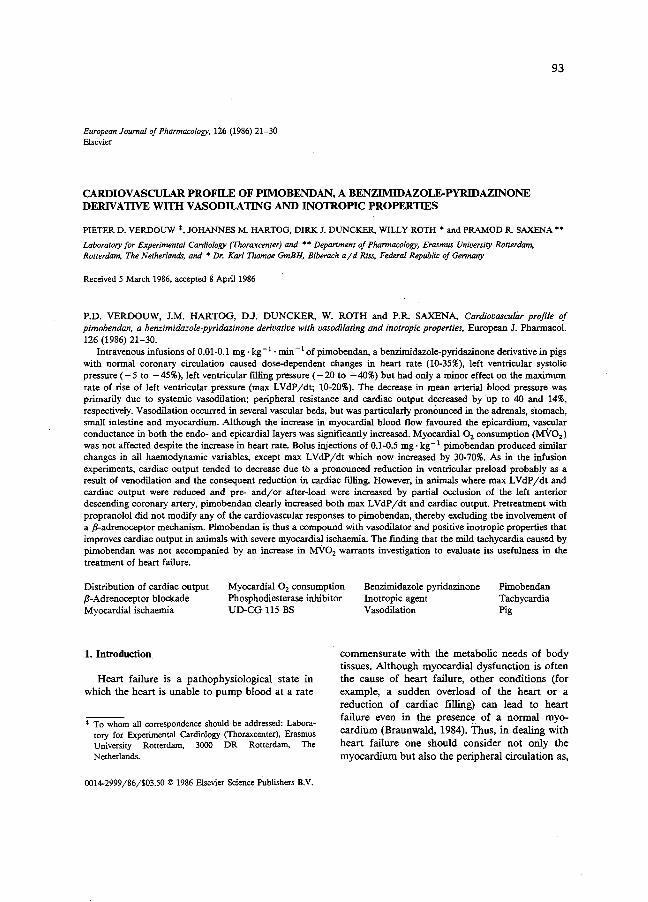
European Journal of Pharmacology, 126 (1986) 21-30 Elsevier
93
CARDIOVASCULAR PROFILE OF PIMOBENDAN, A BENZIMIDAZOLE-PYRIDAZINONE DERIVATIVE WITH VASODILATING AND INOTROPIC PROPERTIES
PIETER D. VERDOUW *,JOHANNES M. HARTOG, DIRK J. DUNCKER, WILLY ROTH* and PRAMOD R. SAXENA ••
Laboratory for Experimental Cardiology (Thoraxcenter) and •• Department of Pharmacology, Erasmus University Rotterdam, Rotterdam, The Netherlands, and • Dr. Karl Thomae GmBH, Biberach ajd Riss, Federal Republic of Germany
Received 5 March 1986, accepted 8 April 1986
P.D. VERDOUW, J.M. HARTOG, D.J. DUNCKER, W. ROTH and P.R. SAXENA, Cardiovascular profile of pimobendan, a benzimidazole-pyridazinone derivative with vasodilating and inotropic properties, European J. Pharmacol. 126 (1986) 21-30.
Intravenous infusions of 0.01-0.1 mg · kg - 1 • min -I of pimobendan, a benzirnidazole-pyridazinone derivative in pigs with normal coronary circulation caused dose-dependent changes in heart rate (10-35%), left. ventricular systolic pressure (- 5 to - 45% ), left ventricular filling pressure (- 20 to -40%) but had only a minor effect on the maximum rate of rise of left ventricular pressure (max LVdPidt; 10-20%). The decrease in mean arterial blood pressure was primarily due to systemic vasodilation; peripheral resistance and cardiac output decreased by ·up to 40 and 14%, respectively. Vasodilation occurred in several vascular beds, but was particularly pronounced in the adrenals, stomach, small intestine and myocardium. Although the increase in myocardial blood flow favoured the epicardium, vascular conductance in both the endo- and epicardial layers was significantly increased. Myocardial 0 2 consumption (MV02 )
was not affected despite the increase in heart rate. Bolus injections of 0.1-0.5 mg · kg- 1 pimobendan produced similar changes in all haemodynamic variables, except max LV dP I dt which now increased by 30-70%. As in the infusion experiments, cardiac output tended to decrease due to a pronounced reduction in ventricular preload probably as a result of venodilation and the consequent reduction in cardiac filling. However, in animals where max LV dP 1 dt and cardiac output were reduced and pre- andjor after-load were increased by partial occlusion of the left anterior descending coronary artery, pimobendan clearly increased both max LV dP 1 dt and cardiac output. Pretreatment with propranolol did not modify any of the cardiovascular responses to pimobendan, thereby excluding the involvement of a ,8-adrenoceptor mechanism. Pimobendan is thus a compound with vasodilator and positive inotropic properties that improves cardiac output in animals with severe myocardial ischaemia. The finding that the mild tachycardia caused by pimobendan was not accompanied by an increase in MV02 warrants investigation to evaluate its usefulness in the treatment of heart failure.
Distribution of cardiac output ,8-Adrenoceptor blockade Myocardial ischaemia
Myocardial 0 2 consumption Phosphodiesterase inhibitor UD-CG 115 BS
Benzimidazole pyridazinone Inotropic agent Vasodilation
Pimobendan Tachycardia Pig
t_ Introduction
Heart failure is a pathophysiological state in which the heart is unable to pump blood at a rate
* To whom all correspondence should be addressed: Laboratory for Experimental Cardiology (Thoraxcenter), Erasmus University Rotterdam, 3000 DR Rotterdam, The Netherlands.
0014-2999j86j$03.50 © 1986 Elsevier Science p;,blishers B.V.
commensurate with the metabolic needs of body tissues. Although myocardial dysfunction is often the cause of heart failure, other conditions (for example, a sudden overload of the heart or a reduction of cardiac filling) can lead to heart failure even in the presence of a normal myocardium (Braunwald, 1984). Thus, in dealing with heart failure one should consider not only the myocardium but also the peripheral circulation as,

94
Fig. 1. Chemical structure of pirnobendan (UD-CG 115 BS).
in addition to a depressed myocardial contractility, elevated left ventricular filling pressure and systemic vascular resistance are among prominent characteristics of heart failure. It is therefore obvious that positive inotropic agents which also cause peripheral vasodilation may be useful in the treatment of heart failure.
In an attempt to provide efficient substitutes for cardiac glycosides, which have a narrow therapeutic margin, many positive inotropic agents acting via different mechanisms including inhibition of cardiac phosphodiesterases, have been described during the past several years (see Farah et al., 1984). One such compound, pimobendan (UD-CG 115 BS; fig. 1) has been shown to increase myocardial contractility and cause peripheral vasodilation (Diederen et al., 1982; Van Meel, 1985) possibly due to elevation of cyclic A~P levels following inhibition of myocardial phosphodiesterase (Honerjager et a!., 1984). In this report we give a detailed description of the cardiovascular profile of the drug, including its effects on regional vascular beds and myocardial performance.
2. Materials and methods
2.1. Experimental set-up
After an overnight fast Yorkshire pigs (22-28 kg) were sedated with 120 mg azaperone i.m. and anaesthetized with 150 mg i.v. metomidate. The levels of arterial blood gases (ABL-3, Acid-Base Laboratory, Radiometer, Copenhagen) were controlled by artificial ventilation with a mixture of 0 2 : N20 (1 : 2). Catheters were placed in the superior vena cava for administration of pentobar-
bital (20 mg · kg- 1 • h- 1
), sodium bicarbonate (8.4%, when needed), pimobendan and haemaccel; the latter was given to replace blood loss. Microtipped Millar catheters were used for measurements of the left ventricular and central aortic blood pressures. An 8 F catheter was positioned in the descending aorta for the withdrawal of arterial blood samples. The muscle relaxant pancuronium ( 4 mg) was administered before the heart was exposed via a midstemal split. Electromagnetic flow probes (Skalar, Delft, The Netherlands) were placed around the ascending aorta and the left anterior descending coronary artery (LAD). The great cardiac vein was cannulated and the left atrial appendage was catheterized for the injection of microspheres.
Myocardial wall thickness was monitored with a 5 MHz ultrasonic transducer (KrautkramerBranson, Lewistown, PA, USA). The wall thick~ ness at end-diastole (EDT) and end-systole (EST) was used to calculate systolic wall thickening (swt) as:
swt (%) = 100 x (EST-EDT)/EDT
and the mean velocity of swt (V,w,) as:
Y,w, = (EST-EDT)jDS
where DS is the duration of the isovolumic contraction phase and the ejection time.
The distribution of cardiac output was measured using the microsphere technique (see Saxena and Verdouw, 1985; Verdouw et al., 1985). About 2 X 10⁶ microspheres (15 ± 1 (S.D.)p.m diameter, NEN Company, Dreieich, West Germany), labelled with either 46Sc, 103 Ru, 141 Ce, 95 Nb or 113Sn, were injected via a cannula placed in the left atrium of the heart. A reference arterial blood sample was withdrawn (flow rate, 10 ml· min- 1 )
starting just before and continuing for a period of about 1 min after the injection of microspheres. At the end of the experiment several organs (see later) were excised, weighed and placed in vials. The heart was removed and fixed in 4% formalin for at least 24 h. The details of the counting of radioactivity and processing of data have been described previously (Saxena et a!., 1980).
Myocardial oxygen consumption (MV02 ) was estimated by multiplying the difference in the 0 2

content of the arterial and coronary venous blood by coronary blood flow.
2.2. Experimental protocols
Four series of experiments were performed to characterize the cardiovascular actions of pirnobendan. In the first series (n = 8), four consecutive 10 min infusions (0.01, 0.025, 0.05 and 0.10 rng · kg- 1 -min- 1 ) were used. At the end of each infusion all cardiovascular variables were recorded and arterial blood was drawn for the determination of plasma concentrations of the parent cornpound and its major metabolite (UD-CG 212 BS). In the second series (n = 9), each animal received three consecutive bolus injections of pirnobendan at 10 min intervals. The doses (0.1, 0.25 and 0.5 rng · kg -I) were chosen such that the animals received the same amount of drug as administered during each comparable infusion period in the first series.
To evaluate cardiac pump function during pathological conditions, blood flow in the left anterior descending coronary artery was reduced in 4 animals (third series) by tightening a J-shaped clamp placed around the vessel until the systolic wall thickening was reduced to 20-30% of the baseline value. After the haernodynamic values had been stable for at least 15 min these animals were given pirnobendan (0.25 rng · kg- 1 + 0.05 rng · kg- 1 . min- 1). Lastly, in order to establish whether or not some of the actions of pirnobendan were caused via ,8-adrenoceptor stimulation, the cardiovascular effects of the drug were studied in 5 pigs (fourth series) before and after pretreatment with propranolol (0.5 rng · kg- 1 followed by 0.5 rng · kg -I · h-I). This dose regimen of propranolol provides adequate ,8-adrenoceptor blockade in ·pigs (Wolffenbuttel and Verdouw, 1983).
2.3. Determination of plasma concentrations
The plasma concentrations of pirnobendan and its 0-dernethylrnetabolite, UD-CG 212 BS were determined using an HPLC assay with fully auto-
95
mated drug preconcentration on solid supports (Roth, 1983). Briefly, the drugs were extracted on a reverse phase column and simultaneously preconcentrated after injection of whole plasma. The compounds were measured by means of fluorescence detection (332 nrn/405 nrn) after HPLC separation on reversed phase ODS-hypersil (particle size: 5 JLm). The eluent composition was methanol/water 590/460 (v/v) + 2.5 g ammonium acetate per liter eluent (total amount 2.625 g). Post column, a mixture of rnethanol/orthophosphoric acid 85%/water (300/100/100, vjv/v) was added with a flow rate of 0.2 rnl · min -I via a T-fitting in order to optimize the fluorescence (increase in fluorescence by a factor of 2). The lower limit of detection for both compounds was about 1 ng/rnl. Pirnobendan and UD-CG 212 BS themselves were used as external standards.
2.4. Data presentation and statistical analysis
All data are presented as means ± S.E.M. Absolute values are given in the tables and in fig. 3, but % changes from baseline values are shown in the other figures to facilitate comparison between different variables. However, all statistical analyses were performed on the actual data values. The significance of the difference between the means of any two groups was compared by applying Duncan's new multiple range test once an analysis of variance (randomized block design) had revealed that the samples represented different populations (Saxena, 1985). Statistical significance was accepted at P < 0.05 (two-tailed).
2.5. Drugs
The only drugs used in this study were the anaesthetics, haernaccel, propranolol hydrochloride and pirnobendan ( 4,5-dihydro-6-[2-( 4-rnethoxypheny 1)-1 H-benzimidazol-5-yl]-5-rnethyl-3(2H)pyridazinone; UD-CG 115 BS). Pirnobendan was dissolved in a mixture of polyethylene glycol 200 and saline (1 : 1 ).

96
3. Results
3.1. Intravenous infusion of pimobendan in animals with normal coronary circulation
3.1.1. Plasma concentrations of pimobendan and UD-CG212 BS
The plasma concentrations of the parent drug increased rapidly during intravenous infusions of pimobendan. There was a more than 14-fold difference between the concentrations noted at the end of the first and the last infusion period. The concentration of the major metabolite, UD-CG 212 BS, increased only 6-fold during the same period (table 1) suggesting saturation of the enzymes metabolizing pimobendan during higher infusion rates.
3.1.2. Systemic haemodynamics The most pronounced effect of pirnobendan
was the vasodilation in systemic vascular beds as the dose-dependent decreases in arterial blood pressure (up to 45%) agreed very closely with those in systemic vascular resistance (table 1 ). Diastolic arterial pressure declined slightly more than systolic arterial blood pressure (up to 52 and 44%, respectively). Pimobendan also decreased the
TABLE 1
left ventricular filling pressure, ·indicating reduced venous return and, therefore, venodilation. This probably accounts for the slight decrease in cardiac output at the two highest infusion rates. Max LVdP/dt was moderately enhanced (up to 20%) with the two lowest doses, but was not different from baseline during the two highest infusion rates.
3.1.3. Regional haemodynamics Table 2 shows that, despite the reduction in
cardiac output, pimobendan increased blood flow to the adrenals, stomach, small intestine and brain, but decreased that to the kidneys and spleen. Hepatic arterial (liver) and skeletal muscle blood flow did not change significantly after pimobendan. Since arterial blood pressure was decreased by the drug, vascular conductance increased dose dependently in most organs (fig. 2). The most marked effect was on the stomach (up to 325%) followed by adrenals (200%), small intestine (190%), myocardium (160%; see following section), skeletal muscles (140%) and brain (130%). No differences existed between the effects of pimobendan on different brain areas (cerebral hemisphe;:es, diencephalon, cerebellum and brainstem; data not shown in the figure). Vascular conduc-
Cardiovascular actions of continuous 10 min pirnobendan infusions in 8 anaesthetized open-chest pigs. Abbreviations: CO, cardiac output; HR, heart rate; SV, stroke volume; LVEDP, left ventricular end-diastolic pressure; LVdP /dt, maximal rate of rise of left ventricular pressure; SVR, systemic vascular resistance; SAP, systolic arterial pressure (mm Hg); DAP, diastolic arterial pressure (mm Hg). All data were obtained after 10 min of infusions. All data are presented as means± S.E.M. • P < 0.05 vs. baseline.
Baseline Pimobendan (mg·kg 1-min- 1)
O.Ql 0.025 0.05 0.1
Total dose injected (mg·kg- 1) 0.1 0.35 0.85 1.85
Plasma concentration (1'&·1- 1)
Pimobendan 100 ± 10 274 ± 19 689 ± 35 1441 ± 61 UD-CG212 BS 3.1 ± 0.3 9.1 ± 0.6 14.4 ± 1.2 20.2 ± 1.8
CO(l·min- 1) 2.16± 0.11 2.15± 0.11 2.01± 0.12 1.90± 0.11* 1.85± 0.14. HR (beats· min_, ) 84 ± 4 92 ± 4* 104 ± 5 • 112 ± 6. 114 ± 7. SV(ml) 26 ± 2 24 ± 1 20 ± 2* 17 ± 2* 17 ± 2* LVEDP (mm Hg) 10 ± 1 ± 1* 6 ± 1* 6 ± 1* 6 ± 1. LVdP /dt (mm Hg·s- 1) 1460 ±160 1680 ±170. 1770 ±210. 1620 ±210 1570 ±230 SVR (mm Hgr 1·min) 39 ± 3 37 ± 3 30 ± 2* 25 ± 1* 22 ± 1. SAP(mmHg) 102 ± 4 95 ± 5. 79 ± 3* 64 ± 3. 57 ± 2* DAP(mmHg) 63 ± 4 59 ± 4 49 ± 4* 37 ± 3. 30 ± 2*

97
TABLE 2
Effect of continuous 10 min pimobendan infusions on organ blood flow (ml· min - 1·100 g - 1) in 8 anaesthetized open-chest pigs. All
data are presented as means± S.E.M.; • P- < 0.05 vs. baseline.
Baseline Pimobendan (mg. kg - 1• min- 1)
Kidneys Liver Spleen Adrenals Stomach Small intestine Skeletal muscles Brain
(!,%) KIDNEY
100~
-4~ .
(6%) STOMACH
(6%) MUSCLE
200Li* 100 0
322±27 30± 9 80±13
136±22 14± 1 21± 7±
22± 2
(6%) LIVER
(6%) SM INTESTINE
200LJ~ 100 . 0
(t.%) BRAIN
J : ... aLA
0.01
351±22 27± 8
101±20 166±24. 17± 2 22± 2 7± 1
22± 1
(6%) SPLEEN
100r . ~ ..i. -4~~
(!,%) ADRENALS
:::ld 100 ..
0 -~· j~;
PlMOBENDAN (mg·k9!mili1)
Fig. 2. Effect of intravenous infusion of pimobendan on regional vascular conductances. Pre-infusion values of vascular conductances (in ml·min- 1·mm Hg- 1·100 g- 1) were: kidneys: 3.98±0.39; liver: 0.37±0.10; spleen: 0.94±0.13; stomach 0.18 ± 0.02; small intestine: 0.25 ± 0.03; adrenals: 1.68 ± 0.29; skeletal muscles 0.08 ± 0.02 and brain: 0.27 ± 0.02. Data are presented as percentage changes from baseJine, but the statistical analysis was performed on the actual data. • P < 0.05 vs. baseline.
O.Q25 0.05 0.1
318±32 217±28. 156±23. 24± 6 22± 4 21± 5 84±17 51± 11. 33± 8.
189±29. 194± 19 • 181±20. 19± 2* 24± 3* 27± 3.
22± 1 26± 2. 28± 2* 6± 7±2 8±
22± 2 23± 1 25± 2*
tance in the kidneys, liver and spleen was much less affected. All three showed increases at lower doses (<50%) but the effect tended to decrease as perfusion pressure started to drop severely.
3.1.4. Myocardial blood flow and performance Coronary blood flow increased up to 30% in
spite of the decline in arterial blood pressure (fig. 3). This increase in flow was not evenly distributed transmurally as the epicardium benefited more (up to 43%) than the endocardial layers (up to 15%) during the first three infusion rates. There was even a slight decrease in the endocardial blood flow at the highest infusion rate (0.1 mg · kg- 1
• min- 1). Consequently, the endo-epi flow ratio decreased gradually from 0.99 ± 0.03 to 0.70 ± 0.02 (P < 0.05) at the end of the highest infusion rate. Coronary venous 0 2 saturation increased dose dependently from 37 ± 5 to 58± 3% (P < 0.05). Hence MV02 was not significantly affected (fig. 3). Regional myocardial function showed no changes for systolic wall thickening but a gradual increase in the velocity of thickening was noticed. Figure 4 shows that, although the vasodilator response to pimobendan in the epicardial layers exceeded that in the endocardial layers, vascular conductance in the latter was also doubled at the end of the highest infusion rate. Though not shown in the figure, the increases in vascular conductance in the right ventricle (up to 180 ± 12%) and left atrium (up to 110 ± 20%) were very similar to those in the left ventricle.

98
transmural t30t LV
blood !low 1lO
(ml.min:g')
0.90
e~i 1.0[
0.7
M~O:z 4.5[ (1Jmol.miri"1.g1
)
2.5
swt (%)
35[
17
PIMOBENDAN (mg.kg:min') 0.01
*
0.1 0.025~
Fig. 3. Left ventricular performance after intravenous infusions of pimobendan at increasing rates. From top to bottom: transmural left ventricular blood flow, the endo-epi blood flow ratio, left ventricular 0 2 consumption (MV02 ), regional systolic wall thickening (swt) and the mean velocity of wall thickening cY,w, ). • P < 0.05 vs. baseline.
3.2. Bolus injections in animals with normal coronary circulation
3.2.1. Plasma concentrations of pimobendan and UD-CG212 BS
Following each bolus injection of pimobendan, the plasma levels of the parent drug increased sharply but, at 10 min, they were considerably lower than those after infusions of the corresponding amounts. As was the case in the infusion experiments, the plasma concentration of the metabolite UD-CG 212 BS increased but less steeply than that of. the parent drug (table 3).
(6%) TRANSMURAL
20l ~ 101AJ
(6%) ENDO
~
PIMOBENDAN
(mg.kg1.min")
I O.Q1 0025 . 0.05 0.1
(6%) EPI
200~: 100 0
Fig. 4. Effect of intravenous infusion of pimobendan on left ventricular vascular conductance. Vasodilation of the epicardial vessels exceeded that of the endocardial vessels, which explains the decrease in the endo-epi blood flow ratio. Preinfusion transmural conductance was 1.26 ± 0.22 mhnin -I. mm Hg- 1·100 g- 1• • P < 0.05 vs. baseline. Data are presented as percentage changes from baseline but the statistical analysis was performed on the actual data -'
3.2.2. Systemic haemodynamics As in the infusion experiments, a pronounced
vasodilation accompanied by tachycardia occurred after bolus injections of pimobendan (table 3). Cardiac output decreased slightly (19%) after the highest dose as the increase in heart rate (up to 35%) was not sufficient to balance the decrease in stroke volume (up to 40%). The latter appeared to be caused by the fall in left ventricular filling pressure (up to 62%) as systemic vascular resistance decreased (up to 40%) and max LVdPjdt increased by almost 70% in spite of the fall in diastolic arterial blood pressure (table 3). The substantial increase in max LV dP / dt after the bolus injections contrasts sharply with the minor changes observed when the drug was infused (series 1). This is best illustrated in fig. 5 which shows that the larger increases occurred at comparable reductions in systemic vascular resistance. Another striking feature was that the increase in max LVdPjdt was lower with 0.5 mg·kg- 1 than with 0.25 mg · kg- 1 despite similar after- and preload reductions.

99
TABLE 3
Cardiovascular actions of cumulative bolus injections of pimobendan in 9 anaesthetiZed open-chest pigs. Abbreviations: CO, cardiac output; HR. heart rate; SV, stroke volume; LVEDP, left ventricular end-diastolic pressure; LVdP/dt, maximal rate of rise of left ventricular pressure; SVR, systemic vascular resistance; SAP, systolic arterial pressure; DAP, diastolic arterial pressure; a peak refers to the time at which the effect on max LVdP/dt was maximum. All data are presented as means±S.E.M.; * P < 0.05 vs. baseline.
Baseline Pimobendan (mg·kg~ 1 )
0.1 (n = 9)
Peak' 10min
Total dose injected (mg·kg- 1) 0.1
Plasma concentration (p.gr'> Pimobendan 124 ±22 34 ± UD-CG212BS 1.7 ± 0.4 5.4 ± 1.6
CO(l·min- 1) 2.46± 0.18 2.46± 0.16 2.37± 0.15 HR (beats·min- 1) 102 ± 5 110 ± 5 * 114 ± 6 * SV(ml) 24 ± 1 24 ± 1 21 ± 1.
LVEDP (rom Hg) 9 ± 1 6 ± 1* ± 1.
LVdP/dt (mm Hg·s- 1) 1750 ±20 2230 ±20. 2080 ±20.
SVR(mmHgr 1
·min) 38 ± 5 32 ± 4* 36 ± 4 SAP(mmHg) 107 ± 5 98 ± 5* 96 ± s• DAP(mmHg) 71 ± 4 63 ± 5* 66 ± 5
75
j\0 50
'*' ...s- 0
: 1 i~~ . -------·
0
0 0
-so
500 1000 1500
PtMOBENOAN ("gll
0.25 (n = 9) 0.5 (n -7)
Peak lOmin Peak 10min
0.35 0.85
484 ±59 129 ±12 757 ±68 273 ±26 7.8 ± 1.6 11.2 ± 1.4 12.4 ± 1.9 17.4 ± 1.8 2.47± 0.12 2.21± 0.17 2.40± 0.20 2.06± 0.16.
121 ± 7. 128 ± s• 127 ± s• 135 ± 6.
21 ± 1 17 ± 1. 19 ± 1. 15 ± 1.
± 1* ± 1* 4 ± 1* 4 ± 1.
3100 ±30 * 2540 ±20. 2690 ±30. 2570 ±20.
26 ± 4* 31 ± 3 * 25 ± 3. 29 ± 3* 80 ± s• 81 ± s• 77 ± 4* 76 ± 3• 47 ± 4* 53 ± 4* 45 ± 4* 48 ± 4*
3.3. Bolus injections in animals with ischaemic hearts
Reduction of the lumen of the left anterior descending coronary artery did not affect heart rate but led to an increase in the left ventricular end-diastolic pressure (from 8.4 ± 1.4 to 19.6 ± 1.7 mm Hg) and diminutions in max LVdP jdt (25%), cardiac output (by 26%), stroke volume (23%) and regional systolic wall thickening (from 24 ± 2 to 9 ± 2%). A large fall in mean arterial blood pressure (9%) was prevented by the vasoconstriction (23%) in systemic vascular beds. After the preparation had been stable for 15 min, pimobendan (0.25 mg · kg- 1 over 2 min+ 0.05 mg · kg- 1 ·
Fig. 5. Changes in max LVdP /dt (top) and total systemic vascular resistance (SVR, bottom) as a function of arterial plasma concentrations of pimobendan after infusions or bolus injections (peak and 10 min later). Notice that the changes in SVR but not those in max LVdP /dt depend on the plasma concentrations. The latter is true even when ·th:e effects on heart rate and preM and afterload are taken into account (compare tables 1 and 3). • P < 0.05 vs. baseline. 0 Pimobendan bolus (peak); 0 pimobendan bolus (10 min); • pimobendan infusion.

100
40 co MAP SVR
{A%)
1 0
[J ~ *+ . •+ -40
40 HR max LV dFYclt 160 LVEOP
•+ {A%) ~ _I + u {A%)
0
-40 160
Fig. 6. Effect of pimobendan (0.25 mg-kg- 1 +0.05 mg·kg- 1•
min- 1) in 4 pigs with a partial stanosis in the left anterior descending coronary artery. The ischemia (D) and pimobendan (•) induced changes are given as percentage change from baseline. Baseline values were: cardiac output (CO, 2.44±0.25 l·min- 1 ); mean arterial blood pressure (MAP, 74±5 mm Hg); systemic vascular resistance (SVR, 31±5 mm Hg·l- 1-min); heart rate (HR, 81±1 beats·min- 1
); max LYdP/dt (1630± 220 mm Hg · s -l) and left ventricular end-diastolic blood pressure (LVEDP, 8.4± 1.4 mm Hg). • P < 0.05 vs. baseline; + P < 0.05 vs. ischaemia. Data are presented as percent changes from baseline but the statistical analysis was performed on the actual data.
min -I for 5 min) was administered. The drug lowered left ventricular filling pressure to 12.6 ± 1.8 mm Hg and systemic vascular resistance (45%), but increased heart rate (20%) and max LVdPidt (30%). Stroke volume did not change significantly, perhaps due to a reduction in left ventricular filling following the increase in heart rate. These observations are akin to those in the animals with a normal coronary circulation. However, the increase in cardiac output in the ischaemic preparation (20%, fig. 6) was at variance with the results of these experiments.
3.4. Cardiovascular actions of pimobendan after /3-adrenoceptor blockade
A bolus of 0.5 mg · kg- 1 propranolol followed by an infusion of 0.5 mg · kg- 1 · h-I lowered heart
rate (from 89 ± 14 to 75 ± 7 beats · min -I), max LV dP I dt (from 1 930 ± 290 to 1 280 ± 110 rnm Hg · s- 1
) and mean arterial blood pressure (from 83 ± 2 to 74 ± 4 mm Hg). Cardiac output was reduced to the same extent as arterial blood pressure so that peripheral resistance did not change. Pimobendan-induced tachycardia, increase in cardiac contractility and systemic vasodilation were not modified in the presence of /3-blockade. The changes caused by 0.5 mg · kg- 1 pimobendan before and after propranolol, respectively, were 27 ± 6 and 27 ± 5% (heart rate), 49 ± 13 and 47 ± 14% (max LVdPidt) and -32 ± 6 and -38 ± 5% (systemic vascular resistance). Coronary venous 0 2 saturation, which had decreased from 18 ± 3 to 13 ± 3% after /3-blockade, increased dose dependently to 39 ± 7% (P < 0.05) during pimobendan administration, a response again not different from that observed with pimobendan alone.
4. Discussion
The investigation showed that pimobendan caused a vasodilation and cardiac stimulation which were not mediated by /3-adrenoceptors. The vasodilator activity of pimobendan was demonstrated in animals that received the drug either as infusions or as bolus injections. The magnitude of the effect was closely related to the arterial plasma concentrations and appeared to be independent of the rate of drug administration. In contrast, the inotropic effects of pimobendan, evaluated by changes in max LV dP I dt, were less obvious. Bolus injections caused marked increases, but when the plasma levels were raised gradually by infusion, the changes in max LV dP I dt were only minimal. One must, however, be careful when using max LVdPidt as an index of contractility as this parameter also depends on heart rate and pre- and afterload, all of which changed considerably with the drug. In pigs, an increase in heart rate from 60 to 100 beats· min- 1 causes a 15% increase in max LV dP I dt, but further increases have no additional effect (Verdouw et al., 1980a,b; Scheffer and Verdouw, 1983). Since the heart rate ranged from 80 to 140 beats· min -I in the present study, it is unlikely that tachycardia alone could account for

the increase in max LVdPidt. Moreover, because of the fall in left ventricular end-diastolic (probably due to venodilation) and arterial blood pressures, the increases in inotropic state were underestimated by the changes in max LVdPidt. At variance with the changes in total systemic vascular resistance, the effects of pimobendan on max LV dP I dt were not related to the plasma concentrations of the drug (fig. 5). During the infusion experiments higher concentrations were accompanied by lower max LV dP I dt increments, even at comparable changes in heart rate, pre- and afterload. It has been suggested that the metabolite UD-CG 212 BS possesses positive inotropic properties (Meyer, unpublished data). However, in our study the concentrations of UD-CG 212 BS increased at a similar rate after infusion or bolus injections of pimobendan (see tables 1 and 3), yet the pattern of max LV dP 1 dt changes differed widely. Therefore, our data do not suggest a positive inotropic action of UD-CG 212 BS in the concentration range attained in this investigation.
A combination of vasodilation and positive inotropy increases cardiac output by facilitation of ventricular emptying and augmentation of the velocity of wall thickening (Cohn and Franciosa, 1978; Miller et a!., 1977; 1981; Verdouw et a!., 1981). The results obtained with pimobendan during coronary artery occlusion-induced myocardial ischemia show that, in a setting where cardiac contractility and output are depressed in the presence of a high pre- andlor afterload, the pimobendan-induced increase in cardiac inotropy is translated into a salutary effect on the pump function of the heart. It would appear, therefore, that the drug may be useful in clinical heart failure where a similar pathophysiological state is encountered. The finding that cardiac output did not increase in the animals with normal hearts emphasizes the role of left ventricular filling and systemic vascular resistance.
The effects of pimobendan on regional vascular beds are relevant to the therapy of congestive heart failure. Vasodilation occurred in all beds but, for the kidneys and spleen, this happened only with the lowest doses. When perfusion pressure started to fall severely, the vascular beds of these organs tended to constrict. For some other
101
organs (stomach, adrenals, heart, brain) the vasodilator response increased dose dependently. In the heart, vasodilation was more marked in the epicardium than in the endocardium ( endo-epi ratio was reduced) and, as a result, perfusion of the epicardium increased and that of the endocardium was maintained despite a marked reduction in perfusion pressure. The smaller effect on the endocardial blood flow may have been at least partly due to an increase in heart rate and the consequent reduction of the duration of diastole caused by pimobendan. Moreover, the vasodilator capacity of the endocardial vessels appears to be less than that of the epicardial vessels (Winbury and Howe, 1979). The reduction in endo-epi ratio does not appear to be deleterious because myocardial function was improved by the drug, even in the animals with an ischaemic heart.
A potential disadvantage of cardiotonic agents is that they may increase MV02 , not least by the accompanying tachycardia. In the case of pimobendan, these untoward effects were apparently balanced by reductions in pre- and afterload since MV02 did not change following drug administration. The data on MV02 also confirm that vasodilation in the coronary vascular bed was due to a direct action of pirnobendan and was not the consequence of an increased myocardial metabolic demand.
In summary, pimobendan presents itself as a compound with vasodilator and positive inotropic properties that improves cardiac output in a model of severe ischaemia. The finding that the mild tachycardia elicited by the drug was not accompanied by an increase in MV02 warrants investigation to evaluate its usefulness in the treatment of heart failure. This is especially true since, in the clinical setting, the heart rate may even be reduced because of a reduction in reflex sympathetic activity as a consequence of an improvement in left ventricular function.
Acknowledgements
Mr. R.J. Rensen is thanked for the art work and Miss. P.H. Vegter for her assistance in the preparation of this manuscript.

102
References
Braunwald, E., 1984, Heart failure: evaluation of therapy based on a consideration of natural history, in: Milrinone, Investigation of New Inotropic Therapy for Congestive Heart Failure, eds. E. Braunwald, E.H. Sonnenblick, L.W. Chakrin and R.P. Schwan, Jr. (Raven Press, New York) p. 1.
Cohn, J.N. and J.A. Franciosa, 1978, Selection of vasodilator inotropic or combined therapy for the management of heart failure, Am. J. Med. 65, 181.
Diederen, W., J. Diimmgen and R. Kadatz, 1982, Cardiovascular profile of UD-CG 115, a new orally and long acting cardiotonic compound, not related to beta-mimetics or cardiac glycosides, N aunyn-Schntiedeb. Arch. Pharmacal. 321 (Suppl.), R36 (Abstr. 141).
Farah, A.E., A.A. Alousi and R.P. Schwan, Jr., 1984, Positive inotropic agents, Ann. Rev. Pharmacal. Toxicol. 24, 275.
HoneJjager, P., A. Heiss, M. Schlifer-Korting, G. Schonsteiner and M. Reiter, 1984, UD-CG 115 - a cardiotonic pyridazinone which elevates cyclic AMP and prolongs the action potential in guinea-pig papillary muscle, NaunynSchntiedeb. Arch. Pharmacal. 325, 259.
Miller, R.R., N.A. Awan, A.N. DeMaria and D.T. Mason, 1977, Importance of maintaining systolic blood pressure during nitroglycerin administration for reducing ischemic injury in patients with coronary disease: Effects of coronary blood flow on myocardial energetics anci left ventricular function, Am. J. Cardiol. 40, 504.
Miller, R.R., A.R. Palomo, T.A. Brandon, C.J. Hartley and M.A. Quinones, 1981, Combined vasodilator and inotropic therapy of heart failure: Experimental and clinical concepts, Am. Heart J. 102, 500.
Roth, W., 1983, Rapid, sensitive and fully automated high-performance liquid chromatographic assay with fluorescence detection for sulmazole and metabolites, J. Chromatogr. Biomed. Appl. 278, 347.
Saxena, P.R.,; 1985, An interactive computer programme for data management and parametric and non-parametric statistical analysis, Proc. Br. Pharmacol. Soc., Edinburg, p. 03.
Saxena, P.R., H.C. Schamhardt, R.P. Forsyth and J. Loeve, 1980, Computer programs for the radioactive microsphere technique. Determination of regional blood flows and other
haemodynarnic variables in different experimental circumstances, Comp. Prog. Biomed. 12, 63.
Saxena, P.R. and P.O. Verdouw, 1985, 5-Carboxamide tryptamine, a compound with high affmity for 5-HT1 binding sites, dilates arterioles and constricts arteriovenous anastomoses, Br. J. Pharmacal. 84, 533.
Scheffer, M.G. and P.O. Verdouw, 1983, Decreased incidence of ventricular fibrillation after an acute coronary artery ligation in exercised pigs, Basic Res. Cardiol. 78, 298.
Van Meel, J.C.A., 1985, Cardiovascular effects of the positive inotropic agents pimobendan and sulrnazole in vivo, Arzneim. Forsch./Drug Res. 35, 284.
Verdouw, P.O., J.M. Hartog and A.M. Rutternan, 1981, Systemic and regional myocardial responses to AR L 115 BS, a positive inotropic imidazo-pyridine, in the absence or in the presence of the bradycardiac action of alinidine, Basic Res. Cardiol. 76, 328.
Verdouw, P.O., H.M. Jennewein, J. Mierau and P.R. Saxena, 1985, N-(3-acetylarninophenyl)piperazine hydrochloride (BEA 1654), a putative 5-HT1 agonist, causes constriction of arteriovenous anastomoses and dilatation of arterioles, European J. Pharmacol. 107, 337.
Verdouw, P.O., P.R. Saxena, H.C. Schamhardt, T.M. Van der Hoek and A.M. Rutteman, 1980a, The effects of alinidine, an N-allyl derivative of clonidine, on regional myocardial perfusion and performance in the pig with or without atrial pacing, European J. Pharmacal. 64, 209.
Verdouw, P.O., FJ. Ten Cate1 H.C. Schamhardt, T.M. Vander Hoek and O.L. Bastiaans, 1980b, Segmental myocardial function during progressive coronary flow reduction and its modification by pharmacologic intervention, in: Advances in Clinical Cardiology, Vol. 1, eds. H. Kreuzer, W.W. Parmley, P. Rentrop and H.W. Heiss (Gerhard Witzstrock, Publishing House, Inc., New York) p. 270.
Winbury, M.M. and B.B. Howe, 1979, Stenosis: regional myocardial ischemia and reserve, in: Ischemic Myocardium and Antianginal Drugs, Vol. 3, eds. M.M. Winbury andY. Abiko (Publisher New York) p. 55.
Wolffenbuttel, B.H.R. and P.O. Verdouw, 1983, Nifedipine and myocardial performance in the presence and absence of beta-blockade with propranolol, Arch. Int. Pharmacodyn. Ther. 266, 83.

CHAPTER 7
USEFULNESS OF PIMOBENDAN IN THE TREATMENT OF HEART FAILURE.
D.J. Duncker, F.J. van Dalen, J.M. Hartog, J.M.J. Lamersl, R.J. Rensen,
P.R. Saxenaz and P.D. Verdouw.
Laboratory for Experimental Cardiology (Thoraxcenter), 1Department of Biochemistry and 2Pharmacology,
Erasmus University Rotterdam, Rotterdam, The Netherlands.
Arznelm. Forsch. I Drug Res. 36(Il) 12, 1740-1744, 1986.
103


105
Reprint
ARZNEIMITTEL-FORSCHUNG I DRUG RESEARCH Arzneim.-Forsch./Dru·g Res. 36 (II), 12,1740-1744 (1986) · Editio Cantor· D-7960 Aulendorf
From the Departments ofCardiology1, Biochennstry2 and Pharmacology', Erasmus University, Rotterdam (The Netherlands)
Usefulness ofPimobendan in the Treatment of Heart Failure
By D. J. Duncker1, F. J. van Dalen1, J. M. Hartog1, J. M. J. Lamers', R. J. Rensen1, P.R. Saxena', and P. D. Verdouw1
Summary: The effects of the benzimidazole-pyridazinone pimobendan (UD-CG 115 BS) on systemic haemodynamics, myocardial performance and the distribution of cardiac output were studied in open-chest anaesthetized'pigs. After intravenous bolus injections (0.1-0.5 mg- kg-1, n = 7) increases in heart rate (up to 37%), LVdPidtmax (up to 54%) and decreases in systemic vascular resistance (up to 33%) and left ventricular filling pressure (up to 50%) were observed, while cardiac output was unchanged. Vasodilation occurred in nearly all regional vascular beds, but was most pronounced in the adrenals (200%), followed by stomach ( 1 50%), small intestines (130%), heart (1 25%) and brain (110%). o,consumption was not ajfocted in spite of the increases in heart rate and myocardial inotropy. To evaluate the direct ejfocts on the myocardium, pimobendan was also infosed (1-5 llg. kg-L mirr-1, n = 7) directly into the left anterior descending coronary artery. In addition to a marked vasodilation of the coronary bed (140%), also a lowertng of the left ventricular filling pressure (up to 20%) and cardiac output (15%) was observed, but no changes in regional myocardial function, LVdP/dtmax and systemic vascular resistance occurred. Immediately after intracoronary bolus injections (1 mg · kg-1, n = 4), vasodilation of the coronary vessels was apparent, but myocardial contractility was not affected. This may explain that cyclic AMP content, determined in biopsies excised 30 s after injection, was unaltered. it may be concluded that pimobendan e;,;erts actions on the cardiovascular ,system which may be useful in the treatment of heart failure.
Zusammenfassung: Nutzlichkeit von Pimobendan bei der Behandlung der Herzinsuffizienz Die Wirkungen des Benzimidazol-pyridazinons Pimob.endan (UD-CG 115 BS) aufsystemische Htimodynamik, myo-
kardiale Leistung und_Verteilung des Herzminutenvolumens wurden an narkotisierten Schweinen mit eriJjfoetem Brustkorb untersucht. Nach intraveniisen Bolusinjektionen (0,1-0,5 mg. kg-1, n = 7) wurde eine Zunahme der Herzfrequenz (bis 37%) und der LVdPidtmax (bis 54%) sowie eine Abnahine des systemischen Gefiif3widerstandes (bis 33%) und des linksventrikultiren Fiillungsdruckes (bis 50%) beobachtet, wtihrend das Herzminutenvolumen unvertindert blieb. Eine Vasodilatation war in fast allen regionalen Gefiij3betten zu verzeichnen, jedoch war sie besonders augenfiillig in den Nebennieren (200%), gefolgt vom Magen (150%), dem Dunndarm (130%), dem Herzen (125%) und Gehim (110%). Der 0,- Verbrauch blieb trotz der Steigerung der Herzfrequenz und des myokardialen Inotropismus unbeeinjluj3t. Urn die unmittelbaren Effokte auf das Myokard zu bewerten, wurde Pimobendan direkt in die Iinke vordere absteigende Koronararterie infondiert (1-5 Jlg- kg-1 · mirr-1, n = 7). Auj3er einer deutlichen Vasodilatation in den Kranzgefiij3betten (140%) wurde auch eine Abnahme des linksventrikultiren Fullungsdruckes (bis 20%) und des Herzminutenvolumens (1 5%) beobachtet, doch es traten keine Vertinderungen bei der regiona/en myokardialen Funktion, der LV dP ldtmax und dem systemischen Gefiij3widerstand ein. Unmittelbar nach intrakoronaren Bolusinjektionen (1 mg . kg-1, n = 4) war die Erweiterung) der Herzkranzgefiij3e augenfiillig, die Kontraktilittit des Myokards wurde jedoch nicht beeinj/uj3t. Dies kiJnnte eine Erk/tirung dafiir sein, daj3 der Gehalt an zyklischem AMP, der in 30 s nach Injektion exzidiertem Biopsiematerial bestimmt wurde, unvertindert war. Daraus kann geschlossen werden, daj3 die Effekte, die Pimobendan auf das kardiovaskultire System ausiibt, bei der Behandlung der Herzinsuffiziertz nutzlich sein kiJnnten.
Key words: Cardiotonic drugs· Pimobendan, pharmacology· UD-CG 115 BS
1. Introduction The ideal drug for the treatment of heart failure should augment cardiac pump function by improving myocardial contractility, and (or) reducing left yentricular pre- and afterload, without increasing myocardial 0 2-demand. In recent years a large series of new compounds (i.e. sulmazole, amrinone, milrinone) has been shown to meet the above mentioned requirements in the experimental animaL In clinical practice; however, the high incidence of side effects has prevented these drugs from totally replacing cardiac glycosides, despite the unwanted properties of the latter. In this study we describe some investigations with the benzimidazolepyridazinone pimobendan (UD-CG 115 BS*), see formula
•l Manufacturer: Dr. Karl Thomae GmbH, Biberach an der Riss (Federal Republic of Germany).
diagram), which is structurally related to sulmazole [1]. Pimobendan increases myocardial contractility in the canine preparation [1, 2], possibly due to phosphodiesterase inhibition [3]. Diederen et aL also provided evidence that the drug is active on the venous vasculature [2]. We have shown that intravenous infusion of the drug also causes a marked systemic vasodilation in the pig [3a]. However, in these experiments the positive inotropic potency of pimo-

106
bendan was of relatively minor importance. This disctepancy in inotropy between our study and those reported by others [1, 2] could not only be due to differences in the species used and experimental model but also be caused by differences in the administration: intravenous bolus injections versus infusions. In an attempt to reconcile these differences we studied the effect of pimobendan after intravenous bolus injections in anaesthetized pigs. Radioactively labelled microspheres were used to determine the effects on organ blood flow. In a second series of experiments we also infused the drug directly into a coronary artery to separate the direct actions on the myocardium from those on the vascular beds. Finally, the effects on myocardial cAMP levels were evaluated after selective coronary artery bolus injections.
2; Materials and methods 2.1. ~rimen131 set-up After an overnight fast Yorkshire pigs (22-28 kg) were sedated with 120 mg azaperone i.m., and anaesthetized with 150 mg iotravenous metomidate [4]. Arterial blood gases (ABL-3, Acid-Base Laboratory, Radiometer, Copenhagen, Denmark) were controlled by artificial ventilation with a mixture of 0, : N,O (I : 2). Catheters were placed in the superior vena cava for administration of pentobarbital (20 mg · kg-1 • h-1), sodium bicarbonate (8.4%, when needed), pimobendan and polygelioe (Haemaccel"', Behringwerke AG, Mahtbwg!Lahn, FR Germany) to replace blood loss. Microtipped Millar catheters (Millar, lnstr., Houston, TX, USA) were used for measurement of the blood pressures in the left ventricular cavity and central aorta. An 8 F catheter was positioned in the aorta descendens for the withdrawal of blood samples. Before the heart was exposed via a midsternal split, 4 mg of the muscle relaxant pancuronium bromide was administered. A precalibrated electromagnetic flow probe (Skalar, Delft, The Netherlands) was placed around the ascending aorta. The great cardiac vein was cannulated and the left atrial appendage was catheterized for the injection of microspheres. In some animals the left anterior descending coronary artery (LADCA) was cannulated for intracoronary administration of pimobendan. Myocardial wall thickness tracings, monitored with a 5 MHz ultrasonic transducer (Krautkramer-Branson, Lewistown, PA, USA), were used to evaluate regional myocardial performance. The wall thickness at end-diastolic (EDT) and end-systole (ES'D were used to calculate systolic wall thickening (swt) as
swt (%) = 100 X (EST -EDT)/EDT and the mean velocity of swt (fl swiJ as
V"" =(EST -EDT)/DS where DS is the duration of the isovolumic contraction phase and the ejection time [4). In some experiments (n = 3) regional function was estimated from segment length changes obtained from a pair of ultrasonic dimension gauges (5 MHz), placed subendocardially [5) and connected to a 4-channel ultrasonic dimension system (Model 40 I, Schuessler and associates, Cardiff by the Sea, CA, USA). The segment length at end-diastole' (EDSL) and at end-systole (ESSL) were used to calculate systolic segment length shortening (sis) as
sis(%)= 100 x (EDSL-ESSL)/EDSL and the mean velocity of sis (fl sts) as
Vsts= (EDSL-ESSL)IDS. Distribution of cardiac output was measured usiog the radioactive microsphere technique [4]. Just before the injection of about 2 x 10⁶
microspheres (15 ± 1 p.m diameter, NEN Company, Dreieich, FR Germany) labelled with either 46Sc, 103Ru, '"Ce, "Nb or "'Sn, into the left atrium, the withdrawal of an arterial reference sample was started (flow rate 10 m1 • min-I) and continued for a period of about 1 min ,after injection of the microspheres was completed. After the experiment organs and tissues were excised, weighed and placed in vials. Details of the counting of the radioactivity and processing of the data have.been described in detail [6, 7].
2.2. Experimen131 protocols Three series of experiments were performed. In the first series (n = 7) 3 consecutive i.v. bolus injections (0.1, 0.25 and 0.5 mg. kg-1) were used at 10-min intervals. Haemodynamic data were obtained duriog peak response and 10 min ·after· administration of the bolus. At this time radioactive microspheres were injected to determine regional blood flows. In a second series of experiments (n = 7) pimobendan was directly iofused into the left anterior descending coronary artery at increasing iofusion rates (1, 2.5 and 5 p.g · kg-1 · min-I), each rate again laSting 10 min. In the last series (n = 4) the role of cAMP was studied. To minimize the effect of the systemic circulation on myocardial performance pimobendan was adminis-
tered directly into the left anterior descendiog coronary artery. Cyclic AMP and cyclic GMP levels were, determined in segments per, fused by the LADCA and remote from this artery. To this end 0.5 g biopsies were rapidly taken from the ventricle areas, and directly frozen in isopentane cooled in liquid N,. The frozen tissue was pulvarized in a mortar while liquid N, was continuously added. The powder was mixed with 2.5 m1 0.9% perchloric acid (PCA), thawed and centrifuged thereafter. The PCA pellet was washed once more with 2.5 m1 0.9% PCA and further used for protein estimation. Cyclic AMP and cyclic GMP were determined, using the Amersham's cyclic AMP and cyclic GMP assay kits (Amersharn, UK), in the combined supernatants after neutralization with 0. 7 mol/1 K,PO, and 10 min centrifugation at 12,000 g [8].
2.3. Statistical analysis Statistical analysis was performed using parametric tests [9]. After each dose of pimobendan, changes from baseline were calculated separately in each experiment and the significance of these changes was determined by using the Duncan ·new multiple-range test, once an analysis of variance had established that the data represented different populations. Statistical significance was accepted at· p < 0.05 (two-tailed). All data have been expressed as mean ± standard error of mean (mean± SEM),.
2.4. Drugs The only drugs used in this study were the anaesthetics, Haemaccel and pimobendan (supplied by'Dr. Karl' Thomae GmbH, Biberach an der Riss, FR Germany) which was dissolved in a mixture of polye,tbylene glyco1400 and saline (I : 1).
·~LdiiHR ·.~lrJtfi~Vd~ldt '!"': 40 • 40 . . . ~ * ~ •
0 0 ~ ~ ~ ~ ~ ~
LVEDP SV
·:~Q· .. Q5 ·~MQ. .. QS
-20 .... . -20 ...
-40 ... . . -40 ...
-eo .....
CD
-~~ 0.1 Q3 0.5
Fig. t: Haemodynamic effects of cumulative bolus i:njections (mg · Icg-1) of pim.obendan (blank columns= i.v. bolus peak; dark columns= i.v. bolus 10 min). The following parameters are shown: heart rate (HR: baseline 99 ± 6 beats · m.m-1); ·maximum rate of rise of left ventricular pressure (LVdP/dtmax: 1680: 140 mmHg ·,-I); mean arterial blood pressure (MAP: 92 ± S mmHg); systemic vascular resistance (SVR: 41 ± 5 mmlfg · 1-1 ·min); left ventricular end-diastolic pressure (LVEDP: 9.7 ± 1.0 mmHg); stroke volume (SV: 24.2: 1.7 mi) and cardiac output (CD: 2.39: 0.23 I· min-). All data are presented as percentage change from baseline (mean ± SEM). • p < 0.05 vs baseline.

3. Results 3.1. Intravenous bolus injections in pigs 3.1.1. Haemodynamics Within minutes after each bolus injection ·mean arterial blood press\rre decreased up to 36o/o in a dose-dependent manner. This was caused by arterial vasodilation as cardiac output was not significantly affected (Fig, 1). From the determinants of cardiac output, heart rate increased up to 37o/o but stroke volume decreased to 62o/o of baseline. This occurred in. spite of the reduction in afterload and tbe increase in LVdP/dtmax (44o/o, Fig. 1). Therefore tbe 50o/o decrease in left .ventricular· filling pressure must have been responsible for tbe decline in stroke volume.
107
3.1.2. Distribution of cardiac output 10 min after administration of the lowest dose (0.1 mg . kg-I), none of the conductances of the regional vascular beds differed from their respective baseline values (Table I), but after 0.25 and 0.50 mg. kg-t, a profound vasodilation of tbe regional beds not only prevented a significant fall in perfuSion of most organs and tissues but even caused sligbt increases in that of tbe myocardium (40o/o, Table 2). The increases in conductance were in declining order: adrenals (200o/o), stomach (150%), small intestine (130o/o), heart (125o/o) and brain· (IIOo/o). In Table I tbe results for total brain are presented because only minor differences existed between tbe effects on tbe hemispheres (IOOo/o), diencepha-
Table 1: Conductance of regional vascUlar beds after cumulatiVe intravenous bolus injections ofpimobendan in 7 anaesthetized pigs.
Baseline Pimobendan (mg · kg- 1)
.o.t 0.25 0.5
svc 27.7 ±3.0 30.0 ±3.3 35.3 :1:4.6" 37.5 ±3.7" Kidneys 4.05 ±0.86 4.34 ±1.02 4.28 ±0.52 5.66 ±0.75 Liver 0.50 ±0.19 0.50 ±0.19 0.42 ±0.18 0.64 ±0.18 Spleen 1.52 ±0.17 1.70' ±0.18 2.01 ±0.43 1.87 ±0.21 Adrenals 2.01 ±0.41 2.47 ±0.45 2.92 ±0.46" 4.31 ±0.31" Stomach 0.22 ±0.06 0.22 ±0.05 0.30 ±0.05° 0.38 ±0.05° Small intestine 0.23 ±.0.03 0.24 ±0.03 0.31' ±0.04° 0.40 ±0.04~ Muscle 0.050±0.015 0.045 ±0.009 0.051 ±0.014 0.067±0.014" Skin 0.015±0.005 0.049±0.011* 0.054±0.022" 0.044±0.011" Left atrium 1.05 ±0.24 1.30 ±0.27 1.69 ±0.28° 2.15 ±0.33° Left ventricle 1.26 ±0.22 1.44 ±0.27 1.85 ±0.37" 2.59 ±0.50" Right ventricle 1.27 ±0.30 1.53 ±0.35 2.26 ±0.50" 3.07 ±0.63" Total brain 0.28 ±0.05 0.32 ±0.05 0.39 ±0.05° 0.50 ±0.05"
All conductances are in mJ. min-I· mmHg-1 · 100 g-1, ~cept those of the systemic vascular tree (SVC. ml· min-I· mmHg-1). All data are presented as mean ± SEM. "' p < 0.05 vs baseline. ·
·Table 2: Perfusion of regional vascular beds after cumulative intravenous bolus injections ofpimobendan in 7 anaesthetized pigs.
Baseline Pimobendan (mg. kg-!)
0.1 0.25 0.5
co 2.49± 0.24. 2.44± 0.20 2.37± 0.23 2.26± 0.20 Kidrieys 359±72 343±63 310±56 351±59 Liver 45.2 ±16.7 40.7 ±13.8 29.8 ±11:7 38.0 ±10.2 Spleen 139±18 141±17 141±31 113±12 Adrenals 182±39 203±35 206±32 259± 11 Stomach 19.6 ± 5.3 17.9 ± 3.7 20.5 ± 3.8 23.0 ± 3.6 Small intestine 19.9 ± 2.7 19.7 ± 1.8 21'.1 ± 2.4 23.7 ± 1.7 Muscle 4.45± 1.34 3.67± 0.73 3.48± 0.94 3.94± 0.72 Skin 1.32± 0.37 4.19± 1.12 3.95± 1.95 2.68± 0.67 Left atrium 93±20 104±17 115±18 130±19 Left ventricle 113±20 118± 19 128±23 157±29 Right ventricle 112±24 123±22 155±32° 183±34" Total brain 25.3 ± 3.8 26.1 ± 3.1 26.0 ± 2.6 29.5 ± 2.2
.. -All flow data are m '1'1· mm 1 · 100 g 1, except cardiac output (CO) which IS presented in I · min- . All data are presented as mean ± SEM. • p < 0.05 vs baseline.
Table 3: Haemodyna.rirlc effects of cumulative 10-min intracoronary infusions of pimobendan in 7 anaesthetized pigs.
Baseline Pimobendan (Jlg · kg-1 · min-1)
1 2.5 5
co 2.10±0.25 1.99±0.22" 1.88±0.22° 1.82±0.22° HR 82±4 85±4 87±4 92±5° sv 26±3 23±3' 22±3' 20±3° LVdP/dtmax 1690±70 1730±100 1780±120 1990±200 .LVEDP 10.0 ±0.4 9.3 ±0.7 8.6 ±0.9 8.0 ±0.9' MAP 84±5 79±5° 76±5" 69±5° SVR 43±4 43±5 43±5 40±4
Abb~vations: CO = cardiac output 0 · min-1); HR = heart rate (beats· min-_); SV =stroke volume (ml)i LVdP/dt"""' = maximw:n rate ofris~ ofleft ventricular pressure (mmHg · ,- ); L VEDP = left ventricular end-diastolic
~;:;::c ~;~~em(~·~~·= AJf~ ~.~~ ~ mean ± SEM. • P< 0.05 vs baseline.
Jon ( 11 Oo/o ), cerebellum ( 130o/o) and brainstem (150o/o ). The vasodilatory response of the vascular beds of the kidneys, liver and spleen did not reach levels of significance.
3.1.3. Myocardial perfonnance Coronary blood flow increased dose-dependently up to 140o/o of the pre-treatment value (113 ± 20 ml. min·! . 100 g-1) because of a doubling oftbe coronary vascular conductance. The increase in flow was most beneficial for tbe epicardial layers as perfusion of tbe endocardial layers remained unchanged (not shown). Consequently tbe endo-epi blood flow ratio decreased from L06 ±' 0.04 to 0.83 ± 0.03 (p < 0.05). The oxygen content in the coromiry venous blood rose as tbe oxygen saturation in tbe great cardiac vein increased from 26 ± 4 to 44 ± 3o/o (p < 0.05). Myocardial Oz·consumption was not significantly affected, which is not surprising because tbe double product (heart rate x systolic arterial· blood pressure) was also not affected as tbe elevations in heart rate were balanced by tbe decreases in arterial blood pressure.
3.2. Intracoronary infusions 3.2.1. Haemodynamics In spite oftbe low doses (1-5 pg · kg-1. min·') the intracoronary infuSions caused dose-dependent decreases in arterial blood pressure (up to 18o/o, p < 0.05) because of a fall in cardiac output (up to 15o/o, p < 0.05) as systemic vascular resistance was not affected (Table 3). This decrease in cardiac output occurred despite a moderate dose-dependent increase in heart rate.' Consequently stroke volume was decreased. Of its determinants only left ventricular filling was reduced, reflected by a 20o/o decrease in LVEDP, while systemic vascular resistance was not affected ·and LV dP I dtmax tended to increase. Although arterial blood pressure decreased, no redistribution of cardiac output occurred as the conductances of tbe regional vascular beds were not significantly affected (Table 4).

108
Table 4: Conductance of regional vascular-beds after cumulative 10-min intracoronary infusions ofpimobendan in 7 anaesthetized pigs.
Pimobendan (Jlg · ~cg- 1 · min-1) Baseline
I 2.5 5
svc 25.0 ±2.6 25.1 ±2.6 24.8 ±2.6 26.3 ±2.4· KidneY. 4.23 ±0.56 3.77 ±0.30 4.52 ±0.85 4.65 ±1.10 Liver 0.43 ±0.13 0.44 ±0.17 0.49 ±0.15 0.47 ±0.17 Spleen 1.74 ±0.38 1.78 ±0.39 2.00 ±0.54 1.46 ±0.36 Adrenals 2.51 ±0.26 2.70 ±0.37 3.03 ±0.47 3.32 ±0.67 Stomach (n = 3) 0.18 ±0.05 0.19 ±0.06 0.18 ±0.06 0.19 ±0.06 Muscle 0.038±0.006 0.036±0.007 0.041 ±0.008 0.039±0.009 Left atrium 1.14±0.13. 1.16 ±0.15 1.33 ±0.19 1.47 ±0.18
-All conductances are m ml· m.m-1. mmHg-1 · 100 g-1, except of the systemic vascular tree (SVC, ml· nun 1 • mmHg 1}. All data are presented as mean ± SEM.
Table 5: Myocardial performance after cumulative I 0-min intra coronary AMP and cyclic GMP detemrinations. Cyclic AMP in the (LADCA) infusions ofpimobendan in 7 anaesthetized pigs. ·LADCA-perfused myocardium tended to he slightly lower
Pimobendan (Jlg · kg- 1 · min-1) Baseline
1 2.5 5
Control segment Flow
epi 125± II 118± 10 129±18 122±14 endo 138± 14 128±12 127± 13 124±11 endo/epi 1.07±0.06 1.09±0.06 1.02±0.08 1.04±0.06
Conductance epi 1.48±0.07 1.49±0.07 1.66±0.17 1.77±0.15 en do 1.62±0.13 1.62±0.11 1.67±0.13 1.81±0.10
LADCA segment Flow
epi 132± 17 143±16 163±19 172±27 endo 143±17 154±19 179±18 191±25
.endo/epi 1.18±0.09 1.16±0.12 1.18±0.09 1.21±0.09
Conductance epi · 1.57±0.16 1.81±0.16' 2.13±0.16' 2.53±0.34' endo 1.68±0.16 1.93±0.16 2.32±0.12' 2.77±0.26'
Cor ven 01-sat 24±4" 44±5* 51±5"' 63±6' 0 2.-consumption 0.53±().07 0.40±0.05 0.39±0.07 0.30±0.03'
~~~~~~~~Oo r:P;~ndu~n~~~nn:l ~:eEF~;~EJ~o~11:~~ ~~rwv! 02-sat in%; 02-consumption in mmol· min~· 100 g- . All data are presented as mean ± SEM. "' p < 0.05 vs baseline.
3.2.2. Myocardial performance Because of the fall in perfusion pressure, a slight decrease in perfusion of the myocardium adjacent to the LADCA perfused segment was observed (Table 5). A minor increase in conductance prevented that ·this decrease in perfusion reached levels of significance. The increase in the conductance of both the endo- and epicardial layers from the area perfused by the LADCA prevented that the fall in perfusion pressure was detrimental to their perfusion. Systolic wall thickening (swt), mean velocity of swt, systolic segment length shortening (sis) and mean velocity of sis did not change significantly from their respective baseline values of 25 ± 4%, 19.8 ± 3.4 mm. s-', 24 ± 5% and 5.6 ± !'.! mm · s-1. The most marked changes were again in the coronary venous 0,-content, as the saturation in the great cardiac vein increased from 24 ± 4 to. 63 ± 6% (p < 0.05). These increases in coronary venous Q,-saturation were caused by a lowering· in regional myocardial o,-consumption (Table 5), which occurred although the double product was hardly affected.
3.3. Myocardial cyclic AMP and cyclic GMP levels after intracorooary pimobendan administration Within seconds after intracoronary bol~s injections (I mg · kg-') a very pronounced vasodilation of the coronary vascular bed occurred (visible by a bright red colour of the myocardium and confirmed by the extreme high o,-saturations in the great cardiac vein (up to 75%)), but LVdP/dt...,. and regional myocardial wall thickening of the area perfused by the LADCA were not significantly affected. At that moment the heart was excised rapidly and treated for cyclic
(6.13 ± 0.42 vs 7.27 ± 0.50 pmoVmg protein, p > 0.05), while cyclic GMP was slightly higher (0.343 ± 0.115 vs 0.143 ± 0.24 pmoVmg protein) compared to respective data obtained from a segment remote from the myocardium perfuSed by the LADCA.
4. Discussion Intracoronary infusions were used to minimize the extracardial effects of pimobendan to enable us to evaluate more directly the drug's inotropic potency. In spite of the low doses (1-5 llg . kg-' . min-I) the intracoronary infusions caused a decrease in cardiac output, as the decrease in stroke volume was not compensated by the acceleration of heart rate. This diminution in stroke volume was the result of a reduction in preload as the responses of afterload and myocardial contractility did not favor such a reduction. Because no dilation of the systemic arterial bed was noted, our data suggest that at these low doses a larger sensitivity. exists
. for the venous than for the arterial vessels, confirming the earlier observations reported by Diederen et a!. [2]. Higher doses (0.1-0.5 mg ·kg-I i. v.) caused a marked systemic arterial vasodilation in addition to the venodilation. It is noteworthy that the effects on the arterial vessels appear to be shorter lasting than on the veins, as after I 0 min the effects on the arterial ·vessels were considerably lower than the peak effects obtained after 2 min, while the effects on the venous vasculature persisted. The arterial vasodilation is one of the factors that prevented a fall in cardiac output during the intravenous bolus injections. Another was the significant rise in myocardial contractility, reflected by the increase in LVdP/dtmax· Although the latter is not a true index for myo.cardial contractility because of its dependence on heart rate and pre· and afterload, the experimental evidence in this study warrants such a conclusion as the pre- and afterload induced-changes in LVdP/dtmax tend to underestimate the inotropic changes [10, 11]. Moreover, we have repeatedly shown that in the anaesthetized pig LVdP/dt.n.x is only slightly modified when heart rate varies between I 00 and 140 beatS • min-I [12, 13]. While intravenous bolus injections (0.1-0.5 mg . kg-') revealed increases in myocardial inotropy, such an action was not noticeable during the intracoronary infusions. That the intracoronary infusion rates were too low is ruled out when one takes into account that after intravenous administration only about 2o/o of the pimobendan passes through the coronary circulation. Moreover, intracoronary bolus injections (up to I mg . kg-1) were also unable to elicit a positive inotropic action, while arterial vasodilation was remarkably present. In these experiments, we excised the hearts approximately 30 s after administration, to avoid extracardial influences of pimobendan on myocardial performance, The absence of significant changes in myocardial cAMP content therefore does not exclude the possibility of phosphodiesterase inhibition as a mechanism for the drug's inotropic potency [3), but rather indicates that the vessels respond much more rapidly (within seconds) than the tissue.
After intravenous bolus injection· vasodilation occ~ed in all regional vascular beds. The increase in conductance was enough to maintain or even augment regional blood flow in spite of the fall in perfusion pressure. In the myocardium, the epicardial layers benefited more than the endocardial layers yielding a 20% decrease in the endo-epi blood flow ratio. Domenech and Goich [14] reported tbat tachycardia does not affect the endo-epi blood flow ratio in normotensive ammals because of the endocardial vasodilatory ·reserve capacity, but in dilated coronary vascular beds raising the heart rate resulted in a decrease of the endo-epi blood flow ratio [14]. Therefore, the pimobendan-induced tachycardia and decrease in perfusion pressure are very likely the responsible factors for the uneven distribution of the flow after administration of the drug. The intracoronary infusions of pimobendan were accompamed by smaller changes in heart rate and arterial blood pressure, which explains why the different myocardial layers were now equally affected. Since the myocardial 0,-consumption was not increased after the intravenous and even decreased during the intracoronary experiments we conclude that the increase in flow was not the result of an autoregulatory response mediated by an increased 0,-demand. The decrease in 0,-consumption after the intracoronary infusions probably underestimates the true vasodilatory response to the pimoberidan infusions because of a metabolic-induced vasoconstriction. Since the double product of heart rate and left ventricular systolic pressure was unchanged in the intracoronary experiments, other factors may have played a role in the lowering of the myocardial 0,-consumption: A decrease in left ventricular wall tension due to the decrease in LVEDP seems to be such a factor. In the intravenous experiments the double product was also unchanged, but now the decrease in L VEDP was balanced by an increase in LVdP/dtm .. resulting in an unchanged myocardial o,-consumption. In conclusion, our data suggest that pimobendan at low doses acts primarily on the venous system but at higher doses the effects on the arterial vessels are also prominent. Depending on the rate of administration, a significant positive inotropic response can be elicited. In view of our observations with pimobendan, ii is of interest that Wilmshurst et al. [15] reported amrinone to be devoid of any beneficial effect in patients with cardiac failure when administered directly into the coronary artery. Intravenous administration, on the other hand, improved cardiovascular performance due to vasodilation of the venous and arterial beds. Only in a small number of patients myocardial contractility increased probably due to catecholamine release. These ob-
109
servations -suggest that the beneficial actions of some of the so-called positive inotropic agents might be due to normalization of left ventricular dimensions as a result of a reduction in both preload and afterload rather than to a significant increase in myocardial contractility. However, the above described properties of pimobendan are useful in the treatment of heart failure and side effects permitting, clinical studies appear to be warranted.
5. References [I] van Meel, J. C. A., Arzneim.-Forsch./Drug Res. 35 (1), 284 (1985) - [2] Diederen, W., Diimmgen, J., Kadatz, R.; NaunynSchmiedeberg's Arch. Pharmacol. 321 (Suppl.), R36, abstr. no. 141 (1982)- [3] Honerjiiger, P., Heiss, A., Schiifer-Korting, M., Schonsteiner, G., Reiter; M., Naunyn-Schmiedeberg's Arch. Pharmacol. 325, 259 (1984)- [3a] Verdouw, P. D., Hartog, J. M., Duncker, D. J., Roth, W., Saxena, P.R., Eur. J. Pharmacol. 126, 21 (1986)- [4] Verdouw, P. D., Ten Cate, F. J., Hugenholtz, P. G., Eur. J. Pharmacal. 63, 209 (1980)- [5] Gallagher, K. P., Osakada, G., Hess, 0. M., Koziol, J. A., Kemper, W. S., Ross, J. Jr., Circ. Res. 50, 352 (1982) - [6] Saxena, P. R., Verdouw, P. D., Brit. J. Pharmacol. 84, 533 (1985) - [7] Verdouw, P. ·D., Jennewein, H. M., Heiligers, J., Duncker, D. J., Saxena, P.R., Eur. J. Pharmacol.102, 499 (1984)[8] Lamers, J. M. J., de Jonge-Stinis, J. T., Hiilsmann, W. C., Verdouw, P. D., J. Molec. Cell. Cardiol. 18, 115 (-1986)- [9] Steel, R. G. D., Torrie, J. H., Principles and Procedures of Statistics - A biomedical approach, 2nd ed., McGraw-Hill Kogakusha Ltd., Tokyo (1980) - [10] Mason, D. T., Am. J. Cardiol. 23, 516 (1969)- [11] Wallace, A. G., Skinner, N. S., Mitchell, J. H., Am. J. Physiol. 205, 30 (1963) - [12] Verdouw, P. D., Ten Cate, F. J., Schamhardt, H. C., van der Hoek, T. M., Bastiaans, 0. L., in: Advances in Clinical Cardiology, Vol. I, H. Kreuzer, W. W. Parmley, P. Rentrop, H. W. Heiss (eds.), p. 270-283, Gerhard Witzstrock Pub!. Inc., New York (1980)- [13] Scheffer, M.G., Verdouw, P. D., Bas. Res. Cardiol. 78, 298 (1983)- [14] Domenech, R. J., Goich, J. Cardiovasc. Res. 10, 224 (1976)- [15] Wiln\shurst, P. T., Thompson, D. S., Juul, S.M., Dittrich, H. C., Dawson, J. R., Walker, J. M., Jenkins, B.S., Coltart, D. J., Webb-Peploe, M. M., Brit. Heart J. 53, 493 (1985)
Acknowledgements Pimobendan was supplied by Dr. Karl Thomae GmbH, Biberach an der Riss (FR Germany). The authors are indebted to Miss P. H. Vegter for her assistance in the preparation of this manuscript.
For the authors: P. D. Verdouw, Ph.D., Laboratory for Experimental Cardiology, Thorax Center, Erasmus University Rotterdam, P.O. Box 17 38, NL-3000 DR Rotterdam (The Netherlands)
Editors responsible: Prof. Or. Hans Georg Classen, Viklor Schramm. Editorinl services: Waltraud Frey. Publisher: Editio Cantor, Verlag rllr Medizi.n und Naturwissenschaften GmbH. P.O. Box 12 55, D-7960 Aulendorf,(Fedcral Republic ofGennany); phone: (0 75 25)4 31-433; telex: 07 32225 vebud. Printing shop: Vereinigtc Buchdruckereien, A. Sandmaier & Sohn, D-7952 Bad Buchau (Federal Republic ofGennany). All rights reserved. Dclivciy conditions: The journal is published monthly. Two volUmes (I, U) per year. One volume includes 6 issues (su):lplcmentnr)' issues free of charge). The journal is sUpplied by the publisher and by booksellers. Subscription rate per volume: 190.- OM including VAT plus postage; foreign subscription: 210.- OM plus postage, Sale per copy: 37 .SO DM including VAT plus postage, Free specimen copies are supplied by the publisher upon req~est, Printed in W. Gennany -ISSN 00044172


CHAPTER 8
CARDIOVASCULAR EFFECTS OF UD-CG 212 CL, A METABOLITE
OF PIMOBENDAN, IN ANAESTHETIZED PIGS.
111
P.D. Verdouw, L. Levinsky1 , D.J. Duncker, A.M. Rutteman and P.R. Saxenaz.
Laboratory for Experimental Cardiology (Thoraxcenter)
and zoepartment of Pharmacology,
Erasmus University Rotterdam, Rotterdam, The Netherlands.
1Present address: Department of Thoracic
and Cardiovascular Surgery,
Beilinson Hospital Medical Center, Petah Tikva, Israel.
Eur. J. Pharmacol. 137, 219-226, 1987.


European Journal of Pharmacology, 137 (1987) 219-226 Elsevier
EJP 00759
113
Cardiovascular effects of UD-CG 212 CL, a metabolite of pimobendan, in anaesthetized pigs
Pieter D. Verdouw *,Leon Levinsky 1, Dirk J. Duncker, A. Marita Rutteman and Pramod R. Saxena 2
Laboratory for Experimental Cardiology (Thoraxcenter) and 2 Department of Pharmacology, Erasmus University Rotterdam, Rotterdam, The Netherlands
Received 20 November 1986, revised MS received 19 January 1987, accepted 3 March 1987
Systemic and regional haemodynamic effects of UD-CG 212 CL (0.5-16 p.g · kg- 1 • min- 1), the major metabolite of
the vasodilator and cardiotonic drug pimobendan, were studied in anaesthetized pigs. The drug caused a dose-dependent decrease in left ventricular (LV) end-diastolic and arterial blood pressures while it increased systemic vascular conductance, heart rate and maxLVdP/dt. The decrease in LV end-diastolic pressure was observed at lower plasma concentrations than the increase in systemic vascular conductance. Cardiac output tended to decrease but statistical significance was achieved only with the highest concentration. These effects of UD-CG 212 CL were not altered by the blockade of ,8-adrenoceptors with propranolol. The vasodilator action of UD-CG 212 CL was noticed in several organs but the effects were relatively more marked (in decreasing order of magnitude) in the adrenals, kidneys, gastrointestinal tract, brain and LV epicardium. Since both arterial pressure and cardiac output decreased, the blood flow increased significantly only in the adrenals and decreased moderately in the spleen, LV endocardium and skeletal muscles. The effects of UD-CG 212 CL on the renal and skeletal muscle haemodynamics were different from those of pimobendan, which causes vasodilatation in the skeletal muscles but not in the kidneys. The results of this study show that, like the parent compound pimobendan, UD-CG 212 CL has independent cardiotonic and vasodilator actions; the latter being more pronounced on the venous side. However, the contribution of this metabolite to the overall pharmacological activity of pimobendan appears to be limited.
UD-CG 212 CL; Pimobendan; Phosphodiesterase inhibitors; Vasodilatation; Regional blood flow; ,8-Adrenoceptor blockade; Myocardial 0 2 consumption; Tachycardia; Inotropic agents (positive); (Pig)
l. Introduction
The last decade has seen considerable attention focused on a new type of cardioactive drugs which could be useful in the treatment of congestive heart failure. These agents are thought to dilate
• To whom all correspondence should be addressed: Laboratory for Experimental Cardiology, Erasmus University Rotterdam, P.O. Box 1738, 3000 DR Rotterdam, The Netherlands.
1 Present address: Dept. of Thoracic and Cardiovascular Surgery, Beilinson Hospital Medical Centre, Petah Tikva, IsraeL
peripheral vascular beds and increase myocardial contractility by elevating cyclic AMP levels following inhibition of phosphodiesterases (Honerjager et a!., 1984; Scholz and Meyer, 1986). One such drug is pimobendan (UD-CG 115 BS) which in vivo experiments have shown to act more potently on the venous than on the arterial side; the positive inotropic action is only moderate (Diederen et a!., 1982; Van Meel, 1985; Verdouw et a!., 1986; Duncker et a!., 1986c). Recently, the major metabolite of pimobendan, UD-CG 212 CL (fig. 1), was found in in vitro studies to increase cardiac contractile force (Meyer eta!., 1985; Scholz
0014-2999/87/$03.50 © 1987 Elsevier Science Publishers B.V. (Biomedical Division)

114
0
0
N
~~0-CH3 Pimobendan (UD-CG 115 BS)
N
N~OH H "=T
UD-CG 212 CL
Fig. 1. Chemical structure of pimobendan and its major metabolite UD-CG 212 CL.
and Meyer, 1986). The major goal of the present study was to investigate the complete systemic and regional haemodynamic profile of UD-CG 212 CL. Additionally, in view of the possible contribution of UD-CG · 212 CL to the systemic haemodynamic effects of pimobendan (Meyer et al., 1985; Scholz and Meyer, 1986), we have measured the plasma concentrations of UD-CG 212 CL and compared them with concentrations achieved following infusions of the parent drug pimobendan (Verdouw et al., 1986) to assess the extent of such a contribution.
2. Materials and methods
2.1. Experimental set-up
After a 24 h fast, Yorkshire pigs (23-28 kg) were sedated with 120 mg azaperone (Stresnil®) i.m., anaesthetized with 150 mg metomidate (Hypnodil®), intubated and ventilated with a mixture of 0 2 and N20 (1 : 2). Respiratory rate and tidal volume were set to keep arterial blood gases, measured with an ABL-3 (Radiometer, Copenhagen, Denmark), within normal limits (7.35 <pH
< 7.45; 35 =Hg < PC02 < 45 =Hg; 90 mmHg < P02 < 150 =Hg). Cathethers placed in the superior vena cava were used for administration of a-chloralose (100 mg · kg- 1) and pentobarbital sodium (5 mg · kg- 1 • h- 1
) for anaesthesia, Haemaccel® (Behringwerke AG, Marburg, FRG) to replace blood loss, and UD-CG 212 CL. An 8F catheter was positioned in the descending aorta for withdrawal of blood samples. Left ventricular and aortic blood pressures were obtained with 8F Millar microtipped catheters (Millar Instruments Houston, Texas, USA). Prior to exposing the heart via a midstemal split, 4 mg of the muscle relaxant pancuronium bromide (Pavulon®) was administered. Ascending aortic blood flow was measured with an electromagnetic flow probe (Skalar, Delft, The Netherlands). Blood samples were collected from the great cardiac vein for the determination of haemoglobin and 0 2 saturation. Myocardial 0 2
consumption was calculated by multiplying left ventricular blood flow by the arterial-coronary venous Oz content difference.
The distribution of cardiac output was determined using the radioactive microsphere technique. Microspheres of 15 ± 1 JLm (mean± S.D.) diameter labeled with either 103Ru, 113Sn, 46Sc, 95 Nb or 141Ce (NEN Chemicals GmbH, Dreieich, FRG), were injected in random order via a cannula inserted into the left atrial appendage. Flow measurements were calibrated by withdrawal of an arterial reference blood sample at a rate of 10 ml· min - 1 starting just before and continuing for 1 min after each injection of microspheres. The aruma! was killed at the end of the experiment and several organs and tissues were excised and treated as described elsewhere (Saxena and Verdouw, 1985). The data were processed using computer progra=es developed for the purpose (Saxena et al., 1980).
2.2. Experimental protocols
Two series of experiments· were performed. In both series pre-drug systemic haemodynamic data were collected after a stabilisation period of 30-45 min. In the first group (n = 9) this was followed by six consecutive 15 min infusions of 0.5, 1, 2, 4, 8 and 16 JLg-kg- 1 -min- 1 of UD-CG 212 CL.

Measurements were repeated at the end of each infusion at a given rate. Radioactive microspheres were injected at baseline and at the end of the four highest 'infusion rates' (2, 4, 8 and 16 p.g · kg- 1 • min- 1), because of the limited number of isotopes available. Since increases in heart rate and maxL V dP I dt in these experinlents were accompanied by a fall in mean arterial blood pressure, the same infusion rates were repeated in a second group of 5 animals after pretreatment with propranolol (0.5 mg · kg -I followed by an infusion of 0.5 mg · kg- 1 ·h-I) to exclude effects of a direct or indirect ,8-adrenoceptor mechanism. Regional blood flows and plasma concentrations were not determined in this series of experinlents. An earlier study from our laboratory (W olffenbuttel and Verdouw, 1983) had shown that the above-mentioned dose regimen for propranolol provides adequate ,8-adrenoceptor blockade and that systemic haemodynamic parameters change less than 5% over a period of 90 min.
2.3. Determination of plasma drug concentrations
The concentration of UD-CG 212 CL in the plasma was measured using high-performance liquid chromatography (HPLC). The details of the HPLC assay have been described earlier (Roth, 1983; Verdouw et a!., 1986). The lower limit for detection of the compound is 1 ng · ml- 1.
2.4. Statistical analysis
Data are presented as means ± S.E.M. Statistical analysis was performed by use of a parametric two-way analysis of variance (randomized block design), followed by Duncan's new multiple range test (Steel and Torrie, 1980). Statistical significance was accepted at P < 0.05 (two-tailed).
2.5. Drugs
The substances used were the anaesthetics, Haemaccel, propranolol hydrochloride (ICIPharma, Rotterdam, The netherlands) and UD-CG 212 CL (Dr. Karl Thomas GmBH, Biberach ajd Riss, FRG). The latter was dissolved in a mixture of polyethylene glycol and saline, such that the
115
infusion rates of polyethylene glycol ranged between 0.5 and 1.0 mi · min- 1• The infusion of the solvent of these rates has no cardiovascular effects (Verdouw eta!., 1983).
3. Results
3.1. Cardiovascular actions of UD-CG 212 CL without ,8-adrenoceptor blockade
3.1.1. Plasma concentrations of UD-CG 212 CL As the infusion rates (0.5, 1, 2, 4, 8 and 16
p.g·kg- 1 ·min- 1) of UD-CG 212 CL were in
creased, the plasma concentrations reached levels of 9±1 ng·ml-\ 21±2 ng·ml-\ 44±5 ng· ml-\ 87 ± 8 ng · ml- 1
; 170 ± 16 ng · ml- 1 and 361 ± 32 ng · mi- \ respectively. The latter was approximately 20 tinies the highest UD-CG 212 CL concentration obtained in pimobendan infusion experiments (Verdouw eta!., 1986).
HR (beats.min- 1 } maxLVdP/dt (mmHg.s -l)
* 170 3800
~/ * 130 ~~ 3000
90 2200
MAP (mmHgl
: l LVEDP (mmHg)
·j ~~ ~ * * * * * * "
CO (l.min- 1) SVC (ml.min - ~mmHg)
3.0 l ... ~ 50 j ~~ 2.2 30 ......
0 10 30 TOO 300 0 10 30 100 300
UD-CC 212 CL (ng.ml- 1 ) UD-CG 212 CL (ng.mJ-ll
Fig. 2. Systemic haemodynamics at increasing UD-CG 212 CL plasma concentrations. HR, heart rate; maxL VdP jdt, maximal rate of rise of left ventricular pressure; MAP, mean arterial pressure; L VEDP, left ventricular end-diastolic pressure; CO, cardiac output; SVC, systemic vascular conductance. Data are presented as means± S.E.M. • P < 0.05 vs. pre-drug values.

116
3.1.2. Systemic haemodynamics UD-CG 212 CL caused dose-related decreases
in mean arterial blood pressure (fig. 2), without affecting pulse pressure (not shown). Although the hypotensive action of UD-CG 212 CL was accompanied by a positive chronotropic action (heart rate increased up to 40%) it was not sufficient to prevent a fall in cardiac output, as stroke volume decreased dose dependently from a pre-drug value of 29 ± 2 to 17 ± 2 ml (not shown). For concentrations less than 30 ng · ml- 1 this decrease in stroke volume was primarily due to a reduced left ventricular filling (left· ventricular end-diastolic pressure decreased up to SO%), as the increases in systemic vascular conductance (flow 1 pressure) by 10% and maxLVdP I dt (15%) would facilitate left ventricular emptying. No additional effects on left ventricular end-diastolic pressure were seen for concentrations higher than 50 ng · ml-I, while maxL V dP I dt increased gradually up to 60% and systemic vascular conductance increased by 45% (fig. 2).
' c
200
kidneys spleen
'E
i "j ld [9 ::C
0 liver stomach
;; .§ "'
, ~J [b CD small brain
intestine
adrenals
owu. skeletal muscle
1:4 ± 5 87 ± 8 170 ± 16 361 ± 32
UD-CG 212 CL
(ng.ml-1)
Fig. 3. Effects of i. v. infusions of UD-CG 212 CL on regional blood flows. Data are presented as means± S.E.M. * P < 0.05 vs. pre-drug values.
3.1.3. Regional blood flows and vascular conductances
Because of the limited number of microspheres available with different radioactive labels, regional blood flows were determined before the start of the infusion of UD-CG 212 CL and at the end of the four highest infusion rates. Figure 3 shows that the decrease in cardiac output was not equally distributed over all organs. Blood flow to the adrenals was increased at each plasma concentration (up to 100%). Renal blood flow initially tended to increase, but started to decrease at concentrations higher than 40 ng · ml- 1 and bad fallen to below pre-drug values at 360 ng · ml- 1• At concentrations higher than 40 ng · ml-I, splenic (up to 45%), skeletal muscle (up to 40%) and left ventricular (up to 20%, for further details see below) blood flow decreased but no significant changes were observed at any concentration in
' g
"' :I: E E
' c •• "E
6.0
4.0
2.0
0.4 ~
~ , ] 0.2 8 ~ . l >
• l5 ]' "' 0.4
I' kidneys spleen
~ stomach
~~ small brain
intestine
adrenals
(1'2J?;tai skeletal muscle
"" ± 5 87 + 8 ~0
170-:!: 16
361 : 32
uo-cc 212 CL
(ng.ml- 1 )
Fig. 4. Effects of i.v. infusions of UD-CG 212 CL on regional vascular conductances. Data are presented as means± S.E.M. • P < 0.05 vs. pre-drug values.

any of the other organs studied (stomach, small intestine, brain, liver and skin (not shown)). Dose-related increases in vascular conductance were observed in the adrenals (up to 300%), stomach (up to 100%), brain (up to 60%) and small intestine (up to 60%; fig. 4). The increase in conductance in the renal bed was independent of the dose, whereas vasodilatation in the spleen only occurred at the lowest two doses. The changes in vascular conductance in skeletal muscle, liver and skin (not shown) did not reach significance.
3.1.4. Coronary circulation As shown in fig. 5 the blood flow to the two
atria and the right ventricle did not change after the administration of UD-CG 212 CL. Since a
right atrium left atrium
100
flow (ml.min-1 .1 oog-1)
conductance (ml.min-1 .mmHg-1 .1 OQg-1)
117
decrease in arterial blood pressure was observed, vascular conductance in these organs increased dose dependently. Transmural left ventricular blood flow decreased by 12% when UD-CG 212 CL reached arterial plasma levels of 40 ng · mi- 1.
A further decline (28%) was observed at 170 ng· ml- 1 (fig. 5). The decreases were confined to the subendocardial layers, yielding a moderately decreased endo-epi blood flow ratio (from 1.23 ± 0.04 to 0.95 ± 0.03, P < 0.05, not shown). Vascular conductances of the subendocardial and subepicardial layers were, respectively, virtually unchanged and increased dose dependently (up to 55%).
The 0 2 saturation in the great cardiac vein increased from 15 ± 2 to 21 ± 1% (P < 0.05) at 40 ng · ml- 1 but was not further affected at higher
right ventricle
total endocard epicard
flow (ml.min-1.10og-1)
conductance (ml.min-1 .mmHg-1 .1 Oog-1)
100
left ventricle
llrll ~rnl~ 02'1816 02tl816 024816
UD·CG 212 CL (!.<g.kg.-1.min-1)
Fig. 5. Myocardial blood flows and vascular conductances with increasing rate of UD-CG 212 CL i.v. infusion. The plasma concentrations at baseline and at the end of the infusions at each rate were: 0 ng·ml-', 44±5 ng·ml-', 87±8 ng·ml-', 170±16 ng·ml- 1 and 361 ±32 ng·ml-', respectively. Data.are presented as means±S.E.M. • P < 0.05 vs. pre-drug values.

118
concentrations (not shown). Myocardial 0 2 consumption therefore decreased gradually from 5.1 ±0.4!1mol· min-1 . g- 1 to 3.6 ± 0.51'mol· min- 1
. g- 1 (P < 0.05; not shown).
3.2. Cardiovascular actions of UD-CG 212 CL after /3-adrenoceptor blockade
When UD-CG 212 CL was infused at rates up to 8 l'g·kg- 1 ·min- 1 neither the responses of heart rate nor those of maxL V dP I dt, mean arterial blood pressure, cardiac output and left ventricular end-diastolic pressure were significantly modified by the presence of propranolol (not shown).
4. Discussion
In the study just described UD-CG 212 CL. dilated both arterial and venous vascular beds and increased myocardial contractility. The data in fig. 2 reveal that at concentrations lower than 30 ng · mi-l· UD-CG 212 CL is primarily a venodilator (decrease in left ventricular end-diastolic pressure) and that the arterial vasodilator and maxL V dP I dt increasing properties only become apparent at higher concentrations. Because maxLVdPidt depends on heart rate and pre- and afterload, its use as an index of inotropy demands caution. In this study the heart rate increased, which could be a factor contributing to the augmentation of maxL V dP I dt (Higgins et al., 1973). However, in the anaesthetized pig, maxL V dP I dt is not significantly affected by a heart rate in the range of 100-150 beats·min-1 (Scheffer and Verdouw, 1983). Furthermore, the decreases in diastolic arterial blood pressure and left ventricular end-diastolic pressure tend to reduce maxL V dP I dt (Mason, 1969). It thus seems that the enhancement of maxL V dP I dt by UD-CG 212 CL represents its positive inotropic effect. The increases in heart rate and maxL V dP I dt induced by UD-CG 212 CL were not modified by propranolol. We therefore conclude that these effects are not mediated by /3-adrenoceptors, either directly or via enhancement of sympathetic nerve activity (due to baroreceptor reflex) but the possibility of withdrawal of vagal tone cannot be ex-
eluded. With respect to the positive inotropic effects of UD-CG 212 CL as well as of pimobendan, evidence from in vitro studies suggests that inlribition of phosphodiesterase is involved, but the extent of this involvement is less than in the case of bipyridine derivatives such as amrinone and milrinone (Scholz and Meyer, 1986).
UD-CG 212 CL caused a pronounced vasodilatation of the systemic arterial bed. Although vasodilatation occurred in most regional beds it was conspicuously absent in skeletal muscle and was of only limited magnitude in the left ventricle. In this respect the effects of UD-CG 212 CL are similar to those of a.Duinone (Hartog et al., 1986), but quite different from those of other vasodilators such as the calcium channel blockers nimodipine and nisoldipine (Duncker et al., 1986a,b; Verdouw et al., in press), the nitrate-like drug nicorandil (unpublished data from our laboratory)· or even pimobendan (Verdouw et al., 1986; Duncker et al., 1986c), which were all evaluated in the same experimental model and elicited moderate to pronounced vasodilatation in these regions. The vasodilatation that occurred in the left ventricle was not sufficient to prevent a reduction in transmural blood flow. As the coronary venous 0 2 content increased slightly it is likely that the reduction in flow reflects the diminished metabolic needs (myocardial ~ consumption decreased up to 30% ). The transmural vasodilatation was almost solely confined to the subepicardial layers; endocardial blood flow and the endolepi ratio decreased. It is known that endocardial blood flow is more susceptible to a decrease in diastolic perfusion time (due to tachycardia in this case) and systemic perfusion pressure (see Feigl, 1983). Alternatively, though less likely, there is the possibility that UD-CG 212 CL has a more potent vasodilator effect on the epicardial layers whereby a 'steal' of endocardial blood flow may take place. Interestingly, a reduction in endo-epi ratio has also been observed for the phosphodiesterase inhibitor amrinone in the same animal model (Hartog et al., 1986), but in animals with ischaemic hearts amrinone significantly increased endocardial blood flow and the endo-epi ratio (Hartog et al., 1987). It therefore appears that the decrease in endocardial blood

flow and endo-epi ratio by UD-CG 212 CL found in this study in animals with a normal heart may not be of much consequence for the clinical situation. Furthermore, in congestive heart failure, the heart rate is usually already high and vasodilator drugs are likely to reduce heart rate, while blood pressure is maintained (due to an increase in cardiac output).
Of particular interest in the treatment of congestive heart failure are the renal and skeletal muscle blood flows. With UD-CG 212 CL vasodilatation occurred in the renal vascular bed at all concentrations. There was even an increase in blood flow at the lower doses despite the fall in arterial blood pressure and cardiac output. However, no vasodilatation occurred in skeletal muscle. In this respect the actions of UD-CG 212 CL were quite different from those of pimobendan which did not affect renal vascular conductance but increased skeletal muscle conductance by 40% at comparable changes in mean arterial blood pressure and cardiac output (Verdouw et al., 1986).
The maximum concentration of UD-CG 212 CL after administration of pharmacologically active doses (10, 25, 50 and 100 p.g · kg- 1
· min- 1) of
pimobendan did not exceed 20 ng · ml- 1 (Verdouw et al., 1986). Around ~his concentration, UD-CG 212 CL caused only small but significant reductions in left ventricular end-diastolic and mean arterial pressures and increases in heart rate. These effects may partly contribute to the systemic effects of pimobendan. However, in view of the much higher plasma concentrations of UD-CG 212 CL required to cause major haemodynarnic effects, it would appear that most cardiovascular changes observed after the administration of pimobendan in the original study (Verdouw et al., 1986) were induced by the parent drug itself.
In summary, the present experiments show that UD-CG 212 CL is a vasodilator agent with positive inotropic actions. The vasodilator effect of the drug is more marked on the venous than on the arterial vascular bed. In addition, the positive inotropic action of UD-CG 212 CL seems to be more potent than that of its parent compound pimobendan.
119
References Diederen, W., J. Dammgen and R. Kadatz, 1982, Cardiovascu
lar profile of UD-CG 115, a new orally and long acting cardiotonic compound, not related to .8-mimetics or cardiac glycosides, Naunyn-Schmiedeb. Arch. Pharmacal 321 (Suppl), R36, Abstr. No. 141.
Duncker, D.J., J.M. Hartog, P.G. Hugenholtz, P.R. Saxena and P.O. Verdouw, 1986a, The effects of nisoldipine (Bay K 5552) on cardiovascular performance and regional blood flow in pentobarbital-anaesthetized pigs with or without beta-adrenoceptor blockade, Br. J. Pharmacal. 88, 9.
Duncker, D.J., J. Heiligers, EJ. Mylecharane, P.R. Saxena and P.O. Verdouw, 1986b, Nimodipine-induced changes in the distribution of carotid blood flow and cardiac output in pentobarbitone-anaesthetized pigs, Br. J. Pharmacal. 89, 35.
Duncker, D.J., F.J. Van Dalen, J.M. Hartog, J.M.J. Lamers, R.J. Rensen, P.R. Saxena and P.O. Verdouw, 1986c, Usefulness of pimobendan in the treatment of heart failure, Armeim. Forsch./Drug Res. 12, 1740.
Feigl, E.O., 1983, Coronary physiology, Physiol. Rev. 63, 1. Hartog, J.M., P.R. Saxena and P.O. Verdouw, 1986, Inotropic
and vasodilating properties of amrinone depend on the mode of administration, European Heart J. 7, 1067.
Hartog, J.M., P.R. Saxena and P.O. Verdouw, 1987b, Cardiovascular actions of amrinone in anesthetized open-<:hest pigs with an acute mild stenosis in the left anterior descending coronary artery, Can. J. Cardiel. (in press).
Higgins, C.B., S.F. Vatner, D. Franklin and E. Braunwald, 1973, Extent of regulation of the heart's contractile state in the conscious dog by alteration of frequency of contraction, J. Clin. Invest 52, 1187.
HoneJjager, P., A. Heiss, M. Schafer-Korting, G. SchOnsteiner and M. Reiter, 1984, UD-CG 115 - a cardiotonic pyridazinone whlch elevates cyclic AMP and prolongs the action potential in guinea-pig papillary muscle, NaunynSchmiedeb. Arch. Pharmacal. 325, 259.
Mason, D.T., 1969, Usefulness and limitations of the rate of rise of left ventricular pressure (dp/dt) in the evaluation of myocardial contractility in man, Am. J. Cardiel. 23, 516.
Meyer, W., C. Berger and H. Scholz, 1985, Comparison of the effects of pimobendan · and UD-CG 212 with those of milrinone on force of contraction and phosphodiesterase activity in guinea pig hearts, Naunyn-Schmiedeb. Arch. Pharmacal. 329, RS1 (Abstr.).
Roth, W., 1983, Rapid, sensitive and fully automated high-performance liquid chromatographic assay with fluorescence detection for sulmazole and metabolites, J. Chromatogr. Biomed. Appl. 278, 347.
Saxena, P.R., H.C. Scharnhardt, R.P. Forsyth and J. Loeve, 1980, Computer programs for the radioactive microsphere technique. Determination of regional blood flows and other haemodynarnic variables in different experimental circumstances, Camp. Progr. Biomed. 12, 63.
Saxena, P.R. and P.O. Verdouw, 1985, 5-Carboxarnide tryptamine, a compound with hlgh affinity for S-HT1 binding sites, dilates arterioles and constricts arteriovenous anastomoses, Br. J. Pharmacal. 84, 533.

120
Scheffer, M.G. and P.O. Verdouw, 1983, Decreased incidence of ventricular fibrillation after an acute coronary artery ligation in exercised pigs, Bas. Res. Cardiol. 78, 298.
Scholz, H. and W. Meyer, 1986, Phosph_odiesterase-inhibiting properties of newer inotropic agents, Circulation 73 (Suppl. 111), 99.
Steel, R.G.D. and J.H. Torrie, 1980, Principles and Procedures of Statistics, A Biomedical Approach, 2nd edn. Tokyo: McGraw-Hill Kogakusha Ltd.
Van Meel, J.C.A., 1985, Cardiovascular effects of the positive inotropic agents pimobendan and sulmazole in vivo, Arzneim. Forsch.jDrug Res. 35, 284.
Verdouw, P.O., B.H.R. Wolffenbuttel and M.G. Scheffer, 1983, Cardiovascular actions of the calmodulin inhibitor felodi-
pine, N aunyn-Schmiedeb. Arch. Pharmacol. 323, 350. Verdouw, P.O., J.M. Hartog, OJ. Duncker, W. Roth and P.R.
Saxena, 1986, Cardiovascular profile of pimobendan, a benzimidazole-pyridazinone derivative with vasodilating and inotropic properties, European J. PharmacoL 126, 21.
Verdouw, P.O., OJ. Duncker, P.R. Saxena, P.W. Serruys and P.G. Hugenholtz, Cardiovascular actions of nisoldipine in health and coronary artery disease, Am. J. CardioL (in press).
Wolffenbuttel, B.H.R. and P.O. Verdouw, 1983, Nifedipine and myocardial performance in the presence and absence of beta-blockade with propranolol, Arch. Int. Pharmacodyn. Ther. 266, 83.

121
VASODILATORY PROFILE OF NICORANDIL


CHAPTER 9
NICORANDIL-INDUCED CHANGES IN THE DISTRIBUTION OF
CARDIAC OUTPUT AND CORONARY BLOOD FLOW IN PIGS.
_ P.O. Verdouw, L.M.A. Sassen, D.J. Duncker, I.O.L. Schmeets,
R.J. Rensen and P.R. Saxenal.
Laboratory for Experimental Cardiology (Thoraxcenter)
and 1Department of Pharmacology,
Erasmus University Rotterdam, Rotterdam, The Netherlands.
Naunyn-Schmiedeberg's Arch. Pharmacol. 336, 352-358, 1987.
123


Naunyn-Schmicdeberg's Arch Pharmacol (1987) 336:352-358
125
Neunyn-Schmledeberg's
Archives of Pharmacology © Springer-Verlag 1987
Nicorandil-induced changes. in the distribution of cardiac output and coronary blood flow in pigs Pieter D. Venloow 1, Loes M.A. Sassen 1, Dirk J. Doncker 1, Dona 0. L. Scbmeets 1, Rob J. Reoseo 1, and Pramod R. Saxena 1
1 Laboratory for Experimental Cardiology and 2 Department of Pharmacology, Erasmus University Rotterdam, P. 0. Box 1738, NL-3000 DR Rotterdam, The Netherlands
Summary. The present investigation was conducted to study systemic and regional haemodynamic effects of nicoraodil, a potent coronary vasodilator, after intravenous or local intracoronary administration in anaesthetized or conscious pigs. Intravenous infusions ofnicoraodil for 10 min in both anaesthetized (15, 30, 75 and 150 )J.g ·.kg- 1
• min- 1) and
conscious (20, 40 and 80 )J.g · kg -• · min -•) pigs reduced arterial blood pressure, stroke volume, left ventricular enddiastolic pressure (L VEDP) and systemic vascular resistance, but increased heart rate and maxLVdP/dt. Since nicoraodil decreased LVEDP at doses' which did not affect arterial blood pressure, the drug may be considered as a more potent venodilator than arterial dilator. Nicoraodil increased cardiac output only in conscious animals due to a more marked tachycardia (85% after 80 )J.g · kg-• · min- 1)
than in anaesthetized animals (30% after 7 5 )J.g · kg -• · min- 1). The nicorandil-induced increase in heart rate and maxL VdP/dt, being substantially attenuated in conscious pigs after treatment with propranolol, can be ascribed to a reflex activation of the sympathetic nervous system following the fall in arterial pressure. Although cardiac output did not change in anaesthetized animals, intravenous infusions of nicorandil did cause a redistribution of blood flow in favour of organs such as the heart, adrenals, spleen, small intestine and brain at the expense of that to the stomach and kidneys; hepatic artery and skeletal muscle blood flow did not change. The increase in myocardial blood flow, primarily to the subepicardial layers, was associated with an enhancement in coronary venous oxygen content and was also noticed after intracoronary infusions ofnicorandil (0.6, 1.5, 3 and 6 )J.g · kg- 1 · min -•). The above cardiovascular proftle suggests a possible usefulness of nicorandil in angina pectoris as well as congestive heart failure. However, caution is needed because the strong hypotensive action and reflexmediated tachycardia may under certain conditions aggravate myocardial ischaemia, particularly in the subendocardial layers.
Key words: Nicorandil - Systemic haemodynamics -Vasodilatation - Coronary circulation - Regional blood flows - Beta-adrenoceptor blockade - Propranolol - Pigs
Introduction
Nicorandil (N-(2-hydroxyethyl) nicotinamide nitrate; SG-75) is a potent directly-acting coronary vasodilator (Uchida
Send offprint requests toP. D. Verdouw
et a!. 1978). Upon intra-arterial injection the di-ug also increases blood flow in the femoral, mesenteric and, to a lesser extent, renal vessels (Sakai'et a!. 1981). The results obtained in several models of myocardial ischaemia (Aono eta!. 1981; Lamping and Gross 1984a, b; Lamping eta!. 1984a, b) and in angina pectoris (Uchida 1978; Thormann et a!. 1982, 1983) have focused special attention on the potential usefulness of the drug as an anti-anginal agent (Sakai et a!. 1983). In addition, since nicorandil decreases both left ventricular end-diastolic and end-systolic volumes and possesses only a negligible negative inotropic action in the therapeutic dose-range, the drug might also be useful in the treatment of heart failure (Belz eta!. 1984). In this condition the decrease in cardiac output leads to cardiovascular adjustments often at th.e expense of renal and skeletal muscle blood flow (Drexler eta!. 1986). Therefore, normalization of perfusion of these organs is particularly beneficial. However, few data on the effects ofnicoraodil on regional blood flows, other than that of the coronary circulation, have so far been reported.
With respect to regional myocardial blood flow, using the radioactive microsphere technique, Gross et a!. (Lamping and Gross 1984a; Lamping eta!. 1984a, b; Preuss et a!. 1985) have reported that, both in anaesthetized and conscious dogs, nicorandil dose-dependently increases transmural left ventricular blood flow with the greatest increases occurring in the subepicardium and midmyocardium. As a result the subendocardial-subepicardial blood flow ratio (endofepi) decreases but even then subendocardial blood flow is elevated by more than two-fold (Preuss eta!. 1985). Moreover, if the fall in aortic pressure after nicorandil is prevented by use of a cuff around the descending thoracic aorta, collateral blood flow to the subendocardial layers of an ischaernic area increases to a similar extent as (Lamping and Gross 1984b) or in excess of (Lamping and Gross 1984a) that to the subepicardial layers.
The object of the present investigation, performed in young Yorkshire pigs, is three-fold. Firstly, we have studied the regional haemodynamic effects of nicoraodil on various tissues. Secondly, an attempt has been made to delineate direct and indirect (secondary to systemic haemodynamic changes) effects of nicorandil on blood flow to different layers of the myocardium. For this purpose the drug was infused directly into a coronary artery. Lastly, since a combination of nicorandil and beta-adrenoceptor antagonists is clinically important, we also report on the systemic haemodynamic effects of nicorandil with or without propranolol in conscious pigs.

126
Materials and methods
Anaesthetized pigs. After an overnight fast Yorkshire pigs of either sex (24-26 kg, n = 17), were sedated with 120 mg azaperone (Stresnil) i. m., anaesthetized with 150 mg metomidate (Hypnodil) i. v., intubated and connected to a respirator for intermittent positive pressure ventilation with a mixture of oxygen and nitrous oxide (1 : 2). Respiratory rate and tidal volume were set to keep arterial blood gases within the normal range: 7.35 <pH< 7.45; 35 mm Hg < PC0 2 < 45 mm Hg and 90 mm Hg < P0 2 < 150 mm Hg. SF catheters were placed in the superior caval·vein for administration of 100 mg · kg- 1 alpha-chloralose followed by an infusion of low dose pentobarbital (5 mg · kg- 1 • h- 1),
administration of the muscle relaxant pancuronium bromide ( 4 mg) prior to thoracotomy, and Haemaceel (Behringwerke A. G., Marburg, FRG) to replace blood loss. Catheters were also positioned in the inferior caval vein for infusion of nicorandil and in the femoral artery for withdrawal of blood samples. Microtipped catheters (SF Millar) were used to measure left ventricular and central aortic blood pressures. After thoracotomy, an electromagnetic flow probe (Skalar, Delft, The Netherlands) was placed around the ascending aorta, while the great cardiac vein was cannulated for collection of blood in which haemoglobin concentration and oxygen saturation were determined (OSM2, Radiometer, Copeu!Iagen, Denmark). In some animals the left anterior descending coronary artery was also cannulated with a 4F catheter for intracoronary infusions of nicorandil. Myocardial oxygen consumption (M V02), was calculated as the product of coronary blood flow and the difference in the oxygen contents of the arterial and coronary venous blood.
To determine regional blood flows, the left atrial appendage was cannulated for injection of a batch of 1-2.106 carbonized plastic microspheres [15 ± 1 IJll1 (SD) in diameter] labeled with either 46Sc, 95Nb, 103Ru, 113Sn or 141Ce. Full details of the procedures and the calculation of flow data have been reported earlier (Saxena et aj. 19SO; Saxena and Verdouw 19S5).
Conscious pigs. After an overnight fast Yorkshire pigs (1S-20 kg, n = 6), pretreated with a mixture of procaine penicilline-G and benzathinepenicilline-G (Duplocilline) both 300.000 U i.m., were sedated with 30 mg · kg- 1
ketarnine · HCl i.m., intubated and connected to a respirator for artificial ventilation with a mixture of oxygen and nitrous oxide (1: 2) to which 1% halothane was added. After the jugular vein and common carotid artery had been cannulated for infusion of drugs and measurement of mean arterial blood pressure, the chest was opened via the left filth intercostal space, the heart exposed and a Konigsberg transducer (Konigsberg Instrument Inc., Pasadena, CA, USA) implanted near the apex for recording ofleft ventricular pressure. The left atrium was cannulated for recording ofleft atrial pressure, used for calibration of the Konigsberg transducer signals. The aorta was approached through the third intercostal space and an electromagnetic flow probe was positioned around the ascending aorta. Catheters and wires were tunneled subcutaneously to the back, the chest was closed and the animals allowed to recover. During the next days the animals received i. v. 500 mg an10xicilline (Clamoxil) and 500 mg kanamycine (Kamynex) to prevent infection. Daily flushment of the catheters was performed to prevent clotting of blood in the lumen. After recovery of surgery at least 4 sessions were held to adapt the animals to
the experimental and laboratory facilities. The experimental protocol was executed 2-3 weeks after surgery.
Experimental protocols. In the anaesthetized animals four consecutive 10 minintravenous(15, 30, ?Sand 150 J.lg · kg- 1
·min -I; n = 12) or coronary (0.6, 1.5, 3.0 and 6.0 J.lg · kg- 1
· min_,; n = 5) infusions were administered. Systemic haemodynarnics were measured and the distribution of coronary blood flow was determined in both series of experiments, but the distribution of cardiac output was ouly determined during the intravenous infusion experiments. In the conscious pigs the systemic haemodynarnic effects of three successive 10-mininfusions (20,40and SO 11g · kg- 1 · min- 1)
without (n = 6) and after (n = 4) beta-adrenoceptor blockade with propranolol (0.5 mg · kg- 1 + 0.5 mg · kg- 1
· h- 1) were employed. Regional blood flows were not deter
mined in these animals.
Drugs. The substances used in this study were the anaesthetics, propranolol (ICI-Pharma, Rotterdam, The Netherlands) and nicorandil (Rhone-Poulenc, Amstelveen, The Netherlands). Nicorandil was dissolved in 1.5 ml ethylalcohol and o:s ml polyethylene glycol and subsequently fmal volume was reached by adding isotonic saline. The solvent has no effect on cardiovascular performance in the pig (Duncker eta!. 19S6b).
Statistical analysis. Analysis was performed by using a parametric two-way analysis of variance (randomized block design) followed by Duncan's new multiple range test (Steel and Torrie 19SO). P-Values less than 0.05 were considered to be statistically significant.
Results
Intravenous infusions of nicorandil in anaesthetized animals
Systemic circulation. The haemodynarnic effects of nicorandil are summarized in Table 1. Mean arterial blood pressure decreased dose-dependently to 55% of the pre-drug value without affecting pulse pressure. This was due to a reduction in systemic vascular resistance, as cardiac output remained unchanged. The maintenance of cardiac output resulted from a reflex-induced tachycardia (heart rate increased up to 30% ), as stroke volume decreased from 24 ± 2 ml to 20 ± 1 mi. This decline in stroke volume must have been the result ofthe reduction in left ventricular filling pressure from 7 ± 1 mm Hg to 4 ± 1 mm Hg as the reduction in arterial blood pressure and the slight increase in maxLVdPfdt would facilitate left ventricular ejection.
Coronary circulation. Although left ventricular transmural blood flow was not affected, its distribution over the myocardium changed in favour of the subepicardial layers, as flow to the subendocardium decreased by 30% of the predrug value of 144 ± 9 mi · min- 1 • 100 g- 1 , whereas that to the subepicardium increased by 65% of its pre-drug value of 120 ± S ml· min- 1
• 100 g- 1 (Fig.1). Transmural blood flow was maintained despite the nicorandil-induced hypotension and, therefore, coronary vasodilation must have taken place; the calculated transmural resistance decreased by up to 45%. The vasodilatation was only limited to the subepicardial layers (60% decrease in vascular resistance) as the resistance of the subendocardial layers was not significantly affected (Fig. 1).

127
Table 1. Systemic haemodynamics after continuous 10 min intravenous infusions ofnicorandil in 12 open-chest anaesthetized pigs
Cumulative total dose (Jlg · kg- 1)
co HR SV LVSP LVEDP maxLVdP/dt DAP MAP SVR
Baseline
2.5 ± 0.2 104 + 5 24 ± 2
101 ± 5 7.1 ± 0.7
2630 ± 190 69 ± 4 87 ± 4 37 ± 3
Nicorandil (!lg · kg- 1 • min- 1)
15 30
150 450
2.4± 0.3 2.7± 0.2 114 ± 6• 120 ± s•
21 ± 2 21 ± 2• 94 ± s• 86 ± 4• 5.6± 0.6• 5.0± 0.6•
'3030 ±3oo• 3040 ±260• 64 ± 4• 56 ± 4• 81 ± s• 70 ± s• 37 ± 5 29 ± 2•
75 150
1200 2700
2.6± 0.2 2.6± 0.2 126 ± 6• 135 ± 7•
21 ± z• 20 ± 1• 78 ± 3• 69 ± 2• 3.6± o.s• 4.2± o.s•
3120 ±38o• 3000 ±350 47 ± 3• 37 ± 3• 70 ± 4• 48 ± 3• 24 ± 2• 19 ± t•
CO =cardiac output (1 ·min-'); HR = heart ate (beats· min- 1); SV = stroke volume (rul); LVSP and LVEDP are the left ventricular
systolic and end-diastolic pressure, respectively (mm Hg); maxLVdP/dt =maximum rate of rise ofleft ventricular pressure (mmHg · s- 1);
DAP and MAP are the diastolic and mean arterial blood pressure (mm Hg), respectively; SVR = systemic vascular resistance (mm Hg · 1- 1
· min); all data are mean ± SEM; • P < 0.05 vs. baseline
Table 2. Myocardial blood flows and resistances after continuous 10 min intravenous infusions ofnicorandil in 11 open-chest anaesthetized pigs
Baseline Nicorandil (!lg · kg_, . min_,)
15 30 75 !SO
Cumulative total dose (llg · kg- 1) !SO 450 1200 2700
Blood flows left ventricle 133 ± 8 142 ± 15 149 ±17 177 ±19• 148 ±23 right ventricle 99 ±15 122 ±10 134 ± 15 169 ±20• 164 ±26• left atrium 99 ±17 119 ±11 119 ±16 133 ±14 113 ±14 right atrium 104 ±22 113 ±10 123 ±16 113 ±11 88 ±11
Resistances left ventricle 0.66± 0.05 0.57± 0.06 0.51 ± O.Q7 0.36± o.os• 0.36± o.o5• right ventricle 1.03± 0.16 0.66± 0.07• 0.57± O.Q9• 0.41 ± 0.07• 0.37± o.o8• left atrium 1.00± 0.16 0.69± 0.09• 0.72± 0.17• 0.51 ± 0.09• 0.45 ± 0.06• right atrium 1.01 ± 0.17 0.74± 0.09 0.62± O.Q7 0.56± 0.06• 0.59 ± 0.09•
Blood flows are in rut · min_, · 100 g _, and resistances in mm Hg · ml- 1 min · 100 g- 1 ; all data are mean ± ~EM; • P < 0.05 vs. baseline
transmural sub
endocardial ....
epicardial
:::] rn. [mt OOw 0 015 3075150 0 1530 75150 0 15 31175150
Nlcorandll (l&g.kg.-1 min.-1; i.v.)
Fig. 1. Left ventricular blood flow and coronary vascular resistance after consecutive 10 min intravenous infusions of nicorandil in 11 anaesthetized pigs. Although transmural blood flow did not change, there was a redistribution in favour of the epicardium. All data have been presented as means ± SEM. • P < 0.05 vs. pre-nicorandil
,'t
Although left ventricular transmural blood flow did not change, myocardial oxygen consumption decreased from 5.5 ± 0.3 to 4.1 ± 0. 7 J.lffiOl ·min - 1 · g- 1. This was reflected by the decrease in myocardial oxygen extraction as coronary venous oxygen saturation increased from 21% ± 3% to 36% ± 4% (P < 0.05).
Right ventricular blood flow increased dose-dependently up to 55%, but perfusion of the left and right atrium were not significantly affected (Table 2). Consequently, the decrease in vascular resistance was more prominent in the right ventricle (up to 65%) than in the right (up to 40%) or left atrium (up to 55%).
Regional blood flows. Although cardiac output did not change during the nicorandil infusions, blood flow to some organs (adrenals, spleen, small intestine and brain) increased whereas that to others was minimally affected (liver and skeletal muscle) or decreased (kidneys and stomach) (Fig. 2). Blood flow to the stomach decreased at the lowest two concentrations (up to 22%), but returned towards the pre-drug

128
* * •oo
- I 200 II 7
~ kidneys adrenals spleen 7
0 80 ~ ..
aFJ e
cd . •o ~ "' [B 8 li stomach small brain
~ Intestine
~ 80
Nlcorandll (1Jg.kg-1.min-l)
~ 10 •o 15
~ 30 75
150
liver skeletal muscle
Fig. 2. Regional blood flows after consecutive 10 min intravenous infusions of nicorandil in 12 anaesthetized pigs. All data have been presented as means ± SEM. • P < 0.05 vs. pre-nicorandil
''l 7 0.3 [I ~ ~ ~
,; ~ 0
7 kidneys adrenals spleen e .;, :z:
' • !
~ ~ ~ ~ . stomach Smllll brain
] Intestine
··o
~ ~ Nleor-andll
5
~ ,-7···-·· ~ 15 t5
30 75
150
liver skeletal muscle
Fig. 3. Regional vascular resistances after consecutive 10 min intravenous infusions of nicorandil in 12 anaesthetized pigs. All data have been presented as means ± SEM. • P < 0.05 vs. pre-nicorandil
Table 3. Systemic haemodynamics after consecutive 10 min intracoronary infusions of nicorandil in 5 open-<:hest anaesthetized pigs
Baseline Nicorandil (llg · kg- 1 • min- 1)
0.6 1.5 3.0 6.0
co 2.9± 0.3 2.9± 0.3 2.8± 0.2 2.8± 0.2 2.9± 0.2 HR 104 ± 7 102 ± 7 101 ± 8 106 ± 7 114 ± 9* sv 28 ± 3 29 ± 3 28 ± 3 27 ± 4 26 ± 4* LVSP 114 ± 9 110 ± 8 106 ± 6* 104 ± 6* 101 ± 6* LVEDP 8.3+ 0.8 7.7± 1.1 6.9± 1.1* 5.7± 1.0* 5.5± 0.8* maxLVdPjdt 2400 ±220 2310 ±200 2360 ±200 2400 ±190 2550 ±220 MAP 96 ± 9 93 ± 8 90 ± 6* 89 ± 7* 86 ± 7* SVR 35 ± 5 34 ± 5 34 ± 4 33 ± 3 31 ± 3
CO =cardiac output (I· min-'); HR = heart rate (beats· min- 1); SV = stroke volume (ml); LVSP and LVEDP are the left ventricular
systolic and end-diastolic pressure (mm Hg), respectively; maxLVdP/dt =maximum rate of rise ofleft ventricular pressure (mmHg · s- 1);
SVR =systemic vascular resistance (mm Hg ·1-1 ·min); all data are mean ± SEM; * P < 0.05 vs. baseline
value at higher concentrations. In the small intestine, blood flow was significantly elevated at the highest concentration.
In view of the hypotensive action of nicorandil, vasodilatation must have occurred in all organs in which flow was increased or remained unchanged (Fig. 3). The resistance of the renal vascular bed was unchanged because the decrease in renal blood flow paralleled the fall in perfusion pressure. The biphasic pattern in flow to the stomach was also reflected by a vasoconstriction at low and a vasodilatation at high concentrations. For the small intestine the increase in resistance at low concentrations was not statistically significant.
lntracoronary infusions ofnicorandil in anaesthetized animals
Systemic circulation. Infusion rates up to 3 Jlg · kg- 1 • min- 1
did not affect cardiac output, heart rate, stroke volume,
maxL V dP fdt and systemic vascular resistance, while a slight ( < 10%) decrease in blood pressure was observed (fable 3). Only left ventricular end-diastolic pressure was markedly affected as there was a drop from 8.3 ± 0.8 mm Hg to 5.7 ± 1.0 mm Hg. At the highest infusion rate (6 J.Lg · kg- 1
· min - 1) cardiac output was maintained because the slight increase in heart rate (10%) compensated for a similar decrease in stroke volume.
Coronary circulation. In the left anterior descending coronary artery (LADCA) perfused area subepicardial blood flow increased with the highest two infusion rates, but subendocardial blood flow remained unchanged (Fig. 4). Consequently, the endofepi decreased dose-dependently from 1.15 ± 0.11 to 0.91 ± 0.08 (P < 0.05). Transmural myocardial resistance decreased slightly which was primarily due to vasodilatation in the subepicardial layers. Because the

blood flow (ml.mln.-1 100g-1)
LADCA
IU~ IU~
transmural endocardial epk:l.rdllll
:[d[dd :::~rn~ [g La
=~~n:·~·OOg-') :::] mtii Cbi ffifi 0
no~~DCA :::l ml ra [91 Fig. 4. Myocardial blood flow and coronary vascular resistance after intracoronary infusions of nicorandil into tbe left anterior descending coronary artery (LADCA) of 5 anaesthetized pigs. No changes were observed in tbe area adjacent to the LADCA perfused myocardium. The intracoronary infusion rates (1'8 · kg - 1 • min - 1)
were: 0 = 0, ~ = 0.6; 111'1 = 1.5; ~ = 3.0 and • = 6.0. All data have been preseo.ted as means ± SEM. • P < 0.05 vs. pre-nicorandil
slight increase in transmural blood flow was accompanied by a decreased coronary arterio-venous oxygen content difference, oxygen consumption of the LADCA-perfused area remained unchanged. In the different layers of the myocardium not perfused by the LADCA no significant changes in either blood flow or vascular resistance were observed. ·
Intravenous infusions of nicorandil in conscious pigs
Systemic circulation. Cardiac output increased dose-dependently from 3.0 ± 0.21· min- 1 to 4.0 ± 0.21· min- 1
(P < 0.05) due to an increase in heart rate (up to 80%, Fig. 5), which completely negated the dose-related diminution in stroke volume from 24 ± 2 m1 to 19 ± 2 m1 (P < 0.05, not shown). The reason for the decrease in stroke volume was the reduction in left ventricular end-diastolic pressure from 10.6 ± 0.8 mm Hg to 4.6 ± 1.0 mm Hg (P < 0.05) as both the reduction in blood pressure (up to 20%) and the increase in maxLVdP/dt (up to 80%) would augment stroke volume. Since the decrease in blood pressure occurred despite an increase in cardiac output, nicorandil caused a vasodilatation of the systemic vascular bed.
The immediate effects of propranolol were similar to those described for pentobarbital-anaesthetized pigs (Wolffenbuttel and Verdouw 1983): decreases in cardiac output (22%), heart rate (19%) and maxLVdP/dt (40%), increases in left ventricular filling pressure (from 10 to 15 mm Hg) and systemic vascular resistance (27%) and no change in mean arterial blood pressure. After betaadrenoceptor blockade with propranolol, the responses of heart rate and maxLVdP/dt to nicorandil infusions were markedly attenuated, whereas those of the other variables were not significantly affected.
200 j ~
... At~·· ••• ::Hg.r-t.mln) 110 J( ~
20 •.•
pp 0 20 "' 10
Nlcorandll (pg.kg.-t.mln-f)
MAP (mmHg) "'l~ . . ..
.... 1 .;J rnaxL.VdP/dt / (mmHg.o-')
2000 ~
LVEDP (mmHg).
,. A •. ~~ .
pp 0 20 40 10
Nk;orandll (l&g.kg""1.mln-1)
129
Fig. 5. The effects of continuous 10 min intravenous infusions of nicorandil witbout (0; n = 6) and after (e; n = 4) betaadrenoceptor blockade in conscious pigs. All data have been presented as mean ± SEM. • P < 0.05 vs. pre-nicorandil ( 0 ). + P < 0.05 vs. pre-propranolol (PP)
Discussion
Systemic haemodynamics
The present study in conscious and anaesthetized pigs confirms the potent vasodilating properties of nicorandil already reported by others (Uchida et al. 1978; Sakai et al. 1981). At lower concentrations nicorandil exerted a more pronounced effect on preload (left ventricular end-diastolic pressure; LVEDP) than on afterload (systemic arterial pressure) suggesting that the drug is a more potent venodilator than arterial dilator. This is also supported by the results obtained in the intracoronary infusion experiments where doses were increased in small steps and L VEDP already decreased at doses which had a negligible effect on systemic arterial pressure.
In general nicorandil produced similar effects in both the anaesthetized and the conscious pigs. Apart from the fact that cardiac output increased in the conscious but not in the anaesthetized animals, in both conditions intravenous administration of nicorandil increased heart rate and maxLVdP/dt and decreased stroke volume, LVEDP, systemic vascular resistance and a:terial pressure. The increase in cardiac output was due to the more marked tachycardia in the conscious (85% after 80 J.Lg · kg- 1 ·min_,) than in the anaesthetized (30% after 75 J.Lg · kg- 1 • min- 1) animals. The nicorandil-induced tachycardia and increase in maxLVdP/dt can be ascribed to a reflex activation of the sympathetic nervous system following the fall in arterial pressure elicited by nicorandil. It is to be appreciated that reflex-activity can be attenuated by anaesthetic agents and, therefore, the magnitude of tachycardia was less in the anaesthetized animals. Moreover, positive chronotropic and inotropic effects were substantially reduced, though tachycardia was not completely

130
eliminated, when nicorandil was administered to the conscious animals after beta-adrenoceptor blockade with propranolol. The propranolol-resistant tachycardia, as already described with a number of other vasodilators in different species, including man (Man in 't Veld et al. 1978; Reid 1979; Nakaya et al. 1983; Bolt and Saxena 1984a; Warltier et al. 1984), are most likely due to a withdrawal of parasympathetic tone. The fall in stroke volume in both conscious and anaesthetized pigs was apparently caused by the decrease in L VEDP as the nicorandil-induced reduction in arterial pressure and enhancement in maxL V dP/dt would tend to facilitate left ventricular ejection.
Regional haemodynamics
Although cardiac output did not change, at lower concentrations there was a redistribution of blood flow in favour of organs such as the adrenals, spleen and brain at the expense of that to the stomach, small intestine and kidneys. At the highest concentration increases in blood flow were observed in the small intestine and brain, while renal blood flow was diminished. Except the kidneys, vascular resistance decreased to different degrees in all organs studied. From a number of studies it is abundantly clear that the direct vascular effects of vasodilators are modified to different extents by counter-regulatory mechanisms, such as tissue autoregulation and baroreceptor activation and, therefore, each vasodilating agent seems to produce a characteristic haemodynamic profile (see Saxena and Bolt 1987).
The reduction in renal blood flow with nicorandil is similar to our previous observations with the dihydropyridine calcium channel blockers nisoldipine (Dunkker et al. 1986a) and nimodipine (Duncker et al. 1986b) as well as the pyridazi.none derivative pimobendan (V erdouw et a!. 1986; Duncker et al. 1986c) which were studied in the same animal model (anaesthetized pigs). However, in contrast, another dihydropyridine calcium channel blocker felodipine, studied in conscious renal hypertensive rabbits, increased renal blood flow (Bolt and Saxena 1984b).
The effects of nicorandil and the calcium channel blockers on skeletal muscle blood flow also differ. Whereas nicorandil and pimobendan (Verdouw et al. 1986; Duncker et al. 1986c) did not affect muscle blood flow, it was markedly increased (100% to 400%) by the calcium channel blockers, felodipine (Bolt and Saxena 1984b), nisoldipine (Duncker et a!. 1986a) and nimodipine (Duncker et al. 1986b).
Myocardial oxygen consumption and haemodynamics
Although myocardial oxygen consumption decreased slightly left ventricular blood flow tended to increase which, in view of the increased coronary venous oxygen content, points towards a vasodilatory action of nicorandil on the coronary arterial bed. Despite the unchanged transmural blood flow there was a redistribution in favour of the subepicardium which is in agreement with the observation by other investigators (Preuss et al. 1985). To investigate whether the decrease in endojepi was due to the hypotension and tachycardia (Domenech and Goich 1976) or to a preference of nicorandil for the subepicardial layers, we infused the substance directly into a coronary artery in order to minimize systemic haemodynamic responses. Except for
the highest intracoronary infusion rate, the systemic haemodynamic changes were minimal and transmural myocardial blood flow, and its distribution, of the control area were not affected. However, nicorandil again selectively increased epicardial blood flow suggesting a preferential susceptibility of the subepicardium to the vasodilatory action of nicorandil.
In conclusion, the cardiovascular profile of nicorandil suggests that the drug may be useful during myocardial ischaemia, but caution is warranted because the strong hypotensive action and the reflex-mediated tachycardia might under certain conditions, especially when the vasodilatory reserve of the subendocardial layers is exhausted, aggravate rather than ameliorate myocardial ischaemia by a coronary steal. Administration of the drug to patients with heart failllre might also be considered. Because of the high activity of the sympathetic nervous system in a large number of these patients, reflex tachycardia is less likely to occur. Furthermore, reduction of both pre- and afterload by nicorandil might normalize dimensions of the heart and thereby reduce myocardial oxygen consumption. Data on renal and skeletal muscle blood flows do not show such a favourable action of the drug, but it is possible that when vascular tone of these beds is increased during heart failure, vasodilatation in these beds may become more prominent.
References
Aono J, Akima M, Sakai K (1981) The effect of N-(2-hydroxyethyl)nicotinamide nitrate (SG-75) on a new experimental model of angina pectoris in the intact anesthetized rat. Jpn J Pharmacol31 :823-830
Belz GG, Matthews J, Heinrich J, Wagner G (1984) Controlled comparison of the pharmacodynamic effects of nicorandil (SG-75) and isosorbide dinitrate in man. Eur J Clin Pharmacol 26:681-685
Bolt GR, Saxena PR (1984a) Interaction of atenolol with the systemic and regional hemodynamic effects of hydralazine in conscious renal hypertensive rabbits. J Pharmacol Exp Ther 230:205-213
Bolt GR, Saxena PR (1984b) Acute systemic and regional hemodynamic effects of felodipine, a new calcium channel antagonist, in conscious renal hypertensive rabbits. J Cardiovasc Pharmacol6:707 -712
Domenech RJ, Goich J (1976) Effect of heart rate on regional coronary blood flow. Cardiovasc Res 10:224-231
Drexler H, Toggart EJ, Glick MR, Heald J, Flaim SF, Zelis R (1986) Regional vascular adjustments during recovery from myocardial infarction in rats. JAm Coli Cardiol8:134-142
Duncker DJ, Hartog JM, Hugenholtz PG, Saxena PR, Verdouw PD (1986a) The effects of i:lisoldipine (Bay K 5552) on cardiovascular performance and regional blood flow in pentobarbital-anaesthetized pigs with or without betaadrenooeptor blockade. Br J Pharmacol 88:9-18
Duncker DJ, Heiligers J, Mylecharane EJ, Saxena PR, Verdouw PD (1986b) Nimodipine-induced changes in the distribution of carotid blood flow and·cardiac output in pentobarbitoneanaesthetized pigs. Br J Pharmacol89:35-46
Duncker DJ, van Dalen FJ, Hartog JM, Lamers JMJ, Rensen RJ, Saxena PR, Verdouw PD (1986c) Usefulness of pimobendan in the treatment of heart failure. Arzncim Forsch/Drug Res 36(ln:1740-1744
Lamping KA, Gross GJ (1984a) Comparative effects of a nicotinamide nitrate derivative, nicorandil (SG-75), with nifedipine and nitroglyceriin on true collateral blood flow following an acute coronary occlusion in dogs. J Cardiovasc Pharmacol 6:601 -608

Lamping KA, Gross GJ (1984b) Improved recovery of myocardial segment function following a short coronary occlusion in dogs by nicorandil, a potential new antianginal agent, and nifedipine. J Cardiovasc Pbannacol 7: 158 -166
Lamping KA, Christensen CW, Gross GJ (1984a) Effects of nicorandil and nifedipine on protection of ischemic myocardium. J Cardiovasc Pharmacal 6: 536-542
Lamping KA, W arltier DC, Gross GJ (1984 b) Effects of nicorandil, a new antianginal agent, and nifedipine on collateral blood flow in a chronic coronary occlusion model. J Pharmacal Exp Ther 229:359-363
Manin'tVeldAJ, WentingGJ, VerhoevenRP,SchalekampMADH (1978) Sympathetic and parasympathetic components of cardiostimulation during vasodilator treatment of hypertension. Clin Sci Mol Med 55:329s-332s
Nakaya H, Schwartz A, Millard RW (1983) Reflex chronotropic and· inotropic effects of calcium channel-blocking agents in conscious dogs. Diltiazem, verapamil and nifedipine compared. Circulation Res 52:302-311
Preuss KC, Gross GJ, Brooks HL, Warltier DC (1985) Hemodynamic actions of nicorandil, a new antianginal agent, in the conscious dog. J Cardiovasc Pharmacal 7: 709- 714
Reid JL (1979) Acute and chronic beta-receptor blockade with propranolol and the cardiovascular response to intravenous sodium nitroprusside in the conscious rabbit. J Cardiovasc Pharmacol1 :403-414
Sakai K, Shiraki Y, Nahata H (1981) Cardiovascular effects of a new coronary vasodilator, N-(2-hydroxyethyl)nicotinamide nitrate (SG-75): Comparison with nitroglycerin and diltiazem. J Cardiovasc Pharmacal 3: 139-150
Sakai N, Nakano H, Nagano H, Uchida Y (1983) Nicorandil. In: Scriabine A (ed) Cardiovascular drugs, new drug annual. Raven Press, New York, pp 227-242
Saxena PR, Bolt GR (1987) Haemodynamic profiles of vasodilators in experimental hypertension. Trends Pharmacal Sci 7:501-506
Saxena PR, Schamhardt HC, Forsyth RP, Loeve J (1980) Computer programs for the radioactive microsphere technique. Determination of regional blood flows and other haemodynamic variables in different experimental circumstances. Comp Progr Biomed 12:63-84
131
Saxena PR, Verdouw PO (1985) 5-Carboxamide tryptamine, a compound with high affmity for 5-hydroxytryptamine1 binding sites, dilates arterioles and constricts arteriovenous anastomoses. Br J Pharmacol84:533-544
Steel RGD, Torrie JH (1980) Principles and procedures of statistics. A biomedical approach, 2nd edn. McGraw-Hill Kogakusha Ltd, Tokyo
Thormann J, Schlepper M, Kramer W, Gottwik M (1982) Effectiveness of nicorandil (SG-75), a new long-acting drug with nitroproperties, in patients with coronary artery disease: improvement in left ventricular function and regional wall motion without pacing-induced angina. Z Kardiol 71:747-753
Thormann J, Schlepper M, Kramer W, Gottwik M, Kindler M (1983) Effectiveness of nicorandil (SG-75), a new long-acting drug with nitroglycerin effects, in patients with coronary artery disease: improved left ventricular function and regional wall motion and abolition of pacing-induced angina. J Cardiovasc Pharmacol5:371-377
Uchida Y, Yoshimoto N, Murao S (1978) Effect of 2-nicotinamidoethyl nitrate (SG-75) on coronary circulation. Jpn Heart J 19:112-124
Uchida Y (1978) Antianginal action of2-nicotinamidoethyl nitrate (SG-75). Saishin lgaku 33:1629-1635
Verdouw PD, Hartog JM, Duncker OJ, Roth W, Saxena PR (1986) Cardiovascular profde of pirnobendan, and benzimidazolepyridazinone derivative with vasodilating and inotropic properties. Eur J Pharmacal 126:21 -30
Warltier DC, Zyvoloski MG, Gross GJ, Brooks HL (1984) Comparative actions of dihydropyridine slow channel calcium blocking agents in conscious dogs: systemic and coronary hemodynamics with and without combined beta-adrenergic blockade. J Pharmacal Exp Ther 230: 367- 375
Wolffenbuttel BHR, Verdouw PD (1983) Nifedipine and myocardial performance in the presence and absence of betablockade with propranolol. Arch lnt Pharmacodyn Ther 266:83-92
Received December 29, 1986/Accepted May 20, 1987


SYSTEMIC HEMODYNAMIC ACTIONS OF VASODILATING DRUGS IN THE
ABSENCE OR PRESENCE OF ~-ADRENOCEPTOR BLOCKADE
133


CHAPTER 10
COMPARISON OF THE SYSTEMIC HEMODYNAMIC ACTIONS
OF DIHYDROPYRIDINE CALCIUM-CHANNEL BLOCKERS
IN CONSCIOUS PIGS WITH OR WITHOUT ~-ADRENOCEPTOR BLOCKADE.
135


137
Chapter 10
Comparison of the systemic hemodynamic actions of dihydropyridine calcium
channel blockers in conscious pigs with or without ~-adrenoceptor blockade.
Introduction
Though the calcium-channel blockers, nifedipine, nisoldipine and
nimodipine, have greater vascular than direct cardiac actions, these drugs may
show variations in the extent of their preferences for vascular versus cardiac
musculature and for venous versus arterial vasculature. Nisoldipine has been
reported to be 4 to 10 times more potent on vascular smooth muscle than
nifedipine, while it is equipotent or less potent with respect to inhibition of
cardiac muscle contraction (Kazda et al., 1980). Nimodipine has been claimed
to cause vasodilation in the cerebral bed with very little effect on arterial
blood pressure (Kazda et al., 1982). In this investigation in conscious pigs
the effects of these calcium-channel blockers on systemic hemodynamic
variables have been compared using cumulative intravenous infusions. Since
these drugs often induce reflex-mediated cardiostimulation (Warltier et al.,
1984; Duncker et al., 1986, 1987b), combination of calcium antagonists with
13-adrenoceptor antagonists may be superior to monotherapy in the treatment
of coronary artery disease (Dargie et al., 1981; Fox et al., 1981). Therefore,
the effects of these dihydropyridine-derivatives have also been studied in
animals after pretreatment with propranolol.
Materials and methods
General
The instrumentation of the animals has been described in detail in an
earlier publication (Duncker et al., 1987a). After recovery from surgery at
least 4 sessions were held to adapt the animals to the experimental and
laboratory facilities, before the experimental protocol was executed.
Experimental protocols
Three consecutive 10 min intravenous infusions were used with each drug:
0.5, 1 and 2 IJg.kg-t.min-1 for nisoldipine (n=7) and 1, 2 and 4 1Jg.kg-1.min-1
for nimodipine (n=6) and nifedipine (n=6). At least 24 hours later these
experiments were repeated 10 min after 13-adrenoceptor blockade with
intravenous 0.5 mg.kg-1 + 0.5 mg.kg-1.h-1 propranolol. This dose-regimen

138
provides adequate ~-adrenoceptor blockade in conscious pigs (Duncker et al.,
1987c).
In another series of experiments (n=9) we studied the cardiovascular
actions of the dihydropyridine solvent to evaluate the hemodynamic stability
of our experimental model. The amount of solvent (intravenous 10 min
infusions of 0.1, 0.2 and 0.4 ml.min-1) corresponded with the amount of
solvent infused during the nimodipine experiments and was twice the volume
administered during nifedipine and nisoldipine infusions. Finally, we evaluated
the hemodynamic stability of ~-adrenoceptor blockade after intravenous
administration of 0.5 mg.kg-1 + 0.05 mg.kg-1.h-1 propranolol (n=6).
In each case data were obtained at baseline and at the end (10 min) of
each infusion period. In the propranolol-treated animals data were also
recorded 10 min after administration of the ~-adrenoceptor antagonist at
which time cardiovascular parameters had reached a new stable level.
Data presentation and statistical analysis
Data have been presented as mean ± S.E. of the mean. In general data
have been presented as absolute values, except for the control experiments
(Tables and 2) which have been presented in percentage changes.
Statistical analysis was performed by use of Duncan's new multiple-range test
once an analysis of variance (randomized block design) had revealed that the
samples represented different populations (Steel and Torrie, 1980). P values
of 0.05 or less (two-tailed) were considered to be statistically significant.
Drugs
Apart from the anesthetics during surgery and the antibiotics and heparin
during the post-surgical period, the drugs used in this study were nisoldipine,
nifedipine and nimodipine (Bayer AG, Wuppertal, FRG) and propranolol
hydrochloride (ICI Farma, Rotterdam, the Netherlands).
Results
Hemodynamic stability of' solvent and propranolol treated animals
Intravenous infusions of solvent did not affect any of the measured
systemic hemodynamic parameters (Table 1). Intravenous treatment with
propranolol reduced heart rate by 15 ± 2%, LVdP/dtmax by 26 ± 4% and
cardiac output by 17 ± 3%, while left ventricular end -diastolic blood pressure

139
Table 1. Systemic hemodynamics during 10 min solvent infusions in 9
conscious pigs.
% by solvent (ml.min-1)
baseline 0.1 0.2 0.4
co 2.4 ± 0.1 -3 ± 2 -1 ± 3 1 ± 3
HR 118±6 -1 ± 1 -2 ± 2 -1 ± 2
SV 21 ± 1 -2 ± 2 1 ±2 2 ± 2
LVdP/dtmax 3060 ± 240 -2 ± 2 2 ±3 5 ± 4
LVEDP1 11.0 ± 1.3 0.9 ± 0.7 1.0 ± 0.4 1.0 ± 0.9
SAP 118±2 -1 ± 1 0 ± 1 0 ± 1
MAP 89 ± 3 -1 ± 1 0 ± 1 0 ± 1
DAP 76 ± 3 -1 ± 1 0 ± 2 2 ± 2
SVR 37 ± 2 3 ± 2 ±3 0 ±2
1changes have been expressed in absolute values. CO = cardiac output (l.min-1); HR = heart rate (beats.min-1); SV = stroke volume (ml); LVdP/dtmax = maximal rate of rise of left ventricular pressure (mmHg.s-1); LVEDP = left ventricular end-diastolic blood pressure (mmHg); SAP = systolic arterial blood pressure (mmHg); MAP = mean arterial blood pressure (mmHg); DAP = diastolic arterial blood pressure (mmHg); SVR = systemic vascular resistance (mmHg.l-l.min).
increased by 4.6 ± 0.9 mmHg within 10 min after injection of the bolus.
Mean arterial blood pressure did not change due to an increase in systemic
vascular resistance (19 ± 6%). The propranolol-induced effects did not further
change during the remainder of the 40 min period (Table 2).
Nisoldipine with or without [3-adrenoceptor blockade
Nisoldipine (0.5, 1 and 2 !Jg.kg-1.min-1) caused a dose-dependent increase
in cardiac output (up to 71 ± 8% with the highest dose) which was entirely
due to the increase in heart rate (up to 86 ± 12%) as stroke volume did not
change with the lower doses and even slightly decreased after the highest
dose (Fig. 1). In spite of the increase in cardiac output, systolic, mean and
diastolic arterial blood pressure decreased up to 12 ± 3%, 19 ± 2% and
23 ± 2%, respectively, due to systemic vasodilation (systemic vascular

140
Table 2. Systemic hemodynamics during propranolol infusions in 6 conscious
pigs
---------------------------------------------------------------------------% by propranolol (0.5 mg.kg-1 + 0.5 mg.kg-1.h-1)
----------------------------------------------------baseline propranolol 10' 20' 30'
----------------------------------------------------------------co 2.6 ± 0.1 2.2 ± 0.1+ 0 ± 2 -1 ± 1 -1 ± 1
HR 127 ± 7 108 ± 5+ -2 ± 1 -2 ± 1 -2 ± 2
sv 21 ± 1 20 ± 1 3 ± 1 1 ± 1 0 ± 2
LVdP/dtmax 3280 ± 430 2400 ± 240+ -2 ± 3 -1 ± 3 -2 ± 3
LVEDP1 12.2 ± 0.9 16.8 ± 1.2+ -0.7 ± 0.5 0.5 ± 0.4 -0.3 ± 0.8
SAP 122 ± 12 119 ± 10 0 ± 2 -3 ± 2 -2 ± 2
MAP 96 ± 9 93 ± 7 0 ± 2 -4 ± 3 -2 ± 3
DAP 84 ± 8 82 ± 6 -1 ± 2 -4 ± 4 -3 ± 3
SVR 37 ± 4 44 ± 5• 0 ± 3 -2 ± 2 0 ± 3
---------------------------------------------------------------------------1changes have been expressed in absolute values. CO = cardiac output (l.min-1); HR = heart rate (beats.min-1); SV = stroke volume (ml); LVdP/dtmax =maximal rate of rise of left ventricular pressure (mmHg.s- 1); LVEDP =left ventricular end-diastolic blood pressure (mmHg); SAP= systolic arterial blood pressure (mmHg); MAP =mean arterial blood pressure (mmHg); DAP = diastolic arterial blood pressure (mmHg); SVR = systemic vascular resistance (mmHg.l-1.min).
resistance decreased by 52 ± 3%). Since the increase in LVdP/dtmax
(105 ± 10%) and the decrease in systemic vascular resistance would favour an
increase in stroke volume, the slight decrease in left . ventricular
end-diastolic pressure (-2.7 ± 1.1 mmHg) must have been responsible for the
reduction in stroke volume after the highest dose.
~-Adrenoceptor blockade did not much affect the nisoldipine-induced
increases in cardiac output, despite an attenuation of the positive inotropic
response (increase in LVdP/dtmax; Fig. I). At the highest dose the increase
in stroke volume partly compensated for the attenuated heart rate response.
Arterial blood pressure responses were not affected by pretreatment with
propranolol, nor was the response of the calculated systemic vascular

141
* 5
~ 8000 co LVdP/dtmax * (l.min-1) (mmHg.s·1)
~ 6000
3 4000
+ 1 2000
HR 250 * LVEDP 18 y (beats.min·l)
~ (mmHg)
14
150
10
~ + 50 6
35 140
sv MAP (ml) * (mmHg)
~ 25 ~ 100
* *
15 60
55 140
~ SVR SAP
(mmHg.1·1.min)
~ (mmHg)
35 100
~ DAP
* 15 * 60 . . . . . . . . .
pp 0 0.5 1 2 pp 0 0.5 2
JLQ.kg·1.min·1 JLQ.kg·1.min·1 nisoldipine nisoldipine
Figure 1. The effects of consecutive 10 min intravenous infusions of 0.5, 1 and 2 1Jg.kg-1.min-1 nisoldipine without ( 0; n=7) and after (e; n=6) 13-adrenoceptor blockade with 0.5 mg.kg-1 + 0.5 mg.kg-1.h-1 propranolol in conscious pigs. CO = cardiac output; LVdP/dtmax. = maximal rate of rise of left ventricular pressure; HR = heart rate; LVEDP = left ventricular end-diastolic blood pressure; SV = stroke volume; MAP = mean arterial blood pressure; SVR = systemic vascular resistance; SAP = systolic arterial blood pressure; DAP = diastolic arterial blood pressure; PP = Pre-propranolol. Data have been presented as mean ± S.E.M.; "nisoldipine-induced changes statistically significant (P<0.05); •propranolol-induced changes statistically significant (P<0.05).

142
resistance. 13-adrenoceptor blockade abolished the reduction in left
ventricular end-diastolic blood pressure caused by nisoldipine.
Nimodipine with or without {3-adrenoceptor blockade
Nimodipine (1, 2 and 4 1Jg.kg-1.min-1) caused dose-related hemodynamic
effects that were similar to those induced by nisoldipine (Fig. 2). Only the
responses of stroke volume and left ventricular end-diastolic pressure were
slightly different as these parameters did not change with nimodipine.
After 13-adrenoceptor blockade responses of cardiac output and heart rate
were attenuated and that of LVdP/dtmax abolished. Changes in arterial blood
pressure were not affected, nor were the actions of nimodipine on stroke
volume. Left ventricular end-diastolic pressure tended to increase but this
did not reach levels of statistical significance. Finally the systemic
vasodilation was slightly attenuated.
Ni:fedipine with or without {3-adrenoceptor blockade
The dose-dependent actions of nifedipine (1, 2 and 4 1Jg.kg-1.min-1) were
similar to that of the other two dihydropyridines. Increases in cardiac
output, heart rate, LVdP/dtmax were observed, and decreases in arterial blood
pressure and calculated systemic vascular resistance. Like nimodipine,
nifedipine did not affect stroke volume and left ventricular end -diastolic
pressure. After 13-adrenoceptor blockade responses of cardiac output and
heart rate were minimally affected but that of LVdP/dtmax was blunted. The
responses of the other variables were not affected by the presence of
propranolol.
Discussion
The most prominent of the hemodynamic responses of the dihydropyridines
was systemic vasodilation, with nisoldipine as the most potent of the three
drugs. Probably due to the pronounced vasodilation, nisoldipine elicited also
the most prominent cardiostimulatory action. The increase in heart rate
might have contributed to the increase in LVdP/dtmax. However, it has been
shown that in pigs, although under anesthesia, raising heart rate from 100 to
160 beats.min-1 by left atrial pacing has only a minor effect on LVdP/dtmax
(Scheffer and Verdouw, 1983). Moreover, preliminary observations from our

143
co 5 * LVdP/dtmax 8000
(l.min-1)
~ (mmHg.s-1) ..
6000 / 3
4000 + ~ ~ ~ -:!
2000 +
250 18
HR LVEDP (beats.min-1) • (mmHg) 14
150 ~. 10 +
50 6
35 140 sv MAP (ml) (mmHg)
25
~ 100 ~
• 15 60 55 140
SVR + SAP ~ (mmHg.f-1 min)
~ (mmHg) .. •
35 DAP 100
~ 15 .. 60 .
pp 0 2 4 pp 0 2 4 ,ug.kg-1.min-1 ,ug.kg-1.min-1
nimodipine nimodipine
Figure 2. The effects of consecutive 10 min intravenous infusions of 1, 2 and 4 IJg.kg-t.min-1 nimodipine without ( o ; n=6) or after ( e ; n=6) propranolol in conscious pigs. See legends of Fig. 1 for further details.

144
5 8000 co * LVdP/dtmax (l.min-1)
~ (mmHg.s-1)
6000 3 *
4000 ~ +
2000 ~ 250 18
HR LVEDP
~ (beats.min-1) (mmHg)
* 14
150 ~ 10 ~ + 50 6
35 140 sv MAP (ml)
44=1 (mmHg)
25 !
100 ~ *
15 60
55 140
~ SVR + SAP (mmHg.1-1 .min)
~ (mmHg) * 35 100
~ DAP
* * 15 60
pp 0 2 4 pp 0 2 4 ,u.g.kg-1 .min-1 ,u.g.kg-1 .min-1
nifedipine nifedipine
Figure 3. The effects of consecutive 10 min intravenous infusion of 1, 2 aJ 4 iJg.kg-t.min-1 nifedipine without ( o; n=6) or after (e; n=6) propranolol conscious pigs. See legends of Fig. 1 for further details.

145
laboratory in conscious pigs, showed that the negative chronotropic compound
UL-FS 49 ( 1,3,4,5-tetrahydro-7 ,8-dimethoxy-3-[3-[2-(3,4-dimethoxyphenyl)
ethyl]methylimino]propyl]-2H -3-benzazepin -2-on hydrochloride; Kobinger and
Lillie, 1984) reduced heart rate from 140 to 90 beats.min-1 without affecting
LVdP/dtmax. It is therefore likely that the increase in LVdP/dtmax is
predominantly due to direct sympathetic stimulation of the left ventricle
rather than mediated by the increase in heart rate. Left ventricular filling
pressure was only minimally affected by the three drugs although the
nisoldipine-induced decrease was statistically significant. However, this
finding has probably no major clinical implication as other substances, like
the pyridazinone-derivatives pimobendan and UD-CG 212 Cl and the
nitrate-like substance nicorandil which have been studied in the same model,
reduced left ventricular filling pressure to a greater extent, while increments
in heart rate were less (Duncker et al., 1987a) or equal (Verdouw et al.,
1987). Furthermore, when preload was elevated by propranolol none of the
three dihydropyridines affected this parameter, in contrast to the above
mentioned other substances.
13-Adrenoceptor blockade did not significantly affect the vasodilatory
responses. The increases in LVdP/dtmax were more potently attenuated than
those in heart rate and cardiac output. An increase in heart rate in the
presence of 13-adrenoceptor blockade has also been reported by other
investigators (Warltier et al., 1984; Silke et al., 1986) and may be due to
parasympathetic withdrawal (Nakaya et al., 1983). Since the left ventricle is
only scarcely innervated by the parasympathetic system (Higgins et al., 1973),
myocardial contractility is unable to increase through such a mechanism. Of
the three drugs, the effects of nimodipine were affected to a greater extent
by the presence of propranolol than were the effects of the two other
calcium-channel blockers.
In conclusion, the dihydropyridine calcium channel blockers (nifedipine,
nisoldipine and nimodipine) appear potent arterial vasodilators with negligible
cardiodepressant actions. The systemic hemodynamical profiles were very
similar for all three drugs. In view of the reflex-tachycardia and the
negligible negative inotropic actions these substances can be safely combined
with 13-adrenoceptor blockade without compromising left ventricular pump
function.

146
References Dargie, H.J., Lynch, P.G., Krikler, D.M., Harris, L. and Krikler, S. Nifedipine
and propranolol: a beneficial drug interaction. Am. J. Med. 71, 676-682, 1981.
Duncker, D.J., Hartog, J.M., Levinsky, L. and Verdouw, P.D. Systemic haemodynamic actions of pimobendan (UD-CG 115 BS) and its 0-demethylmetabolite UD-CG 212 Cl in the conscious pig. Br. J. Pharmacol. 91, 609-615, 1987a.
Duncker, D.J., Saxena, P.R. and Verdouw, P.D. The effects of nisoldipine alone and in combination with beta-adrenoceptor blockade on systemic haemodynamics and myocardial performance in conscious pigs. Eur. Heart J. 1987b (in press).
Duncker, D.J., Saxena, P.R. and Verdouw, P.O. Systemic haemodynamic and beta-adrenoceptor antagonistic effects of bisoprolol in conscious pigs: a comparison with propranolol. Arch. Int. Pharmacol. Ther. 290, 54-63, 1987c.
Fox, K.M., Jonathan, A. and Selwyn,· A.D. The use of propranolol and nifedipine in the medical management of angina pectoris. Clin. Cardiol. 4, 125-129, 1981.
Higgins, C.B., Vatner, S.F. and Braunwald, E. Parasympathetic control of the heart. Pharmacol. Rev. 25, 119-148, 1973.
Kazda, S., Garthoff, B., Meyer, H., Schlossmann, K., Stoepel, K., Towart, R., Vater, W. and Wehinger, E. Pharmacology of a new calcium antagonistic compound, isobutyl methyl 1,4-dihydro-2,6-dimethyl-4-(2-nitrophyenyl)-3,5-pyridinedicarboxylate (Nisoldipine, Bay K 5552). Arzneim. Forsch. I Drug Res. 30, 2144-2162, 1980, ·
Kazda, S., Garthoff, B., Krause, H.P. and Schlossmann, K. Cerebrovascular effects of the calcium antagonistic dihydropyridine derivative nimodipine in animal experiments. Arzneim. Forsch. I Drug Res. 32, 331-337, 1982.
Kobinger, W. and Lillie, C. Cardiovascular characterization of UL-FS 49 •. 1, 3,4, 5-tetrahydro-7 ,8-dimethoxy-3- [3-([2- (3,4-dimethoxyphenyl) ethyl]methylimino ]propyl]- 2H -3-benzazepin -2 -on · hydrochloride, a new "specific bradycardic agent". Eur. J. Pharmacol. 104, 9-18, 1984.
Nakaya, H., Schwartz, A. and Millard, R.W. Reflex chronotropic and inotropic effects of calcium channel-blocking agents in conscious dogs. Diltiazem, verapamil and nifedipine compared. Circ. Res. 52, 302-311, 1983.
Scheffer, M.G. and Verdouw, P.D. Decreased incidence of ventricular fibrillation after an acute coronary artery ligation in exercised pigs. Basic Res. Cardiol. 78, 298-309, 1983.
Silke, B., Verma, S.P., Midtbo, K.A., Muller, P., Frais, M.A., Reynolds, G. and Taylor, S.H. A haemodynamic study of the effects of combined slow-channel blockade (nisoldipine) and beta-blockade (metoprolol) in coronary heart disease. Int. J. Cardiol. 13, 231-241, 1986.
Steel, R.G.D. and Torrie, J.H. Principles and procedures of statistics. A biomedical approach, 2nd edn. McGraw-Hill Kogakusha Ltd, Tokyo, 1980.
Verdouw, P.O., Sassen, L.M.A., Duncker, D.J., Schmeets, I.O.L., Rensen, R.J. and Saxena, P.R. Nicorandil-induced changes in the distribution of cardiac output and coronary blood flow in pigs. Naunyn-Schmiedeberg's Arch. Pharmacol. 336, 352-358, 1987.
Warltier, D.C., Zyvoloski, M.G., Gross, G.J.and Brooks, H.L. Comparative actions of dihydropyridine slow channel calcium blocking agents in conscious dogs: systemic and coronary hemodynamics with and without combined beta-adrenergic blockade. J. Pharmacol. Exp. Ther. 230, 367-375, 1984.

CHAPTER 11
SYSTEMIC HAEMODYNAMIC ACTIONS OF PIMOBENDAN
(UD-CG 115 BS) AND ITS 0-DEMETHYLMETABOLITE
UD-CG 212 CL IN THE CONSCIOUS PIG.
D.J. Duncker, J.M. Hartog, L. Levinsky1 and P.O. Verdouw.
Laboratory for Experimental Cardiology (Thoraxcenter),
Erasmus University Rotterdam, Rotterdam, The Netherlands.
1Present address: Department of Thoracic
and Cardiovascular surgery,
Beilinson Hospital Medical Center, Petah Tikva, Israel.
Br. J. Pharmacol. 91, 609-615, 1987.
147


Br. J. Pharmac. (1987), 91, 609-615
Systemic haemodynamic actions of pimobendan (UD-CG 115 BS) and its 0-demethylmetabolite UD-CG 212 Cl in the conscious pig Dirk J. Duncker, Johannes M. Hartog, 1Leon Levinsky & iPieter D. Verdouw
Laboratory for Experimental Cardiology, Thoraxcentre, Erasmus University Rotterdam, Rotterdam, The Netherlands ·
1 The cardiovascular effects of the pyridazinone-derivatives pimobendan and its 0-demethylmetabolite UD-CG 212 Cl (2-( 4-hydroxy-phenyl)-5-(5-methyl-3-oxo-4,5-dihydro-2H-6-pyridazinyl) benzimidazole HCl) were studied in conscious pigs, employing consecutive intravenous 10 min infusions of 10, 25; 50 and IOOJ!gkg-• min-• and 2, 4 and 8J!gkg-• min-• respectively. 2 Pimobendan caused dose-dependent increases in LVdP/dt"""' (up to 115%) and heart rate (up to 30%), while cardiac output was slightly elevated (up to 15%) and stroke volume decreased by 12%. Left ventricular end-diastolic pressure decreased in a dose-related manner from 8. 7 ± 1.0 mmHg to 2. 7 ± 1. 7 mmHg. Mean arterial blood pressure was not signific.antly affected because systemic vascular resistance decreased dose-dependently up to 15%. 3 After jJ-adrenoceptor blockade, the pimobendan-induced increases in heart rate and cardiac output were attenuated and the increase in LV dPfdt"""' almost abolished. The responses ofleft ventricular enddiastolic and mean arterial blood pressure, systemic vascular resistance and stroke volume were not modified. 4 UD-CG 212Cl caused dose-related increases in LVdPfdt"""' (up to 100%) and heart rate (up to 25%). Cardiac output was minimally elevated (up tQ 8%) as stroke volume decreased dosedependently up to 15%. As systemic vascular resistance decreased up to 12%, mean arterial blood pressure was slightly reduced (5%). Left ventricular end-diastolic blood pressure decreased dosedependently from 9.0 ± 0.8 mmHg to 3.8 ± 1.3 mmHg. 5 After IJ-adrenoceptor blockade, the UD-CG 212 Cl-induced increases in heart rate and LV dPfdtmax were attenuated and almost abolished and amounted up to 15% and 20%, respectively. The responses of the other systemic haemodynamic parameters were not significantly modified. 6 We conclude that pimobendan and UD-CG 212 Clare compounds with marked positive inotropic and venodilator properties in the conscious pig. The attenuation of 'the inotropic effects by pretreatment with propranolol strongly suggests that, in the conscious pig, the jJ-adrenergic system is significantly involved in the positive inotropic actions. The lack of effect of jJ-adrenoceptor blockade on the vasodilator responses to both compounds suggest a mechanism not related to jJ-adrenergic activity.
Introduction
149
The pyridazinone-derivative pimobendan (UDCG 115 BS) has been shown to possess veno- and arteriodilator as well as positive inotropic properties in a number of animal models (Diederen eta/., 1982; van Meel, 1985; Verdouw eta/., 1986; Duncker et al., 1986). Although the precise mechanism of action of
' Present address: Dept. of Thoracic and Cardiovascular Surgery, Beilinson Hospital Medical Centre, Petah Tikva, Israel. 2 Author for correspondence.
pimobendan is still largely unknown, phosphodiesterase inhibition and an increased sensitivity of contractile proteins to calcium may contribute to its cardiovascular actions (Riiegg eta/., 1984; Honeljiiger eta/., 1984; Scholz & Meyer, 1986). It has also been reported that UD-CG212CI (2-(4-hydroxy-phenyl)-5-(5-methyl-3-oxo-4,5-dihydro-2H-6-pyridazinyl) benzimidazole HCI), the 0-demethylmetabolite of pimobendan, is more potent at increasing myocardial contractile force than the parent drug itself (Scholz & Meyer, 1986). Confusion therefore exists as to what
©The Macmillan Press Ltd 1987

150
D.J. DUNCKER et a/.
extent the cardiovascular actions ascribed to pimobendan are in fact secondary to the presence of the metabolite. In the present study we describe the cardiovascular actions of pimobendan and UDCG212Cl in the conscious pig and on the possible contribution of UD-CG 212 Cl to the actions of pimobendan. In addition, to obtain information on the relative contribution of factors other than those involving the j!-adrenergic system, we also studied the effects of pimobendan and UD-CG 212 Cl after pretreatment with propranolol.
Methods
General
After an overnight fast, Yorkshire pigs (18-20 kg, n = 6), pretreated with a mixture of procaine penicillin-G and benzathinepenicillin-G (Duplocillin, Gist-Brocades N.V., Delft, The Netherlands)' both 300000units i.m., were sedated with 30mgkg- 1
ketamine HCI i.m. (Aescoket, Aesculaap B.V., Boxtel, The Netherlands). The animals were intubated and connected to a respirator for artificial ventilation with a mixture of 0 2 and N20 (I : 2) to which 1% halothane was added. A jugular vein and common carotid artery were cannulated for infusion of drugs and measurement of mean arterial blood pressure, respectively. The chest was opened via the left fifth intercostal space to expose the heart. A transducer (P._,, Konigsberg Instruments Inc. Pasadena, California, USA) was implanted into the left ventricle ofthe heart through its apex for recording of left ventricular pressure. The left atrium was cannulated for recording of left atrial pressure which, together with the aortic blood pressure, was used for calibration of the Konigsberg transducer signals. The aorta was approached through the third intercostal space and an electromagnetic flowprobe (Skalar, Delft, The Netherlands) was positioned around the ascending aorta. Catheters and wires were tunnelled subcutaneously to the back, the chest was closed and the animals allowed to recover. During the next 14 days the animals received daily intravenous bolus injections of 500 mg amoxicilline (Clamoxil; Beecham Farma B.V., Amstelveen, The Netherlands) and in addition, during the first week, 500mg kanamycin (Kamynex; Gist-Brocades N.V., Delft, The Netherlands) to prevent infection. Daily flushing of catheters with an isotonic saline solution containing 500 iu heparin per ml (Thromboliquine; Organon Teknika B.V., Boxtel, The Netherlands) was performed to avoid clotting of blood in the lumen. After one week for recovery from surgery, at least 4 sessions were held to adapt the animals to the experimental and laboratory facilities. The experimental protocol was executed 2-3 weeks after the opera-
tion. All tracings were on a Graphtec Linearcorder (F WR 3701; Ankersmit, Breda, The Netherlands). Arterial acid-base balance and oxygenation during the experiments were not significantly different from those observed for young conscious Yorkshire pigs by Lagerwey (1973): pH= 7.41 ± 0.04, Pco2 = 44 ± 4mmHg, Po2 = 87 ± 6 mmHg and Hb02-saturation of 91 ± 2%.
Experimental protocols
Four series of experiments (6 pigs in each series) were performed. In two series, consecutive I 0 min infusions of either drug were administered. For pimobendan the infusion rates were 10, 25, 50 and 100!'gkg-1 min- 1
and for UD-CG212Cl, 2, 4 and 8!'gkg- 1min- 1•
Corresponding volumes were 0.2, 0.5, 1.0 and 2.0mlmin- 1 and 0.5, 1.0 and 2.0mlmin- 1 for the pimobendan and UD-CG212Cl infusions, respectively. At the end of each 10 min infusion period, when parameters .had reached a stable level, tracings of left ventricular pressure and its first derivative (LV dP/dt; obtained by electronic differentiation), arterial blood pressure, stroke volume and cardiac output were recorded and arterial blood samples were withdrawn for the determination of plasma concentrations of pimobendan and UD-CG212Cl. In the other two series of experiments the same protocols were repeated after jl-adrenoceptor blockade with propranolol. The latter was dissolved in isotonic saline and administered intravenously as a bolus injection of0.5 mg kg- 1 (given over 2 min), immediately followed by a continuous infusion of0.5mgkg- 1 h- 1 at a rate of0.2mlmin- 1
•
The infusions of the pyridazinone-derivatives were started 10 min after the bolus administration of propranolol. At this time haemodynamic parameters had reached a stable level. In other experiments we have shown that the isoprenaline dose-ratio for heart rate and LVdP/dt'""" for this dose of propranolol is more than 20 (unpublished data from this laboratory). Since the volume that was infused during propranolol administration was small (0.2 m1 min - 1
), isotonic saline was not administered to the animals which did not receive propranolol.
Determination of plasma concentrations
The plasma concentrations of pimobendan and UDCG 212 Cl were determined by use of an h.p.l.c. assay with fully automated drug preconcentration on solid support (Roth, 1983). Briefly, the drugs were extracted on a reverse phase column and simultaneously preconcentrated after injection of whole plasma. The compounds were measured by means of fluorescence detection (332 nm/405 nm) after h.p.l.c separation on reversed phase ODS-hypersil (particle size; 5 I'm). The eluent composition was methanol/water (590/460, v/

v) + 2.5 g ammonium acetate per litre eluent (total amount 2.625 g). Post column, a mixture of methanol/ orthophosphoric acid 85%/water (300/100/100,vfv/v) was added with a flow rate of 0.2 m1 min -• via a Tfitting in order to optimize the fluorescence (increase in fluorescence by a factor of 2). The lower limit of detection for both compounds was about I ngml- 1•
Pimobendan and UD-CG212CI themselves were used as external standards.
Statistical analysis
Data have been presented as mean of 6 experiments ± s.e.mean. Statistical analysis was performed by use of a parametric two-way analysis of variance (randomized block design), followed by the Duncan new multiple range test (Steel & Torrie, 1980). Statistical significance was accepted at P<O.OS (two-tailed).
Drugs
The only substances used were propranolol hydrochloride (ICI-Pharma, Rotterdam, The Netherlands), pimobendan and its 0-demethylmetabolite (2-(4-hydroxy-phenyl)-5-(5-methyl -3-oxo-4,5-dihydro-2H-6- pyridazinyl)benzimidazole HCI). Both of the latter compounds were kind gifts from Dr Karl Thomae GmBH, Biberach afd Riss, FRG and they were dissolved in a mixture of polyethylene glycol 200 and saline (1: 1 ).
PIMOBENDAN AND UD-CG 212 CL
Results
Plasma concentrations ofpimobendan and UDCG212Cl
151
Although duration and rate of infusion of pimobendan were the same as for the anaesthetized pigs (Verdouw eta/., 1986), the arterial plasma concentrations ofpimobendan were considerably less (30- 50%) in the conscious animals. On the other hand, the concentrations of UD-CG 212 Cl were very similar in the two preparations. With the lowest two infusion rates of UD-CG 212 Cl, UD-CG 212 Cl plasma concentrations were similar to the UD-CG 212 Cl plasma concentrations measured after the highest two infusion rates of pimobendan (Table 1).
Effects of pimobendan
Pimobendan caused a mild increase in cardiac output (up to IS%) due to a moderate tachycardia (heart rate increased up to 30%), as stroke volume decreased by 12% (Figure 1). LV dPfdt~ increased dose-dependently and was more than doubled after the highest infusion rate. Since mean arterial blood pressure was unchanged in the presence of an increased cardiac output, systemic arterial vasodilatation (reflected by a decrease in systemic vascular resistance up to 15%) must have occurred. The effect on the systemic venous
Table 1 Plasma concentrations ofpimobendan and UD-CG 212 Cl after continuous intravenous IOmin infusions of pimobendan and UD-CG212Cl in conscious pigs
Pimobendan {l!gkg- 1 min-')
10 25 so 100 Total dose administered {l!g kg-') tOO 350 850 1850
Plasma concentration (ngml- 1)
Pimobendan 65 ± 3* 157± 7* 364 ± 16* 828 ± 32* Pimobendan + 66± 3* 172± 6* 399 ± 17* 918 ± 35* UD-CG212CI 0±0 9± t• 14±2* 22±4* UD-CG212Cl + I± 1 7±2* 13 ± 2* 20± 3*
UD-CG212Cl {l!gkg-• min-')
2 4 8 Total dose administered {l!g kg-') 20 60 140
Plasma concentration (ng mJ- 1)
UD-CG212Cl 14± t• 29± I* 62±2* UD-CG2t2Cl + 18 ± 2*t 33 ± t•t 70 ± 2*t
Values are given as mean of6experiments ± s.e.mean; (-)indicates that propranolol was not present and (+)that the animals were pretreated with propranolol (0.5 mg kg- 1 + 0.5 nig kg- 1 h- '); • P < 0.05 versus each of the lower plasma concentrations in the same series of experiments. t P < 0.05 versus plasma level at comparable infusion rate without /ladrenoceptor blockade.

152
D.J. DUNCKER et al.
co *
~ 'i 3 c ·e ~ ;
2 t
sv
]. ~:t~ m "'[ MAP ~100~ _g *
w *
-a :t: E _g
12
8
4
t LVEDP
pp 0 10 25 50 100
f.L9 kg-' min_, Pimobendan
'i c ·e 150
!l ., ., 8
100
8000
I .. 6000 Ol :t:
i 4000
2000
60
c ·e !... 40 C)
:t: E _g
20
J ~
t.
7 ~
t SVR
~ *
pp 0 10 25 50 100
f.L9 kg_, min-' Pimobendan
Figure 1 Effects of continuous intravenous 10 min infusions ofpimobendan, before (0) or after (e) jl-adrenoceptor blockade with propranolol, on systemic haemodynamics in conscious pigs. CO = cardiac output; HR = heart rate; SV =stroke volume; MAP= mean arterial blood pressure; LVdP/dt_= maximal rate of rise of left ventricular pressure; SVR = systemic vascular resistance. Data have been presented as mean of6 experiments with s.e.mean shown by vertical lines; t P < 0.05 vs pre-propranolol (PP) values; • P < 0.05 vs baseline (0).
vasculature was, however, much more pronounced as left ventricular end-diastolic pressure was reduced from 8.7± l.OmmHg to 2.7± 1.7mmHg.
Pretreatment with propranolol attenuated the increases in heart rate and cardiac output, almost abolished the response of LV dPfdtm~ but had no effect on the reductions in pre- and afterload (Figure I).
Effects of UD-CG 212 Cl
UD-CG212Cl (0-60ngml- 1) produced cardiovas
cular effects similar, both qualitatively and quantitatively, to pimobendan (0-900ngml-'), as shown in Figure 2. After J;l-adrenoceptor blockade the UDCG212Cl-induced increases in heart rate and LVdP/ dtm~ were attenuated and almost abolished, respectively, whereas there was no effect on the changes in systemic vascular resistance, mean arterial blood pressure or left ventricular end-diastolic pressure.
Discussion
In a number of animal models, pimobendan has been shown to dilate the venous and arterial vasculature, as well as exert positive inotropic and chronotropic effects. However, differences in the potency of this drug with respect to the vasodilator and cardiac stimulatory effects have been reported, which may be due to the absence or presence of anaesthesia as well as to differences in species. Diederen et a/. (1982) reported that in conscious dogs there was a more potent effect on the myocardium than on the vasculature, while in anaesthetized baboons prominent effects on both the vasculature and the myocardium were observed. Van Meel (1985) described in anaesthetized cats a strong venodilator effect besides a potent positive inotropic action. In anaesthetized pigs, pimobendan proved to be a more potent vasodilator, in particular of the venous bed, than a positive

153
PIMOBENDAN AND UD-CG 212 CL
co HR 3 *
~ I
~ 'i c: 150 c: ·e ·e !!l - ca
~ CD
2 t e
SV 100 t
~~ ! ~~ 8000 LVdP/dtm•x
l *
/ 16 * 'i 6000 ..
C)
MAP :I:
Ci 110t
E :I:
~ .s 4000
E .s 90 * ~
16 t 2000 t
SVR c t
Ci 12 ·e
~ * L :I: * 50
E
~ CD .s :I:
8 E ~ g * *
4 30
pp 0 2 4 8 pp 0 2 4 8
fl-9 k9- 1 min_, fl-9 k9- 1 min_, UD-CG 212 Cl UD-CG 212CI
Figure 2 Effects of continuous intravenous 10 min infusions of UD-CG 212 Cl, before (0) or after (e) fladrenoceptor blockade with propranolol, on systemic haemodynamics in conscious pigs. CO = cardiac output: HR =heart rate; SV =stroke volume; MAP= mean arterial blood pressure; LVDPfDT -=maximal rate of rise of left ventricular pressure; L VEDP = left ventricular end-diastolic pressure; SVR = systemic vascular resistance. Data have been presented as mean of6 experiments with s.e.mean shown by vertical lines; t P < 0.05 vs pre-propranolol (PP) values; • P < 0.05 vs baseline (0).
otropic agent (Verdouw et al., 1986; Duncker et al., 186). In the present study pimobendan caused a ·amatic increase in LV dP/dt_, while the vasodilator feet was primarily confined to the venous vasilature. The use of LVdPfdt .... , as an index of myocardial ontractility is often subject to criticism because of its :pendence on heart rate, preload and afterload lfason, 1969). The question therefore remains to nat extent the increase in LVdPjdtmax induced by .ese pyridazinone derivatives reflects true positive otropy. In anaesthetized pigs we have shown that creasing heart rate, by atrial pacing, from I 00 beats in-• to 160beatsmin-• has no effect on LVdPjdtmd< icheffer & Verdouw, 1983). Although we have no 1ch data in the conscious animal, it appears unlikely
that LVdPjdt_ would more than double when heart rate increases only by 30%. Furthermore, the reduction in left ventricular filling pressure leads to a underestimation of myocardial contractility changes by using LVdPjdt_.
The precise mechanism of action of the pyridazinone derivatives is still largely unknown. However, phosphodiesterase inhibition, an increased sensitivity of contractile proteins to calcium and a prolongation of duration of the action potential, allowing more calcium to enter the cell, have been demonstrated to be involved in their actions in a number of in vitro preparations (Riiegg et al., 1984; Honerjiiger et a/., 1984; Berger eta/., 1985; Scholz & Meyer, 1986). The marked attenuation of the pyridazinones-induced increases in LVdPjdtmd< after pretreatment with

154
D.J. DUNCKER eta/.
propranolol implies that in the present study the p.. adrenergic system, possibly via phosphodiesterase inhibition, contributed significantly to the actions of these drugs. In contrast, in pentobarbitone-anaesthetized pigs, JJ-adrenoceptor blockade did not modify the responses to pimobendan (Verdouw eta/., 1986). One must keep in mind, however, that in the anaesthetized animals, pimobendan caused not only much smaller increases in LVdPfdt_ compared to the effects in conscious animals, but also that LVdPfdt .... was already severely depressed (baseline 1500 mmHg s- 1
by the presence of pentobarbitone. Barbiturates have been reported to decrease sympathetic outflow (Roberts, 1980) and this would render phosphodiesterase inhibition less effective than in the conscious state with a higher JJ-adrenergic activity. Therefore, in the conscious pigs phosphodiesterase inhibition might be involved in the positive inotropic action of pimobendan and UD-CG 212 Cl. However, the increase in LVdPfdt,..,. which was insensitive to Padrenoceptor blockade in the anaesthetized animals as well as the pyridazinone-induced increase in LVdP/ dt .... after fj-adrenoceptor blockade in the conscious animals, strongly suggest that other mechanisms are also involved in the inotropic actions of these drugs. It is of interest that another pyridazinone-derivative (sulmazole) increased LVdPfdt,... by 75% from its baseline value of 2400 mmHg s- 1 in anaesthetized pigs (Verdouw et a/., 1981). Surprisingly the vasodilator actions of pimobendan and UD-CG 212 Cl were not affected by propranolol suggesting that these effects are not mediated by a fj-adrenergic mechanism.
UD-CG 212 Cl is more potent at increasing contractile force than its parent compound (Scholz & Meyer, 1986). In the present study UD-CG 212 Cl exerted a cardiovascular action similar to that of pimobendan,
References
BERGER, C., MEYER, W., SCHOLZ, H. & STARBATIY, J. ( 1985). Effects of the benzimidazole derivatives pimobendan and 2-(4-Hydroxyphenyl)-5-(5-methyl-3-oxo-4,5-dihydro-2H-6-pyridazinyl) benzimidazole HCI on phosphodiesterase activity and force of contraction in guineapig hearts. Arzneim. Forsch.fDrug Res., 35(11), 1668-1673.
DIEDEREN, W., DAMMGEN, J. & KADATZ, R. (1982). Cardiovascular profile of UD-CG 115, a new orally and long acting cardiotonic compound, not related to betamimetics or cardiac glycosides, Nawzyn-Schmiedebergs Arch. Pharmac., 321, R36, 141.
DUNCKER, D.J., VAN DALEN, F.J., HARTOG, J.M. LAMERS, J.M.J., RENSEN, R.J., SAXENA, P.R. & VERDOUW, P.O. (1986). Usefulness of pimobendan in the treatment of heart failure. Arzneim. Forsch.fDrug Res., 36(11), 1740-1744.
HONERJAGER, P., HEISS, A., SCHAFER-KORTING, M., SCHONSTEINER, G. & REITER, M. (1984). UD-CG 115-
but at much lower plasma concentrations. It is therefore feasible that the metabolite contributed to the positive inotropic actions ofpimobendan. During the pimobendan infusions, UD-CG 212 Cl plasma levels did not exceed 22 ng mJ- 1
• When this concentration was attained during infusion of the metabolite itself, heart rate and LV dPfdt,... were only moderately elevated but left ventricular filling pressure was alr~ady markedly reduced. However, at the end of the first infusion period of pimobendan, there was already a pronounced reduction in left ventricular filling pressure, while UD-CG 212 Cl could not yet be detected. It therefore appears that the effects during pimobendan infusion are primarily due to the parent drug itself.
In conclusion pimobendan as well as UDCG 212 Cl are, in conscious pigs, potent positive inotropic agents and venodilators. The vasodilator effects on the systemic arterial vasculature are much less pronounced than in anaesthetized animals. Since pretreatment with propranolol strongly attenuated, but did not abolish, the increases in myocardial contractility caused by both compounds, phosphodiesterase inhibition could well be involved but this does not account completely for their positive inotropic actions. In contrast, the vasodilator effects are not affected by the presence of fj-adrenoceptor blockade and other mechanisms than those operating through the JJ-adrenergic system must be involved.
We would like to express our gratitude to the Laboratory for Surgery for their skilful assistance during surgery and the post-surgical period. We would like to thank Dr W. Roth for determining the plasma concentrations of pimobendan and UD-CG212Cl. Miss P.H. Vegter is gratefully thanked for her assistance in the preparation of the manuscript.
a cardiotonic pyridazinone which elevates cyclic AMP and prolongs the action potential in guinea-pig papiUary muscle. Nawzyn-Schmiedebergs Arch. Pharmac., 325, 259-269.
LAGERWEY, E. (1973). Anaesthesia in swine for experimental purpose. Ph.D. dissertation, University of Utrecht, The Netherlands.
MASON, D.T. (1969). Usefulness and limitations of the rate of rise ofleft ventricular pressure (dpfdt) in the evaluation of myocardial contractility in man. Am. J. Cardiol., 23, 516-527.
ROBERTS, J.G. (1980). Intravenous anaesthetic agents, in the circulation. In Anaesthesia. ed. Prys-Roberts, C. pp. 460-489. Oxford: Blackwell Scientific Publications.
ROTH, W. (1983). Rapid, sensitive and fully automated high performance liquid chromatographic assay with fluorescence detection for sulmazole and metabolites. J. Chromatogr. Biomed. Appl., 218,347-358.
R0EGG, J.C., PFITZER, G., EUBLER, D. & ZEUGNER, C.

(1984). Effect on contractility of skinned fibres from mammalian heart and smooth muscle by a new benzimidazole derivative, 4,5-dihydro-6-[2-(4-methoxyphenyl)-1 H -benzimidazol-5-yl)-5-methyl-3(2H)-pyridazinone. Arzneim. Forsch./Drug Res., 34(11), 1736-1738.
SCHEFFER, M.G. & VEROOUW, P.O. (1983). Decreased incidence of ventricular fibrillation after an acute coronary artery ligation in exercised pigs. Basic Res. Cardiol., 78, 298-309.
SCHOLZ, H. & MEYER, W. (1986). Phosphodiesterase inhibiting properties of newer inotropic agents Circulation, 73 (suppl. III), 99-108.
STEEL, R.G.O. & TORRIE, JJ. (1980). Principles and Procedures of Statistics, A Biomedical Approach. 2nd edn.
155
PIMOBENDAN AND UD-CG 212 CL
Tokyo:McGraw-Hill Kogakusha Ltd. VAN MEEL, J.C.A. (1985). Cardiovascular effects of the
positive inotropic agents pimobendan and sulmazole in vivo. Arzneim. Forsch.jDrug Res., 35, 284-288.
VEROOUW, P.O., HARTOG,J.M. & RUTTEMAN,A.M. (1981). Systemic and regional myocardial responses to ARL 115 BS, a positive inotropic imidazo-pyridine, in the absence or in the presence of the bradycardiac action of alinidine. Basic Res. Cardiol., 76, 328-343.
VEROOUW, P.O., HARTOG, J.M., OUNCKER, O.J., ROTH, W. & SAXENA, P.R. (1986). Cardiovascular profile of pimobendan, a benzimidazole-pyridazinone derivative with vasodilating and inotropic properties. Eur. J. Pharmac., 126, 21-30.
(Received November 19, 1986. Revised February 17, 1987.
Accepted March 2, 1987).


157
VASODILATOR THERAPY AND MYOCARDIAL ISCHEMIA


CHAPTER 12
NISOLDIPINE AND PERFUSION OF POST-STENOTIC MYOCARDIUM
IN CONSCIOUS PIGS WITH DIFFERENT DEGREES OF CONCENTRIC STENOSIS.
D.J. Duncker, J.P.C. Heiligersl, P.R. Saxenal and P.D. Verdouw.
Laboratory for Experimental Cardiology (Thoraxcenter)
and 1Department of Pharmacology,
Erasmus University Rotterdam, Rotterdam, The Netherlands.
Brit. J. Pharmacol., 1988 (in press).
159


161
Chapter 12
Nisoldipine and perfusion of post-stenotic myocardium in conscious pigs with different degrees of concentric stenosis.
Summary The effects of oral nisoldipine on the perfusion and wall function of a
myocardial segment distal to a fixed coronary artery stenosis were studied in
2 groups of conscious pigs with different degrees of stenosis. In group 1
(n=S) systolic wall thickening (SWT) of the post-stenotic segment was more
than 15% (27 ± 4%); in group 2 (n=7) SWT was less than 10% (7 ± 1%).
The systemic hemodynamic profiles at baseline and during nisoldipine were
similar in both groups. Dose-titrations of nisoldipine (0.24 ± 0.02 mg.kg-1 and
0.47 ± 0.04 mg.kg-1) were performed to obtain increases in heart rate of 25%
and 50%, respectively. These increases were accompanied by increases in
cardiac output (up to 50%) and LVdP/dtmax (60%), while systemic vascular
resistance (35%) and mean arterial blood pressure (10%) were reduced. Left
ventricular systolic and end-diastolic blood pressure and stroke volume were
not affected.
In both groups, nisoldipine caused increases in blood flow to the
non-stenotic area which favoured the subepicardium more than the
subendocardium. Blood flow to the post-stenotic area of group 1 was normal
at baseline and was only slightly enhanced (preferentially to the
subepicardium) by nisoldipine. In the post-stenotic area of group 2
transmural and subendocardial blood flow were lower at baseline compared to
the control area. Nisoldipine did not affect subepicardial blood flow but
reduced subendocardial blood flow.
In spite of the reflex-mediated positive chronotropic actions of nisoldipine
the acute post-stenotic systolic wall thickening was not affected by
nisoldipine in either group.
We conclude that, under the experimental conditions employed (concentric
stenosis, no coronary collaterals and acute drug administration), nisoldipine
does not have a useful effect on post-stenotic myocardial blood flow,
particularly in animals with severe stenosis. In view of a possible resetting
of the baroreceptors (subsiding of the tachycardia) with chronic treatment
and the presence of eccentric stenosis in many patients, additional studies are
warranted.
162
Introduction Since the myocardial vasculature possesses a great capacity for
autoregulation, a coronary artery stenosis reduces coronary perfusion pressure
but not necessarily perfusion (Berne and Rubio, 1979; Feigl, 1983). The
autoregulatory capacity is most pronounced in the subepicardial layers of the
myocardium as vasodilator reserve in these layers is greater than that in the
subendocardium. Consequently, progressive narrowing of a coronary artery
affects perfusion and causes ischemia in the subendocardium earlier than in
the subepicardium.
Until recently it was believed that during myocardial ischemia, coronary
vasodilator reserve is completely exhausted as vasodilation in the
post-stenotic segment is maximal (Berne and Rubio, 1979). Pharmacological
interventions were therefore primarily aimed at reducing myocardial oxygen
demand and at increasing myocardial oxygen supply by elevation of perfusion
pressure or prolongation of diastolic perfusion time. Unless vasodilation at
the site of stenosis is possible (eccentric stenosis or spasm), vasodilation is
generally regarded as being potentially harmful since it may induce "coronary
steal" from ischemic to non-ischemic transmurium (Warltier et al., 1980) or
from post-stenotic subendocardium to subepicardium (Weintraub et al., 1981;
Gewirtz et al., 1984). However, evidence is now emerging that coronary
vasodilation may not be maximal in ischemic myocardium. In dogs (Aversano
& Becker, 1985; Canty and Klocke, 1985) as well as in pigs (Pantely et al.,
1985) with myocardial ischemia resulting from a fixed stenosis in a coronary
artery, intracoronary administration of adenosine can increase myocardial
blood flow. Furthermore, during exercise-induced ischemia in dogs, both
a2-adrenoceptor blockade (Seitelberger et al., 1986) and nifedipine (Heusch et
al., 1987) improve blood flow to and function of the myocardium distal to the
stenosis.
In the present investigation we studied the effects of the dihydropyridine
calcium channel blocker nisoldipine on regional myocardial blood flow
distribution and function of the post-stenotic myocardium in conscious pigs
with a fixed chronic coronary artery stenosis. To investigate the possible
dependency of these effects upon the severity of the stenosis the animals
were divided into two groups, one with slight or almost no and the other
with marked attenuation of myocardial wall motion. Nisoldipine was
administered orally in doses up to 0.5 mg.kg-1 which corresponds with doses

163
used in the clinical setting (Lam et al., 1985; Lopez et al., 1985).
Furthermore, Drexler et al. (1986) showed that after oral administration of
nisoldipine a more pronounced vasodilation of the coronary bed is obtained
than after intravenous application.
Materials and Methods General
After an overnight fast Yorkshire pigs (18-20 kg), pretreated with a
mixture of procaine penicillin-G and benzathine penicillin-G (Duplocillin,
Gist-Brocades N.V., Delft, The Netherlands) both 300,000 units i.m., were
sedated with 30 mg.kg-1 ketamine HCl i.m. (Aescoket, Aesculaap B.V., Boxtel,
The Netherlands). The animals were intubated and connected to a respirator
for artificial ventilation with a mixture of oxygen and nitrous oxide (1 :2) to
which 1% halothane was added. A jugular vein and a common carotid artery
were cannulated for infusion of drugs and measurement of mean arterial blood
pressure, respectively. The chest was opened via the left fifth intercostal
space to expose the heart. A transducer (PM, Konigsberg Instruments Inc.
Pasadena, California, USA) was implanted into the left ventricle of the heart
through its apex for recording of left ventricular blood pressure. The left
atrium was cannulated for the injection of radioactive microspheres (see
later) and for recording of left atrial pressure which, together with the aortic
blood pressure, was used for calibration of the Konigsberg transducer signals.
Regional myocardial function was assessed by sonomicrometry (Triton
Technology, San Diego, Ca, USA). One pair of ultrasonic crystals (5 MHz)
was implanted in the myocardial area perfused by the left anterior descending
coronary artery (LADCA) to measure regional myocardial wall thickness. The
wall thicknesses at end-diastole (EDT) and end-systole (EST) were used to
calculate systolic wall thickening (SWT) as:
SWT (%) = (EST - EDT)/ EDT x 100%
and the mean velocity of SWT (Vswt) as:
Vswt (mm.s-1) = (EST - EDT) I DS

164
where DS is the duration of systole (isovolumic contraction phase and
ejection time). Systolic wall thickening at the time of surgery (open-chest
state) was 32 ± 4%.
The aorta was approached through the third intercostal space and an
electromagnetic flow probe (Skalar, Delft, The Netherlands) was positioned
around the ascending aorta. The proximal segment of the LADCA was
dissected free from its surrounding tissue and a teflon constrictor, with an
internal diameter varying from 1.0 to 2.0 rnrn, was positioned around the
LADCA which resulted in different degrees of loss of systolic wall
thickening. Catheters and wires were tunnelled subcutaneously to the back.
The chest was closed and the animals allowed to recover. During the next
two days the animals received intravenous bolus injections of 500 mg
amoxicilline per day (Clamoxil, Beecham Farma B.V., Amstelveen, The
Netherlands) to prevent infection. Catheters were flushed daily with an
isotonic saline solution containing 500 I.U. heparin per ml (Thromboliquine,
Organon Teknika B.V., Boxtel, The Netherlands) to avoid blood clotting. Prior
to surgery the animals had been adapted to the laboratory and experimental
facilities. An additional adaptation procedure was performed on the second
day after surgery to confirm hemodynamic stability. This was the case in all
but two animals showing ventricular arrhythmias (see results). On the next
day the arrhythmias had disappeared and the experimental protocol could be
executed. The animals were fasted for 18 hours prior to the experiments.
All tracings were written on a Graphtec Linearcorder (F WR 3701, Ankersmit,
Breda, The Netherlands). Arterial acid-base balance and oxygenation during
the experiments were within the following limits: 7.37 < pH < 7.49;
35 mmHg < PC02 < 45 mmHg; 75 rnrnHg < P02 < 95 mmHg. These values are
in accordance with earlier reports (see Tumbleson and Schmidt, 1986).
Regional myocardial blood flows
Carbonized plastic microspheres (15 ± 1 (s.d) 1Jm in diameter) labelled with
141Ce, uasn, toaRn or 90Nb (NEN Chemicals GmbH, Dreieich, FRG) and
suspended in saline containing a drop of Tween 80, were injected in random
order into the left atrium over a period of 30 seconds, while an arterial
reference sample was drawn for calibration of the microsphere data.
At the end of each experiment the animal was killed with an overdose of
pentobarbitone sodium, the heart excised, the left anterior descending

165
Group 1 Group 2 Group 1 Group 2
HR 200 200 co 4
:r (beats.min-1) (l.min'1)
100 [ill 100 [[IJ 2 [IJJ] [[I] 0 0 0 ol
'LVdP/dt max MAP (mmHg.s-1) (mmHg)
rnlJ 4000 4000 80 [ill
80
0
OJ] 0
[ill 0 0
SWT 40
[ill 40 SVR
(%) (mmHg.r' .min)
40
[ill 40
[ill 20 20
0 0 ~ 0 0
Fig. 1. Systemic haemodynamics of untreated animals of group 1 (SWT>15%; n=6) and group 2 (SWT<10%; n=6). The four bars represent: baseline, 30 min, 90 min and 150 min after baseline, respectively. HR=heart rate; CO=cardiac output; LVdP/dt.,ax=maximal rate of rise of left ventricular blood pressure; MAP=mean arterial blood pressure; SWT=systolic wall thickening; SVR=systemic vascular resistance; Data have been presented as mean ± SEM.
coronary artery (LADCA) cannulated for injection of methylene blue dye to
delineate between LADCA and non-LADCA perfused areas, and the heart fixed
in 10% formalin for at least 48 hours. The left ventricle was then divided
into LADCA and non-LADCA perfused areas. To avoid mixture of both areas,
border zone tissue was not sampled. Both areas were separated into three
layers of equal thickness from endocardium to epicardium. Details of the
radioactive microsphere method and of the calculation of flow data have been
reported earlier (Saxena et al., 1980; Verdouw et al., 1985).
166
Experimental protocols
The experiments were performed in 18 instrumented pigs. In 12 of these
animals stability of systemic hemodynamic variables and regional myocardial
wall function was evaluated over a 150 min period. The effects of orally
administered nisoldipine on systemic hemodynamics, myocardial blood flows
and regional left ventricular wall function of the LADCA perfused segment
were studied in 15 pigs. The two doses of nisoldipine (0.24 ± 0.02 and
0.47 ± 0.04 mg.kg-1 ) were selected in each animal prior to surgery and were
such that elicited peak tachycardia (30-60 min after drug administration) of
approximately 30 and 60 beats.min-1 , respectively. Measurements consisting of
systemic hemodynamics and regional myocardial wall function were made and a
batch of microspheres injected at baseline and at peak heart rate effect of
the first dose of nisoldipine. In all but the first three animals systemic
hemodynamic measurements were repeated at 30 min, 60 min and 120 min
after peak-effect. Twentyfour hours later, when it is known that previously
administered nisoldipine is no longer detectable in the plasma (see Duncker et
al., 1987) the same protocol was performed, but now employing the higher
dose of the drug.
Data presentation and statistical analysis
Animals were categorized into two groups defined by the loss of systolic
wall thickening of the post-stenotic segment at the time of the experiment;
each group was analyzed separately. Group 1 consisted of animals with a
systolic wall thickening of 15% or more; group 2 consisted of animals with a
systolic wall thickening of 10% or less. No animals with a systolic wall
thickening between 10% and 15% were present.
All data have been presented as mean ± SE of mean. Statistical analysis
was performed using Duncan's New Multiple range-test once a parametric
two-way analysis of variance (randomized block design) had revealed that the
samples represented different populations.
Drugs
Except for the anesthetics during surgery and the antibiotics during the
post-surgical period the only drug used in this study was nisoldipine
(Bayer A.G., Wuppertal, F.R.G.).

167
Group 1 Group 2
200 * HR
(beats.min·1)
100
0
6000 _LVdP/dt max (mmHg.S"')
4000
2000
0
4
co (l.min-1)
0
'"~ MAP (mmHg)
50
0
so SVR
(mmHg.J-1.min) 40
20
0 0.22±0.02 0.44±0.05 0.25±0.02 0.51 ±0.05
Nlsoldlplne (mg.kg-1; p.o.)
Fig. 2. Effects of nisoldipine on systemic haemodynamics in group 1 (SWT>I5%; n=8) and in group 2 (SWT<IO%; n=7). The five bars represent: baseline <0>. peak-response to nisoldipine ( .) and 30 min (~). 60 min (~) and 120 min (rffij) after peak-response, respectively. HR = heart rate; LVdP/dtmax = maximal rate of rise of left ventricular blood pressure; MAP = mean arterial blood pressure; CO = cardiac output; SVR = systemic vascular resistance. Data have been presented as mean ± SEM; "P<0.05 vs baseline.

168
Results Arrhythmias during the post-surgical period
Two animals died the night following surgery. Because post-mortem
examination was negative ventricular fibrillation might have been the cause of
death. In two other animals ventricular arrhythmias (>5 premature ventricular
contractions.min-1) were observed during the first adaptation session after
surgery, but not on the following days. Arrhythmias were not observed in
any of the other animals, neither during adaptation, nor during the course of
the experiments.
Stability of systemic hemodynamics and regional myocardial wall function
Hemodynamical data obtained during 150 min in which the animals received
no treatment have been presented in Fig. 1. In both group 1 and group 2
none of the parameters changed significantly from its baseline value during
the course of the experiment. Moreover, there were no differences between
the systemic hemodynamic parameters of the two groups despite the
difference in regional systolic wall function.
Nisoldipine-induced responses
Systemic hemodynamics. The nisoldipine-induced increases in heart rate
were accompanied by responses that were very similar in both groups (Fig. 2).
Calculated systemic vascular resistance decreased dose-dependently (up to
36 ± 4% and 37 ± 4% after the highest dose in group 1 and 2, respectively),
-as mean arterial blood pressure was reduced from 98 ± 5 to 88 ± 5 mmHg and
from 95 ± 4 to 83 ± 5 mmHg after the highest dose and cardiac output
increased considerably (43 ± 5% and 46 ± 7%). The increase in cardiac output
resulted primarily from a dose-dependent increase in heart rate (up to
53 ± 6% and 50 ± 7%, in group 1 and 2, respectively) as stroke volume was
unchanged from its baseline value (21 ± 1 ml and 19 ± 2 ml). Left
ventricular systolic (124 ± 8 mmHg and 122 ± 2 mmHg) and end-diastolic
(12.1 ± 1.0 mmHg and 12.8 ± 1.9 mmHg) blood pressure were also unchanged
from their respective baseline values. Finally LVdP/dtmax increased
dose-dependently up to 67 ± 8% and 53 ± 7% in group 1 and 2 respectively.
The doses needed to elicit these responses were
(0.22 ± 0.03 mg.kg-1 and 0.44 ± 0.05 mg.kg-1,
(0.25 ± 0.02 mg.kg-1 and 0.51 ± 0.05 mg.kg-1, p.o.).
similar for group 1
p.o.) and group 2
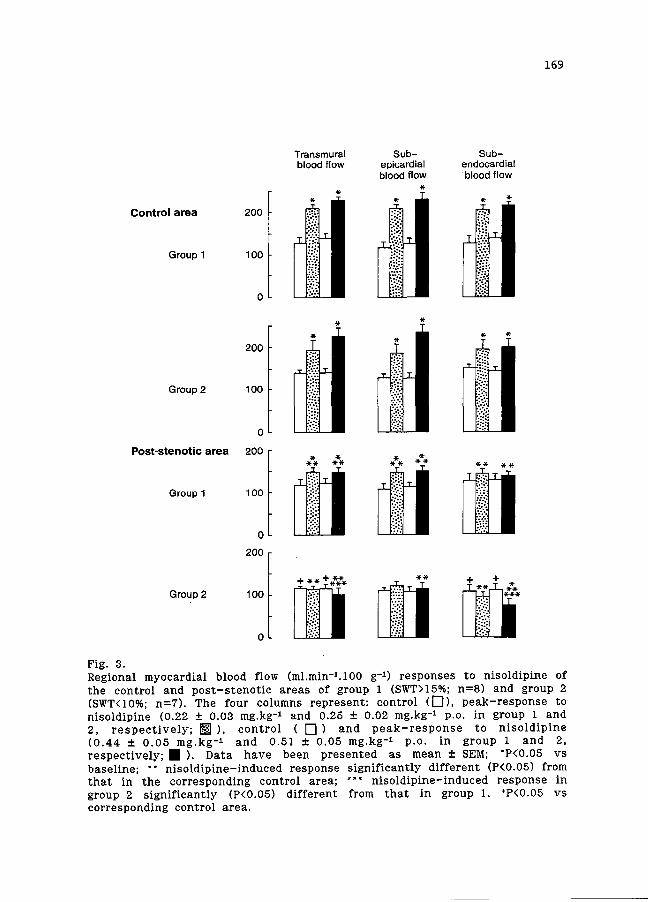
Fig. 3.
Control area 200
Group 1 100
0
200
Group 2 100
0
Post-stenotic area 200
Group 1 100
Group 2
0
200
100
0
Transmural blood flow
* *
*
Sub-epicardial blood flow
*
* *
**
[(J .
.
.
169
Sub-endocardial blood flow
* *
*
Regional myocardial blood flow (ml.min-1.100 g-1 ) responses to nisoldipine of the control and post-stenotic areas of group 1 (SWT>15%; n=8) and group 2 (SWT<10%; n=7). The four columns represent: control (0), peak-response to nisoldipine (0.22 ± 0.03 mg.kg-1 and 0.25 ± 0.02 mg.kg-1 p.o. in group 1 and 2, respectively; ~ ), control ( D ) and peak-response to nisoldipine (0.44 ± 0.05 mg.kg-1 and 0.51 ± 0.05 mg.kg-1 p.o. in group 1 and 2, respectively; • ). Data have been presented as mean ± SEM; ·p<o.o5 vs baseline; • • nisoldipine-induced response significantly different (P<0.05) from that in the corresponding control area; • • • nisoldipine-induced response in group 2 significantly (P<0.05) different from that in group 1. •P<0.05 vs corresponding control area.

170
Regional myocardial blood flows. Baseline myocardial blood flow values
and the nisoldipine-induced responses in the control area of both groups were
similar (Fig. 3). Myocardial blood flow to the control areas was
dose-dependently enhanced in both groups (up to 76 ± 18% and 64 ± 12% after
the highest dose in group 1 and 2, respectively). The increase in transmural
blood flow favoured the subepicardial (96 ± 22% and 88 ± 13%, respectively)
over the subendocardial (70 ± 21% and 40 ± 13%) layers. In the post-stenotic
area of group 1 baseline blood flow values were very similar to those in the
control segment, but the nisoldipine-induced responses were markedly
different. Nisoldipine caused only slight increases in transmural (25%) and
subepicardial (40%) blood flows; however, subendocardial flow remained
unchanged (Fig. 3). In the post-stenotic segment of group 2 transmural and
subendocardial blood flows at baseline were significantly lower compared with
flows of the control area. Nisoldipine did not affect transmural and
subepicardial blood flow but reduced subendocardial blood flow by 30% with
the highest dose.
Regional myocardial wall function. The effects of nisoldipine on regional
wall function are shown in Fig. 4. In neither of the two groups myocardial
wall thickness at end-diastole and at end-systole (baseline values:
10.0 ± 0.8 mm and 12.8 ± 0.9 mm in group 1; 10.6 ± 1.1 mm and
11.3 ± 1.2 mm in group 2) was affected. Systolic wall thickening (27 ± 4% for
group 1 and 7 ± 1% for group 2 during baseline) was also not affected by the
drug in either group. Velocity of wall thickening was enhanced in group 1
but unaffected in group 2.
Discussion
In this investigation nisoldipine was administered orally at two doses which
were titrated in each animal based on peak heart rate responses of 30 and
60 beats.min-1, respectively. The tachycardiac effect of the lower dose (25%
of baseline value) corresponds well with heart rate changes observed in the
clinical setting (Silke et al., 1985; Serruys et al., 1985). Moreover, the peak
tachycardiac responses to nisoldipine correlate with peak drug concentrations
in the plasma (Duncker et al., 1987).
As reported by many investigators (see Verdouw et al., 1988) we also
observed that nisoldipine induced a pronounced systemic vasodilation, which

SWT (%)
VSWT (mm.s-1)
Fig. 4.
40
20
0
20
*
10
0 0.22±0.02
Group 1
0.44±0.05
Nisoldipine (mg.kg·1; p.o.)
0.25±0.02
171
Group 2
~ 0.51 ±0.05
Regional left ventricular myocardial wall function responses to nisoldipine of pigs in group 1 (SWT>15%; n=8) and group 2 (SWT<10%; n=7). The five columns represent: baseline CO>. peak re!Ponse to nisoldipine (•) and 30 min (l§§j), 60 min (~) and 120 min (~) after peak-response, respectively. SWT = normalized systolic wall thickening; Vswt = mean velocity of wall thickening. Data have been presented as mean ± SEM; "P<0.05 vs baseline.
resulted in only a moderate reduction in mean arterial blood pressure as
cardiac output was markedly elevated. The increase in cardiac output was the
result of a reflex-mediated increase in heart rate as stroke volume was
maintained. Due to the baroreceptor reflex LVdP/dtmax was also .enhanced.
Left ventricular end-diastolic blood pressure was unchanged, which is in
accordance with reported earlier findings (see Verdouw et al., 1988).
Nisoldipine, like other dihydropyridine calcium channel-blockers, does not have
an effect on preload unless elevated end-diastolic pressures are present
(Verdouw et al., 1984; Kimchi et al., 1985). In both groups the cardiovascular
profile of nisoldipine was not different from that observed in conscious pigs
with a normal coronary circulation (Duncker et al., 1987), which implies that
the myocardial ischemia in the present study was not severe enough to
produce chronic reduction in global left ventricular pump function.

172
Nisoldipine potently enhanced myocardial blood flow to the normally
perfused areas which is in agreement with data obtained in animals with a
normal coronary circulation (for reference see Verdouw et al., 1988) or in the
control area of ischemic canine hearts (Warltier et al., 1981). The increase in
blood flow favoured the subepicardial layers, a finding observed with many
vasodilators (Verdouw et al., 1986, 1987a,b), arid this most likely resulted from
the moderate hypotension and the reflex-mediated tachycardia (see Feigl,
1983).
In group 1 the stenosis did not affect basal blood flow which correlates
well with the almost normal systolic wall thickening of the post-stenotic
area. In group 2, however, the stenosis severely reduced systolic wall
thickening, while basal transmural and subendocardial but not subepicardial
blood flow were decreased. These findings are supported by Gallagher et al.
(1985) who reported a severe impairment of systolic wall thickening in the
presence of a coronary stenosis despite a normal subepicardial blood flow
and subepicardial function. In the post-stenotic segment of group 1 in
.which the stenosis caused almost no or slight loss of wall function,
nisoldipine caused a moderate increase in transmural blood flow which was
solely confined to the subepicardial layers. In the post-stenotic myocardial
area of the animals in which the stenosis caused marked loss of wall function
(group 2), nisoldipine failed to cause an increase in blood flow to the
subepicardial layers whereas subendocardial blood flow was decreased. This
decrease probably resulted from the hypotension and increase in heart rate.
Although subepicardial blood flow was not affected by the drug vasodilation
must have occurred as mean aortic blood pressure and hence perfusion
pressure was decreased.
Our findings lend further support to the concept that during myocardial
ischemia due to a fixed concentric coronary artery stenosis, vasodilators,
may not be beneficial (Weintraub et al., 1981; Gross and Warltier, 1981;
Gewirtz et al., 1984). However, recent reports have shown that vasodilator
reserve may be present in ischemic myocardium distal to a severe coronary
artery stenosis and that vasodilators may improve myocardial blood flow and
myocardial function (Heusch and Deussen, 1984; Aversano and Becker, 1985,
Seitelberger et al., 1986; Heusch et al., 1987). The different observations in
those studies and in the present one might be the result of (i) different
routes of administration, (ii) absence or presence of collateral circulation and

173
(iii) different duration of ischemia. In most studies intracoronary
administration of drugs was used to minimize systemic effects (Heusch and
Deussen, 1984; Aversano and Becker, 1985; Canty and Klocke, 1985; Pantely et
al., 1985; Seitelberger et al., 1986). We, on the other hand, used oral
administration like in the clinical situation. Systemic administration of a
vasodilator results in hypotension which together with a reflex-tachycardia
reduces autoregulatory capacity of the myocardial vasculature especially in the
subendocardial layers (see Feigl, 1983). Only in the study of Heusch et al.
(1987) intravenous administration of nifedipine was employed. However,
during exercise-induced ischemia, systemic hemodynamics of the untreated and
the nifedipine-treated group did not differ. Furthermore, systemic
administration of nifedipine might have enhanced blood flow through
collateral vessels, which may be abundantly present in canine hearts. For
example, Warltier et al (1981) observed an increase in flow to a totally
collateral-dependent area in acutely ischemic dog hearts after intravenous
administration of nisoldipine. Since pigs possess very few collaterals and it is
unlikely that after induction of ischemia extensive collateral formation has
taken place within 5 days (Ramo et al., 1970), such a beneficial effect of
nisoldipine was not to be expected in our study. Finally, in all previous
studies measurements were made up to maximally 3 hours after induction of
ischemia, whereas in our study ischemia was present for more than 2 days
when the protocol was executed. To our knowledge, no information is
available on the extent of vasodilator reserve during prolonged (more than 3
hours) ischemia. Therefore, it might be that in our animals vasodilator
reserve was no longer present at the time of the experiment.
The nisoldipine-induced responses of myocardial blood flow were not
accompanied by a worsening of wall function in group 1 and, surprisingly,
also not in group 2. An explanation for this observation is not readily
found. Nisoldipine might have decreased oxygen demand of the post-stenotic
area although the increase in heart rate suggests an increase in oxygen
demand rather than a reduction. Another possibility arises from the
investigation of Berdeaux et al (1984} who observed an increase in wall
function of severely ischemic myocardium after a low dose of prenalterol, a
13-adrenoceptor agonist, causing an increase in heart rate of 15 beats.min-1,
while after a high dose of prenalterol an increase in heart rate of 40
beats.min-1 was accompanied by an unchanged wall function. This suggests

174
that the reflex-mediated increase in sympathetic activity can cause an
increase in wall function even in severely ischemic myocardium, provided that
the increase in heart rate is minimal. In our experiments it is possible that
these reflex-mediated chronotropic and inotropic actions of nisoldipine balance
one another with respect to their effects on wall function.
In conclusion, the findings in the present study, although obtained in
normotensive animals with a normal cardiac pump function, suggest that in
patients with myocardial ischemia caused by a concentric coronary artery
stenosis and with few collaterals, vasodilators may not be beneficial.
Therefore, it is important to stratify patients according to the status of their
coronary circulation, i.e. presence or absence of collaterals and severity and
type of stenosis. Because of its potent systemic vasodilator properties, the
drug will most likely also be used in patients with hypertension. In view of
this and the probable resetting of the baroreceptors, long-term studies in
models with hypertension and myocardial ischemia appear to be worth-while.
Acknowledgements
We would like to thank Bayer A.G. (Wuppertal, F.R.G.) for providing us
generously with nisoldipine. The Laboratory for Experimental Surgery is
greatfully acknowledged for their skilful assistance during surgery and the
post-surgical period. Finally we thank miss P.H. Vegter for her assistance in
the preparation of this manuscript.
References
Aversano, T. and Becker, L.C. Persistence of coronary vasodilator reserve despite functionally significant flow reduction. Am. J. Physiol. 248, H403-H411, 1985.
Berdeaux, A., Bonhenry, C., Duhaze, P., Giudicelli, J.F. and Thuillez, C. Influence of heart rate on the effects of prenalterol on regional myocardial blood flow and function during coronary stenosis in dogs. Br. J. Pharmacal. 83, 203-210, 1984.
Berne, R.M. and Rubio, R. Coronary circulation. In: Handbook of physiology. Section 2: The Cardiovascular System, Vol. I: The heart, Eds: Berne, R.M., Sperelakis, N. and Geiger, S.R., Waverly Press, Inc., Baltimore, 1979, pp. 873-952.
Canty, J.M. and Klocke, F.J. Reduced regional myocardial perfusion in the presence of pharmacologic vasodilator reserve. Circulation 71(2), 370-377. 1985.

175
Drexler, H., Truog, A.G., Zelis, R. and Flaim, S.F. Regional vascular and hemodynamic effects of orally administration nisoldipine in conscious rats. J. Cardiovasc. Pharmacal. 8, 151-155, 1986.
Duncker, D.J., Saxena, P.R. and Verdouw, P.O. The effects of nisoldipine alone and in combination with beta-adrenoceptor blockade on systemic haemodynamics and myocardial performance in conscious pigs. Eur. Heart J. Hl87 (in press).
Feigl, E.O. Coronary physiology. Physiol. Rev. 63, 1-205, 1983. Gallagher, K.P., Stirling, M.D., Choy, M., Szpunar, C.A., Gerren, R.A.,
Botham, M. and Lemmer, J.H. Dissociation between epicardial and transmural function during acute myocardial ischemia. Circulation 6, 1279-1291, 1985.
Gewirtz, H., Gross, S.L., Williams, D.O. and Most, A.S. Contrasting effects of nifedipine and adenosine on regional myocardial flow distribution and metabolism distal to a severe coronary arterial stenosis: observations in sedated, closed-chest, domestic swine. Circulation 5, 1048-1057, 1984.
Gross, G.J., Warltier, D.C. Coronary steal in four models of single or multivessel obstruction in dogs. Am. J. Cardiol. 48, 84-92, 1981.
Heusch, G. and Deussen, A. Nifedipine prevents sympathetic vasoconstriction distal to severe coronary stenoses. J. Cardiovasc. Pharmacal. 6, 378, 1984.
Heusch, G., Guth, B.D., Seitelberger, R. and Ross, J. JR. Attenuation of exercise-induced myocardial ischemia in dogs with recruitment of coronary vasodilator reserve by nifedipine. Circulation 75(2), 482-490, 1987.
Kimchi, A., Ellrodt, A.G., Charuzi, Y., Shell, W. and Murata, G.H. Salutary hemodynamic and sustained clinical beneficial effects of nisoldipine, a new calcium channel blocker, in patients with recurrent ischemia and severe heart failure. Am. Heart. J. 110, 496-498, 1985.
Lam, J., Chaitman, B.R., Crean, P., Blum, R. and Waters, D.O. A dose-ranging, placebo-controlled, double-blind trial of nisoldipine in effort angina: duration and extent of antianginal effects. J. Am. Coil. Cardiol. 6, 447-452, 1985.
Lopez, L.M., Rubin, M.R., Holland, J.P., Mehta, J.L. Improvement in exercise performance with nisoldipine, a new second-generation calcium blocker, in stable angina patients. Am. Heart J. 110, 991-996, 1985.
Pantely, G.A., Bristow, J.D., Swenson, L.J., Ladley, H.D., Johnson, W.B. and Anselone, e.G. Incomplete coronary vasodilation during myocardial ischemia in swine. Am. J. Physiol. 249, H638-H647, 1985.
Ramo, B.W., Peter, R.H., Ratliff, N., Kong, Y., Mcintosh, H.D. and Morris, J.J.Jr. The natural history of right coronary arterial occlusion in the pig. Am. J. Cardiol. 26, 156-161, 1970.
Saxena, P.R., Schamhardt, H.C., Forsyth, R.P. and Loeve, J. Computer programs for the radioactive microsphere technique. Determination of regional blood flows and other haemodynamic variables in different experimental circumstances. Comp. Prog. Biomed. 12, 63-84, 1980.
Seitelberger, R., Guth, B.D., Heusch, G., Katayama, K., Lee, J.D. and Ross, J.Jr. Regional alpha2 blockade improves function and flow in the ischemic dog myocardium during exercise. J. Am. Coil. Cardiol. 7 (suppl.A), 252A, 1986.
Serruys, P.W., Suryapranata, H., Planellas, J., Wijns, W., Vanhaleweyk, G.L.J. Soward, A., Jaski, B.E. and Hugenholtz, P.G. Acute effects of intravenous nisoldipine on left ventricular function and coronary hemodynamics. Am. J. Cardiol. 56, 140-146, 1985.

176
Silke, B., Frais, M.A., Muller, P., Verma, S.P., Reynolds, G., Taylor, S.H. Haemodynamic dose-response effects of intravenous nisoldipine in coronary artery disease. Br. J. Clin. Pharmacal. 20, 675-680, 1985.
Tumbleson, M.E. and Schmidt, D.A. Swine clinical chemistry. In: Swine in Biomedical Research, Ed: Tumbleson M.E., ·Plenum Press, New York, 1986, pp. 783-807.
Verdouw, P.D., Slager, C.J., Van Bremen, R.H. and Verkeste, C.M. Is nisoldipine capable of reducing left ventricular preload? Eur. J. Pharmacal. 98, 137-140, 1984.
Verdouw, P.D., Jennewein, H.M., Mierau, J. and Saxena, P.R. N-(3-acetylaminophenyl) piperazine hydrochloride (BEA 1654), a putative 5-HTt agonist, causes constriction of arteriovenous anastomoses and dilatation of arterioles. Eur. J. Pharmacal. 107, 337-346, 1985.
Verdouw, P.D., Hartog, J.M., Duncker, D.J., Roth, W. and Saxena, P.R. Cardiovascular profile of pimobendan, a benzimidazole-pyridazinone derivative with vasodilating and inotropic properties. Eur. J. Pharmacal. 126, 21-30, 1986.
Verdouw, P.D., Levinsky, L., Duncker, D.J., Rutteman, A.M. and Saxena, P.R. Cardiovascular performance after intravenous infusion of the pimobendan metabolite UD-CG 212 CL in anaesthetized pigs;. Eur. J. Pharmacol. 137, 219-226, 1987a. !
Verdouw, P.D., Sassen, L.M.A., Duncker, D.J., Schmeets, I.O.L., Rensen, R.J. and Saxena, P.R. Nicorandil-induced changes in the distribution of cardiac output and coronary blood now in pigs. Naunyn-Schmiedeberg's Arch. Pharmacal. 336, 352-358, 1987b.
Verdouw, P.D., Duncker, D.J., Saxena, P.R., Serruys, P.W. and Hugenholtz, P.G. Cardiovascular actions of nisoldipine in health and coronary artery disease. Am. J. Cardiol. 1988. (in press).
Warltier, D.C., Gross, G.J. and Brooks, H.L. Coronary steal-induced increase in myocardial infarct size after pharmacological coronary vasodilation. Am. J. Cardiol. 46, 83-90, 1980.
Warltier, D.C., Meils, C.M., Gross, G.J. and Brooks, H.L. Blood flow in normal and acutely ischemic myocardium after verapamil, diltiazem and nisoldipine (Bay k 5552), a new dihydropyridine calcium antagonist. J. Pharmacal. Exp. Ther. 218, 296-302, 1981.
Weintraub, W.S., Hattori, S., Agarwal, J., Bodenheimer, M.M., Banka, V.S. and Helfant, R.H. Variable effect of nifedipine on myocardial blood now at three grades of coronary occlusion in the dog. Circ. Res. 48, 937-942, 1981.

CHAPTER 13
EXERCISE-INDUCED ISCHEMIA IN PIGS: EFFECTS OF NISOLDIPINE
WITH OR WITHOUT PROPRANOLOL.
D.J. Duncker, J.P.C. Heiligers1, P.R. Saxena1 and P.D. Verrdouw.
Laboratory for Experimental Cardiology (Thoraxcenter)
and Department of Pharmacology,
Erasmus University Rotterdam, Rotterdam, The Netherlands.
Submitted for publication.
177


179
Chapter 13
Exercise-induced myocardial ischemia in pigs: cardiovascular actions of
nisoldipine with or without ~-adrenoceptor blockade.
Summary
We evaluated the effects of oral nisoldipine with or without propranolol on
exercise-induced myocardial ischemia in conscious pigs with a coronary artery
stenosis. Treadmill-running up to 4 km.h-1 increased cardiac output (90%),
heart rate {90%), LVdP/dtmax (80%), left ventricular systolic (15%) and
end-diastolic blood pressure (13 ± 3 mmHg), while systolic wall thickening of
the post-stenotic left ventricular myocardium was reduced from 29 ± 8% to
19 ± 6%. Nisoldipine neither affected the systemic hemodynamic profile
during exercise nor the exercise-induced reductions in myocardial wall
function. Propranolol attenuated both the positive chronotropic and inotropic
effects and the deterioration of wall function caused by the
treadmill-exercise. Combined treatment with the two drugs resulted in a
cardiovascular profile similar to propranolol during exercise, but the loss of
wall function was now completely prevented. We conclude that, unlike
propranolol, nisoldipine was not effective against the exercise-induced
ischemia but may have beneficial actions when combined with [3-adrenoceptor
antagonists.
Introduction
In clinical studies both [3-adrenoceptor antagonists and calcium-channel
blockers are effective as anti -anginal agents (Prichard et al., 1970; Livesly
et al., 1973; Kaltenbach et al., 1979). These two classes of drugs act through
different mechanisms and in a number of studies their combination has proven
superior to monotherapy with either class (Leon at al., 1980; Dargie et al.,
1981; Fox et al., 1981). In this respect the dihydropyridine calcium-channel
blockers appear particularly attractive to combine with [3-adrenoceptor
antagonists since the latter drugs block the baroreceptor reflex-mediated
tachycardia. However, the effects of the dihydropyridines are not always
predictable as vasodilation per se, depending on the experimental set-up and
dose of the vasodilator used, may either induce "coronary steal" (Warltier
et al., 1980; Weintraub et al., 1981) or increase blood flow to ischemic regions
(Canty and Klocke, 1985; Pantely et al., 1985; Heusch et al., 1987). In the

180
present study we evaluated the effects of nisoldipine, a dihydropyridine
derivative with potent vasodilator and minimal direct negative inotropic
properties (Kazda et al., 1980; Duncker et al., 1987a), on systemic
hemodynamics and post-stenotic myocardial wall function during
exercise-induced myocardial ischemia in pigs with a subacute coronary artery
obstruction. Since nisoldipine may be used in the clinical setting in
combination with ~-adrenoceptor antagonists, we also investigated the actions
of nisoldipine in the presence of propranolol.
employed in view of the clinical use of these drugs.
Methods and Materials
Oral administration was
Surgical procedure for instrumentation and coronary artery stenosis
After an overnight fast 9 Yorkshire pigs (18-20 kg), pretreated with a
mixture of procaine penicillin-G and benzathine-penicillin-G (Duplocillin,
Gist-Brocades N.V., Delft, The Netherlands) both 300,000 units i.m., were
sedated with 30 mg.kg-1 ketamine HCl i.m. (Aescoket, Aesculaap B.V., Boxtel,
The Netherlands). The animals were intubated and connected to a respirator
for artificial ventilation with a mixture of oxygen and nitrous oxide ( 1 :2) to
which 1% halothane was added. A jugular vein and common carotid artery
were cannulated for infusion of drugs and measurement of arterial blood
pressure, respectively. The chest was opened via the left fifth intercostal
space to expose the heart. A transducer (P4.~, Konigsberg Instruments Inc.
Pasadena, California, USA) was implanted into the left ventricle of the heart
through its apex for recording of left ventricular pressure. The left atrium
was cannulated for recording of left atrial pressure which, together with the
arterial blood pressure, was used for calibration of the Konigsberg transducer
signals. Regional myocardial function was assessed by sonomicrometry (Triton
Technology, San Diego, Ca, USA). A pair of ultrasonic crystals (5 MHz) was
implanted in the left ventricular myocardial segment perfused by the left
anterior descending coronary artery (LADCA) to measure regional myocardial
wall thickness. The wall thickness at end-diastole (EDT) and end-systole
(EST) were used to calculate percentual systolic wall thickening (SWT) as
reported earlier (Verdouw et al., 1983). Subsequently, the aorta was
approached through the third intercostal space and an electromagnetic flow
probe (Skalar, Delft, The Netherlands) was positioned around the ascending
aorta. The proximal segment of the LADCA was dissected free from its

181
surrounding tissue and a teflon constrictor with an internal diameter varying
from 1.0 to 2.0 mm was positioned around the LADCA which resulted in a loss
of systolic wall thickening to different degrees. Lastly, catheters and wires
were tunnelled subcutaneously to the back and the chest was closed. The
animals were allowed to recover from the surgery for 7-8 days during which
they were adapted to the laboratory facilities.
During the postoperative period the animals received daily intravenous
bolus injections of 500 mg amoxicilline (Clamoxil, Beecham Parma B.V.,
Amstelveen, The Netherlands) to prevent infection. The catheters were
flushed daily with an isotonic saline solution containing 500 I.U. per ml
heparin (Thromboliquine, Organon Teknika B.V., Boxtel, The Netherlands) to
avoid clotting of blood in the lumen.
Experimental protocol
This study in conscious instrumented pigs with a fixed coronary artery
stenosis was conducted in two parts: one part dealt with the effects of
nisoldipine on exercise-induced changes in systemic hemodynamic variables and
post-stenotic myocardial wall motion in control (untreated) animals and the
other part dealt with similar effects of nisoldipine in animals treated with
propranolol. For the first part of the study the animals were placed on a
treadmill and systemic hemodynamic variables and post-stenotic myocardial
wall motion were recorded. The speed of the treadmill was gradually
increased first to 3 km.h-1 and then to 4 km.h-1. These speeds were
maintained for a period of 2 min each during which hemodynamic variables
became stable and data were collected. Both systemic hemodynamics and wall
thickness variables returned towards baseline values within 60 min after
cessation of the treadmill-running. Subsequently, the animals were orally
administered 10 mg (0.54 ± 0.01 mg.kg-1) of nisoldipine and, at the
drug-induced peak-response of systemic hemodynamic parameters (30-60 min
after administration), measurements were made and the exercise protocol
repeated.
For the second part of the study the first exercise-test was performed
60 min after treating the animals with 80 mg (4.36 ± 0.10 mg.kg-1)
propranolol. In a pilot-study (n=3) we established the extent of
~-adrenoceptor blockade at various times after oral administration of 80 mg of
propranolol; the tachycardiac responses to isoprenaline (0.1 ~g.kg-1; i. v.) were

182
37 ± 3 beats.min-1 at baseline and 26 ± 6, 14 ± 1, 6 ± 4, 14 ± 7 and
7 ± 4 beats.min-1 at 30, 60, 120, 180 and 240 min after propranolol
administration. After a 60 min period of recovery from treadmill-exercise
nisoldipine (10 mg) was administered and, approximately 30-60 min later (at
nisoldipine peak-effect), exercise-tests were again performed. The two parts
of the study, as outlined above, were executed on consecutive days, but the
order of the two parts was varied. We have shown earlier that nisoldipine
(10 mg orally) is completely eliminated from the plasma within 24 hours after
administration (Duncker et al., 1987a). Furthermore, the isoprenaline-induced
increases in heart rate had returned to baseline levels 24 hours after
administration of 80 mg of propranolol orally: 37 ± 3 beats.min-1 at baseline
versus 33 ± 4 beats.min-1 24 hours after propranolol. We did not perform
control experiments to establish the reproducibility of the exercise-induced
responses, but studies in dogs (Matsuzaki et al., 1984a) reveal that the
responses are identical during exercise performed 3 hours after the first
exercise test.
Two of the 9 animals, which did not run on the treadmill satisfactorily,
were excluded from the study protocol. Arterial blood pressure measured with
a fluid-filled catheter could not be obtained during the exercise period due to
disturbances in the signals induced by the running; this parameter was
therefore excluded from analysis.
Drugs
Except for the anesthetics during surgery and the antibiotics, the only
drugs used in this study were nisoldipine (Bayer A.G., Wuppertal, F.R.G.) and
propranolol hydrochloride (ICI-Farma, Rotterdam, The Netherlands).
Data presentation and statistical analysis
All data have been presented as mean ± SE of mean. Statistical analysis
was performed using Duncan's New Multiple range test once a parametric
two-way analysis of variance (randomized block design) had revealed that the
samples represented different populations.

183
Control Nisoldipine Propranolol Nisoldipine
300 + Propranolol
* +
200 * HR (beats.min-1)
100
0
6
4
co (l.min-1)
2
0 30
20
sv (ml) 10
0 0 3 4 0 3 4 0 3 4 0 3 4
Running speed (km.h-1)
Fig. 1. Systemic haemodynamic effects of exercise without and after medication with nisoldipine, propranolol or their combination. HR=heart rate; CO=cardiac output; SV=stroke volume. *P<0.05 vs corresponding pre-exercise; *"P<0.05 pre-exercise after medication vs pre-exercise without medication; +,x,o exercise-induced response after medication significantly different (P<0.05) from exercise without treatment (+), after nisoldipine (x), or propranolol (O).

184
Results Blood chemistry
Arterial acid-base balance and oxygenation at the time of the experiments
were within the following limits: 7.37 < pH < 7.50; 35 mmHg < PC02 <
45 mmHg; 75 mmHg < P02 <95 mmHg, corresponding well with earlier reports
(Tumbleson and Schmidt, 1986).
Arrhythmias during the post-surgical period
In two animals ventricular arrhythmias (>5 min-1) were observed during
the first adaptation session after surgery but not on the following days.
Arrhythmias were not observed in any of the other animals, neither during
adaptation, nor during the course of the experiments.
Exercise without treatment
The exercise-induced changes observed in our study are very similar to
those reported by other investigators (Sanders et al., 1977). Treadmill
exercise up to 4 km.h-1 almost doubled heart rate, cardiac output and
LVdP/dtmax as those parameters had increased at the highest running speed
by 91 ± 6%, 92 ± 8% and 83 ± 11%, respectively. Increases were also observed
in left ventricular end-diastolic blood pressure (from 7 ± 1 to 20 ± 4 mmHg)
and left ventricular systolic pressure (15 ± 3%), while stroke volume was
minimally affected (Figs. 1 and 2). Treadmill-running caused speed-dependent
reductions in systolic wall thickening (from 29 ± 8% to 19 ± 6% at 4 km.h-1 )
which were primarily due to the decrease in end-systolic wall thickness as
end-diastolic wall thickness was not significantly affected (Fig. 3).
Exercise in the presence of nisoldipine
During exercise in the presence of nisoldipine the hemodynamic parameters
reached levels very similar to those obtained during the control run, except
for left ventricular end-diastolic pressure which was, at both running speeds,
consistently lower than in the untreated animals (Figs. 1 and 2). Since
nisoldipine increased pre-exercise levels of heart rate, cardiac output and
LVdP/dtmax, the exercise-induced changes in these variables after nisoldipine
were less than those observed in the control period. Nisoldipine tended to
reduce the exercise-induced decrease in systolic wall thickening (from 26 ± 8%

8000
LVdP/dtmax (mmHg.s-1)
4000
0
150 LVSP (mmHg) 100
50
0 30
LVEDP (mmHg)
20
10
0
Fig. 2.
Control Nisoldipine Propranolol
0 3 4 0 3 4 0 3 4
Running speed (km.h-1)
185
Niso!dipine +
Propranolol
0 3 4
Systemic haemodynamic effects of exercise without and after medication with nisoldipine, propranolol, or their combination. LVdP/dtmax=maximal rate of rise of left ventricular pressure; LVSP=left ventricular systolic blood pressure; LVEDP=left ventricular end -diastolic blood pressure. *P<0.05 vs corresponding pre-exercise; **P<0.05 pre-exercise after medication vs pre-exercise without medication; +,x.o exercise-induced responses after medication significantly (P<0.05) different from exercise-induced responses without treatment (+), after nisoldipine (x), or propranolol (0).

186
to 19 ± 8%) but this effect did not reach levels of statistical significance
(Fig. 3).
Exercise in the presence of propranolol
After ~-adrenoceptor blockade with propranolol the exercise-induced
increases in heart rate (up to 81 ± 11 %), cardiac output (82 ± 13%) and
LVdP/dtmax (38 ± 5%) were lower than those observed during exercise without
medication. Stroke volume increased more (P<0.05) than in the untreated
animals, while the responses of left ventricular end-diastolic pressures were
similar (Figs. 1 and 2). Propranolol also significantly reduced the
exercise-induced loss of wall function as systolic wall thickening decreased
only from 29 ± 8% to 24 ± 7% (Fig. 3).
Exercise in the presence of propranolol and nisoldipine
Systemic hemodynamic parameters reached levels similar to those obtained
during exercise-tests in propranolol-treated animals (Figs. 1 and 2). However,
the combination completely prevented the exercise-induced loss of wall
function of the post-stenotic segment (Fig. 3).
Discussion
Systemic hemodynamics
The exercise-induced responses of systemic hemodynamics were very similar
to those described for exercising pigs with a normal coronary circulation
(Sanders et al., 1977; Scheffer and Verdouw, 1983) or with a gradually
occluded coronary artery (Bloor et al., 1984; White and Bloor, 1986). The
increase in cardiac output was almost exclusively due to the tachycardia as
stroke volume remained virtually unchanged. LVdP/dtmax was markedly
enhanced indicating that global left ventricular function increased. Left
ventricular systolic pressure was slightly elevated, while a pronounced
increase in left ventricular end-diastolic pressure was observed.
The pre-exercise cardiovascular actions of nisoldipine, propranolol or their
combination under resting conditions are in accordance with the findings of
other investigators (Warltier et al., 1984; Scriabine and Taylor, 1984; Silke
et al., 1985, 1986) as well as those from our laboratory (Duncker et al.,
1987a,b). Despite the changes induced by nisoldipine at rest, during exercise
the systemic hemodynamic variables reached levels that were similar in

MWT (mm)
EST
EDT
14
12
10
40
SWT 20 (%)
0
Fig. 3.
Control
0 3 4
Nisoldipine Propranolol
0 3 4 0 3 4
Running speed (km.h-1)
187
Nisoldipine +
Propranolol
0 3 4
Effects of exercise on post-stenotic wall function in the absence or presence of medication with nisoldipine, propranolol, or their combination. MWT=left ventricular myocardial wall thickness; EST = end-systolic thickness; EDT = end-diastolic thickness; SWT= normalized systolic wall thickening. *P<O.OS vs corresponding pre-exercise; **P<O.OS pre-exercise after medication vs pre-exercise without treatment; +,x exercise-induced responses after medication significantly (P<O.OS) different from exercise-induced responses without treatment (+), or after nisoldipine (x).

188
untreated and nisoldipine-treated animals. Only left ventricular systolic and
end-diastolic pressures were lower when compared to the group without
medication. Another dihydropyridine-derivative, nifedipine, has also been
shown not to modify the changes in systemic hemodynamic variables during
exercise in dogs with critical coronary stenosis (Heusch et al., 1987). In
contrast, Silke et al. (1985, 1986} reported that during exercise
nisoldipine-treated patients with coronary artery disease achieved higher heart
rate levels but had a lower arterial blood pressure than untreated control
patients. These patients were apparently subjected to a mild exercise, as
heart rates of only 100 beats.min-1 were reached during the control-exercise
period. At such a low level of exercise the baroreceptor-reflex is still
operative so that nisoldipine could induce further tachycardia.
Pretreatment with propranolol attenuated but not completely eliminated the
exercise-induced increases in heart rate, cardiac output, LVdP/dtmax and left
ventricular systolic blood pressure. These findings, suggesting the
involvement, at least in part, of the withdrawal of parasympathetic tone
during exercise, are in agreement with data reported earlier in dogs with
propranolol (Heyndrickx et al., 1980) and atenolol (Matsuzaki et al., 1984b).
Combined treatment with nisoldipine and propranolol resulted during exercise
in a hemodynamic state similar to that obtained after propranolol alone.
Silke et al. (1986) observed in their patients that addition of nisoldipine to
metoprolol caused an increase in cardiac output, heart rate and stroke volume.
The reason for this observation might again be the low exercise level to
which their patients were subjected and at which the baroreceptor-reflex
(parasympathetic withdrawal) is still operative.
Regional myocardial wall function
None of the drug regimen caused a significant increase in wall function at
rest, but different patterns emerged during exercise. In our study nisoldipine
was ineffective in antagonizing the exercise-induced decrease in systolic wall
thickening. In contrast, Heusch et al. (1987) observed an improvement of
myocardial performance after nifedipine in exercise-induced myocardial
ischemia in dogs. The reason for the difference in observations may be that
pigs, unlike dogs, possess few native coronary collaterals. Our experiments
were performed 7-9 days after implantation of a constrictor which caused
severe impairment of wall function (systolic wall thickening less than 15%) in

189
only two out of seven animals. Since myocardial ischemia is an important
inductor of collateral formation it is not likely that extensive collateral
formation took place in most animals in this study. Warltier et al. (1981)
have also shown that in anesthetized dogs nisoldipine increased blood flow to
a myocardial area totally dependent upon collateral circulation. Therefore,
the reason for the difference between our observations and those of Heusch
et al. (1987) might be related to the absence of extensive coronary
collaterals in our animals.
As we found in this study with pigs, 13-adrenoceptor antagonists have been
shown to be effective against myocardial ischemia induced by treadmill
exercise in dogs (Kumada et al., 1980; Matsuzaki et al., 1984b; Tomoike et al.,
1987). This beneficial effect is most likely due to a reduction in heart rate
(Guth et al., 1987) which not only decreases myocardial oxygen demand but
also prolongs the diastolic coronary perfusion time to improve myocardial
blood flow, especially to the subendocardial layers (Schamhardt et al., 1981;
Saxena, 1983; Guth et al., 1987). Though propranolol effectively antagonized
(but not eliminated) the exercise-induced loss of systolic wall function,
nisoldipine did not. The reason for this difference in the efficacy of the two
drugs may lie in their respective pharmacological profile. Propranolol can
decrease myocardial oxygen demand (due to negative chronotropic and
inotropic effects) as well as enhance blood supply to the jeopardized
myocardium (due to increased diastolic perfusion time; Schamhardt et al.,
1981). In case of nisoldipine the decrease in myocardial oxygen demand (due
to peripheral vasodilation) can be offset by an exercise-induced increase in
sympathetic activity. Since the last effect is antagonized by propranolol, the
combination of the two drugs may be more effective in averting the
deterioration of systolic wall function of post-stenotic myocardium during
exercise. Indeed, this seems to be the case in our experiments where after
propranolol alone the animals did show some deterioration in systolic wall
thickening in the post-stenotic myocardium during treadmill exercise; such
changes were completely prevented after the combined treatment with
propranolol and nisoldipine.

190
Acknowledgements We would like to thank Bayer A.G. (Wuppertal, F.R.G.) for their financial
support. The Laboratory for Experimental Surgery is greatfully acknowledged
for their skilful assistance during surgery and the post-surgical period.
Finally we thank miss P.H. Vegter for her assistance in the preparation of
this manuscript.
References Bloor, C.M., White, F.C., Sander, T.M. Coronary collateral development and
myocardial tissue salvage induced by exercise in pigs with coronary occlusion. J. Appl. Physiol. 56, 656-665, 1984.
Canty, J.M., Klocke, F.J. Reduced regional myocardial perfusion in the presence of pharmacologic vasodilator reserve. Circulation 71(2), 370-377, 1985.
Dargie, H.J., Lynch, P.G., Krikler, D.M., Harris, L., Krikler, S. Nifedipine and propranolol, a beneficial drug interaction. Am. J. Med. 71, 676-682, 1981.
Duncker, D.J., Saxena, P.R. Verdouw, P.O. The effects of nisoldipine alone and in combination with beta-adrenoceptor blckade on systemic haemodynamics and myocardial performance in conscious pigs. Eur. Heart J. 1987a (in press).
Duncker, D.J., Saxena, P.R., Verdouw, P.O. Systemic haemodynamic and beta-adrenoceptor antagonistic effects of bisoprolol in conscious pigs, a comparison with propranolol. Arch. Int. Pharmacodyn. 290, 54-63, 1987b.
Fox, K.M., Jonathan, A., Selwyn, A.D. The use of propranolol and nifedipine in the medical management of angina pectoris. Clin. Cardiol. 4, 125-129, 1981.
Guth, B.D., Reusch, G., Seitelberger, R., Ross, J.jr. Mechanism of beneficial effect of beta-adrenergic blockade on exercise-induced myocardial ischemia in conscious dogs. Circ. Res. 60, 738-746, 1987.
Heusch, G., Guth, B.D., Seitelberger, R., Ross, J.jr. Attenuation of exercise-induced myocardial ischemia in dogs with recruitment of coronary vasodilator reserve by nifedipine. Circulation 1987 75(2), 482-490, 1987.
Heyndrickx, G.R., Pannier, J.L., Muylaert, Ph., Mabilde, C., Leusen, I. Alteration in myocardial oxygen balance during exercise after beta-adrenergic blockade in dogs. J. Appl. Physiol. 49(1), 28-33, 1980.
Kaltenbach, M., Schultz, W., Kober, G. Effects of nifedipine after intravenous and intracoronary administration. Am. J. Cardiol. 1979 44, 832-838, 1979.
Kazda, S., Garthoff, B., Meyer, H., Schlossmann, K., Stoepel, K., Towart, R.,Vater, W., Wehinger, E. Pharmacology of a new calcium antagonistic compound, Isobutyl Methyl! ,4-Dihydro-2,6-dimethyl-4-(2-nitro-phenyl)-3,5-pyridinedicarboxylate (nisoldipine, Bay k 5552). Arzneim. Forsch. I Drug Res. 30, 2144-2162, 1980.
Kumada, T., Gallagher, K.P., Shirato, K., McKown, D., Miller, M., Kemper, W.S., White, F., Ross, J.jr. Reduction of exercise-induced regional myocardial dysfunction by propranolol. Studies in a canine model of chronic coronary artery stenosis. Circ. Res. 46, 190, 1980.

191
Leon, M.B., Bonow, R.O., Rosing, D.R., Barach, S.L., Green, M.V., Epstein, S.E. Effects of verapamil alone and combined with propranolol on left ventricular systolic function in patients with coronary artery disease. Circulation 62(abs)III, 233, 1980.
Livesly, B., Catley, P.F., Campbell, R.L., Oraur, S. Double blind evaluation of verapamil, propranolol and isosorbide dinitrate against a placebo in the treatment of angina pectoris. Br. Med. J. 1, 375-378, 1973.
Matsuzaki, M., Gallagher, K.P., Patri8tti, J., Tajimi, T., Kemper, W.S., White, F.C., Ross, J.jr. Effects of a calcium-entry blocker (diltiazem) on regional myocardial flow and function during exercise in conscious dogs. Circulation 69:801, 1984.
Matsuzaki, M., Patritti, J., Tajimi, T., Miller, M., Kemper, W.S., Ross, J.jr. Effects of beta-blockade on regional myocardial flow and function during exercise. Am. J. Physiol. 1984(1P2), H52-H60, 1984.
Pantely, G.A., Bristow, J.D., Swenson, L.J., Ladley, H.D., Johnson, W.B., Anselone, C.G. Incomplete coronary vasodilation during myocardial ischemia in swine. Am. J. Physiol. 249, H638-H647, 1985.
Prichard, B.N.C., Aellig, W.H., Richardson, G.A. The action of intravenous oxprenolol, practolol, propranolol and sotalol on acute exercise tolerance in angina pectoris, the effect on heart rate and the electrocardiogram. · Postgrad. Med. J. 46(suppl), 77-85, 1970.
Sanders, M., White, F., Bloor, C. Cardiovascular responses of dogs and pigs exposed to similar physiological stress. Biochem. Physiol. 58A, 365-370, 1977.
Saxena, P.R. Beta-adrenoceptor antagonists and blood flow to the jeopardized myocardium. Eur. Heart J. 4 (suppl. D), 101-108, 1983.
Schamhardt, H.C., Verdouw, P.D., Sa,xena, P.R. Improvement of perfusion and function of ischaemic porcine myocardium after reduction of heart rate by alinidine. J. cardiovasc. Pharmacal. 3, 728-738, 1981.
Scheffer, M.G., Verdouw, P.D. Decreased incidence of ventricular fibrillation after an acute coronary artery ligation in exercised pigs. Basic Res. Cardiol. 78, 298-309, 1983.
Scriabine, A., Taylor, D.G. Antihypertensive Drugs. In: Cardiovascular Pharmacology, second edition. Ed: Antonaccio M. New York, Raven Press, 1984, 257-294, 1984.
Silke, B., Frais, M.A., Muller, P., Verma, S.P., Reynolds, G., Taylor, S.H. Haemodynamic dose-response effects of intravenous nisoldipine in coronary artery disease. Br. J. Clin. Pharmacal. 20, 675-680, 1985.
Silke, B., Verma, S.P., Midtbo, K.A., Muller, P., Frais, M.A., Reynolds, G., Taylor, S.H. A haemodynamic study of the effects of combined slow-calcium channel blockade (nisoldipine) and beta-blockade (metoprolol) in coronary heart disease. Int. J. Cardiol. 13, 231-241, 1986.
Tomoike, H., Ross, J., Franklin, D., Crozatier, B., McKown, D., Kempers, W.S. Improvement by propranolol of regional dysfunction and abnormal coronary flow pattern in conscious dogs with coronary narrowing. Am. J. Cardiol. 41, 689-696, 1987.
Tumbleson, M.E., Schmidt, D.A. Swine clinical chemistry. In: Swine in Biomedical Research, vol 2. Ed: Tumbleson, M.E. New York, Plenum Press, 1986, pp. 783-807.
Verdouw, P.D., Wolffenbuttel, B.H.R., Scheffer, M.G. Cardiovascular actions of the calmodulin inhibitor felodipine. Naunyn-Schmiedeberg's Arch. Pharmacal. 323, 350-354, 1983.

192
Warltier, D.C., Gross, G.J., Brooks, H.L. Coronary steal-induced increase in myocardial infarct size after pharmacological coronary vasodilation. -Am. J. Cardiol. 46, 83-90, 1980.
Warltier, D.C., Meils, C.M., Gross, G.J., Brooks, H.L. Blood flow in normal and acutely ischemic myocardium after verapamil, diltiazem and nisoldipine (Bay k 5552), a new dihydropyridine calcium .antagonist. J. Pharmacol. Exp. Ther. 218, 296-302, 1981.
Warltier, D.C., Zyvoloski, M.G., Gross, G.J., Brooks, H.L. Comparative actions of dihydropiridine slow channel calcium blocking agents in conscious dogs, systemic and coronary hemodynemics with and without combined beta adrenergic blockade. J. Pharmacol. Exp. Ther. 230, 367-375, 1984.
Weintraub, W.S., Hattori, S., Agarwal, J., Bodenheimer, M.M., Banka, V.S., Helfant, R.h. Variable effect of nifedipine on myocardial blood flow at three grades of coronary occlusion in the dog. Circ. Res. 48, 937-942, 1981.
White, C.W., Bloor, C.M. The pig as a model for myocardial ischemia. In: Swine in Biomedical Research, vol 1. Ed: Tumbleson, M.E. New York, Plenum Press, 1986, pp. 481-490.

193
DISCUSSION
CHAPTER 14
GENERAL DISCUSSION AND CONCLUSIONS


DISCUSSION
Chapter 14
General discussion and conclusions
14.1 Methodological considerations
The experimental animal
195
The experimental animal used in the studies presented in this thesis was
the farm-bred Yorkshire pig. The use of swine in scientific research has
increased rapidly over the last decade. Ample studies demonstrate that the
pig is closely related to the human, anatomically and physiologically (Pond
and Houpt, 1978; Sack, 1982; Swindle and Bobbie, 1983; Peng et al., 1983;
Pluth, 1983; Swindle, 1984). From a cardiovascular point of view the
similarities in size and distribution of coronary arteries (Weaver et al., 1986),
blood pressure and heart rate (Smith et al., 1964), plasma lipoprotein profile
(Mahley and Weisgraber, 1974) and responses to hyperlipidemic diets (Thomas
et al., 1983) are of interest. In pigs atherosclerosis occurs spontaneously
(Skold and Getty, 1961) and can also be easily induced (Thomas et al., 1983).
However, for cardiovascular studies in vivo the dog is perhaps still the most
often employed animal, partly because it is easier to handle. However, a
disadvantage of the dog for cardiovascular research is that it has an
extensive native collateral circulation in the heart and even complete
occlusion of coronary arteries may result in no infarction or variable infarct
sizes (Schaper, 1971). In contrast pigs, like humans, possess very few native
collaterals; the formation of new collaterals can, however, be easily induced
(Ramo, 1970; Millard, 1981). Moreover, for exercise studies, swine compare
more favorably to human than dogs with respect to heart weight-body weight
ratio, V02max, cardiac index, regional distribution of cardiac output and
acid-base balance (see Mckirnan et al., 1986).
The experiments in this thesis were performed in anesthetized as well as
conscious animals. The anesthetized animal model can be used for acute,
relatively short-lasting experiments with drugs administered intravenously or
intra-arterially (intracoronary or intracarotid). However, the presence of
anesthesia may interfere with the autonomic regulation of the cardiovascular
system and, therefore, may alter the actions of drugs. For example,
pentobarbital anesthesia is known to reduce inotropy of the heart (Sawyer

196
et al., 1971) which is an undesirable action especially in a model of
myocardial ischemia. Also, the baroreceptor reflex appears to be attenuated
in the presence of barbiturates (Roberts, 1980; Montgomery et al., 1982;
Zimpfer et al., 1982). Although the conscious animal model is more laborious
than the anesthetized model, it enables chronic studies, exercise studies, or
studies in which oral administration of a drug is employed. Furthermore, in
conscious animals there is no interference of anesthesia with circulatory
dynamics.
Model tor the study of myocsrdlsl lschemls
Myocardial ischemia has been defined as the imbalance between the supply
of oxygenated blood and the oxygen requirements of the myocardium. The
consequence of this imbalance is: 1) a lack of oxygen and metabolic substrates
for the production of energy in the form of high energy phosphates such as
adenosine triphosphate (ATP) and creatine phosphate and 2) the diminished
"wash out" of metabolites of the ensuing anaerobic glycolysis. Anaerobic
glycolysis is insufficient to meet the energy requirements of the myocyte.
Furthermore, the accumulation of metabolic products, such as lactate, H+ and
purine bases in the heart inhibits anaerobic glycolysis and other metabolic
pathways.
Functional features of myocardial ischemia are: 1) loss of contractile
function (within seconds after a flow reducing intervention) and 2) electrolyte
and electrophysiological changes which may lead to rhythm disturbances (Hillis
and Braunwald, 1977). In this thesis we used systolic wall thickening as a
measure of regional myocardial function. Systolic wall thickening of the left
ventricle is a good measure of local myocardial function (Sasayama et al.,
1976) which is closely related to disturbances in oxygen demand/oxygen supply
(Kerber et al., 1975; Verdouw et al., 1980; Ross and Franklin, 1976). Another
measure of myocardial ischemia, frequently used in the clinical setting, are
ST-segments changes. Although ST-segment changes are used to quantitate
infarct size (Maroko et al., 1972) poor correlations have been reported
between ST-segment changes and infarct size or the severity of myocardial
ischemia (Vincent et al., 1977). Metabolic markers (lactate and
hypoxanthines) have also been used as a measure of the severity and extent
of myocardial ischemia (Parker et al., 1969; Opie et al., 1973; Remme et al.,
1977) but they have the disadvantage of being time-dependent as their

197
production fades during prolonged ischemia (Rovetto et al., 1975; De Jong
et al., 1977; Verdouw et al., 1979), probably due to deterioration of cell
function.
In chapters 12 and 13 we investigated the effects of nisoldipine on
perfusion and/or wall function of a myocardial area distal to a coronary
artery stenosis in the proximal part of the left anterior descending coronary
artery. The stenosis was produced by use of a teflon ring, implantation of
which resulted in a fixed concentric obstruction. In a clinical study by
Vlodaver and Edwards (1971) 30% of all (200) atherosclerotic lesions studied
at autopsy were of the concentric type, whereas 70% belonged to the
eccentric type, either polymorphous or slit-like. In contrast to eccentric
stenoses, the vessel in the case of concentric obstructions is circumferentially
affected by the atherosclerotic process and responds less well to vasodilatory
interventions than eccentric stenoses (see Lichtlen and Ebner, 1986).
Another important factor that determines the effectiveness of vasodilatory
treatment is the presence or absence of collateral vessels supplying the
post-stenotic myocardium. In the experiments described in chapter 12 the
stenosis in the animals was present for a period of 5 days which is not
sufficient to allow significant collateral formation (Ramo et al., 1970). In
pigs with a longer period of stenosis (7-9 days) as used in chapter 13, 5 out
of 7 animals showed no signs of myocardial ischemia under resting conditions.
Since myocardial ischemia is a potent inductor of collateral formation
(Schaper, 1971), it is not likely that an extensive collateral circulation was
present in that group of animals. In a clinical study by Schwartz et al.
(1978) collaterals were not observed when the stenosis obstructed less than
90% of the cross-sectional area of the coronary arterial lumen. With a
stenosis of 90% to 99% of the luminal area, 22% of the patients demonstrated
collaterals; of patients with a complete occlusion, 83% showed collateral
supply. In another study {Kober et al., 1978) collaterals were not present
when the stenosis was less than 60%; half the patients, with a stenosis of
90% to 99%, showed collaterals and again with complete coronary occlusion
collateral vessels were apparent in nearly all patients. These clinical data
correspond well with experimental findings (Schaper and Wusten, 1979;
Marcus, 1983) in which collateral formation becomes apparent when the
stenosis has reduced the luminal area for more than 80%-90%. At this stage
the vasodilator reserve of the post-stenotic bed is exhausted even at rest {see

198
Klocke et al., 1987) and myocardial ischemia will ensue, reflected by a loss of
wall function (Schwartz et al., 1978), thereby promoting collateral-formation.
In the above mentioned clinical studies (Schwartz et al., 1978; Kober et al.,
1978) no collaterals could be visualized in approximately 30% of all patients.
In the study of Kober et al. (1978) 30% of all patients had an obstruction in
the left anterior descending coronary artery, without concomitant
atherosclerotic processes in other coronary vessels, or collaterals. In the
study by Schwartz et al. (1978), 50% of patients with one-vessel disease had
no collaterals. In view of the above findings in patients with coronary artery
disease, our model seems to correlate well with a subgroup of patients who
have one-vessel disease and no or few collaterals.
In chapter 13 acute myocardial ischemia was induced by exercise on a
treadmill one week after surgery. The animals were adapted to the laboratory
facilities prior to the operation and during the post-surgical period but were
not trained on the treadmill to avoid a training-effect per se. We know
from earlier experiments in our laboratory (Scheffer and Verdouw, 1983) as
well as from other reports (Sanders et al., 1978; Moores et al., 1986) that
pigs can be easily trained to perform treadmill exercise. Six out of 9 animals
performed the complete exercise protocol on the first day, while in one
animal the first day exercise-adaptation was necessary. In two animals no
exercise-protocol could be executed because the animals were incapable of
running at a constant pace and they were excluded from further study. In
only two out of the seven animals employed in the exercise-protocol the fixed
stenosis caused a significant loss of systolic wall· function at rest and
therefore collateral circulation may have been present in these two animals.
However, based on the findings by Ramo et al. (1970) the collateral
circulation would probably not have been extensive after 1 week of
implantation of the constrictor. This also suggests that our animal
experimental model mimics patients with one-vessel disease with only poor or
no collateral formation.
14.2 Systemic hemodynamic actions of vasodllating drugs in conscious pigs.
In order to facilitate the comparison of the systemic hemodynamic actions
of the substances studied in this thesis the relative changes of the different
systemic hemodynamics have been depicted along the diagonals of a hexagon
(Fig. 1). One must be careful when using percentage changes from baseline

SVR (- .6. O/o)
MAP (- ~ 0/o)
Fig. 1
HR (~ O/o)
LVEDP (- .0. mmHg)
co (.6. O/o)
199
LVdP/dtmax (.6. o/o)
Schematic representation of the effects on systemic hemodynamic parameters as used in Figs 2 and 3. HR = heart rate; CO = cardiac output; LVdP/dtmaz = maximal rate of rise in left ventricular pressure; LVEDP= left ventricular end-diastolic pressure; MAP = mean arterial pressure; SVR = systemic vascular resistance. All changes have been expressed as percentage change from baseline (as specified for heart rate), except LVEDP. The changes in the latter have been expressed in absolute values because of the marked effect of 13-adrenoceptor blockade on this parameter.
as an indicator of the magnitude of the response of a parameter because such
changes depend on the baseline values. Therefore a minimum requirement is
that the respective baseline values of a parameter are similar for each drug.
No significant differences between any of the baseline values of the
parameters existed before 13-adrenoceptor blockade. 13-adrenoceptor blockade

200
Fig. 2
Without beta·adrenoceptor blockade
(1) (2)
(2) (4)
(2) (4)
Nisoldipine
Nifedipine
Nimodipine
After beta·adrenoceptor blockade
(1) (2)
(2)
(2) (4)
Systemic hemodynamic responses to three dihydropyridines without and after 13-adrenoceptor blockade in conscious pigs. See Fig. 1 for explanation of the diagrams. The numbers in parenthesis indicate the infusion rate of the drugs in !Jg.kg-l.min-1.
had a relatively minor effect on all parameters except for LVEDP. The
changes in the latter have therefore been expressed in absolute values to
allow comparison between the hemodynamic actions of a drug without and
after 13-adrenoceptor blockade. Inspection of Fig. 2 reveals that in conscious
pigs the dlhydropyridines nisoldipine, nimodipine and nifedipine have simi.lar

201
systemic hemodynamic profiles, with arteriodilation (indicated by the decrease
in systemic vascular resistance) being the most prominent feature, but that
Fig. 3
Without beta·adrenoceptor blockade
(40) (80)
(25) (100)
(2) (8)
Nicorandil
Pimobendan
UD·CG 212 Cl
After beta·adrenoceptor blockade
(40) (80)
(25) (100)
(2) (8)
Systemic hemodynamic responses to nicorandil, pimobendan and UD-CG 212 Cl without and after 1)-adrenoceptor blockade in conscious pigs. See Fig. 1 for explanation of the diagrams. The number in parenthesis indicate the infusion rate of the drugs in IJg.kg-l,min-1 •
they differ somewhat in potency. At variance with the data of the
dihydropyridines are those of the three substances presented in Fig. 3.
Nicorandil as well as pimobendan and UD-CG 212 Cl markedly decreased left
ventricular filling pressure, indicating venodilation. Because of this action

202
the increase in cardiac output was consistently less than the increase in heart
rate with these drugs.
c. HR C. LVdP/dtmax (beats/min) (mmHg/s)
100 4000
so 3000
60
2000
40
1000 20
0 0 0 -10 -20 -30 0 -10 -20 -30
c. MAP (mmHg) c. MAP (mmHg)
Fig. 4 Changes in heart rate ( HR) and the rate of rise in left ventricular pressure ( LVdP/dtmax) are plotted as a function of the decrease in mean arterial blood pressure ( MAP) for conscious pigs (open symbols) and dogs (closed symbols). The vasodilatory drugs were nifedipine (tJ •• ) nisoldipine (0,.) and nicorandil ( V , T ). The data on pigs and dogs were obtained in the laboratories of Dr. Verdouw (Verdouw et al., 1987, chapter 10 this thesis) and Dr. Gross (Warltier et al., 1984; Preuss et al., 1985), respectively.
13-adrenoceptor blockade had no major effects on the arterio- and
venodilatory responses of the compounds (Figs. 2 and 3), but attenuated in
all cases the positive inotropic and also, to a large extent, the positive
chronotropic actions.
If we compare the actions of vasodilating drugs in conscious pigs to those
in conscious dogs, it is clear that in both species the drop in blood pressure
elicits a similar marked baroreceptor reflex-mediated tachycardia (Fig. 4). In
contrast, a reflex-mediated increase in myocardial contractility is much more
pronounced in pigs than in dogs. An explanation for this observation is not
readily found but one of the possibilities is a denser sympathetic innervation
in left ventricular porcine myocardium resulting in a more pronounced
reflex-mediated increase in LVdP/dtmax. It is unlikely that these drugs exert
a greater negative inotropic action in dogs than in pigs since after

203
pretreatment with 13-adrenoceptor blockade no significant reductions in
LVdP/dtmax were observed in conscious dogs (not shown in Fig. 4).
14.3 Influence of anesthesia on the systemic hemodynamic profile of
vasodllatlng drugs.
The influence of anesthesia on systemic hemodynamic responses to
vasodilators are indicated in Table 1. In anesthetized animals a certain
degree of systemic vasodilation resulted in a much greater fall in arterial
blood pressure, apparently due to an attenuation of the baroreceptor reflex
which prevented an increase in cardiac output. It is known for some time
that pentobarbital anesthesia attenuates the baroreceptor-reflex (Roberts,
1980; Montgomery et al., 1982; Zimpfer et al., 1982). In an earlier study
TABLE 1
Systematic hemodynamic responses induced by intravenously administered vasodilating drugs at comparable reductions in systemic vascular resistance1 in pigs.
·-· lpg/kg{mln) MAP SVR co HR sv LVdP/dtmu LVEDP
Nisoldipine A 0.25-1.0 . ' H • t ~ c 0.5-1.0 ! H • • t tt •
Nimodipine A 0.25-1.25 H • ! ~ ! c 1.0-2.0 ~ H • • tt
Nicorandil A 15-75 ., H • ! • . ' c 20-80 • H tt tH • Ht H+ Pimobendan A 10-100 • • ·~ • t • t .,
c 10-100 ! • ! Ht +H UD·CG 212CI A 0.5-8 . ' ·~ • t ., • t .. ,
c 2-8 ~ • ' tH H!
Abbreviations: A = anesthetized; C = conscious; MAP = mean arterial pressure; SVR = systemic vascular resistance; CO = cardiac output; HR =heart rate; SV = stroke volume; LVdP/dtmax =maximal rate of rise of left ventricular pressure; LVEDP =left ventricular end-diastolic pressure. 1 Pimobendan and UD-CG 212 Cl administration in conscious pigs induced only a slight reduction in systemic vascular resistance.

204
Booth et al. (1960) had shown that a-chloralose anesthesia, which we
employed in the nicorandil and UD-CG 212 Cl studies, did not affect the
baroreceptor-reflex in pigs. The low dose of pentobarbital (5 mg.kg-1.h-1 )
which we added to the a-chloralose anesthesia may therefore be responsible
for the attenuated positive chronotropic responses to nicorandil.
At variance with the other three compounds presented in Table 1 the
pyridazinone-derivatives showed a somewhat larger percentage increase in
heart rate in anesthetized animals. The slightly lower baseline value in
anesthetized pigs only partly explains this observation. Another contributing
factor might be the minimal systemic vasodilation in conscious animals, which
in anesthetized animals was prominent, inducing therefore only a slight
increase in heart rate. Furthermore, it cannot be excluded that the positive
chronotropic action of these drugs in anesthetized pigs were di.rect rather
than reflex-mediated. The increments in heart rate were, namely, not
affected by pretreatment with propranolol.
Finally, in contrast with the similarities between the systemic hemodynamic
profiles of nisoldipine and nimodipine in conscious pigs, nimodipine exerted in
anesthetized pigs a direct cardiodepressant action while nisoldipine did not.
Similar differences in action between nimodipine and nisoldipine have been
reported in pentobarbital anesthetized dogs (Maxwell et al., 1982; Satoh et al.,
1984).
14.4 Vasodllatory profile of vasodllating drugs.
The responses of regional vascular conductances induced by five
vasodilating drugs have been presented along the diagonals of a hexagon
(Fig. 5). As can be seen in Fig. 6, the dihydropyridines nisoldipine and
nimodipine caused their greatest vasodilatory response in skeletal muscles.
Nicorandil, at the middle dose having a profile similar to nimodipine, elicited
at the highest dose a more evenly distributed vasodilation in the peripheral
beds. This profile at the highest dose was similar to that after the highest
dose of pimobendan. Despite the similarity to pimobendan with respect to the
systemic hemodynamic actions, UD-CG 212 Cl did not cause significant
vasodilation in the left ventricular myocardium and the skeletal muscle
vasculature. However, of all drugs studied this compound was the only
substance that caused a slight increase in renal vascular conductance.

Kidneys (.6 °/o}
Left ventricle
(.6 °/o)
Fig. 5
Brain (.6 °/o}
400
Skeletal muscle (.6 °/o}
Small intestine
(.6 °/o}
Skin (.6 °/o}
20S
Schematic representation of the effects on six regional vascular conductances as used in Fig. 5. All changes have been expressed as percentage change from baseline.
In the carotid circulation, intra-arterial infusions of nimodipine and
nifedipine produced almost identical vasodilatory profiles (Fig. 7). Here again,
the dihydropyridines induced the greatest response in the skeletal muscles.
When the vasodilatory actions of nisoldipine after intravenous
administration in anesthetized animals are compared with those of the drug

206
Nisoldipine Nimodipine Nicorandil Pimobendan UD-CG 212 Cl
(0.25) (0.25) (30) (25) (4)
@@@@@ (0.5) (1.25) (75) (50) (B)
@@@@@ (1.0)
Fig. 6 Regional vasodilatory responses to five vasodilators. See Fig. 5 for explanation of the diagrams. The numbers in parenthesis indicate the infusion rates of the drugs in i-Jg.kg-1 .min-1 • Blood pressure reductions at each dose were, respectively, for nisoldipine 5, 15 and 25 mmHg, for nimodipine 21, 38 and 52 mmHg, for nicorandil 18, 28 and 40 mmHg, for pimobendan 21, 35 and 43 mmHg and for UD-CG 212 Cl 28, 34 and 39 mmHg.
after oral administration in conscious pigs (Fig. 8), the most striking feature
is the absence of preferential vasodilation in skeletal muscles in the conscious
animals. Based on the responses of systemic vascular conductance, the
gastrointestinal tract, left ventricle and brain responded dose-dependently. It
is not clear whether the differences with regard to vascular conductance
responses in the skeletal muscle, and also skin, are due to the anesthesia or
the route of administration. A slight response of the skin vasculature to a
vasodilator agent in conscious animals is a finding also observed with
nisoldipine in rats (Drexler et al., 1985, 1986) and felodipine in rabbits (Bolt
and Saxena, 1984) and is possibly due to reflex-mediated sympathetic
vasoconstriction. Although the skeletal muscle responses might also be due to
the absence of anesthesia, Drexler et al. (1985, 1986) also observed that in

Fat
Salivary glands
Fig. 7
Nimodipine
Skeletal muscle
Brain
207
Nifedipine
Ears
Skin
Effects of intracarotid infusions of nimodipine (0.25 (.Jg.kg-l.min-1) and nifedipine (0.75 (Jg.kg-1 .min-1 ) on regional vascular conductances of ipsilateral tissues and organs in the pig head. Data have been expressed as percentage change from baseline.
conscious rats the largest vasodilatory response in the skeletal muscle
occurred after intravenous administration of nisoldipine. In contrast, after an
oral dose eliciting similar systemic hemodynamic actions. the greatest
vasodilatory response was observed in the gastrointestinal tract. These
findings are in agreement with our data and it is therefore possible that the
differences that we observed are due to different routes of administration
rather than absence or presence of anesthesia.
Finally, in contrast to some reports claiming selectivity of nisoldipine and
nimodipine for the coronary and cerebral vascular beds, respectively (Kazda
et al.. 1980, 1982; Serruys et al.. 1985). we observed that all three
dihydropyridines exerted their most potent vasodilating action on skeletal
muscle vasculature.

208
14.6 Conclusions and perspectives The cardiovascular responses ~nduced by the vasodilating drugs studied in
this thesis indicate that these drugs might play a role in the treatment of
800 1 700*
b.%
400
300
200
100
0
Fig. 8
J svc Stomach Small
intestine Muscle Skin
~ 0.25
0.5
1.0
0.4
LV
JLQ/kg/min i.v.
mg/kg p;o.
Kidneys Brain
Comparison of the changes in regional vascular conductances induced by intravenous nisoldipine in anesthetized and oral nisoldipine in conscious pigs. SVC = systemic vascular conductance, LV = left ventricle.
cardiovascular disorders, in particular, coronary artery disease, hypertension
and heart failure.
With respect to coronary artery disease and hypertension, the
dihydropyridines and nicorandil caused systemic and coronary arteriolar
vasodilation. The acute administration of nicorandil and the dihydropyridines
induced a baroreceptor reflex-tachycardia which may limit their usefulness,
since the combination of hypotension and an increase in heart rate may lead
to an underperfusion of the subendocardial layers in the myocardium distal to
a coronary artery stenosis. However, during prolonged treatment with
vasodilators the baroreceptors usually reset and the reflex-tachycardia fades
(Kiowski et al., 1983). But, since pretreatment with the 13-adrenoceptor
antagonist propranolol attenuated the reflex-tachycardia and in addition

209
detrimental effects on cardiac pump function and contractility were not
observed, a combination of vasodilators and 13-adrenoceptor blockade might be
more attractive than monotherapy with either drug. Furthermore, during
chronic treatment with 13-adrenoceptor antagonists, after an initial
reflex-mediated compensatory increase in systemic vascular resistance, blood
pressure is decreased and this effect may add to the hypotensive actions of
vasodilating drugs.
Though most vasodilators, in particular after acute administration, cause a
greater vasodilation in the subepicardial than in the subendocardial layers due
to their systemic actions and the smaller vasodilatory reserve in the
subendocardial layers, they do not seem to have an intrinsic preference for
either layer. In this respect, an interesting observation was recently made by
Gross and co-workers (Pelc et al., 1987) who reported that so called
"endothelium-dependent" vasodilators (acetylcholine and ATP) preferentially
caused vasodilation in the subendocardial regions after intracoronary
administration whereas "endothelium-independent" vasodilators like nifedipine
did not have such a preference. We found that the "endothelium-independent"
vasodilator pimobendan did not have a preference, while nicorandil even
favoured the subepicardial layers after intracoronary infusions. It might be
speculated that, provided the systemic actions are mild, "endothelium
dependent" vasodilators are less likely to inpuce so called "steal" distal to a
coronary artery stenosis due to vasodilation in subepicardial layers and that
they may favorably influence myocardial oxygen balance preferentially in the
subendocardial layer where ischemia occurs first. Other factors that may
determine the possible benefits of vasodilators in coronary artery disease are
the nature of the coronary artery stenosis and the absence or presence of a
collateral circulation. In this thesis, like in all other experimental models
with a coronary artery stenosis, a concentric (fixed) stenosis which is not
amenable to vasodilation was used. In the present experiments in pigs, where
only few collaterals could have been present, nisoldipine caused an impairment
of blood flow to the subendocardial layers during rest, whereas the drug was
ineffective against exercise-induced ischemia. It seems therefore important to
stratify patients on the basis of the nature of coronary stenosis (concentric
or eccentric) and the presence of a collateral circulation in order to obtain a
patient population that is most likely to benefit from vasodilating therapy.
Furthermore, studies in experimental models with eccentric stenoses and/or

210
collaterals employing chronic treatment with vasodilators are necessary to
obtain more detailed information on the possible usefulness of a certain drug
in a particular patient population.
In patients with heart failure a reduction in left ventricular filling
pressure is associated with a reduction in mortality (see Packer et al., 1987).
Therefore, it appears that nicorandil and the pyridazinone-derivatives are of
greater promise in this disorder than are the dihydropyridines of which
nisoldipine only after i. v. administration caused a slight decrease in filling
pressure. The use of calcium-channel blockers has also not been advised for
treatment of congestive heart failure in a recent review article (Packer et al.,
1987). Although the "unloading" of the heart by the pyridazinone-derivatives
and nicorandil caused a reduction in stroke volume and/or cardiac output in
animals with a normal circulation, in animals with failing hearts pimobendan
unloaded the left ventricle and increased cardiac pump function, thereby
normalizing these parameters (chapter 6). Reflex-tachycardia does not seem
to be important in heart failure since the reduction in afterload is often
unaccompanied by increases in heart rate due to the already enhanced
sympathetic drive (Higgins et al., 1972; Levine et al., 1982). Furthermore, the
increase in cardiac pump function prevents a fall in blood pressure.
Another aspect of heart failure is the systemic arteriolar vasoconstriction
in the peripheral vascular beds especially in the kidneys, skin, skeletal muscle
and gastro-intestinal tract. Nicorandil and pimobendan caused vasodilation in
the latter three beds, but failed to increase vascular conductance in the renal
bed. On the other hand UD-CG 212 Cl increased renal vascular conductance,
but was relatively ineffective on the skeletal muscle vasculature.
Interpretation of these regional vasodilatory profiles of drugs that are of
potential usefulness in the treatment of congestive heart failure must be done
with caution since the vasodilatory profile of a drug in a normal circulation
may not be the same as in a circulation with high vascular tone (Hof, 1983;
Hof et al., 1985). Further research in experimental models of congestive
heart failure seems therefore necessary to elucidate whether the drugs studied
in this thesis still exert their potentially beneficial actions.
Finally, marked effects of anesthesia on circulatory control mechanisms
suggest that conscious rather than anesthetized animals should be employed in
cardiovascular research. This seems particularly true for animal models

211
dealing with myocardial ischemia and heart failure where counterregulatory or
compensatory mechanisms are integral parts of the profile of the disease.
References
Bolt, G.R. and Saxena, P.R. Acute systemic and regional haemodynamic effects of felodipine, a new calcium antagonist, in conscious renal hypertensive rabbits. J. Cardiovasc. Pharmacol. 6, 707-712, 1984.
Booth, N.H., Bredeck, H.E. and Herin, R.A. Baroreceptor and chemoreceptor reflex mechanism in swine. In: Swine in medical research, Ed: Bustad, L.K. and McClellan, R.O., Frayn, Seattle, 1965, pp. 331-346.
De Jong, J.W., Verdouw, P.O. and Remme, W.J. Myocardial nucleoside and carbohydrate metabolism and hemodynamics during partial occlusion and reperfusion of pig coronary artery. J. Mol. Cell. Cardiol. 9, 297-312, 1977.
Drexler, H., Flaim, S.F., Fields, R.H. and Zelis, R. Effects of nisoldipine on cardiocirculatory dynamics and cardiac output distribution in conscious rats at rest and during treadmill exercise. J. Pharmacol. Exp. Ther. 232, 376-381, 1985.
Drexler, H., Truog, A.G., Zelis, R. and Flaim, S.F. Regional vascular and hemodynamic effects of orally administered nisoldipine in conscious rats. J. Cardiovasc. Pharmacol. 8, 151-155, 1986.
Higgins, C.B., Vatner, S.F., Eckberg, D.L. and Braunwald, E. Alterations in the baroreceptor reflex in conscious dogs with heart failure. J. Clin. Invest. 51, 715-724, 1972.
Hillis, L.D. and Braunwald, E. Myocardial ischaemia. New Eng. J. Med. 296, 971-978, 1034-1041, 1093-1096, 1977.
Hof, R.P. Calcium antagonists and the peripheral circulation: differences and similarities between PY 108-068, nicardipine, verapamil and diltiazem. Br. J. Pharmacol. 78, 375-94, 1983.
Hof, R.P., Hof, A. and Kalkman, H.O. Modification of the vasoconstrictor effects of noradrenaline and serotonin by the selective calcium antagonist PY 108-068 in the peripheral circulation of anesthetized cats. J. Cardiovasc. Pharmacal. 7 (Suppl.6), S61, 1985.
Kazda, S., Garthoff, B., Meyer, H., Schlossmann, K., Stoepel, K., Towart, R., Vater, W. and Wehinger, E. Pharmacology of a new calcium antagonistic compound, isobutyl methyl 1,4-dihydro-2,6-dimethyl-4-(2-nitrophyenyl)-3,5-pyridinedicarboxylate (Nisoldipine, Bay K 5552). Arzneim. Forsch. I Drug Res. 30, 2144-2162, 1980.
Kazda, s.. Garthoff, B., Krause, H.P. and Schlossmann, K. Cerebrovascular effects of the calcium antagonistic dihydropyridine derivative nimodipine in animal experiments. Arzneim. Forsch. I Drug Res. 32(1), 331-337, 1982.
Kerber, R.E., Martins, J.B., Marcus, M.L. Effect of acute ischemia, nitroglycerin and nitroprusside on regional myocardial thickening, stress and perfusion. Experimental echocardiographic studies. Circulation 60, 121-129, 1979.
Kiowski, W., Berte!, 0., Erne, P., Bolli, P., Hulthem, U.L., Ritz, R. and Buhler, F.R. Hemodynamic and reflex responses to acute and chronic antihypertensive therapy with the calcium entry blocker nifedipine. Hypertension 5 (Suppl.l), 70-74, 1983.

212
Kober, G., Kuck, H., Lentz, R.W. and Kaltenbach, M. Angiographic Evidence of Collateral Circulation and Its Effect on Left Ventricular Function in Coronary Heart Disease. In: Coronary Heart Disease, Ed: Kaltenbach, M., Lichtlen, P., Balcon, R. and Bussman, W.D., Georg Thieme Publishers, Stuttgart, 1978, pp. 48-55.
Klocke, F.J., Ellis, A.K. and Canty Jr, J.M. Interpretation of changes in coronary flow that accompany pharmacologic interventions. Circulation 75 (Suppl.V), V34-V38, 1987. .
Levine, T.B., Francis, G.S. and Goldsmith, S.R. Activity of the sympathetic nervous system and renin-angiotensin system assessed by plasma hormone levels and their relation to hemodynamic abnormalities in congestive heart failure. Am. J. Cardiol. 49, 1659-1666, 1982.
Lichtlen, P.R. and Ebner, F. Nifedipine - historical aspects. In: 6th International Adalat Symposium, New Therapy of Ischaemic Heart Disease and Hypertension, Ed: Lichtlen, P.R., Excerpta Medica, Amsterdam, Hong Kong, Princeton, Syndey, Tokyo, 1986, pp. 3-19.
Mahley, R.W. and Weisgraber, K.H. An electrophoretic method for the quantitative isolation of human and swine plasma lipoproteins. Biochemistry 13, 1964-1969, 1974.
Marcus, M.L. The Coronary Circulation in Health and Disease. McGraw-Hill Book Company, New York, 1983.
Maroko, P.R., Libby, P., Covell, J.W., Sobel, B.E., Ross, J. Jr, Braunwald, E. Precordial S-T segment elevation mapping: An atraumatic method for assessing alterations in the extent of myocardial ischemic injury. The effects of pharmacologic and hemodynamic interventions. Am. J. Cardiol. 29, 223-230, 1972.
Maxwell, G.M., Crompton, S. and Rencis, V. Effect of nisoldipine upon the general and coronary hemodynamics of the anesthetized dog. J. Cardiovasc. Pharmacal. 4, 393-397, 1982.
McKirnan, M.D., White, F.C., Guth, B.D. and Bloor, C.M. Cardiovascular and metabolic responses to acute and chronic exercise in swine. In: Swine in Biomedical Research, Ed: Tumbleson, M.E., Plenum Press, New York and London, 1986, pp. 1379-1394.
Millard, R.W. Induction of functional collaterals in the swine heart. Basic Res. Cardiol. 76, 468-473, 1981.
Montgomery, S.R., Jose, P.A. and Eisner, G.M. The role of anesthesia and catecholamines in the renal response to mild hemorrhage. Circ. Shock 9, 433-444, 1982.
Moores, W.Y., White, F.C., Bloor, C.M., Willford, D.C. and Guth, B.D. Hemodynamic measurements in exercising swine. In: Swine in Biomedical Research, Ed: Tumbleson, M.E., Plenum Press, New York and London, 1986, pp. 1371-1378.
Opie, L.H., Owen, P. Thomas, M. and Samson, R. Coronary sinus lactate measurements in assessment of myocardial ischemia. Comparison with changes in lactate/pyruvate and beta-hydroxybutyrate/acetoacetate ratios and with release of hydrogen, phosphate and potassium ions from the heart. Am. J. Cardiol. 32, 295-305, 1973.
Packer, M., Kessler, P.D. and Lee, W.H. Calcium-channel blockade in the management of severe chronic congestive heart failure: a bridge too far. Circulation 75 (suppl.V), V56-V64, 1987.
Parker, J.O., Chiong, M.A., West, R.O., Roxroy, O.W. and Case, R.B. Sequential alterations in myocardial lactate metabolism, S-T segments, and left ventricular function during angina induced by atrial pacing. Circulation 40, 113-131, 1969.

213
Pelc, L.R., Gross, G.J. and Warltier, D.C. Preferential increase in subendocardial perfusion produced by endothelium-dependent vasodilators. Circulation 76, 191-200, 1987.
Peng, C.F., Kane, J.J., Jones, E.M., Murphy, M.L., Straub, K.D., Doherty, J .E. The adverse effect of systemic hypertension following myocardial reperfusion. J. Surg. Res. 34, 59-67, 1983.
Pluth, J.R. What is the status of coronary revascularization for acute myocardial infarction? Cardiovasc. Clin. 13, 183-190, 1983.
Pond, W.G. and Houpt, K.A. The biology of the pig. Comstock, Ithaca, New York, 1978.
Preuss, K.C., Gross, G.J., Brooks, H.L. and Warltier, D.C. Hemodynamic actions of nicorandil, a new antianginal agent, in the conscious dog. J. Cardiovasc. Pharmacol. 7, 709-714, 1985.
Ramo, B.W., Peter, R.H., Ratliff, N., Kong, Y., Mcintosh, H.D. and Morris, J.J. jr. The natural history of right coronary arterial occlusion in the pig. Am. J. Cardiol. 26, 156-161, 1970.
Remme, W.J., De Jong, J.W. and Verdouw, P.O. Effects of pacing-induced myocardial ischemia on hypoxanthine efflux from the human heart. Am. J. Cardiol. 40, 55-62, 1977.
Roberts, J.G. Intravenous anaesthetic agents, in the circulation. In: Anaesthesia, Ed: Prys-Roberts, c., Blackwell Scientific Publications, Oxford, pp. 460-489, 1980.
Ross, J. jr and Franklin, D. Analysis of regional myocardial function, dimensions, and wall thickness in the characterization of myocardial ischemia and infarction. Circulation 53, 88-92, 1976.
Rovetto, M.J., Lamberton, W.F. and Neely, J.R. Mechanisms of glycolytic inhibition in ischemic rat hearts. Circ. Res. 37, 742-751, 1975.
Sack, W.O. Essentials of pig anatomy. Veterinary textbooks, Ithaca, New York, 1982.
Sanders, M., White, F.C., Peterson, T.M. and Bloor, C.M. Effects of endurance exercise on coronary collateral blood flow in miniature swine. Am. J. Physiol. 234, H614-H619, 1978.
Sasayama, S., Franklin, D., Ross, J. Jr, Kemper, W.S. and McKown, D. Dynamic changes in left ventricular wall thickness and their use in analyzing cardiac function in the conscious dog. Am. J. Cardiol. 38, 870-879, 1976.
Satoh, K, Kawada, M., Wada, Y. and Taira, N. Cardiovascular actions of the dihydropyridine calcium antagonist nimodipine in the dog. Arzneim. Forsch. I Drug Res. 34(1), 563-568, 1984.
Sawyer, D.C., Lumb, W. V. and Stone, H.L. Cardiovascular effects of halothane, methoxyflurane, pentobarbital and thiamylal. J. Appl. Physiol. 30, 36-43, 1971.
Schaper, W. The collateral circulation of the heart. North-Holland Publishing Company, Amsterdam, 1971.
Schaper, W. and Wusten, B. Collateral circulation. In: The pathophysiology of myocardial perfusion, chapter 13, Ed: Schaper W., Elsevier, North-Holland Biomedical Press, Amsterdam, 1979.
Scheffer, M.G. and Verdouw, P.O. Decreased incidence of ventricular fibrillation after an acute coronary artery ligation in exercised pigs. Basic Res. Cardiol. 78, 298-309, 1983.

214
Schwartz, F., Flameng, W. and Thidemann, K.U. Vascular compensatory changes in· obstructive coronary artery disease. In: Coronary Heart Disease. Eds: Kaltenbach, M., Lichtlen, P.. Balcon, R. and Bussmann, W.D. Georg Thieme .Publishers Stuttgart, 1978, pp. 33-39.
Serruys, P.W., Suryapranata, H., Planellas, J., Wijns, W., Vanhaleweyk, G.L.J., Soward, A., Kaski, B.E. and Hugenholtz, P.G. Acute effects of intravenous nisoldipine on left ventricular function and coronary hemodynamics. Am. J. Cardiol. 56, 140-146, 1985.
Skold, B.H. and Getty, R. Spontaneous atherosclerosis of swine. J. Am. Vet. Med. Assoc. 139, 665-660, 1961.
. Smith, G.S., Smith, J.L., Mameesh, M.S., Simon, J. and Johnson, B.C. Hypertension and cardiovascular abnormalities in starved-refed swine. J. Nutr. 82, 172-182, 1964.
Swindle, M.M. Swine as replacements for dogs in the surgical teaching and research laboratory. Lab. Anim. Sci. 34, 383-385, 1984.
Swindle, M.M. and Bobbie, D.L. Basic surgical exercises using swine. Praeger, New York, 1983.
Thomas, W.A., Kim, D.I.N., Lee, K.T., Reiner, J.M. and Smee, J. Population dynamics of arterial cells during atherogenesis. XIII Mitogenic and cytotoxic effects of a hyperlipidemic diet on cells in advanced lesions in the abdominal aortas of swine fed HL diet for 270-345 days. Exp. Mol. Pathol. 39, 257-270, 1983.
Verdouw, P.D., Remme, W.J., De Jong, J.W. and Breeman, W.A.P. Myocardial substrate utilization and hemodynamics following repeated coronary flow reduction in pigs. Basic. Res. Cardiol. 74, 477-493, 1979.
Verdouw, P.D., Cate, F.J. ten, Schamhardt, H.C., Bastiaans, O.L. and Hoek, T.M. van der. Segmental myocardial functi"on during progressive coronary flow reductions and its modification by pharmacological interventions. In: Adv. Clin. Cardiol. Vol. 1: Quantification of myocardial ischemia, Ed: Kreuzer H., Parmley W.W., Rentrop P. and Weiss H.W., G. Witzstrock Publ. House, New York, 1980, pp. 270-284.
Vincent, G.M., Abildskov, J.A. and Burgess, M.J. Mechanism of ischemic ST-segment displacement. Evaluation by direct current recordings. Circulation 56, 599-566, 1977.
Vlodaver, Z. and Edwards, J.E. Pathology of coronary atherosclerosis. Progr. Cardiovasc. Dis. 14, 256-274, 1971.
Warltier, D.C., Zyvoloski, M.G., Gross, G.J. and Brooks, H.L. Comparative actions of dihydropyridine slow channel calcium blocking agents in conscious dogs: Systemic and coronary hemodynamics with and without combined beta adrenergic blockade. J. Pharmacol. Exp. Ther. 230, 367-375, 1984.
Weaver, M.E., Pantely, G.A., Bristow, J.D. and Ladley, H.D. A quantitative study of the anatomy and distribution of coronary arteries in swine in comparison with other animals and man. Cardiovasc. Res. 20, 907-917, 1986.
Zimpfer, M., Manders, W.T., Barger, A.C. and Vatner, S.F. Pentobarbital alters compensatory neural and humoral mechanisms in response to hemorrhage. Am. J. Physiol. 243, H713-H721, 1982.

215
SUMMARY


217
SUMMARY
Chapter 1 presents a brief review of literature concerning the mechanism
of action, the vasodilator profile and some clinical uses of vasodilating drugs.
Thereafter, this thesis describes an investigation in anesthetized or conscious
pigs dealing with the cardiovascular pharmacology of representatives of three
groups of vasodilating drugs. The selected drugs were: the dihydropyridine
calcium-channel blockers nisoldipine, nimodipine and nifedipine, the
pyridazinone-derivatives pimobendan and UD-CG 212 Cl (which may act via
phosphodiesterase-inhibition), and the nitrate-like drug nicorandil (which also
opens potassium-channels).
The Vasodilator Profile of Dihydropyridine Calcium-channel Blockers Nisoldipine has been described as having a particularly pronounced and
selective vasodilator action on the coronary circulation. In order to evaluate
this claim we studied the effects of this substance on distribution of cardiac
output. In anesthetized pigs intravenous infusions of nisoldipine caused dose
dependent coronary and systemic vasodilation and increases in heart rate and
LVdP/dtmax as a result of baroreceptor reflex activation (Chapter 2).
Systemic vasodilation was most pronounced in the skeletal muscle vasculature.
Coronary vasodilation occurred particularly in the subepicardial layers of the
left ventricle. After pretreatment with the 13-adrenoceptor antagonist
propranolol, vasodilator responses to nisoldipine were relatively unaffected,
but the reflex -mediated responses were abolished. In conscious pigs
(Chapter 3) oral administration of nisoldipine resulted in marked systemic
vasodilation which was most pronounced in the gastrointestinal tract and
skeletal muscles. The increase in cardiac output (reflex-mediated) prevented a
major drop in arterial blood pressure. Probably due to the attenuated
increase in heart rate after combined treatment with propranolol and
nisoldipine, the subendocardial-subepicardial blood flow ratio was better
maintained than after nisoldipine alone.
We conclude that: (i) nisoldipine is a substance with a potent vasodilator
action, (ii) this vasodilation is most pronounced in the skeletal muscles and,
(iii) the drug can be safely combined with 13-adrenoceptor antagonists
without compromising left ventricular pump function.

218
In view of the claimed selectivity for cerebral vessels and effectiveness in
migraine, the effects of nimodipine on the distribution of cardiac output and
carotid artery blood flow as well as on the vascular responses to
5-hydroxytryptamine (5-HT) were evaluated (Chapters 4 and 5). Intravenous
infusion of nimodipine caused hypotension, bradycardia and a vasodilator
response that was most marked in the skeletal muscles. Upon intracarotid
administration, both nimodipine and nifedipine redistributed carotid blood flow
in favor of the capillary compartment, again particularly to the skeletal
muscles. Intracarotid infusion of 5-HT also redistributed carotid artery blood
flow, but in favor of the capillary blood flow to the skin. Arteriovenous
anastomotic (AVA) blood flow was severely reduced by 5-HT, while the
calcium-channel blockers affected it only mildly. The vasoconstrictor
response to 5-HT was not affected by the two calcium-channel blocking drugs.
In conclusion: (i) nimodipine lacks selectivity for the cerebral vasculature,
(ii) the vasodilator actions of nimodipine and nifedipine are most pronounced
in the skeletal muscles, (iii) the comparatively mild reductions in AVA
conductance may be one of the reasons for the inability of dihydropyridine
derivatives to abort acute attacks of migraine and (iv) in contrast to
'in-vitro' studies, 5-HT induced vasoconstriction 'in-vivo' is antagonized
neither by nimodipine nor by nifedipine.
Vasodilator Profile of Pyrldazlnone-derlvatives
In heart failure peripheral vasoconstriction may play an important role in
maintaining or aggravating the failing state of the heart. Vasoconstriction
may be particularly pronounced in the renal, dermal, gastrointestinal and
skeletal muscle vasculature. We therefore evaluated the effects of the
pyridazinone-derivatives pimobendan and UD-CG 212 Cl on cardiovascular
dynamics, in particular the distribution of cardiac output. Intravenous
infusions of pimobendan in anesthetized pigs caused venous and arterial
dilation and an increase in heart rate but had only a minor effect on
LVdP/dtmax (Chapter 6). Arterial vasodilation was most pronounced in the
adrenals, gastrointestinal tract, skeletal muscles and myocardium
(subepicardium > subendocardium). Intravenous bolus injection of pimobendan
produced similar changes in all systemic and regional hemodynamic variables
except that LVdP/dtmax now increased markedly (Chapter 7). Cardiac output
was slightly reduced in animals with a normal coronary circulation.

219
However, in animals with an ischemic heart, the drug clearly increased both
LVdP/dtmax and cardiac output while pre- and afterload were reduced.
During intracoronary infusion no preference for the subepicardial layer was
observed and the endo-epi blood flow ratio was unaffected.
Intravenous infusion of the 0-demethyl metabolite of pimobendan,
UD-CG 212 Cl, caused nearly similar systemic hemodynamic actions as the
parent drug but at much lower doses (Chapter 8). The plasma concentrations
of UD-CG 212 Cl obtained during pimobendan infusions were however too low
to ascribe the actions of the parent drug to the formation of the metabolite.
The regional arterial vasodilation was most pronounced in the adrenals,
kidneys and gastrointestinal tract but was conspicuously absent in the
skeletal muscles.
It is concluded that: (i) the pyridazinone-derivatives exert vasodilator and
positive inotropic actions which improve cardiac pump function in pigs with
severe myocardial ischemia, (ii) although the contribution of the metabolite
to the actions of pimobendan seems negligible during acute experiments, it is
conceded that it may become important during prolonged treatment; this is
particularly interesting in view of the vasodilation by pimobendan in the
skeletal muscles and gastrointestinal tract and the vasodilation by UD-CG
212 Cl in the renal vasculature.
Vasodilator Profile of Nlcorandll
The vasodilator pro,file of the nitrate-like substance nicorandil was
evaluated in the light of its potential usefulness in coronary artery disease,
hypertension and congestive heart failure. Intravenous infusions of nicorandil
in anesthetized pigs reduced arterial blood pressure, stroke volume, left
ventricular end-diastolic pressure and systemic vascular resistance, but
increased heart rate and LVdP/dtmax (Chapter 9). Cardiac output was not
affected as the increase in heart rate was balanced by the reduction in stroke
volume. The nicorandil-induced increases in heart rate and LVdP/dtmax were
most likely due to a reflex activation of the sympathetic nervous system
following the fall in arterial blood pressure. Although cardiac output did not
change in animals, intravenous infusions of nicorandil did cause a
redistribution of blood flow in favor of organs such as the heart, adrenals,
spleen, small intestine and brain at the expense of that to the stomach and
kidneys. The increase in myocardial blood flow, primarily to the subepicardial

220
layers, was associate4 with an enhancement in coronary venous oxygen
content and was also noticed after intracoronary infusions of nicorandil.
We conclude that the cardiovascular profile of nicorandil suggests possible
usefulness of the drug in coronary artery disease as well as in congestive
heart failure and hypertension. However, the combination of the marked
hypotensive effect and reflex-mediated tachycardia may, under certain
conditions, aggravate rather than ameliorate myocardial ischemia, particularly
in the subendocardial layers.
Systemic hemodynamic actions of vasodllating drugs in the conscious pig in
the absence or presence of ~-adrenoceptor blockade
Since vasodilating drugs induce reflex-mediated cardiostimulatory
responses which may partially offset their beneficial actions, these agents are
often combined with ~-adrenoceptor blockade. Furthermore, patients with
hypertension and/or coronary artery disease which are considered for
treatment with vasodilator therapy may already be on ~-adrenoceptor
antagonist medication. Therefore, we studied the hemodynamic effects of the
vasodilators nicorandil, nisoldipine, nimodipine and nifedipine in combination
with propranolol.
Intravenous infusions of nicorandil caused a marked systemic vasodilation
which led, however, to only a moderate fall in arterial blood pressure due to
an increase in cardiac output (Chapter 9). This increase in cardiac output
was due to tachycardia as stroke volume and left ventricular filling pressure
were reduced. After pretreatment with propranolol the vasodilation was not
affected but increases in heart rate and LVdP/dtmax were attenuated.
Intravenous infusions of nisoldipine, nimodipine and nifedipine resulted in
similar systemic hemodynamic profiles. Of the three dihydropyridines
nisoldipine was the most potent substance (Chapter 10). Dose-dependent
reduction in systemic vascular resistance induced marked baroreceptor reflex
mediated counterregulation. Systemic vasodilation was not affected by
pretreatment with propranolol, but reflex-mediated actions were attenuated
(cardiac output and heart rate) or abolished (LVdP/dtmax). The effects of
the dihydropyridines on left ventricular filling pressure were minimal and
independent of pretreatment with propranolol.
To evaluate the importance of 13-adrenergic tone for the cardiovascular
actions of the pyridazinone-derivatives, we studied the effects of pimobendan
221
and its 0-demethyl metabolite UD-CG 212 Cl in combination with propranolol
in conscious pigs (Chapter 11). Intravenous infusions of both compounds
caused prominent dose-dependent increases in LVdP/dtmax and, to a far lesser
extent, in heart rate and cardiac output. Stroke volume was slightly reduced
due to the decrease in left ventricular end-diastolic pressure. Mean arterial
blood pressure was not significantly affected. In contrast to the observations
in anesthetized pigs there was a slight decrease in the systemic vascular
resistance slightly decreased of the conscious animals. After ~-adrenoceptor
blockade, increases in heart rate and cardiac output induced by the
pyridazinone-derivatives were attenuated and those in LVdP/dtmax were
almost abolished. The responses of left ventricular end-diastolic and mean
arterial blood pressure, systemic vascular resistance and stroke volume were
not modified.
In conclusion: (i) combination treatment of nicorandil or the
dihydropyridines with ~-adrenoceptor blockade seems useful as it attenuates
the reflex-mediated cardiostimulatory responses, (ii) these reflex responses
are much more pronounced in conscious than in anesthetized animals, and (iii)
13-adrenergic tone seems to be necessary for the cardiostimulatory actions of
pyridazinone-deri va ti ves.
Vasodllator therapy and myocardial ischemia
Recent studies indicate that vasodilator reserve may still be present
despite severe myocardial ischemia. Thus we investigated the effects of
nisoldipine on the performance of the myocardium distal to a fixed coronary
artery stenosis in conscious pigs at rest (Chapter 12) and during exercise
(Chapter 13).
In Chapter 12 the animals studied at rest were divided into two groups
depending upon the severity of stenosis as judged by systolic wall thickening
(SWT) of the post-stenotic segment: Group 1 (SWT>l5%) and Group 2
(SWT<10%). The systemic hemodynamic profiles at baseline and after oral
administration of nisoldipine were similar in both groups. Increases in heart
rate were accompanied by increases in cardiac output and LVdP/dtmax, while
systemic vascular resistance and mean arterial blood pressure were reduced.
Left ventricular systolic and end -diastolic blood pressure and stroke volume
were not affected. In both groups, nisoldipine caused increases in blood flow
to the control (non-stenotic) myocardial area which favored the subepicardium

222
over the subendocardium. Blood flow to the post-stenotic area in group 1
animals was normal at baseline but was only slightly enhanced (preferentially
to the subepicardium) by nisoldipine. In the post-stenotic area of group 2
animals, transmural and subendocardial blood flows were lower at baseline as
compared to the control area. Nisoldipine did not affect subepicardial blood
flow but reduced subendocardial blood flow. It is concluded that, under the
experimental conditions employed (concentric stenosis, no coronary collaterals
and acute drug administration) nisoldipine does not have a beneficial effect
on post-stenotic myocardial blood flow, particularly in animals with severe
stenosis.
In Chapter 13 the effects of oral nisoldipine with or without propranolol
on exercise-induced myocardial ischemia in pigs with a coronary artery
stenosis were evaluated. Treadmill-running up to 4 km.h-1 increased cardiac
output, heart rate, LVdP/dtmax, left ventricular systolic and end-diastolic
blood pressure, while systolic wall thickening of the post-stenotic left
ventricular myocardium was reduced. Nisoldipine neither affected the
systemic hemodynamic profile during exercise nor the exercise-induced
reductions in myocardial wall function. Propranolol attenuated both the
positive chronotropic and inotropic effects and the deterioration of wall
function caused by the treadmill-exercise. Combined treatment with the two
drugs resulted in a cardiovascular profile during exercise similar to that with
propranolol alone, but the loss of wall function was now completely
prevented. Thus, unlike propranolol, nisoldipine was not effective against the
exercise-induced ischemia but may have beneficial actions when combined with
13-adrenoceptor antagonists.
overall conclusions
The pharmacological responses induced by the vasodilating drugs studied in
this thesis indicate that these drugs have a hemodynamic. profile
commensurate with their therapeutic use in cardiovascular disorders like
coronary artery disease, hypertension and heart failure. However there are
certain aspects which should be further elaborated. Firstly, with respect to
the first two disease entities, the baroreceptor reflex mediated actions may
potentially offset the benefits of nicorandil and the dihydropyridines. To
circumvent these negative effects a combination with 13-adrenoceptor
blockade appears useful. Secondly, based on the findings with nisoldipine in

223
our model of myocardial ischemia, nisoldipine may not be effective in patients
with concentric stenosis lacking coronary collaterals. It may therefore be
important to select patients for therapy with such drug on the basis of the
nature of their stenosis and the state of their coronary collateral circulation.
Also, chronic studies in experimental models with eccentric stenoses and/or
coronary collaterals are necessary to obtain more detailed information on
possible benefits from these drugs in a particular patient population. Thirdly,
with respect to heart failure the pyridazinone-derlvatives present greater
promise than other vasodilator drugs studied in this thesis. These drugs
reduce preload and enhance myocardial contractility which, in animals with
ischemic hearts, normalized cardiac pump function. Finally, the marked
effects of anesthesia on circulatory control mechanisms shows that conscious
rather than anesthetized animals should be preferred in cardiovascular
research especially in experimental models dealing with myocardial ischemia
and heart failure.


225
SAMENV ATTING


227
SAMENV ATTING
In hoofdstuk 1 is een kort overzicht gegeven van het
werkingsmechanisme, het vasodilator profiel en een aantal klinische
toepassingen van vasodilatantia. Hierna volgt een beschrljving van onderzoek
naar de cardiovasculaire pharmacologie van drie groepen van vasodilatantia in
genarcotiseerde en niet-genarcotiseerde varkens. De geselecteerde stoffen
waren: de dihydropyridine calcium-kanaal blokkeerders nisoldipine, nlmodipine
and nifedipine, de pyridazinone-derivaten pimobendan en UD-CG 212 Cl
(werkingsmechanisme mogelijkerwijs via phosphodiesterase-inhibitie) en de
nitraat-achtige stof nicorandil (werkingsmechanisme tevens via kalium
kanalen).
Het vasodilator profiel van dihydropyridine calcium-kanaal blokkeerders Van nisoldipine is beschreven dat het een selectieve verwljding geeft van
het coronaire vaatbed. Teneinde deze eigenschap te toetsen, bestudeerden we
de effecten van deze stof op de verdeling van het hartminuutvolume. In
genarcotiseerde varkens veroorzaakten intraveneuze infusies van nisoldipine
dosis afhankelljke verwijding van zowel het coronaire alsook het systemische
vaatbed. Tevens namen tengevolge van activatie van de baroreceptor reflex
hartfrequentie en LVdP/dtmax toe (Hoofdstuk 2). In het systemische vaatbed
trad de dilatatie voornamelijk op in de skeletspieren. In de linker ventrikel
was de dilatatie van het coronaire vaatbed grater in de subepicardiale dan in
de subendocardiale lagen. Voorbehandeling met de 15-adrenoceptor antagonist
propranolol, had geen invloed op de vasodilator responsies van nisoldipine
maar de reflectoire responsies waren sterk verminderd. Orale toediening van
nisoldipine aan varkens (Hoofdstuk 3) leidde in het systemische vaatbed vooral
tot dilatatie van de tractus gastro-intestinalis en de skeletspieren. De
reflectoire toename in het hartminuutvolume (reflectoir) voorkwam een grate
daling van de arteriele bloeddruk. De subendocardiale-subepicardiale perfusie
ratio nam minder af na gecombineerde behandeling met propranolol en
nisoldipine dan na nisoldipine aileen, ten gevolge van de geringere toename in
hartfrequentie.
Concluderend: (i) nisoldipine is een stof met een potent vasodilaterende
werking, (ii) deze vasodilatatie is niet-selectief en het meest uitgesproken in
de skeletspier en (iii) de stof kan gecombineerd worden met 13-adrenoceptor

228
antagonisten zonder de pompfunctie van de linker ventrikel nadelig te
beinvloeden.
In het licht van de gepropageerde selectiviteit voor de cerebrale vaten en
effectiviteit in migraine, werden de effecten van nimodipine op de distributie
van het hartminuutvolume en de bloedstroom door de arteria carotis communis
zonder en tijdens infusies van 5-hydroxytryptamine (5-HT) bestudeerd in
genarcotiseerde varkens (Hoofdstukken 4 en 5). Intraveneuze infusie van
nimodipine veroorzaakte hypotensie, bradycardie en een vaatverwijding welke
het meest uitgesproken was in de skeletspieren. Na toediening in de arteria
carotis induceerden nimodipine en nifedipine een redistributie van de carotis
bloedstroom ten gunste van het capillaire compartiment, en in het bijzonder
van de skeletspieren. Infusie van 5-HT in de carotis gaf een redistributie van
de carotis bloedstroom, waarvan nu vrijwel uitsluitend de huid profiteerde.
De bloedstroom door de arterioveneuze anastomosen (AVA) werd sterk
gereduceerd door 5-HT, terwijl de calcium-kanaal blokkeerders de AVA
doorbloeding slechts in geringe mate beinvloedden. De vasoconstrictor
responsie na infusie van 5-HT werd niet door de calcium-kanaal blokkeerders
geantagoneerd.
We concluderen dat: (i) nimodipine geen selectiviteit ten opzichte van de
cerebrale vasculatuur bezit, (ii) de vasodilator effecten van nimodipine en
nifedipine het meest uitgesproken zijn in de skeletspieren, (iii) de relatief
mil de afnamen van de AVA geleiding na toediening van nifedpine en
nimodipine wellicht een van de mogelijke verklaringen is voor het onvermogen
van dihydropyridine-derivaten om acute aanvallen van migraine te couperen,
en (iv) in tegenstellig tot 'in-vitro' studies, de vasoconstrictie door 5-HT
'in-vivo' niet door nimodipine en nifedipine geantagoneerd wordt.
Het vasodilator proflel van pyrldazlnone-derlvaten Bij hartfalen kan een toename in periphere vaatweerstand leiden tot een
verslechtering van de pompfunctie van het hart. Vasoconstrictie is dan vooral
sterk in de circulatie van de nier, skeletspier, huid en tractus gastro
intestinalis. We evalueerden daarom de effecten van de pyridazinone
derivaten pimobendan en UD-CG 212 Cl op de cardiovasculaire dynamica en in
het bijzonder op de distributie van het hartminuutvolume. Intraveneuze
infusies van pimobendan in genarcotiseerde varkens veroorzaakten veneuze en
arteriele dilatatie en een toename in hartfrequentie, maar hadden slechts een

229
gering effect op LVdP/dtmax (Hoofdstuk 6). Arteriele vasodilatatie was het
meest uitgesproken in de bijnieren, tractus gastro-intestinalis, skeletspieren
en myocardium (subepicardium > subendocardium). Intraveneuze bolus injecties
van pimobendan produceerden dezelfde veranderingen in aile systemische en
regionaal haemodynamische variabelen met uitzondering van LVdP/dtmax die
nu sterk toenam (Hoofdstuk 7). In dieren met een normale coronair circulatie
was er een geringe daling van het hartminuutvolume. Daarentegen leidde
toediening van pimobendan aan dieren met een ischaemisch hart tot een
stijging van het hartminuutvolume en de LVdP/dtmax. Gedurende
intracoronaire infusies nam de myocard doorbloeding toe zonder voorkeur voor
de subepicardiale lagen.
Intraveneuze infusie van UD-CG 212 Cl, de O-demethyl metaboliet van
pimobendan, veroorzaakte systemisch haemodynamische veranderingen die
vrijwel gelijk waren aan die van pimobendan. UD-CG 212 Cl veroorzaakte
deze veranderingen echter al bij veel lagere doseringen (Hoofdstuk 8). De
plasma concentraties van het geformeerde UD-CG 212 Cl tijdens pimobendan
infusies zijn echter te laag om de effecten van pimobendan toe te schrijven
aan de formatie van de metaboliet. De perifere vasodilatatie was het meest
uitgesproken in de bijnieren, nieren en tractus gastro-intestinalis maar
opvallend afwezig in de skeletspieren.
Concluderend: (i) de pyridazinone-derivaten bezitten een vasodilator en
een positief inotrope werking, (ii) in varkens met een ischaemisch hart leidt
dit tot een verbetering van het hartminuutvolume, (iii) hoewel de bijdrage
van de metaboliet aan de werking van pimobendan verwaarloosbaar is
gedurende acute experimenten, wordt deze wellicht belangrijk bij chronische
toediening. Dit is vooral interessant indien men bedenkt dat pimobendan
vaatverwijding induceert in de skeletspieren en tractus gastro-intestinalis en
UD-CG 212 Cl in de nieren.
Het vasodilator proflel van nlcorandU
Het vasodilator profiel van de nitraat-achtige stof nicorandil werd
geevalueerd vanwege mogelijke toepassing in coronaire hartziekten,
hypertensie en hartfalen. Intraveneuze infusies van nicorandil in
genarcotiseerde varkens reduceerde de arteriele bloeddruk, slagvolume, linker
ventrikel eind-diastolische bloeddruk en de systemische vasculaire weerstand,
maar induceerde een toename in hartfrequentie en LVdP/dtmax (Hoofdstuk 9).
230
Het hartminuutvolume veranderde niet omdat de afname in slagvolume werd
gecompenseerd door de toename in hartfrequentie. Er was wel een
verandering in de distributie van het hartminuutvolume ten gunste van het
hart, dunne darm en hersenen ten koste van de maag en nieren. De toename
in de linker ventrikel doorbloeding, voornamelijk naar de subepicardiale lagen,
ging gepaard met een toename in de coronair veneuze zuurstof saturatie. Na
intracoronaire toediening van nicorandil werden dezelfde resultaten voor het
hart gevonden.
Concluderend: het cardiovasculaire profiel van nicorandil suggereert een
mogelijke toepassing in coronaire hartziekten, hartfalen en hypertensie. De
combinatie van een sterke bloeddruk daling en een reflectoire tachycardie kan
onder bepaalde condities, vooral in de subendocardiale lagen, ischaemia
induceren.
Systemische haemodynamische werking van vasodilatantia in het niet
genarcotiseerde varken voor en tijdens 13-adrenoceptor blokkade.
Aangezien vaatverwijders reflex-gemedieerde cardiostimulerende responsies
kunnen induceren die de gunstige effecten geheel of gedeeltelijk opheffen,
worden deze stoffen vaak gecombineerd . met 13-adrenoceptor antagonisten.
Tevens worden patienten met een ischaemisch hartziekte en/of hypertensie
die voor behandeling met vaatverwijders in aanmerking komen al vaak
behandeld met 13-adrenoceptor antagonisten. Wij bestudeerden daarom de
haemodynamische effecten van de vaatverwijders nicorandil, nisoldipine,
nimodipine en nifedipine na voorbehandeling met propranolol. Om mogelijke
effecten van anaesthesie uit te sluiten werden de experimenten in niet
genarcotiseerde dieren uitgevoerd.
In traveneuze infusies van nicorandil induceerden een opmerkelijke
systemische vasodilatatie met slechts een geringe daling in de arteriele
bloeddruk, omdat het hartminuutvolume toenam (Hoofdstuk 9). Deze toename
was het gevolg van een tachycardie omdat het slagvolume afnam. Na
voorbehandeling met propranolol was de systemische vasodilatatie ongewijzigd
maar de toenamen in hartfrequentie en LVdP/dtmax verminderd.
Nisoldipine, nimodipine en nifedipine vertoonden na intraveneuze toediening
een overeenkomstig systemisch haemodynamisch profiel met nisoldipine als de
meest potente stof (Hoofdstuk 10). Dosis afhankelijke reducties in
systemische vasculaire weerstand leidden tot opmerkelijke baroreceptor reflex

231
gemedieerde effecten. Systemische vasodilatatie werd niet beinvloed door
voorbehandeling met propranolol, maar de reflectoir geinduceerde effecten
waren verminderd (hartminuutvolume en hartfrequentie) of zelfs volledig
geinhibeerd (LVdP/dt~aax). De effecten van de dihydropyridines op de
vullingsdruk van de linker ventrikel waren zeer gering en onafhankelijk van
voorbehandeling met propranolol.
Om de invloed van de 13-adrenerge tonus op de cardiovasculaire werking
van de pyridazinone-derivaten te evalueren, bestudeerden we de effecten van
pimobendan en UD-CG 212 Cl in niet-genarcotiseerde varkens in combinatie
met propranolol (Hoofdstuk 11). Intraveneuze infusies van beide pyridazinone
derivaten veroorzaakten sterke toenamen in LVdP/dtmax en in mindere mate
in hartfrequentie en hartminuutvolume. Het slagvolume nam een weinig af
door de daling van de linker ventrikel vullingsdruk. De gemiddelde arteriele
bloeddruk veranderde niet. In tegenstelling tot de bevindingen bij de
genarcotiseerde varkens nam de systemische vaatweerstand slechts weinig af
in de niet-genarcotiseerde dieren. Na [3-adrenoceptor blokkade waren de door
de pyridazinone-derivaten geinduceerde stijgingen in hartfrequentie en
hartminuutvolume geringer en in LVdP/dtmax bijna volledig verdwenen.
Onveranderd waren de responsies van de linker ventrikel vullingsdruk, de
arteriele bloeddruk, de systemische vasculaire weerstand en het slagvolume.
Samenvattend: (i) combinatie therapie van nicorandil of de dihydropyridine
derivaten met 13-adrenoceptor antagonisten lijkt zinvol aangezien dit de
reflex -gemedieerde cardiostimulerende resp.onsies verzwakt, (ii) deze
reflectoire responsies zijn veel sterker in niet-genarcotiseerde dan in
genarcotiseerde dieren, en (iii) de 13-adrenerge tonus is van belang voor de
cardiostimulerende werking van pyridazinona-derivaten.
Vasodilator theraple en myocard lschaemle Uit recente studies is gebleken dat vasodilator reserve nog aanwezig kan
zijn in ischaemisch myocard. Daarom bestudeerden we de effecten van
nisoldipine op het myocard distaal van een gefixeerde coronair stenose in
varkens in rust (Hoofdstuk 12) en tijdens inspanning (Hoofdstuk 13). In
hoofdstuk 12 waren de dieren die bestudeerd werden verdeeld in twee
groepen gebaseerd op de ernst van de stenose. Deze werd beoordeeld met
behulp van de procentuele systolische wandverdikking (SW) van het post
stenotische segment: groep 1 (SW>15%) en groep 2 (SW<10%). De systemisch

232
haemodynamische profielen van groep 1 en 2 waren voor en na orale
toediening van nisoldipine hetzelfde. Toenamen in hartfrequentie gingen
vergezeld van stijgingen in hartminuutvolume en LVdP/dtmax , terwijl de
systemisch vasculaire weerstand en de gemiddelde arteriele bloeddruk daalden.
Linker ventrikel systolische- en eind-diastolische bloeddruk en slagvolume
bleven onveranderd. In beide groepen vero_orzaakte nisoldipine een toename
in doorbloeding van het controle (niet-stenotische) gebied van de linker
ventrikel waarvan de subepicardiale lagen het meest profiteerden. De
doorbloeding van het post-stenotische gebied in de varkens behorende tot
groep 1 was normaal tijdens de controle waarneming maar werd slechts gering
verhoogd (preferentieel naar het subepicardium) door nisoldipine. In het post
stenotische gebied van de dieren van groep 2 waren de transmurale en
subendocardiale doorbloeding tijdens de controle meting verlaagd ten opzichte
van het niet-stenotische gebied. Nisoldipine had geen effect op de
subepicardiale bloedstroom maar reduceerde de subendocardiale doorbloeding.
Concluderend: nisoldipine heeft onder de experimentele condities zoals die
door zijn ons gebruikt (concentrische stenose, geen coronair collateralen en
acute toediening van de stof), geen gunstige effecten op post-stenotische
myocardiale bloedstroom, vooral in dieren met een ernstige stenose.
In hoofdstuk 13 werden de effecten van oraal toegediend nisoldipine
geevalueerd voor en tijdens 15-adrenoceptor blokkade met propranolol op
inspannings-gebonden myocard ischaemie in varkens met een coronair stenose.
Lopen op een tredmolen tot een snelheid van 4 km.h-1 veroorzaakte een
toename in hartminuutvolume, hartfrequentie, LVdP/dtmax, linker ventrikel
systolische en eind-diastolische bloeddruk, terwijl de systolische
wandverdikking van de post-stenotische linker ventrikel wand werd
gereduceerd. Nisoldipine beinvloedde noch het haemodynamisch profiel noch
de inspannings-gebonden afname in wandfunctie tijdens inspanning.
Propranolol remde zowel de positief chrono- en inotrope effecten als de
inspannings-gebonden reductie in wandfunctie. Combinatie therapie
resulteerde tijdens inspanning in een cardiovasculair profiel dat gelijk was
aan dat verkregen met propranolol.
echter volledig voorkomen. Dus,
De afname in wandfunctie werd nu
nisoldipine had, in tegenstelling tot
propranolol, geen effect op het verlies aan functie van het post-stenotische
myocard tijdens inspanning, maar heeft eventueel wel een gunstige werking
na combinatie met 13-adrenoceptor antagonisten.

233
Eindconclusies
De farmacologische responsies op de vaatverwijders die in dlt proefschrift
werden bestudeerd geven aan dat deze stoffen een haemodynamisch profiel
bezitten dat beantwoord aan de eisen voor een therapeutisch gebruik in
cardiovasculaire aandoeningen zoals coronaire hartziekten, hypertensie en
hartfalen. Er zijn echter een aantal aspecten die meer aandacht vereisen.
Ten eerste , wat betreft hypertensie en coronaire hartziekten, kunnen de
baroreceptor reflex gemedieerde effecten mogelijkerwijs de gunstige effecten
van nicorandil en de dihydropyridines te niet doen. Om deze negatieve
effecten te vermijden lijkt een combinatie met een (3-adrenoceptor antagonist
nuttig. Ten tweede, gebaseerd op de bevindingen in ons model van
myocardiale ischaemie zou het kunnen zijn dat nisoldipine niet effectief is in
patienten met een concentrische coronair stenose en een gering ontwikkelde
collateraal circulatie. Het lijkt daarom belangrijk om patienten te selecteren
voor behandeling met een stof als nisoldipine op basis van de aard van hun
stenose en de aanwezigheid van een collateraal circulatie. Daarbij zijn
studies in experimentele modellen met eccentrische vernauwingen en/of
collateralen noodzakelijk om hieromtrent meer informatie te verkrijgen. Ten
derde, wat betreft hartfalen lijken de pyridazinone-derivaten de stoffen met
het meest geschikte profiel voor deze entiteit. De pyridazinone-derivaten
verlagen de vullingsdruk en verhogen de contractiliteit van de linker
ventrikel, wat in dieren met ischaemische harten leidde tot een normalisering
van de pompfunctie van het hart. Tenslotte, de sterke effecten van
anaesthesie op de cardiovasculaire reflexen laten zien dat genarcotiseerde
boven niet-genarcotiseerde dieren te prefereren zijn in het onderzoek van
vooral myocard ischaemie en hartfalen.


235
LIST OF PUBLICATIONS


237
LIST OF PUBLICATIONS
Verdouw, P.O., Jennewein, H.M., Heiligers, J., Duncker, D.J. and Saxena, P.R. Redistribution of carotid artery blood flow by 5-HT: Effects of the 5-HT2 receptor antagonists ketanserin and wal 1307. Eur. J. Pharmacol., 102, 1984, 499-509.
Verdouw, P.O., Duncker, D.J. and Saxena, P.R. Poor vasoconstrictor response to adrenergic stimulation in the arteriovenous anastomoses present in the carotid vascular bed of young yorkshire pigs. Arch. Int. Pharmacodyn. Ther., 272, 1984, 56-70.
Verdouw, P.O., Hartog, J.M., Wolffenbuttel, B.H.R., Berk, L., Duncker, D.J., Schmeets, I.O.L., Sassen, L.M.A., Rensen, R.J., Bremen, R.H. van and Hugenholtz, P.G. Intracoronary and intravenous effects of calcium antagonists on cardiac efficiency without and with beta-adrenoceptor blockade. In: New Therapy of Ischaemic Heart Disease and Hypertension. Ed. P.R. Lichtlen, 1986, 347-356.
Saxena, P.R., Duncker, D.J., Bom, A.H., Heiligers, J. and Verdouw, P.O. Effects of MDL 72222 and methiothepin on carotid vascular responses to 5-hydroxytryptamine in the pig: Evidence for the presence of "5-hydroxytryptamine1 - like" receptors. Naunyn-Schmiedeberg's Arch. Pharmacol., 333, 1986, 198-204.
Verdouw, P.O., Hartog, J.M., Duncker, D.J., Roth, W. and Saxena, P.R. Cardiovascular profile of pimobendan, a benzimidazole-pyridazinone derivative with vasodilating and inotropic properties. Eur. J. Pharmacol., 126, 1986, 21-30.
Duncker, D.J., Hartog, J.M., Hugenholtz, P.G., Saxena, P.R. and Verdouw, P.D. The effects of nisoldipine (Bay K 5552) on cardiovascular performance and regional blood flow in pentobarbital-anaesthetized pigs with or withoutbeta-adrenoceptor blockade. Br. J. Pharmacol., 88, 1986, 9-18.
Duncker, D.J., Heiligers, J., Mylecharane E.J., Saxena, P.R. and Verdouw, P.O. Nimodipine-induced changes in the distribution of carotid blood flow and cardiac output in pentobarbitone-anaesthetized pigs. Br. J. Pharmacol., 89, 1986, 35-46.
Duncker, D.J., Dalen, F.J. van, Hartog, J.M., Lamers, J.M.J., Rensen, R.J., Saxena, P.R. and Verdouw, P.O. Usefulness of pimobendan in the treatment of heart failure. Arzneim. Forsch./Drug Res., 36(II), 1986, 1740-1744.
Duncker, D.J., Yland, M.J., Van der Weij, L.P., Saxena, P.R. and Verdouw, P.D. Enhancement of vasoconstrictor and attenuation of vasodilator effects of 5-hydroxytryptamine by the calcium channel blockers nimodipine and nifedipine in the pig. Eur. J. Pharmacol. 136, 11-21, 1987.
Verdouw, P.O., Levinsky, L., Duncker, D.J., Rutteman, A.M. and Saxena, P.R. Cardiovascular effects of UD-CG 212 CL, a metabolite of pimobendan, in anaesthetized pigs. Eur. J. Pharmacol. 137, 219-226, 1987.
Duncker, D.J., Hartog, J.M., Levinsky, L. and Verdouw, P.D. Systemic haemodynamic actions of pimobendan (UD-CG 115 BS) and its 0-demethylmetabolite UD-CG 212 Cl in the conscious pig. Br. J. Pharmacol. 91, 609-615, 1987.
Verdouw, P.D., Sassen, L.M.A., Duncker, D.J., Schmeets, I.O.L., Rensen, R.J. and Saxena, P.R. Nicorandil-induced changes in the distribution of cardiac output and coronary blood flow in pigs. Naunyn-Schmiedeberg's Arch. Pharmacol. 336, 352-358, 1987.


239
Duncker, D.J .. Saxena, P.R. and Verdouw, P.O. Systemic haemodynamic and beta-adrenoceptor antagonistic effects of bisoprolol in conscious pigs: a comparison with propranolol. Arch. Int. Pharmacol. Ther. 290, 54-63, 1987.
Verdouw, P.O., Duncker, D.J .. Saxena, P.R. Serruys, P.W. and Hugenholtz, P.G. Cardiovascular actions of nisoldipine in health and coronary artery disease. Am. J. Cardiol. (in press).
Born, A.H., Duncker, D.J., Saxena, P.R. and Verdouw, P.O. 5-hydroxytryptamine-induced tachycardia in the pig: mediation by a new type of 5-hydroxytryptamine receptor? Br. J. Pharmacol. (in press).
Duncker, D.J .. Saxena, P.R. and Verdouw, P.O. The effects of nisoldipine alone and in combination with beta-adrenoceptor blockade on systemic haemodynamics and myocardial performance in conscious pigs. Eur. Heart J. (in press).
Verdouw, P.O., Duncker, D.J.. Heere, Th.J. and Saxena, P.R. Changes in systemic haemodynamics, regional myocardial function and perfusion induced by alinidine in pigs with and without narrowed coronary arteries. Eur. Heart J. (in press).
Verdouw, P.O., Duncker, D.J .. Schmeets, I.O.L., Woerkens, L. van, and Saxena, P.R. Changes in regional blood flows and myocardial performance after administration of bisoprolol to pigs. Eur. Heart J. (in press).
Verdouw, P.O., Berk, L. and Duncker, D.J.. Experimental models for the study of venticular arrhythmias. Proceedings of the Asian Congress of Pharmacology (in press).
Saxena, P.R., Duncker, D.J. and Verdouw, P.O. Mechanisms underlying the vasodilatator effects of serotonin. In: Vanhoutte, P.M. (ed.) "Vasodilatation", Raven Press, New York (in press).
Duncker, D.J .. Heiligers, J.P.C., Saxena, P.R. and Verdouw, P.O. Nis.oldipine and perfusion of post-stenotic myocardium in conscious Pii~s with different degrees of concentric stenosis. Br. J. Pharmacol. (in press).
Duncker, D.J.. Heiligers, J.P.C., Saxena, P.R. and Verdouw, P.O. Exercise-induced myocardial ischaemia in pigs: Cardiovascular actions of nisoldipine with or without beta-adrenoceptor blockade. (submitted).


241
CURRICULUM VITAE


1960
1973 - 1979
1979
1982
1984
1985 - 1988
CURRICULUM VITAE
16 december geboren te Rotterdam
St Laurenscollege, Rotterdam
Atheneum b
Erasmus Universiteit Rotterdam
Aanvang studie Geneeskunde
Kandidaa tsexamen Geneeskunde
Aanvang student assistentschap afdeling
Experimentele Cardiologie, Thoraxcentrum
Doctoraal examen Geneeskunde
243
Erasmus Universiteit Rotterdam, Thoraxcentrum,
Experimentele Cardiologie, wetenschappelijk onderzoeker.


245
DANKWOORD


247
DANKWOORD
Vanaf maart 1985 heeft het Thoraxcentrum (Prof. P.G. Hugenholtz en Prof. Dr. J.R.T.C. Roelandt) mij de kans geboden om mezelf "full time" te bekwamen in het wetenschappelijk onderzoek, hetgeen uiteindelijk heeft geleid tot dit proefschrift. Hierbij wil ik een ieder bedanken die een bijdrage heeft geleverd aan het tot stand komen daarvan, in het bijzonder:
Prof. Dr. P.R. Saxena, u wil ik bedanken voor uw begeleiding als promotor. Door de nauwe samenwerking die bestaat tussen de afdelingen Farmacologie en Experimentele Cardiologie, d.w.z. tussen u en dr. P.O. Verdouw, heb ik de luxe gekend door twee ervaren wetenschappers geestelijk te worden "gekneed".
Dr. P.O. Verdouw, u wil ik bedanken voor al hetgeen u mij geleerd heeft en voor het vertrouwen dat u in mij stelde. Het enthousiasme en de energie waarmee u mij stimuleerde ervoer ik soms als "gesel", meestal als stimulans, maar wekten altijd mijn bewondering.
Prof. Dr. Roelandt, Prof. Dr. Struyker Boudier en Prof. Dr. van Zwieten, u dank ik voor uw bereidheid het proefschrift te willen beoordelen op zijn wetenschappelijke waarde.
Grote dank gaat uit naar mensen die mij bij het uitvoeren van de acute experimenten geholpen hebben: Mw A.M. Rutteman, Dhr R.H. van Bremen, Dhr. R.J. Rensen en Dhr. J.P.C. Heiligers.
Voor de chronische experimenten ·ben ik allereerst de medewerkers van het Laboratorium voor Experimentele Chirurgie grote dank verschuldigd, Dhr. J. Kasbergen, Mw. J. de Kam, Dhr. E. Ridderhof, Dhr. E.C.C. Collij en "last but not least" Dhr. R.C. Spruyt, voor hun hulp bij de operaties en de postoperatieve zorg voor de varkens. Verder ben ik Dhr. J.M. Hartog, Dhr. R.H. van Bremen en Dhr. J.P.C. Heiligers bijzonder erkentelijk voor hun bijdrage tijdens operaties en/of uitvoer van het .experimentele protocol.
Zonder de vliegensvlugge vingertjes van Mw P.H. Vegter, welke zij wist te combineren met opgewektheid en geduld was dit proefschrift waarschijnlijk niet verschenen. Mijn dank hiervoor is groot.
De medewerkers van de Centrale Research Werkplaats, ben ik zeer erkentelijk voor de fabricage van enkele van de geimplanteerde materialen.
Bayer Nederland B.V. wordt hartelijk bedankt voor de genereuze donaties van farmaca en ondersteunende middelen.
Het audiovisueel centrum dank ik voor de vervaardiging van de illustraties van dit proefschrift.
Tot slot wil ik mijn ouders bedanken voor hun liefde en zorg zonder welke dit alles niet mogelijk zou zijn geweest.