reproductive physiology and endocrinology of normal …
TRANSCRIPT

Ondmtepoort ]. vet. Res. 38 (1) 1-62 (1971)
REPRODUCTIVE PHYSIOLOGY AND ENDOCRINOLOGY OF NORMAL AND HABITUALLY ABORTING
ANGORA GOATS*
S. J. VAN RENSBURG(l), Veterinary Research Institute, Onderstepoort
SuMMARY
VAN RENSBURG, S. J. Reproductive physiology and endocrinology of normal and habitualfy aborting Angora goats. Onderstepoort ]. vet. Res. 38 (1), 1-62 (1971).
Recurrent abortion of non-infectious or non-organic origin is exceedingly prevalent in many species including man, yet virtually no information on possible metabolic and endocrine causes was available. In order to study the pathogenesis of this form of gestational failure, an experimental flock was constituted which consisted of normal and habitually aborting Angora goats, a species in which the high incidence of abortion constitutes a significant economic problem. The investigation was initially complicated by the fact that at the time of its inception there were no acceptable theories regarding the cause of the initiation of normal parturition. For this reason experimental work on relevant fundamental aspects was included in the study.
Comparative studies on normal and aborting goats entailed: (i) Investigations of sexual behavioural patterns and of breeding performance in mature animals,
followed by physiological, clinical and pathological observations on mature does, foetuses and kids.
(ii) Development of suitable methods for the precise chemical assay of steroid hormone metabolism in goats. The methods used facilitated detailed studies on luteal function, cortisol metabolism and the excretion of oestrogens.
(iii) Investigation of the mohair growth rate as well as its fibre characteristics in relation to reproductive capabilities.
(iv) Experimental reproduction in normal animals of the aberrations found and the investigation of the significance of such aberrations in causing gestation termination.
Gestational failure was more prevalent in the heavier, older type of goats which were found to have enlarged pituitaries and which occasionally exhibited clinical signs of disturbed adrenal function. Animals that had aborted exhibited abnormally short oestrous cycles which appeared to be responsible for a lowered conception rate. Their ovaries contained cystic corpora lutea and displayed excessive follicular growth; experimental studies indicated that these changes were secondary to adrenal hyperplasia.
Abortions were most frequent during the early part of the fourth month of gestation, a time which coincided with the most rapid increase in the rate of foetal growth and also with the cessation of placental growth. The condition of the foetus destined to be aborted suggested placental insufficiency; growth was retarded, anaemia was usually present and the concentration of some elements in the liver was abnormally low.
Changes noted prior to abortion included excessive or deficient urinary oestrogen excretion, excessive ovarian follicular growth, the sudden onset of maternal adrenal atrophy, accumulation of excessive foetal fluids and degeneration of the placentomes.
Control of gestation maintenance by the corpus luteum was confirmed; removal of the corpus luteum from Angora goats at any stage of pregnancy resulted in abortion 40 to 60 hours later. Variations in the level of luteal function during gestation are postulated to be due to a placental lact.ogen-type hormone secreted by the growing placenta. Peripheral plasma levels of progesterone tended to be higher than usual in pregnant aborters, but were depressed shortly prior to abortion in only some individuals. Studies on ovarian secretion rates suggested that this reduction was partly due to a lowered adrenal contribution, which may be expected in view of an observed concurrent adrenal atrophy. Signs of impending abortion were, however, evident while luteal function was still quite normal.
The markedly aberrant oestrogen excretion rate of aborters could only be ascribed to an abnormal supply of steroid precursors resulting from altered metabolism in the maternal adrenal glands.
Newborn kids destined to perpetuate the abortion defect tended to be heavier than normal and had finer birthcoats. The quantity of mohair produced by the young animal born from aborter stock was exceptional and the young males produced 30 per cent more than usual. Adrenal function in young highproducing aborter stock was lower than usual. However, established regular aborters had enlarged adrenal cortices and produced smaller quantities of finer mohair. Such findings are consistent with experimental results obtained with other species, the results demonstrating that corticosteroids inhibit the rate of hair growth and the fibre diameter.
Evidence is presented which suggests that the adrenal enlargement found in aborters is an adaptive response favouring the foetus at the expense of hair production characteristics; aborter does which could maintain a higher level of adrenal function throughout gestation carried their foetuses successfully to term.
Experimental administration of small doses of corticosteroids to goats during pregnancy prolonged gestation by several days, a situation which resembled successfully adapted aborter does. Dose-related prolongation of gestation was also obtained when small amounts of corticosteroid were administered to the sheep or goat foetus, but slightly higher dosages lead to rapid expulsion of the foetus. When administered maternally to sheep, these steroids caused a moderate reduction of placental progesterone synthesis. However, the same dosage rate given to the foetus caused a more drastic progesterone block, rapidly followed by expulsion of the foetus . Adrenalectomy of the foetus caused indefinite prolongation of gestation in sheep, but not in goats.
The investigations have contributed to the concept that normal birth is initiated by the foetal hypothalamus-pituitary-adrenal axis; when the hypothalamus is adequately sensitive to ensure viability
*Thesis submitted in partial fulfilment of the requirements for the degree of Doctor of Veterinary Science, in the Department of Physiology, Faculty of Veterinary Science, University of Pretoria, May 1970, Promotor: Prof.]. M. M. Brown, Department of Physiology, Faculty of Veterinary Science.
(1
) Present address: Medical Research Council, Private Bag 380, Pretoria.
Received 27 January 1971-Editor
1

REPRODUCTIVE PHYSIOLOGY AND ENDOCRINOLOGY OF ANGORA GOATS
of the foetus, it responds to the usual prenatal deterioration of the foetal nutritional environment hy stimulating the foetal adrenal g lands and the elevated steroid secretions have effects on the foetoplacental unit, resulting in the initiation of parturition, possibly by means of blocking both the production and the action of progesterone.
The cause of abortion in Angora goats appears to be intimately related to a h igh metabol ic prio rity for harr growth, artificially induced by intensive selection and inbreeding . A n abnormally low level of adrenal function, coupled with some qualitative changes in adrenal steroid biosynthesis seems to be the responsible mechanism. Physiological adaptation involves adrenal hyperplasia in order to assist the transfer of maternal nutrients to the foetus. Abortion is a consequence of the failure of this mechanism.
INTRODUCTION
Angora goats are maintained primarily for the production of the quality luxury fibre, mohair, a.s opposed to the usual basic use of the ruminant animal for meat, milk and wool production. It is also a very old breed, since Angora goats appeared to have existed several thousand years B.C. This distinctive breed hybridizes readily with any type of goat, yet it has retained its essential fleece characteristics. Historical literature attributes this survival to careful selection in order to maintain the highly cherished qualities of the fleece. Vogt & Specht (1889) state: "The Angora goat has been brought, in certain mountainous countries with a raw. climate, by careful in-and-in breeding to produce an mvaluable kind of long wool, which envelopes almost the entire body of the animal, and is unsurpassed br delicacy and softness."
Fashion dictates during the first half of the century necessitated ~rastic reductions of the goat population m South Afnca, consequently only those animals with the best production characteristics were retained, and this nucleus was inbred for many generations (Van Heerden, 1963). The resurgence of the demand for mohair since 1949 was accompanied by spectacular rises in the price of mohair, and attempts to increase the Angora goat population met with considerable ~ifficulties because of the emerging problem of abortion; only at this time was the incidence ascertained to exceed 50 per cent frequently. Such losses barely leave sufficient offspring for normal flock replacement, and therefore further selection for improvement is precluded. The greatest demand is for hair from young animals, and it is therefore economically advantageous to keep the flock as young as possible. This combination of factors has been a serious handicap to the mohair industry in South Africa.
There is no evidence of a similar problem in other mohair producing countries such as Turkey, Madagascar, India, Albania and Russia; but in Texas, U.S.A., reliable _information indicates that in flocks consisting predommantly of the smaller South African type of goat which has been selected for quality hair, the abortwn rate may be as high as 40 per cent. The majority ~f Texan goats are, however, of the large robust type which produce relatively smaller quantities of fairly c?arse hair, and which rarely abort. All types are considerably more sensitive to stress factors in comparison to sheep, and the sudden onset of cold wet weather results in serious mortality if artificial shelter is not provided.
The cause of abortions in Angora goats has been the subject of an exceedingly thorough field study by Van H eerden (1963). His data eliminate the possibility that infection plays any role, and show that many advers~ ~nv_iron~ental influences seem to participate by preop1tating Impending abortion. The occurrence of abortion was independent of the genotype of the foetus, and was associated with an inherent maternal factor. Van Heerden also postulated that the abortions were due to a hereditary defect of the anterior hypophysis as regards the maintenance of the corpus luteum
2
of pregnancy by luteotrophic hormone secretion. This work and that of others (Short, 1960, 1969) suggest that corpus luteum reg ression is invariably necessary prior to normal or abnormal expulsion of the foetus and its role seems essentially permissive. Many early changes, indicating abortion at a later stage, were found in this w ork w hilst luteal function was still assessed to be normal or even above the norm. Nevertheless as a result of Van Heerden's work the problem has been contained by eliminating affected animals from the breeding flock, both in South Africa (Van Heerden, 1964) and in Texas (personal communications).
This work was primarily an attempt to define the nature of the defect more accurately in order to facilitate future control. The research value of statistical groups of habitually aborting experimental animals is also unique since even the nature of factors initiating the normal termination of gestation was unknown at the commencement of this pro ject. As an experimental animal the goat has many distinct advantages (Fletcher, Rogers & Donaldson, 1964) which make it suitable for physiological studies.
An association with the individual mohair production potential of a goat and the occurrence of abortion has been established in this study. I t is a variable syndrome and the evidence suggests gestational failure to be a quantitative tendency in certain Angora blood lines, rather than a specific defect of certain individuals. The exceedingly constant involvement of the adrenal gland and glucocorticosteroid biosynthesis found in afflicted animals may represent a mechanism of hormonal control of gene expression; there is evidence that cortisol may act in this way as an effector molecule in induction and repression. As far as the actual cause of abortions is concerned the results of this study conform most acceptably to the theory of Spiegelberg (1891), who suggested that parturition was initiated through the action of substances secreted by the foetus and passed into the maternal blood and that the exciting substances were elaborated as a result of insufficiency of nutrition and were an indication that the foetus required other substances than those supplied to it through the placenta.
Animals
CHAPTER
MATERIALS AN D METHODS
Small groups of Angora goats were purchased from several farmers during the earlier part of this w ork and during the latter stages the flock consisted predominantly of the offspring of these does. Some of the purchased groups were alleged by farmers to be does which had aborted while others were stated to have bred normally. In all instances, however, the farmers' breeding history was disregarded. In this work the term "aborter" is applied to a doe which had been observed to have aborted at least once while in our flock, regardless of how many normal kids she may have produced. A "normal" doe was an individual which had produced at least two live term kids, and which had never been

observed to have aborted. These definitions differ from the human "habitual aborter" as abortion is exceedingly common in women (15 to 20 per cent) when compared to the rare occurrence of abortion of non-infectious aetiology in small ruminant animals.
T he goats were kept in pens equipped with adequate shelter throughout the year and were maintained under intensive laboratory supervision. Under these circumstances relatively few goats (25 to 65 adult does) could be kept efficiently, and since Angora goats breed only once a year, the resulting limited supply of experimental animals with known breeding capabilities necessitated the extension of these studies over eight years.
Dried lucerne hay and a concentrate meal containing fish meal, salt, bone meal, ground maize, teff and lucerne were fed throughout and were supplemented with a small quantity of green lucerne, barley, or oats daily. This very constant ration in no way resembles the natural browsing diet, yet we found no indication that reproductive performance was altered in any way.
Internal parasites were controlled by biannual drenching and daily cleaning of pen floors; periodic faeces examinations invariably yielded negative results. All goats were shorn and dipped to control external parasites every six months and the date of shearing and accurate weight of each fleece were recorded. We found it most convenient to shear at the commencement of the breeding season and again six months later when kidding commenced. In this way copulation and suckling by the resulting kid were facilitated, and the stress of shearing during gestation was avoided. In addition, animals with excessive hair covering of the face were trimmed at three-monthly intervals as this condition is deleterious to the productive performance of Angora goats (Shelton, 1960).
Bucks were housed adjacent to the breeding does throughout the year and at the beginning of February a buck was allowed into the doe pen twice daily at 8 a.m. and 4 p.m. The males were observed constantly while with tl-te does and were removed after a brief period. Copulation with a fertile buck was usually permitted at the first oestrus. A single buck was used to cover all the does when experiments such as studies on steroid metabolism during gestation were performed. Teaser males were used to assist with oestrus detection and their sterility as well as the fertility of intact males was evaluated as previously described (Van Rensburg, McFarlane & van Rensburg, 1963). Daily teasing was discontinued after July each year but the pen floors and does were inspected daily throughout the year for signs of gestation termination. Clinical examination of the doe, including the use of a vaginal speculum, and even laparotomy, was resorted to if there was any doubt about the diagnosis of gestation termination. Kids were weaned individually when 18 weeks old.
Sampling procedures Animals from which periodic blood samples were
taken were housed immediately adjacent to the laboratory and were quite accustomed to being handled and bled prior to the commencement of the experiment. Samples were always drawn between 9 and 10 a.m. ; the doe was immobilized where she happened to be standing in the pen and immediately bled with a 16 gauge needle directly into a 75 ml centrifuge tube containing a few drops of heparin solution. The blood was centrifuged rapidly for 20 minutes and the plasma frozen; it was always stored at - 15°C for at least two days prior to steroid assay. For haematology, 5 ml samples were
3
S. J. VAN RENSBURG
collected in bottles containing EDT A and the methods used were those described by Morgenthal (1966).
Urine samples were collected routinely continuously for 48 hours while the doe was confined in a metabolism cage. The animals were trained to the cages by confining them for at least four 48 hour t'eriods, after which time they usually entered the cages voluntarily and appeared quite contented. Collecting vessels were surrounded by solid C0 2 and embedded in an insulated container; the urine was therefore instantly frozen and no chemical preservatives were used. A t the end of 48 hours the sample was fully thawed, the volume recorded and a filtered aliquot was stored at - 15°C for assay.
All foetuses and dead kids were weighed and dissected rapidly after their discovery. Smaller organs were dissected free of connective tissue on moistened filter paper and weighed on an analytical balance. Stillbirths were not diagnosed unless both lungs failed to float in water. Goats to be slaughtered were never fasted but were herded 91,4 m to the Institute abattoir immediately prior to being killed by captive-bolt stunning followed by exsanguination. Corpora lutea removed after slaughter or surgery were dissected free of connective tissue, weighed, incised for inspection of structure; one half was then frozen on solid C02 and the other half preserved in 10 per cent formalin. Corpora lutea collected at a field station for the nutritional stress experiment were preserved quite successfully in 5 per cent formalin.
Surgical procedures Operations were performed during the course of this
work with the purpose of inspecting the genital tract, cannulating the ovarian vein, removing the corpus luteum or ovaries, injecting hormones into the foetuses and performing foetal adrenalectomies. Animals were not subjected to any form of presurgical fasting if the surgery was to be terminal but when recovery was necessary, access to water was denied from the evening before the surgery.
No sedative premedications were used. Anaesthesia was induced to effect with pentobarbitone and 5 per cent dextrose-saline was administered continuously as an intravenous drip during the operation. Tracheal intubation was routine but since no gaseous anaesthetic apparatus was available the subject was maintained on small periodic doses of barbiturate conveniently administered through the dextrose-saline infusion tube. The frequent occurrence of apnoea in goats with this system was particularly troublesome but good results were obtained by using thiopentone induction and at the same time injecting one mg acetyl-promazine intramuscularly. A tropine was only used when prolonged surgery and recovery were necessary. Local anaesthetics were not used as their effectiveness in animals which are incapable of expressing pain clearly is difficult to assess. Usual aseptic precautions of theatre standard were always observed, but since the atmosphere was inclined to be dusty and wounds were not dressed penicillin was administered intramuscularly as a routine before the start of each operation.
General laboratory procedures Glassware was subjected to acid treatment after use;
sulphuric acid containing 5 per cent of a saturated solution of sodium dichromate was used initially but the bulk of this work was done with glassware washed with methanol and hydrochloric acid as described by France, Rivera, McNiven & Dorfman (1965).

REPRODUCTIVE PHYSIOLOGY AND ENDOCRINOLOGY OF ANGORA GOATS
"Analytical Reagent" quality reagents* were usually used, volatile chemicals were fractionally distilled and residues of aliquots of some of the purified fractions were examined by gas chromatography before use. If necessary, the solvents were purified further, usually as described by Bush (1961). Particular care is necessary with solvents used for cortisol assay as volatile impurities were present in some batches of methylene chloride which caused severe losses; such batches were discarded. Reference steroids were obtained from Steraloids Ltd., and various authentic steroids were also donated by the British M.R.C. steroid reference collection.
Paper chromatography was performed at 32°C in a thermostatically controlled constant temperature room equipped with an air turbulence fan. Whatman No. 20 chromatography paper sheets were laned into three 1,5 em wide strip s for the blank, sample and authentic reference steroid and henceforth the "no touch" technique was used. T he sheets were washed in a Soxhlet extractor with methanol for 12 hours and immediately before use by descending chromatography in an all-glas tank.
A Beckman model GC 4 gas chromatograph fitted with 1,83 m glass column packed with 3 per cent S.E. 30 on Gaschrom Q t was found highly satisfactory for routine steroid gas-liquid chromatography. This instrument was used to quantitate all the urinary oestrogen and peripheral blood progesterone extracts and also in many of the pregnanediol and corpora lutea analyses. Steroid peak areas were quantitated by planimetry ; though a somewhat laborious technique, it is most accurate and this parameter remains constant with slight variations in retention time. Operating conditions were usually as follows: on-column inlet, 300°C; column, 210°C ; flame ionization detector, 270°C; nitrogen carrier flow, 50 to 60 mlfmin.
Mohair samples were clipped from the shoulder over the distal part of the scapula. The diameter of the fibre was determined as described by Malan, Carter & van Wyk (1938) from the proximal portion of the removed locks and should therefore represent growth during the preceding month or two. T wo technicians each made 125 lanometer diameter readings independently on each coded sample and each result represents therefore the mean of 250 diameter readings.
Various elements were determined on samples of formalin-fixed liver by means of atomic absorption spectrophotometry. Wet digestion using a combination of perchloric-sulphuric and nitric acids was used to prepare the samples for analyses using the Beckman atomic absorption system and DB-G grating spectrophotometer.
Progesterone assqy
Plasma: An aliquot of thawed plasma (5 to 25 ml) was extracted twice with five times its volume of freshly distilled ether and the pooled ether extracts washed twice with a 15 per cent volume IN NaOH. Two further washes with an approximately 7 per cent volume of deionized, redistilled water followed. A small quantity of anhydrous sodium sulphate was added and each flask was allowed to stand for 15 minutes.
The extract was dried in a vacuum rotary evaporator§ at 43°C, and transferred to small 2,5 ml test tubes with
*Riedel-De-Haen, A.G., Seelze-Hannover and E . Merck, A.G., Darmstadt
t Applied Sciences Laboratory, Inglewood, California §W. Buchi Scientific Apparatus, Flawil, Switzerland
4
three successive rinses of 1 ml methanol. Each rinse was evaporated under a stream of nitrogen. The residue was redissolved in 0,1 ml of methanol which was then transferred to the origin of the paper, followed by two further rinses of 0,1 ml. The descending chromatographic system used consisted of 90 per cent aqueous methanol as the stationary phase and petroleum ether (60 to 80°C fraction) as mobile phase; the sheets were allowed to equilibrate for at least one hour and then run for 3 hours. Progesterone spots were located by ultraviolet reflex contact photography and eluted in a simplified all-glass Zander-Simmer type apparatus with 4 ml methanol. The dried residue was acetylated with 0,05 ml acetic anhydride and pyridine for 1,5 hours at 43°C, in an attempt to alter the Rf of the impurities. After evaporating under nitrogen the extract was rechromatographed and eluted as before. Cholestane (0,3 J.Lg) was added as an internal standard before the eluate was evaporated. Freshly distilled tetrahydrofuran (100 J.Ll) was used to dissolve the residue for gas-liquid chromatography; when it had evaporated to approximately 10 to 25 J.Ll a 5 J.Ll aliquot was chromatographed.
Procedural losses were assessed by adding duplicates of 11 goat plasma samples to flasks containing 0,112 J.Lg of dry progesterone and assaying the samples as above. A mean of 87 ± 0,007 per cent (mean ± standard error) of the added progesterone was recovered, consequently the results were multiplied by a factor of 1,15 to correct for losses.
Corpora lutea : The majority of samples were assayed by the method of Short as slightly modified by Van Rensburg & van Niekerk (1968) and the same recovery rate of 74,4 per cent was assumed, therefore the results were corrected for losses by multiplying by a factor of 1,33. Latterly, samples were assayed by the plasma method above using an alkaline hom ogenate of a very small aliquot of luteal tissue with very similar results.
The isolated fractions from both methods from goat plasma and corpora lutea were characterized as previously described by Van Rensburg & Van Niekerk (1968) and no evidence was found to indicate that the isolated substance was anything other than progesterone. During the course of this work no evidence was found which suggested the presence of considerable amounts of 201X-hydroxypregn-4-en-one in goats ; a steroid known to occur in the corpora lutea of certain species.
Cortisol assqy In ruminant animals the very low plasma concentra
tion of cortisol and the many interfering compounds present in the plasma considerably complicate this determination. Cortisol is also exceedingly labile in comparison to the sex steroids and therefore the utmost care must be paid to procedural details. During the early part of the project, a paper chromatographic method was used (Van Rensburg, 1965). For the main experiments, a rapid thin layer chromatographic method was evolved, which has been used extensively on samples from goats, sheep and cattle. The method entails ethyl acetate extraction, partition between benzene and water, re-extraction with methylene chloride, TLC, and finally performance of the Porter-Silber reaction directly on the silica gel containing the isolated cortisol.
Method: A frozen sample is fully thawed by placing the polythene storage bottle in a waterbath at 43°C and then an aliquot (25 to 100 ml, 75 ml was usually used) is placed in a separating funnel for assay. Two volumes of ethyl acetate are added and the mixture is shaken for approximately 30 seconds and then allowed to stand for 5 minutes while the phases separate. The ethyl acetate is removed and the plasma is extracted with a further two volumes of ethyl acetate. The combined ethyl acetate extracts are washed twice with 5 per cent of their volume of water, again allowing the emulsion to separate for 5 minutes before discarding the water. The extract is then dried on a rotary evaporator with a good vacuum, taking care not to exceed a waterbath temperature of 43°C.
Five ml benzene is added to the residue, which redissolves more readily if the flask is rotated briefly in the waterbath and then transferred to a 100 ml separating funnel. A 5 ml benzene rinse is also added to the separating funnel and the benzene is shaken gently three times with 10 ml portions of water, avoiding the formation of emulsions. The benzene is discarded and the pooled water extracts are extracted twice with 75 ml methylene chloride. The pooled methylene chloride is washed with 15 ml cold 0,1N NaOH, followed by a 15 ml water wash. A small quantity of anhydrous sodium sulphate is added to the methylene chloride and after 10 minutes the extract is dried in a rotary evaporator. Two volumes of 0,1 ml methanol, followed by 0,05 ml, are used to transfer the residue to the origin of a silica gel plate, evaporation being assisted by a stream of nitrogen.
Authentic reference cortisol is applied to each lateral lane of the plate, and a blank lane left for each sample. Immediately the transfers are completed, the plate is run in the system ethanol-chloroform, 23:77 at a constant temperature; 32°C beibg usually used and the run was generally completed within 50 minutes. The plates are briefly scanned without delay under ultraviolet light to locate the cortisol sports and these are removed with a square-tipped spatula while the plate is inclined over weighing paper at the edge of a bench. A blank of the same weight and Rf value as that of the sample is also taken and each is transferred to small test tubes with ground-glass stoppers. To each tube 0,8 ml ethanol is added and the tube is agitated for 1 minute in a mechanical shaker. After adding 1,2 ml freshly prepared Porter-Silber reagent (1 mg recrystallized phenylhydrazine hydrochloride per ml added to a mixture of 190 ml water and 310 ml concentrated sulphuric acid*), the mixture is again agitated for 1 minute and left overnight in the dark at room temperature. The following morning the tubes are again shaken and then centrifuged. A Hitachi-Perkin Elmer spectrophotometer was used for quantitation. It was necessary in this case to raise the standard 1 em cuvettes slightly in their basket in order to obtain full traverse of the incident light. This operation is simple to perform in the apparatus used. Absorption was measured at 370, 410 and 450 mfL and the corrected optical density at 410 mfL was obtained by using the formula of Allen (1950).
Comment: The main disadvantage of the method is a lack of sensitivity, necessitating large volumes of plasma, although in species such as the horse and dog 5 to 10 ml was found to be adequate. Fluorimetry was not attempted on the final extract but where sensitivity
*BDH Micro-analytical grade
5
S. ]. VAN RENSBURG
is required the use of the newer competitive proteinbinding radioassays is indicated.
Eight recovery experiments were performed by adding a duplicate plasma sample to a flask containing 5 fLg of dry cortisol and assaying the sample. The amount recovered averaged 72,0 ± 3,65 per cent (mean ± SE) and therefore all values were multiplied by a factor of 1,4. The duplicates of 10 samples assaying between 1 to 7 fLg and averaging 3,4 fLg all yielded very similar results; the standard deviation (SD) and standard error (SE) of the difference of the results from their means were 0,071 and 0,022 respectively. Losses are mainly sustained during the chromatographic step and it is important to keep the time of contact of cortisol with silica gel to a minimum; leaving the hormone on the plate overnight will result in loss of most of the sample. Heating the extract above 45°C and the use of impure reagents, particularly methylene chloride, also resulted in severe losses; no advantage could be found in rendering extracts slightly acidic before drying and hence this step was omitted.
The Porter-Silber reaction was extensively checked and optimal conditions for the proportions of reagents, time and temperature factors previously found (see Peron, 1962) were confirmed. Heating the extract to 60°C for one hour to hasten the formation of the chromogen, resulted in high blank values and a reduced optical density when compared to samples left overnight; there was no change in the corrected optical density in the latter samples between 17 and 23 hours. Considerable difficulty was experienced in eluting the steroid completely from the silica gel and this problem was overcome by adding the reagents directly to the powder. Premixing the ethanol and Porter-Silber reagent gave somewhat erratic results but first shaking the silica gel for 1 to 30 minutes with ethanol before adding the reagent yielded satisfactory results.
An excellent correlation between the results obtained and the physiological state of the animal has been noted in several species, and in this work fluctuations in the size of the goat adrenal cortex were consistently detected by the above method of cortisol chemical assay. Adrenocorticotrophin markedly elevated the plasma concentration; two Angora goats injected intravenously and assayed every half hour up to two hours after injection showed maximal values at one-half and one hour after injection. The response to ACTH was therefore evaluated routinely one hour after injection. Adrenal suppression was also detected in a doe treated with exogenous steroids for two days. At 8 a.m. on each day she received 30 mg prednisolone and at 4 p.m. 10 mg betamethasone was injected intramuscularly. Assay of cortisol levels in samples taken at 8 a.m. each day was as follows:
Day 1 -before treatment: 0,91 fLg/100 ml Day 2- treated : 0,78 fLg /100 ml Day 3 -no treatment : 0,24 fLg/100 ml Day 4- no treatment : 1,46 fLg/100 ml
The isolated steroid from goats plasma was characterized further by eluting it from the silica gel and subjecting it to gas chromatography. Characteristic dissociation changes found with increasing increments of column temperature were identical to that observed with authentic cortisol. The ultraviolet absorption of an ethanolic solution as well as the sulphuric acid chromogen also provided identical spectra to that obtained with authentic cortisol.
REPRODUCTIVE PHYSIOLOGY AND ENDOCRINOLOGY OF ANGORA GOATS
Urinary oestradiol-17a and oestrone assqy
In the goat, this assay is complicated by the fact that oestradiol is in the unstable 17a form (Klyne & Wright, 1957). By using a combination of enzymic and acidic hydrolysis, we were able to show in our preliminary studies that oestradiol-17a was the major steroid present during gestation, together with fair amounts of oestrone, traces of oestriol, and no detectable oestradiol-17 ~· For routine purposes, therefore, only oestradiol-17a and oestrone were assayed. The method below is based on principles which have been well established by numerous workers.
Method: The pH of a 10 ml aliquot of thawed urine is adjusted to 4,8 with cone. HCl and one ml of 0,1 M acetate buffer containing 11 mg EDT A is added. The enzyme ~-glucuronidase (mollusc-BDH) is added at the rate of 300 Fishman units per ml urine and the sample is incubated at 37°C for at least 4 hours before being extracted three times with 30 ml ether. Acid hydrolysis is then performed on the remaining aqueous p~ase by adding 1,5 ml cone. HCl and refluxing for 18 mmutes. The sample is rapidly cooled under running water and again extracted three times with 30 ml of ether.
The pooled ether extracts are washed with one-sixth volume of 1N NaOH saturated with NaCI, followed by a similar' "1ume of water. The ether is evaporated on a rotating Su.J and the residue redissolved in 114 ml of a ~ixture of ether-chloroform 1 :18. This organic solvent 1s extracted three times with 50 ml 1N KOH and once with 10 ml water. The pooled aqueous phase is acidified to pH 3,0 and extracted once with 100 and twice with 80 ml portions of ether. Washing of the pooled ether extract is performed once .vith 50 ml of 1N NaOH saturated with NaCL, once with 25 ml 0,1N HCl and finally with 50 ml water. The ether is dried with a small quantity of anhydrous sodium sulphate, evaporated on a rotary evaporator and the residue transferred to small tubes by dissolving it in one ml methanol and then evaporating it under nitrogen at 55°C and repeating with two further rinses of one mi.
Sheets of chromatography paper are laned into two 1,5 em. wide strips; on the origin of one strip some authentlc oestrone and oestradiol-17a are spotted and the sample is transferred to the origin of the other strip. The descending chromatographic system used was toluene: petroleum ether: methanol: water 5:5:8:2. After one hour equilibration, the chromatograms were run for 2 hours, air dried and the reference strips dipped thr~ugh a f~eshly mixed aqueous solution of 2 per cent ferne chlonde and 2 per cent potassium ferricyanide. The oestradiol and oestrone zones in the sample strip were eluted separately with 2,5 ml methanol, which was evaporated under nitrogen. The remaining portions of the sample strips were dipped through the detecting reagents to establish that the hormone spots had been fully removed.
Acetates for GLC were formed by adding 0,1 ml of both pyridine and acetic anhydride to the residue and leavinfS the mixture for one hour at 65°C or alternatively overmght at room temperature. After evaporation, the residue is redissolved in an accurate volume of tetrahydrofuran according to the expected amounts of hormone present (50 to 200 ~-tl) and 5 ~-tl is chromatographed; standard curves are constructed with authentic steroids in the same manner. The most convenient co_nditions found for the Beckman GC 4 used during th1s work were: on-column inlet 290°C; column temp-
6
erature 225°C; flame ionization detector block 240°C, and nitrogen carrier flow 60 ml jmin.
Comment: Exceedingly clean chromatograms were obtained with the method and the very minor impurities present were also obtained from acetylated blank paper eluate residue. Recovery experiments were not performed, as steroid conjugates were not available. A simpler method used in this laboratory for the assay of oestradiol in follicular fluid (Van Rensburg & Van Niekerk, 1968) incorporated several similar steps and a mean of 80 per cent of added hormone was recovered. Twelve urine specimens were assayed in duplicate and the oestrone value were found to range from 9 to 239 ~-tg/24 hours, with a mean of 76; the SD and SE of the difference of the results from their means were 5,72 and 1,65 respectively. The oestradiol fraction varied from 2 to 709 ~-tg/24 hours with a mean of 177; the SD and SE of the difference of these results from their means were 15,9 and 4,5 respectively.
The procedures previously used (Van Rensburg & Van Niekerk, 1968) were all applied to characterize the oestrone and oestradiol-17a isolated from goats urine. In addition, the Kober chromogens were characteristic, and identical retention times to the authentic compounds were obtained on GLC of the free steroid, the acetates, and trimethylsilyl ethers.
CHAPTER 2
SEXUAL BEHAVIOUR AND BREEDING PERFORMANCE
Angora goats are strictly seasonal breeders; the decreasing length of daylight in autumn stimulates the appearance of oestrous behaviour in the female and the male also exhibits increased libido in conjunction with the characteristic buck odour. The breeding season lasts about four months (March to July in the southern hemisphere), during which time the average doe will exhibit six or seven oestrous cycles if not bred. The length of the oestrous cycle exhibited by goats in Texas has been reported to average 19,5 days with 80 per cent in the range of 19, 20 or 21 days (Shelton, 1961) and was found to be of similar duration in this country (19,4 days, Marincowitz, 1962). This is considerably shorter than the duration of oestrous cycles in milk producing goats which average 23 days (Phillips, Simmons & Schott, 1943). This study shows that abnormally short cycles are frequent amongst aborters and are associated with infertility.
Duration of gestation in goats is virtually identical to the five month period of sheep and our mean of 149,4 days resembles the Texas value of 149,2 days. An interesting finding reported below is slight prolongation of the occasional successful pregnancy experienced by the aborter ewe; as demonstrated in Chapter 9 this may well be due to a protective action of hypercortisolism. Much of the characteristics of the gestational failure syndrome resembled the field observations of Van Heerden (1963), but under our experimental conditions more detailed observations were possible.
Results Oestrous behaviour The time of commencement of the breeding season: The mean dates_ when the first oestrus of the year was exhibited by normal and aborter does were calculated separately for seven years. "Aborter" does were those in which the breeding season under study was destined to end in abortion. This value may reveal any possible differences

in the activity of the hypothalamic-pituitary gona~otrophic axis following stimulation, chiefly by decreasmg daylight leng th and association with males.
Management of the flock was standardized as far as possible, yet the seasonal variation was from the 1st of March to the 25th of April for various groups. For any particular year, however, the dates for normal and aborter groups were in close agreement. ~borters tended to exhibit oestrus slightly later, but no differences were significant in any of the seven years studied (Table 1).
TABLE 1 Average time 1vhen first oestrus of each breeding season 1vas exhibited
Normals Aborters
Year No. of A verage SE No. of Average goats day (days) goats day
1962 14 25 April 4,3 6 12 May 1963 21 28 March 3,0 6 3 A pril 1964 23 21 April 5,6 7 21 A pril 1965 30 23 March 3,9 12 27 March 1966 34 28 March 1,6 20 2 A pril 1967 30 3 A pril 5,7 19 25 March 1968 33 7 March 3,6 22 1 March
Differences fo r each season not significant (P < 0,05) SE = Standard Error
SE (days)
9,8 7,2
13,9 4,5 4,2 6,2 6,1
Oestrus duration : The goats were observed twice daily for signs of oestrus and the results of four seasons w~re pooled for analysis, allowing 12 hours per poslt!ve observation .
A mean duration of 22,3 hours was found for 157 oestrous periods exhibited by normal does; 117 periods of aborter does lasted a mean of 21,4 hours. O f the total of 274 oestrous periods recorded, only six were as long as 48 hours. Our goats have therefore a consistently short oestrous period of a day or less and no differences between normal and aborters were apparent.
Duration of the oestrous rycle: The occurrence of shorter oestrous cycles in aborter does has been previously reported (Van Rensburg , 1964). A nalysis of a total of 58 cycles showed that the average duration for normal does was 20,6 days, whereas for aborters it was 16,2 days (P< 0,01). Further studies have confirmed this difference. The total of 124 cycles now studied comprises 76 no rmal doe cycles and 48 exhibited by aborter does and the distribution of their duration is presented in Fig . 1. .
Peak cycling frequency in normals was 20 days and In aborters 19 days. However, only nine per cent of normals' cycles were 19 days or less, whereas 69 per cent of aborter cycles were in this category. Short cycles of 5 to 10 days were common and laparotomy of two ewes exhibiting oestrus after such short cycles revealed only very small regressed corpora lutea in the ovaries, together with pronounced follicular growth.
Unlike normal does, unusually prolonged cycles were virtually absent in aborter animals. The four cycles of 37 and 38 days duration were probably double cycles resulting from "silent" or missed oestrous periods. T hese basic differences between the two groups suggest that luteal regression is premature in the aborter group and that follicular growth and ovarian oestrogen secretion is normal or excessive.
Oestrus duringpregnanry : T ypical oestrus was encountered in approximately 6 per cent of all gestations and w~s twice as frequent in aborters as in normal ewes. This
7
S. J. VAN RENSBURG
phenomenon was, however, not a~sociat~d . with abortion as some potential aborters which exhibited oestrus during pregnancy, carried thei~ kids to term. The occurrence of this oestrous behaviOur was evenly distnbuted between 12 to 60 days after conception.
Oestrus after abortion : More than 60 per cent of the ~oes observed exhibited oestrus within a month of abortmg. The majority of these were receptive to the ram within a week, but the intervals were quite irregular and ranged from the day of abortion up to 29 days later. Those does showing oestrus shortly after ~bortion _usually repeated oestrous behaviour about twice, Invan ably at abnormally short intervals. Only about 25 per cent of those bred were later proved to have conceived, the earliest conception after abortion being the 15th da~. Surprisingly, three out of four such conceptl?ns ultimately resulted in the productiOn of a v iable kid, whrle the fourth conception again terminated in a late abortion .
Conception rate and intervals of return to oestrus after un-successful breeding
C onception rates: Anoestrus during an entire breeding season was no t encountered in reasonably healthy does. When it did occur, it was due to immaturity, extreme senility or severe organic disease.
A total of 205 breedings with bucks known to be fertile was analyzed. Breedings with aborter ammals totalled 98 and the remaining 107 does bred were classed as normal. Does were served twice daily as long as they remained receptive.
When bred at a single oestrus, 77 per cent of the normal does and 60 per cent of the aborters were proved to have conceived. After those that returned had been bred at tw o further periods, a total of 91 per cent normal and 80 per cen t aborter does had conceived. In the normal flock, 9 per cent were barren, as compared with 20 per cent in the aborter group.
Interval of return to oestrus after unsuccessjul breeding: This analysis was made to obtam some mdrcauon of t he importance of abnormally short cycles and the possible occurrence of foetal resorption, usually manifested by prolonged cycles. Incidental failure to conceive should be reflected by returns at normal cycle lengths.
Figure 2 shows that the majority of returns (58 per cent) exhibited by normal does occurred at 19 to 22 or 38 to 44 days after fertile service. These returns at normal cycle lengths are assumed to be lar!Sely due to fertilization or nidation failure from miscellaneous causes. T he existence of considerable foetal loss within the normal group is suggested by the 27 per cent returning to oestrus between 25 and 32 days. Following sterile service, only 5 per cent were found to have such prolonged cycles.
Returns after short intervals of 18 days or less accounted for 58 per cent of the failures on the aborter group. A striking peak oc~urred at seven days . a~ter service, suggesting early farlure of adequate lutermzation. Short cycles were also observed m the group w hic_h was subjected to sterile service and this phenomenon Is clear evidence of a basic defect in the aborter group.
Evidence of resorption of foetuses was ~lsc;> presen t as in the normal group, but the data on the rnc~de?ce of resorption are not comparable, as. the maJon~y of aborters returned without the endocnne opportumty of conceiving . It must therefore be assumed that the _i~cidence of resorption in aborters capable of mamtammg one normal oestrous cycle length after fertilization is very much higher than in the normal group.

RE PRODUCTIVE PHYSIOLOGY AND ENDOCRINOLOGY OF ANGORA GOATS
40
30
20
., ..! u 10 >. u
0
~ .... 0 5 10 15 20 25 Q) 0
.E c: Q) 30 u Q; Q.
ABORTERS - 48 CYCLES
20
10
5 10 15 20 25
Duration of cycles (days)
FrG. 1 The length of oestrous cycles in Angora goats
0
~ -0 Q)
01 0
~ 15 ~ ,f
10
5
5 10
5 10
NORMAL- 33 RETURNS
15 20 25
ABORTERS- 43 RETURNS
15 20 25
Duration of return interval (days)
FrG. 2 Intervals of return to oestrus after unsuccessful breeding
8
30
30
30
30
35 40
35 40 45
35 40 45
35 40 45

Miscellaneous causes of conception failure, deduced from returns at normal cycle lengths, accounted for 16 per cent failures in the aborter group, as compared with 58 per cent in the normal group.
Duration of gestation
Only does which terminated their pregnancies after the 140th day were considered to be within the normal range, as viability was not recorded before this time and shorter gestations were therefore classed as abortions.
A total of 106 single gestations within the normal range lasted 149,4 ± 0,21 (mean ± SE) days. The range was from 143 to 153 days. Contrary to many previous reports, not a single gestation of the total of 245 studied was longer than 153 days. These observations were made over many years in small groups and under strict laboratory control. With less intensive supervision, the high frequency of cycles lasting a week or less may well result in "recorded" gestation period of up to 160 days if these short cycles are overlooked.
The frequency distribution of these gestation lengths will be seen to be skewed in Figure 3. The maximum number of births does not occur on the arithmetical mean of 149 days. Furthermore, it was found that an additional 15 does which concluded a successful single gestation after having previously aborted, had gestations significantly longer (151 ,0, SE = 0,41 , P < O,OOS) than the normal does. These differences suggested that there is an unusual factor which slightly prolongs gestation in potential aborters and also in some does which were considered to be normal.
Twin pregnancies averaged a day less (148,4, SE = 0,81, 16 observations) than single gestatio ns, but the difference was not significant.
Occurrence of gestational failure Incidence of gestational failure: The constitution of the experimental flock cannot strictly be considered representative of the breed, as aborter does were at times specifically selected for purchase. On the other hand, concerted efforts to obtain normally breeding groups were made, but were never fully successful.
Of a total of 245 gestations studied, no viable kids were produced in 101 instances (41,2 per cent). Twentyone kids (8,6 per cent) died within three days of birth and nine (3, 7 per cent) were stillborn. Gestations ending in abortion before 140 days amounted to 71 (29,0 per cent).
T his incidence of 29 per cent aborters is quite usual for commercial flocks (Van Heerden, 1963, 1964). However, the unusually high 12 per cent non-viable kids born at term was possibly due to the optimal management and nutrition of the flock which allowed some pregnancies, that would otherwise have been aborted to proceed to term.
T he incidence of abortion, kidding and barren seasons with each successive gestation was then examined. The does in this study consisted of both purchased mature animals that had bred previously and maiden does bred in the experimental flock. Only gestations recorded in the experimental flock were analysed. Table 2 clearly shows that age did not influence the incidence of barren seasons. Abortions, however, increased dramatically from 13 per cent in the first gestation studied, to some 70 per cent in the fifth successive pregnancy. I t must be emphasized that those does exhibiting a 70 per cent incidence of abortion during the fifth gestation were in excellent condition, and not showing signs of senility such as emaciation.
9
S. J. VAN RENSBURG
TABLE 2 The incidence of kidding, abortion and barren seasons in successive gestations
Percentage of total
Gestation Total Kidded Barren Aborted
1st 97 66 21 13 2nd 77 65 16 20 3rd 56 54 21 25 4th 33 55 15 30 5th 14 21 7 71 6th 5 20 20 60
Stage of pregnancy at which abortion occurs: The earliest abortions confirmed by recovery of some of the conceptus tissues were two on the 34th day. Abortions were frequent from this time up to the 46th day, but thereafter only rarely encountered up to the 90th day. Figure 4 illustrates the high incidence of abortion between 90 and 139 days, with a sharp peak shortly after the 100th day of gestation. As will be seen later, the peak incidence of abortion coincides exactly with a rapid increase in the growth rate of the foetus. Consecutive breeding patterns of individual goats: The outcome of gestation and duration of pregnancy in all 20 goats, on which a complete breeding record of at least four seasons was available are presented in Table 3. These does largely represent the troublesome intermediate type of goat, as those which had only aborted or kidded up to three times in succession were usually used for experiments and not retained longer.
TABLE 3 Consecutive breeding patterns of individual does
1 2 3 4 5
K -153 KT- 151 K T- 146 K T- 144 K - 148 KD-151 K -150 K -151 K -150 A -100 AT-105 K -151 KT-148 A -101 A - 105 K -152 B B K - 153 KT-153 K -152 A - 104 A - 98 A - 96 A - 91 K - 146 KS - 148 A -119 A - 117 A - 109 A - 108 B A - 46 K - 150 A -104 KS - 149 KD- 149 K - 152 A-91 ; K-148 A - 91 K - 148 KD- 145 A - 44 KD- 151 A - 38 K - 147 KT - 147 KT- 147 KT- 147 K - 150 K - 152 KS - 150 A - 133 K S -148 A - 105 A - 118 A - 123 K -151 A - 45 KD-148 K -151 K -151 A - 119 K - 149 B K - 150 B K - 152 K - 148 KD- 150 K -149 A - 123 B
B KD- 149 K - 151 K - 150 B B K - 150 A - 93
K - 149 A - 92 K - 152 K - 151 B A- 34; A- 118 A - 100 KS - 151
K = Kidded ; D = Kid died within 3 days; S = Stillborn ; A = Aborted ; T = Twins Numerals adjacent to letters indicate duration of gestation
The data in Table 3 emphas iLe the variable breeding performance of our type of animal; it is not unusual for an "aborter" to produce a normal kid. N evertheless it does seem that non-viable kids and stillbirths are related to the problem of abortion and generally the breeding performance is inclined to become less productive with each successive gestation, with a strong repetitive tendency for the majority of individuals.
Sex and birth weights of offspring Sex ratios of f oetuses and kids : In sheep selection for fineness of birth coat can lead to increased loss of particularly female embryos when the sheep attain adulthood and are bred (Schinckel, 1955).

REPRODUCTIVE PHYSIOLOGY AND ENDOCRINOLOGY OF ANGORA GOATS
25
.. .. . ., 20 c:
0 c:
"' ~ Q. - 15 0 ~ .. .0 E ::> 10 z
5
143 145 147 149 151
Duration of gestation (days)
FIG. 3 The duration of gestation in normal Angora goats
25
20
"' c 0 ... ~
0 .a 15 c -0 ~ Q) .a 10 E ~
z
5
30 39
40 49
50 59
60 70 80 90 100 69 79 89 99 109
Range within gestation (days)
F IG. 4 The stage of pregnancy at which abortion occurs
10
153
110 119
120 129
130 139

Of the 53 aborted Angora goat foetuses which were sexed, 26 were male and 27 female.
Little difference also existed between the 138 kids sexed; 70 were males and 68 females. Birth Jveights of kids: Male kids tended to be slightly heavier (2487 g; 46 observations) than female kids (2365 g; 41 observations).
The duration of gestation between 143 and 153 days also did not have a marked effect on birth weight, though kids carried beyond 150 days tended to average about 250 grams more than those dropped earlier.
Kids born at full term from aborters and from normal does were then grouped and the following differences were apparent:
Number Mean SE !/)eight (g)
Normal does 57 2439 67 Births before dam aborted . 19 2292 83 Births after dam aborted 14 2708 93
Birth weights of kids from known aborter ewes were significantly heavier than kids from normal does (P< 0,01) and also heavier than kids from does which were destined to abort at a subsequent gestation (P < 0,025). This difference is probably largely the result of the longer gestation exhibited by such does and shows that gestation is certainly not prolonged due to a poorly developed foetus.
CHAPTER 3
CLINICAL AND MORPHOLOGICAL PATHOLOGICAL
CHANGES IN ABORTER DoEs
The role of deviations of the maternal endocrine system in the aetiology of reproductive failure remains largely hypothetical in both man and animals. In man it appears that only exceedingly severe endocrine disease is recognized as being deleterious to the foetus (Franklin & Alexander, 1963) while the aetiology of the majority of abortions in apparently healthy individuals, not known to suffer from cervical incompetence, remains unknown. Incompetence of the cervix is not a problem in ruminant animals, because of their horizontal posture and many Angora goats have been found carrying dead foetuses while the cervix was still closed.
Experimental work and observations on various syndromes in farm animals have revealed some relationships between adrenal function and fertility (Van Rensburg, 1965). Adrenal cortical adenomata have been reported to be common in the goat, and as many as 24 per cent of castrated male Angoras were reported to be affected (Richter, 1958), though in this country cortical adenomata have not been observed. The thyroid appears to have less specific effects on reproduction; hypothyroidism seems to cause reduced gonadotrophin secretion in most species, yet thyroidectomized sheep are capable of successful reproduction (Brooks, Ross & Turner, 1964). This study shows that the adrenal gland of aborters was more consistently altered in weight than any other endocrine organ, and that the thyroid glands were not consistently abnormal. The existence of specific interrelationships between adrenocortical and thyroid function are controversial, but there is little doubt that in most species the administration of cortisone will inhibit thyroid function, whereas excess thyroid hormone will cause an absolute increase in endogenous cortisol secretion and accelerate the disappearance of glucocorticosteroids from the blood. It has been postulated that lack of adrenal response to stressful stimuli may induce hyperthyroidism.
11
S. ]. VAN RENSBURG
At the commencement of the study the development of satisfactory trophic hormone assays was still in its infancy, therefore the practical approach adopted consisted of clinical observations and the slaughter and dissection of anoestrous, cycling, and pregnant does. In the presence of normal histology, the weight of some endocrine organs was later demonstrated to be an excellent index of their functional level.
The work reported below shows that abortion is more prevalent in the larger, older type of doe, which not infrequently shows clinical signs of hypercortisolism. The does suffer from pituitary hypertrophy and in anoestrous and cycling animals the adrenal cortex is enlarged; however, at least 10 days prior to abortion the adrenal glands are smaller than normal. Adult bucks, which are the off-spring of aborter dams, also have small adrenal glands. Some pre-abortion changes were noted and included excessive ovarian follicular growth, adrenal atrophy, the accumulation of excessive foetal fluids, pale coloured placentomes with histological regressive changes and retarded mammary development.
Results Size of doe and reproductive performance
Some observers have generalized that it is mainly undersized or emaciated animals that are inclined to abort. This may be so under conditions of malnutrition, as for instance after droughts in commercial flocks, but this is not our experience. Too frequently an outstanding doe in robust condition aborts, and this fact has been confirmed for us by frequent complaints from breeders that their "best" animal aborts repeatedly.
All females were weighed immediately before the breeding season and in Figure 5 the incidence of kidding, abortion and barren does within the different weight groups is illustrated.
Lighter does had a surprisingly high kidding percentage but the percentage declined progressively in groups weighing more than 29 kg. This decline in the kidding percentage was due to a progressive increase .in the percentage aborting, which reached a maximum in the 38 kg group.
Age of the animal was no doubt an important variable influencing the results of this study. Animals weighing less than 22,7 kg were invariably young maiden ewes. These maidens conceived very poorly after service by fertile rams as evidenced by the high percentage that were barren. The incidence of barren seasons showed little other variation apart from a slight rise in the heaviest group. The rapid rise in the abortion rate with increasing age has been demonstrated in the previous chapter and Figure 5 indicates that in our flock, abortion is less prevalent among lighter does.
H aematological investigations At an early stage of this work, blood examinations
revealed a highly significant increase in the numbers of neutrophiles and a reduction in lymphocytes and eosinophiles in aborter animals. These leucocytic alterations were more pronounced in pregnant animals and were interpreted as being suggestive of adrenal hyperactivity (Van Rensburg, 1963). No significant alterations in the numbers of leucocytes were found in nine normal and eight aborter ewes when examined during oestrus and the luteal phase (10 to 15 days) of the cycle.
Of 13 pregnant animals studied, eight were observed to kid normally and five aborted. Samples examined during early gestation revealed a higher percentage

REPRODUCTIVE PHYSIOLOGY AND ENDOCRINOLOGY OF ANGORA GOATS
Number of animals per class
20 70
32 62 46 33 21
60 D KIDDED
• ABORTED
~ BARREN 50
40 Q)
"" 2 c: Q) 30 ~ Q)
a..
20
10
<22,7 24,5 29,0 33,6 38, 1 > 40,8
Body weight (class midpoint, kg )
FrG. 5 The rate of kidding, abortion and barren seasons in relation to body weight
neutrophiles in the aborters (63 per cent) as compared with the normal does ( 42 per cent, P < 0,01 ). Lymphocytes were correspondingly significantly reduced to 34 per cent as compared to 52 per cent in normal animals. The percentage eosinophiles in normal does was 3,3 and in aborters 1,2 (P < O,OS).
Total leucocyte counts were higher in the pregnant aborter group, but not significantly so. The calculated number of neutrophiles for normal animals was 3 400 per mm3 ; this value was significantly increased in the aborter group (5 580, P < O,OS).
Mean haemoglobin values obtained from 17 normal and 13 aborter does were found to be virtually identical.
The haematological aberrations which were found have been amply confirmed and extended by work on the same flock (Morgenthal, 1966). In addition to the blood cytological parameters suggesting adrenal hyperactivity, blood glucose and plasma sodium were higher and plasma potassium lower in aborter does. These findings apply to pregnant individuals, but Morgenthal (personal communication) has established the same aberrations, to a somewhat lesser degree in anoestrous aborters.
Clinical hyp ercortisolism in Angora goats In five instances of asthenia and death observed in
our flock over eight years, adrenocortical hyperplasia appeared to be the primary cause of death.
Progressive loss of condition and muscular weakness failed to respond to symptomatic treatment and the patients were usually destroyed in extremis within weeks of the condition first being noticed. On post mortem examination, extreme muscular atrophy with abundant abdominal fat deposits was striking. The fat appeared to be free of cachectic changes and was most prominent in the dorsal lumbar, renal and pericolonic areas; the omentum usually contained only a moderate amount of
12
fat. Notwithstanding this, these usually average sized goats generally weighed less than 20 kg at death, a fact emphasizing the extreme muscular atrophy.
Adrenal glands were enlarged to about twice the size of those of normal anoestrous animals. On section, the cortex was a dark hyperaemic colour and very large in relation to the small medulla. In contrast, the weights of the thyroids and anterior and posterior pituitaries tended to be normal in proportion to body size.
Blood was taken from one case (Plate 1) for cortisol determinations. A week before the disease proved fatal the resting value was 3,7 /kg per 100 ml plasma. A day before death this value was 4,4 and an hour after the intravenous administration of 40 i. u. adrenocor ticotrophin, it rose to 11,0 /kg per 100 ml plasma. These were the highest values recorded in goats, yet the adrenals were not greatly enlarged (2,5 g) when compared with other similar clinical cases.
A feature commonly seen in otherwise thriving aborter does was abdominal distention (Plate 1 ), which, on slaughter, proved to be entirely due to fatty deposits, particularly involving the mesenteries and omentum. Slaughtermen complained that when removing the skin from such animals it was inclined to shred and tear. A similar defect of the skin was induced by injecting cortisol acetate into sheep (Spurlock & Clegg, 1962). '
Pendulous abdomens certainly occurred within the normal groups, but appeared less prevalent and pronounced. This is a wellknown symptom of "Cushings" syndrome in several species, including man, and as seen here, may well be due to a mild degree of hypercortisolism. The other features of the disease such as muscular atrophy and weakness, skin changes and the doubled or trebled plasma cortisol levels in conjunction with hyperplastic adrenals are all typical of "Cushings" syndrome as described in man.

S. J. VAN RENSBURG
PLATE 1 A Advanced clinical hypercortisolism with muscular wasting B Abdominal distention in an anoestrous aborter doe (right) compared with a normal doe (left)
13

REPRODUCTIVE PHYSIOLOGY AND ENDOCRINOLOGY OF ANGORA GOATS
Relative morphology of boc!J organs The first group slaughtered for study in 1963 con
sisted of 18 does, of which half were in anoestrus. The others were luteal phase cycling animals being used for a collateral ovarian study. The body weight, taken immediately prior to slaughter, and the endocrine organ weights after dissection, were recorded (Table 4).
TABLE 4 Mean weights and SE of endocrine organs from anoestrous and cycling normal and aborter does- 1963
Parameter Normal Aborter t-test
Number per group 8 10 -Body weight (kg) . 31,5-1,9 28,8-2,4 N.S. Adenohypophysis (mg) 323-15 330-31 N.S. Neurohypophysis (mg) 72,9-9,0 83,3-8,8 N.S. Thyroid (g) 2,59-0,3 3,20-0,4 N.S. Ai:lrenals (g) 1,53- 0,1 2,10-0,1 P < 0,01
N.S. = P > 0,05
Aborter does were a mean of 2,7 kg lighter, yet both the anterior and posterior pituitaries and the thyroids tended to be heavier than in non-aborters. The adrenal weight was increased by a significant 37 per cent. A fairly regular bilateral hyperplasia of the cortex appeared to be responsible for the increase, as the glands were smooth and focal fascicular hyperplasia and nodule formation were absent on macroscopic and microscopic examination.
A further group of 20 relatively aged does were slaughtered five years later during 1968 while in anoestrus. In addition to the endocrine organs, various body organs were dissected, examined and weighed. As evident from the results in Table 5, significant differences were confined to the endocrine organs. The heavier anterior and posterior pituitaries were again present and proved to be most significant.
Adrenal medullary hyperplasia was found in several of these aged normal and aborter does. These particular individuals had a fairly high incidence of barren seasons in their latter years. As demonstrated earlier, the incidence of barren seasons was similar in aborters and normals, and we have no evidence which may suggest an association between adrenal medullary hyperplasia and abortion. The adrenal medulla was separated from the cortices by dissection of the formalin fixed glands and the weight of the aborter adrenal cortex was consistently heavier than that of normals. The increase was 34 per cent, which was similar to the 37 per cent found in the first study done five years earlier.
TABLE 5 Mean 1veights and SE of boc!J organs from anoestrous does - 1968
Parameter Normal Aborter t-test
Number per group 10 10 -Body weight (kg) . 31,3-5,4 30,3-0,9 N.S. Liver (g) 463- 26 453-21 N.S. K idneys (g) 95,0-7,1 91,9-3,8 N.S. Spleen (g) . 48,6- 5,6 56,8- 3,7 N.S. Uterus (g) 20,1- 2,8 22,4-2,4 N.S. Ovaries (g) 2,23- 0,4 2,33-0,1 N.S. Adenohypophysis (mg) 323-22 439-28 P<0,01 Neurohypophysis (mg) 46,3-2,3 63,2-2,9 P<0,001 Thyroid (g) . 2,33- 0,2 2,55-0,2 P<0,05 Adrenals (g)* . 1,95-0,2 2,63- 0,4 N.S. Adrenal cortex (g) 1,39-0,01 1,86-0,14 P < 0,005
*Marked medullary hyperplasia, probably incidental, in one normal and two aborters
N .S. = P > 0,05
14
The third set of data concerning maternal body organs was obtained from 21 pregnant does. The majority were about 90 days pregnant and were killed by exsanguination under anaesthesia after su!gery to obtain ovarian vein and foetal blood samples.
The results in Table 6 show that at this critical stage of pregnancy, the only significant changes demonstrable are once more in the adrenal weights. However, both the total gland weights and weights of cortex only from aborters were significantly lower than those of normal does. Histologically, these glands showed marked regression with prominent pyknosis of cortical cell nuclei, particularly at the junction of the glomerular and fascicular zones. The very early instances of placentome degeneration described below also showed marked pyknosis and cellular atrophy in the fascicular zone; the obvious impression was that adrenal regresswn precedes placentome degeneration. In the majority of aborters the glomerular zone was relatively more prominent and the formation of concentric aggregations of these cells surrounded by delicate stands of connective tissue was frequent. The anterior pituitary still averaged a heavier weight in the aborters, but to a very much lesser extent than the difference found in anoestrous does.
The uterus of one aborter contained a foetus which had died recently. Its adrenals were the lightest of all those in the 21 animals used in the experiment, but other endocrines were of average size. A second animal carrying a dead foetus 90 days after conception was encountered in a serial study consisting of 22 does dissected at various stages (between 16 to 140 days gestation) of pregnancy. Her adrenals were also the smallest in the experiment. In addition, the anterior and posterior pituitaries were both considerably heavier than those of all other animals. The serial study of gestation did not reveal any clear variations of adrenal weights throughout pregnancy and the thyroids, anterior and posterior pituitary weights similarly remained constant.
We therefore have the position where the anoestrous and cycling aborter does exhibit adrenal hyperplasia, but in pregnant aborters adrenal atrophy is apparent at least 10 days before abortion and is most marked in animals carrying foetuses already dead. The pituitaries are consistently larger than normal.
The reproductive tract of normal and aborter does at three months gestation
These data were obtained from the same does as those noted in Table 6 where it is shown that the adrenals of pregnant aborters were reduced in size. Only animals that were between 92 and 100 days pregnant were included, except for two in the normal
TABLE 6 Mean weights and SE of boc!J organs from pregnant normal and aborter does - 1968
Parameter Normal Aborter t-test
Number per group 10 11 -
Gestation stage (days) 99,0-4,9 87,1-5,9 N.S. Body weight (kg) . 43,4- 2,0 41,3- 1,1 N.S. Liver (g) 658-28 582-24 N.S. Kidneys (g) 122-8,6 111-4,6 N.S. Spleen (g) 88,8-10 74,8-9 N.S. Adenohypophysis (mg) 364-34 384-23 N.S. Thyroid (g) 3,12-0,4 2,96-0,2 N.S. Adrenals (g) 3,12-0,2 2,51-0,1 P < 0,05 Adrenal cortex (g) 2,36- 0,1 1,99-0,1 P < 0,025
N.S. = P > O,OS

group, one of which was 80 and the other 110 days pregnant. Less complete data from an earlier study of 15 does, pregnant between 80 and 140 days, were also available.
It is clear from the results presented in Table 7 that there are many considerable differences, but none except the size of the mammary gland are statistically significant. The erratic presence of some changes believed to precede abortion is no doubt due to the variability in the time of abortion. Furthermore, with groups of this nature, at least one or two "normal" does can be expected to abort, whereas there is no doubt that several aborter does would have carried their conceptus to term.
The average gestation stage of 95 days was 10 days earlier than the anticipated peak abortion day of 105 days gestation. At this stage foetal weight was undoubtedly less in the majority of aborters, since five normal foetuses aged between 92 and 94 days weighed 435 g as compared to seven aborter foetuses within the same age range which weighed a mean of 370 g. Foetal growth will be dealt with later, but it is as well to note that the rate of growth increases rapidly at this time -the foetus increases its weight by 10 fold from 80 to 110 days of gestation.
Uterine weight was shown in the serial study to increase gradually from 200 gat 80 days to 300 gat about 100 days, thereafter more slowly to 350 g at 130 days and then rapidly again to 500 g at 140 days. Growth of the uterus in aborters appeared to be normal while the foetus lived and as could be expected both uteri examined which contained dead foetuses were unusually light.
Several uteri containing foetuses which had recently died were seen during the course of the work and were found to be excessively distended with allantoic fluid. The only one measured contained 1 020 ml foetal fluids, whereas the normal figure for this time appears to be in the region of 400 mi. The uteri of five of the nine aborters contained more than 550 ml; only one of seven normals exceeded this figure. It therefore seems likely that pre-abortion distention of the uterus occurs and may prove embarrassing to the foetus.
In this study, it was noted that placentome weight reached its maximum at about 85 to 90 days of age. As the foetus is still very small at this time, the functional efficiency of the placentome unit will have to increase rapidly from 90 days gestation. This coincides exactly with the time when abortions commence in earnest (Fig. 4).
TABLE 7 Reproductive tract data of pregnant normal and aborter does - means and S E
Parameter Normal Aborter t-test
Number per group 9 9 Gestation stage (days) 95,7 ± 3,9 94,0 ± 0,7 N .S. Foetal weight (g) . 509 ± 85 381 ± 20 N .S. Uterus weight (g) . . 327 ± 24 286 ± 19 N .S. Foetal fluid volume (ml) 453 ± 58 588 ± 73 N.S. Placentomes -
weight (g) 360 ± 25 373 ± 47 N.S. number 99 ± 8,7 93 ± 6,0 N.S. mean weight (g) 3,72 ± 0,2 3,98 ± 0,4 N.S.
Ovarian weight (g) 2,53 ± 0,16 2,94 ± 0,27 N.S. Follicles-
number • 0 •• 11,8 ± 2,3 15,8 ± 2,4 N.S. diam. largest (mm) . . 3,79 ± 0,24 4,86 ± 0,50 N.S. diam. 2nd largest (mm) 3,25 ± 0,22 3,91 ± 0,30 N.S.
Mammary gland (g) 120 ± 19 65 ± 7 P < 0,025
N.S. = P> O,OS
15
S. J. VAN RENSBURG
The number of placentomes present averaged less in the aborter group, but this was more than adequately compensated for by increased size. The total and mean weight of individual placentomes varied considerably in the aborter group which had both the lightest and heaviest weights. It was furthermore noticel:i that many aborters' placentomes were lighter in colour than usual and separated from the foetal placentome more readily than in the case of normal animals.
On examination of the ovaries, the presence of unusually large follicles in four of the aborter group was striking. These follicles measured between 5,2 and 6,6 mm and were not always associated with an increased number of follicles. Many normal does had numerous follicles in the ovaries but the largest encountered in this group was 4,5 mm. The large follicles in the aborter group appeared to be almost mature and are the obvious explanation for the frequent occurrence of oestrus shortly after abortion. A t this stage the corpora lutea appeared perfectly normal (they are fully dealt with under "Luteal Function" later).
Marked regression of the mammae was found during anoestrus, these structures consisting only of a few grams of tissue, even in normal does that had raised kids during the previous season. It is, however, logical to expect the mammae in those does which had raised several kids to develop faster than the glands of aborters. In this study of pregnant animals, normal glands were as much as twice the size of those of aborters and watery-milky secretions were present in the mammary tissue. An endocrine difference seems a likely explanation for this severely retarded growth, in view of the fact that the majority of our aborters had, at some stage, raised a kid.
It has been shown in heifers and in unpublished work by the author on Boergoats, that corticosteroids can induce remarkable udder growth and secretion of milk at this stage of gestation; conversly adrenalectomy in goats causes rapid inhibition of lactation, which may then be restored by the administration of corticosteroids. A low level of these steroids at this time in aborters may be the limiting factor in mammary development.
Histopatholof!J of placentomes Histopathological examination of two placentomes
from each of the 18 animals recorded in Table 7 revealed remarkable regressive changes in some placentomes which were still supporting live foetuses, at a time which was 10 to 15 days prior to the expected peak abortion period. Regressive changes were noted in three of the nine normal animals and in eight of nine aborters; such an incidence of gestational failure would be quite usual for what could be expected for these groups.
The earliest change noted was degeneration in the reticulum structure on the periphery of the maternal villi, which is in close apposition to the foetal trophoblast cells. Foots' modification of the Bielschowsky reticulum staining method revealed frequent interruptions in the normally continuous reticulum network which in H-E sections appeared perfectly normal. At this time the trophoblast giant cells also exhibited slight regression; the cytoplasmic granules stained less intensely and were frequently coalesced at one or two poles of the cell. These apparent early changes appeared to be followed by regression of the maternal vascular capillary bed and hyaline degeneration of all the supporting connective tissue. In some animals where these changes were at an early stage there appeared to be some proliferations of trophoblast cells invading the degenerating maternal villus. One animal at this stage
REPRODUCTIVE PHYSIOLOGY AND ENDOCRINOLOGY OF ANGORA GOATS
had an unusually heavy combined placentome weight. Histologically frequent foci of haemorrhage or oedema which separated the foetal and maternal syndesmochorial junction was observed.
The disappearance of the small capillaries and hyalinization of maternal villi seems progressive and ultimately complete; the earlier stages of the degenerative process particularly, were clearly demonstrated with Masson's trichrome stain for collagen. At no time were regressive changes seen in the reticulum or collagen fibres within the foetal villi, even after death in one foetus. T rophoblast epithelial cells were generally in a better state of preservation than the maternal cells, but showed considerable vacuolar degeneration at an advanced stage ; at this time the cytoplasm of the binucleate giant cells was greatly reduced. Two live foetuses were found to be maintained by exceedingly degenerated placentomes, whose appearance was almost identical to those from the one recently dead foetus encountered.
Male progetry organ morphology Buck kids were raised under consistent conditions of
management and at the age of three-and-a-half-years, 17 were slaughtered and the endocrine glands and reproductive tract dissected. Four of these bucks were born from does that had aborted one or more foetuses and at the termination of the experiment they weighed a mean of 46 kg. The dams of 10 bucks had kidded normally between two to four times and all kids had survived ; these 10 males weighed a mean of 50 kg but this difference was not significant. Endocrine organ weights (anterior and posterior pituitaries, thyroids, adrenals) differed only significantly as far as the adrenals were concerned. These glands were 25 per cent lighter in the four bucks born of aborter stock (2,25 and 1, 73 g, P < 0,025).
Weights of the testes of three bucks indicated the existence of a moderate hypoplasia; their dams had been observed to kid only normally and no relationships with the size of endocrine organs were present. The four animals whose dams were aborters had consistently normal testes and no evidence has ever been observed which could suggest an association between hypogonadism and the abortion problem. Seminal vesicle and epididymis weights were similarly good in the abnormal group.
Numerous semen examinations showed that Angora buck semen was invariably of excellent quality, and generally superior to that of our Merino sheep rams as far as density, motility and percentage live spermatozoa were concerned. Libido, however, was poorer and showed a much more marked seasonal variation than in the case of sheep rams or Boergoat bucks kept under similar management conditions.
CHAPTER 4
MOHAI R G ROWTH IN RELATION TO REPRODUCTIVE
PERFORMANCE
Virtually no experimental data on factors affecting mohair production is available, but these factors may reasonably be expected to b.: similar to those factors having an effect on wool pt~ .:'\uction in sheep. The amount of wool produced is mainly dependent on feed intake, conversion efficiency at,J the rate of nitrogen retention. By virtue of metabolic priority, reproduction generally depresses the rate of wool growth and the demands of lactation are quantitatively more pronounced than those of foetal growth. In these respects,
16
many endocrine mechanisms are implicated which have been discussed elsewhere (Van Rensburg, 1964, 1968).
The remarkable inhibitory influence of the adrenal cortex on hair growth was demonstrated some 30 years ago in laboratory animals and the suppressive effects of underfeeding were found to be abolished by adrenalectomy. Hair growth therefore seems to have an inherently high metabolic priority, which is altered by adrenocortical hormones under circumstances such as underfeeding. In sheep, excessive adrenal function has been demonstrated to reduce both fibre diameter and length ; furthermore the levels of cortisol in the blood necessary to cause inhibition are within physiological limits. Adequate thyroid function · is necessary for good wool production and additional thyroxine increases yield, fibre characteristics remaining unchanged. Sex steroids have milder effects on wool g rowth. Oestrogens are inhibitory and there is strong evidence that this action is due to the stimulatory effect of oestrogens on the adrenal cortex. Testosterone tends to suppress the adrenal gland and has been found to increase wool growth in ewes, but not in wethers.
A surprisingly intimate relationship between the growth characteristics of the hair and reproductive performance is demonstrated below. The young progeny of aborter stock and young maiden does which later become aborters produce very much more mohair than usual and the two highest mohair-producing adult bucks bred in our flock were the sole surviving offspring from two of our worst aborters. Adult does which abort at an early stage during the second month of gestation are particularly excellent producers of mohair, but the more usual aborter who loses her foetus during the fourth month of gestation has a depressed mohair growth rate and fibre diameter. Assessment of adrenal function elsewhere in this work shows that it is invariably inversely proportional to the mohair growth rate and fibre diameter.
R esults Mohair production fry mature does
Most groups produced more mohair during summer than in winter. Normal does that kidded produced approximately 20 per cent less mohair during winter than summer, a difference which was significant (mg per kg body weight per day, P < 0,001; g per day, P < O,OOS). The data for summer and winter production are therefore presented separately in Tables 8 and 9 respectively. The three aborter groups studied also produced less mohair during winter, but these differences were n ot significant.
Comparison of normal and aborter does noted in these tables shows that the latter invariably produce less mohair than normal does, generally to the extent of 10 to 30 per cent. Undoubtedly our results confirm the retarding effect of pregnancy and lactation on mohair growth, yet normal ewes subjected to these metabolic stresses still produced more hair than aborters, the majority of whose pregnancies were terminated at an early stage. Two groups that are directly ,comparable are aborters that kidded after full term and normals that kidded. Under these circumstances the aborters only produced 75 per cent of the normal production both during winter (g per day, P < O,OOS; mg per kg body weight per day, P < 0,001) and during the following summer (g per day, P < 0,001; mg per kg per day P < 0,001).
In the course of this study it was noticed that all six does that lost their foetuses during the second month of

TABLE 8 Summer mohair production m relation to reproductive performance
Daily summer mohair Doe groups No. per production
group g SE mg/kg SE
Normals - kidded 42 10,4 0,40 333 12 Normals - barren 15 9,2 0,51 282 16 Normals - twins . 8 8,4 0,78 223 21 Aborters - aborted 34 8,7 0,43 243 12 Aborters - barren 6 8,3 0,82 236 19 Aborters - kidded 22 7,7 0,54 227 16
TABLE 9 Winter mohair production in relation to reproductive performance
D aily winter mohair D oe groups No. per production
group g SE mg/kg SE
Normals- kidded 38 8,7 0,31 278 9,3 Normals - barren 12 9,6 0,78 306 25 Normals - twins 7 7,7 1,07 223 32 Aborters - aborted 34 7,9 0,35 229 11 Aborters - barren 7 6,9 0,93 196 22 Aborters - kidded 13 6,6 0,62 194 18 Aborted 2nd month . 7 11,7 0,64 364 27
gestation had exceptionally high rates of mohair production (Table 9). This minor peak of abortion, probably the result of improper nidation, is quite distinct from the more usual type of abortion investigated (Fig. 4), and its pathogenesis may therefore be quite different. The high rate of mohair production was furthermore only associated with the particular season in which the early abortion occurred. If the doe subsequently aborted at the usual later stage, mohair growth rate was typically low. Yet the majority were not very typical aborters and a moderate incidence of kidding was apparent as may be seen from their breeding histories (the code used is explained in Table 3):
1. A-108, B, A-46, K-150, A-104. 2. K-148, KD-145, A-44, KD-151, A-38. 3. K-151, A-45, KD-148, K-151. 4. A -34, A-118, A-100, KS-151. 5. A-37, KD, K-150. 6. A-53, B, B. Abortions during the second month are too early to
be noticed in the usual commercial flocks. Intensive inadvertent selection by breeders for these excellent producers of mohair can be expected. Some, however, subsequently become typical fourth month aborters and as is associated with gestational failure at this time, the mohair production rate decreases. These apparently paradoxical changes become quite logical when the effects of hormones on hair growth are studied; small increases or decreases of hormone levels may stimulate growth, whereas excessive changes will exert an in-
S. J. VAN RENSBURG
hibiting action. If such a mechanism is operative, it must be concluded from these studies that those goats, whose gestations are liable to fail, suffer from an inherent hormonal imbalance and that the degree of this imbalance is inclined to change from season to season.
Mohair production by progerry A group of five weaner kids was purchased from a
commercial farmer who kept no records and they were raised under identical conditions. The outcome of the first two breeding seasons was compared with their average daily mohair production rate during the summer prior to their first breeding, with results as follows (for explanation of the code see Table 3) :
1. 7,5 g- 310 mg/kg - K , K. 2. 10,4 g- 398 mgfkg - K, K. 3. 8,9 g- 411 mg/kg - K, K. 4. 9,2 g - 475 mg /kg - B, B. 5. 11,2 g - 497 mg /kg- A , K.
This small series suggested that young maiden does with an outstanding production in proportion to body size were very likely to suffer from gestational failure when bred at a later stage. Mohair production in young pre-pubertal maiden does and bucks bred by us was then examined, together with a group of mature rams which were born 3,5 years previously from known does (Table 10).
Female kids born from dams which had aborted or were destined to abort produced 10 per cent more hair than kids from normal does, but the groups are small and the difference was only significant at the 10 per cent level.
The most exceptional production was seen in young male kids born of aborter does. Their production averaged 30 per cent more than normal doe male kids. It is as well to note that young female goats generally produce more hair per unit body weight than young male goats.
Lifetime mohair production was exceptionally high in three of the four "aborter" bucks. It is significant that the two highest producers of all 14 bucks came from the two worst aborters - both were the first offspring and only survivors of five gestations studied in both dams. The other two were successful gestations after a single abortion was observed in each of their dams.
The scope of this study was not intended to include genetic aspects; these results, however, seem to eliminate any doubt that the problem .of abortion is related to production characteristics, which show a strong heritable tendency. The conscientious breeder who selects his rams on a basis of production is most likely to select the offspring of aborter does. Similarly, young maiden does that are destined to become aborters tend to produce larger amounts of mohair per unit body weight. Once, however, they attain maturity and have bred, the position becomes reversed and their hair growth is significantly reduced.
TABL E 10 Dai(y mohair production by the progetry of known normal and aborter does
Normal dam Aborter dam Group Parameter t-test
No. Mean SE No. Mean SE
Female kids production between 12 to 18 months g per day 8 9,4 0,6 9 10,6 0,7 N.S. mg per kg 8 423 26 9 500 31 P < 0,10
Male kids production between 12 to 18 months g per day 14 10,1 0,5 7 13,0 0,9 P < 0,01 mg per kg 14 362 15 7 494 24 P < 0,001
Lifetime production of bucks aged 3,5 yr mg per kg 10 355 15 4 406 24 N .S.
17

REPRODUCTIVE PHYSIOLOGY AND ENDOCRINOLOGY OF ANGORA GOATS
TABLE 11 Mean mohair fibre diameters (microns)
Group
1. Mature does - 1963 2. Mature does - 1968 • • • • 0 •
3. "Normal" does that later kidded /aborted 4. Kids at birth . . . . . . . .
Mohair fibre diameter
At an early stage during this work it was shown that known aborters produced a finer quality mohair than the normal doe (Group 1, Table 11). These results were confirmed with different does five years later, with almost identical results (Group 2). The average fibre di~meter would appear to be rather coarse in comparison wlth high quality standards, but this is due to the relatively advanced age of our groups. Normally fibre diameter increases rapidly with age, therefore only valuable stud animals would be retained to this age in a flock.
The third group consisted of 20 does which had all been observed to kid normally once. Fibre diameters were measured soon after kidding and in subsequent years 11 does again kidded normally at least once, whereas nine aborted; some only after a further normal kid was produced. As may be seen in Table 11, a significant difference . already existed between these groups pnor to aboruon and potential aborters grew hair intermediate between that of normal and established aborter does.
Hair samples of Group 4 were taken from kids within three days of birth, and probably reflect environmental influences within the uterus more clearly than the true genetic potential of the individual. As may be seen from the results in Table 11, the texture of the birthcoat is determined by the breeding potential of the doe and not by her current performance. These differences in fibre diameter further illustrate the fact that an unusual foetal environment is experienced in utero. It has been shown that kids which aborter does manage to carry to term have longer gestations and are heavier. From a production viewpoint it may be assumed that these heavier kids with finer birthcoats may be favoured by the breeder.
The possibility that fibre diameter may be related to the thickness of the skin thus providing a rapid method o~ identifying fine-hair individuals, was investigated. Eight measurements were made with calipers at various predetermined sites on each of 30 does, and the mean calculated. The skin of the aborter does averaged only fiye per cent thinner than normal animals, and the difference was not significant.
CHAPTER 5
PATHOLOGI CAL CHANGES lN THE FOETUSES AND KIDS
The concept of the relatively autonomous nature of the foetoplacental endocrine system has been supported by much work during the last decade. Pituitary trophic hormones may be detected in the young human foetal pituitary, virtually from the time it ceases to be an embryo (Kastin, Gennser, Arimura, Miller & Schally, 1968), and gonadal steroid synthesis has been detected prior to completion of cell differentiation. The biosynthesis of corticosteroids by foetal sheep adrenal tissue appears to follow the same pathways as in the adult from an early stage in gestation (Vinson & White-
18
Normal Aborter t-test
No. Mean SE No. Mean SE
18 38 0,7 7 33 1,7 P<0,025 12 40 1,0 12 33 1,1 P < 0,001 11 38 1,4 9 35 0,8 P < 0,05 11 40 1,5 5 33 1,6 P < 0,01
house, 1967). Foetal liver tissue may be regarded as an endocrine organ as it is capable of various hydroxylating and aromatisation transformations of steroid precursors. Normal development of the foetal thyroid is dependent on the presence of the foetal pituitary and inadequate amounts of thyroid hormone are known to cross the placenta; there is little doubt that foetal and maternal thyroid h ormone secretions are regulated independently (Robin, Refetoff & Selenkow, 1969).
Adrenal cortical hyperplasia, as judged by morphology and electrolyte changes in the work recorded below, was the salient alteration in the Angora aborter foetuses. There is much experimental w ork which suggests that maternal corticosteroids cross the placenta and prevent the foetal pituitary from stimulating the foetal adrenal (D eanesly, 1961). E lsewhere in this work the marked regression of the maternal adrenals and reduced blood cortisol levels p rior to abortion is demonstrated; thus the probable maternal inhibi tory influence may be removed and the foetal adrenal hypertrophies, possibly due to a combination of physiological corticosteroid requirements and the stress of inadequate placental function. The significance of a high level of foetal adrenal function in precipitating termination of gestation in goats will be discussed at a later stage.
In addition to adrenal hyperplasia, foetuses from aborters show retarded growth, anaemia, and thymic, splenic and renal atrophy. There is some suggestion of hepatomegaly in aborted foetuses and kids born uneventfully at full term from aborter does have unusually small livers. Such kids that die shortly after birth also have adrenal hyperplasia, in contradistinction to n onviable kids from normal stock, which seem to suffer from hypopituitarism.
The work reported below shows that the rate of increase in the foetal growth momentum is most rapid between the 90th and 100th day of gestation, yet growth of the placenta is completed by the 90th day. The condition of the aborter foetus shortly after this time suggests the existence of placental insufficiency. The growth rate is retarded and the concentration of some elements in the liver is deficient. Elements such as potassium, iron, copper and selenium depend on the biological activity of the placentome unit for their transfer to the foetus and it is some elements in this group that were found to be deficient. The administration of progesterone and to a lesser extent, oestrogens has been claimed to actually slow down the passage of iron across the placenta (Serluca, Conti, Cattaeno & Ballicu, 1964).
Experimental malnutrition in sheep has no effect on foetal weight prior to the 90th day of gestation and after this time foetal weight may be reduced by approximately 10 per cent (Everitt, 1964). The plane of nutrition in pigs does not affect the weight of the foetal thyroid or adrenal (Hafez, Mauer & Ensminger, 1958). Post-natal iron deficiency is common in piglets, yet anaemia in the pig foetus seems rare (Cummings, 1968).

Haemorrhages have been noted in malnourished sheep foetuses and clotting defects do occur in low-birthweight infants (Gray, Ackerman & Fraser, 1968); hepatic haemorrhages were seen in some aborted Angora foetuses and in goat foetuses aborted after injection of corticosteroids.
Results Abortion and state of foetuses
Small embryos dropped during the first minor abortion peak extending from 34 to 46 days post conception were always very fresh and intact within the membranes. Foetal cotyledon formation was advanced but it is doubtful if normal foetal maternal attachments were ever established in these cases. No pre-abortion symptoms were observed. During and after abortion these does appeared to be unconcerned or unaware of the event. A small amount of haemorrhagic mucus on the vulva and within the vagina was usually observed for up to 24 hours after loss of the foetus at this early stage.
Abortion at the more usual 100 day gestational stage was occasionally preceded by slight vulvular oedema, hyperaemia and only small amounts of tenacious, clear mucus. The invariably dead foetus was expelled without any difficulty, always followed by the membranes within a few hours. No post-abortion metritis or retention of the placenta was ever observed following abortion at any stage. Active oestrogen secretion from the large ovarian follicles, usually present at abortion, probably accounts for the absence of post-abortion uterine complications and the frequent occurrence of oestrus at this time. Maternal interest in the conceptus was proportional to its development; the hairless 400 g foetus only received a few seconds scrutiny from its dam before being ignored, whereas the dam of an approaching term foetus may stay with it for several hours. Experience of maternal behaviour following abortion from other causes in sheep and Boergoats illustrated the fact that maternal instinct in the aborter doe was generally exceedingly poor.
The condition of the foetus in the majority of cases was good and the author was of the opinion that death had occurred within the 48 hours prior to abortion. Little difference between these foetuses and normal material obtained at slaughter was evident, except for the fact that they were pale in colour but nevertheless more pink than the cyanotic slaughter material. Subcutaneous vasculature was prominent and imparted a mottled appearance to the skin.
Also most frequent were foetuses showing variable degrees of autolysis and dehydration; a somewhat advanced example is the smaller foetus shown in Plate 2a. The zygomatic arch became prominent due to collapse of the eyeball, skin colour became progressively darker brown and the internal organs were soft, rather dry and pasty. The weights (Fig. 6) and crown-rump lengths of these foetuses indicated that they died anything from a few days up to two weeks or more before expulsion.
The third most frequently observed type of foetus was very fresh with extensive subcutaneous oedema, generally involving the entire body except the ears. A relatively mild example is the larger twin shown in Plate 2a. Oedematous foetuses were usually seen with late abortions, most frequently in the 110 to 125 day stage. It is possible that oedema is also common prior to foetal death at earlier stages, but the latter are usually retained longer and resorption of fluid may take place.
19
S. J. VAN RENSBURG
Accumulation of excessive foetal fluids has already been described as a pre-abortion change.
Least frequently seen were small distorted and dehydrated foetuses usually described clinically as "mummified" (Plate 2b). The evidence indicates th"t they were retained within the uterus for at least a month after death (the example in Plate 2b was removed by hysterotomy from a doe exhibiting no signs of impending abortion). Vaginal examinations were not performed as a routine in our flock, yet several instances were encountered where speculum examination revealed placental and foetal debris being discharged through the cervix. These animals never exhibited external signs of abortion. This type of foetal loss may well have occurred in some does recorded as " barren".
The abortion rate in multiple gestations was not very much higher than that encountered in single gestations. Frequently it was seen that one foetus had obviously died some time before the other and while it was unusual to find advanced autolysis in both foetuses, two fresh foetuses were more frequently seen in the case of late abortions. In some species it is not unusual for one twin to die at an early age and yet be carried to term. This phenomenon was never observed in our goats and the rule appeared to be that if one foetus died it was a matter of time before both were aborted.
The only pathological changes observed on dissection of foetuses were the occasional occurrence of anasarca and extensive hepatic subserosa! haemorrhages. At times the hepatic serosa ruptured and large amounts of blood were then present in the peritoneal cavity. These changes may merely be agonal but both have been seen after abortion induced by the experimental administration of glucocorticosteroids to the foetus.
Foetal growth The weights of 20 normal foetuses, obtained at
various times by slaughter or caesarean section, are graphically represented in relation to gestational age in Fig. 6. This material was obtained from normal does and surprisingly little variation in the rate of growth was evident. Data obtained in a similar manner from potential aborter does generally showed a depressed rate of growth in foetuses that were probably destined to be aborted.
As may be seen from Fig. 6, foetuses that were actually aborted were very much lighter than could be expected for their gestational age. This also applied to fresh foetuses that had died only recently. The three aborted foetuses that were within or above the normal range were grossly oedematous. N otwithstanding considerable oedema in some aborted foetuses, their weights were still below normal.
Corpora lutea were surgically removed from five known normal does and they all aborted within 72 hours (see "Luteal function"). The weights of these foetuses were only slightly retarded, to the equivalent of one to three days growth (Fig. 6). From this observation the inference may be made that g rowth of the foetus ceases virtually immediately if exposure of the placenta to progesterone is stopped. I t also emphasizes the fact that aborted foetuses show considerable growth retardation prior to their death and expulsion.
The growth rate of the normal foetus was found to average 11 g per day between 70 and 80 days of gestation. There is a slight increase to 14 g per day between the 80th and 90th days. Thereafter there is a rapid increase to 24 and 35 g per day in the 90th to 100th and 100th to 110th day gestational age periods. However, placentome growth is completed at approximately 80

REPRODUCTIVE PHYSIOLOGY AND ENDOCRINOLOGY OF ANGORA GOATS
B
PLATE 2 A Twins aborted on the 115th day of gestation. The fresh foetus above shows. mild anasarca, and the smaller foetus below has undergone autolysis which IS commonly observed.
B A small dehydrated foetus found with its detached placenta at caesarean section on the 99th day of gestation. Corpus luteum function assessed to be normal. Scales in em.
20

S. ]. VAN RENSBURG
£ NORMAL CONTROL
0 ABORTED AFTER REMOVAL OF CORPUS LUTEUM
-Q)
0 u...
I ABORTED FOETUSES
• • • • •
140
Gestation stage (days)
FrG. 6 Normal Angora foetal growth rate (estimated curve) as compared to the weight of aborted foetuses
to 90 days, therefore at this time there must be a tremendous increase in the functional efficiency. This close association of increased demands on the placenta followed by foetal death and the demonstrated severely stunted foetal growth, suggests that foetal death is due to placental insufficiency. From 110 days to term there were no further increases in the rate of foetal growth, which remained constant at about 46 to 47 g per day. The abortion rate was also reduced and remained relatively constant during this latter period of gestation.
Morphology of aborted foetus organs Hypophysis: It was not possible to obtain accurate
data on the neurohypophysis due to the soft texture of this organ in the foetus. For the same reason it was readily separated from the firmer adenohypophysis, which was enucleated intact from the surrounding capsule without difficulty. Colloid material within the pars intermedia, commonly encountered in adults, was not seen in any foetus. The growth of the adenohypophysis appeared to be quite normal in relation to body size (Fig. 7).
Thyroid: In the majority of cases the thyroid gland was darker in colour and also atrophic. Figure 7 shows that the atrophy was absent in very small foetuses, but became progressively worse in larger foetuses. This change is probably incidentally induced by adrenal hyperfunction, as the thyroid has a "see-saw" functional relationship with this gland.
Adrenals: Gross hyperplasia, to a similar extent in both glands, was obvious in most instances. The en-
21
largement was seen on section to be exclusively due to cortical hyperplasia. This change was the most regu lar and pronounced feature found in aborted foetuses of all ages (Fig. 8).
Thymus: Small and brownish thymic glands were particularly noticeable in larger aborted foetuses (Fig. 8). The weights reflect only the thoracic portion of the thymus, which may be more accurately dissected. Thymic atrophy in these animals was most likely induced by corticosteroids from the hyperplastic adrenal glands.
L iver: Lighter foetuses were inclined to have proportionally heavier livers (Fig. 9), but in the 400 to 600 g range the liver weights were quite normal. This apparent hepatomegaly might have been due to severe growth retardation of the remaining carcass. The chronic administration of corticosteroids, for instance, will result in livers constituting a larger proportion of the body weight than normal (Van Rensburg & Cupps, 1967).
Kidneys: The combined renal weight showed progressive and severe reduction in size (Fig. 9). No obvious explanation of this atrophy exists, but it may be related to the grossly excessive amounts of allantoic fluid present at foetal death.
Gonads: These appeared to be of normal size and changes such as ovarian follicular growth were absent.
Twin abortions: Five pairs of twins were dissected; one was always smaller and more autolyzed than its twin and therefore was presumed to have died first. Some

REPRODUCTIVE PHYSIOLOGY AND ENDOCRINOLOGY OF ANGORA GOATS
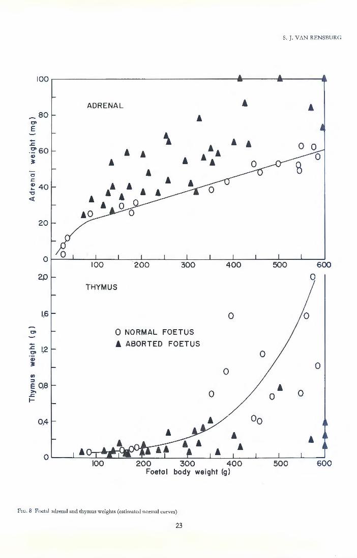
S. J. VAN RENSBURG
0 600
20 0 THYMUS
1,6 0
Ct 0 NORMAL FOETUS
- i ABORTED FOETUS ..c: IJ2 Ct 0 Q)
~ 0 0 en
::J E OJS ' >. 0 0 0 ..c: I-
oo
100 200 300 400 Foetal body weight (g)
FrG. 8 Foetal adrenal and thymus weights (estimated normal curves)
23

REPRODUCTIVE PHYSIOLOGY AND ENDOCRINOLOGY OF ANGORA GOATS
--~ Q) >
..J
-01 --.c 01 Q)
~
>-Q) c
"C
~
LIVER
30
10
0 100
10
KIDNEY
8
6
4
2
FIG . 9 Foetal liver and kidney weights
'
£
'' ' ' ' ' ' ' ' •' '
200 300
t·· ' '
Foetal body weight (g)
24
£ 0
0 0
0 NORMAL FOETUS A ABORTED FOETUS
400 500 600
0
0 0
0 0
Ao
' '
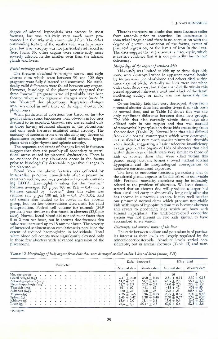
degree of adrenal hyperplasia was present in most foetuses, but was relatively very much more pronounced in the twin which succumbed first. The other outstanding feature of the smaller twin was hepatomegaly, but renal atrophy was not particularly advanced in these twins. N o other organs were consistently more severely affected in the smaller twin than the adrenal glands and livers.
Foetal pathology prior to "in utero" death The foetuses obtained from eight normal and eight
aborter does which were between 90 and 100 days pregnant were fully dissected and compared. No statistically valid differences were found between any organs. However, histology of the placentome suggested that three " normal" pregnancies would probably have been aborted whereas no regressive changes were found in one "aborter" doe placentome. Regressive changes were advanced in only three of the eight aborter doe placentomes. .
When prediction of abortions was based on histological evidence some tendencies were obvious in foetuses destined to be expelled. Growth rate was retarded only in those having advanced changes in the placentome and only such foetuses exhibited renal atrophy. The majority of foetuses from does showing any degree of placentome regression exhibited mild adrenal hyperplasia with slight thymic and splenic atrophy.
T he sequence and extent of changes found in foetuses suggest that they are possibly all secondary to nutr~tional stress caused by placental dysfunctwn. There 1s no evidence that any alterations occur in the foetus prior to histologically detectable regressive changes in the placentome.
Blood from the above foetuses was collected by intracardiac puncture immediately after exposure by caesarean section, and was transferred to vials containing EDTA. H aemoglobin values for the "normal" foetuses averaged 9,0 g per 100 ml (SE = 0,4) but in foetuses carried by " aborter" does this value was reduced (7,5 g per 100 ml, SE = 0,4, P < 0,05). Red cell counts also tended to be lower in the aborter group, but too few observations were made for valid interpretation. Packed cell v olume for normals (34,9 per cent) was similar to that found in aborters (33,0 per cent). Normal foetal-blood did not sediment faster than 0 to 2 mm per hour, but in aborter doe foetuses this value was increased up to 15 mm per hour. The severity of increased sedimentation rate intimately paralleled the extent of reduced haemoglobin in individuals. Total white blood cell counts were significantly elevated only in those few aborters with advanced regression of the placentome.
S. J. VAN RENSBURG
There is therefore no doubt that most foetuses suffer from anaemia prior to abortion. Its occurrence is somewhat irregular and there is no correlation with the degree of growth retardation of the ~oetu.s, exter:t of placental regression, or the levels of u on m the hver. The data suggest that the anaemia is macrocytic, which is further evidence that it is not primarily due to iron deficiency.
Morphology of the organs of newborn k ids This study was limited to kids up to three days old;
some were destroyed when in apparent norJ?al h~al~h by intravenous pentobarbitone and others dted wlthm three days of birth. Virtually no kids were lost when older than three days, but those that did die within this period appeared inherentlY: weak and a la~k of ~he dams' mothering ability, or mtlk, was n ot mvan ably the cause.
Of the healthy kids that were destroyed, those from potential aborter dams had smaller livers than kids born of normal does, and as Table 12 shows this was the only significant difference betwe~n .these two groups. The kids that died naturally w1thm three days also differed only in one respect and that was marked adrenocortical hyperplasia in those kids emanating from aborter does (Table 12). N ormal kids that died differed from their normal counterparts which were destroyed, in that they had very much srr:aller pitu.itar~es, thy~oids and adrenals, suggesting a baste endocnne msufficter:-cy in this group. The organs of kids of aborters that dted within three days of birth were very si.milar t<;> t~ose <;>f kids of aborter dams that were killed wlthm thts period, except that the former sh?wed marked adrenal hyperplasia and the morphological consequences of excessive corticosteroid exposure.
The level of endocrine function, particularly that of the adrenal gland, appears to be disturbed in r:o~-viable kids. Perinatal mortality therefore may be mttmately related to the problem of abortion. We have demo~strated that an aborter doe will produce a larger ktd than usual and carry it abnormally long only after she has aborted in a previous season. It may well be. that our presumed normal dams which produce non-v1able kids with signs of hypopituitarism may become abort~rs and revert to producing kids which are born w!th adrenal hyperplasia. The under-developed endocnne system was not present in two kids known to have succumbed to starvation.
Electro(yte and mineral status of the liver The ratio between sodium and potassium is of particu
lar interest as their levels are largely regulated by the mineralocorticosteroids. Absolute levels varied considerably, but in normal foetuses (Table 13) and new-
TABLE 12 Morphology of boc!J organs from kids that were destrf!)led or died within 3 days of birth (means, SE )
Kids - destroyed K ids - died Parameter
Normal dam Aborter dam Normal dam Aborter dam
No. per group 6 6 10 5 Foetal weight (kg) . . 2,47 ± 0,20 2,90 ± 0,45 2,31 ± 0,14 2,39 ± 0,15 Adenohypophysis (mg) . 64,2 ± 5,7 61,7 ± 4,0 46,5 ± 2,3 56,7 ± 5,3 Neurohypophysis (mg) 18,7 ± 2,7 20,2 ± 2,4 14,0 ± 2,0 22,0 ± 5,2 Thyroids (mg) 567 ± 60 461 ± 62 415 ± 42 476 ± 83 Adrenals (mg) 320 ± 21 320 ± 25 278 ± 23 488* ± 80 Thymus (g) 2,76 ± 0,65 3,96 ± 0,63 2,25 ± 0,65 2,00 ± 0,48 Spleen (g) 3,45 ± 0,42 3,38 ± 0,48 2,68 ± 0,57 2,67 ± 0,25 Kidneys (g) 18,5 ± 2,0 15,1' ± 2,4 13,6 ± 0,4 16,6 ± 2,2 Liver (g) 78,2 ± 3,1 65,4* ± 2,8 62,6 ± 6,4 62,3 ± 5,6
*P < 0,025
25
----- - -- --- - - - --
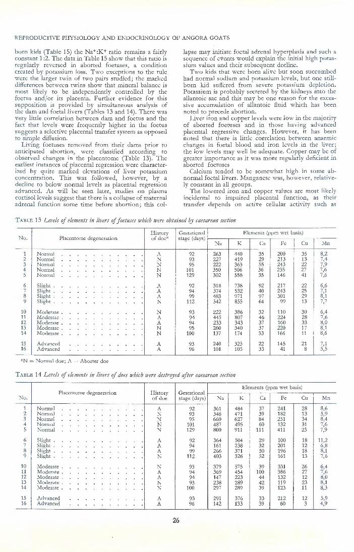
REPRODUCTIVE PHYSIOLOGY AND E NDOCRINOLOGY OF ANGORA GOATS
born kids (Table 15) the Na+:K+ ratio remains a fairly constant 1 :2. The data in Table 15 show that this ratio is regularly reversed in aborted foetuses, a condition created by potassium loss. Two exceptions to the rule were the larger twin of two pairs studied; the marked differences between twins show that mineral balance is most likely to be independently controlled by the foetus andfor its placenta. Further evidence for this supposition is provided by simultaneous analysis of the dam and foetal livers (Tables 13 and 14). There was very lit tle correlation between dam and foetus and the fact that levels were frequently higher in the foetus suggests a selective placental transfer system as opposed to simple diffusion.
Living foetuses removed from their dams prior to anticipated abortion, were classified according to observed changes in the placentome (Table 13). The earliest instances of placental reg ression were characterized by quite marked elevations of liver potassium concentration. This was followed, however, by a decline to below normal levels as placental regression advanced. As will be seen later, studies on plasma cortisol levels suggest that there is a collapse of maternal adrenal function some time before abortion; this col-
lapse may initiate foetal adrenal hyperplasia and such a sequence of events would explain the initial high potassium values and their subsequent decline.
Two kids that were born alive but soon succumbed had normal sodium and potassium levels, but one stillborn kid suffered from severe potassium depletion. Potassium is probably secreted by the kidneys into the allantoic sac and this may be one reason for the excessive accumulation of allantoic fluid which has been noted to precede abortion.
Liver iron and copper levels were low in the majority of aborted foetuses and in those having advanced placental regressive changes. However, it has been noted that there is little correlation between anaemic changes in foetal blood and iron levels in the liver ; the low levels may well be adequate. Copper may be of greater importance as it was more regularly deficient in aborted foetuses
Calcium tended to be somewhat high in some abnormal foetal livers. Manganese was, however, relatively constant in all groups.
The lowered iron and copper values are most likely incidental to impaired placental function, as their transfer depends on active cellular activity such as
TABLE 13 L evels of elements in livers of foetuses which were obtained by caesarean section
History Gestational E lements (ppm wet basis) No. Placentome degeneration of doe* stage (days)
N a K Ca Fe Cu Mn
1 Normal A 92 263 440 35 200 35 8,2 2 Normal N 93 227 419 29 213 13 7,4 3 Normal N 95 222 363 55 243 22 7,9 4 Normal N 101 350 506 36 235 27 7,6 5 Normal N 129 302 558 35 146 41 7,6
6 Slight A 92 318 738 92 217 22 6,6 7 Slight A 94 374 532 40 243 25 7,1 8 Slight A 99 483 971 97 301 29 8,1 9 Slight N 112 542 855 64 99 13 7,7
10 Moderate . N 93 222 386 32 110 30 6,4 11 Moderate . A 94 445 807 46 224 28 7,6 12 Moderate . A 94 233 343 37 160 33 8,0 13 Moderate . N 95 260 340 37 220 17 8,1 14 Moderate . N 100 137 174 33 166 11 8,6
15 Ad vanced A 93 240 325 22 145 21 7,1 16 Advanced A 96 101 105 33 41 8 5,5
*N = Normal doe ; A= Aborter doe
TABLE 14 L evels of elements in livers of does which were destroyed after caesarean section
E lements (ppm wet basis) Placentome degeneration History G estational
No. of doe stage (days) Na K Ca Fe Cu Mn
1 Normal A 92 361 484 37 241 28 8,6 2 Normal N 93 346 471 39 182 13 5,9 3 Normal N 95 660 627 84 251 34 8,4 4 Normal N 101 487 495 60 132 31 7,6 5 Normal N 129 800 911 111 411 23 7,9
6 Slight A 92 364 504 29 100 18 11,2 7 Slight A 94 161 238 32 201 12 6,8 8 Slight A 99 266 371 so 196 18 8,1 9 Slight N 112 403 326 52 161 13 7,6
10 Moderate . N 93 379 575 39 331 26 6,4 11 Moderate . A 94 369 454 100 386 27 7,6 12 Moderate . A 94 147 223 44 132 12 8,0 13 Moderate . N 95 238 289 42 119 23 8,1 14 Moderate . N 100 297 289 39 123 11 8,3
15 Advanced A 93 291 376 33 212 12 5,9 16 Advanced A 96 142 133 39 60 3 4,9
26

S. J. VAN RENSBURG
TABLE 15 Levels of elements in the livers of aborted foetuses and newborn kids
No. Gestation termination
1 Aborted 2 Aborted 3 Aborted 4 Aborted-oedema . 5 Aborted 6 Aborted 7 Aborted 8 Aborted-twin 9 Aborted-twin
10 Aborted 11 Aborted 12 Aborted 13 Aborted 14 Aborted-oedema . 15 Aborted-twin 16 Aborted 17 Kid died 18 Kid died 19 Stillborn 20 Kid destroyed 21 Kid destroyed 22 Kid destroyed 23 Kid destroyed 24 Kid destroyed 25 Kid destroyed 26 Kid destroyed
phagocytosis. The more soluble sodium and potassium ions are under endocrine control, and it may well be that the foetal adrenal incidentally secretes excessive mineralocorticosteroids when in fact it is attempting to produce glucocorticosteroids in response to stress.
CHAPTER 6
LuTEAL FuNCTION
T here is no doubt that at abortion luteal regression is usually more advanced than at normal kidding (Van Rensburg & Van Rensburg, 1961). Extensive alterations in the appearance of hypophyseal acidophiles and basophiles are also present at the time of abortion, and defective luteotrophic hormone secretion has been suggested as the primary cause of the gestation failure (Van Heerden, 1961, 1963). This work has now demonstrated that foetal death may occur as much as a month before abortion, and under such circumstances advanced regressive changes are to be expected. The work recorded below supports the notion that, in those species whose placentas do not produce progesterone such as the goat (Short, 1967), some regressive changes in the corpus luteum are a necessary prerequisite for expulsion of the foetus. In these species the blood progesterone levels decline prior to normal parturition (Short, 1960), as they also appear to do prior to abortion induced experimentally by infectious venereal disease (Osburn, Stabenfeldt & Ewing, 1969). Obviously, inadequate luteal function must be demonstrated at an early stage, even prior to the occurrence of retarded foetal growth, before luteotrophic failure can be incriminated as the primary cause of abortion.
Little work on the mechanisms controlling the functional activity of the caprine corpus luteum has been reported, yet with a little extrapolation of data obtained on similar species a rational working hypothesis may be constructed as follows. As is the case in the sheep, the embryo extends the life of the corpus luteum by virtue of its anti-luteolytic action (Short, 1969), which presumably it exerts until parturition or its death. A similar effect can be obtained by hysterectomy;
27
Elements (ppm wet basis) Gestation
stage Na K Ca Fe Cu Mn
89 707 353 56 82 15 9,0 91 394 229 115 120 24 4,9 91 396 266 71 102 14 7,5 93 760 181 81 123 31 4,9 93 457 372 43 62 13 8,9 95 398 155 94 56 13 8,3
104 810 446 61 95 22 10,0 105 519 615 108 110 12 5,1 105 264 227 98 62 25 7,5 105 125 433 165 200 19 9,2 108 896 788 55 82 18 10,5 109 170 104 116 172 14 6,6 110 127 60 151 240 18 8,3 115 358 92 120 155 20 9,0 115 325 485 81 221 14 10,5 123 299 109 90 132 19 9,1 143 991 1114 24 384 15 9,3 143 510 797 40 233 16 8,3 150 457 150 65 161 17 8,3 147 570 926 64 236 33 6,5 148 565 1071 34 276 42 7,6 151 432 734 23 146 23 6,5 152 495 1087 38 243 23 7,4 152 585 806 30 161 20 12,4 153 657 819 33 214 60 7,8 153 463 999 23 209 112 7,1
in this situation the lifespan of the corpus luteum is usually slightly shorter than in normal gestation, and its metabolic activity declines progressively (Deanesly & Perry, 1969). After hypophysectomy or stalk section, these corpora lutea will regress, and the time sequences between the operations and abortion eliminate any doubt as to the existence of an essential luteotrophic hormone secreted by the pituitary (Cowie, Daniel, Prichard & Tindal, 1963). The luteotrophin is secreted under continuous hypothalamic control and therefore is not prolactin. Presumably the hypothalamus is "timeprogrammed" to withdraw the trophic influence prior to parturition in pregnant animals, or its expected time in hysterectomized animals.
The corpus luteum in the pregnant goats studied showed considerable reduction in size and metabolic activity by the 40th day of gestation. Hysterectomized goats may be expected to exhibit a similar reduced level of luteal function at this time. From the 40th day up to the 90th or 100th day, the placenta grows to a large size and becomes metabolically active as far as steroid biosynthesis is concerned; during th~s period of placental growth the corpus luteum was found to increase its biosynthetic activity. After cessation of placental growth, there was a steady decline in the level of luteal function. These variations could be accounted for by a placental hormone, secreted by the proliferating trophoblast into the maternal circulation, and having both growth hormone and luteotrophic-like actions. Human placental lactogen is one known substance with these properties. Such a placental lactogen in goats may synergise with the pituitary luteotrophin in determining the level of luteal function, and ensuring its maintenance until interrupted by the mechanism initiating parturition. Synergism between placental and pituitary gonadotrophins seems a logical explanation for the mechanism by which a higher level of ovarian luteal function is attained in horses (Van Rensburg & Van N iekerk, 1968).
The continued maintenance of the corpus luteum in the pregnant goat would, according to the above hypothesis, depend essentially on three factors :

REPRODUCTIVE PHYSIOLOGY AND ENDOCRINOLOGY OF ANGORA GOATS
1. Removal andjor suppression of the uterine luteolysin by the conceptus,
2. Trophic hormone secretion from the pituitary. 3. Placental lactogen hormone production. If the production of placental lactogen ceased in the
dead and dying Angora foetuses studied below, it would account for the slightly reduced level of luteal function found in some such cases. Continued secretion of pituitary trophic hormone may maintain the dead foetus within the uterus for some time. Factors which may induce uterine luteolysin production are unknown, but our observations indicate that abortion of a dead foetus is usually dependent on the presence of considerable follicular growth in the ovaries, suggesting an altered progesterone: oestrogen ratio. It may be argued, that pituitary FSH is released at the expense of LH, but oestrogens are known to exert a luteolytic action under certain experimental circumstances in ruminants, and furthermore, follicular growth prior to luteal regression is a normal feature of the oestrous cycle in several species. The participation of the foetal adrenal in precipitating abortion, by means of the mechanism considered to be involved in the normal termination of gestation, may be important in late abortions and will be discussed later.
Luteinization in cycling aborter animals was found to be slightly slower than normal and the young corpus luteum was usually cystic. The cysts disappear early in gestation and blood progesterone levels even tend to be above normal. At about the time of foetal death, no evidence of ovarian luteal insufficiency was found, yet peripheral blood plasma progesterone levels tended to be somewhat low only in some animals. These fluctuations from the normal blood progesterone levels could have been largely due to variations in the level of adrenal function, as the gland's size in aborters parallels the blood progesterone deflections. The adrenal glands of aborters seem to respond to ACTH in an erratic manner as far as the synthesis and release of progesterone and pregnanediol-like compounds are concerned, suggesting some alteration in the biosynthetic pathway.
Corpus luteum morphology T he degree of corpus luteum development and the
occurrence of cysts in corpora lutea exactly 10 and 15 days after oestrus in cycling does were examined. After ovulation in normal animals luteinization appears to proceed more rapidly and is virtually complete by the 10th day after ovulation. Corpora lutea of aborters appear retarded at this stage, although at 15 days post ovulation the weight of luteal tissue was similar in both groups. Extraordinarily large cysts were present in aborter corpora lutea, whereas normal corpora lutea were usually solid structures (Table 16.)
T hose cysts examined 10 days after ovulation contained large blood clots, whereas at 15 days the cysts contained amber coloured fluid. Normal solid corpora showed a prominent ovulatory point or "rosette" caused by luteinization of everted granulosa cells at the ovulation point. Rosettes were absent in the cystic structures, which had a smooth surface, but close examination revealed that all had developed from ovulated follicles. It seems that pressure within the cystic structure was increased, probably by excessive haemorrhage, and this prevented eversion of granulosa cells at the point of ovulation.
Histological examination of all these corpora lutea showed that, when present, the cysts were lined with a substantial layer of fibroblastic tissue. No clear differences between the types and condition of luteal cells of
28
normal and corpora lutea of aborters could be discerned. Functionally, these cystic corpora lutea must regress earlier,, as aborter does have shorter cycles. Such premature regression does not necessarily indicate luteal insufficiency, as luteal regression is now known to be initiated by uterine factors.
TABLE 16 Weight of corpus luteum and diameter of its caviry in cycling Angora does
Normal Aborter Day of cycle
Weight Cavity Weight Cavity (mg) (mm) (mg) (mm)
10 468 0 315 5,5 10 815 0 522 3,0 10 957 0 312 5,0 15 728 0 762 6,0 15 727 0 788 4,5 15 742 0,4 606 1,5 15 825 0 889 9,0 15 802 0 563 4,0 15 702 6,0 595 0
Pregnant does showed marked variations in the size of the corpus luteum, usually to the extent of 100 per cent for any given stage of gestation. The individual weights of corpora lutea, together with monthly means of normal and aborter groups are presented in Fig. 10. This figure clearly demonstrates that there are no appreciable differences between normal animals and those expected to abort, at any stage of gestation. The data do, however, suggest regression up to the 45th day, and that there may be a normal increase in luteal size after this time, reaching a maximum between 90 and 100 days, whereafter there may be a steady decline to term.
Corpora lutea of two aborters were examined 30 days after fertile service for central cavities. One doe was barren and her corpus luteum had a 3mm cavity, whereas the other animal's corpus luteum had a 1 mm cavity and she was pregnant. No other cavities were found in pregnant potential aborters, although a large amount of central fibroblastic tissue was usually present. These findings suggest that initially the corpus luteum of aborters is invariably cystic, but that the cyst rapidly regresses with advancing gestation. No cysts or cavities were found in normal does known to be pregnant.
Histologically, the luteinized granulosa cells tended to be spindle-shaped with the long axis radially arranged in cycling and recently pregnant animals. Small blood capillaries with prominent endothelial cell nuclei and small cells of presumably thecal origin surrounded most sides of each luteinized cell. As gestation advanced the cytoplasmic mass increased and became more basophilic, the cells became more hexagonal and were arranged in groups or cords of several adjacent cells surrounded by prominent capillaries of a sinusoidal nature.
Such architectural details are typical of endocrine glands like the adrenal cortex or adenohypophysis, and efficiency of function may be expected to have increased. An increased synthetic activity of the luteal cells in the latter half of gestation is also indicated by their size, which continqes to increase until approximately the 100th day.
A corpus luteum was surgically removed from a doe which exhibited imminent signs of kidding when 149 days pregnant; she did in fact kid normally 14 hours after the operation. This corpus luteum was pale in colour and showed signs of relatively advanced re-

S. J. VAN REN SBURG
1,5
0 Normal
• Aborter
~ Monthly means -Normal 0 1,3 • Monthly means -Aborter • .. 0 0
• 00 0 0 • :. 0 01 • 1,1 0
..c. 01
·-"' 3: 0 E :::J
0,9 0 0 "' 00 - • -----8----- -:::J
0\ 0 Cl)
0 0 :::J • a. • .... • •• 0 0 u 0 0,7 • • 0 0 0 0 0
0 0 0,5
20 40 60 80 100 120
Days gestation
FrG. 10 Weights of corpora lutea obtained from pregnant normal and aborter does
70
•• Abortion peak 0
l "' 66 3.. 0 Normal pregnant does E • aborter does 0 :::J Pregnant .,
50 • - • ~ 0 ., • • :::J
0 Q. • .... 0 40 •• 0 ()
.... ., a.
., 0 c::
0 0 ....
•o• ., 0 ., • .,
0 • 0 "' 0 ~ Q.
0 ~ 0 0
~ t-
0 20 40 60 80 100 120 140
Days gestation
FIG. 11 Progesterone content of corpora lutea during gestation (estimated curve)
29

REPRODUCTIVE PHYSIOLOGY AND ENDOCRI!'-iOLOGY OF ANGORA GOATS
gression on histologic:1l examination. A corpus luteum of similar macroscopic appearance was obtained a few hours after abortion of a fresh foetus at 106 days gestation. Histological regressive changes were present but on! y to an extent similar to the normal 149 day prenatal corpus Juteum. A third corpus luteum was also surgically removed from a Bocrgoat within a fe w hours after she had aborted on the 110th day of gestation as a consequence of a severe coccidiosis episode. Its corpus lutcum was also pale and firmer than usual and microscopically exhibited regressive changes similar to the previous two animals.
These and other observations show that in the goat some degree of luteal regressive change invariably precedes expulsion of the foetus, and therefore, such changes at about the time of abortion do not necessarily indicate luteal failure as the primary cause of the unsuccessful gestation. There is no doubt that at abortion luteal regression is usually more advanced than at normal kidding but this work has now demonstrated that foetal death may occur as much as a month before abortion. Under such circumstances advanced regression is to be expected.
JVecessity of corpus !ute!lm for the maintenance of gestation
It has been shown by several workers that the presence of a functional corpus luteum is necessary for m:1intenance of gestation in goats (Van Heerden, 1963). A study was undertaken to confirm this fact in Angora goats, to obtain luteal material for study, and to examine the time between surgery and abortion.
This latter value may also indicate that pregnancy is more dependent on the corpus luteum at some stages than others. In the animals listed below, the time of abortion after surgery was observed to be as follows:
Da)'S gestation Time bet1veen at removal C.L. removal
No. of C.L. and abortion
1 30 (hours)
45 2 60 90 3 95 60 4 95 60 5 105 60 6 115 52 7 120 36 8 125 47 9 135 39
10 135 40 11 145 60
This study shows that abortion occurs at a surprisingly consistent time after removal of the corpus luteum at any stage of gestation. The last three foetuses in the series could have been expected to be born alive, yet a~! were dead and considerably autolyzed. Function of the placenta must be exceedingly dependent on progesterone as there is no doubt that within a few hours of the blood levels dropping to the basal levels maintained by the adrenal, foetal death occurs.
A doe was found to be carrying twins at 75 days gestation with a corpus luteum in each ovary. The corpus luteum that appeared the largest was removed and found to weigh 589 mg; its removal did not disturb the course of gestation and normal twins were born at term. Another doe, 85 days pregnant, had a single average-sized corpus luteum which was incised and a portion which appeared to constitute at least twothirds of the gland weighing 480 mg was removed. Absorbant sponge material was sutured over the pro-
30
fusely haemorrhaging cavity, and this doe also carried her kid to term. Considering the rapid lethal effects of removal of the whole gland, these two instances suggest that the level of progesterone is not critical and most does gestate with a considerable physiological excess of progesterone.
Progesterone content of rorpora lutea
Similar results were obtained from three independent studies undertaken in 1963, 1965 and 1968. These were pooled for presentation in Fig. 11. The corpora I utca were all obta.ined at laparotomy; some by making a circular incision of the capsule at the junction between ovarian stroma and the projecting portion of the corpora, and expressing it manually, but the majority were obtained by immediate dissection from the excised ovary after cannulation of the ovarian vein and collection of its effluent for about 10 minutes. Each corpus was sectioned longitudinally to examine its structure, rapidly weighed, frozen on solid C0 2, and stored at -20°C for assay.
When progesterone results were expressed as the concentration or the total amount calculated to be present in each corpus lutcum, very similar tendencies were revealed. Figure 11 shows that the amounts in the corpus luteum remain relatively constant until the 80th day of gestation, whereafter there is an appreciable increase up to the 11 Oth day which is followed by a slow decline.
Paradoxically, maximal luteal levels coincide precisely with the time of highest foetal death rate and abortion. Furthermore, at this time the majority of potential aborters have a higher luteal progesterone content than the mode for normal animals. Included in the data of Fig. 11 are corpora lutea from two does carrying dead foetuses. One at 96 days was extremely fresh and its corpus luteum contained 36,2 fLg progesterone, whereas the other had been dead for at least three weeks (Plate 2b) and her corpus luteum still contained 17,2fLg progesterone. Also included were two animals, 92 and 93 days pregnant, which had extremely advanced degenerative changes in the placentomes similar to one whose foetus had already died. Their corpora contained as much as 40,8 and 38,1 [Lg progesterone, respectively. At about the usual time of foetal death there is therefore no evidence of any luteal insufficiency from this study.
Progesterone output lry the ovaries
The ovarian vein draining the ovary containing the corpus luteum was cannulated in a total of 30 does, 11 of which were aborters. The amount of progesterone secreted per minute was calculated. These animals were not subjected to presurgical fasting or disturbed in any other way prior to anaesthesia and laparotomy. In four does the ovary not containing the corpus luteum, but having follicles whose maximum size ranged from three to six millimetres, was cannulated. In each instance the concentration of progesterone in the plasma was similar to that found in peripheral blood. On the other hand the progesterone concentration in plasma draining the ovary containing the corpus luteum was usually increased to the order of 50 to 200 times above that of peripheral plasma.
Ovarian secretion rate is shown to decrease gradually to about the 40th day of gestation by the estimated curve of regression in Fig. 12. This decrease parallels that exhibited by the size of the corpus luteum (Fig. 10) and is in agreement with the histological findings of Harrison (1948). He demonstrated a decrease in the

average diameter of luteal cells from 35 microns at 12 days after ovulation, down to 30 at 20 days and 25 at the 35th and 45th day of pregnancy. This low point in the rate of progesterone secretion is also reflected in our peripheral blood values and may be important, since a minor abortion peak occurs at this time. Animals aborting at this stage frequently exhibit oestrous behaviour simultaneously with abortion, which indicates luteal insufficiency. Those aborting embryos at this early stage differ from animals aborting later, in so far as having exceptionally good mohair growth rates (Chapter 4).
Harrison (1948) unfortunately indulged in excessive speculation with his small histological series and divided his luteal material, which only represented the first 60 days of the five month gestation period, into four "phases of luteal development". The first stage he defined as "hypertrophy extending from ovulation until about the 35th day", notwithstanding the fact that his measurements demonstrate the converse, which is in full agreement with the histological findings of this study. Also in accordance with our results is the finding by Harrison that luteal cells had again increased in size at the 45th and 55th day, but he claimed early regression of the goat corpus luteum on the basis of a single animal examined at the 60th day. This would be precisely similar to the sequence of events in sheep, as this species is not dependent on the ovaries for gestation maintenance after the 50th day, since placental progesterone production reaches adequate levels at this time.
As we have observed in this study, the luteal cells in the Angora goat continue to enlarge and only reach their maximal size after the 100th day. It is only after mid-pregnancy that the structure of the corpus luteum becomes well organized as a typical endocrine gland and at this time all the usual cytoplasmic characteristics, which may be seen with the light microscope and are indicative of a highly active endoplasmic reticulum, are most pronounced.
Heap & Linzell (1966) interpreted their own results rather unsatisfactorily according to the findings of Harrison. The data of the four goats which they presented in detail all exhibit a considerable decline in carotid artery plasma progesterone levels at about the 35th to 45th day of gestation, and thereafter the levels increase until a few days prior to term. The implication of this earlier work that luteal activity decreases with advancing gestation in the goat is therefore incorrect.
Physically robust goats such as used by Heap & Linzell show an exceptionally high incidence of multiple ovulation and therefore unusually high progesterone values are encountered in cycling and early pregnant does. We and other workers have evidence that in goats, sheep and cattle, when an excessive number of corpora lutea are present in the ovaries of pregnant individuals, they will regress to a limited extent at an early stage, suggesting the existence of some form of "negative feedback" system. Animals with a single corpus luteum are clearly more suitable to study the variations in the level of luteal function. In this respect we are fortunate, as the incidence of multiple ovulation in our Angora flock is only in the region of 10 per cent.
Figure 12 shows quite clearly that ovarian secretion of progesterone increases steadily up to approximately the 100th day and then exhibits a gradual decline to term. While the minor first abortion peak occurs at the low point of progesterone secretion, the major abortion peak coincides with maximal secretion rates. Two
31
S. J. VAN RENSBURG
potential aborters had particularly high secretion rates of 14 and 9,1 f.Lg per minute (equivalent to 19 and 13 mg per day) yet exhibited many signs of imminent abortion such as placentome degeneration, foetal anaemia, and large ovarian follicles. Such daily production rates are higher than those necessary to maintain gestation in does experimentally deprived of their corpora lutea (Meites, Webster, Young, Thorp & Hatch, 1951) and it is difficult to visualize them as having suffered from a deficiency at any stage. One of the aborters with the lowest output was the one at 99 days carrying a mummified dead foetus (Plate 2b) and even it had a probable adequate production rate of 4,03 f.l-g per minute (5, 7 mg per day).
Progesterone levels in peripheral plasma Peripheral plasma progesterone in the goat is known
to originate only from the corpus luteum and adrenal gland. To investigate the levels produced by the latter gland, six does were examined during the mid-anoestrous period. Immediately after collection of these samples, 40 i.u. of adrenocorticotrophin was injected intravenously and a second sample was collected an hour later. The results of the three normal and three aborters (f.l-g progesteronef100 ml plasma) were as follows:
Breeding history K;K;KTD KT;K;B K;K K;K;A;B A; A; A; A A; A; A
Resting level After ACTH 0,21 0,29 0,23 0,44 0,13 0,19 0,22 0,17 0,22 0,19 0,16 0,62
All these animals, and those used in the next gestation study, were quite accustomed to being bled. This procedure was routinely performed with a minimum of pain and apprehension where the doe happened to be standing in the pen. Nevertheless there were considerable amounts of plasma progesterone present in such anoestrous goats and our mean value of 0,20 f.Lg per 100 ml is even less than the 0,34 f.l-g found by Heap & Linzell (1966) in eight anoestrous and oestrous Saanen goats. Adrenocorticotrophin increased plasma progesterone slightly to moderately in the normal individuals, but in two aborters the amounts found after adrenocorticotrophin were actually less and in the third it was greatly increased. Similar strange behaviour by the adrenal in respect of the output of incidental metabolites is noted in the section on pregnanediol excretion. It is nevertheless obvious that the adrenal can influence the plasma levels significantly. This effect is clearly seen when the blood cortisol levels of aborters is compared with their plasma progesterone levels in the experiment below.
A total of 14 goats were bled from the jugular vein at precisely 15 day intervals throughout gestation, or for a 150 day period. Of the 14 does, six produced kids at term, one of which was stillborn. Seven aborted and gestation was not observed to terminate in one, but judging from oestrogen excretion data she is presumed to have been pregnant, and to have resorbed the foetus. Mean progesterone values from the six does that produced a term kid and the seven that aborted are compared in Fig. 13.
Progesterone values in normal animals were again found to increase from the 45th day and reach maximal values between 90 and 120 days after which the levels declined to term. The increase during this period is virtually a symmetrical parabola. This independent
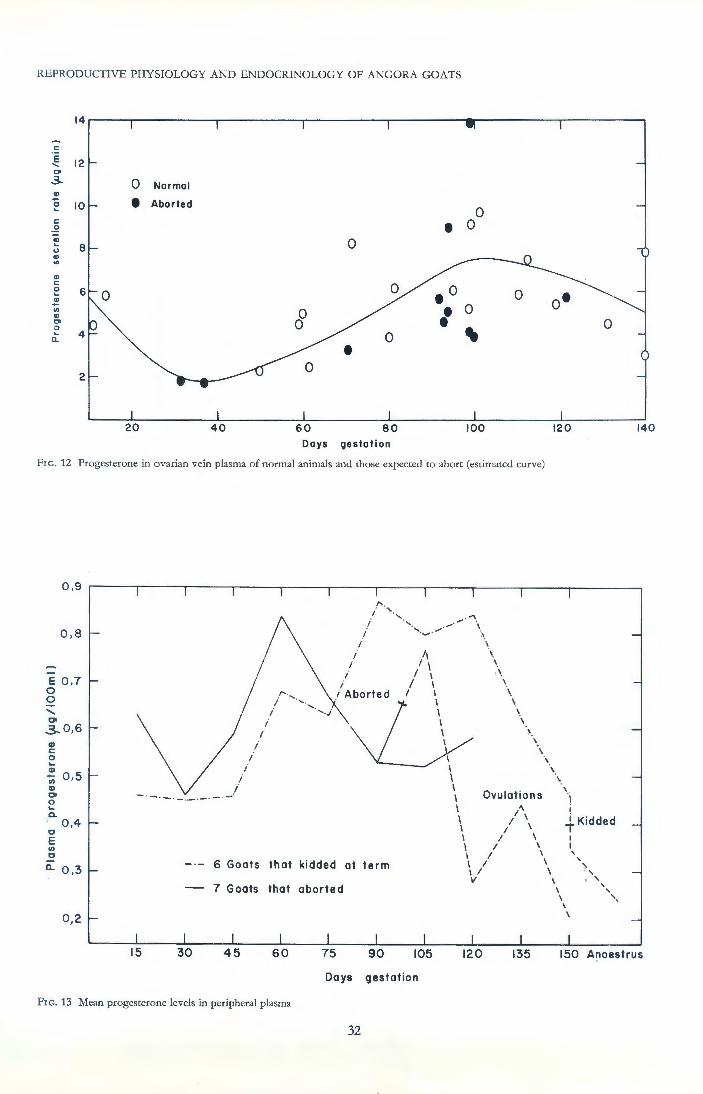
REPRODUCTIVE PHYSIOLOGY AND ENDOCRINOLOGY OF ANGORA GOATS
1 4~--~----------r----------.---------.--------~r----------.---------,
c:
~ 12 "" ..3-
c: . !2
~ (.)
CD en
., <:
e ~ en CD
"" 0 ... 0..
0 Normal
e Aborted
20 40
0
0 0
0 •
60 80 Days gestation
0
• 0
.o 0 • 0 • .. 0
100 120
FrG. 12 Progesterone in ovarian vein plasma of normal animals and those expected to abort (estimated curve)
0 ,9
0 ,8
E 0 ,7 0 0
' co ,3...0,6 ., c: 0 .... CD
0 ,5 -Ul CD co 0 .... Q. ' 0,4 0 E Ul 0
Cl. 0 ,3
0 ,2
-·-- -·-·- ·-
i i
/
i
i /
I I
I I
/ I
·" I
i /Aborted
6 Goats that kidded at term
7 Goats that aborted
II I I
I I I I
I I I I I I I
'. \ \ \ \
\
\ \
\ \
\
\
\ \ I
I I
\
I I Ovulations
I /' I I \ I I \ I I \ I I ' I I \ I I \ I I \ v \
\
I
.i Kidded
' ' \
I I I
'
\
' ' . ' ' ' ' '·
140
90 105 135 150 Anoest rus
Days gestation
FrG. 13 Mean progesterone levels in peripheral plasma
32
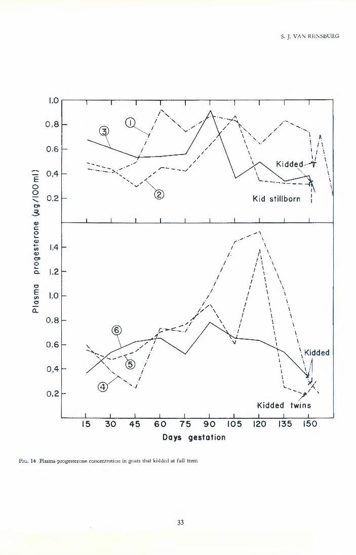
S. J. VAN RENSBURG
0,8 ..... ~ ....
// ~. / ..... ' \ ' · / .....
0,6
~ 0,4
0 0
' 01
,3. Q) c 0 ~
Q) -V)
Q)
01 0 ~
a.
0 E V)
0
a..
0,2
1,4
1,2
1,0
0,8
0,6
0,2
I I
/ /
/ /
/
--- ,.· / - .... / -·----->,...., / r--- --/
15 30
',,v~
45
I I
I i
I I
//\ // \
/ \ /j \
,t;; ,.. :::. ::. i \
""
60 75 90
Days gestation
FrG. 14 Plasma progesterone concentration in goats that kidded at full term
33
r I
I
\ '· / I ~ \ ' ·,/ I i \ \\ I ., . I
\ . ~ i \ ' K1dded _,.. i
Kid stillborn
.,..,.""" '\ \
\ 1\ \ II \ I I \
I . I I \
I I \ I I \
,' \ \ I I I I I I I \ I I \ I I \ I I I I
I I
\
\
I I I
I I I I I
\Kidded \
I I
I.. X. ........ /, ·,
Kidded twins
105 120 135 150
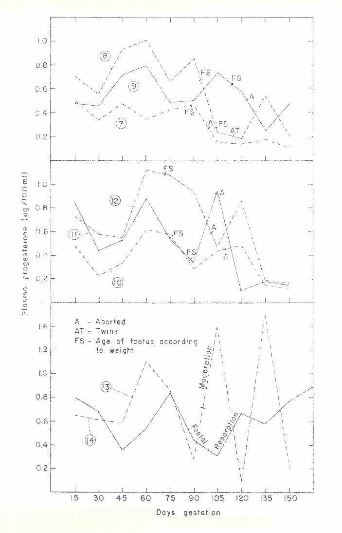
E 1.0 0 0 ......
0 .8 0" <-. -<I> 0 ,6 c 0 .... <IJ .... en 0.4 <IJ 0" 0 .... 0. 0,2 0
E en 0
0...
1,4
1,2
1,0
0.8
0,6
0,4
0,2
,..-"\ ..... - \
® /.., \ I \ /\
I \ / \ I \ // I FS
', I \ \/ ;._
' I ® \ '-... I
'I \ \
/' _ . -t · I ', -< ', ..,- FS' 1 " / /-:::;\7 .... .... ..... \ \
\.!.) ....... \AI FS I
1\ -~'~.,._ AT / \ .._,_./ '---- -·-- .....
~ I I
I I I \
I \ I I \ \ - \ \
I \ \
\ I
\ \
\ \
~ A - Aborted AT - Twins FS - Age of foetus
to weight
15 30 45 60
according
I
/I I \
c: j I 0 .
:.;: I I
~j I <IJ . 1. of o; I ~i
I ./.
I I
I I i
\ II I. . \ I .
I \
I I
I I
I \ I
\ I. \ \
\ \
\
I
75 90 105 120 135 150
Days gestation
1 I


REPRODUCTIVE PHYSIOLOGY AND ENDOCRINOLOGY OF ANGORA GOATS
pregnanediol is present in goats' urine but with these critical methods we have never found more than 0,5 mg, whereas the more usual amount seems to be less than 100 fLg per 24-hour sample.
A method involving enzymic hydrolysis, acetylation, thin layer and gas-liquid chromatography, similar to that of Kirscher & Lipsett (1964) was used to assay pregnanediol at six points during the first 115 days of gestation in six goats, three of which were aborters. This experiment did not reveal any obvious differences between normal and aborter animals, and the rate of excretion remained relatively constant during the first two thirds of gestation. Excretion rate in most animals :.:emained somewhat below 100 fLg per day in five does, while the sixth doe excreted approximately 250 fLg per day.
The effect of adrenocorticotrophin administration was investigated with the same method in four ovariectomized Angora does, whose excretion was found to remain relatively constant, usually in the region of 15 fLg per day. Treatment consisting of 40 i.u. ACTH three times a day for two days significantly elevated the excretion rate in only one normal goat, which reached 90 fLg during the second day and rapidly returned to normal in the 48 hours after cessation of treatment. A second "normal" doe showed virtually no change, but she was a young animal that had produced only one kid. The two aborters similarly showed virtually no change. One of these, however, was showing early clinical signs of hyperadrenocorticism and had the lowest excretion rate which remained around 5 fLg per day.
The later method used by us demonstrates that the earlier double column chromatographic method of Klapper et a!. (1955) gave gross overestimates and is not valid when applied to animal urine. However, the remarkable association between the amount of contaminating chromogens and the outcome of gestation first reported by Brown eta!. (1963) has been confirmed and extended. There is now little doubt that these contaminants are largely metabolites of adrenal steroidogenesis. The significant point is that they decrease under conditions of adrenal stimulation, particularly in aborters, suggesting specific effects on enzymes by ACTH, at a more advanced point in the steroidogenesis pathway which favours the synthesis of corticosteroids, rather than incidental steroid metabolites.
CHAPTER 7
CORTISOL LEVELS IN PERIPHERAL
PLASMA
The presence of the adrenals is necessary for normal reproduction in female rats and in adrenalectomized rats a satisfactory reproductive performance was only attained by administering both gluco- and mineralocorticosteroids (Cupps, 1955). When, however, the diet contains adequate protein and salt, satisfactory reproduction is possible and is not further improved by the administration of cortisone (Aschkenasy & Aschkenasy, 1957). If the diet contains no protein, only intact rats which receive supplementary progesterone can maintain pregnancy. Progesterone supplemented adrenalectomized rats on a protein-free diet invariably aborted, unless cortisone was also administered.
Later work confirmed the abortifacient effects of a protein-free diet and the fact that in intact animals reproduction may be largely restored by supplementary progesterone and oestrogens (Fisher & Leathem, 1965; Hazelwood & Nelson, 1965). There seems little doubt
36
that adequate corticosteroids, together with a high level of sex steroids, are necessary to protect the foetus from a nutritional crisis. Corticosteroids probably act by assisting transfer of maternal protein to the developing foetus, largely by means of mobilizing skeletal muscle protein and stimulating its degradation to amino acids. The additional progesterone may increase placental efficiency, although this hormone or its metabolites are known to have anabolic effects. Adrenal glands of ruminant animals seem to secrete particularly large amounts of progesterone. Short (1960) concluded that this source may represent a significant proportion of the total progesterone production.
Experimental stress in rats, or the administration of ACTH, will induce reproductive quiescence identical to that obtained by the administration of adrenal androgens. Excessive amounts of injected glucocorticosteroid will inhibit the normal uterine responses to oestrogen and progesterone, but foetal death only occurs if the dosage is high enough to induce weight loss. Species other than rodents and humans seem to secrete very little adrenal androgens. Many observations, including the assay of pituitary FSH, show that stress or the injection of corticosteroids have an FSH releasing action. This effect does not occur in rats (Van Rensburg, 1965 ; Van Rensburg & Cupps, 1967).
The unequivocal demonstration of adrenal hyperplasia in aborter Angora goats at an early stage in this work suggested that the above inhibitory effects of excessive corticosteroids may participate in causing abortion. Experimental evidence showed that if the cortisol levels were maintained at pathologically high values, foetal death would be a consequence.
This work confirms that cortisol levels are high in the majority of aborters, during the first half of gestation. Abortion, however, only occurs when blood cortisol values are exceedingly low at a time when the foetal nutrient demands are increasing rapidly, such low levels resulting in abortion need not necessarily be preceded by high levels. Advanced adrenal atrophy prior to anticipated foetal death was then demonstrated on morphological grounds, and confirmed the revised notion that abortion was primarily associated with adrenal insufficiency.
Evidence now exists that the hyperadrenocorticism is a protective response favouring the foetus, reducing the rate of mohair growth, and presumably increasing the maternal resistance to adverse environment. The hyperplastic adrenal produces cortisol with increased efficiency. Goats which abort very early in gestation produce exceedingly large amounts of mohair during the particular season. Young maiden does which later become aborters produce large amounts of mohair and have low blood cortisol values.
R esults Resting plasma cortisol levels of various groups
Using specific methods and well trained animals, cortisol levels were found to be somewhat lower than previously reported for goats. Normal goat plasma usually contained approximately 1 fLg cortisol per 100 ml, whereas aborter does yielded very variable results. The majority of aborters were higher than normal, but occasional exceptionally low values were also encountered.
Different groups within the female breeding flock were assayed during various times of the year, and the results are presented in Fig. 16. High plasma cortisol levels were encountered particularly in aborters during

~ z 1-en w a::
I 1-(.)
<t
a:: w IlL.. <t
E 0 0
' 0>
3--0
"" -... 0 u
0
E "" ~ a..
2,0
1,5
1,0
0 ,5
0
9
8
Before I January
Animals: 9 10 N.S.
Before I
Animals 7 7 P<0,05
February
S. ]. VAN RENSBURG
breeding Pregnant Post-natal I I
March June-Sept November 6 7 4 7 6 6 p <0,01 N.S. N. S.
breeding Pregnant Post- natal I
6 6 18 8 5 5 P<O,OOI P<O,OOI N. S.
March June November
FrG. 16 Resting and stimulated plasma cortisol levels in various groups of Angora does. Crossed bars represent aborters
37
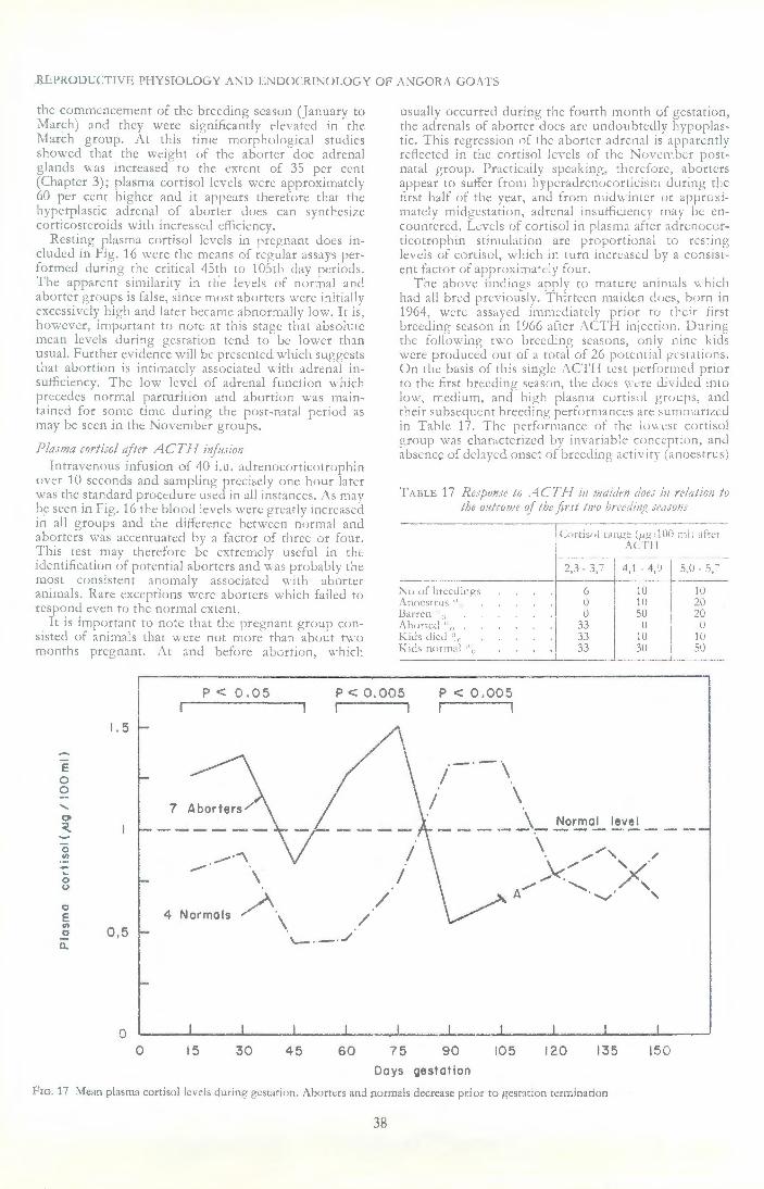
REPRODUCTIVE PHYSIOLOGY A"'D ENDOCR1"0LOGY OF AJ'\GOR:\ GO.\TS
the commencement of the breeding season (January to \!arch) anJ they were significantly clc:vatc.J in the March group. i\ t this t imc m(Jrphnlogical studies showed that the wciPht of the aborter doc aJrcn:tl g!anJs \\as 1nncascd'' to the extent of 35 per cent (Chapter 3); plasma cortisol levels were approxinutely 60 per cent higher and it appc;,rs therefore rbat the bypcrplastic adrenal c,f aborter does can synthesize corticosteroids with increasl'd dl1ciency.
Resting plasma cortisol levels in pregnant docs included in Fig. 16 were the means of regular <1Ssays performed during tht: critical 45th ro 105rh day periods. The: apparent sirnibrity in the levels of norm::tl and abtJrrer i'roups is Cdsc, since must ahoners \\'ere initially excessively high and later became :lbnorn·,ally low. It is, however, important to note at this stag<:: thar absolute mean levels during gestation tend tD he Jm, er rhan usuaL further evidence will Gc presented which suggests that abortion is intimately associated with adrenal insufficiency. The low lev~! uf adrenal function which precedes normal parturition and abortion was n;aint<tined for some time during rhc post-natal period as may be seen in the Novcmbn groups.
Plasma cortisol ajttr ACT! I mfmion Intravenous infusion of 40 i,u, adrcnoClJtticotrophin
over 10 seconds and sampling precisely one h,;ur later was the standard procedure used in all instances. ;\s may be seen in Fig. 16 the blood levels were grL·arly increased in all gruups and the difference between nllrmal and :tborters was accentuated bv a flctor of three or four. This test rnav therefore l;c extremely useful in rhe iclentification ~Jf potc:nttal aborters and ,~·as probably the must consJstcnt anomaly associated with :tb(Jttcr animals. Rare exceptions were abortcrs which failed w respond even tfJ the normal extent.
It is import:1nt tu note tlut the pregnanr group consisted of animals that were nnt more rhan about two months pregnant. ,\t anJ before abortion, which
usually occurred during the fourrh monrh of gesration, rhc adrc:nais of aborter docs arc unduubtedly hypoplastic. This regrcssinn of the aborter adren:d is appar..:nrly reflected in the cortis<ll levels of the i\<>\'enJber postnatal group. Practically speaking, thcrcfure, ahorters appear to surfer from hyperadrenocorticism during the tlrst balf of the year, and from midwinter or apprw:im:trcly midgcst~Jion, aurcnai insuH1cicncy may be encountcr~.:d. Levels of cortisc,J in plasma after ::drcn<>cnrricotrnphin stimulation arc prr•portiunal w resting levels uf Cllrtisol, \v hich in turn incre<lscd by a cunsistent f:1ctor of apprr:ximall'ly four.
The above findings a[>ply to mature animals which had all bred previously. Thirteen maidw d(leS, b')rll in 1964, were assayed imml'diarely prim tu their first breeding scasun in 1966 after .'\C:Tl I injection. During the: following two breeding se.1sons, oniv lllrle kids were produced out of a total uf 26 pr,tcmial gt·srations, On the basis of this single .\CTi l test pertormcd prior w the first brcedin!r season, rhc· docs \\'ere divided int<> low, medium, and. high plasma cortisol groups, anc.l their subscgucnt breeding performances arc sumnnrizcd in Table 17. Thc r>crformancc: of rhc ltl\\ est conisol group \Yas chaDctcrizcd by in varia bil- concept ion , and absence of dchycd onset or breeding acti\ it\" (anoestrus)
TABLE 17 RejjJfJIIjf tu .··1CTi I til JJ!atdrn dar.: in rflation to the ulftcu;;;e of t!Je jir.r/ 111'0 bu~di1\~ .rra.roiJJ
r Ctlrtist;! Llll?C' (;Lfl, 100 :n!; Jfrvr .\C.T/1
----------+r1 _2_-.~_, _-6-_3-._7 -_--
1
r--, _4~ 1 _ 4. ') 1
s:u _ s.-;-.,",,fhrt·.dirws 10 ' lU ,\tlocstrus" . . :I U I 10 I 20 !Ja rren ' " I 0 I 50 · ?0
:\--b-o-rt_c,_J_··_·· ____ w· . . 3~ .J.I ___ l~~ol ___ l' ___ -_u __ Kids died '1 ., 1iJ Kitls norm:il ' ' .. SU
p < 0,05 p < 0.005 p < 0.005 r -
1.5
E 0 0
' 01 ~
0 _.-·"'\ "' - .--· . ... \ 0
0
0 ~. / E 4 Normals
\ / VJ 0,5 0
\.- . - . ../ 0..
0 0 15 30 45 60 75 90 105 120 135 !50
Days gestation
Frc. 17 Mean plasma cortisol levels during gestation. Abortcrs and normals decrease prior to gestation t<:rminarion
38

A
2
3 B.
E l 0 0
' 2 01
~
0 1/) -~ 0 u
0 E 1/)
0
0...
3
L 2
I I Normal- kidded
\\~ \
® \ I \ \ I
"l-. - - - - I / ~-·-- ..... 1
Normal- aborted following year
K .,
( .
j I
i I . I
I I I
\
I! 3
I
~ 1/~ ·~ ...... K .... · ... ·- ... ' . .,.·
C. Aborter does- did not abort
15
I i
I
30 45
I
I I
i I
'w·- ·- · -·"'
I I
I
I I
I i
; /
60 75 90 105 120 135
Days gestation
fK i
I i
I
150
Fxc. 18 Plasma cortisol levels Juring gestJtion in d(>eS which did not abort
i
J I
l
I
l I

i KS
D. Normal does- first unsuccesful ; 8,4 -
3 gestation ! t ! 5,6 I ! I ;
2 I ;
9 ;
I . . -·-
,.~·(®
?' // ',, / "
E E. Aborted 2nd time KS- stillbirth 0
2 A- aborted 0 ........ AT- aborted twins 01
:::t
'·~ -/ 0 ' 1/) ' / - A /
~ / 0 / (.) ,-...,/
0 7 E AT 1/)
0
a. F. Aborted 3 rd time 9,8 I I
3 I ! I
I I I ,/, i I
2 .' '. I I .'\ i I . . \ i I .
I i
I I
\
! ./..A
\\,\ I I . . I ~·
........ ,
Macerated foetus
15 30 45 60 75 90 105 120 135 150
Days gestation
FrG. 19 Plasma cortisol levels in gestations that failed. Note invariably low values at 90th or 105th day

and also does which failed to kid after service (barren). These latter two failures occurred in 50 per cent of animals in the higher groups. However, the only two animals of the 13 does which aborted were in the lowest cortisol group and a further third of the animals in this group produced non-viable kids. Other evidence, which will be discussed later, also exists which indicates that adrenal corticosteroid synthesis is at a lower level in young aborter stock which have not yet bred.
Plasma cortisol during gestation
Two separate studies were performed; for the first a paper chromatographic method (Van Rensburg, 1965) was used to assay cortisol and in the second the TLC method described in this work was applied. Both experiments yielded essentially similar results.
The first study performed in 1965 consisted of observations on six normal and six aborter goats. Samples were taken at random intervals during gestation; some animals were not trained to bleeding at this time and furthermore the does were moved approximately 50 metres to an indoor clinic for bleeding. Consequently, levels were generally higher and more variable than found with the use of the more refined techniq).les in the 1966 study, and are therefore reported only briefly.
Normal animals averaged 1,26 fl-g per 100 ml plasma (40 observations, SE = 0,12) and aborter does were significantly elevated to a mean of 1,9 fl-g (34 observations, SE = 0,18). Levels generally increased moderately to reach a maximum between 100 and 115 days, then decreased to term, particularly during the last 20 days. Seven assays on normal pregnant goats performed after the 130th day averaged only 0,69 !Lg /100 ml plasma, as compared to the average of 1,42 fl-g found from 24 assays on the same animals between the 85th and 130th day (P < 0,005). At the actual time of abortion levels were high, but in each instance a considerable sudden decrease to at least pre-kidding levels had occurred prior to abortion. ·
The second study performed during 1966 was more detailed, as the above findings suggested variations of considerable consequence. The same 14 animals used for the progesterone and oestrogen excretion studies were used and these well-trained animals were bled for assay at precisely 15 day intervals, commencing 15 days after service. Mean cortisol values obtained are compared in Fig. 17, which shows that the abnormally high levels in aborters reach their maximum between the 60th and 75th days. At this stage the placenta grows exceedingly rapidly and reaches the final stages of maturation under the protective trophic influence of progesterone. Evidence that excessive corticosteroids may inhibit this influence has been mentioned in the introduction, and such a hypothesis on the cause of abortion seems attractive but is not fully sustained by examination of cortisol levels in individual animals (Fig. 18 and 19). On the 60th day, two of the eight abnormal goats whose results are presented in Fig. 19 were in the normal range. All aborters tended to be high on the 75th day, but one normal animal (No. 4) exhibited a deflection from the expected curve which placed it in the range of the aborters. These "high" levels in the aborters at this time are merely relative, as the difference is chiefly ascribable to the normal animals whose cortisol levels are depressed below non-pregnant resting levels. Half of the aborters did not exceed 1,5 fl-g during the first half of gestation and there is no evidence that such levels are unphysiological. Increases to this extent and even higher are usually associated with a
41
S. J. VAN RENSBURG
defensive or protective response of the body ar:d whe_n cortisol levels of individuals are compared w1th their progesterone curves, several _instance_s are ~pparent which indicate that unusually high cort1coster01d levels increase progesterone concentration. The koown ab_orter animal which kidded normally (No. 6) had the hlghest average cort1sol blood levels during gestation of all the does that kidded.
Adrenal function was assessed in 11 similar known aborters, which maintained their gestations to term, by Morgenthal (1966), who used blood cellular and electrolyte parameters. These indicated that potential aborters remained in a state of relative adrenal hyperactivity throughout gestation and that the difference was most marked from 100 to 140 days, which time coincides with the period of greatest foetal loss. Yet these animals successfully maintained their gestations and this work has shown that such gestations are maintained slightly beyond the normal duration, and that the kids are heavier at birth than normal. These considerations are strong evidence that the hyperadrenocorticism seen in aborters is a protective response favouring the foetus. . .
Quite contrary to the marked mcrease 1n adrenal output in normal and aborter does wh~ch maintained their gestations, those that actually d1d abort were found to have very low cortisol levels on the 90th and 105th days (Fig. 17, P < 0,005). A study of the in?ividu_al plasma cortisol values reveals that all e1ght ammals m Fig. 19 whose gestations failed, decreased to below the normal range on the 90th or 105th, or both days. All except one were lower than the range of normals on the 90th day, and the one exception had an extraordinari!y high value which was well above that encountered ~n advanced Cushing's syndrome. Although the level m this goat was very low at the 105th day and she aborted only 10 days later, a blood level in the region of 10 fl-g per 100 ml may be expected, from experimental studies, to rapidly induce foetal death.
Functional efficiency of the placenta and hence the metabolic activity of its cells must be greatly increased around the 90th day due to the rapid increase in the rate of foetal growth and cessation of placental growth (Chapters 3 and 5). Such a physiological state will require corticosteroids which are well known to be essential for reproduction in most species (Cupps, 1955; Van Rensburg, 1965, and others). The exceedingly low cortisol levels encountered in our goats at this time must be interpreted as adrenal insufficiency, a state which was followed by abortion in all animals in which it occurred.
Mean cortisol levels in normal animals were virtually doubled during the 90th and 105th day period (1,33 fl-g), but had decreased rapidly by the 120th and 135th days (0, 72 fl-g, P < 0,005, Fig. 17), thus c~mfirming the pr~parturient decreases in maternal cort1sol levels noted m the earlier experiment.
Cortisol in the foetus and neonate The small size of the foetus before the 100th day
necessitated the pooling of several samples for reliable quantitative assays. The composition of these samples, together with the results obtained are detailed in Table 18. Unfortunately these samples were constituted and assayed on a basis of the goats breeding history, whereas later histological examination of the placentome provided what was considered to be more reliable evidence of gestation outcome, thus some groups consist of both aborter and normal individuals.

REPRODUCTIVE PHYSIOLOGY AND ENDOCRINOLOGY OF ANGORA GOATS
Foetal cortisol levels seem to resemble that of the dam during the fourth month of gestation, but in the last month there is a very substantial increase. This may be partly due to increased foetal ability to respond to the stress of surgery, but it is exceedingly doubtful if such a marked response is possible in the short time. The foetus was usually bled within half-an-hour of the goat being fetched from her usual pen. Within a few hours of birth some kids had extraordinarily high cortisol blood levels, but each of their dams had aborted previously and clear evidence of adrenal anomalies in such offspring exists (Chapter 5).
Very low values were found in three of the samples included in Table 18. The four animals constituting Samples No. 1 and 4 all had early histological regressive changes in the placentomes and very low plasma levels of cortisol. Number 6 had similar low levels but this doe was not examined histologically. These low levels are likely to be a consequence of maternal adrenal insufficiency which has been shown to occur prior to abortion and seemingly is the cause of potassium accumulation in the same foetuses. Samples 2 and 3 comprised equal numbers of normal and aborter foetuses and they contained moderate amounts of cortisol. Most of the plasma in sample 5 was from aborters and it had the highest cortisol level at this stage. These scanty results are direct support for all the earlier evidence which suggests that the foetus becomes acutely embarrassed as a result of maternal adrenal insufficiency and its own adrenal then undergoes hyperplasia. Finally, at expulsion of the foetus, it is in a state of hyperadrenocorticism, as is also shown by morphological and electrolyte studies.
TABLE 18 Plasma cortisol in foetal and neu;born kid blood
No. Subjects Age (days) 1-'g/100 ml
1 2 Foetuses 92, 93 0,13 2 4 Foetuses 92,92,94,94 1,04 3 2 Foetuses 92, 95 1,06 4 2 Foetuses 94, 99 0,16 5 3 Foetuses 93, 95, 100 2,53 6 1 Foetus 100 0,24 7 2 Foetuses 110, 112 1,33 8 1 Foetus 121 1,75 9 1 Foetus 129 3,78*
10 1 Foetus 140 3,46 11 1 Foetus 140 6,24
12 Kid 0 8,60 13 Kid 0 0,2 14 Kid 0 12,0 15 Kid 0 1,76 16 Kid 2 5,58
Blood obtained by intracardiac puncture, at laparotomy, from foetuses and under pentobarbitone anaesthesia from kids *Umbilical venous blood
CHAPTER 8
EXCRETION OF 0ESTROGENS IN THE
URINE
By means of a biological assay, Kiist & Vogt (1934) identified a pattern of urinary oestrogen secretion rather similar to that found in this study with precise chemical methods. Klyne & Wright (1957) identified oestrone (0,3 mg/1) as the major oestrogen in the urine of pregnant goats and also found considerable quantities of oestradiol-17a (0, 1 mg/1). The small quantities of oestradiol detected were no doubt due to the acid hydrolysis used by these early workers, and we have confirmed the fact that oestradiol-17a is
42
quantitatively very much more important than oestrone. Daily excretion in our pregnant Angora goats re
mained low until up to the 40th day and then slowly increased up to the 100th day. After this time there was a rapid increase up to a few days before term. Such a pattern of excretion is precisely proportional to the rate of foetal growth and not to that of the placenta which reaches its maximum size on approximately the 90th day of gestation. Some aborter does had excessively high oestrogen excretion values prior to abortion, whereas in others the oestrogen excretion normally associated with pregnancy failed to occur. These divergent patterns of excretion were not associated with differences in the time of abortion or with the state of the foetus, therefore urinary oestrogen excretion does not simply represent an index of placental function.
Recent studies on steroidogenesis in the human placenta has led to the concept of the foeto-placental unit and the necessity of the integrity and viability of this unit for steroid biosynthesis and metabolism during pregnancy. The placenta itself apparently carries out little de novo steroid biosynthesis, and its activity is attributed to efficient conversion of ~ 5-3~ hydroxysteroid precursors reaching it from the foetal and maternal circulations. Thus, for example, the placenta cannot synthesize dehydroepiandrosterone, but can convert it to oestrone and oestradiol. Oestriol appears to be formed from 16a hydroxylated neutral precursors elaborated by the foetus, largely in its liver. There was some suggestion of hepatomegaly in some of our foetuses and an hepatic origin of oestradiol precursors in goats may explain the excessive amounts excreted from an early stage in some unsuccessful gestations.
Since dehydroepianodrosterone originates largely from the adrenal gland, the injection of ACTH increases oestrogen excretion in pregnant women. Conversely, dexamethasone suppression will decrease oestrogen excretion. Variations in the level of adrenal function in aborter Angora goats did not seem, on the above premise, to be the explanation for their anomalous excretion patterns. High oestrogen excretors had only recently become aborters and their cortisol values were relatively low, whereas low excretors were usually well established aborters which had exceedingly variable blood cortisol values with frequent high levels. Qualitative alterations in various adrenal biosynthetic pathways, as has been suspected with alterations in adrenal progesterone and pregnanediol synthesis, may account for an unusual supply of oestrogen precursors and hence the extraordinary relationship of oestrogen excretion and adrenal function.
The function or necessity of the high level of maternal oestrogens during gestation largely remains a matter for speculation. The only apparent differences in the aborted foetuses obtained from high and low excretors were reduced foetal iron and copper stores in the foetuses of high excretors; in those where foetalplacental oestrogen production was virtually absent, the placental transfer of heavy metals appeared to be quite normal.
Oestradiol has repeatedly been demonstrated to increase pituitary prolactin secretion in vitro, and there is good evidence that it can act physiologically at the pituitary level (Ben-David, Dikstein & Sulman, 1964). Such an action may influence mammary growth, but since prolactin does not appear to be luteotrophic in the goat, it has no known effect on the maintenance of gestation.

Res nits LVormaf oestradiol-1 7a excretion
Urine samples ,,·ere.: collected from 14 animals at a time which averaged three \\·ccks before breeding. Occasional docs had exhibited one oestrus period and were sampled during the mid-luteal phase, but in the majority the first oestrus was imminent, and such docs arc known to ha v<:: considerable follicular gro\vth (Shelton, 1960). Daily excretion of oestradiol remained under 20 11g in these animals and in the six docs which produced a term kid, It vari<::d from 3 to 19 p.g per 24 hours. , \ftcr f<::rtilc breeding, no signihcant changes were noted on the 10th, 25th and 40th days.
Distinct evidence of placental oestrogen production was only present o n the 55th day of gestation, when k,·els in most indi,·iduals \n:rc in the region of 40 f1g. This stage is similar to the time in sheep when placental stcroiJogcnesis attains adcguatc levels to maintain gestation, without the assistance of the ovaries.
Excretion rates increased relatively slowly to the vicinity of 300 flg per 24 hours at the 100th day and thereafter , -<::ry rapidly to approximately 1 000 flg shortly before term in normal animals (Fig. 20). i\ decrease prior to parturition was only noticed in the last one to three Jays of gestation (No. 4, 5 and 2). Within 24 to 48 hours of parturition, excretion rates recorded in the six animals ,,·hich reached term had dropped to between 11 and 145 {J-g oestradiol.
The maximal rate of excretion recorded ranged from 400 to 1 200 {J-g in the six term docs. Since the two highest excrctors of oestradiol \\-ere the only consistent normal breeders, the variation and also the lower values encountered in the other four (Fig. 21, 22) are guite likely to be related to the abortion problem. Even animals which had not aborted but were destined to do so, suffered from an apparently reduced rate of placental steroidogenesis. The one normal twin gestation (No. 5) produced no more oestradiol than usual, nevertheless the rate of excretion was generally proportional to the foetal g rowth rate.
Oestradiol excretion in unsuccessful gestations Prior to breeding and up to the 40th day of gestation
the excretion of oestr~diol in the majority of aborter docs remained low at similar levels to those found in normal animals. Slight increases in the majority of poor breeders at the 55th day indicated the onset of placental steroidogenesis as in normal individuals. Dramatic differences were, however, evident at the critical 70th and 85th day stages; in five poor breeders the excretion rate of oestradiol was abnormally high, whereas in four individuals there was very little evidence of placental oestrogen production at all. These extreme differences were neither related to the time of abortion, nor to the condition of the expelled foetus.
The two animals which kidded normally but became aborters during the following year (No.1 and 3; Fig. 21) had relatively normal patterns of excretion, but placental steroidogenesis was somewhat less active from the 115th day to term. One aborter doe which kidded normally (No. 6) had considerably depressed levels, particularly up to the 100th day. The gestation resulting in a stillbirth also had initial depressed levels, but on the 100th day the excretion rate was higher than usual (No. 2; Fig. 22).
Excessively high initial levels of excretion on the 70th and 85th days (No.2, 14, 9, 12 and 8) are associated with invariable failure of gestation and also with animals which had only recently become aborters.
43
Furthermore, plasma cortisol levels \H:rc inclined to be low up to the lOOth day in this group ,,-hilc oestrogen cxcrctJOn \\·as excess! ve.
Diametrically opposite results \\·ere obtaJncd in well established aborter indi\·iduals (::--\o. II , 7, 10 and 13 ; Fig. 23) \vhich excreted Yirtually no oestradiol at all. Three of these does had very hif!:h fluctuating plasma cortisol lcYels and the other had the lo\\·cst average cortisol levels found during gestation. The single h igh value in this group exh ibiteJ by No. 10 on the H5th day was associateJ \\·irh an extraordin:ui!y h1gh plasma cortisol level of approximatdy 10 /.Lg per JU() ml ; the simultaneously incrcaocd oestradiol could well be of adrenal and ovarian origin and we therefore have no proof of any placental oestrogen synthesis in this group. The steroidogenic ability of the placenta is clearly seen to be impaircJ in these animals as early as tb<:: 55th and 70th days, yet the foetus is only expelled 40 to 55 days later, after having undergone considerable further normal growth and development.
The mineral content of the livers of aborted foetuses, which resulted from gestations wher<:: no oestrogen synthesis by the placenta could be demonstrated, differed from that usually found in aborted foetuses (Chapter 5). Sodium concentration was exceedingly lo\\' and calcium exceptionally high. The content of iron \\·as not depressed as in the majority of aborted foetuses, and copper levels were also only slightly lo,,·er than usual.
Defective steroidogenesis of the placenta was the earliest pathology detected in the conceptus. lt is, however, preceded by abnormally high maternal plasma cortisol levels during the first half of gestation and also by unusually high plasma progesterone. Some correlation exists between the anomalous levels of oestrogen excretion and plasma cortisol, but no such association exists with plasma progesterone.
Since levels of progesterone declined well after the detection of abnormal oestrogen excretion, this study supports the view that the decreased progesterone levels demonstrated prior to abortion arc secondary manifestations.
Oestrone excretion At the time of maximum oestradiol excretion during
gestation, oes tronc levels were general! y five times lower. The fact that oestrone is considerably less active biologically than oestradiol, renders it quantitatively even more inconsequential.
The level of excretion was relatively constant and usually somewhat lower than oestradiol up to the 55th day. Thereafter, placental production gradually increased excretion values to the region of 100 {J-g at the 85th day. A feature found in all five animals which delivered a viable term foetus was a considerable decrease in excretion of oestrone between the 85th and 100th day. Since the rate of oestradiol excretion increases appreciably at this time and considering the fact that oestradiol is synthesised via oestrone, it seems that the necessary hydroxylases favouring oestradiol production are responsible for the decreased oestrone output in favour of oestradiol. Subsequent slight increases were again recorded, but excretion declined some days prior to parturition or abortion.
Aborter animals generally excreted more oestrone during early gestation; such values frequently exceeded the oestradiol value and this oestrone is most likely to be of ovarian origin . Excessive levels of probable placental origin were only detected in No. 8 (Fig. 23) which later aborted twins.

0'
3-c 0 -Q) ._ u X Q)
c Q)
0' 0 ... -1/)
Q)
0
>. ... c c ... :::3
>.
c 0
I 2 00 ~,~-----.-,--..---~--.1--"T"~--.--~-..... ~- ----.---.....----.----., I I I
I 000 ~
800 ,_
1--
600 ,....
400 -
1-
200 1-
1-
0 800
600 ~
1-
400 1-
200 1-
4. Normal
Oestradiol
Oestrone
I I
I I
I I
I I
I I
I i
I I
I
i i
I I
i I i
/i /
I / / -
I I
/ -I
I
-
I -I
! I -
-
-
-
-
Kidded \ . -
/I'' ~! I
I .- / ', "\ I /-.. / ;, \ -,........ / .......... / . . \
_.... .... / '- / I \. \ ,.... / -- i \ \ 1-.--.----~--~:....-- I---;- I I I i I \. I
i \ i
i \ I
\ -i \
\ i \
5. Normal I i -
I I
/ ! I I
i I -I I
! I I I -
I i ! i
I -i .J
"' "
-
I \ I \
I \ / I ' . / I \ I / \ .
/ .... -.... I \I
-
/ __ ........ ..... ..... , _ __ Kidded twins~,!
Q L-LJ·-~=~~~--=·-=~~·~~·=~·--· -.,~~· -=:~;~~.~~..,.--~ __ L_I ___ L_I ___ ~I--~1----~~----~· ,~~· --~ 0 25 40 55 70 85 100 115 130 145 160
Days gestation FIG. 20 Oestrogen excretion in normal animals during gestation

c::: 0 -a> .... u X a>
>. .... 0 c:::
0 a
800
600
4 00
200
400
200
400
200
/
_/
/
/I l I
/ I /
I. Normal- aborted the nex t se ason /
I
Oestradiol
Oestrone
/ /
)
I
I
/ A I 11
I I I I I' I I I
/ ' I I . I I ', I I I
I / , I · ./ __ ...... I ../ II
,.-:,. -- ............ / \
. = ·-=:..:-.:...:--~-,:..; =·= ·~..:...-+-.:::........ ........./ Kidded -----Ji.
3. Normal- aborted the next season
6. Aborter doe - kidded
i i
/ I
/ /
I I
I ;-·- . i
i - .
I
I
I
I I I I
\ \ \
! I I I
Kidded~ ...... "\ i
...... \. ............. \!
'-- - ---·-- ' Days gesta t ion
FIG. 21 Urinary oestrogen excretion during pregnancy by potential aborters

0'
.3-c 0 +-Q) .... u )(
Q)
c Q)
0' 0 .... IJ)
Q)
0
>. .... 0 c ·-....
:::::l
>. ·-0
0
I I I I I I I I \ I
700 / \ - /
I \
\ I
\
600 - 2 . Normal - kid stillborn and I \
aborted next season I \
I I
500 I I - I I
I \ I
I 400 ~ I - · - · - · Oestradi ol I
I
I
300 - - -- Oestrone I I ~
I I
I I
200 ~ I I
I Kid stillborn --...:.. I _..., I
/ _, \ .
100 ~ I -/ \ I _...., -- \I ) - - --/-- / ' - -- -/ ---- / ' - ...... \
/ ---- _,_ . ..... ...... 0 f- ~ . - · - · -·- · - · - ·- · - - ·- · , ___ ........., \
I I I I I I I I l _l
300 f- 14 · Aborter doe - failed to kid - · """\ - ' .
r
200 f-/
\ Resorption /
/ ' assumed / \
'j 100 ~ /
/ / ...... -- - ------. ...... ......
1-- -~ ....... -:---:-.:: .-::...7--:==·=~ ...... ..... - ·- · - · ......
0 ........ _ --- _ ·::::-.::.,.
I
300 -
200 -
100 -
I
300 1--
200 ~
100 ~
9 .
12 .
I I I I I
Normal f i rs t abortion / ,... .-· I
I I
I i
/ /
/ -· I I I I I
Norma I - fir st abor- /'-1 \ tio n
/
I
/
/ I
/ .,.,----
\
\
I I I I
.-- ' ' ' ' \ \
\ Aborte d J,o
\123 rd day \
\
l I I
' Aborted IOist day
\,\ _____ ./ _........- ..... , ..........
------:.::::.....::::- ·- · __ ...,. ...... ...._ ..... . ___ ,_ .... - ,.,.., ..... __.,..- ' 0 - - -- . ..-·--- ...... ...... _ _ _ -----1 I I I 1 1 I I I
-
-
-
I I
~
-
-
-
-
-
-
-
-
-
-
-
-
-
-
-
-0 25 40 55 70 85 100 115 130 145 160
Days gestation FrG. 22 Urinary oestrogen excretion by poor breeders. Note excessive excretion between the 70th and tOOth days

c 0 ..... cv .... (.) )(
cv
c cv Cl 0 .... ..... 1/)
cv 0
>. .... 0 c
0 a
I I
600 r-a.
500 f.-
400 r-
300 r-
200 f.-
100 r-
200 r- II.
100 r-
I
Aborter abortion
Oestradiol
Oestrone
I I
doe - 2nd
I I
I I I
Aborter doe - 2nd
I I
I
I
" i \ \
\
\ \
\
I
\ \
\
abortion
\ \
I I I
\ Aborted t w1 n s )(- 115th day
\ \
\
Aborted 105th day
0 r- -~..:..:-...:..=-....:...-.~.:: .. : :.·.::-:. -..:-~--- :·;;::-_:_-,......-.-....... -:_·: ·...,. :..:--'-·---~....:..... I I I I I I I I I I
200 1- 7. Aborter doe - 2nd abortion
100 1-
_......... - · Aborted IOOth day / ~ ......... ,.., ·":,-,..-._........ . ....._ -..::::...~ --
0 r :::-:._~~----= -~·.....:-- ' ·r·~--- ---....-...:..=-~---.:....--.:..:
I I I I I I _l_ _l_ _l I
200 r- I 0 . Aborter doe- 3rd abortion .'\
I \ / \
-
-
-
-
-
-
-
-
-
-
-
-
-
-
100 r- / \ -/ /- ...... ....:\,. Aborted II 0 t h da y
/ , ...... ...... ---........ --~-·-·-· .J / ·- . ::::.~--i...·- · - · -·-. 0 ~ _ ......... ---~.....:....--::::._......... ---_...;I' .._. __ ---~ -I I I I I I I I l I
200 1- 13. Aborter doe- 3rd abortion -
100 1-Macerated foetus from 96th day
-
._. .-- ·-·-. -- --.::=- ~~...:-:_.._Jc..:= ·..:::.- .. :_--....... --::::.. 0 1- --...-:-_,... _____ -....__ _____ . ·---.........._-.- - -
I I I I I I l _l _l I
0 10 25 40 55 70 85 100 115 130 145 160
Days gestation FrG. 23 Urinary oestrogen excretion by well established aborters. Evidence of virtual absence of placental oestrogen synthesis

REPRODUCTIVE PHYSIOLOGY AND ENDOCRINOLOGY OF ANGORA GOATS
CHAPTER 9
EXPERIMENTAL STUDIES ON POSSIBLE FACTORS
INFLUENCING GESTATION TERMINATION
Review of clinical ryndromes characterized by pathological disturbances of gestation length
Clinical syndromes characterized by abnormal gestation lengths have provided experimental models from which it may be concluded that the foetus plays a vital role in determining its own destiny.
The genetically conditioned forms of prolonged gestation in cattle (reviewed by Holm, 1967) are well known. One of the two main clinical entities which are recognized is described in the Guernsey breed, whose postmature foetuses are premature in development and always exhibit adenohypophyseal aplasia. HolsteinFriesian postmature calves, on the other hand, are unusually large at delivery and the adenohypophysis is present but smaller than normal.
Foetuses with these genetic defects may remain in the uterus for a remarkably long time. Holm quotes a case described by Andres in 1931 of a 218-pound postmature calf in utero for 369 days. This foetus had remarkably small adrenal glands. Instances of postmature HolsteinFriesian calves delivered by caesarean section also clearly suffered from adrenal insufficiency, which appeared to be the cause of their deaths within a few hours of delivery (Holm, Parker & Galligan, 1961).
Congenital malformations in Iambs have been associated with unduly prolonged gestation periods by Binns, Anderson & Sullivan (1960). An 11,8 kg Iamb was obtained from a ewe after a 230 day gestation period. Abnormalities are mainly confined to the head and brain, cyclops formation is frequent and the hypothalamus is most certainly affected. This condition is caused by a teratogen in the skunk cabbage and the critical stage for ingestion is 14 days after conception (Binns, Shupe, Keeler & James, 1965).
In South West Africa, the Karakul pelt industry has been hampered by the frequent occurrence of "big Iamb disease" which results in worthless overgrown pelts. The period of prolongation varies from a few days to months, similar to the extreme cyclopian cases described . by Binns et a/. (1960). Postmature "big Iamb disease" foetuses appear normal except for increased size and are characterized by progressive hypophyseal and adrenal atrophy (Morgenthal & Basson, 1966; Basson, Morgenthal, Bilbrough, Marais, Kruger & van der Merwe, 1969). The syndrome appears to be caused by a shrub, Sa/sola tubercula/a (Moq.) Schinz, Chenopodiaceae, when ingested during the last 50 days of gestation. The experimental evidence strongly suggests that the active principle acts as a foetal hypothalamic inhibitor.
Human anencephalic foetuses have caused considerable confusion as an experimental model, because of the variable nature of the malformation and associated hydramnios which appears capable of precipitating parturition by virtue of volumetric effects. The development of the adenohypophysis also varies, but the adrenals are usually grossly hypoplastic. In the absence of hydramnios gestation may be very prolonged, sometimes lasting more than 52 weeks with a living foetus (Turnbull & Anderson, 1969). Excretion of oestriol during such gestations is exceedingly low, presumably because ofthe lack of steroid precursors supplied to the placenta by the foetal adrenal.
Distinct clinical entities constituting a problem of premature delivery of non-infectious etiology have only
48
rarely been recognized. The problem of recurrent abortion in Angora goats is the only syndrome that has been studied on a physiological basis. Various disturbances in the level of the maternal adrenocortical function occur, but most consistently the expelled foetuses suffer from adrenal hyperplasia.
The occurrence of excessively large adrenal glands in prematurely delivered human foetuses has been recorded since at least 1909. Striking increases in the weight of the adrenals in many infants delivered as a result of "unexplained" premature labour after the 20th week are frequently present (Turnbull & Anderson, 1969). Cyst formation in the outer adrenal cortex of human foetuses has also been associated with in utero stress and prematurity (Oppenheimer, 1969).
Consideration of the above models has led to the rather obvious hypothesis that the foetal hypothalamicpituitary-adrenal axis plays an important role in the termination of gestation by virtue of foetal adrenal corticosteroid action on the placenta (Van Rensburg, 1964, 1965; Holm, 1966).
Discussion of experimental studies on the cause of birth The potent inhibitory influence of glucocortico
steroids on induced progestational decidual tissue formation is well known (Velardo, 1957; a'nd others). The uterine growth response to oestrogens is also readily inhibited by glucocorticosteroids (Velardo & Sturgis, 1956). Impairment of the basic trophic actions of these steroids by glucocorticosteroids has received little attention until the realization that the level of foetal adrenal function was implicated in the termination of gestation.
Administration of cortisol acetate to pregnant rats results in a selective inhibition of the uterine decidual tissue weight that is greater than the extent of placental or foetal inhibition (Van Rensburg & Cupps, 1967). The experiments of these workers also suggest that there is a factor in the cycling non-gravid rat uterus which limits the progesterone concentration in the ovaries. This inhibitory influence was fully eliminated only by gestation or hysterectomy. Cortisol injections readily reduced the elevated ovarian progesterone in pregnant rats, but did not alter the high values in hysterectomized rats. Such experiments do not show that cortisol is luteolytic, but demonstrate that it has an action on the gravid uterus which results in a reduction of ovarian progesterone; the significance of such a mechanism should be investigated further.
The remarkable experiments of Liggins, Holm & Kennedy (1966) on sheep demonstrated an essential role for the foetal hypothalamic-pituitary system in the timely initiation of parturition. D estruction of the hypothalamus, pituitary, or stalk section performed on the foetus resulted in prolonged gestation. Infusion of one overdue hypophysectomized foetus for six days with ACTH resulted in termination of the gestation.
Cortisol, the major glucocorticoid in sheep, was found readily to alter the gestation length when administered to the foetus (Van Rensburg, 1967). A single injection of 5 to 30 mg given during the third or fourth month significantly prolongs gestation, but when injected during the last month parturition occurred prematurely. Slightly higher dosages induced rapid termination of gestation at any stage. The studies reported in this work confirm the dose-related prolongation of gestation when glucocorticosteroids are administered to the foetus during the third and fourth month; gestation is prolonged for up to a week and the foetuses at birth are

abnormally large. During the last month corticosteroids seem capable only of shortening gestation and those lambs born after the 140th day survived well. Methylprednisolone acetate exhibited approximately six times the activity of cortisol acetate, thereby confirming the conclusion of Liggins (1969) that the activity of cortisol is associated with its glucocorticoid activity rather than with its mineralocorticoid actions.
The above effects of glucocorticosteroids have all been found to apply to the goat, a species whose gestation is dependent on ovarian luteal function rather than placental progesterone biosynthesis as in the sheep. Goats appear to be more sensitive since abortion is induced with lower dosages, gestation is prolonged longer and the viability of treated offspring is poorer. The interval between injection and abortion is, however, longer.
Halliday & Buttle (1968) showed that excess cortisone given to near-term foetuses also causes sudden death and expulsion of the foetus, whereas slightly less would induce premature delivery of a viable foetus. The suddenness of death after high dosages points to a breakdown in placental function, and the idea that cortisol interferes with the action of progesterone is favoured. Our experimental evidence certainly shows that there is a dose-related drastic reduction in maternal plasma progesterone after such cortisol injections into the foetus.
Quantitative reduction in progesterone levels is not necessarily present prior to induced premature or normal delivery. On the other hand the goat takes longer to respond and as progesterone invariably declines prior to delivery in this species, the possibility that cortisol induces the foeto-placental unit to release a luteolysin-like factor must be considered. Such a mechanism may also apply to sheep which are induced to abort at an early stage while the corpus luteum is still active, although ovarian luteal function after cortisolinduced termination of gestation has not been studied.
A convenient explanation for the mechanism by which small doses of cortisol administered to previable foetuses prolong gestation is that it suppresses the maturation of the hypothalamic-pituitary-adrenal axis. Since the period of prolongation approximates the estimated duration of effective action of the steroid and is dose-related, any interaction with progesterone metabolism seems unlikely. Experimental hypoglycaemia with insulin as reported below significantly prolonged one gestation, although interactions such as compensatory adrenal hypertrophy could have occurred.
Much evidence has accumulated which shows that the foetal adrenal gland and cortisol levels in the foetal circulation of sheep and goats increase to several times the normal species level some days before birth (Alexander, Britton, James, Nixon, Parker, Wintour & Wright, 1968; Van Rensburg, 1968; Bassett & Thorburn, 1969). The elegant studies of the latter authors particularly, suggest that at least five days of elevated levels are necessary to initiate labour. In the human foetus there is no such evidence and the foetal adrenals seem to be suppressed until birth. The. onset of high cortisol levels in foetal ruminants seems to coincide with the attainment of foetal viability which is virtually only reached a few days before term, whereas the human foetus is viable some months before term. The "evolution" of human term uterine activity (Csapo, 1969) appears to be influenced by several factors and is a very gradual process, consequently the gestation period
49
S. J. VAN RENSBURG
is exceedingly variable in this species when compared to the sheep or goat.
Maternal cortisol levels in sheep are known to decrease about two weeks prior to term (Saba, 1965; Bas son eta!, 1969). Cortisol levels were founti to remain low during the 50 days of prolonged gestation in one ewe carrying an adrenalectomized foetus. Normal goats studied during this work exhibited a similar preparturient decline in plasma cortisol levels. Maternal injections of excess corticosteroids during this period and even earlier, readily induce delivery and there is little difference in the susceptibility of sheep and goats.
When goats were injected prior to the last month of gestation, significant prolongation of gestation similar to that obtained by injection of the foetus occurred. The same maternal treatment in sheep had the reverse effect and shortened gestation by a few days; those foetuses tended to be smaller. Since the ovine gestation period is also prolonged when the foetus is treated, the difference in effect after maternal treatment may lie in an altered metabolism of the administered steroid at the placental barrier. There seems little doubt that this barrier must be traversed since this work shows that a higher dosage rate must be used to achieve effect when the hormone is administered to the mother. The extreme potency of some synthetic steroids in precipitating premature labour may be due to their stability at the placental barrier (Adams, 1969; Skinner, J ochle & Nel, 1970).
Adrenalectomy experiments performed on the foetus in utero eliminate any doubt that the substance usually responsible for initiating parturition is secreted from this source. In 1967, Van Rensburg reported signs of postmaturity in a sheep foetus whose gestation was prolonged by five days following adrenalectomy on the 142nd day of gestation. It was soon found that if foetal adrenalectomy was performed prior to the time when foetal plasma cortisol levels commenced to rise, then pregnancy was prolonged for remarkably long periods (Van Rensburg, 1968; Drost & Holm, 1968). During the prolonged stage of gestation, which may last as long as 50 days, the foetus exhibits severely retarded somatic growth and hypertrophy of both the neuroand adenohypophysis.
Only after death of the foetus and presumably cessation of placental progesterone production, does spontaneous delivery commence. Goats do not produce placental progesterone and the results of this study show that it is not possible to prolong their gestations by means of foetal adrenalectomy. In such instances the maternal hypophyseal luteotrophin is probably withdrawn, due to hypothalamic "timing" which can be illustrated by hysterectomy.
Elimination of progesterone production and secretion by means of removing the corpus luteum in goats will result in birth of the foetus 40 to 60 hours later; the time interval is quite independent of the stage of gestation. Initiation of labour after death of a postmature adrenalectomized sheep foetus may occur in a similar fashion. The administration of cortisol, oestrogens and gonadotrophins is capable of the same effects. There are, however, experimental objections to the possibility that substances such as glucocorticosteroids and gonadotrophins act primarily by blocking the production of progesterone. A biologically significant decline in the levels of circulating progesterone has not been demonstrated in several species (Short, 1969). Continuous infusion of progesterone into the foetus or mother does not block or delay the labour initiated by dexametha-

REPRODUCTIVE PHYSIOLOGY AND ENDOCRINOLOGY OF A GORA GOATS
sone (Liggins, 1969). Injection of gonadotrophins into pregnant sheep induces abortion but foetal death has been demonstrated to occur prior to luteal regression (Moor, Rowson, Hay & Caldwell, 1969). Increasing preparturient foetal corticosteroid levels were interrupted and eliminated by means of adrenalectomizing two foetuses on the 138th and 144th days of gestation; at this time progesterone production is still undoubtedly good yet the "message" for labour initiation 10 days later had been conveyed and removing further stimuli resulted in a delay of only five days. In these cases active progesterone production and its action must have ceased in the presence of very little corticosteroid.
The above considerations are consistent with the concept that foetal adrenal secretions, probably corticosteroids, normally initiate labour by means of inducing one or more substances within the foetalplacental unit which leads to birth. This mechanism leads to preparturient alterations in the reproductive tract which are similar to those ascribed to relaxin and oestrogens; alterations in the ratio of biologically active oestrogens and progesterone may be largely responsible. In corpus luteum dependent species luteal regression is also initiated, possibly by the removal of inhibition on uterine luteolysin production. Alternatively pituitary LH secretion may be suppressed (assuming that LH is the luteotrophin) and FSH increased, since the ovaries of parturient animals usually exhibit some follicular growth. An essential role for the pituitary and ovaries in parturition has been excluded in sheep and man, as ablation of either organ has no effect on parturition, although in the goat the integrity of both are necessary for the maintenance of gestation. There is experimental evidence that the trophic action of progesterone on the placenta may be blocked directly by corticosteroids, although such an action may result in foetal death and occur only under pathological circumstances. The progesterone "myometrical block" is finally eliminated by luteal regression in corpus luteum dependent species, or by possible combinations of interference with its actions and reduced production and release in placentalprogesterone species.
The primary initiator of termination of gestation according to the above hypothesis, would be the foetal hypothalamus. The time at which it commences to release adequate ACTH-releasing factor which will result in termination of the gestation may be ascribed to an inbuilt "clock" mechanism which would be influenced by factors such as cell growth dynamics. This possibility is not supported by the observations on foetal adrenal hyperplasia in aborted Angora goat and human foetuses. Liggins (1968) has shown that infusing small sheep foetuses, at a similar stage of gestation to the usual aborted Angora foetuses, results in adrenal hypertrophy and parturition. The nutritional environment of the sheep foetus commences to deteriorate from the 120th day (Mitchell, 1962) and the concept of Spiegelberg (1891) who suggested that parturition is initiated through the action of substances secreted by the foetus in response to insufficiency of nutrition, is favoured. From experimental evidence it seems possible that the hypothalamic threshold to "nutritional stress" slowly declines as the foetus matures and when it is adequately sensitive to ensure viability of the foetus, it may initiate parturition in response to relatively mild suboptimal environmental stimuli.
Many factors are known to influence the period of gestation slightly. The effects of the majority of variables such as maternal and foetal size, sex, parity and number
50
of foetuses can all be explained in terms of the concepts above. Some seemingly contradictory influences of under-nutrition may also theoretically be reconciled in terms of the outlined scheme. A sudden change to a low plane of nutrition during the last month of pregnancy in sheep hastens parturition by several days (Alexander, 1956), probably bv increasing corticosteroid secretion in response to adaptive nutritional stress. A continuous low plane of feeding has no effect on gestation length in adapted ewes (Davies, Southey & Goodall, 1966). The time of retention of nutritionallystressed Angora foetuses prior to their expulsion is exactly proportional to their development and hence their ability to secrete adrenal steroids.
Results (A) The pituitary-gonad axis and hyperadrenocorticism
Influence of adrenocorticotrophin on the ovarian response to gonadotrophin.
For this experiment, four Angora goats in anoestrus and six sheep were pretreated with progesterone for 10 days before receiving a single injection of exogenous gonadotrophin. Five 48-hourly intramuscular injections of progesterone (0,55 mg/kg) were followed by a subcutaneous injection of pregnant mares' serum (25 i. u. f kg) 24 hours after the last progesterone injection. Commencing with the first progesterone injection and continuing until slaughter, half of the animals were treated with 0,75 i.u. ACTH per kilogram ("Action Prolongatum"; Frederiksberg Chern. Labs.) by deep intramuscular injection administered 48-hourly.
Rams were used twice daily to detect oestrus. ACTH did not alter the time when oestrus was first exhibited or the duration of the induced oestrus. The response of the ovaries was examined (Table 19) at slaughter nine days after the first signs of oestrus were exhibited.
No differences in the rate of superovulation were evident, but the weights of corpora lutea were considerably less in all ACTH treated animals. Luteal central cavities tended to be larger in the ACTH treated group and increased follicular growth was also
TABLE 19 Influence of ACTH on the ovarian response to gonadotrophin
Mean No. of No. of Mean C.L. diameter intact fol-
Breed Treatment* corpora weight of C.L. licles > lutea (mg) cavities 4mm
(mm)
Angora Control 1 26 368 1,8 3 2 5 451 0,4 2
Angora ACTH 1 17 280 3,1 5 2 2 405 2,0 10
Merino Control 1 6 799 0,3 4 2 5 621 0 0 3 8 616 0,4 1
Merino ACTH 1 6 525 4,2 5 2 17 425 3,2 2 3 5 489 0,4 4
*See text
evident. Pituitary and thyroid weights were similar, but adrenal weight was increased by 40 per cent following the ACTH treatment. Thus adrenocorticotrophin treatment for 20 days resulted in alterations identical to those found in luteal phase aborter animals.
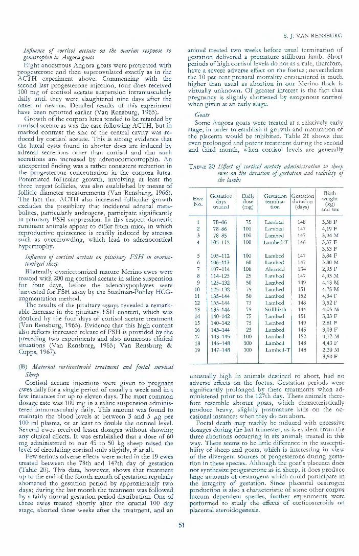
Influence of cortisol acetate on the ovarzan response to gonatrophin in Angora goats Eight anoestrous Angora goats were pretreated with
progesterone and then superovulated exactly as in the ACTH experiment above. Commencing with the second last progesterone injection, four does received 100 mg of cortisol acetate suspension intramuscularly daily until they were slaughtered nine days after the onset of oestrus. Detailed results of this experiment have been reported earlier (Van Rensburg, 1965).
G rowth of the corpora lutea tended to be retarded by cortisol acetate as was the case following ACTH, but in marked contrast the size of the central cavity was reduced by cortisol acetate. This is strong evidence that the luteal cysts found in aborter does are induced by adrenal secretions other than cortisol and that such secretions are increased by adrenocorticotrophin. An unexpected finding was a rather consistent reduction in the progesterone concentration in the corpora lutea. Potentiated follicular growth, involving at least the three largest follicles, was also established by means of follicle diameter measurements (Van Rensburg, 1966). The fact that ACTH also increased follicular growth excludes the possibility that incidental adrenal metabolites, particularly androgens, participate significantly in pituitary FSH suppression. In this respect domestic ruminant animals appear to differ from mice, in which reproductive quiescence is readily induced by stresses such as overcrowding, which lead to adrenocortical hypertrophy.
Influence of cortisol acetate on pituitary FSH in ovariectomized sheep Bilaterally ovariectomized mature Merino ewes were
treated with 200 mg cortisol acetate in saline suspension for four days, before the adenohypophyses were harvested for FSH assay by the Steelman-Pohley HCGaugmentation method.
The results of the pituitary assays revealed a remarkable increase in the pituitary FSH content, w hich was doubled by the four days of cortisol acetate treatment (Van Rensburg, 1965). Evidence that this high content also reflects increased release of FSH is provided by the preceding tw o experiments and also numerous clinical situations (Van Rensburg, 1965; Van Rensburg & Cupps, 1967).
(B) Maternal corticosteroid treatment and foetal survival Sheep Cortisol acetate injections were given to pregnant
ewes daily for a single period of usually a week and in a few instances for up to eleven days. The most common dosage rate w as 100 mg in a saline suspension administered intramuscularly daily. This amount was found to maintain the blood levels at between 3 and 5 11-g per 100 ml plasma, or at least to double the normal level. Several ewes received lesser dosages without showing any clinical effects. It was established that a dose of 60 mg administered to our 45 to 50 kg sheep raised the level of circulating cortisol only slightly, if at all.
Few serious adverse effects were noted in the 19 ewes treated between the 78th and 147th day of gestation (Table 20). This data, however, shows that treatment up to the end of the fourth month of gestation regularly shortened the gestation period by approximately two days; during the last month the treatment was followed by a fairly normal gestation period distribution. One of three ewes treated shortly after the crucial 100 day stage, aborted three weeks after the treatment, and an
51
S. J. VAN RENSBURG
animal treated two weeks before usual termination of gestation delivered a premature stillborn lamb. Short periods of high cortisol levels do not as a rule, therefore, have a severe adverse effect on the foetus; nevertheless the 10 per cent prenatal mortality encountered is much higher than usual as abortion in our Merino flock is virtually unknown . Of greater interest is the fact that pregnancy is slightly shortened by exogenous cortisol when given at an early stage.
Goats Some Angora goats were t reated at a relatively early
stage, in order to establish if g rowth and maturation of the placenta would be inhibited. Table 21 shows that even prolonged and poten t treatment during the second and third month, when cortisol levels are generally
T A BLE 20 Effect of cortisol acetate administration to sheep eu;es on the duration of gestation and viability of the lambs
Gestation D aily Gestation Gestation Birth Ewe weight No. days dose termina- duration (kg) treated (mg) tion (days) and sex
1 78- 86 75 Lambed 148 3,38 F 2 78- 86 100 Lambed 147 4,19 F 3 78- 85 100 Lambed 147 3,94M 4 105-112 100 Lambed-T 146 3,37 F
3,53 F 5 105-112 100 Lambed 147 3,84 F 6 106-113 60 Lambed 147 3,80 M 7 107-114 100 Aborted 134 2,95 F 8 114-125 25 Lambed 147 4,03 M 9 125-132 50 Lambed 149 4,13 M
10 125- 132 75 Lambed 151 4,78 M 11 135-144 50 Lambed 152 4,34 F 12 135-144 75 Lambed 148 3,52 F 13 135-144 75 Stillbirth 144 4,05 M 14 140-142 75 Lambed 151 3,33 F 15 140-142 75 Lambed 149 2,81 F 16 143-144 25 Lambed 145 3,03 F 17 143-145 100 Lambed 152 4,72 M 18 146- 148 100 Lambed 148 4,43 F 19 147-148 100 Lambed-T 148 2,30 M
3,50 F
unusually high in animals destined to abort, had no adverse effects on the foetus. Gestation periods were significantly prolonged by these treatments when administered prior to the 127th day. These animals therefore resemble aborter goats, which characteristically produce heavy, slightly postmature kids on the occasional instances when they do not abort.
Foetal death may readily be induced with excessive dosages during the last trimester, as is evident from the three abortions occurring in six animals treated in this way. There seems to be little difference in the susceptibility of sheep and goats, which is interesting in view of the divergent sources of progesterone during gestation in these species. Although the goat's placenta does not synthesize progesterone as in sheep, it does produce large amounts of oestrogens which could participate in the integrity of gestation. Since placental oestrogen production is also a characteristic of some other corpus luteum dependent species, further experiments were performed to study the effects of corticosteroids on placental steroidogenesis.

REPRODUCTIVE PHYSIOLOGY AND ENDOCRINOLOGY OF ANGORA GOATS
TABLE 21 Effect of glucocorticosteroid administration to goats on the duration of gestation and viabiliry of the kids
Gestation Steroid** and Gestation Gestation Birth weight Doe No. and breed* days duration treated daily dose (mg) termination (days) (kg) and sex
1-A 40-73 M-S0/4 days Kidded 154 2,9M 2-A 4S-79 M-S0/4 days Kidded 1SO 2,3 F 3-A S1-84 M-S0/4 days Kidded 1S4 3,0 F 4-A 47-80 P-S0/4 days Kid died 149 1,8 F S-B S0-61 C-1SO daily Kidded 1SS 3,4 M 6-B 78-85 C-100 daily Kidded T 1S2 2,6 F
2,9 F 7-B 10S- 112 C- 100 daily Kidded T 148 4,1 M
3,0 F 8-B 111 M-240 Aborted 117 0,3 M 9-B 113-120 C-100 daily Aborted 12S -
10-B 120-127 C-100 daily Kidded T 1S4 2,4 F 2,1 F
11-B 12S M-270 Aborted T 131 2,2M 1,9 F
12- B 136-149 C-100 daily Kidded T 149 3,3 M 3,1 F
13-B 137-144 C-7S daily Kidded 148 2,6 F
*A= Angora; B = Boergoat **M = Methylprednisolone acetate; P = Prednisolone; C = Cortisol acetate
TABLE 22 Effect of injection of glucocorticosteroids into sheep foetuses on their survival and the duration of gestation
Gestation Ewe No. day Dose (mg) and steroid
treated
1 77 3 betamethasone 2 77 10 cortisol a c. 3 77 20 cortisol ac. 4 78 10 cortisol ac. s 79 25 cortisol ac. 6 105 2,S methylprednisolone ac. each twin .
7 105 5 methylprednisolone ac. each twin
8 10S S methylprednisolone ac. 9 10S 10 methylprednisolone ac.
10 10S 10 methylprednisolone ac. 11 10S 40 methylprednisolone ac. 12 107 S cortisol ac. One twin
13 107 10 cortisol ac. 14 107 30 cortisol ac. 1S 120 2S cortisol ac. 16 122 20 cortisol ac. 17 120 SO cortisol ac. 18 120 SO cortisol ac. 19 122 60 cortisol ac. 20 120 7 S cortisol ac. 21 136 S cortisol ac. 22 141 7 S cortisol ac.
One twin . 23 143 2S cortisol ac.
NB. ac. = acetate
(C) Effect of foetal corticosteroid administration on gestation Sheep
A total of 23 ewes was laparotomized and corticosteroids were injected through the maternal uterine wall directly into the gluteal muscles of the foetus. Regular and dose-dependent effects were observed at all of the three principal stages of gestation (77, 105 and 120 days; Table 22) which were studied.
Small doses of corticosteroid had no apparent effect, but larger single doses prolonged gestation for periods of up to one week beyond the normal mean of 149 days established for our flock. Prolongation of gestation by corticosteroids was possible during various times in the third and fourth month of gestation, but could not be induced from the 120th day. Postmature foetuses were
52
Gestation Gestation Birth
termination duration weight (kg) and sex
Dystocia 1SO 4,3M Lambed 1S4 S,7 M Dystocia 154 4,S F Lambed 1S2 4,0 M Aborted 86 0,3 M Lambed 1SO 3,6 F
4,4M Lambs 1S6 2,8 F Died 3,0 F Aborted 111 1,2 F Aborted 109 1,1M Aborted 107 1,0 F Aborted 107 0,9 F Lambed 1S1 3,2 F
3,1 F Dystocia 1S4 4,8 M Lambed 1SS 3,8 F Lambed 147 3,S F Lambed 146 2,7 M Lambed 149 3,8 F Aborted 123 1,7 M Aborted 131 1,9M Aborted 123 1,8M Lambed 147 3,6 F Lambed 143 2,4 F
2,2 F Lambed 147 3,S F
abnormally large and the incidence of dystocia, which appeared to be caused by mal presentations in two of the three instances observed, was high. '
The administration of only slightly more corticoSteroid than was found necessary to prolong gestation, induced abortion. Gestation terminated · without difficulty within two to seven days of the operation and no complications such as placental retention or infections were noted. Although pregnancy could not be prolonged during the last month, adequat~ doses readily terminated gestation prematurely and the lambs were invariably born alive, but only those born after a gestation period of 140 days survived. The two lambs which were aborted on the 123rd day only lived for several hours, but that of ewe No. 19 which was born 18 days

S. J. VAN RENSBURG
TABLE 23 Effect of i1!jection of glucocorticosteroids into Boergoat foetuses on their survival and the duration of gestation
Doe No. Gestation Dose (mg) and steroid day treated
1 80 10 cortisol a c. each twin .
2 105 20 cortisol ac. 3 105 20 cortisol ac. 4 111 10 cortisol a c.
each twin . 5 129 20 cortisol ac.
each twin . 6 137 75 cortisol ac.
each twin . 7 139 100 cortisol a c.
one twin 8 140 40 methylprednisolone ac.
one twin
NB. ac. = acetate
prematurely survived for 50 hours; the only post mortem lesion found being thymic atrophy.
Methylprednisolone acetate* was also used, because of its extremely low mineralo-corticoid activity in comparison with cortisol acetate and its prolonged action. The results suggest that it is approximately six times more potent than co rtisol acetate as far as prolongation of gestation and abortifacient activities are concerned. This fact precludes the possibility that the above actions are due to electrolyte changes, and possibly favours the incrimination of immunological phenomena in gestation termination.
Goats Eight pregnant Boergoats were available for this
experiment, but the limited data obtained (Table 23) confirmed all the important findi ngs concluded from the sheep experiment. Spontaneous abortion is virtually un known in the hardy type of goat used, yet they were considerably more sensitive to the adverse effects of administered corticosteroid. The one gestation exhibiting prolongation was longer than any recorded in sheep, and lack of viability in treated nearterm foetuses was particularly evident. The injection-abortion intervals also tended to be longer and one of the twins from doe No. 4 showed the typical anasarca and hepatic subserosa! haemorrhages noted in several spontaneous abortions amongst A ngoras.
(D ) Effect of methylprednisolone acetate on ovine placental steroidogenesis
Of particular interest in this study was the comparison of the route of administration of the corticosteroid, i.e., maternal or foetal, intramuscular injection. Oestrogen biosynthesis by the goat placenta was of pertinent interest, but as suitable goats were not available, the experimental model chosen was the ovariectomized pregnant Merino ewe, and the parameter measured was peripheral blood levels of progesterone. The adrenal progesterone source is a distinct weakness in this system, but in sheep appears to contribute only slightly to peripheral blood levels (Linzell & Heap, 1968).
The first control blood sample was draw n on the 120th day of gestation, which was five to seven days after ovariectomy. Prior to treatment two or three days later, a second blood sample was withdrawn for progesterone assay. Two controls bearing single foetuses were laparotomized on the 123rd day of gestation and the foetus was injected with saline. In one, the progesterone level rose transiently by 0,2 fLg per 100 ml, but in
*Depomedrol, Upjohn
53
Gestation Gestation Birth weight termination duration (j<g) and sex
Aborted 91 0,3 M 0,3 M
Kidded 158 3,6 M Aborted 115 1,2 F Aborted 135 2,0M
1,2M Kidded-1 146 2,1 F stillborn 1,9 F Kidded 140 2,2 F
2,3 M Kidded-2 142 2,1 F died 2,3M Kidded-1 142 2,6 M died 2,3 F
the second control the procedure did not cause any significant fluctuations (Fig. 24).
Ewes No. 3 and 4 were not laparotomized but both were injected with hormone at the rate of 6 mg/kg body weight. Both exhibited a transient rise in progesterone levels 24 hours later, similar to the one laparotomized control, but thereafter a progressive decline in blood levels lasting for four days occurred. The progesterone levels were, however, never extremely low and no adverse effects were observed on the foetus. Part of the slow decline in progesterone levels could have been due to suppression of the adrenal source.
If the weights of the foetus and placenta are taken into account, then a dosage rate of 15 mg to the foetus is similar to that administered to the above ewes. After this dose was injected into the single foetuses carried by Ewes No. 5 and 6 a rapid decline to levels below 0,1 fLg per 100 ml plasma was followed by abortion in both instances (Fig. 25). The higher dose rate used for both twins of ewe No. 7 appeared to induce a similar precipitous drop in the circulating progesterone level. When only one twin was injected as in ewes No. 8 and 9, the levels only declined moderately, yet one ewe aborted.
Treated lambs rapidly removed at slaughter were considerably weaker than the controls. The placentomes of ewe No. 7, particularly, were heavier than usual and her cervix was double the usual weight, admitting three fingers. Histologically, the foetal trophoblastic epithelium showed vacuolar degeneration, the giant cells were smaller and fewer cells appeared binucleate. Their PAS positive cytoplasmic granules stained paler with this method, o r appeared to be condensed excentrically on one side of the cell, forming a dark relatively homogeneous mass.
(E) Foetal adrenalectomy The surgical technique previously described (Van
Rensburg, 1967) was used and a further two "controls" in the form of unilateral foetal adrenalectomies were performed on sheep which were 78 and 110 days pregnant. Both ewes gave birth to normal healthy lambs on the 148th and 146th days which weighed 3,2 and 4,0 kg, respectively. Two goats were also subjected to "control" operations, one adrenal was removed from a foetus of a single gestation on the 125th day and a normal 2,4 kg kid was delivered on the 149th day. One of twins was bilaterally adrenalectomized on the 111th day and both were stillborn on the 150th day.
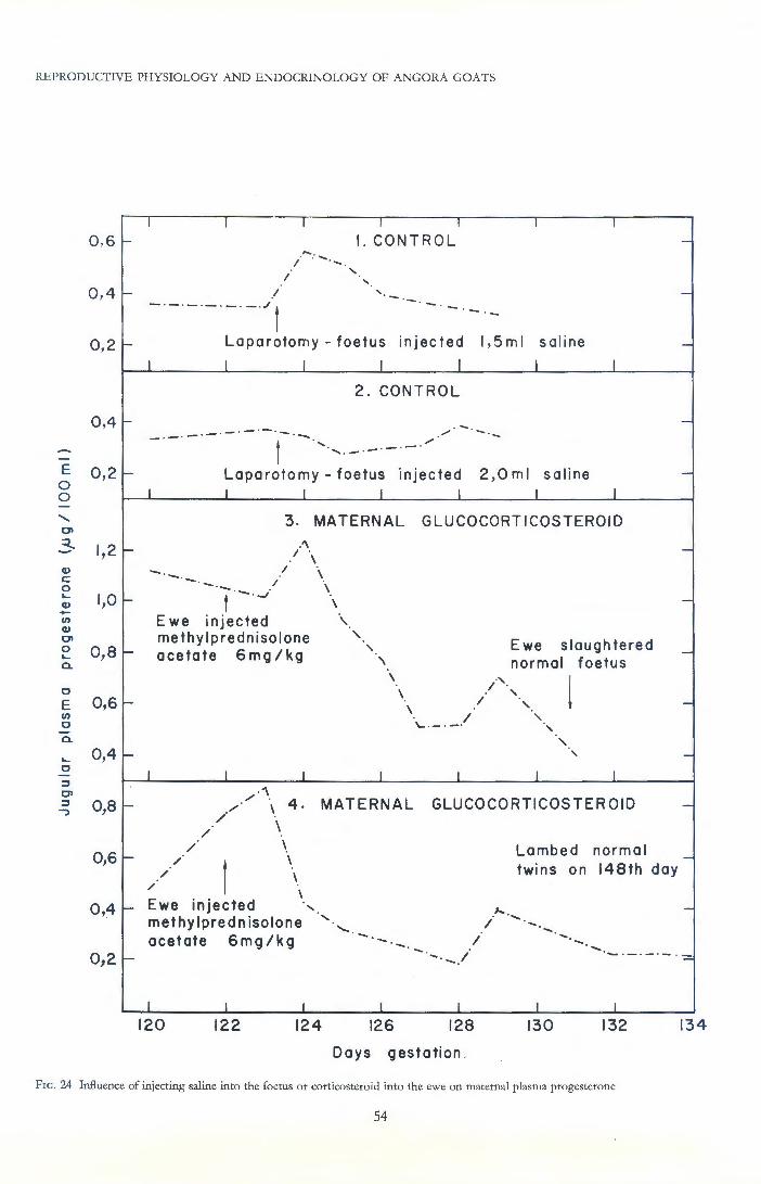
REPROD UCTIVE PHYSIOLOGY AND ENDOCRINOLOGY OF ANGORA GOAT S
E 0 0
Q)
c: 0 ~
Q) -(/) Q)
01 0 ~
a.
0
E (/)
0
a.
0 ,6 ~
0 , 4 f-
0 , 2 f-
0,4~
0,2 f-
1,2 f-
1,0 t-
0,8 t-
0,6-
0,4-
0,8-
0,6-
I I I I I I
I. CONTROL
I ' - ·- - . - ·-·-Laparotomy- foetus injected 1,5ml sal ine
I I I I I I I
2 . CONTROL
- · - · -·- · -·-·- · - ·~ / - ~-- - ~
l ..... ·-·- ·"' I
' ~ - · -
Laparotomy- foetus injected I I I I
2 ,0ml saline I I
3. MATERNAL GLUCOCORTICOSTEROJD !\
I \
- · - · - . I \ - · I \ T-·-,. '
Ewe injected \ methylprednisolone '
' acetate 6mg/kg '\
I I I I
Ewe slaughtere d normal foetus
\ ., \ / ·,
/ ' \ I '-·- ·-·
I
'
I
' ' ' I
-
-
-
-
-
-
-
-
-
-
,., ......-·\ 4. MATERNAL GLUCOCORTICOSTEROID -
/
/ /
/ /
/ \
\ \ \
Lambed norma l _ twins on 148th day
\ 0 4 - Ewe inJ· ected ' ' .J.. ...... -
' methylpred n isotone I ........
0,2-acetate 6mg/kg -. I
- ·- ·- .1 .... ..... . - ·- ·-· - ·=
I I I I I I I
120 122 124 126 128 130 132 134
Days gestation ~
FrG. 24 Influence of injecting saline into the foetus or corticosteroid into the ewe on maternal plasma progesterone
54

E 0 0
(l)
c 0 ...... (l) -VI (l)
0> 0 ...... a.
c E VI c a.
..... c ::J 0> ::J
--_,
S. J. VAN RENSBURG
0,3 1--
I I I 5
I I
-
0 , I 1--
- -·-·-·-·- -·- ·-·-·r·-· -·,, Foetus injected 15mg ·,. methylprednisolone a c. ',
Aborted live foetus
-I I I I I
6 0,6 1--
...... . ...... -
0,4 1---- -- ·-·l..... '-,
- ·- - ·-. -· F~etus injected 15mg' methylprednisolone ac. \ Aborted live foetus
-
0 ,2 1- = 6mg /kg conceptus '
\'·,.1 -
I I I . .., I
1,0 1-7 / .................. ·1·- -·-.,
/ I ' -
Both foetal twins\ 0,8 1- injected 30 mg ' -
meth yIp red n isolone
0,6 1- / / ac. = 12mg /kg ' conceptus \ Abortion imminent -
0 ,4 1-
0,2 1-
I
0,6-
0,4-
0,2-
I
0,4 1-
0,2 1-
I
120
\ ewe slaughtered \ \foetuses weak -
\ l I -
I I I I
-·-· -.-.-.-.-.-.-.-. - t-. ' ' ' 8 -One twin injected '
' 30mg methylprednisolone ', -ac. '...._
t Aborted -
I l I I
9 Ewe slaughtered
..... one foetus weak -.... ..-- ......-One twin injected '...._ ~,, f
30mg methylprednisolone ' ...._ ~ a c.
I
122 I
124
Days gestation
--- / . ........ ...., · I
126
-I
128
FIG. 25 Influence of injecting corticosteroid into the foetus on maternal plasma progesterone
55

REPRODUCTIVE PHYSIOLOGY AND ENDOCRINOLOGY OF ANGORA GOATS
MEAN CONTROL GESTATION
progesterone Foetus
GOATS - dvarian progest"rone
120 140 160
Days gestation
FrG. 26 The duration of gestation after bilateral foetal adrenalectomy
Complications which were encountered in the technique were, firstly, death of the foetus from haemorrhage when the right adrenal is dissected from the vena cava; this problem could no doubt be overcome by utilizing electrocoagulation. Secondly, infection within the amniotic cavity occurred readily. Several hundred operations had been performed in our clinic without a single septic incident, yet the first two ewes on which foetal surgery was attempted rapidly succcumbed to septic metritis, notwithstanding all normal aseptic surgical techniques. Instilling antibiotics into the foetal wound and treating the ewe, were also not entirely successful in controlling infection, but all instances which had a small amount of penicillin instilled into the amniotic cavity, survived. We were forced to the conclusion that amniotic fluid is an excellent medium for the multiplication of airborne bacteria (our clinic is surrounded by animal facilities) against which it has no natural defensive mechanism.
Results of successful bilateral adrenalectomies in sheep and goats are compared in Fig. 26. All six gestations were single, every foetus exhibited obvious further growth after the operation and their wounds were quite healed at the termination of gestation.
All the sheep pregnancies exhibited undoubted signs of prolonged gestation; however, for this to be virtually indefinite, the foetal adrenal glands had to be removed prior to the attainment of foetal viability. The time of viability coincides with the onset of elevated foetal plasma cortisol levels. Delivery of the lambs, which all appeared to have succumbed during birth, was accomplished with considerable difficulty by the ewes and
56
was spontaneous in all except one which was subjected to caesarean section on the 200th day. The lamb had recently died and appeared slightly dehydrated, yet it was at least double the normal birth weight (7,20 kg). During the 50 days of prolonged gestation the ewe remained perfectly normal, but abdominal distension was progressive and she gained 5 kg during the last 10 days. Slight discomfort was apparent immediately prior to the caesarean, not unlike a ewe at termination of a normal multiple gestation. A week after the caesarean she weighed 9 kg less than at gestation termination.
Cortisol blood levels were generally relatively low during the prolonged period (0,8 J-Lg /100 ml plasma) and the unusually low value of 0,38 J-Lg /100 ml plasma was recorded on the 190th day. A week after delivery the more usual value of 1,2 f-Lg was found.
The second longest overdue ewe commenced delivery spontaneously on the 189th day; delivery of the normally presenting 6.1 kg foetus was only accomplished with assisted traction.
The lambs of the two gestations terminating on the 154th day (5 days overdue) weighed 4,9 and 4,3 kg. The lightest of these two lambs had been adrenalectomized three days earlier than the heavier lamb. Normal lambs are capable of doubling their birth weight during the first two weeks of life and therefore growth was severely retarded during the advanced postmature period. This retardation of growth is illustrated by comparison of the 200 day foetus with two normal lambs below; one was selected because it weighed the same as the postmature foetus and the other was the same post-conception age:

Normal Adrenalectomized Normal
Age (days) 12 50 postmature so Weight (kg) 7,2 7,2 13,2 Liver (g) 177 131 240 Kidneys (g) . 46 60 54 Spleen (g) 30 9 38 Thymus (g) . 24,8 7,1 0,8 Adrenals (mg) 797 Absent 794 Thyroids (mg) 595 670 873 Testes (g) 2,4 2,1 5,4 Adenohypophysis (mg) 132 220 175 Neurohypophysis (mg) 137 535 243
These data suggest that somatic growth generally was depressed, and also particularly the liver and spleen. Both the anterior and posterior pituitaries were markedly hypertrophied in the postmature lamb, and its kidneys were also unduly heavy.
The two adrenalectomized goat foetuses were delivered spontaneously slightly prematurely; their wounds had also healed perfectly and their body weights were similar to normal newborn kids.
These observations suggest that in some species where placental progesterone is produced, the resulting pregnancy block is normally overcome by foetal adrenal secretions. Injection of corticosteroids into goats, which do not produce placental progesterone, also results in birth of the foetus. Hysterectomy of females of several species is known to result in prolongation of the lifespan of the corpus luteum to a period approximately equal to or slightly shorter than that of gestation; the luteolytic mechanism operative after such prolongation may be identical to that causing termination of gestation in our goat foetal adrenalectomy experiments. These considerations do not support the notion that the foetal adrenal secretions act by terminating placental progesterone synthesis, but rather that a dominant mechanism leading to birth is initiated following strictly quantitative exposure to adrenal glucocorticosteroids. This same mechanism could also be initiated by the disappearance of corpus luteum progesterone in those species dependent on this source.
(F) Influence of insulin administration on the course of gestation
Morgenthal (1966) demonstrated that blood glucose levels of potential aborter goats were considerably elevated throughout successful gestation periods exhibited by such animals. This finding was one of several parameters measured which indicated elevated adrenocortical function. This work has shown that in those which abort the level of adrenal function invariably reverts from hyperfunction to hypofunction prior to abortion. Therefore, maternal hypoglycaemia was experimentally induced in pregnant Boer goats; blood glucose was not determined as typical symptoms of insulin shock, such as trembling and muscular twitching were exhibited virtually daily after the second injection.
Two goats were treated with 2,5 i.u. protamine zinc insulin ('Wellcome') per kg .daily for eight days. The first was treated from the 89th to the 97th day of gestation; a large normal kid was born after a gestation period of 157 days. The second doe was treated from the 109th to 116th days of gestation. She kidded small twins on the 151st day; one was stillborn and the other suffered from posterior paresis. It therefore seems that although excess insulin may induce developmental disturbances in the foetus, it was not abortifacient under the circumstances used.
57
S. J. VAN RENSBURG
CHAPTER 10
DISCUSSION
The evidence accumulated in this study shows that the immature animal destined to perpetuate the abortion defect is outstanding in several respects. Newborn kids tend to be heavier and have finer birthcoats. The quantity of valuable kids' mohair produced during the first 18 months is exceptional. Young males produce an average of 30 per cent more mohair than usual, which is a quantity easily assessed by subjective means; furthermore this excellent production rate is maintained to at least 3 to 5 years of age. The "aborter" maiden does in the experimental flock produced only 10 per cent more than normal controls, but the former also tended to show oestrus, conceive and breed better during the first breeding season. Thus the conscientious breeder who is anxious to increase the production potential of his stock will inadvertently select for the abortion defect, regardless of the time when he selects his breeding stock.
Selection for outstanding young mature males, whose mothers in the experimental flock were generally aborters, may be the main reason why the abortion tendency is not more self-limiting. Limited studies on such bucks revealed that their adrenal glands were smaller than normal, similarly maiden does destined to abort later tended to have low blood cortisol levels.
The potent inhibitory effects of glucocorticoids on the rate of hair growth has been discussed; of particular interest is the evidence in rats that the depression of hair growth during malnutrition is mediated via the adrenal gland. If this mechanism is applicable to goats, then the evidence suggests that artificial selection has in reality been for a low level of adrenal function to remove inhibitory effects on hair growth, thereby creating a metabolic priority for the latter. Differences in production rate of mohair may be expected to be more marked under natural free-range farming conditions due to the seasonal variations in availability of adequate feed. The low level of adrenal function may explain the lack of resistance of the breed to inclement weather when calorific demands are suddenly increased, such as is caused by simultaneous rain and cold.
If the feed conversion rate remains relatively unchanged, animals having the abortion tendency may be expected to be smaller. The results recorded seem to suggest that such a slight tendency exists; however, in the U.S.A. abortion has been noted to be almost exclusively confined to the smaller types, and furthermore has greatly increased since criteria for selection have largely been based on hair characteristics (Shelton, 1970, personal communication). The usual American type of Angora goat is considerably larger than the South African type, produces less hair and rarely aborts.
Abortion is seldom observed during the first gestation period but the incidence in our experimental flock increased linearly up to 70 per cent during the fifth gestation studied. The majority of does will commence to abort during their fourth or fifth year of life, approximately at the time when the doe reaches physical maturity and weight gains cease. Factors causing growth in young vigorous goats may confer some protection to the developing foetus and placentome; certainly senility and senescence are accompanied by a tremendous increase in the abortion incidence.
Changes associated with the first abortion have not been particularly studied due to the difficulties in predicting such an event. Some of our "normal" animals,

REPRODUCTIVE PHYSIOLOGY AND ENDOCRINOLOGY OF ANGORA GOATS
which were intended to be controls, aborted for the first time while under study and they did not display the typical high level of adrenal function and low oestrogen excretion of known aborter individuals but had fairly normal cortisol levels and somewhat excessive oestrogen excretion rates. Abortion itself or the possible stresses preceding and associated with this event may induce various metabolic changes of lasting nature in the doe and as far as the pathogenesis is concerned, it is important to distinguish "established" aborters, those aborting for the first time and those destined to abort but which have never done so.
Several physiological differences were apparent in known aborters which were not pregnant. The hair growth rate was less than usual and of a finer character. Animals destined to become aborters were intermediate between normals and aborters as far as hair characteristics were concerned. Anoestrous and cycling aborters had increased levels of plasma cortisol which were consistently raised higher than in normal animals by ACTH. Aborters responded erratically to ACTH as far as the secretion of adrenal progesterone and pregnanediol-like compounds were concerned, these frequently being reduced in quantity. Dissection studies revealed pituitary hypertrophy, involving both the neurohypophysis and adenohypophysis, in conjunction with consistent adrenocortical hypertrophy. On a weight basis, the enlarged adrenal cortices of aborters synthesized cortisol with increased efficiency, assuming that the half-life of the hormone is equal in normal and aborter animals.
It thus seems possible that the outstanding young animal becomes metabolically embarrassed by its exceptional hair production rate and therefore an adaptive endocrine mechanism develops to inhibit hair growth and alter the system of metabolic priorities in favour of more essential physiological functions. Further evidence in support of this interpretation will emerge below.
In all species studied, cortisol inhibits the rate of hair growth and the fibre diameter and in the experimental goats the functional state of the adrenal cortex was always correlated with hair growth characteristics. Unpublished observations on the hardy short-coated Boergoat breed suggest that their adrenal gland size and plasma cortisol levels more closely resemble that of aborter Angora goats. This observation suggests that, as a breed, the Angora has a relatively low level of adrenal function, except for established aborters which have adapted to a more physiological level to the detriment of hair growth. This interpretation would imply that the aborter also has other mechanisms establishing a metabolic priority for production characteristics apart from an unusual low level of adrenal function.
There is no suggestion of a gonadotrophic deficiency in aborter does since they exhibit normal seasonal oestrus activity and a normal oestrus period. The majority of cycling aborters, however, exhibit a shorter luteal phase period, indicating premature luteal regression. Luteinization after ovulation, though complete by the 15th day, appears to be slower and virtually all corpora lutea of aborters are cystic. Excessive follicles were present in the ovaries of cycling aborters and the oestrogen excretion during very early gestation tended to be high. When an amount of ACTH which was adequate to induce a similar degree of adrenocorti-
*Dept. of Physiology, Faculty of Animal Science, University of Stellenbosch
58
cal hypertrophy to that seen in aborters, was administered to normal animals, retarded luteinization, luteal cysts and excessive ovarian follicular growth identical to that observed in aborters were reproduced. Cortisol alone retarded luteinization and reduced luteal progesterone concentration, but also reduced the size of the luteal cavities. It must be concluded that adrenal secretions other than cortisol, as has been demonstrated with dehydroepiandrosterone in cows (Rahlmann & Cupps, 1962) are responsible for the development of cystic corpora lutea. A possible mechanism is the inhibition of pituitary LH or other luteotrophic factors by sex steroids incidentally secreted from the hypertrophied adrenal glands . Mnch evidence has been accumulated which indicates that cortisol is capable of greatly increasing FSH synthesis and release in ruminant animals ; the resulting follicular growth and oestrogen secretion may contribute to the retarded luteinization and premature luteal regression by virtue of sex steroid influences on pituitary gonadotrophic function. Unpublished work done in collaboration with C.H. van Niekerk* has shown that acute and subacute malnutrition during the first month of gestation in Angora goats and in Merino sheep will result in high embryonic mortality. Luteal progesterone content in malnourished animals tended to be higher than normal but no significant differences were found . It therefore seems that all the aberrations of ovarian function detected in cycling aborters may merely be incidental side-effects induced by increased adrenocortical secretions.
The conception rate of aborters was approximately 20 per cent lower than usual. These failures could almost exclusively be accounted for by animals returning to oestrus after abnormally short cycle lengths. Adrenal hyperplasia may indirectly be responsible for creating unfavourable endocrine conditions for conception as discussed above, thus creating an adaptive mechanism to protect the mother against the nutritional demands of gestation when the metabolism is predominantly catabolic.
In agreement with Van Heerden's (1963) observations, the major abortion peak occurred soon after the 1 OOth day of gestation. This stage coincides with the time of cessation of placental growth and the time of greatest increase in the rate of foetal growth. During this work a minor abortion peak during the second month of gestation was evident. Van Heerden suggested that abortion at this time may be more common than realized. Breeding records of the early aborters suggested that they did not always display the typical characteristics of established aborter does, such as short oestrous cycles and fine hair growth. Their outstanding feature was an exceedingly high rate of hair g rowth, from which it may be inferred that a low level of adrenal function exists. The same animals may later maintain a gestation into the fourth month but during such a season the hair growth rate will be reduced. These data also suggest that a higher level of adrenal function is associated with more successful maintenance of gestation.
Studies on luteal function indicated that ovarian progesterone production declines steadily to reach minimum values around the 40th day of gestation. The resulting lowered progesterone: oestrogen ratio may well contribute to early gestational failure. Oestrous behaviour in pregnant aborters occurred twice as frequently than normal and was commonly exhibited a

short time after abortion. After the 40th day, all criteria used to assess luteal function indicated that it increased to reach maximum levels, which equalled or slightly exceeded luteal phase levels, at approximately the 100th day. Paradoxically, these high levels coincide with the major abortion peak. The exact levels of progesterone necessary to maintain gestation are uncertain, as is the concept of a quantitative or "all or none" biological action of the hormone. Peripheral plasma progesterone levels were higher than usual in most aborters up to at least the 70th day of gestation, thereafter some, but not all, tended to be rather low. The decrease was not obvious in the ovarian vein plasma values and corpus luteum studies, and the extent to which variations in the adrenal contribution were responsible must be considered. Reductions in the production of a possible placental lactogen-like hormone by a marasmic conceptus may also have contributed to lowered peripheral plasma progesterone levels observed prior to abortion. There is in either event no evidence that luteal function is impaired prior to the onset of retarded foetal growth and in fact dead in utero foetuses may be found with apparently normally functioning corpora lutea. The very long time that dead foetuses may be retained in utero illustrates the adequacy of this luteal function, since retention of the foetus in the absence of a fu nctional corpus luteum in a species where this organ is necessary for gestation maintenance, is contrary to the author's experience.
The time which elapses between death and expulsion of the foetus is generally proportional to its development and presumably to the ability of the foetal adrenal glands to secrete adequate glucocorticosteroids in order to initiate parturition as discussed in Chapter 9. This is a strictly dose-time related effect and very little foetal steroid stimulus may initiate the event, as is illustrated by foetal adrenalectomy around about the 140th day of gestation, which was apparently too late to interrupt the initiation mechanism 10 days later. The implication in the same experiment is that a foetus which may have been dead for several days could nevertheless have been responsible for initiating luteolysis and parturition. o clear evidence exists to suggest that foetuses which die before the 80th or 90th day are capable of terminating their own gestations and it is these that are retained in utero the longest. Adequate maternal corticosteroids could initiate expulsion of such dead foetuses and this mechanism would explain th.e "abortion storms" noted by Van Heerden (1963) following adverse environmental circumstances such as cold windy weather, shearing and dipping. This worker was also of the opinion that such stresses promoted the expulsion of already dead foetuses, rather than acting as primary causes of abortions.
Plasma cortisol levels were generally lower than normal during gestation and the majority of individuals showed a steady decline up to mid-gestation. With the increasing foetal g rowth rate after this time, all normal animals were found to have high levels which fell into the normal non-pregnant range or above it, when assayed on the 90th and 105th days of gestation. In all instances of abortion the values were exceedingly low on one or both of these intervals. During early gestation some aborters were relatively normal and others had unusually high and erratic cortisol levels. A study on pre-abortion changes revealed marked maternal adrenal atrophy in the presence of the very earliest signs of degenerative changes in the placentome, namely fragmentation of the reticulum network.
59
S. J. VAN RENSBURG
The association of abortion with the acute onset of adrenal insufficiency is consistent with the idea that the problem centres around metabolic competition since aborters whose cortisol levels remain high have successful gestations and deliver normal viable kids . The abortifacient effects of a protein-free diet in laboratory animals and the fact that high levels of sex steroids and glucocorticosteroids are necessary to protect the foetus against a nutri tional crisis have been discussed in Chapter 9. The Angora goat is presumed to be more sensitive to protein restriction because of the unusual demands of hair growth, yet many very thriving animals which are gaining weight still abort. Simple metabolic competition also does not explain the sudden onset of adrenal insufficiency which is temporary and is probably the vital factor in causing abortion by ceasing to assist the transfer of maternal protein to the developing foetus. The foetus either dies or it is adequately stressed to initiate the parturition process. This sequence of events seems to be a logical adaptive situation to protect the mother from further metabolic stress, yet in view of the usually high foetal nutritional priority it can only be explained as an overriding effect of a genetically altered system of metabolic priorities favouring production characteristics. The possibility that immunological phenomena are implicated in these quantitative metabolic interactions would be a somewhat novel concept.
The evidence that adrenal hypertrophy is essentially an adaptive adjustment favouring reproduction and essential physiological functions seems logical. One of the most puzzling features of the abortion syndrome was the fact that when aborters are transferred from one locality to another, they usually breed successfully during their first gestation and thereafter recommence aborting. It is logical to expect that the strange environment will stimulate further adrenal adaptive change, thus facilitating normal reproduction. The adrenal adjustment does appear to be accompanied by some loss of stable control of the hypophyseal-adrenal axis, since old established aborters have high and exceedingly variable cortisol levels, which occasionally progress to clinical hypercortisolism reminiscent of Cushing's disease. Early studies of these animals led to the hypothesis that foetal death was caused by excessive maternal corticosteroids (Van Rensburg, 1964). In the 14 animals studied in detail during the course of this work, one aborted and another kid was stillborn after extraordinarily high maternal cortisol levels, which were within the range that could be expected to be caused by the injection of sufficient cortisol to induce foetal death. Thus it seems likely that when the conceptus is exposed to a marginal nu trition and possibly marginal sex steroid environment, moderate amounts of cortisol are necessary to ensure success of the gestation. Inadequate corticosteroids will result in "starvation" of the foetus, while excessive amounts may block the trophic actions of the sex steroids at the placental level and initiate the parturition process.
Altered oestrogen excretion was the earliest anomaly which could be attributed to the conceptus destined to be aborted. Animals that had recently become aborters generally had low plasma cortisol levels and excessively high oestrogen excretion rates; whereas in established aborters the position was reversed. Known aborters which delivered full-term kids also had a tendency to low oestrogen excretion values and their blood cortisol values were maintained at high levels.

REPRODUCTIVE PHYSIOLOGY AND ENDOCRINOLOGY OF ANGORA GOATS
If it is assumed that placental oestrogen synthesis in goats is also dependent on the supply of precursor steroids from the maternal and foetal adrenal glands, then an attractive hypothesis which could explain the abnormal oestrogen levels would include the variation in the supply of precursors. The fact that these precursors are a limiting factor in human placental oestrogen synthesis may be demonstrated by the use of ACTH. If this concept is applicable to Angora goats it implies that when ACTH stimulation is at a low level then larger amounts of incidental steroids of the sex steroid type are secreted. However, under intense stimulation the biosynthetic pathway will be quantitatively altered in favour of glucocorticosteroid synthesis at the expense of sex steroid secretion.
Ample evidence is available that ACTH can reduce the adrenal secretion of pregnanediol-like compounds in Angora goats, which is contrary to what is usually thought to be the case in humans. In one unpublished study of cycling animals, little "pregnanediol" of luteal origin was detected but during the stress of oestrus the excretion rate in normal Boergoats was increased, whereas values were decreased at this time in Angora goats. Variations during gestation in the latter breed were invariably inversely proportional to the blood cortisol levels and sudden marked increases, coincidental with the onset of adrenal insufficiency were recorded a short while prior to abortion. Scanty evidence has also been presented which indicates that the secretion of adrenal progesterone tends to be reduced in aborters before and after ACTH injection. The fact that regular aborters excrete extremely small amounts of oestrogen suggests that the relative changes in the adrenal biosynthetic pathways are part of the adrenal adaptation process previously suggested, since such animals have a higher level of adrenal function. The extent of involvement of the foetal liver is uncertain in a species which excretes little or no oestriol, but the fact remains that some aborted foetuses exhibited hepatomegaly, whereas normal kids born from aborter does have small livers.
The main possible purpose of placental oestrogen biosynthesis is thought by some to be a trophic influence on placental function itself and a luteotrophic role has also been suggested. Some of our aborters exhibited excessively high oestrogen excretion rates and in others there was virtually no evidence of placental oestrogen synthesis, yet no differences in the time of abortion or condition of the foetuses were apparent -there was also little relationship between plasma progesterone concentration and oestrogen excretion values. Amniotic and allantoic fluid volumes in sheep do, however, seem to be quite sensitive to oestrogen, particularly in the presence of marginal progesterone (Alexander & Williams, 1968) and abnormalities in the amounts of these hormones may well have been responsible for the accumulation of excessive intrauterine fluids noted prior to abortion.
The possibility of the occurrence of "inborn errors of metabolism" such as specific enzyme disorders controlling protein synthesis, is enormous (Harris, 1970), yet no evidence for the existence of such specific defects has been detected in the Angora flock studied. This is in accordance with the labile nature of the problem.
A more acceptable genetic concept is that the abortion problem may be attributed to a continuously variable character that will not allow clear differentiation between one genotype and another. The current concept is that continuous variation is controlled by poly-
60
genic systems, that is, by complexes of genes, each component of which contributes in some small measure to the total sum of gene controlled variability. The different combinations of these polygenes allow a wide variety of genotypes, which have been shown to be responsible for considerable quantitative variation in adrenal structure and function (Spickett, Shire & Stewart, 1967). These workers state that certain physiological parameters such as the quantity of steroid produced may show a high degree of constancy but the genetic system underlying this similarity may be highly variable. This fact is of importance because it allows a population to carry the variability which will enable it to respond to selection, natural or otherwise. Thus environmental change will cause individuals which are apparently similar, to be revealed as different.
In referring to the mechanism of adaptation to low protein intakes, Waterlow (1968) stated that the only way to recognize the beginning of the abnormal is to understand the mechanisms of adaptation and in particular by identifying those properties or characteristics which the body tries to maintain constant at the expense of others. For instance, adaptive enzyme changes during malnutrition involve increased amino acid activation and the activity of the urea-cycle enzymes is decreased. Waterlow also stated that the cause of such enzyme changes are still speculative but there is much evidence that they are induced by cortisone. The adaptive lag period is shorter in children than in adults and for example, growth stasis of hair, presumably to conserve nitrogen, is readily seen in marasmic children (Bradfield, Cordano & Graham, 1969). In rats and probably most species, this adaptation is clearly mediated via the adrenal gland, removal of which will permit unrestricted growth during malnutrition. Alterations in the metabolic priority of hair growth induced by artificial selection coupled with defects in adrenal adaptive capabilities, which fail to maintain the developing foetus in a viable state, seem to be currently the most acceptable explanation for the Angora abortion syndrome. Metabolic conflicts may be expected to arise when selection for increased production and the necessary physiological adaptation that follows its consequences involves basically the same tissue.
AcKNOWLEDGEMENTS
The facilities for this work were generously provided by the Director of the Veterinary Research Institute, Onderstepoort. I am indebted to the South African Medical Research Council for the opportunity to complete the project.
Advice and guidance from authorities under whom I have been privileged to work, in particular Prof. S. W. ]. van Rensburg, Dr. R. V. Short and Prof. P. T. Cupps have been invaluable.
Technical assistance at various times was excellently provided by Messrs. F . ]. Schutte, B. F. Erasmus, W. Barwise and Miss R. Scheele. Mr. E. L. Barrett kindly performed atomic absorption spectrophotometry on numerous liver samples.
Prof. J. M. M. Brown, as promoter, has been exceedingly helpful.
REFERENCES
ADAMS, W. M., 1969. The elective induction of labour and parturition in cattle.Jl Am. vet. med. Ass., 154,261-265.
ALEXANDER, G ., 1956. Influence of nutrition upon duration of gestation in sheep. Nature, Lond., 178, 1058-1059.

ALEXANDER, D. P., BRITTON, H. G., JAMES, v. H. T., NIXON, D. A., PARKER, R. A., WINTOUR, E. M. & WRIGHT, R. D., 1968. Steroid secretion by the adrenal gland of foetal and neonatal sheep.]. Endocr., 40, 1-13.
ALEXANDER, G., & WILLIAMS, D., 1968. Hormonal control of amniotic and allantoic fluid volume in ovariectomized sheep. ] . Endocr., 41, 477-485.
ALLEN, W. M., 1950. A simple method for analyzing complicated adsorption curves, of use in the calorimetric determinations of urinary steroids.]. clin. Endocr., 10, 71-83.
AscHKENASY, P. & AscHKENASY, A., 1957. Des surrenales dans Ia gestation en fonction de Ia teneur du regime en proteines. Archs Sci. physiol., 11, 125-150.
BASSETT, J . M. & THORBURN, G. D., 1969. Foetal plasma corticosteroids and the initiation of parturition in sheep.]. Endocr., 44, 285-286.
BASSON, P. A., MoRGENTHAL, J. C., BILBROUGH, R. B., MARAIS, J. L., KRuGER, S. P. & VANDER MERWE, J. L. de B., 1969. "Grootlamsiekte", a specific syndrome of prolonged gestation in sheep caused by a shrub, Sa/sola tubercula/a (Fenyl ex Moq) Schinz var. tomentosa C. A. Smith ex Aellen. Onderstepoort ]. vet. Res., 36, 59-104.
BEN-DAVID, M., DIKSTEIN, S. & SuLMAN, F. G., 1964. Effect of different steroids on prolactin secretion in pituitary-hypothalamus organ co-culture. Proc. Soc. exp. Bioi. Med., 117, 511-513.
BINNS, W., ANDERSON, W. A. & SuLLIVAN, D. J., 1960. Further observations on a congenital cyclopian-type malformation in lambs. ]I Am. vet. med. Ass., 137, 515-521.
BINNS, W., SHUPE, J. L., KEELER, R. F. & JAMES, L. F., 1965. Chronologie evaluation of teratogenicity in sheep, Veratum californicum.jl Am. vet. med. Ass., 147, 839-842.
BRADFIELD, R. B., CoRDANO, A. & GRAHAM, G. G., 1969. Hairroot adaptation to marasmus in Andean Indian children. Lancet, ii, 1395-1397.
BROOKS, J. R., Ross, C. V. & TuRNER, C. W., 1964. Effect of thyroidectomy on reproductive performance of ewes and semen quality of rams.]. Anim. Sci., 23, 54-58.
BROWN, J. M. M., VAN RENSBURG, S. J. & GRAY, R., 1963. The urinary excretion of 5 8-pregnane-3cx: 20cx-diol and gestational failure in Angora goats. Onderstepoort]. vet. Res., 30,179-190.
BuSH, I. E., 1961. The chromatography of steroids. London: Pergamon Press.
CowiE, A. T., DANIEL, P. M., PRICHARD, M. M. L. & TINDALL, J. S., 1963. Hypophysectomy in pregnant goats, and section of the pituitary stalk in pregnant goats and sheep. ]. Endocr., 28, 93-102.
CowiE, A. T. & TINDALL, J. S., 1958. Adrenalectomy in the goat. Replacement therapy and the maintenance of lactation. ]. Endoctr., 16, 403-414.
CsAPO, A., 1969. The four direct regulatory factors of myometrical function. In: Progesterone: its regulatory effect on the myometrium. Ed. by G. E . W. Wolstenholme &J. Knight. London: J. & A. Churchill.
CuMMINGS, J. N., 1968. Foetal anaemia and hypovolemia in swine. Can. vet.]., 9, 61.
CuPPS, P. T., 1955. The ability of certain adrenal steroids to restore reproduction in adrenalectomized female rats. Endocrinology, 57, 1-4.
DAVIES, L. H., SouTHEY, I. N. & GooDALL, D. W., 1966. Effect of stocking rate and lambing time on gestation length in sheep. Nature, Lond., 211, 998-999.
DEANESLY, R., 1961. Foetal endocrinology. Br. med. Bull., 17, 91-95.
DEANESLY, R. & PERRY, J. S., 1969. Independent regression of normal and induced corpora lutea in hysterectomized guineapigs.]. Reprod. Pert., 20, 503-508.
DRosT, M. & HoLM, L. W., 1968. Prolonged gestation in ewes after foetal adrenalectomy.]. Endocr., 40, 293-296.
EvERITT, G. C., 1964. Maternal undernutrition and retarded foetal development in Merino sheep. Nature, Lond., 201, 1341.
FISHER, C. J. & LEATHEM, J. H., 1965. Effect of a protein-free diet on protein metabolism in the pregnant rat. Endocrinology 76, 454-462.
FLETCHER, W. S., RoGERS, A. L. & DoNALDSON, S. S., 1964. The use of the goat as an experimental animal. Lab. Anim. Care, 14, 65-90.
FRANCE, J. T., RIVERA, R., McNivEN, N. L. & DoRFMAN, R.I., 1965. Determination of androsterone, etiocholanolone and dehydro-epiandrosterone in urine by gas-liquid chromatography. Steroids, 5, 687-697.
FRANKLIN, R. R. & ALEXANDER, J. A., 1963. Endocrine disorders complicating pregnancy. Am. ]. Med. Sci., 245, 718-725.
GRAY, 0. P., AcKERMAN, A. & FRASER, A.]., 1968. Intracranial haemorrhage and clotting defects in low-birth-weight infants. Lancet, i, 545-548.
61
S. J. VAN RENSBURG
HAFEZ, E. s. E., MAUER, R. E. & ENSMINGER, M. E., 1958. Maternal environment and foetal development in the pig. Grmvth, 22, 269-289.
HALLIDAY, R. & BuTTLE, H. R. L., 1968. The effects of cortisone acetate on the length of gestation period and the survival of foetal Scottish Blackface lambs.]. Endocr., 41, 447-448.
HARRIS, H., 1970. Genetical theory and the "inborn errors of metabolism." Br. med.j., i, 321--327.
HARRISON, R. J ., 1948. The changes occurring in the ovary of the goat during the oestrous cycle and in early pregnancy. ]. Anal., 82, 21-48.
HAZELWOOD, R. L. & NELSON, M. M., 1965. Steroid maintenance of pregnancy in rats in the absence of dietary protein. E ndocrinolog)'_, 77, 999-1013.
HEAP, R. B. & LINZELL, J. L., 1966. Arterial concentration, ovarian secretion and mammary uptake of progesterone in goats during the reproductive cycle.]. Endocr., 36, 389-399.
HOLM, L. W., 1966. The gestation period of mammals. In: Comparative biology of reproduction in mammals. Ed. by I. W. Rowlands. London and New York: Academic Press.
HoLM, L. W., 1967. Prolonged pregnancy. Adv. vet. Sci., 11, 159-205.
HoLM, L. W., PARKER, H. R. & GALLIGAN, S. J., 1961. Adrenal insufficiency in postmature Holstein calves. Am. ]. Obstet. Gynec., 81, 1000-1008.
KASTIN, A. J., GENNSER, G. G., ARIMURA, A., MILLER, M. c. & ScHALLY, A. V., 1968. Melanocyte-stimulating and corticotrophic activities in human foetal pituitary glands. Acta endocr., Copenh., 58, 6-10.
KIRSCHNER, M. A. & LIPSETT, M. B., 1964. Quoted in: Gas chromatography in the analysis of steroid hormones; H. H. Wotiz & S. J. Clark, Plenum Press, New York, 1966.
KLOPPER, A., MITCHIE, E. A. & BROWN, J. B., 1955. A method for the determination of urinary pregnanediol. ]. Endocr., 12, 209-219.
KLYNE, W. & WRIGHT, A. A., 1957. Steroids and other lipids of pregnant goats urine. Biochem. ]., 66, 92-101.
KusT, D. & VoGT, V., 1934. Nachweis der sexualhormone bei der Zeige und ihre austwertungsmoglichkeit fUr die hormonale trachtigkeitsfestellung. Tierdrztl. Rdsch., 40, 589.
LIGGINS, G. C., 1968. Premature parturition after infusion of corticotrophin or cortisol into foetal lambs. ]. Endocr., 42, 323-329.
LIGGINS, G. C., 1969. The foetal role in the initiation of parturition in the ewe. In: Foetal autonomy. Ed. by G. E. W. Wolstenholme & M. O'Connor. London: J. & A. Churchill.
LIGGINS, G. C., HoLM, L. W. & KENNEDY, P. C., 1966. Prolonged pregnancy following surgical lesions of the foetal lamb pituitary. ]. Reprod. Pertil., 12, 419.
LINZELL, J. L. & HEAP, R. B., 1968. A comparison of progesterone metabolism in the pregnant sheep and goat; sources of production and an estimation of uptake by some target organs. ]. Endocr., 41, 433-438.
MALAN, A. P., CARTER, H . B. & VANWYK, C. M., 1938. T he uniformity of a series of fibre thickness measurements on a small sample of medium Merino wool. Onderstepoort ] . vet. Sci. Anim. Indust., 10, 445-466.
MARINCOWITZ, G., 1962. Geslagsaktiwiteit van Angorabokooie op gemengde Karooveld. S. Afr.J. agric. Sci., 5, 211-223.
MErTES, J., WEBSTER, H. D., YouNG, F. W., THORP, F. J. & HATCH, R. N., 1951. Effects of corpora lutea removal and replacement with progesterone on pregnancy in goats. ]. Anim. Sci., 10, 411-416.
MITCHELL, H. H., 1962. Comparative nutrition of man and domestic animals. London and New York: Academic Press.
MooR, R. M ., RowsoN, L. E. A., HAY, M. F. & CALDWELL, B. V., 1969. The effect of exogenous gonadotrophins on the conceptus and corpus luteum in pregnant sheep.]. Endocr., 44,495-499.
MoRGENTHAL, J. C., 1966. The haematology of the Angora goat with special reference to the habitual aborter. Onderstepoort]. vet. Res., 33, 363-378.
MoRGENTHAL, J. C. & BASSON, P. A., 1966. Sekere aspekte van die oortydsindroom in Karakoelskape in Suidwes-Afrika. Geneeskunde, 8, 93-94.
NICOLL, C. S. & MErTES, J., 1964. Prolactin secretion in vitro : Effects of gonadal and adrenal cortical steroidE. Proc. Soc. exp. Bioi. Med., 117, 579- 583.
OPPENHEIMER, E. H., 1969. Cyst formation in the outer adrenal cortex. Studies in the human fetus and newborn. Arch. Path., 87, 653-659.
OsBURN, B. I., STABENFELDT, F. H. & EwiNG, L. L., 1969. Relation of plasma progesterone to mid and late term bovine abortions due to Vibrio fetus infection. ]. Reprod. Pert., 20, 77-83.
PERON, F. G., 1962. Adrenocorticosteroids. In: Methods in hormone Research. Ed. by R. I. Dorfman, Academic Press, New York.

REPRODUCTIVE PHYSIOLOGY AND ENDOCRINOLOGY OF ANGORA GOATS
PHILLIPS, R. W., SIMMONS, V. L. & SCHOTT, R. G., 1943. Observations on the normal estrous cycle and breeding season in goats and possibilities of modification of the breeding season with gonadotrophic hormones. Am.]. vet. Res., 4, 360-367.
RAHLMANN, D. F. & CuPPS, P. T ., 1962. Effects of estradiol and dehydro-epiandrosterone on the reproductive cycle of the cow. ]. Dairy Sci., 45, 1011-1014.
RICHTER, W. R., 1958. Adrenal cortical adenomata in the goat. Am.]. vet. Res., 19, 895-901.
RoBIN, N . I., REFETOFF, V. F. & SELENKOW, H. A., 1969. Parameters of thyroid function in maternal and chord serum at term pregnancy.]. clin. Endocr. Metab., 29, 1276-1280.
SABA, H., 1965. Plasma cortisol levels in pregnant ewes. ]. agric. Sci., Camb., 64, 11- 14.
SCHINCKEL, P. G ., 1955. Inheritance of birthcoats in a strain of Merino sheep. Aust.j. agric. R es., 6, 595-607.
SERLUCA, P., CoNTI, M., CATTAENO, G. & BALLICU, P., 1964. Endocrine control of iron metabolism in pregnancy. Effect of the sex steroids on the transplacental passage of iron. Q uad. Clin. ostet. ginec., 19, 225-237. (Abs. in Excerpta med., 19, Section 10, 150.).
SHELTON, M., 1960. Influence of the presence of a male goat on the initiation of estrous cycling and ovulation of Angora does. ]. Anim. Sci., 19, 368- 375.
SHELTON, M., 1960a. The relation of face covering to fleece weight, body weight and kid production of Angora does. ]. Anim. Sci., 19, 302- 308.
SHELTON, M., 1961. Kidding behaviour of Angora goats. Progre.rs report No. 2189 ~( the Agricultural and Mechanical Co!!ege of Texas.
SHORT, R. V., 1957. Progesterone and related steroids in the blood of domestic animals. Ciba Fdn. Co!!oq. Endocr., 11, 362- 363.
SHORT, R . V. , 1960. Blood progesterone levels in relation to parturition.]. Reprod. Ferti!., 1, 61-70.
SHORT, R. V., 1960a. The secretion of sex hormones by the adrenal gland. Biochem. Soc. Symp., No. 18, 59-83.
SHORT, R. V., 1969. Implantation and the maternal recognition of pregnancy. In: Foetal autonomy, Ed. by G. E. W. Wolstenholme & M. O 'Connor; ]. & A. Churchill, London.
SHORT, R. V., 1969a. Discussions of papers in : Progesterone : its regulatory effect on the myometrium. Ed. by G. E. W. Wolstenholme & J. Knight. London: J. & A. Churchill.
SKINNER, J. D., JocHLE, W . & NEL, ]. W., 1970. Induction of parturition in Karakul and cross-bred ewes with flumethasone. Agroanimalia, 2, 99-100.
SPICKETT, S. G., SHIRE, J. G. M. & STEWART, J., 1967. Genetic variation in adrenal and renal structure and function. In: Endocrine genetics. Ed. S. G. Spickett. Cambridge: University Press.
SPIEGELBERG, 0., 1891. Die D auer der Geburt. Lehrbuch der Geburtshilfe, 2. (Quoted by F. H. A. Marshall & ]. Chassar Moir in: Marshall's physiology of reproduction. Ed. A. S. Parkes, 3rd edition, 1952. London: Longmans, Green & Co.)
SPURLOCK, G. M. & CLEGG, M. T ., 1962. Effect of cortisone acetate on carcass composition and wool characteristics of weaned lambs. ]. Anim. Sci., 21, 494- 500.
TuRNBULL, A. C. & ANDERSON, A. B. M., 1969. The influence of the foetus on myometrial contractility. In: Progesterone: its regulatory effect on the myometrium. Ed. by G. E. W. Wolstenholme & J . Knight. London: J. & A. Churchill.
VAN HEERDEN, K . M., 1961. Luteal failure as a cause of abortion in Angora goats in South Africa. Proc. IVth int. Congr. Anim. R eprod., The Hague, 586-589.
VAN HEERDEN, K. M., 1963. Investigations into the cause of abortions in Angora goats in South Africa. Onderstepoort ]. vet. Res., 30, 23-84.
VAN HEERDEN, K. M., 1964. The effect of culling aborting ewes, on the abortion rate in Angora ewes. ] 1 S. Afr. vet. med. Ass., 35, 19-20.
VAN RENSBURG, S. ]., 1963. Endocrinological aspects of habitually aborting Angora goat ewes. S. Afr. med. j. , 37, 1114-1115.
VAN RENSBURG, S. J ., 1964. Possible endocrine mechanisms in gestational failure. Proc. V. int. Congr. Anim. Reprod., Trento., 3, 375-378.
VAN RENSBTJRG, S. J., 1965. Adrenal function and fertility . .f! S. Afr. vet. med. Ass., 36, 491-500.
VAN RENSBURG, S. J. , 1967. Gestation in sheep after foetal adrenalectomy and cortisol acetate administration.]. Endocr., 38, 83-84.
VAN RENSBURG, S. ]., 1968. Die belang rikheid van die fctale byniere in die beeindiging van swangerskap. Geneeskunde, 10, 239.
VAN RENSBURG, S. J., 1968a. Endocrinological relationships between fertility and production. In: Die skaap en sy vag. Ed. ]. C. Swart. Cape Town: Nasionale Boekhandel Beperk.
VAN RENSBURG, S. J. & CuPPS, P. T., 1967. Alteration of reproductive function in the rat by cortisol acetate. Onderstepoort]. vet. R es., 34, 673-688.
VAN RENSBURG, S. J., McFARLANE, l. S. & VAN RENSBURG, S. W. J., 1963. Sterilization of teaser male ruminants - the reliability of surgical methods. ] 1 S. Afr. vet. med. Ass., 34, 249-253.
VAN RENSBURG, S. J. & VAN NIEKERK, C. H., 1968. Ovarian function, follicular oestradiol -17 B and luteal progesterone and 20ot-hydroxy-pregn-4-en-3-one in cycling and pregnant equines. Onderstepoort ]. vet. Res. , 35, 301-318.
VAN RENSBURG, S. W. J. & VAN RENSBURG, S. ]., 1961. Regression of the corpus luteum graviditatis in Angora ewes. Proc. I V th int. Congr. Anim. Reprod., The Hague, 597- 600.
VELARDO, J . T., 1957. Influence of purified adrenal hormones and adrenocorticotrophin on progesterone in decidual tissue formation. Am.]. P~ysiol., 190,408-412.
VELARDO,]. T . & STURGIS, S. H., 1956. Suppression of uterine growth by purified hydrocortisone acetate, 9ot-flurohydrocortisone acetate and corticotrophin. ]. clin. Endocr. Metab., 16, 496.
VINSON, G. P. & WHITEHOUSE, B. ]., 1967. The biosynthesis of corticosteroids by sheep and human adrenal tissue. Acta. mdocr., Copenh., Suppl. 119, 68.
VoGT, C. & SPECHT, F ., 1889. The natural history of animals -class Mammalia. London: Blackie & Son.
WATERLOW, J. C., 1968. Observations on the mechanism of adaptation to low protein intakes. L ancet, ii, 1091- 1097.
Printed in the Republic of South Africa for the Government Printer, Pretoria, by Heer Printing Co. (Pty) Ltd., Pretoria.
62