reproductive axis gene regulation during photostimulation and photorefractoriness … ·...
TRANSCRIPT

RESEARCH Open Access
Reproductive axis gene regulation duringphotostimulation and photorefractorinessin Yangzhou goose gandersHuanxi Zhu1, Zhe Chen1, Xibin Shao2, Jianning Yu1, Chuankun Wei1, Zichun Dai1 and Zhendan Shi1*
Abstract
Background: The Yangzhou goose is a long-day breeding bird that has been increasingly produced in China.Artificial lighting programs are used for controlling its reproductive activities. This study investigated the regulationsof photostimulation and photorefractoriness that govern the onset and cessation of the breeding period.
Results: Increasing the daily photoperiod from 8 to 12 h rapidly stimulated testis development and increasedplasma testosterone concentrations, with peak levels being reached 2 months after the photoperiod increase.Subsequently, testicular activities, testicular weight, spermatogenesis, and plasma testosterone concentrationsdeclined steadily and reached to the nadir at 5 months after the 12-hour photoperiod. Throughout the experiment,plasma concentrations of triiodothyronine and thyroxine changed in reciprocal fashions to that of testosterone. Thestimulation of reproductive activities caused by the increasing photoperiod was associated with increases ingonadotropin-releasing hormone (GnRH), but decreases in gonadotropin-inhibitory hormone (GnIH) and vasoactiveintestinal peptide (VIP) gene messenger RNA (mRNA) levels in the hypothalamus. In the pituitary gland, the levels offollicle-stimulating hormone (FSH) and luteinizing hormone (LH) mRNA abruptly increased during the longer 12-hourphotoperiod. The occurrence of photorefractoriness was associated with increased GnIH gene transcription by over250-fold, together with increased VIP mRNA levels in the hypothalamus, and then prolactin and thyroid-stimulatinghormone in the pituitary gland. FSH receptor, LH receptor, and StAR mRNA levels in the testis changed in waysparalleling those of testicular weight and testosterone concentrations.
Conclusions: The seasonal reproductive activities in Yangzhou geese were directly stimulated by a long photoperiodvia upregulation of GnRH gene transcription, downregulation of GnIH, VIP gene transcription, and stimulation ofgonadotrophin. Development of photorefractoriness was characterized by hyper-regulation of GnIH gene transcriptionin the hypothalamus, in addition of upregulation of VIP and TRH gene transcription, and that of their receptors, in thepituitary gland.
Keywords: Yangzhou goose ganders, Reproductive activities, Photoperiod, Gene mRNA expressions
BackgroundMost birds exhibit well-defined seasonal changes in go-nadal development, body mass, molting, metabolism, andother physiological parameters [1]. Such seasonal physio-logical changes are a means of coping with the seasonalfluctuations of environmental factors, such as temperatureand food availability, in order to improve survival capacity[2–7]. Seasonality of reproductive activity and that of
other physiologic processes in the majority of birds andmammals is presumed to arise from an interaction be-tween endogenous circannual rhythms and a variety ofenvironmental changes, the most important of which isthe daily photoperiod [8].The classic theory of photoperiodic regulation of sea-
sonal reproductive activities in birds proposes that lightsignals are perceived by photoreceptors in the deepbrain, and induce the secretion of thyroid-stimulatinghormone (TSH) from the pars tuberalis, which then actson ependymal cells to induce the thyroid hormone-activating enzyme type 2 deiodinase. This enzyme
* Correspondence: [email protected] of Animal Improvement and Reproduction, Institute of AnimalScience, Jiangsu Academy of Agricultural Sciences, Nanjing 210014, ChinaFull list of author information is available at the end of the article
© The Author(s). 2017 Open Access This article is distributed under the terms of the Creative Commons Attribution 4.0International License (http://creativecommons.org/licenses/by/4.0/), which permits unrestricted use, distribution, andreproduction in any medium, provided you give appropriate credit to the original author(s) and the source, provide a link tothe Creative Commons license, and indicate if changes were made. The Creative Commons Public Domain Dedication waiver(http://creativecommons.org/publicdomain/zero/1.0/) applies to the data made available in this article, unless otherwise stated.
Zhu et al. Frontiers in Zoology (2017) 14:11 DOI 10.1186/s12983-017-0200-6

catalyzes the conversion of thyroxine (T4) to triiodothyr-onine (T3) [9], which initiates the nervous impulses thatlead to the synthesis and release of gonadotropin-releasing hormone (GnRH) [10, 11]. GnRH is thentransported by portal blood circulation to the anteriorpituitary gland, where it stimulates the synthesis and re-lease of the gonadotropins, luteinizing hormone (LH)and follicle-stimulating hormone (FSH) [12–14]. LH andFSH are responsible for the stimulation of gonad growthand development, as well as for the production of sexsteroid hormones [15–18]. The photoperiodic regulationof the annual reproductive cycle requires two kinds ofphysiologic responses, namely photostimulation andphotorefractoriness. The former leads to the activationof the reproductive system and brings animals into thebreeding season, and the latter inhibits reproductive ac-tivities, terminating the breeding season [3, 14]. Inaddition to positive regulation by GnRH, the reproduct-ive system is negatively regulated by gonadotropin-inhibitory hormone (GnIH), whose secretion is alsosubject to photoperiodic regulation [13, 19, 20].GnIH, which can inhibit LH secretion [20, 21], is pro-
duced from the hypothalamic paraventricular nucleus(birds) [22–24] or the dorsomedial nucleus of the hypo-thalamus (mammals) [25], and is contained in nerve fi-bers extending to various brain regions, including thepre-optic area and the median eminence, where GnRHperikarya are located. Prolactin (PRL) and its releasinghormone, vasoactive intestinal peptide (VIP), which issecreted by the hypothalamus, are also involved in theregulation of seasonal reproductive activities [26]. Forexample, the secretion of VIP and PRL, as well as theirgene expression, are highly responsive to increases inphotoperiod [27–29], and peak PRL concentrations inblood coincide with the onset of gonadal regression [14,30, 31]. Furthermore, the long photoperiod regulatedtesticular regression, which was severely retarded, whilemolting of feathers was blocked in European starlingsactively immunized against VIP, which inhibited pituitaryPRL secretion [32].Moreover, it is well established that thyroid hormones
play an important role in the regulation of seasonalbreeding and other physiological activities such asgrowth and molting [33, 34]. In many seasonal breedinganimals and birds, there is a reciprocal relationshipbetween the plasma concentrations of thyroid hormonesand testosterone [34–38]. Based on previous findingsshowing that thyroidectomy could prevent the develop-ment of photorefractoriness in both avian and mamma-lian species [34, 39, 40], recent investigations in Japanesequail (Coturnix japonica) showed that light-induced thy-roid hormone synthesis in the mediobasal hypothalamus(MBH) is responsible for the regulation of the neuroen-docrine axis involved in seasonal reproduction [41, 42].
The thirty some Chinese geese (Anser cygnoides)breeds throughout the country all exhibit strong season-ality in breeding activities, despite the fact that they havebeen domesticated for more than six thousand years[43]. Although these breeds are considered to be of thesame genetic origin, as suggested by mitochondrial DNAtyping [44], they exhibit divergent breeding seasonality,depending on the geographical location of their habitat.For example, northern breeds are typically long-daybreeding birds, whereas the southern breeds are short-day types (Fig. 1) [45]. Such diverse breeding seasonalitymakes the Chinese geese breeds good model fowls forstudying the photoperiodic regulation of seasonalreproduction. Yangzhou goose, a synthetic breed that is
Fig. 1 Representative laying patterns of four domestic geese breeds ofdifferent reproductive seasonality. Type 1 (a) laying in the long-dayseasons of March to May (from J.H. Li, unpublished laying record of aflock of 500 geese). Type 2 (b) lays entirely during the long-day seasonsof spring and early summer (from W. Wu, unpublished laying record ofa flock of 650 geese). Type 3 (c) starts laying during autumn, but peaksin spring and ends in early summer (from Z.D. Shi, unpublished layingrecord of a flock of 520 geese). Type 4 (d) starts laying in summer whenthe day length shortens, and ends in the following spring after springequinox when daily photoperiod increases (from Z.D. Shi, unpublishedlaying record of a flock of 850 geese)
Zhu et al. Frontiers in Zoology (2017) 14:11 Page 2 of 15
widely produced in China, is a long-day breeding fowl,whose egg laying activity starts in autumn when thedaily photoperiod decreases, peaks between Februaryand March when the photoperiod lengthens, and endsbetween May and June when the daily photoperiod fur-ther increases and becomes greater than 14 to 16 h [45].The reproductive seasonality of the Yangzhou goose canbe regulated by an artificial photoperiod, and this hasbeen used to induce out-of-season breeding in order toimprove economic efficiency of production. In spite ofthis, the mechanisms of development of photosensitivityand photorefractoriness, which coordinate the formationand length of the reproductive state and are thereforeimportant for breeding and economic efficiency, remainunknown, as do the endocrine, neuroendocrine, and mo-lecular mechanisms that underlie the transduction ofphotoperiodic signals during the profound fluctuationsin reproductive activities. Previous studies and informa-tion are scarce, but a preliminary study [46] showed thata long photoperiod of 14 h (14 L:10D) rapidly inducedphotorefractoriness, and caused reproductive activity tocease not long after it was stimulated. Therefore, in ourstudy, we used the equatorial 12L:12D photoperiod toinduce and to maintain reproductive activity. However,to further test the effects of a short-to-long, and a-long-to-longer photoperiod, we designed different photopro-tocols in two experimental groups.This study was designed to unravel the mechanisms of
photostimulation and photorefractoriness by investigatingthe changes in the concentration of plasma testosteroneand thyroid hormones, the artificial photoprogram-induced waxing and waning of the testes, and the tran-scription patterns of relevant genes in the hypothalamus,pituitary gland, and testes.
MethodsExperimental design and animalsThe experiments were carried out on Sunlake SwanFarm (119°58′E, 31°48′N), Henglin Township, Changzhou,Jiangsu Province, China. Two mechanically ventilatedgoose barns with an automatic lighting program were usedto host the ganders used in the study. On March 1, 2015, aflock of Yangzhou goose ganders (n = 210) of 112 days ofage and the same genetic origin were equally divided intotwo groups: A and B.Group A ganders were initially exposed to a short
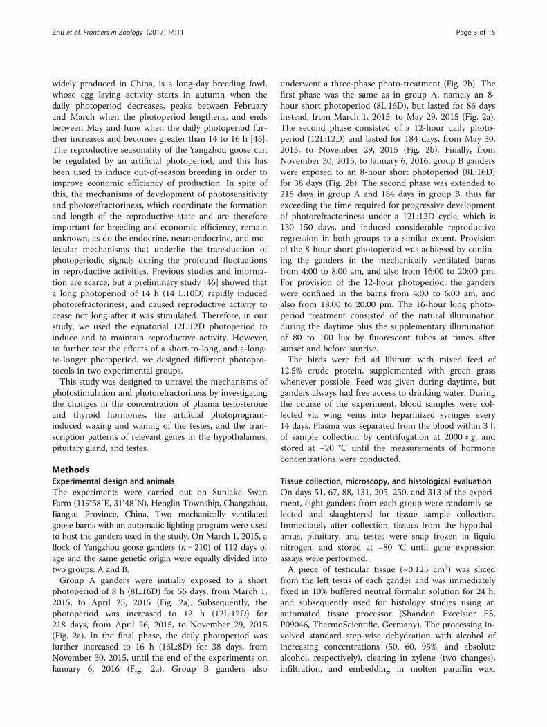
photoperiod of 8 h (8L:16D) for 56 days, from March 1,2015, to April 25, 2015 (Fig. 2a). Subsequently, thephotoperiod was increased to 12 h (12L:12D) for218 days, from April 26, 2015, to November 29, 2015(Fig. 2a). In the final phase, the daily photoperiod wasfurther increased to 16 h (16L:8D) for 38 days, fromNovember 30, 2015, until the end of the experiments onJanuary 6, 2016 (Fig. 2a). Group B ganders also
underwent a three-phase photo-treatment (Fig. 2b). Thefirst phase was the same as in group A, namely an 8-hour short photoperiod (8L:16D), but lasted for 86 daysinstead, from March 1, 2015, to May 29, 2015 (Fig. 2a).The second phase consisted of a 12-hour daily photo-period (12L:12D) and lasted for 184 days, from May 30,2015, to November 29, 2015 (Fig. 2b). Finally, fromNovember 30, 2015, to January 6, 2016, group B ganderswere exposed to an 8-hour short photoperiod (8L:16D)for 38 days (Fig. 2b). The second phase was extended to218 days in group A and 184 days in group B, thus farexceeding the time required for progressive developmentof photorefractoriness under a 12L:12D cycle, which is130–150 days, and induced considerable reproductiveregression in both groups to a similar extent. Provisionof the 8-hour short photoperiod was achieved by confin-ing the ganders in the mechanically ventilated barnsfrom 4:00 to 8:00 am, and also from 16:00 to 20:00 pm.For provision of the 12-hour photoperiod, the ganderswere confined in the barns from 4:00 to 6:00 am, andalso from 18:00 to 20:00 pm. The 16-hour long photo-period treatment consisted of the natural illuminationduring the daytime plus the supplementary illuminationof 80 to 100 lux by fluorescent tubes at times aftersunset and before sunrise.The birds were fed ad libitum with mixed feed of
12.5% crude protein, supplemented with green grasswhenever possible. Feed was given during daytime, butganders always had free access to drinking water. Duringthe course of the experiment, blood samples were col-lected via wing veins into heparinized syringes every14 days. Plasma was separated from the blood within 3 hof sample collection by centrifugation at 2000 × g, andstored at −20 °C until the measurements of hormoneconcentrations were conducted.
Tissue collection, microscopy, and histological evaluationOn days 51, 67, 88, 131, 205, 250, and 313 of the experi-ment, eight ganders from each group were randomly se-lected and slaughtered for tissue sample collection.Immediately after collection, tissues from the hypothal-amus, pituitary, and testes were snap frozen in liquidnitrogen, and stored at −80 °C until gene expressionassays were performed.A piece of testicular tissue (~0.125 cm3) was sliced
from the left testis of each gander and was immediatelyfixed in 10% buffered neutral formalin solution for 24 h,and subsequently used for histology studies using anautomated tissue processor (Shandon Excelsior ES,P09046, ThermoScientific, Germany). The processing in-volved standard step-wise dehydration with alcohol ofincreasing concentrations (50, 60, 95%, and absolutealcohol, respectively), clearing in xylene (two changes),infiltration, and embedding in molten paraffin wax.
Zhu et al. Frontiers in Zoology (2017) 14:11 Page 3 of 15
Tissue sections (5 μm) were mounted on glass slides andstained with hematoxylin and eosin using an automatedslide stainer (Shandon VaristainGermini ES, A78000013,ThermoScientific, Germany). Stained sections were indi-vidually examined under a bright field Olympus BX63light microscope (OLYMPUSBX63, Olympus Corpor-ation, Tokyo) at 10× and 40× magnification for changesin the diameter of the seminiferous tubule, and the
numbers of spermatogonia, spermatocytes, and elon-gated spermatids.
Measurements of hormone concentrationsPlasma testosterone concentrations were determined byenzyme-linked immunosorbent assay using the Quanti-tative Diagnostic Kit for testosterone (North Institute ofBiological Technology, Beijing, China). The assay
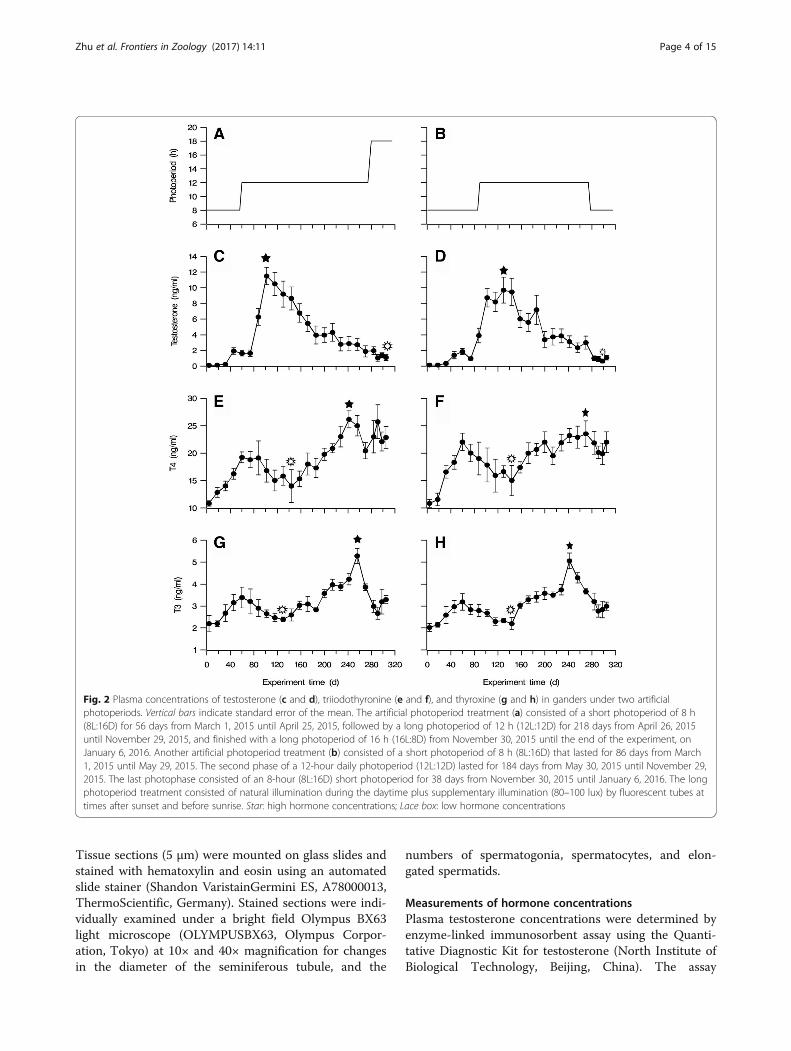
Fig. 2 Plasma concentrations of testosterone (c and d), triiodothyronine (e and f), and thyroxine (g and h) in ganders under two artificialphotoperiods. Vertical bars indicate standard error of the mean. The artificial photoperiod treatment (a) consisted of a short photoperiod of 8 h(8L:16D) for 56 days from March 1, 2015 until April 25, 2015, followed by a long photoperiod of 12 h (12L:12D) for 218 days from April 26, 2015until November 29, 2015, and finished with a long photoperiod of 16 h (16L:8D) from November 30, 2015 until the end of the experiment, onJanuary 6, 2016. Another artificial photoperiod treatment (b) consisted of a short photoperiod of 8 h (8L:16D) that lasted for 86 days from March1, 2015 until May 29, 2015. The second phase of a 12-hour daily photoperiod (12L:12D) lasted for 184 days from May 30, 2015 until November 29,2015. The last photophase consisted of an 8-hour (8L:16D) short photoperiod for 38 days from November 30, 2015 until January 6, 2016. The longphotoperiod treatment consisted of natural illumination during the daytime plus supplementary illumination (80–100 lux) by fluorescent tubes attimes after sunset and before sunrise. Star: high hormone concentrations; Lace box: low hormone concentrations
Zhu et al. Frontiers in Zoology (2017) 14:11 Page 4 of 15

sensitivity was 0.1 ng/mL, and the intra- and inter-assayvariation coefficients were both below 15%. Serial dilu-tions of gander plasma samples resulted in an inhibitioncurve parallel to the standard curve. The r-values of theassay standard curves were greater than 0.99.Blood concentrations of total T3 were also measured
by enzyme-linked immunosorbent assay using the Quan-titative Diagnostic Kit for 3,5,3′-triiodothyronine (NorthInstitute of Biological Technology, Beijing, China). Thesensitivity of the assay was 0.5 ng/mL, and the intra-and inter-assay variation coefficients were both below10%. Similarly, blood concentrations of total T4 weremeasured by enzyme-linked immunosorbent assay usingthe Quantitative Diagnostic Kit for thyroxin (NorthInstitute of Biological Technology, Beijing, China). Thesensitivity of the assay was 0.5 ng/mL, and the intra-and inter-assay variation coefficients were both below15%. The r-values of the assay standard curves were allgreater than 0.99.
RNA isolation, complementary DNA synthesis, andquantitative real-time polymerase chain reactionTotal RNA from hypothalamus, pituitary, and testis tis-sues was extracted with Trizol using a commercial kitaccording to the manufacturer’s instructions (RNAisoPlus, Code No. 9108, Takara, Japan). For RNA extrac-tion, chloroform (0.2 mL) was added to the Trizol re-agent (Code.no 9108, Takara, Japan), the mixture wasvigorously shaken, and after 15 min, centrifuged at12,000 × g for 15 min at 4 °C. Isopropanol (0.5 mL) wasthen added to the separated upper aqueous phase andcentrifuged at 12,000 × g for 10 min at 4 °C. The pelletobtained was washed with 75% ethanol, air dried, anddissolved in 20 mL diethylpyrocarbonate-treated sterilewater. RNA quality was assessed by electrophoresis on a1.2% agarose gel, and the total RNA concentration wasdetermined by measuring the absorbance at 260 nmusing a spectrophotometer (NanoDrop 2000c Thermo-Scientific, Germany). A total of 5 μg RNA was reverselytranscribed using the Takara PrimerScriptTM RTreagent kit (Perfect Real Time) (RR037, Takara, Japan)according to the manufacturer’s instructions.Quantitative real-time polymerase chain reaction ana-
lysis was used to measure the levels of messenger RNA(mRNA) of various genes in samples from the hypothal-amus, pituitary, and testes from ganders in each photo-period phase (Fig. 2a and b). The species source andfunction of the genes tested are shown in Table 1. Gene-specific primers were designed using the Primer 3.0 soft-ware (www.ncbi.nlm.nih.gov/tools/primer-blast/) basedon the BLAST, Ensemble, and GenBank databases(Table 2). Expression of β-actin mRNA was used as areference gene. All polymerase chain reactions (totalvolume of 20 μL) consisted of 10 mL SYBR Premix Ex
TaqII (Takara, Japan), 1 mL complementary DNA, 10pmole of each forward and reverse primers (Table 2),and 7 mL ultrapure water. The thermal cycling profileused was 95 °C for 30 s, 40 cycles of 94 °C for 5 s, and60 °C for 30 s. Fluorescence yields obtained from threereplicate reactions of each complementary DNA samplewere analyzed using the Mastercycler ep realplex(Eppendorf, Germany); furthermore, eight biologicalreplicates were used to ensure the validity and accuracyof the experimental results. The relative expressionlevels of different genes in the tissues were calculatedaccording to the 2−ΔΔCT method [47].
Statistical analysisThe results were mean values ± standard error of 12 (Fig. 2)and 8 (Figs. 3, 4, 5 and 6) replicate samples in each treat-ment groups. Differences of plasma concentrations of tes-tosterone, T3, and T4 between photo-programs wereanalyzed with two-way analyses of variance (two-wayANOVA). Gene expression levels were analyzed with one-way analyses of variance (one-way ANOVA) with maintreatments of the two photo-programs and the serial sam-pling of the experiment. Differences of each effect meanswere compared by the mean ± standard error of mean andconsidered significant at P < 0.05. Statistical analyses wereperformed using IBM SPSS software (ver. 11.0; IBM SPSS,Armonk, NY, USA).
ResultsConcentrations of plasma testosteroneIn the first half of the 56-day 8-hour short photoperiodphase, plasma testosterone concentrations were low,below 0.5 ng/mL, in ganders from both groups, A and B(Fig. 2c–d). With the continuation of the 8-hour photo-period, the concentrations were slightly elevated to ap-proximately 2 ng/mL, and at the end of the 8-hourphotoperiod phase, on day 86 of the experiment, to4 ng/mL in group B (Fig. 2d). At the same time, in groupA ganders, which were already exposed to a 12-hourphotoperiod since one month, plasma testosterone con-centrations had already increased to significantly higherlevels of approximately 6 ng/mL (P = 0.024, F = 10.743)(Fig. 2c). Subsequently, as both gander groups continuedto be exposed to the second 12-hour photoperiod phase,plasma testosterone concentrations continued toincrease rapidly, and they reached peak levels (8–11 ng/mL) from day 100 to day 140 of the experiment.Toward the end of the 12-hour photoperiod phase oron day 274 of the experiment, testosterone concentra-tions in both groups progressively decreased to lowerlevels, approximately 4 ng/mL. Thereafter, in the thirdphotoperiod phase (days 275–313 of the experiment)plasma testosterone concentrations further decreased
Zhu et al. Frontiers in Zoology (2017) 14:11 Page 5 of 15

to levels below 2 ng/mL in both groups (Fig. 2c–d),despite their divergent treatments.
Concentrations of plasma T4 and T3The general pattern of the concentrations of plasma T4and T3, and that of their relative ratio (T4/T3), wasreciprocal to that of testosterone concentrations. For ex-ample, in the first short 8-hour photoperiod phase, bothplasma T4 and T3 concentrations increased from theirinitial low levels and peaked on day 60 of theexperiment. Similarly, the values of the T4/T3 ratio (5.86and 7.07 for groups A and B respectively) were signifi-cantly higher than those at the beginning of the experi-ment (P = 0.031, F = 20.121 and P = 0.047, F = 25.339, forgroups A and B respectively). The concentrations of both
hormones started then to decrease, reaching a lowplateau value at around day 140 of the experiment(2–3 months into the 12-hour photoperiod phase),but they subsequently increased, reaching peak levelsaround days 240 to 260 of the experiment, or almostthe end of the 12-hour photoperiod phase, especiallyfor T3. Until day 270, the values of the T4/T3 ratiowere maintained at relatively high levels: 9.5611 and7.27 for groups A and B, respectively. Then theystarted to decrease again, especially for T3, but dis-played a small increase during the final phase ofphotoperiod change, around days 297 to 313 of theexperiment (Fig. 2f ). Throughout the experiment, thechanging patterns of T4 and T3 concentrations werehighly similar in both groups A and B.
Table 1 Species source and functions in literature of the genes tested in this study
Gene name Species Tissue distribution Gene function
GnRH-I Anser cygnoides Hypothalamus GnRH regulates the secretion of the gonadotropins LH and FSH [66].
GnIH Anser cygnoides Hypothalamus GnIH inhibits LH secretion and reduces testis weigh [20].
VIP Gallus gallus Hypothalamus VIP in the brain acts as a neuroendocrine factorand regulates PRL secretion [51].
TRH Gallus gallus Hypothalamus TRH regulates thyroid stimulating hormone secretion [67].
GnRH Receptor Gallus gallus Pituitary GnRH is a hypothalamic decapeptide that centrally controlsreproduction by binding to GnRH receptors on pituitarygonadotropes and stimulating the secretion of LH and FSH [68].
GnIH Receptor Anser cygnoides Pituitary GnIH acts directly on the pituitary via the GnIH receptor and inhibitsgonadotropin release [69].
VIP Receptor-I Gallus gallus Pituitary VIP is a hypothalamic polypeptide that controls reproduction bybinding to VIP receptors on pituitary gonadotropes and stimulatingPRL secretion [70].
TRH Receptor Gallus gallus Pituitary TRH acts directly on the pituitary via the TRH receptor and controlsthyroid-stimulating hormone (TSH) secretion e [71].
FSH beta Anser cygnoides Pituitary FSH stimulates gonadal growth and estrogen secretion by Sertoli cells.
LH beta Anser cygnoides Pituitary LH controls estrogen and androgen production by mature ovarianfollicles, and regulates androgen production by Leydig cells [72].
PRL Anser cygnoides Pituitary PRL inhibits gene expression of steroidogenic enzymes and reducestestis weight [73, 74].
TSH beta Anser cygnoides Pituitary TSH is a glycoprotein released from the adenohypophysis that activatesiodine uptake, thyroid hormone synthesis, and the release of thyroidhormones from the thyroid gland [75].
LH Receptor Anser cygnoides Testis LH receptor is one of the three glycoprotein hormone receptors thatis necessary for critical reproductive processes, including gonadalsteroidogenesis, oocyte maturation and ovulation, and male sexdifferentiation [76].
FSH Receptor Anser cygnoides Testis FSHR is a transmembrane receptor that interacts with FSH, and itsactivation is necessary for the hormonal functions of FSH [77].
3-beta HSD Anser cygnoides Testis 3-beta HSD catalyzes an obligatory step in the biosynthesis of all classesof hormonal steroids, namely, the oxidation/isomerization of 3-beta-hydroxy-5-ene steroids into the corresponding 3-keto-4-ene steroidsin gonadal as well as in peripheral tissue [78].
StAR Anser cygnoides Testis StAR plays a critical role in steroid hormone synthesis, and it is thoughtto increase the delivery of cholesterol to the inner mitochondrialmembrane where P450scc resides [79].
Abbreviations: GnRH gonadotropin-releasing hormone, GnIH gonadotropin-inhibitory hormone, VIP vasoactive intestinal peptide, TRH thyrotropin releasinghormone, FSH follicle-stimulating hormone, LH luteinizing hormone, PRL prolactin, TSH thyroid-stimulating hormone, 3-beta HSD three beta-hydroxysteroiddehydrogenase, StAR steroidogenic acute regulatory protein
Zhu et al. Frontiers in Zoology (2017) 14:11 Page 6 of 15

Body weight, testis morphometry, and histologyIn both gander groups, body weight exhibited a two-phasevariation (Fig. 3a). Before day 131 of the experiment, theweight of live ganders was maintained around 3.95 kg, buttoward the end of the experiment on day 313, it increasedto significantly higher values, in the range of 4.73–4.93 kg(P = 0.005, F = 16.215), for both gander groups (Fig. 3a).The weight of the left testis in both groups was below
2 g on day 51, during the 8-hour photoperiod (Fig. 3b).For the ganders of group B, it remained the same untilday 67 of the experiment. In the ganders of group A
however, the increase in the photoperiod from 8 to 12 hfor just 12 days promptly stimulated testicular growth,and the weight of the left testis increased to approxi-mately 6 g (Fig. 3b), and continued to increase until theday 88 of the experiment, when it reached a peak valuesignificantly higher than that observed in the ganders ofgroup B (P = 0.000, F = 17.952). In the latter group, thepeak weight of 11 g was reached only on day 131 of theexperiment (Fig. 3b). Thereafter, testicular weight de-creased in both groups. However, at the end of the ex-periment (day 313) the testicular weight of ganders in
Table 2 Primers used in the real-time quantitative PCR assay of gene transcription
Gene name Accession number Primer sequences (5′-3′) Annealing temperature (°C) PCR product (bp)
β-actin L08165.1 upstream: TGACGCAGATCATGTTTGAGA 60 159
downstream: GCAGAGCGTAGCCCTCATAG
GnRH-I EF495207.1 upstream: CTGGGACCCTTGCTGTTTTG 60 232
downstream: AGGGGACTTCCAACCATCAC
GnIH KC514473.1 upstream: ATCTACCTAGGCATGCTCCAA 58 115
downstream: ACAGGCAGTGACTTCCCAAAT
VIP DQ023159 upstream: ACCAGTGTCTACAGCCATCTTTTG 58 204
downstream: AGGTGGCTCAGCAGTTCATCTACA
TRH NM_001030383.2 upstream: GCAAGAGGGGCTGGAATGAT 58 133
downstream: ATGGCAGACTGCTGAAGGTC
GnRH Receptor KJ659046.1 upstream: TCTGCTGGACCCCCTACTAC 60 127
downstream: TCCAGGCAGGCATTGAAGAG
GnIH Receptor KF696709.1 upstream: GTCGTCATGTACACCCGCAT 56 103
downstream: TCTTGCGAGACACCTTCCTC
VIP Receptor-I NM_001097523.1 upstream: TACTGCGTCATGGCCAACTT 58 153
downstream: TGTCCAAGCGGTGATGAACA
TRH Receptor NM_204930.1 upstream: CTATGGCTATGTGGGGTGCC 60 189
downstream: ACTGAGGCGAAAGACCAGAC
FSH beta EU252532 upstream: GTGGTGCTCAGGATACTGCTTCA 60 209
downstream: GTGCAGTTCAGTGCTATCAGTGTCA
LH beta DQ023159 upstream: GACCCGGGAACCGGTGTA 58 90
downstream: AGCAGCCACCGCTCGTAG
PRL DQ023160 upstream: TGCTCAGGGTCGGGGTTTCA 56 218
downstream: GCTTGGAGTCCTCATCGGCAAGTT
TSH beta FJ797681.1 upstream: CTCTGTCCCAAAACGTGTGC 56 121
downstream: CCACACTTGCAGCTTATGGC
LH Receptor XM_013192443.1 upstream: CGGATACACAACGATGCCCT 60 74
downstream: GACTCCAGTGCCGTTGAAGA
FSH Receptor KC477215.1 upstream: CCTAGCCATTGCTGTGCATTT 60 106
downstream: TGCCAGGTTGCTCATCAAGG
3-beta HSD KC310447.1 upstream: TGTGACGTTCCTGTACCGTG 153
downstream: TTACAACGGGTACACGCCTC
StAR KF958133.1 upstream: GGAGCAGATGGGAGACTGGA 56 177
downstream: CGCCTTCTCGTGGGTGAT
Zhu et al. Frontiers in Zoology (2017) 14:11 Page 7 of 15

group A was significantly lower than that of ganders ingroup B, when the two groups were exposed tophotoperiods of 16 and 8 h, respectively (Fig. 3b).Microscopic observation of testis histological sections
(Fig. 4) revealed that for both gander groups, the semin-iferous tubule diameter (Fig. 3c) and the number ofspermatogonia (Fig. 3d), spermatocytes (Fig. 3e), and
elongated spermatids (Fig. 3f ) changed in a similar fash-ion throughout the experiment: they increased from lowvalues on day 67, to peak values on day 131, and de-creased thereafter to low values again until the end ofexperiment (Fig. 4). It should be noted that on day 67,the spermatocyte number (Fig. 3e) was significantlyhigher in group A, which was already exposed to a
Fig. 3 Body (a) and testicular weights (b), and testicular histological data (c-f) of Yangzhou ganders maintained under two artificial photoperiods.Changes in the diameter of the seminiferous tubules (c), the number of spermatogonia (d), spermatocytes (e), and elongated spermatids (f) weremeasured within 8 individial seminiferous tubules. Each bar represents the mean value of six determinations including the standard error. *, **and *** indicate statistical significance based on P < 0.05, P < 0.01 and P < 0.001, respectively
Fig. 4 Histological analysis of sections from Yangzhou goose gander testis. Sections from Yangzhou goose gander were collected on days 67,131, 250, and 313 and were stained with hematoxylin and eosin Black arrowhead: spermatogonia; black arrow: primary spermatocyte; black arrowwith tail: elongated spermatid; star: Sertoli cell vacuolation. Scale bar represents 100 μm in 10 × and 20 μm in 40 ×
Zhu et al. Frontiers in Zoology (2017) 14:11 Page 8 of 15

Fig. 5 Hypothalamic (a-d) and pituitary (e-l) mRNA levels in Yangzhou goose ganders under two artificial photoperiods. Graphs a to d representmRNA expression levels of hypothalamic releasing hormones GnRH-I, GnIH, VIP and TRH, respectively, while e to h their corresponding receptorsin the pituitary gland, and i to l the FSH, LH, PRL and TSH pituitary hormones or beta subunits wherever applicable. Each value represents theaverage of data from eight independent culture experiments. Data are shown as mean values ± standard error of the mean. *, ** and *** indicatestatistical significance based on P < 0.05, P < 0.01and P < 0.001, respectively, between the treatments
Fig. 6 Testicular mRNA levels of LH receptor (a), FSH receptor (b), 3-beta HSD (c) and StAR (d) in Yangzhou goose ganders under two artificialphotoperiods. Each value represents the average of data from eight independent culture experiments. Data are shown as mean values ± standarderror of the mean. *, ** and *** indicate statistical significance based on P < 0.05, P < 0.01and P < 0.001, respectively. between the treatments
Zhu et al. Frontiers in Zoology (2017) 14:11 Page 9 of 15

12-hour photoperiod, than in group B, which was stillexposed to an 8-hour photoperiod (P = 0.000, F = 24.965),as was also the case with the seminiferous tubule diameter(Fig. 3c). On day 313, when the experiment ended, therewere still more elongated spermatocytes in group A thanin group B (Fig. 4 and Fig. 3f). In addition to the changesin diameter, the number of vacuoles in the seminiferoustubules was higher both at the beginning and end of theexperiment than at the peak of the reproductive activityon day 131, where the seminiferous tubules were morecompact and in a cell-filled state.
Gene expression levelsHypothalamus and pituitary genesAll of the 16 genes tested exhibited photoperiod-dependent mRNA level regulation throughout the experi-ment (Fig. 5). The hypothalamic GnRH-I mRNA levelschanged similarly in ganders from groups A and B(Fig. 5a): they increased from the beginning of the experi-ment to reach a peak level on day 205, during the 12-hourphotoperiod, and then steadily decreased, with an excep-tion on day 88, when they were significantly higher ingroup A than in group B. Similarly, GnIH gene transcrip-tion levels varied in a strikingly similar manner in bothgroups (Fig. 5b): they steadily decreased from their alreadylow levels at the beginning of the experiment to a mini-mum value on day 131, during the 12-hour photoperiod,and then remarkably increased by 200-fold to a maximumfrom day 205 to day 250, before they finally decreasedagain. The changes in VIP and TRH gene transcription inthe hypothalamus exhibited changes similar to that ofGnIH throughout the experiment (Fig. 5c–d): their mRNAlevels decreased to or remained at low levels until day 88of the experiment, and then increased to higher levels onday 131, during peak reproductive activities. However, ontwo occasions, namely on days 131 and 313 of theexperiment, the VIP mRNA levels were significantlyhigher in group A than in group B (P = 0.013, F = 13.542and P = 0.002, F = 20.189), while the same was true for theTRH mRNA levels on day 131.In the pituitary gland, the GnRH receptor mRNA
levels increased steadily from day 51, during the short 8-hour photoperiod, to a plateau value at day 250 in groupA, whereas in group B, they further increased to a peakvalue at the end of the experiment (Fig. 5e). The GnIHreceptor mRNA levels (Fig. 5f ) remained low before day88, but they started to rise from day 131, and reached apeak level on day 205, then decreased in both groups.However, on days 250 and 313, the post-peak mRNAlevels were higher in group A than in group B (P = 0.001,F = 24.773 and P = 0.004, F = 7.200 for days 250 and 313respectively) (Fig. 5f). Throughout the experiment, themRNA levels of the VIP receptor changed in a manner
similar to that of the GnIH receptor, as did the TRHreceptor mRNA levels (Fig. 5g–h).FSH beta mRNA (Fig. 5i) expression levels were ini-
tially low under the 8-hour photoperiod, and graduallyincreased to the peak on day 131, and then decreased tothe nadir on day 313 in Group B ganders. The FSHmRNA levels on days 67 and 88 were significantly higherin ganders of group A than in those of group B (P =0.000, F = 11.710 and P = 0.005, F = 6.744 for days 67and 88 respectively). Similarly, in group A, the LH betamRNA levels (Fig. 5j) increased from their low levelsduring the 8-hour photoperiod on day 51 to peak levelson day 67, just 12 days after the photoperiod was in-creased from 8 to 12 h. This peak was maintained untilday 131 and then gradually decreased to a minimum onday 313 (end of the experiment). In addition, on day 67,they were significantly higher in group A than in groupB (P = 0.000, F = 25.221), whose LH beta transcriptionpatterns changed similarly to those of FSH beta. ThePRL mRNA levels (Fig. 5k) remained low before day 88in both groups, then started to increase from day 131 toa peak level on day 205, and thereafter decreased. Ingroup A, on days 131 and 313, PRL transcription was sig-nificantly higher than in group B (P = 0.002, F = 12.867 andP = 0.000, F = 21.107 for days 131 and 313 respectively).The levels of TSH beta mRNA (Fig. 5l) changed in bothgroups in a manner similar to that of GnIH, except on day131, where the expression was abruptly and significantlyhigher in group A than in group B (P = 0.000, F = 20.946).
Testicular genesThe FSH receptor mRNA levels (Fig. 6a) increasedsteadily from the beginning of the experiment; theyreached peak levels during days 205–250, and then de-creased to low levels at end of experiment. In bothgroups of ganders, the LH receptor mRNA levels(Fig. 6b) also increased, but they peaked on day 131, andthen decreased until the end of experiment. This chan-ging pattern was also true for the StAR mRNA levels(Fig. 6c), except that on day 67 they were significantlyhigher in group A than in group B (P = 0.012, F = 5.480).The mRNA levels of 3-beta hydroxysteroid dehydrogen-ase remained low or decreased before day 88, and thenincreased to peak levels from day 131 to 250, and finallydecreased again to low levels at the end of experiment(Fig. 6d). On days 88 and 131, the levels were also higherin group A than in group B (P = 0.000, F = 16.329 andP = 0.037, F = 3.879 for days 88 and 131 respectively).
DiscussionThis study systematically investigated the molecularmechanisms associated with testicular development andregression in response to artificial photoprograms inYangzhou goose ganders. A wide spectrum of variables
Zhu et al. Frontiers in Zoology (2017) 14:11 Page 10 of 15

was assessed, including testis histology, hormone plasmaconcentrations, and gene transcription patterns. Inaddition to the general responses to an increase inphotoperiod (i.e. testicular development and subsequentregression), some minor responses in the timing of tes-ticular development and gene transcription were alsoobserved in relation to subtle changes in photoperiod.These results reinforce the proposition that theYangzhou goose is a long-day breeding bird, and providenew insights into the molecular mechanisms thatmediate the photoperiod-regulated seasonal changes inreproductive activities.It was also noted that after 40 days of exposure to an
8-hour short photoperiod, testosterone concentrationsslightly increased in both gander groups studied (A andB). This phenomenon may be linked to the weak expres-sion of gonadal or reproductive activities during shortdays after dissipation of refractoriness to the long days[4, 45, 48]. In addition, in group B and on day 88 of theexperiment, refractoriness to a short photoperiod alsoled to spontaneous testicular development immediatelybefore the photoperiod was increased. Subsequently, anincrease in daily photoperiod from 8 to 12 h resulted inrapid growth of the testes, accompanied by an increasein testicular weight from less than 2 g during the 8-hourshort photoperiod to approximately 6 g in just 10 days,and to 12 g in 21 days. The delay of 28 days in photo-period increase for group B with respect to that of groupA postponed testicular growth at a similar pace. Follow-ing 40 days of exposure to a 12-hour photoperiod, theganders reached peak reproductive activities as repre-sented by the high levels of plasma testosterone andactive spermatogenesis in the testis at days 100–150 ofthe experiment. After that time point, refractoriness tothe long photoperiod gradually commenced, as reflectedby a decrease in testicular weight, disappearance ofspermatozoa in the seminiferous tubules of the testes,and especially by the steady decrease in the concentra-tion of plasma testosterone. The 12-hour equatorialphotoperiod was adopted in this study not only to in-duce the reproductive activities of ganders, but also totest how photorefractoriness develops in the Yangzhougoose. Clearly, ganders develop a kind of “absolutephotorefractoriness” similar to that reported in starlings[49], in contrast to the “relative photorefractoriness”observed in quails [49] and sheep [50]. Therefore, at theend of the experiment, the testosterone concentrationdecreased to minimal levels in both groups A and B.Such changes should be the result of different mecha-nisms, i.e. the further refractoriness to long photoperiodin group A by an increase in photoperiod to 16 h (aswas indicated by upregulation of VIP and PRL mRNAlevels), and the inhibition of reproductive activity ingroup B by a decrease in photoperiod.
It has been proposed that the endocrine mechanismmediating the photoperiod-dependent regulation of sea-sonal reproduction is through the coordinated produc-tion and secretion of the gonadotrophins LH and PRLby the pituitary gland [14]. Increases in photoperiod firstdrive LH secretion, which stimulates gonadal develop-ment and initiates the breeding season, and then PRLsecretion, which initiates the development of photore-fractoriness, and after reaching peak levels, reduces LHsecretion and leads to gonad regression. Since produc-tion and secretion of LH is regulated by thehypothalamic-releasing hormones GnRH and GnIH [13,21, 25], and that of PRL by VIP [32, 51], these hormonesmust be involved in the photoperiod-dependent regula-tion of seasonal breeding [3, 13, 14]. Therefore, in thepresent study, the mRNA levels of GnRH, GnIH, andVIP were measured in the hypothalamus of the ganders,and those of their receptors in the pituitary gland. Inboth groups of ganders, initiation of reproductive activ-ities after prolonged exposure to a short 8-hour photo-period, or a state of refractoriness to the shortphotoperiod, concomitantly occurred with a steady de-crease in the mRNA levels of VIP and GnIH. Switchingfrom an 8-hour to a 12-hour photoperiod upregulatedthe transcription of hypothalamic GnRH, but furtherdownregulated that of GnIH, which reached a minimumwhen ganders were in a fully active reproductive state.Changes in GnRH/GnIH gene transcription in responseto an increase in photoperiod were accordingly reflectedby changes in FSH and LH mRNA levels in the pituitarygland. It must be noted that the earlier exposure to a 12-hour long photoperiod in group A was associated withan earlier upregulation of both FSH and LH mRNAlevels. This could explain the earlier testicular growthand rise in the concentrations of plasma testosteroneobserved in the ganders of group A.Notwithstanding the above observations, the post-peak
decrease in reproductive activities after a prolonged ex-posure to a 12-hour long photoperiod (day 205 of theexperiment) was associated with a greater than 300-foldupregulation in the mRNA levels of GnIH. This increasein GnIH gene transcription and hence secretion of thehormone may be the driving force in inducing repro-ductive regression during the development of refractori-ness, despite a 2- to 3-fold increase in GnRH mRNAlevels after continued exposure to a long photoperiod.The temporal elevation of GnRH mRNA levels from day205 to day 250 may not be photoperiod-driven, butcould rather be caused by a decreased negative controlfeedback arising from the diminished plasma testoster-one concentrations. Whereas the declined GnRHexpression at end of experiment could be the true effectof photoperiodic, by the refractoriness under 16 hphotoperiod or by the inhibition under 8 h photoperiod.
Zhu et al. Frontiers in Zoology (2017) 14:11 Page 11 of 15

On the other hand, the inhibition by GnIH of pituitarygonadotrophin synthesis could be mediated via twopathways, one by the direct effect on pituitary gland andthe other indirectly via reducing GnRH secretion byinhibiting GnRHneurons in the hypothalamus [13]. Theinhibitory effect of GnIH on gonadotrophin expressionand secretion could be at a maximum level starting fromday 205 of the experiment, that is approximately 150 daysafter switching to the long 12-hour photoperiod, if pitu-itary GnIH receptor mRNA levels were also included.Another factor that is important for the development ofrefractoriness to a long photoperiod is PRL, whosemRNA levels reached their highest value on day 205 ofthe experiment, when the mRNA levels of the GnIH/GnIH receptor also reached their highest value andcould exert their maximal inhibitory impact on gonado-trophin secretion. The mRNA levels of VIP and the VIPreceptor, which stimulate pituitary PRL secretion, werealready upregulated from day 131 of the experiment. Asganders in group A were exposed to a 12-hour photo-period one month earlier than ganders in group B, theVIP/VIP receptor mRNA levels were also observed torise earlier in the former group. Furthermore, at the endof the experiment on day 313, both VIP and PRL mRNAlevels were further upregulated in group A, in responseto an increase in photoperiod from 12 h to 16 h 35 daysearlier than in group B. Such an upregulation did notoccur in the ganders of group B, which experienced adecrease of photoperiod from 12 h to 8 h.The photostimulation and refractoriness of reproductive
activities were also analyzed in terms of testicular ste-roidogenesis gene transcription patterns. The transcrip-tion of LHR, which mediates the gonadotrophic effects ofLH, displayed a typical rise-and-fall pattern, following thatof the changes in testicular weight and plasma testoster-one concentration. A similar effect was observed for StARand 3-beta hydroxysteroid dehydrogenase transcription.Thus, toward the end of the experiment, when both LHbeta and LH receptor mRNA levels significantly subsided,so did the mRNA levels of the steroidogenic genes StARand 3-beta hydroxysteroid dehydrogenase. Depletion ofthese key enzymes of steroidogenesis would result in di-minished testosterone production, as shown by the steadydecline of plasma testosterone concentration toward theend of the experiment. This, in turn, would impair sperm-atogenesis, resulting in testis atrophy and reduced testisweight. Of the testicular genes tested, the FSH receptormRNA levels peaked during days 205–250 of the experi-ment; that is, during the testicular regression process. Thisillustrates that the biological role of FSH in the regulationof testicular functions may occur during the early stagesof spermatogenesis (i.e., germ cell mitosis before thespermatocyte stage) [52]. Testosterone is more importantduring the later stages of spermatogenesis, when
morphologic changes lead to spermatid formation. As aresult, on day 313 of the experiment, when testosteroneand FSH levels were minimal, no spermatids were ob-served in the testes of ganders in either group.The thyroid hormones function as molecular switches
in the regulation of seasonal reproduction [11, 53], andare involved in both photoperiod-induced reproductivestimulation and refractoriness. In Japanese quail, re-moval of thyroid hormones by thyroidectomy reducesthe maximal extent of testis development induced by along photoperiod [49] In the quail, it was discovered thatTSH is synthesized in the pars tuberalis of the pituitarygland in response to an increase in photoperiod, andthrough retrograde uptake, it stimulates the ependymalcells in MBH to express type 2 iodothyroninedeiodinase,which converts T4 to T3 [41, 54]. Such locally producedT3, stimulates the release of GnRH into the portal vas-cular system by inducing morphological or plasticitychanges in GnRH-secreting neurons [41], resulting inelevated LH secretion and full reproductive activities[11, 13]. On the other hand, thyroid hormones have apermissive role in the development of photorefractori-ness that leads to reproductive regression in both mam-mals and birds [34, 39, 55–57]. However, when there is acomplete lack of thyroid hormones, reproductive activ-ities still change in response to daily photoperiod in boththyroidectomized birds [49] and red deer [34]. There-fore, it appears that the photoperiod-dependentregulation of seasonal reproduction or a GnRH pulsegenerator should still be operating at brain centershigher than MBH, while local T3 production in theMBH might amplify or modify this central effect. It ispossible that systemic effects, in contrast to the above-discussed local actions of thyroid hormones in the MBH,may mediate reproductive inhibiting actions. In severalanimal species living in temperate zones, such as ducks[36], Moscovy drakes [37] and geese [38], there was aseasonal rise of plasma thyroid hormones around thetime of reproductive regression. In domestic ganders, ithas been shown that the concentrations of T4 and T3decreased rapidly with increasing testosterone concen-tration, and with the approach of the non-breeding sea-son, testosterone concentration decreased rapidly,whereas T4 concentration showed the opposite trend[38]. In the present study, the changes in plasma con-centrations of both T4 and T3 exhibited a patternopposite to that of testosterone. The seasonal fluctua-tions of thyroid hormones might be an aspect of thegeneral seasonal physiological changes; alternatively,high thyroid hormone levels during the non-breedingseason might facilitate, if not altogether cause, the ter-mination of the breeding season, and facilitate the mani-festation of other seasonal events such as growth [34], asdiscussed further below. It should also be noted that the
Zhu et al. Frontiers in Zoology (2017) 14:11 Page 12 of 15

involvement of thyroid hormones in the refractoriness toa 12-hour long photoperiod, observed in this study, oc-curred with a concomitant upregulation of the TRH,TRH receptor, and TSH mRNA levels, as well as of theVIP, VIP receptor, and PRL mRNA levels. Indeed, inEuropean starlings, abolishing the development ofphotorefractoriness by thyroidectomy also prevented thecorresponding increase in PRL secretion [39].Thyroid hormones, PRL, and GnIH not only stimulate
the development of photorefractoriness that leads to re-productive regression, but can also regulate feed intakeand growth. GnIH has been shown to stimulate feed in-take in both animals [58] and fowls [59, 60]. Based on this,it was proposed that may be a molecular switch betweenfeeding, and hence somatic growth, and reproduction[58]. Furthermore, both thyroid hormones and PRL areessential for long photoperiod-induced seasonal increasesin feed intake and live weight gain simultaneously toreproductive regression [34, 61], whereas PRL has beenfound to be orexigenic in domestic cows [62, 63]. It ap-pears that thyroid hormones and PRL also fulfill the rolesof molecular switches between growth and reproduction.Consequently, at the late stages of the experiment, thereproductive system regression accompanied by a signifi-cant weight increase in ganders could well be the result ofactions of these three molecular switches. It was proposedthat the seasonal somatic growth in wild animals that occurat the same time of reproductive regression [34, 64, 65]promotes animal survival throughout the hardships of win-ter. It appears that this strategy remains operative in thedomestic goose as well, and involves the same endocrineregulatory factors.
ConclusionsIn summary, reproductive stimulation of long-day breedingYangzhou goose ganders was characterized by GnRH up-regulation and GnIH and VIP downregulation in the hypo-thalamus during long or increasing photoperiods. This, inturn, stimulated the transcription of the gonadotrophingene, whereas inhibited that of the PRL gene in the pituit-ary gland, which is known to stimulate testicular develop-ment by upregulating genes important for steroidogenesisand spermatogenesis. On the other hand, development ofphotorefractoriness was characterized by hyper-regulationof GnIH transcription in the hypothalamus, in addition totranscription upregulation of VIP, TRH, and their receptorsin the pituitary gland. Consequently, gonadotrophin tran-scription was downregulated, whereas that of PRL andTSH was upregulated. The combined effects of thesechanges resulted in reproductive system regression.
AcknowledgementsWe thank all anonymous reviewers for their suggestions and criticisms whichimproved the quality of the manuscript
FundingThis study was supported by the National Science Foundation of China(grant no. 31372314), Fund for Independent Innovation of AgriculturalSciences in Jiangsu Province (CX(15)1008), and Research Fund of JiangsuAcademy of Agricultural Science (ZX(15)1007). HXZ was supported byPostdoctoral Research Funding Program of Jiangsu Province (1501079C).
Availability of data and materialsThe datasets supporting the conclusions of this article are included within the article.
Authors’ contributionsHXZ, XBS and ZDS designed the study. HXZ, ZC, JNY, CKW and ZCD carriedout the animal experiment, collected and analyzed the samples. HXZ andZDS prepared the manuscript with correction input by ZC. All authors readand approved the final manuscript.
Competing interestsThe authors declare that they have no competing interests.
Consent for publicationAll authors have agreed to publish this work.
Ethics approval and consent to participateThe owners of the geese agreed to the research being carried out on theiranimals. The experiment was approved by the Research Committee of theJiangsu Academy of Agricultural Sciences and was conducted withadherence to the Regulations for the Administration of Affairs ConcerningExperimental Animals (Decree No. 2 of the State Science and TechnologyCommission on November 14, 1988).
Author details1Laboratory of Animal Improvement and Reproduction, Institute of AnimalScience, Jiangsu Academy of Agricultural Sciences, Nanjing 210014, China.2Sunlake Swan Farm, Changzhou 213101, China.
Received: 1 August 2016 Accepted: 21 February 2017
References1. Wingfield JC, Farner DS. Endocrinology of reproduction in wild species.
Avian Biol. 1993;9:327.2. Dixit AS, Singh NS. Photoperiod as a proximate factor in control of
seasonality in the subtropical male Tree Sparrow, Passer montanus. FrontZool. 2011;8:44–56.
3. Dawson A, Sharp PJ. Photorefractoriness in birds—photoperiodic and non-photoperiodic control. Gen Comp Endocrinol. 2007;153:378–84.
4. Hahn TP, Boswell T, Wingfield JC, Ball GF. Temporal flexibility in avianreproduction. In Current ornithology. Springer; 1997: 39–80.
5. Hahn T, Pereyra M, Katti M, Ward G, MacDougall-Shackleton S. Effects offood availability on the reproductive system. New Delhi: Functional AvianEndocrinology Narosa Publishing House; 2005. p. 167–80.
6. Dawson A. Seasonal differences in the secretion of luteinising hormone andprolactin in response to n‐methyl‐dl‐aspartate in starlings (sturnus vulgaris).J Neuroendocrinol. 2005;17:105–10.
7. Dawson A. The effect of temperature on photoperiodically regulatedgonadal maturation, regression and moult in starlings–potentialconsequences of climate change. Funct Ecol. 2005;19:995–1000.
8. Sharp PJ. Photoperiodic control of reproduction in the domestic hen. PoultSci. 1993;72:897–905.
9. Ikegami K, Yoshimura T. Circadian clocks and the measurement ofdaylength in seasonal reproduction. Mol Cell Endocrinol. 2012;349:76–81.
10. Yoshimura T. Neuroendocrine mechanism of seasonal reproduction in birdsand mammals. Anim Sci J. 2010;81:403–10.
11. Yoshimura T. Thyroid hormone and seasonal regulation of reproduction.Front Neuroendocrinol. 2013;34:157–66.
12. Chaiseha Y, Halawani MEE. Neuroendocrinology of the female turkeyreproductive cycle. J Poult Sci. 2005;42:87–100.
13. Bédécarrats GY, Baxter M, Sparling B. An updated model to describe theneuroendocrine control of reproduction in chickens. Gen Comp Endocrinol.2015;227:58–63.
Zhu et al. Frontiers in Zoology (2017) 14:11 Page 13 of 15
14. Sharp PJ, Blache D. A neuroendocrine model for prolactin as the keymediator of seasonal breeding in birds under long- and short-dayphotoperiods. Can J Physiol Pharmacol. 2003;81:350–8.
15. Burns JM. The influence of chicken and mammalian luteinizing hormoneson ovarian estrogen synthesis in the pullet (Gallus domesticus). CompBiochem Physiol A Physiol. 1972;42:1011–7.
16. Brown N, Baylé J-D, Scanes C, Follett B. Chicken gonadotrophins: theireffects on the testes of immature and hypophysectomized Japanese quail.Cell Tissue Res. 1975;156:499–520.
17. Maung SL, Follett BK. The endocrine control by luteinizing hormone oftestosterone secretion from the testis of the Japanese quail. Gen CompEndocrinol. 1978;36:79–89.
18. Phillips A, Scanes CG, Hahn DW. Effect of androgens and gonadotropins onprogesterone secretion of chicken granulosa cells. Comp Biochem Physiol APhysiol. 1985;81:847–52.
19. Bédécarrats GY, McFarlane H, Maddineni SR, Ramachandran R.Gonadotropin-inhibitory hormone receptor signaling and its impact onreproduction in chickens. Gen Comp Endocrinol. 2009;163:7–11.
20. Ciccone NA, Dunn I, Boswell T, Tsutsui K, Ubuka T, Ukena K, Sharp P.Gonadotrophin inhibitory hormone depresses gonadotrophin α and follicle-stimulating hormone β subunit expression in the pituitary of the domesticchicken. J Neuroendocrinol. 2004;16:999–1006.
21. Tsutsui K, Saigoh E, Ukena K, Teranishi H, Fujisawa Y, Kikuchi M, Ishii S, SharpPJ. A novel avian hypothalamic peptide inhibiting gonadotropin release.Biochem Biophys Res Commun. 2000;275:661–7.
22. Bentley G, Perfito N, Ukena K, Tsutsui K, Wingfield J. Gonadotropin-inhibitorypeptide in song sparrows (melospiza melodia) in different reproductiveconditions, and in house sparrows (passer domesticus) relative to chicken‐gonadotropin‐releasing hormone. J Neuroendocrinol. 2003;15:794–802.
23. Ukena K, Ubuka T, Tsutsui K. Distribution of a novel avian gonadotropin-inhibitory hormone in the quail brain. Cell Tissue Res. 2003;312:73–9.
24. Ubuka T, Bentley GE, Ukena K, Wingfield JC, Tsutsui K. Melatonin inducesthe expression of gonadotropin-inhibitory hormone in the avian brain. ProcNatl Acad Sci U S A. 2005;102:3052–7.
25. Kriegsfeld LJ, Mei DF, Bentley GE, Ubuka T, Mason AO, Inoue K, Ukena K,Tsutsui K, Silver R. Identification and characterization of a gonadotropin-inhibitory system in the brains of mammals. Proc Natl Acad Sci U S A.2006;103:2410–5.
26. Sharp PJ. Photoperiodic regulation of seasonal breeding in birds. Ann N YAcad Sci. 2005;1040:189–99.
27. Mauro LJ, Youngren OM, Proudman JA, Phillips RE, El Halawani ME. Effectsof reproductive status, ovariectomy, and photoperiod on vasoactiveintestinal peptide in the female turkey hypothalamus. Gen CompEndocrinol. 1992;87:481–93.
28. Silverin B, Viebke P. Low temperatures affect the photoperiodically inducedLH and testicular cycles differently in closely related species of tits (Parusspp.). Horm Behav. 1994;28:199–206.
29. Deviche P, Saldanha CJ, Silver R. Changes in brain gonadotropin-releasinghormone-and vasoactive intestinal polypeptide-like immunoreactivityaccompanying reestablishment of photosensitivity in male dark-eyed juncos(Junco hyemalis). Gen Comp Endocrinol. 2000;117:8–19.
30. Sharp P, Sreekumar K. Photoperiodic control of prolactin secretion. In: ADawson and CM Chaturvedi (Eds), Avian Endocrinology. New Delhi: Narosa;2001. p. 245–255.
31. Shi ZD, Huang YM, Liu Z, Liu Y, Li XW, Proudman JA, Yu RC. Seasonal andphotoperiodic regulation of secretion of hormones associated with reproductionin Magang goose ganders. Domest Anim Endocrinol. 2007;32:190–200.
32. Dawson A, Sharp PJ. The role of prolactin in the development ofreproductive photorefractoriness and postnuptial molt in the Europeanstarling (Sturnus vulgaris). Endocrinology. 1998;139:485–90.
33. Nicholls T, Goldsmith A, Dawson A. Photorefractoriness in birds andcomparison with mammals. Physiol Rev. 1988;68:133–76.
34. Shi ZD, Barrell GK. Requirement of thyroid function for the expression ofseasonal reproductive and related changes in red deer (Cervus elaphus)stags. J Reprod Fertil. 1992;94:251–9.
35. Boissinagasse L, Maurel D, Boissin J. Seasonal variations in thyroxine andtestosterone levels in relation to the moult in the adult male mink (Mustelavison Peale and Beauvois). Can J Zool. 1981;59:1062–6.
36. Jallageas M, Assenmacher I. Further evidence for reciprocal interactionsbetween the annual sexual and thyroid cycles in male Peking ducks. GenComp Endocrinol. 1979;37:44–51.
37. Sharp PJ, Klandorf H, Mcneilly AS. Plasma prolactin, thyroxine,triiodothyronine, testosterone, and luteinizing hormone during aphotoinduced reproductive cycle in mallard drakes. J Exp Zool.1986;238:409–13.
38. Zeman M, Košutzký J, Miček Ľ, Lengyel A. Changes in plasma testosterone,thyroxine and triiodothyronine in relation to sperm production and remexmoult in domestic ganders. Reprod Nutr Dev. 1990;30:549–57.
39. Goldsmith AR, Nicholls TJ. Thyroidectomy prevents the development ofphotorefractoriness and the associated rise in plasma prolactin in starlings.Gen Comp Endocrinol. 1984;54:256–63.
40. Follett B, Nicholls T. Acute effect of thyroid hormones in mimickingphotoperiodically induced release of gonadotropins in Japanese quail. JComp Physiol B. 1988;157:837–43.
41. Yoshimura T, Yasuo S, Watanabe M, Iigo M, Yamamura T, Hirunagi K, EbiharaS. Light-induced hormone conversion of T4 to T3 regulates photoperiodicresponse of gonads in birds. Nature. 2003;426:178–81.
42. Nakao N, Ono H, Yamamura T, Anraku T, Takagi T, Higashi K, Yasuo S, KatouY, Kageyama S, Uno Y. Thyrotrophin in the pars tuberalis triggersphotoperiodic response. Nature. 2008;452:317–22.
43. Bo WC. The origin of Chinese domestic geese. Agric Archaeol. 1996;3:268–72.44. Wang JW, Qiu XP, Zeng FT, Shi XW, Zhang YP. Genetic differentiation of
domestic goose breeds in China. Acta Genet Sin. 2005;32:1053–9.45. Shi ZD, Tian YB, Wu W, Wang ZY. Controlling reproductive seasonality in the
geese: a review. Worlds Poult Sci J. 2008;64:343–55.46. Rousselot-Pailley D, Sellier N. Influence de quelques facteurs zootechniques
sur la fertilité des oies. In: BRILLARD, J.P. (Ed.) Control of fertility in domesticbirds. Les Colloques de L'INRA 54. 1990; p. 145–155.
47. Livak KJ, Schmittgen TD. Analysis of relative gene expression data using real-time quantitative PCR and the 2− ΔΔCT method. Methods. 2001;25:402–8.
48. Péczely P, Czifra G, Sepr”Odi A, Teplán I. Effect of low light intensity ontesticular function in photorefractory domestic ganders. Gen CompEndocrinol. 1985;57:293–300.
49. Follett BK, Nicholls TJ. Influences of thyroidectomy and thyroxinereplacement on photoperiodically controlled reproduction in quail. JEndocrinol. 1985;107:211–21.
50. Jackson G, Jansen H, Kao C. Continuous exposure of Suffolk ewes to anequatorial photoperiod disrupts expression of the annual breeding season.Biol Reprod. 1990;42:63–73.
51. Sharp PJ, Sterling RJ, Talbot RT, Huskisson NS. The role of hypothalamicvasoactive intestinal polypeptide in the maintenance of prolactin secretionin incubating bantam hens: observations using passive immunization,radioimmunoassay and immunohistochemistry. J Endocrinol. 1989;122:5-NP.
52. Mclachlan RI, O’Donnell L, Meachem SJ, Stanton PG, Kretser DMD, Pratis K,Robertson DM. Hormonal regulation of spermatogenesis in primates andman: insights for development of the male hormonal contraceptive. JAndrol. 2002;23:149–62.
53. Dardente H, Wyse CA, Birnie MJ, Dupré SM, Loudon ASI, Lincoln GA,Hazlerigg DG. A molecular switch for photoperiod responsiveness inmammals. Curr Biol. 2010;20:2193–8.
54. Nakao N, Ono H, Yoshimura T. Thyroid hormones and seasonal reproductiveneuroendocrine interactions. Reproduction. 2008;136:1–8.
55. Wieselthier AS, Tienhoven AV. The effect of thyroidectomy on testicular sizeand on the photorefractory period in the starling (Sturnus vulgaris L.). J ExpZool. 1972;179:331–8.
56. Pant K, Chandola-Saklani A. T3 fails to mimic certain effects of T4 in muniabirds: physiological implications for seasonal timing. Comp Biochem PhysiolC Pharmacol Toxicol Endocrinol. 1995;111:157–64.
57. Dahl G, Evans N, Thrun L, Karsch F. Thyroxine is permissive to seasonaltransitions in reproductive neuroendocrine activity in the ewe. Biol Reprod.1995;52:690–6.
58. Smith JT, Ross Young I, Veldhuis JD, Clarke IJ. Gonadotropin-inhibitoryhormone (GnIH) secretion into the ovine hypophyseal portal system.Endocrinology. 2012;153:3368–75.
59. Tachibana T, Sato M, Takahashi H, Ukena K, Tsutsui K, Furuse M.Gonadotropin-inhibiting hormone stimulates feeding behavior in chicks.Brain Res. 2005;1050:94–100.
60. Fraley GS, Coombs E, Gerometta E, Colton S, Sharp P, Li Q, Clarke I.Distribution and sequence of gonadotropin-inhibitory hormone and itspotential role as a molecular link between feeding and reproductivesystems in the Pekin duck (Anas platyrhynchos domestica). Gen CompEndocrinol. 2013;184:103–10.
Zhu et al. Frontiers in Zoology (2017) 14:11 Page 14 of 15
61. Curlewis JD, Loudon AS, Milne JA, Mcneilly AS. Effects of chronic long-acting bromocriptine treatment on liveweight, voluntary food intake, coatgrowth and breeding season in non-pregnant red deer hinds. J Endocrinol.1988;119:413–20.
62. Lacasse P, Ollier S. The dopamine antagonist domperidone increasesprolactin concentration and enhances milk production in dairy cows. JDairy Sci. 2015;98:7856–64.
63. Lacasse P, Ollier S, Lollivier V, Boutinaud M. New insights into theimportance of prolactin in dairy ruminants. J Dairy Sci. 2016;99:864–74.
64. Hoffman R, Davidson K, Steinberg K. Influence of photoperiod andtemperature on weight gain, food consumption, fat pads and thyroxine inmale golden hamsters. Growth. 1981;46:150–62.
65. Vriend J, Borer K, Thliveris J. Melatonin: its antagonism of thyroxine’santisomatotrophic activity in male Syrian hamsters. Growth. 1986;51:35–43.
66. Dunn IC, Millam JR. Gonadotropin releasing hormone: forms and functionsin birds. Avian Poult Biol Rev. 1998;9:61–85.
67. Wang JT, Xu SW. Effects of cold stress on the messenger ribonucleic acidlevels of corticotrophin-releasing hormone and thyrotropin-releasinghormone in hypothalami of broilers. Poult Sci. 2008;87:973.
68. Sun YM, Flanagan CA, Illing N, Ott TR, Sellar R, Fromme BJ, Hapgood J,Sharp P, Sealfon SC, Millar RP. A chicken gonadotropin-releasing hormonereceptor that confers agonist activity to mammalian antagonists.Identification of D-Lys(6) in the ligand and extracellular loop two of thereceptor as determinants. J Biol Chem. 2001;276:7754–61.
69. Yin H, Ukena K, Ubuka T, Tsutsui K. A novel G protein-coupled receptor forgonadotropin-inhibitory hormone in the Japanese quail (Coturnix japonica):identification, expression and binding activity. J Endocrinol.2005;184:257–66.
70. Kansaku N, Shimada K, Ohkubo T, Saito N, Suzuki T, Matsuda Y, Zadworny D.Molecular cloning of chicken vasoactive intestinal polypeptide receptorcomplementary DNA, tissue distribution and chromosomal localization. BiolReprod. 2001;64:1575–81.
71. De GB, Geris KL, Manzano J, Bernal J, Millar RP, Abou-Samra AB, Porter TE,Iwasawa A, Kühn ER, Darras VM. Involvement of thyrotropin-releasinghormone receptor, somatostatin receptor subtype 2 and corticotropin-releasing hormone receptor type 1 in the control of chicken thyrotropinsecretion. Mol Cell Endocrinol. 2003;203:33.
72. Leska A, Dusza L. Seasonal changes in the hypothalamo-pituitary-gonadalaxis in birds. Reprod Biol. 2007;7:99–126.
73. Buntin JD, Advis JP, Ottinger MA, Lea RW, Sharp PJ. An analysis ofphysiological mechanisms underlying the antigonadotropic action ofintracranial prolactin in ring doves. Gen Comp Endocrinol. 1999;114:97–107.
74. Tabibzadeh C, Rozenboim I, Silsby JL, Pitts GR, Foster DN, El HM. Modulationof ovarian cytochrome P450 17 alpha-hydroxylase and cytochromearomatase messenger ribonucleic acid by prolactin in the domestic turkey.Biol Reprod. 1995;52:600.
75. Nakamura K, Iwasawa A, Kidokoro H, Komoda M, Zheng J, Maseki Y, Inoue K,Sakai T. Development of thyroid-stimulating hormone beta subunit-producing cells in the chicken embryonic pituitary gland. Cells TissuesOrgans. 2004;177:21–8.
76. Puett D, Angelova K, Costa MRD, Warrenfeltz SW, Fanelli F. The luteinizinghormone receptor: Insights into structure-function relationships andhormone-receptor-mediated changes in gene expression in ovarian cancercells. Mol Cell Endocrinol. 2011;329:47–55.
77. Simoni M, Gromoll J, Nieschlag E. The follicle-stimulating hormone receptor:biochemistry, molecular biology, physiology, and pathophysiology. EndocrRev. 1997;18:739.
78. Zhang Y, Liu H, Yang M, Hu S, Li L, Wang J. Molecular cloning, expressionanalysis and developmental changes in ovarian follicles of goose 3β-hydroxysteroid dehydrogenase 1. Anim Prod Sci. 2014;54:992.
79. Lin D, Sugawara T, Clark BJ, Stocco DM, Saenger P, Rogol A, Miller WL. Roleof steroidogenic acute regulatory protein in adrenal and gonadalsteroidogenesis. Science. 1995;267:1828.
• We accept pre-submission inquiries
• Our selector tool helps you to find the most relevant journal
• We provide round the clock customer support
• Convenient online submission
• Thorough peer review
• Inclusion in PubMed and all major indexing services
• Maximum visibility for your research
Submit your manuscript atwww.biomedcentral.com/submit
Submit your next manuscript to BioMed Central and we will help you at every step:
Zhu et al. Frontiers in Zoology (2017) 14:11 Page 15 of 15