reproductive aspects of the flyingfish, hirundichthys ... · braz. j. biol., 2015, vol. 75, no. 1,...
TRANSCRIPT

Braz. J. Biol., 2015, vol. 75, no. 1, p. 198-207198198
http://dx.doi.org/10.1590/1519-6984.11513 Original Article
Reproductive aspects of the flyingfish, Hirundichthys affinis from the Northeastern coastal waters of Brazil
Oliveira, MR.a*, Carvalho, MM.b, Silva, NB.c, Yamamoto, ME.a and Chellappa, S.b
aPostgraduate Program in Psychobiology, Center of Bioscience, Universidade Federal do Rio Grande do Norte – UFRN, Av. Salgado Filho, 3000, Lagoa Nova, CEP 59072-970, Natal, RN, Brazil
bDepartment of Oceanography and Limnology, Center of Bioscience, Universidade Federal do Rio Grande do Norte – UFRN, Via Costeira Senador Dinarte Medeiros Mariz, Mãe Luíza, s/n, CEP 59014-002, Natal, RN, Brazil
cDepartment of Morphology, Center of Bioscience, Universidade Federal do Rio Grande do Norte – UFRN, Av. Salgado Filho, 3000, Lagoa Nova, CEP 59072-970, Natal, RN, Brazil
*e-mail: [email protected]
Received: July 4, 2013 – Accepted: August 20, 2013 – Distributed: March 31, 2015(With 6 figures)
AbstractThe epipelagic flyingfish, Hirundichthys affinis is a major artisanal fishery resource from the Northeastern coastal waters of Brazil. However, biological information about this species has been poorly documented. This paper presents data on the length-weight relationship, sex ratio, length at first sexual maturity, gonadal development and fecundity of H. affinis sampled from the coastal waters of Rio Grande do Norte, Brazil. The total body length and weight for both sexes ranged from 23.4 to 29.4 cm and from 89 to 188g, respectively. The allometric coefficient of males was 2.208 and that of females was 2.985, indicating negatively allometric growth. The sex ratio was 1M:1.6F thus differing from the expected ratio of 1:1 (χ2 = 18.63). The total length at first sexual maturity was estimated at 27.3 cm for males and 27.1 cm for females. The macroscopic characteristics of the gonads indicated four maturation stages. Histological studies of gonads of H. affinis showed seven phases of oocyte development and four phases of spermatocyte development. The mean absolute fecundity was 9092 vitelogenic oocytes. Spawning occurred during the months of March to July. The microscopic descriptions of the stages of gonad maturation indicate that the study area is an important spawning ground of H. affinis.
Keywords: sex ratio, histology of gonads, fecundity, reproductive period.
Aspectos reprodutivos do peixe voador, Hirundichthys affinis das águas costeiras do Nordeste do Brasil
ResumoO peixe epipelágico voador, Hirundichthys affinis é um importante recurso de pesca artesanal das águas costeiras do Nordeste do Brasil. No entanto, as informações biológicas sobre esta espécie tem sido pouco documentada. Este trabalho apresenta dados sobre a relação peso-comprimento, proporção sexual, comprimento de primeira maturação sexual, desenvolvimento gonadal e fecundidade de H. affinis amostrados das águas costeiras do Rio Grande do Norte, Brasil. O comprimento total e o peso total de ambos os sexos variaram 23,4-29,4 cm, e de 89 a 188g, respectivamente. O coeficiente alométrico dos machos foi de 2,208 e o de fêmeas foi 2,985, indicando um crescimento alométrico negativo. A proporção entre os sexos foi 1M:1.6 F, diferindo da proporção esperada de 1:1 (χ2 = 18.63). O comprimento total da primeira maturação sexual foi estimado em 27,3 centímetros para os machos e 27,1 cm para as fêmeas. As características macroscópicas das gônadas indicaram quatro estádios de maturação. Estudos histológicos das gônadas de H. affinis mostram sete fases de desenvolvimento do ovócito e quatro fases de desenvolvimento do espermatócito. A fecundidade absoluta média foi de 9.092 ovócitos vitelogênicos. A desova ocorreu durante os meses de março a julho. As descrições microscópicas dos estágios de maturação gonadal indicam que a área de estudo é uma importante área de desova de H. affinis.
Palavras-chave: proporção sexual, histologia das gônadas, fecundidade, período reprodutivo.
Braz. J. Biol., 2015, vol. 75, no. 1, p. 198-207 199199
Reproduction of the flyingfish, Hirundichthys affinis
1. Introduction
The flying fish belongs to the family Exocoetidae and are distributed in tropical and subtropical waters around the world (Oxenford et al., 1995; Monteiro et al., 1998). They are considered as commercially and ecologically important species irrespective of their geographical distribution (Gomes et al., 1998; Parin, 2002). The flying fish, Hirundichthys affinis (Günther, 1866) is a commercially important marine fish in the western Atlantic oceanic and coastal waters. Although the flesh of H. affinis is of a good quality, it is frequently used as bait to capture larger predatory fish species, but their eggs are marketed for the production of local caviar. Besides their commercial importance, the flying fishes are crucial components in the epipelagic food chain, where they are preferred prey of predators, such as, Coryphaena hippurus, Thunnus albacares, Tetrapturus albidus, Makaira nigricans and Prionace glauca which are of high commercial value (Araújo and Chellappa, 2002; Parin, 1960).
It is of fundamental importance to know the average length at which the individuals reach sexual maturity, in order to manage an effectively exploitable population, since they can be used to determine a maximum sustainable yield (Chen and Paloheimo, 1994; King, 1997). This work reports on the length-weight relationship, sex ratio, length at first sexual maturity, gonadal development and fecundity of H. affinis sampled from the coastal waters of Rio Grande do Norte, Northeastern Brazil. The results of the present study could provide data for the conservation of natural stocks of this important epipelagic fishery resource.
2. Material and Methods2.1. Study site and fish capture
Fish were captured monthly from the coastal waters of Caiçara do Norte, Rio Grande do Norte, Brazil, during the period of May, 2011 to April, 2012. The fish samples were captured with the help of artisanal fisherman of the region, who used motor boats and a local fishing gear popularly known as “jereré”. This simple traditional fishing gear has a triangular form which is made of wood, and serves as a support for the suspended nylon material. Fishing operations were carried out on the continental shelf at depths more than 600m, approximately 14 nautical miles from the coast of Caiçara do Norte. Fish samples were numbered, the total body length of each fish was measured to the nearest centimeter (cm) and the body weight was measured to the nearest gram (g). The samples were then dissected to identify the sex. T test was used verify the significant difference between the total length and total weight of males and females.
2.2. Length-weight relationshipThe length-weight relationship was determined by
the equation, W = a Lb, where W is the total weight (g), L is the total length (cm), a is the intercept (initial rate of growth or condition factor) and b is allometric coefficient (coefficient of growth or relative growth rate of fish) (Le
Cren, 1951; Hayes et al., 1995; Jobling, 2002). The t test was performed to confirm whether the value of b departed significantly from the isometric value of 3 (Sokal and Rohlf, 1987).
2.3. Sex ratioThe gonads were removed for sex identification based
on the macroscopic observations of gonad characteristics (Mackie and Lewis, 2001). Sex ratio was given as males: females (M: F), calculated using the equation: total number of males / total number of females. The chi-square (χ²) was used to verify the existence of significant differences between the sex ratio of the study species and commonly expected 1:1 sex ratio (Sokal and Rohlf, 1987).
2.4. Length at first sexual maturityThe first sexual maturation of the species was determined
by the relative frequency distribution of adult males and females in total length classes registered during this study (Moreno et al., 2005).
2.5. Macroscopic description and histology of gonadsThe characteristics used for macroscopic classification
of the gonads were based on the following external aspects: size, shape, color, presence of blood vessels, turgidity and the space occupied in the body cavity (Murua et al., 2003). In order to avoid possible variation in the developmental stage of oocytes due to their position in the ovaries, histological examinations were carried out on sections from the anterior (cephalic), middle (central), and posterior (caudal) regions of 20 ovaries in different developmental stages. These data were later compared in order to determine whether samples taken from mid-section of the ovary were representative of oocyte development (Yoshida, 1964). Histological verification indicated that gonads were at the same stage of maturation throughout their length. Subsequently, only a central transverse section was prepared for each gonad (n= 20).
Fragments of ovaries and testicles selected for histological analysis were fixed in Bouin solution for 12-24 h (depending on size), washed for 24 hours in running water to remove excess fixative and were later preserved in 70% Ethyl alcohol. Fragments of ovaries selected for histological study were embedded in paraffin, sectioned at 3-5 μm thickness, and stained with Hematoxylin-Eosin (HE) and periodic acid Schiff (PAS). The histological description of the developmental stages and classification were performed using the existing terminology (Wallace and Selman, 1981; West, 1990; Grier and Taylor, 1998; Schulz et al., 2010).
2.6. FecundityThe oocytes from mature ovaries were dissociated
using Gilson solution and three sub-samples of 0.1 g were extracted and the oocytes were counted using Bogorov counting chamber, a stereo-microscope and an ocular micrometer. Total fecundity was estimated for the total weight of the ovaries. Fecundity = [(number of mature oocytes in the fragments of ovary) x (total weight of

Braz. J. Biol., 2015, vol. 75, no. 1, p. 198-207200
Oliveira, MR. et al.
200
ovary)] / (weight of the fragment of the ovary) (Hunter and Goldberg, 1980; Murua et al., 2003).
2.7. Gonadosomatic index (GSI) and reproductive period
The gonadosomatic index (GSI) was calculated according to Wootton et al. (1978). Period of breeding was determined by the temporal relative frequency distribution of the different stages of ovarian maturation (Martini and Fountain, 1981).
3. Results
A total of 310 individuals of H. affinis were captured (117 males and 193 females). Total body lengths of all individuals of grouped sex varied from 23.4 to 29.5 cm (mean 26.7 ± 1.2), and the body weights from 70 to 189g (mean 129.2 ± 19.5). Male body lengths varied from 23.4 to 29.1 cm (mean 26.3 ± 1.2) and body weights from 89 to 184.6 g (mean 126.6 ± 17.7). Female body lengths ranged from 23.8 to 29.5 cm (mean 27.05 ± 1.1) and body weights from 70 to 189g (mean 130.8 ± 20.4). The females were larger than males with significant difference in total lengths (t = –5.093; df = 308; p < 0.05). The class distribution of total lengths and weights are shown in Figure 1.
3.1. Length-weight relationshipThe length-weight relationship function determined
for males was Wt(g)=0,0916 Lt(cm)2.208 and for females it was
Wt(g)= 0,0068 Lt(cm)2.985. The allometric coefficient of male
H. affinis was 2.208 and of female was 2.985, indicating negatively allometric growth. The t-test indicated that the b-values found for both sexes departed significantly from the isometric value (3). The parameters of the equation of the length-weight relationship are presented in Table 1.
3.2. Sex ratioThe sex ratio was 1M:1.6F thus differing from the
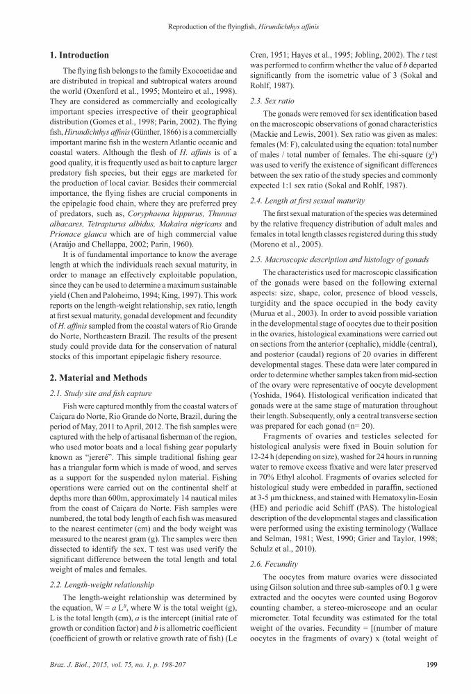
expected ratio of 1:1. This difference was statistically significant (χ2 = 18.63). The females of H. affinis had slight predominance in the sampled population. The females predominated during June to October and the males in the month of May, with significant differences (Figure 2).
3.3. Length at first maturityThe total length at first sexual maturity was estimated
at 27.3 cm for males and 27.1 cm for females. The males of H. affinis mature before the females, shown by the significant difference in size of gonadal maturation of both sexes (t = –5.081; df = 210; p < 0.05).
3.4. Gonad developmentH. affinis had paired elongated gonads, the females
with lobed ovaries and the males with flattened testes. The testicular walls were fragile when compared to the ovarian walls, and did not show much modification between the different stages of development, unlike the ovaries. The volume, coloration, thickness and blood vessels of ovaries varied according to the stage of maturation, presenting shades of light pink to dark yellow, due to the color of the
Figure 1. Distribution by total body length classes (a) and (b) total weight classes of Hirundichthys affinis.
Table 1. Parameter estimates of the length-weight relationship of Hirundichthys affinis (Günther, 1866) (Beloniformes/Exocoetidae).
Species Sex n a b Equation Parameters r2 t- test (d.f.) GrowthS.E. of b (95% C.I. of b)Hirundichythys affinis
M 117 0.0916 2.208* 0.181 (1.8493 to 2.566) 0.56 t = – 8.646 (113) negatively allometric
F 193 0.0068 2.985* 0.359 (2.2765 to 3.6935) 0.26 t = – 2.205(188) negatively allometric
M, males; F, females; n, sample size; a, intercept; b, slope; C.I., confidence interval; r², coefficient determination; *significant differences from 3 (P < 0.05).
Braz. J. Biol., 2015, vol. 75, no. 1, p. 198-207 201201
Reproduction of the flyingfish, Hirundichthys affinis
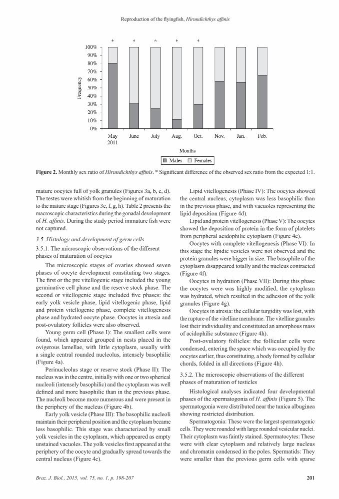
mature oocytes full of yolk granules (Figures 3a, b, c, d). The testes were whitish from the beginning of maturation to the mature stage (Figures 3e, f, g, h). Table 2 presents the macroscopic characteristics during the gonadal development of H. affinis. During the study period immature fish were not captured.
3.5. Histology and development of germ cells3.5.1. The microscopic observations of the different phases of maturation of oocytes
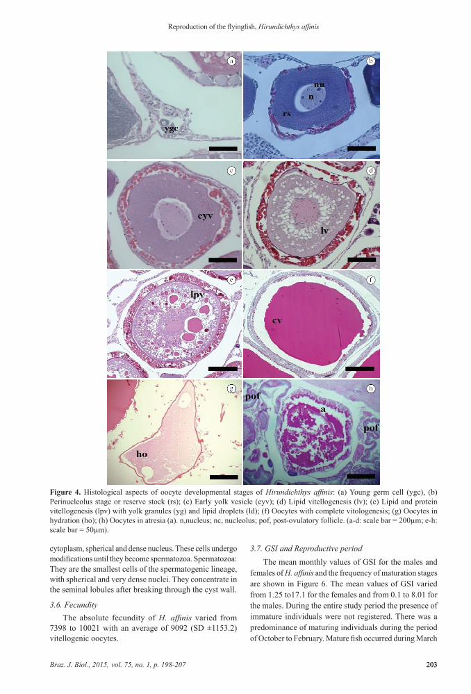
The microscopic stages of ovaries showed seven phases of oocyte development constituting two stages. The first or the pre vitellogenic stage included the young germinative cell phase and the reserve stock phase. The second or vitellogenic stage included five phases: the early yolk vesicle phase, lipid vitellogenic phase, lipid and protein vitellogenic phase, complete vitellogenesis phase and hydrated oocyte phase. Oocytes in atresia and post-ovulatory follicles were also observed.
Young germ cell (Phase I): The smallest cells were found, which appeared grouped in nests placed in the ovigerous lamellae, with little cytoplasm, usually with a single central rounded nucleolus, intensely basophilic (Figure 4a).
Perinucleolus stage or reserve stock (Phase II): The nucleus was in the centre, initially with one or two spherical nucleoli (intensely basophilic) and the cytoplasm was well defined and more basophilic than in the previous phase. The nucleoli become more numerous and were present in the periphery of the nucleus (Figure 4b).
Early yolk vesicle (Phase III): The basophilic nucleoli maintain their peripheral position and the cytoplasm became less basophilic. This stage was characterized by small yolk vesicles in the cytoplasm, which appeared as empty unstained vacuoles. The yolk vesicles first appeared at the periphery of the oocyte and gradually spread towards the central nucleus (Figure 4c).
Lipid vitellogenesis (Phase IV): The oocytes showed the central nucleus, cytoplasm was less basophilic than in the previous phase, and with vacuoles representing the lipid deposition (Figure 4d).
Lipid and protein vitellogenesis (Phase V): The oocytes showed the deposition of protein in the form of platelets from peripheral acidophilic cytoplasm (Figure 4e).
Oocytes with complete vitellogenesis (Phase VI): In this stage the lipidic vesicles were not observed and the protein granules were bigger in size. The basophile of the cytoplasm disappeared totally and the nucleus contracted (Figure 4f).
Oocytes in hydration (Phase VII): During this phase the oocytes were was highly modified, the cytoplasm was hydrated, which resulted in the adhesion of the yolk granules (Figure 4g).
Oocytes in atresia: the cellular turgidity was lost, with the rupture of the vitelline membrane. The vitelline granules lost their individuality and constituted an amorphous mass of acidophilic substance (Figure 4h).
Post-ovulatory follicles: the follicular cells were condensed, entering the space which was occupied by the oocytes earlier, thus constituting, a body formed by cellular chords, folded in all directions (Figure 4h).
3.5.2. The microscopic observations of the different phases of maturation of testicles
Histological analyses indicated four developmental phases of the spermatogonia of H. affinis (Figure 5). The spermatogonia were distributed near the tunica albuginea showing restricted distribution.
Spermatogonia: These were the largest spermatogenic cells. They were rounded with large rounded vesicular nuclei. Their cytoplasm was faintly stained. Spermatocytes: These were with clear cytoplasm and relatively large nucleus and chromatin condensed in the poles. Spermatids: They were smaller than the previous germ cells with sparse
Figure 2. Monthly sex ratio of Hirundichthys affinis. * Significant difference of the observed sex ratio from the expected 1:1.
Braz. J. Biol., 2015, vol. 75, no. 1, p. 198-207202
Oliveira, MR. et al.
202
Figure 3. Macroscopic stages of gonad development in Hirundichthys affinis: (a) ovary in the maturation process, (b) mature ovary, (c) spent ovary, (d) resting ovary, (e) testis in the early maturation process, (f) testis in the late maturation process, (g) mature testis, (h) spent. (Immature individuals were not captured during this study).
Table 2. Macroscopic description of the gonad development stages of Hirundichthys affinis.Stage Females Males
Maturing Ovaries with a clear orange color and ovaries still small in size, reaching 1/3 of the coelomic cavity, with superficial vascularization and small oocytes.
Early maturation: Testes were filiform, reduced in size and whitish in color. Mid maturation: Testes slightly bigger in size, whitish in color and with few blood vessels.
Mature (ripe) Ovaries were big, dark yellowish orange in color occupying almost the entire coelomic cavity. The mature ovaries were turgid, highly vascularized with numerous big oocytes visible to the naked eye.
Testes were turgid, white in color, occupying a large space in the coelomic cavity.
Spent Ovaries were hemorrhagic, flaccid, shrunk and wrinkled in appearance and were of faded yellow color.
Testes were smaller than the previous stage, with hemorrhagic and flaccid aspects and brownish in color.
Resting Ovaries were of reduced size with a light pink color.
Braz. J. Biol., 2015, vol. 75, no. 1, p. 198-207 203203
Reproduction of the flyingfish, Hirundichthys affinis
cytoplasm, spherical and dense nucleus. These cells undergo modifications until they become spermatozoa. Spermatozoa: They are the smallest cells of the spermatogenic lineage, with spherical and very dense nuclei. They concentrate in the seminal lobules after breaking through the cyst wall.
3.6. FecundityThe absolute fecundity of H. affinis varied from
7398 to 10021 with an average of 9092 (SD ±1153.2) vitellogenic oocytes.
3.7. GSI and Reproductive periodThe mean monthly values of GSI for the males and
females of H. affinis and the frequency of maturation stages are shown in Figure 6. The mean values of GSI varied from 1.25 to17.1 for the females and from 0.1 to 8.01 for the males. During the entire study period the presence of immature individuals were not registered. There was a predominance of maturing individuals during the period of October to February. Mature fish occurred during March
Figure 4. Histological aspects of oocyte developmental stages of Hirundichthys affinis: (a) Young germ cell (ygc), (b) Perinucleolus stage or reserve stock (rs); (c) Early yolk vesicle (eyv); (d) Lipid vitellogenesis (lv); (e) Lipid and protein vitellogenesis (lpv) with yolk granules (yg) and lipid droplets (ld); (f) Oocytes with complete vitologenesis; (g) Oocytes in hydration (ho); (h) Oocytes in atresia (a). n,nucleus; nc, nucleolus; pof, post-ovulatory follicle. (a-d: scale bar = 200µm; e-h: scale bar = 50µm).

Braz. J. Biol., 2015, vol. 75, no. 1, p. 198-207204
Oliveira, MR. et al.
204
to July. Variations in IGS and the monthly frequency of maturation stages demonstrate that H. affinis reproduces during the period of March to July.
4. Discussion
It is of fundamental importance to have information regarding the length, weight, sex and length at first sexual maturity, in order to manage an effectively exploitable
fishery population. Small sized immature individuals of H. affinis were not captured during this study. The immature individuals frequently occur in the oceanic region and only the adults migrate to the coastal waters for breeding. However, the effect of the selectivity of the fishing gear employed cannot be ruled out (Araújo and Chellappa, 2002). The absence of individuals less than 19 cm and greater than 24.5cm of the same species, in
Figure 5. Histological aspects of spermatocyte developmental stages of Hirundichthys affinis: a) cross section of the testis; b) testis showing stages of spermatogenesis; c) spermatogonia (spg), spermatocyte (spc), spermatid (spt); d) spermatozoid (spz). (a-b: scale bar = 50µm; c-d: scale bar = 200µm).
Figure 6. a) Temporal distribution of GSI (± SE) of females and males Hirundichthys affinis; b) monthly frequency of maturity stages for pooled sex.

Braz. J. Biol., 2015, vol. 75, no. 1, p. 198-207 205205
Reproduction of the flyingfish, Hirundichthys affinis
the Caribbean coastal waters, was due to the same reason (Khokiattiwong et al., 2000).
The females of H. affinis in this study were larger and heavier than the males. The females are usually heavier than the males due to their gonads which tend to have a greater mass in relation to the testicles (Araújo and Chellappa, 2002; Murua et al., 2003). For the same species in the Caribbean, it was observed that the average length of males and females was not different when fish were immature, but later the females grew faster than males and became larger and heavier (Khokiattiwong et al., 2000). The present study registered a predominance of females which could be due to the selectivity of the fishing gear used with the interest of capturing the bigger individuals. However, females were observed more in the months of June and July, and this period corresponds to the peak of the reproductive activity of H. affinis in Rio Grande do Norte, Brazil (Araújo and Chellappa, 2002; Araújo et al., 2011).
It is possible to determine the type of growth of a species through the allometric coefficient (b), which is isometric when b = 3, positive allometry when b> 3 and negative allometry when b <3 (Jobling, 2002). Isometric growth indicates that the body increases in all dimensions in the same proportion during growth, whereas positive allometry indicates that the body becomes more rotound as it increases in length, and negative allometry indicates a slimmer body (Jobling, 2002). In this study, the allometric coefficient of the length-weight relationship of males and females of H. affinis was estimated to be lower than 3 indicating negatively allometric growth. These results are in agreement with a study conducted for the same species in the oceanic waters of Northeastern Brazil (Lessa and Bezerra Junior, 2004). For this same species in Barbados, the values of b=2.98 for males, b= 3.03 for females and b=3.01 for grouped sex (Khokiattiwong et al., 2000). The parameters of length-weight relationship in fish could be influenced by environmental conditions, gonadal maturity, sex, condition factor, season, population and variations between species (Froese, 2006). In this study, the gonadal development of females could have possibly influenced the body mass.
For rational management of fishery stocks which are subjected to exploitation, it is important to know the size at first gonadal maturation (L50), since it provides information for determining the minimum size at capture and mesh dimensions of the fishing gear. In this study, H. affinis presented a mean length of 27 cm at first maturity, which is larger than that registered in the Caribbeans, where they attained maturity below 18 cm (Khokiattiwong et al., 2000). In the oceanic waters of Northeastern Brazil, the minimum size of capture which was registered for H. affinis is 22.7 cm (Lessa and Bezerra Junior, 2004). The size at first maturity is not fixed and may vary between individuals of the same species, whose populations are subject to different environmental conditions (Wootton, 1990). The length of first sexual maturation may be directly affected through changes in the quantity of energy reserves available for gonad development (Morgan, 2004).
The macroscopic analyses of gonads of H. affinis showed four stages each of ovarian and testicular development. For the same species in Barbados, five ovarian and testicular maturation stages were observed, which included the immature stage (Khokiattiwong et al., 2000). During this study, immature individuals were not captured since the migratory stocks were in the gonad maturation process. Possibly the younger fish were in the oceanic region. Additionally, the fishermen tend to capture the bigger individuals for commercial purposes. The macroscopic description of gonads shows the stages in reproductive cycle, which could be useful to understand the gross reproductive biology of this species.
The histological observations of this study register for the first time the presence of hydrated oocyte phase in the mature ovaries and oocytes in atresia and post-ovulatory follicle phase in the spent ovaries for H. afinnis. The maturation of the oocyte ends in ovulation, with the rupture of the follicle, liberating the oocytes in the lumen of the ovary (Lowerre-Barbieri et al., 2011). The ruptured follicles are known as post-ovulatory follicles (POFs) and they remain in the ovary till they are reabsorbed (Hunter and Macewicz, 1985). The females which are capable of spawning are those which appear during the reproductive cycle with vitelogenic oocytes or with histological indicators of imminent spawning (late migration of germinal vesicle, germinal vesicle breakdown and hydration) or recent spawning (POFs of recent collapse). This study registered the presence of H. affinis in the coastal waters with hydrated oocytes and POFs, which suggests that the females use this area as spawning grounds. This species migrates from the oceanic waters to the coastal waters of Caiçara do Norte in Northeastern Brazil to spawn and complete its reproductive cycle.
This study describes for the first time the histological analyses of spermatogenesis in the testes of H. affinis in the coastal waters of Rio Grande do Norte, Brazil. Observing the distribution of spermatogonia in the germinal compartment of testes H. affinis shows restricted distribution in which the spermatogonia are distributed near the tunica albuginea (Grier, 1981). This type of organization is found in teleost orders such as Atheriniformes, Cyprinodontiformes and Beloniformes (Parenti and Grier, 2004).
This study registered an average of 9092 vitellogenic oocytes for H. affinis, although a lower value has been registered in an earlier study (Araújo and Chellappa, 2002). Fecundity is a specific reproductive tactics and is adapted to the conditions of the life cycle of the species, varying with the growth, population density, body size, food availability, and rate of mortality (Witthames et al., 1995; Murua et al., 2003).
Spawning of H. affinis occurrs during the months of March to July. For the same species, the reproductive period registered was from May to June in Northeastern Brazil (El-Deyr, 1998; Araújo and Chellappa, 2002; Araújo et al., 2011), and in Barbados was from December to June, with a peak from March to June (Khokiattiwong et al., 2000). Breeding season may be influenced by local climatic

Braz. J. Biol., 2015, vol. 75, no. 1, p. 198-207206
Oliveira, MR. et al.
206
conditions. Information pertaining to the reproductive period of H. affinis is important for the conservation of natural stocks of this epipelagic fishery resource. The microscopic descriptions of the stages of gonad maturation indicate that the study area is an important spawning ground of H. affinis. This information could be used for adequate management of exploitable fishery stocks of this commercially and ecologically important fish.
Acknowledgements
This study was supported by the by the National Council for Scientific and Technological Development of Brazil (CNPq) in the form of Research grants and by the Post-Graduate Federal Agency CAPES/MEC, Brazil.
References
ARAúJO, AS. and CHELLAPPA, S., 2002. Estratégia reprodutiva do peixe voador, Hirundichthys affinis Günther (Osteichthyes, Exocoetidae). Revista Brasileira de Zoologia, vol. 19, no. 3, p. 691-702. http://dx.doi.org/10.1590/S0101-81752002000300006.
ARAúJO, AS., OLIVEIRA, MR., CAMPOS, CEC., YAMAMOTO, ME. and CHELLAPPA, S., 2011. Características morfométricas-merísticas, peso-comprimento e maturação gonadal do peixe voador, Hirundichythys affinis (Günther, 1866). Biota Amazônia., vol. 1, no. 2, p. 33-40.
CHEN, Y. and PALOHEIMO, JE., 1994. Estimating fish length and age at 50% maturity using a logistic type model. Aquatic Sciences, vol. 56, no. 3, p. 206-219. http://dx.doi.org/10.1007/BF00879965.
EL DEYR, ACA., 1998. Biologia reprodutiva do peixe voador Hirundichtys affinis da região de Caiçara-RN. Recife: Universidade Federal Rural de Pernambuco. 92 p. Dissertação de Mestrado em Oceanografia.
FROESE, R., 2006. Cube law, condition factor and weight-length relationship: history, meta-analysis and recommendations. Journal of Applied Ichthyology, vol. 22, no. 4, p. 241-253. http://dx.doi.org/10.1111/j.1439-0426.2006.00805.x.
GRIER, HJ. and TAYLOR, RG., 1998. Testicular maturation and regression in the common snook. Journal of Fish Biology, vol. 53, no. 3, p. 521-542. http://dx.doi.org/10.1111/j.1095-8649.1998.tb00999.x.
GRIER, HJ., 1981. Cellular organization of the testis and spermatogenesis in fishes. American Zoologist, vol. 21, p. 345-357.
GOMES, C., DALES, RBG. and OXENFORD, HA., 1998. The application of RAPD markers in stock discrimination of the four-wing flyingfish, Hirundichthys affinis in the central western Atlantic. Molecular Ecology, vol. 7, no. 8, p. 1029-1039. http://dx.doi.org/10.1046/j.1365-294x.1998.00427.x.
HAYES, DB., BRODZIAK, JKT. and O’GORMAN, JB., 1995. Efficiency and bias of estimators and sampling designs for determining length-weight relationships of fish. Canadian Journal of Animal Science, vol. 52, p. 84-92.
HUNTER, JR. and GOLDBERG, SR., 1980. Spawning incidence and batch fecundity in northern anchovy, Engraulis mordax. Fish Bulletin, vol. 77, p. 641-652.
HUNTER, JR. and MACEWICZ, BJ., 1985. Measurement of spawning frequency in multiple spawning fishes. NOAA Technical Report NMFS, vol. 36, p. 79-94.
JOBLING, M., 2002. Environmental factors and rates of development and growth. In HART, PJ. and REYNOLDS, JD. (Eds.). Handbook of fish biology and fisheries. Oxford: Blackwell Publishing Ltd. p. 97-122. Fish Biology, vol. 1. http://dx.doi.org/10.1002/9780470693803.ch5.
KING, MG., 1997. Fisheries biology, assesment and management. Oxford: Osney Mead. 341 p. Fishing news books.
KHOKIATTIWONG, S., MAHON, R. and HUNTE, W., 2000. Seasonal abundance and reproduction of the fourwing flyingfish, Hirundichthys affinis, of Barbados. Environmental Biology of Fishes, vol. 59, no. 1, p. 43-60. http://dx.doi.org/10.1023/A:1007647918255.
LE CREN, ED., 1951. The length-weight relationship and seasonal cycle in gonadal weight and condition in the perch (Perca fluviatilis). Journal of Animal Ecology, vol. 20, no. 2, p. 201-219. http://dx.doi.org/10.2307/1540.
LESSA, RP. and BEZERRA JUNIOR, JL., 2004. Dinâmica de populações e avaliação dos estoques dos recursos pesqueiros do nordeste do Brasil: peixe voador, Hirundichthys affinis. In LESSA, RT., NÓBREGA, MF. and BEZERRA JUNIOR, JL. Dinâmica de populações e avaliação dos estoques dos recursos pesqueiros da região nordeste do Brasil. Recife: Programa REVIZEE; Relatório Executo. p. 39-50. vol. 2.
LOWERRE-BARBIERI, SK., GANIAS, K., SABORIDO-REY, F., MURUA, H. and HUNTER, JR., 2011. Reproductive Timing in Marine Fishes: Variability, Temporal Scales, and Methods. Marine and Coastal Fisheries, vol. 3, no. 1, p. 71-91. http://dx.doi.org/10.1080/19425120.2011.556932.
MACKIE, MC. and LEWIS, PD., 2001. Assessment of gonad staging systems and other methods used in the study of the reproductive biology of the narrow-barred Spanish mackerel, Scomberomorus commerson, in Western Australia. Western Australia: Fisheries Department. p. 1-32. Fisheries Research Report, vol. 136.
MARTINI, EE. and FOUNTAIN, R., 1981. Ovarian cycling frequency and batch fecundity in the queenfish, Seriphus politus: attributes representative of serial spawning fishes. Fish Bulletin, vol. 79, no. 3, p. 547-559.
MONTEIRO, A., VASKE JUNIOR, T., LESSA, RP. and EL-DEYR, ACA., 1998. Exocoetidae (Beloniformes) off north-eastern Brazil. Cybium, vol. 22, no. 4, p. 395-403.
MORGAN, MJ., 2004. The relationship between fish condition and the probability of being mature in American plaice (Hippoglossoides platessoides). ICES Journal of Marine Science, vol. 61, no. 1, p. 64-70. http://dx.doi.org/10.1016/j.icesjms.2003.09.001.
MORENO, T., CASTRO, JJ. and SOCORRO, J., 2005. Reproductive biology of the sand smelt Atherina presbyter Cuvier, 1829 (Pisces:Atherinidae) in the central-east Atlantic. Fisheries Research, vol. 72, no. 1, p. 121-131. http://dx.doi.org/10.1016/j.fishres.2004.06.016.
MURUA, H., KRAUS, G., SABORIDO-REY, F., WITTHAMES, PR., THORSEN, A. and JUNqUERA, S., 2003. Procedures to Estimate Fecundity of Marine Fish Species in Relation to their Reproductive Strategy. Journal of Northwest Atlantic Fishery Science, vol. 33, p. 33-54. http://dx.doi.org/10.2960/J.v33.a3.
OXENFORD, HA., MAHON, R. and HUNTE, W., 1995. Distribution and relative abundance of flyingfish (Exocoetidae)

Braz. J. Biol., 2015, vol. 75, no. 1, p. 198-207 207207
Reproduction of the flyingfish, Hirundichthys affinis
in the eastern Caribbean. I. Adults. Marine Ecology Progress Series, vol. 117, p. 11-23. http://dx.doi.org/10.3354/meps117011.
PARENTI, LR. and GRIER, HJ., 2004. Evolution and phylogeny of gonad morphology in bony fishes. Integrative and Comparative Biology, vol. 44, no. 5, p. 333-348. http://dx.doi.org/10.1093/icb/44.5.333. PMid:21676719
PARIN, NV., 1960. Flying fish (Exocoetidae) of the northwestern part of the Pacific Ocean. Trudy Instituta Okeanologie Akademii Nauk SSS, vol. 31, p. 205-285.
PARIN, NV., 2002. Exocoetidae: flyingfishes. In CARPENTER, KE. (Ed.). The living marine resources of the western Central Atlantic. Rome: FAO Species Identification Guide for Fishery Purposes and American Society of Ichthyologists and Herepetologists Special Publication no. 5. p. 1116-1134. Bony fishes part 1 (Acipenseridae to Grammatidae), vol. 2.
SOKAL, RR. and ROHLF, FJ., 1987. Introduction to biostatistics. 2nd ed. New York: Freeman. 363 p.
SCHULZ, RW., DE FRANçA, LR., LAREYRE, JJ., LE GAC, F., CHIARINI-GARCIA, H., NOBREGA, RH. and MIURA, T., 2010. Spermatogenesis in fish. General and Comparative Endocrinology, vol. 165, no. 3, p. 390-411. http://dx.doi.org/10.1016/j.ygcen.2009.02.013. PMid:19348807
WALLACE, RA. and SELMAN, K., 1981. Cellular and dynamic aspects of oocyte growth in teleosts. American Zoologist, vol. 21, p. 325-343.
WEST, G., 1990. Methods of assessing ovarian development in fishes: a Review. Australian Journal of Marine and Freshwater Research, vol. 41, no. 2, p. 199-222. http://dx.doi.org/10.1071/MF9900199.
WITTHAMES, PR., GREER WALKER, M., DINIS, MT. and WHITING, CL., 1995. The geographical variation in the potential annual fecundity od dover sole, Solea solea, from European shelf waters during 1991. Netherlands Journal of Sea Research, vol. 34, no. 1-3, p. 45-58. http://dx.doi.org/10.1016/0077-7579(95)90013-6.
WOOTTON, RJ., EVANS, GW. and MILLS, LA., 1978. Annual cycle in female three-spined sticklebacks (Gasterosteus aculeatus L.) from an upland and lowland population. Journal of Fish Biology, vol. 12, no. 4, p. 331-343. http://dx.doi.org/10.1111/j.1095-8649.1978.tb04178.x.
WOOTTON, RJ., 1990. Ecology of Teleost Fishes. 1st ed. Londres: Chapman & Hall. 404 p.
YOSHIDA, HO., 1964. Skipjack tuna spawning in the Marquesas Islands and Tuamotu Archipelago. Fish Bulletin, vol. 65, p. 479-488.