report of six cases - ccjm.org
TRANSCRIPT

ANOMALOUS LESIONS
OF T H E U P P E R U R I N A R Y T R A C T Report of Six Cases
ROBERT HUGHES, M.D., and B. H. NICHOLS, M.D.
Before the use of excretory urography and retrograde pyelography, anomalous lesions of the upper urinary tract were seldom diagnosed. In this paper we are presenting illustrated case histories of several of these anomalies, namely, ectopic kidney, fused kidney, solitary kidney, and ureteropelvic obstruction producing hydronephrosis. Some general concepts are applicable to anomalies of the upper urinary tract:
1. These anomalies are seldom suspected from the history or physical examination.
2. Urinary signs and symptoms may be absent. 3. Secondary complications are common. 4. These anomalies frequently produce referred abdominal symp-
toms ranging from vague dyspepsia and backache to symptoms of peptic ulcer, appendicitis, and gallbladder disease.
5. If not diagnosed before operation, these anomalies will complicate renal surgery.
6. Diagnosis is established by intravenous urography or retrograde pyelography.
The value of excretory urography cannot be overemphasized. It is a simple means of studying the urinary tract. The accuracy of diagnosis by this means depends on obtaining satisfactory roentgenograms and using a proper technic. Two general rules should be followed, namely, (1) no dye should be injected before a plain film of the abdomen is made, and (2) excretory urography should not be done within twenty-four hours of cystoscopy or catheterization of the ureters. Opaque shadows may be obscured, or false ones assumed, if a plain film is not available; irritability of the ureter and kidney from instrumentation can cause both to have an abnormal appearance. In exceptional instances a nor-mal kidney may fail to visualize with urography, possibly because of hyperactive excretion of the dye or because of temporary dysfunction of the kidney. In contrast to retrograde pyelography, this study is widely applicable and, if more generally used, would result in the earlier diagno-sis of many urological problems. Retrograde pyelography is frequently necessary to make or confirm the diagnosis, but should be reserved for the specialist.
Anomalous lesions of the upper urinary tract become urological problems if symptoms or complications develop. Any pathological con-
9
require permission. on April 4, 2022. For personal use only. All other useswww.ccjm.orgDownloaded from

ROBERT HUGHES AND B . H . NICHOLS
A B FIG. 1 (Case 1) A—Plain roentgenogram of the abdomen showing a large calcification
in the normal course of the right ureter. B—Urogram demonstrating that this calcification is a large stone in
an ectopic right kidney rather than a ureteral calculus. The congenital short ureter is well visualized.
dition that may occur in the normal kidney or ureter may develop in these anomalies. The more common complications are calculus forma-tion, infection, and hydronephrosis.
ECTOPIC KIDNEYS
Ectopia is reported to occur once in every thousand autopsies and to account for 16.9 per cent of renal anomalies. It may occur as a unilat-eral or bilateral condition. Unilateral ectopic kidneys have been re-ported in many locations, such as between the crura of the diaphragm, between the layers of the mesosigmoid or broad ligament, and between the uterus and rectum. In cases of crossed ectopia the involved kidney lies below the normal one to which it may be fused.
The abnormal position of ectopic kidneys tends to interfere with the drainage of urine. This predisposes such anomalies to disease. Without disease, however, they may cause referred pressure pain or complicate pregnancy.
10
require permission. on April 4, 2022. For personal use only. All other useswww.ccjm.orgDownloaded from

ANOMALOUS LESIONS
In the diagnosis a congenital short ureter is an important finding and serves to distinguish an cctopic kidney from a ptoscd kidney.
Case 1. (4-18-39) A man, aged 38, gave a history of pain in the right side in 1923, for which appendectomy was done. Pain recurred in 1926, at which time a routine roentgenogram of the abdomen demonstrated a calcification in the course of the lower right ureter. This was interpreted as being a ureteral calculus, and laparotomy was advised. The attempt to remove this stone at operation was unsuccessful. Despite this failure, the patient never reported any subsequent pain or other urinary symptoms. The calcification was again demonstrated on a routine roentgenogram made four years later.
In 1939 the patient entered the Clinic because of symptoms that were diagnosed as being due to neurocirculatory asthenia and hypometabolism. Because of the past his-tory and presence of many white blood cells in the urine, intravenous urography was done. This study demonstrated that the calcification, previously considered in the ureter, was actually present in a hydronephrotic right pelvic kidney. Surgery was advised but refused.
Case 2. (3-21-40) A man, aged 50, had an appendectomy performed in 1912. At this operation a mass of undetermined origin was found in the right lower quadrant.
FIG. 2 (Case 2) A—Plain roentgenogram showing a large staghorn type of calcifica-tion at the level of the right sacrum.
B—Pyelogram demonstrating this staghorn calcification in a lo'dro-nephrotic right ectopic kidney. The congenital short ureter is visualized.
11
require permission. on April 4, 2022. For personal use only. All other useswww.ccjm.orgDownloaded from

ROBERT HUGHES AND B . H . NICHOLS
He remained in good health until March, 1939 when he began to have recurrent attacks of dull pain in the lower right abdomen. There was no history of urinary symp-toms.
He entered the Clinic in March, 1940 because of pain in the abdomen and neck and the finding of pus and blood in the urine. Examination at the Clinic revealed evi-dence of periarthritis and confirmed the report of white blood cells and red blood cells in the urine. Plain films and retrograde pyelography showed a large calculus in an ectopic right kidney. Nephrectomy was done. Two months later the patient was symp-tom free.
These two cases point out that symptoms resembling appendicitis may be caused by an ectopic kidney. The first case also demonstrates that a complete examination of the urinary tract is necessary before a positive diagnosis of ureteral calculus can be made.
In the differential diagnosis of ectopic kidney there are two helpful clinical signs, namely, the presence of an atypical pelvic or abdominal mass and the history of urinary symptoms. In these cases there was no information that either sign was elicited before appendectomy was done. In case 2 the finding of an unidentified mass in the right lower quadrant at operation should have led to subsequent intravenous urography or retrograde pyelography. By these studies a positive diagnosis of ectopia could have been established; and, in this instance, early diagnosis might have avoided subsequent nephrectomy.
In case 1 an attempt to make a positive diagnosis of ureteral calculus on the plain roentgenogram probably was responsible for the surgical complications. This wrong assumption emphasizes that the presence of any calcification in the course of the urinary tract is not adequate evi-dence for a positive diagnosis of stone. The presence and location of a calculus must be confirmed by intravenous urography or retrograde pyelography. These studies also provide additional information on the status of each kidney, without which elective surgery should not be done. In this case an incomplete examination of the urinary tract was most likely responsible for an unsuccessful operation.
URETEROPELVIC OBSTRUCTION
Anomalous lesions producing partial or intermittent obstruction at the ureteropelvic junction may cause a hydronephrosis. Such congenital lesions are strictures, periureteral bands, high insertion of the ureter, torsions, and aberrant vessels. Symptoms resulting from these anomalies usually appear in young persons. This diagnosis should be suspected in the presence of a hydronephrosis without apparent cause, especially if the ureter fails to visualize or is normal in size. The exact etiology of the
12
require permission. on April 4, 2022. For personal use only. All other useswww.ccjm.orgDownloaded from
ANOMALOUS LESIONS
hydronephrosis is not evident on intravenous urography or retrograde pyelography and can only be determined at operation. The earlier these surgical lesions are suspected, the greater the opportunity for plastic repair. The results depend more on the degree of damage and infection in the kidney than on the type of anomaly.
One of the following cases illustrates a hydronephrosis due to stenosis or stricture at the ureteropelvic junction, and the other, hydronephrosis secondary to the presence of aberrant vessels.
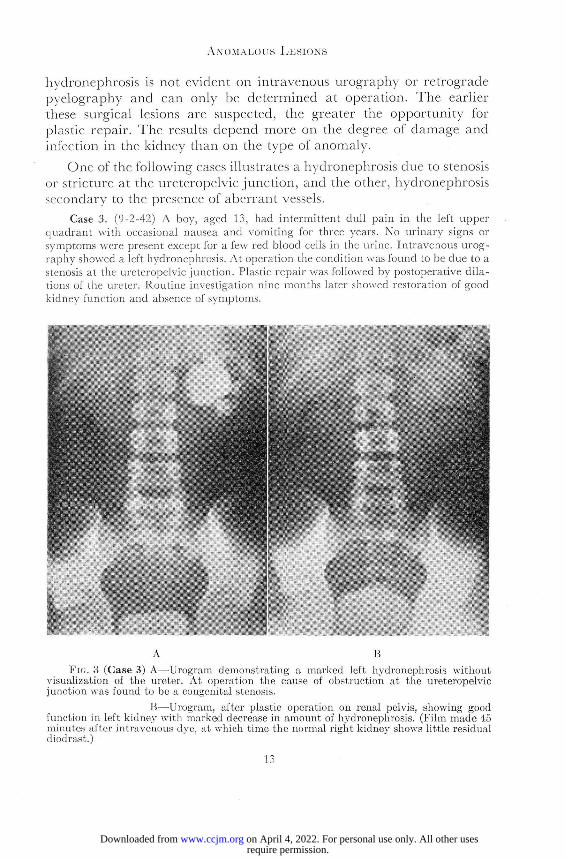
Case 3. (9-2-42) A boy, aged 13, had intermittent dull pain in the left upper quadrant with occasional nausea and vomiting for three years. No urinary signs or symptoms were present except for a few red blood cells in the urine. Intravenous urog-raphy showed a left hydronephrosis. At operation the condition was found to be due to a stenosis at the ureteropelvic junction. Plastic repair was followed by postoperative dila-tions of the ureter. Routine investigation nine months later showed restoration of good kidney function and absence of symptoms.
FIG. 3 (Case 3) A—Urogram demonstrating a marked left hydronephrosis without visualization of the ureter. At operation the cause of obstruction at the ureteropelvic junction was found to be a congenital stenosis.
B—Urogram, after plastic operation on renal pelvis, showing good function in left kidney with marked decrease in amount of hydronephrosis. (Film made 45 minutes after intravenous d3re, at which time the normal right kidney shows little residual diodrast.)
13
require permission. on April 4, 2022. For personal use only. All other useswww.ccjm.orgDownloaded from

ROBERT HUGHES AND B . H . NICHOLS
A B FIG. 4 (Case 4) A—Urogram showing a nonfunctioning left kidney. The right kidney
is partially obscured but normal. B—Pyelogram revealing a huge left hydronephrosis. The left ureter
contains some dye and appears normal in size. At operation the cause of obstruction at the ureteropelvic junction was found to be aberrant vessels. (The course of the upper left ureter demonstrated on the pyelogram suggested this possibility.)
Case 4. (8-18-42) A woman, aged 21, had intermittent attacks of severe pain in the left side for five years. This pain radiated to the abdomen and was frequently associated with nausea and vomiting. She had no urinary symptoms until gross hematuria oc-curred with a recent attack. Her family physician found a nonfunctioning left kidney on intravenous urography and referred the patient to the Clinic for further study.
The general examination and repeated urinanalyses were normal. Intravenous urography was repeated and confirmed the diagnosis of a nonfunctioning left kidney. Retrograde pyelography disclosed a huge left hydronephrosis due to obstruction at the ureteropelvic junction. At operation the cause was found to be aberrant vessels. Exten-sive kidney damage was present and necessitated nephrectomy. After operation, the patient was symptom free.
These two cases illustrate that hydronephrosis may develop with minimal symptoms or may produce atypical attacks of severe pain with or without urinary symptoms. The clinical picture of congenital hydro-nephrosis does not vary from that of acquired hydronephrosis; the symp-toms may be referred to the gastrointestinal tract or resemble gall-bladder disease in either case. The possibility of hydronephrosis should be considered in all patients having atypical abdominal distress.
14
require permission. on April 4, 2022. For personal use only. All other useswww.ccjm.orgDownloaded from

ANOMALOUS LESIONS
FUSED KIDNEYS Fused kidneys are reported to occur once in every 710 autopsies.
They are the most common of all renal anomalies except for reduplica-tion of the renal pelves. Fused kidneys are divided into two groups, the asymmetric and symmetric. The asymmetric type is rare and refers to such anomalies as unilateral fused kidneys or crossed ectopia with fusion. The symmetric, type, or horseshoe kidney, is common and shows fusion of the poles, usually the lower, by an isthmus of parenchymatous or fibrous tissue which crosses the lumbar spine. In this instance the fused organ is referred to as "sitting on the spine."
These renal anomalies are associated with abnormal arrangement of the pelves, ureters, and calices. The pelves are anterior; the ureters lie in front and usually cross the isthmus; the calices are not constant in position but tend to lie directed toward the midline.
Gutierrez,* in his monograph on horseshoe kidney, considered this anomaly an important clinical entity. He listed the symptoms as a
A B FIG. 5 (Case 5) A—Pyelogram showing an asymmetric fused kidney. Marked hydro-
nephrosis is demonstrated in one of the two pelves present in the left half of this kidney. B—Urogram showing normal renal function after heminephrectomy
and division of capsular fusion. "•Gutierrez, Robert: The Clinical Management of Horseshoe Kidney. (New York: Paul B
Hoeber, 1934) p. 125. 15
require permission. on April 4, 2022. For personal use only. All other useswww.ccjm.orgDownloaded from

ROBERT HUGHES AND B . H . NICHOLS
syndrome: "(a) nephralgia or pain in the middle of the abdomen re-ferred to the epigastrium or umbilical region; (b) gastrointestinal dis-orders with a long history of marked chronic constipation, and (c) long-standing intermittent attacks of urinary disturbances." These symptoms may likewise be present with an asymmetric fusion.
The case presented is of an asymmetric fused kidney with an anoma-lous bifid pelvis in the left half. The left kidney was further altered by marked hydronephrosis in one of the two pelves.
Case 5. (5-10-43) A woman, aged 25, had intermittent attacks of pain in the lower left back for the past five years. This pain was frequently associated with chills and fever, and occasionally with cloudy urine. There was no history of typical colic, frequency, or hematuria. Two years previously she went through pregnancy without developing any complications. After delivery, however, a mild attack of chills and fever occurred.
Examination at the Clinic revealed some tenderness and the suggestion of a mass in the midabdomen. Many white blood cells and a few red blood cells were found upon repeated urinalyses. Intravenous and retrograde pyelography showed an asymmetric fused kidney with the left half ectopic and lying in the midline. The left half showed a bifid pelvis with a marked hydronephrosis involving one of the two pelves. Dr. C. C. Higgins performed a heminephrectomy and removed the hydronephrotic portion of the left kidney. At operation both kidneys were found to be united by capsular fusion, which was severed. Follow-up two months later showed entirely satisfactory progress.
SOLITARY KIDNEYS
Solitary kidneys have been reported to constitute approximately 15 per cent of renal anomalies and to occur once in every 2500 births.
Solitary kidney means a complete or partial lack of development of the urinary tract on the affected side. The corresponding ureter, ureteral orifice, and trigone are usually absent along with the kidney. The fact that a part or all of the ureter may persist in the absence of the kidney, however, is important to remember in the differential diagnosis. The diagnosis of this anomaly requires that both intravenous urography and retrograde pyelography be done.
Solitary kidneys are frequently associated with malformations of the genital tract; rarely occupy an ectopic position; usually are associated with some compensatory hypertrophy; and are subject to all renal complications. It is obvious that disease in a solitary kidney is extremely serious. Operative procedures are justified only when planned with the knowledge that a single kidney is present. Despite these serious implica-tions, this anomaly is consistent with normal health and an average span of life.
In the case reported here, a normal solitary kidney was discovered in a woman who had pelvic inflammatory disease.
16
require permission. on April 4, 2022. For personal use only. All other useswww.ccjm.orgDownloaded from

ANOMALOUS LESIONS
Case 6. (8-25-43) A woman, aged 33, stated that she had a pelvic abscess on the right side, which was incised and drained seven years ago. The associated distress was never completely relieved by this operation. In the past year both the pain and pelvic mass recurred. Intravenous urography was done in the course of her examination and was interpreted as showing a nonfunctioning right kidney. There was no history of urinary symptoms.
The patient was referred to the Clinic for further investigation. The physical exami-nation was normal except for confirming the presence of a mass in the right side of the pelvis. Urinalyses and blood urea determinations were nor-mal. Intravenous urography was repeated and demonstrated a normal left kidney but showed no evidence of a right kidney or right ureter. Retrograde studies were then done and revealed an absence of the right ureteral orifice and right trigone. By correlating these findings, a positive diagnosis of an absent right kidney was made.
This patient was advised to have an exploratory laparotomy but operation was refused. The pelvic pathology was clinically considered inflammatory in character.
FIG. 6 (Case 6) Urogram showing a normal solitary left kidney. (The absence of the right kidney was confirmed at cystoscopy, which failed to show either the right ureteral orifice or right trigone.)
1. Six cases of anomalous lesions in the upper urinary tract arc presented.
2. These lesions become urological problems when symptoms or complications develop.
3. These anomalies may be misinterpreted as appendicitis, gastro-intestinal disease, or gallbladder disease.
4. Surgical operations upon the kidney or ureter will be complicated if an anomaly is present.
5. Adequate investigation of the urinary tract must be made for accurate diagnosis.
SUMMARY
17
require permission. on April 4, 2022. For personal use only. All other useswww.ccjm.orgDownloaded from