replicating myoblasts express amuscle-specific phenotype
TRANSCRIPT
Proc. Nati. Acad. Sci. USAVol. 85, pp. 9606-9610, December 1988Developmental Biology
Replicating myoblasts express a muscle-specific phenotype(desmin/muscle membrane glycoprotein H36/5-bromo-2'-deoxyuridine/regulation/skeletal muscle development)
STEPHEN J. KAUFMAN AND RACHEL F. FOSTERDepartments of Microbiology and Cell Biology, University of Illinois, 131 Burrill Hall, 407 South Goodwin Avenue, Urbana, IL 61801
Communicated by C. Ladd Prosser, September 6, 1988
ABSTRACT During the terminal stage of skeletal myogen-esis, myoblasts stop replicating, fuse to form multinucleatefibers, and express the genes that encode the proteins thatconvey contractile capacity. Because of this dramatic shift inproliferative state, morphology, and gene expression, it hasbeen possible to readily identify and quantitate terminallydifferentiating myoblasts. In contrast, it is not clear whetherthe proliferating cells that give rise to postmitotic myoblasts areequally distinct in their phenotype and in fact whether distinctstages in skeletal myogenesis precede the onset of terminaldifferentiation. To address these questions, monoclonal anti-bodies and immunofluorescence microscopy were used todetermine that replicating myoblasts from newborn rats doexpress a muscle-specific phenotype. To identify replicatingcells, incorporation of 5-bromo-2'-deoxyuridine (BrdUrd) intoDNA was assayed by using anti-BrdUrd antibody. The devel-opmentally regulated, muscle-specific, integral membrane pro-tein H36 and the intermediate-filament protein desmin werescored as markers of the myogenic phenotype. The percentageof BrdUrd' (i.e., proliferative) cells among H36' and desmin'myoblasts was equal to the percentage of BrdUrdM cells in theentire population, indicating that the expression of H36 anddesmin is uniformly characteristic of replicating myoblasts.Inhibition of protein synthesis before and during growth inBrdUrd did not alter the frequency of desmin and H36immunofluorescence in BrdUrdM cells. Thus, desmin and H36were present in the replicating myoblasts prior to the onset ofgrowth in BrdUrd. These results were confirmed using H36'cells selected by flow cytometry: these purified H36' myoblastsreplicate, express desmin, and differentiate. Similar resultswere obtained with mouse myoblasts. Desmin expression inthese mammalian cells differs from that in chicken embryomyoblasts: only a small proportion of replicating chickenembryo myoblasts express desmin. That replicating mamma-lian myoblasts have a muscle-specific phenotype serves todefine a distinct stage in myogenic development and a specificcell in the myogenic lineage. Further, it implies that there is aregulatory event activated during myogenesis that precedesterminal differentiation and that is required for expression ofthose genes whose products distinguish the replicating myo-blast.
In what has come to be known as the terminal stage of skeletalmuscle differentiation, myoblasts cease replicating and ex-press a panoply of phenotypic characteristics intrinsic to thisstage of development. During this same transition in myo-genic development, skeletal myoblasts fuse to form multinu-cleate fibers in which assembly of myofibrillar proteins takesplace. Once assembled in these fibers, these proteins providethe structural basis and enzymatic capacities that render thefibers functional contractile units, capable of responding toneuronal excitation. Expression of the genes that encodethese proteins is largely regulated at the level of transcription
(1-6); alternative splicing provides additional diversity and isoften involved in the switching of isoforms (6, 7). Translationof muscle-specific transcripts may also be regulated (8-11).The earliest expression of the terminally differentiated
phenotype takes place in myoblasts that normally do notreplicate further (12, 13). However, there appears to be aperiod in which these postmitotic, terminally differentiatingmyoblasts can be manipulated to replicate again (14, 15):when replication is reinitiated, expression of the differenti-ated phenotype stops. Analysis of developmental mutantsand the use oftumor viruses and growth factors likewise haveled to the conclusion that continued proliferation is incom-patible with terminal myogenic development (16-23). Studiesusing heterokaryons suggest that soluble trans-acting factorsmay mediate or initiate terminal differentiation (24, 25).Transfection studies support this concept and indicate thatthe product of a single gene may mediate the onset of thisstage of skeletal muscle development (26, 27).By using antibodies and nucleic acid probes, and by
measuring isozyme activities and myoblast fusion, it has beenpossible to identify and quantitate terminally differentiatingmyoblasts. In contrast, it is not clear whether the replicatingcells that give rise to these postmitotic myoblasts have anequally distinct phenotype that distinguishes this particularstage of myogenic development. We believe that this is thecase and demonstrate here that expression of the muscle-specific intermediate-filament protein desmin (28, 29) and themuscle-specific cell surface glycoprotein H36 (30, 31) arehallmarks of the population of replicating myoblasts that giverise to terminally differentiating myoblasts.
Previous work (32) demonstrated that detection of incor-porated 5-bromo-2'-deoxyuridine (BrdUrd) by immunofluo-rescence, in conjunction with other antibodies, can be usedto sequence the molecular events in single cells as they relateto the transition in DNA synthesis that accompanies myo-genic differentiation. The selective prolongation by lamininof the proliferative phase of myogenic development was alsodemonstrated by using BrdUrd incorporation as an index ofcell replication (33). We have adopted this same strategy inthese experiments to show that replicating myoblasts fromnewborn rats express H36 and desmin and that replicatingmyoblasts from newborn mice likewise express desmin. Thisdemonstration of a distinct muscle-specific phenotype inreplicating myoblasts distinguishes these cells within themyogenic lineage and necessitates consideration of addi-tional regulatory steps in the pathway of skeletal myogenesis.
In contrast with results found in mammalian cells, rela-tively few replicating myoblasts from the chicken embryoappear to express desmin, indicating that the regulation ofdesmin expression and the expression of a muscle-specificphenotype in replicating myoblasts may differ during avianand mammalian myogenesis.
Abbreviations: mAb, monoclonal antibody; DPBS, Dulbecco'sphosphate-buffered saline; BrdUrd, 5-bromo-2'-deoxyuridine.
9606
The publication costs of this article were defrayed in part by page chargepayment. This article must therefore be hereby marked "advertisement"in accordance with 18 U.S.C. §1734 solely to indicate this fact.

Proc. Natl. Acad. Sci. USA 85 (1988) 9607
METHODS AND MATERIALSCell Culture. Cultures from the hindlimbs of newborn
Sprague-Dawley rats and newborn BALB/c mice (Holtz-mann, Madison, WI) were prepared and grown in Dulbecco'smodified Eagle's medium (DMEM) supplemented with 10%horse serum, 10% fetal bovine serum, and antibiotics, asdescribed (33). Chicken embryo hindlimb cultures wereprepared from fertilized White Leghorn eggs (University ofIllinois Poultry Farm) by dissociation of minced tissue byVortex mixing (11- and 12-day embryos) (34); tissue from18-day embryos was preincubated for 15 min at 370C in 0.25%trypsin in calcium-free phosphate-buffered saline. Thechicken cells were grown on gelatin-coated or laminin-coated(33) dishes in medium further supplemented with 2% chickenembryo extract. Expression of desmin in replicating chickencells grown on laminin or gelatin was the same. Growth mediaand supplements were purchased from GIBCO.Immunofluorescence Microscopy. The muscle-specific in-
termediate-filament protein desmin was detected with DE-R-11 monoclonal antibody (mAb), generously provided byM. Osborn (Max-Planck Institute, Goettingen, F.R.G.) (35).Anti-H36 mAb reacts with a developmentally regulated,muscle-specific, integral membrane protein on the surface ofrat skeletal myoblasts and cardiac myocytes (30, 31) but noton smooth muscle. mAb 76.7, reactive with BrdUrd, was thegenerous gift of T. Ternynck (Institute Pasteur, Paris) (36).To render incorporated BrdUrd accessible to antibody, fixedcells were rinsed twice in Dulbecco's phosphate-bufferedsaline (DPBS), treated with 2 M HCl for 30 min at roomtemperature, neutralized by three 20-min washes in 50 mMNaCl/100mM Tris HCl, pH 7.4, and rinsed twice with DPBS.For simultaneous immunofluorescence labeling of desminand BrdUrd, cells grown on coverslips were incubated for 90min with 40 ,uM BrdUrd, fixed for 10 min with 95% ethanol,treated with 2 M HCl as cited above, and stained aspreviously described (32, 33). Cells labeled for H36 andBrdUrd were incubated with 40 ,uM BrdUrd for 90 min orwith 4 ,M BrdUrd for 15 hr, rinsed twice with cold DMEM,treated with fresh 1% formaldehyde for 10 min, and thenincubated at 5°C with mAb (5 ,g/ml) H36 followed byfluorescein isothiocyanate-conjugated rabbit antimouse IgG(FITC-RaMIg; Miles) diluted 1:50. The cells were washed,fixed with 95% ethanol, treated with 2 M HCl as above, andincubated with anti-BrdUrd mAb followed by FITC-RaMIg.All preparations were stained with 10 ,uM Hoechst 33342 toaid in enumeration of nuclei, mounted in glycerol/phosphate-buffered saline, 9:1 (vol/vol), pH 8.5, containing 0.01 Mp-phenylenediamine (Eastman), sealed with FLO-TEXX(Fisher), and examined with a Zeiss Universal microscopeequipped with epi-illumination optics and an HBO 100-Wmercury lamp. Photomicrographs were made with KodakTri-X film and a Planapochromat x63 objective. Handling ofcultures grown in BrdUrd was done in gold light (GeneralElectric F15T8-GO) prior to fixation.Flow Cytometry. Cells to be sorted by flow cytometry were
grown for 24 hr in DMEM supplemented with 30% fetalbovine serum and 2% chicken embryo extract on laminin-coated dishes to maximize proliferation (33). The cultures,containing both myogenic cells and fibroblasts, were rinsedwith calcium-free phosphate-buffered saline and dissociatedwith 0.05% trypsin. The cells were washed twice in DPBScontaining glucose at 1 mg/ml and bovine serum albumin at40 mg/ml (DPBS/BSA). The cells were resuspended to 107per ml in DPBS/BSA containing H36 mAb or normal mouseserum IgG at 50 ,ug/ml and then were incubated at 5°C for 15min, washed three times, incubated for 15 min in DPBS/BSAwith FITC-RaMIg, washed, and resuspended to 106 cells perml in DPBS/BSA for sorting. Myoblasts (i.e., H36' cells)were selected with a Coulter EPICS 751 cell sorter with an
argon laser adjusted to emit 400 mW at 488 nm, using a 457-to 502-nm laser-blocking filter and a 515-nm long-pass filter.Cells in the H36-labeled sample were collected if brighterthan 93% of the negative control cells.
RESULTSTo resolve whether replicating myoblasts express a distinctmuscle-specific phenotype, we determined whether the mus-cle-specific proteins desmin and H36 were expressed in vitroin replicating myoblasts from newborn rat thigh muscle. At24-hr intervals the cells were incubated for 90 min withBrdUrd at a final concentration of 40 ,M. Incorporation ofBrdUrd was detected by immunofluorescence using anti-BrdUrd antibody and scored in individual cells as an index oftheir proliferation. The specificity of the anti-BrdUrd mAbused is stringent and is restricted to the iodo and bromoderivatives of thymidine (36); this assay is equivalent insensitivity to that achieved by [3H]thymidine incorporationand autoradiography and it is by far more convenient (S.J.K.and M. Robert-Nicoud, unpublished data). Incubations withBrdUrd at the concentrations and times indicated had noeffect on subsequent myogenic development.Desmin and H36 were detected by using the respective
mAbs DE-R-11 and anti-H36. The specificity of DE-R-11 fordesmin has been established by immunoblotting with avianand mammalian desmin (35); the reactivity of anti-H36 isrestricted to cardiac and skeletal muscle (30). Culturesprepared from fetal rat hindlimb contain both myoblasts andfibroblasts. Only the myoblasts stain with anti-desmin oranti-H36, whereas both fibroblasts and myoblasts react withanti-vimentin antibody. Immunofluorescence was used todefine replicating cells that expressed H36 and desmin. Thatthese muscle-specific proteins are expressed in replicatingmyoblasts is illustrated in Fig. 1. Quantitation and furtherdocumentation of this follows.
Expression of Desmin in Replicating Rat Myoblasts. Thefrequency of expression of desmin by replicating cells from
FIG. 1. Expression of desmin (A) and H36 (B) by proliferatingcells from newborn rat hindlimb. (A) Cells were grown for 24 hr andincubated with 40 MM BrdUrd for 90 min, fixed with 95% ethanol,and stained with anti-BrdUrd and anti-desmin antibodies. Cells werephenotyped with respect to cytoplasmic desmin and BrdUrd incor-poration into DNA. Examples are shown of desmin' BrdUrd' (a)and desmin' BrdUrd- (b) cells and of a desmin- BrdUrd' cell (c).For data see Table 1. (B) A culture grown for 24 hr was enriched formyoblasts (i.e., H36' cells) by flow cytometry, replated, andincubated with 4 AM BrdUrd for 15 hr, restained with anti-H36, andthen fixed and stained with anti-BrdUrd antibody. Most cells (84%)incorporated BrdUrd and expressed H36 on their surface (BJ),indicating that the myoblast population was replicating. B2 shows aphase-contrast image of the cells in BJ. For data see Table 2.(Planapochromat x63 objective; bar = 20 ,m.)
Developmental Biology: Kaufman and Foster
9608 Developmental Biology: Kaufman and Foster
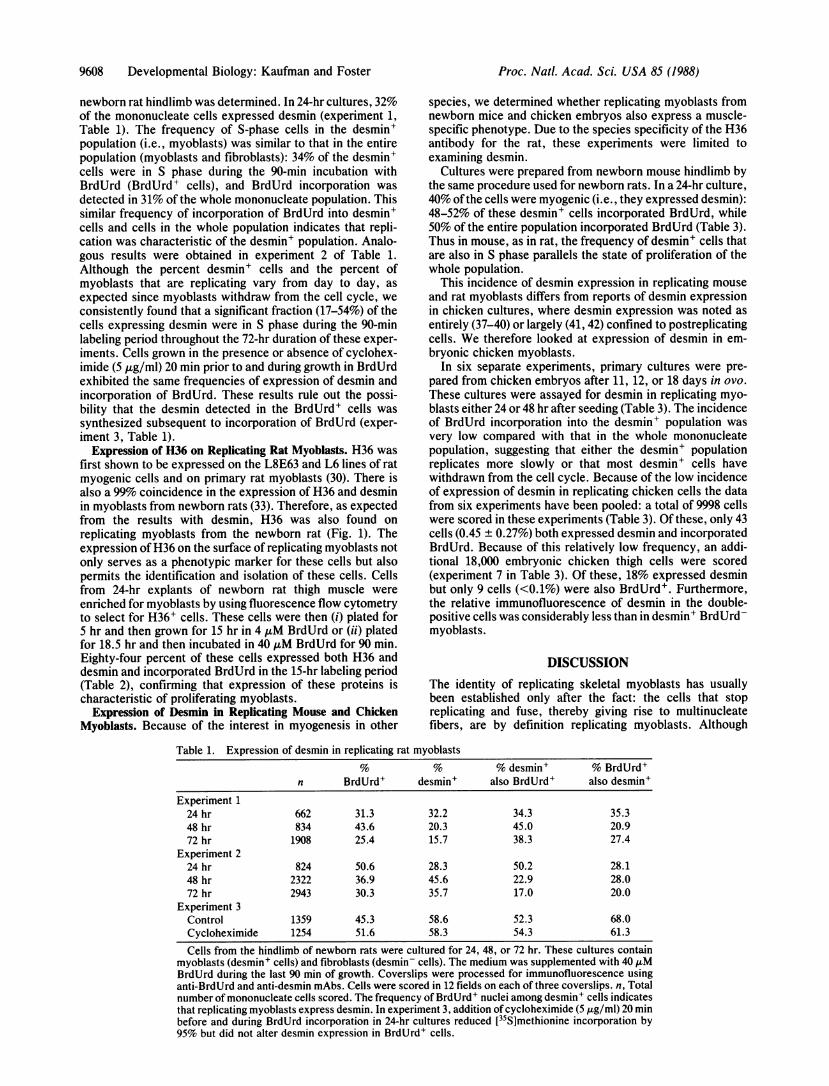
newborn rat hindlimb was determined. In 24-hr cultures, 32%of the mononucleate cells expressed desmin (experiment 1,Table 1). The frequency of S-phase cells in the desmin+population (i.e., myoblasts) was similar to that in the entirepopulation (myoblasts and fibroblasts): 34% of the desmin+cells were in S phase during the 90-min incubation withBrdUrd (BrdUrd+ cells), and BrdUrd incorporation was
detected in 31% of the whole mononucleate population. Thissimilar frequency of incorporation of BrdUrd into desmin+cells and cells in the whole population indicates that repli-cation was characteristic of the desmin+ population. Analo-gous results were obtained in experiment 2 of Table 1.Although the percent desmin+ cells and the percent ofmyoblasts that are replicating vary from day to day, as
expected since myoblasts withdraw from the cell cycle, we
consistently found that a significant fraction (17-54%) of thecells expressing desmin were in S phase during the 90-minlabeling period throughout the 72-hr duration of these exper-iments. Cells grown in the presence or absence of cyclohex-imide (5 ,ug/ml) 20 min prior to and during growth in BrdUrdexhibited the same frequencies of expression of desmin andincorporation of BrdUrd. These results rule out the possi-bility that the desmin detected in the BrdUrd+ cells was
synthesized subsequent to incorporation of BrdUrd (exper-iment 3, Table 1).
Expression of H36 on Replicating Rat Myoblasts. H36 was
first shown to be expressed on the L8E63 and L6 lines of ratmyogenic cells and on primary rat myoblasts (30). There isalso a 99% coincidence in the expression of H36 and desminin myoblasts from newborn rats (33). Therefore, as expectedfrom the results with desmin, H36 was also found onreplicating myoblasts from the newborn rat (Fig. 1). Theexpression of H36 on the surface of replicating myoblasts notonly serves as a phenotypic marker for these cells but alsopermits the identification and isolation of these cells. Cellsfrom 24-hr explants of newborn rat thigh muscle were
enriched for myoblasts by using fluorescence flow cytometryto select for H36+ cells. These cells were then (i) plated for5 hr and then grown for 15 hr in 4 ,uM BrdUrd or (ii) platedfor 18.5 hr and then incubated in 40 ,uM BrdUrd for 90 min.Eighty-four percent of these cells expressed both H36 anddesmin and incorporated BrdUrd in the 15-hr labeling period(Table 2), confirming that expression of these proteins ischaracteristic of proliferating myoblasts.
Expression of Desmin in Replicating Mouse and ChickenMyoblasts. Because of the interest in myogenesis in other
species, we determined whether replicating myoblasts fromnewborn mice and chicken embryos also express a muscle-specific phenotype. Due to the species specificity of the H36antibody for the rat, these experiments were limited toexamining desmin.
Cultures were prepared from newborn mouse hindlimb bythe same procedure used for newborn rats. In a 24-hr culture,40% ofthe cells were myogenic (i.e., they expressed desmin):48-52% of these desmin+ cells incorporated BrdUrd, while50% of the entire population incorporated BrdUrd (Table 3).Thus in mouse, as in rat, the frequency of desmin+ cells thatare also in S phase parallels the state of proliferation of thewhole population.
This incidence of desmin expression in replicating mouseand rat myoblasts differs from reports of desmin expressionin chicken cultures, where desmin expression was noted asentirely (37-40) or largely (41, 42) confined to postreplicatingcells. We therefore looked at expression of desmin in em-bryonic chicken myoblasts.
In six separate experiments, primary cultures were pre-
pared from chicken embryos after 11, 12, or 18 days in ovo.
These cultures were assayed for desmin in replicating myo-blasts either 24 or 48 hr after seeding (Table 3). The incidenceof BrdUrd incorporation into the desmin+ population was
very low compared with that in the whole mononucleatepopulation, suggesting that either the desmin+ populationreplicates more slowly or that most desmin+ cells havewithdrawn from the cell cycle. Because of the low incidenceof expression of desmin in replicating chicken cells the datafrom six experiments have been pooled: a total of 9998 cellswere scored in these experiments (Table 3). Of these, only 43cells (0.45 ± 0.27%) both expressed desmin and incorporatedBrdUrd. Because of this relatively low frequency, an addi-tional 18,000 embryonic chicken thigh cells were scored(experiment 7 in Table 3). Of these, 18% expressed desminbut only 9 cells (<0.1%) were also BrdUrd+. Furthermore,the relative immunofluorescence of desmin in the double-positive cells was considerably less than in desmin BrdUrM-myoblasts.
DISCUSSIONThe identity of replicating skeletal myoblasts has usuallybeen established only after the fact: the cells that stopreplicating and fuse, thereby giving rise to multinucleatefibers, are by definition replicating myoblasts. Although
Table 1. Expression of desmin in replicating rat myoblasts% % % desmin' % BrdUrd'
n BrdUrd' desmin' also BrdUrd' also desmin'Experiment 1
24 hr 662 31.3 32.2 34.3 35.348 hr 834 43.6 20.3 45.0 20.972 hr 1908 25.4 15.7 38.3 27.4
Experiment 224 hr 824 50.6 28.3 50.2 28.148 hr 2322 36.9 45.6 22.9 28.072 hr 2943 30.3 35.7 17.0 20.0
Experiment 3Control 1359 45.3 58.6 52.3 68.0Cycloheximide 1254 51.6 58.3 54.3 61.3
Cells from the hindlimb of newborn rats were cultured for 24, 48, or 72 hr. These cultures containmyoblasts (desmin' cells) and fibroblasts (desmin- cells). The medium was supplemented with 40 jxMBrdUrd during the last 90 min of growth. Coverslips were processed for immunofluorescence usinganti-BrdUrd and anti-desmin mAbs. Cells were scored in 12 fields on each of three coverslips. n, Totalnumber of mononucleate cells scored. The frequency of BrdUrd' nuclei among desmin' cells indicatesthat replicating myoblasts express desmin. In experiment 3, addition of cycloheximide (5,ug/ml) 20 minbefore and during BrdUrd incorporation in 24-hr cultures reduced [35S]methionine incorporation by95% but did not alter desmin expression in BrdUrd+ cells.
Proc. Natl. Acad. Sci. USA 85 (1988)

Proc. Natl. Acad. Sci. USA 85 (1988) 9609
Table 2. H36 and desmin expression in sorted rat myoblastsTime withBrdUrd, hr n % BrdUrd' % H36+ % desmin+
15 988 84.2 ND ND0 610 ND 84.1 84.811/2 1043 43.6 ND ND
Cells were selected by fluorescence flow cytometry on the basis ofH36 expression: myoblasts express H36 and desmin, fibroblasts donot. The enriched H36+ population was subsequently grown in 4 JIMBrdUrd for 15 hr or in 40 ,uM BrdUrd for the last 90 min of incubation.The cells were then stained for immunofluorescence with anti-H36,anti-BrdUrd, and anti-desmin antibodies. The frequency of eachphenotype was determined by scoring at least 30 fields on each oftwocoverslips. n, Number of cells scored; ND, not determined.
expression ofmany muscle-specific myofibrillar proteins andisozymes and the acetylcholine receptor have been used ashallmarks of the postproliferative phase of myogenesis, nodistinct phenotype has been associated with replicatingmyogenic cells.
Since terminal differentiation can be readily identified bythe capacity of postmitotic myoblasts to fuse, conditions thatpromote the cessation of proliferation and terminal differen-tiation have most often been used in studies of myoblasts invitro. Therefore the relative lack of focus on the replicativephase of myogenic development in vitro is not surprising. Thereplicating cells in the developing limb that are not directlymyogenic are predominantly fibroblasts. Differences in mor-phology and developmental capacity between myoblasts andfibroblasts suggest that replicating myoblasts should alsohave distinguishing muscle-specific biochemical markers.However, in the absence of known functions specificallyattributable to these cells, such biochemical markers havelargely gone undetected. Furthermore, the absence of thisinformation has fostered the notion that such markers andfunctions may not exist and that the preeminent, if not sole,mechanism underlying the differentiation of skeletal muscleis associated with the terminal phase of development andcessation of replication. The expression of two muscle-specific proteins, desmin and H36, establishes that replicat-ing mammalian myoblasts do have a muscle-specific pheno-type and establishes criteria for enumerating another cell inthe myogenic lineage. This expression of muscle-specificgenes prior to the onset of terminal differentiation suggeststhat there are additional regulatory events expressed atearlier stages of development.
Expression of desmin was previously reported to berestricted to postmitotic differentiated myoblasts (37-40) andto rhabdomyosarcoma cells (43). Desmin+ cells have beenreported in day-3 to -5 embryonic chicken limb bud (44), but
the proliferative state of these cells was not determined.More recently, desmin expression was found in embryonicchicken myoblasts that were denoted as replicating. Dugloszet al. (42) reported that expression of desmin in chickenmyoblasts in vitro generally follows what they suggest is theterminal DNA synthesis in the myoblasts. They did find thatabout 1% of cells grown with [3H]thymidine for 45 min bothexpress desmin and incorporate thymidine. This frequencycould be increased by growth of cells in the presence of thetumor promoter phorbol 12-myristate 13-acetate. However,unlike the untreated controls, these cells also expressedmyosin heavy chain and thus were more analogous torhabdomyosarcoma cells, which also exhibit aberrant growthcontrol and express desmin as well as other characteristics ofthe differentiated myogenic phenotype. More recently, Hillet al. (45) stated that 7% of fibroblastic cells in 4- to 8-dayprimary cultures of 12-day chicken embryo breast muscleexpress desmin (but not myosin or titin) and concluded thatthese cells are most probably replicating presumptive myo-blasts. In that experiment no attempt was made to demon-strate that these cells were proliferating; however, it wasstated that 8% of metaphase-arrested cells in 2-day culturestreated with Colcemid for 4 hr did express desmin. Thissuggested a population of desmin+ replicating cells. How-ever, the desmin in these cells may have been synthesizedsubsequent to S phase, in the G2 period, during which thesecells were treated with Colcemid. Yablonka-Reuveni andNameroff (41), using the same antiserum against desmin asthat of Duglosz et al. (42) and Hill et al. (45), showed thatabout 5% ofmyoblasts from 18-day chicken embryo hindlimbcells both incorporated [3H]thymidine and stained with anti-desmin antibodies. They also raised their concern, in theabsence of any independent marker, whether these labeledcells were indeed skeletal myoblasts or were of smoothmuscle origin. As with the other studies of chicken cellsreferred to above, it is possible that the cells in theseexperiments that expressed desmin did so after they hadincorporated [3H]thymidine. The relatively low frequency ofreplicating, desmin+ chicken embryo cells in these and in ourexperiments, the possibility that desmin expression followedterminal replication, and the lack of an independent markerfor chicken skeletal myogenic cells leave unresolved thequestion of whether or not replicating chicken myoblastsexpress a distinct phenotype.Our analysis of fetal rat skeletal muscle has avoided the
ambiguities of the avian system and allowed the definition ofa population of replicating myoblasts with a muscle-specificphenotype. The frequency of desmin expression and ofreplication in newborn rat myoblasts suggests that most if notall myoblasts at this stage of development express desmin.
Table 3. Expression of desmin in replicating mouse and chicken myoblasts
% of total cells% desmin+ % BrdUrd+ desmin+
n % BrdUrd+ % desmin+ also BrdUrd+ also desmin+ BrdUrd+Mouse
Experiment 1 1,889 53.5 40.2 48.5 14.6 19.5Experiment 2 1,987 50.2 32.3 51.6 33.2 16.7
ChickenExperiments 1-6 9,998* 16-35t 11-18t ND ND 0.45 ± 0.27VExperiment 7 -18,000 30 18 ND ND <0.1For experiments with mouse myoblasts, cells from newborn mouse hindlimb were cultured for 24 hr and grown in 40 ,M
BrdUrd for the last 90 min of incubation. The frequency of BrdUrd incorporation and of desmin expression was determinedby immunofluorescence in 15 fields on each of two coverslips. For chicken myoblasts, data were pooled from sixexperiments in which 11-, 12-, and 18-day chicken embryos were used; an additional experiment (no. 7) used cells from11-day embryos. n, Number of cells scored; ND, not determined.*Total in six experiments.tRange in six experiments.tMean ± SD from six experiments.
Developmental Biology: Kaufman and Foster

9610 Developmental Biology: Kaufman and Foster
Expression ofdesmin in replicating rat myoblasts was unaffectedby inhibition of protein synthesis during the S phase in whichmyoblast DNA was labeled with BrdUrd, indicating that desminexpression is truly characteristic of these replicating cells. H36,an independent cell surface marker specific for skeletal andcardiac smooth muscle myogenic cells, was readily identified onreplicating (BrdUrd+) rat myoblasts, removing any ambiguity inidentifying skeletal myoblasts. Myoblasts enriched by flow cy-tometry based on expression of H36 continued to replicate andto express H36 and desmin. Since the coincidence in expressionof H36 and desmin in newborn rat myoblasts is >99%, eachmarker provides an independent confirmation of the myogenicnature of these cells.Due to the species specificity of the anti-H36 antibody for
rat cells, detection of a distinct myogenic phenotype inreplicating myoblasts from other species is presently limitedto desmin. Our results show that replicating mouse myoblastsalso generally express desmin. This is also true of humansatellite myoblasts (data not shown), and thus it may be trueof mammalian myoblasts in general. The low frequency ofexpression of desmin in replicating chicken myoblasts mayreflect differences in the regulation of desmin expressionduring avian and mammalian myogenesis or differences in theproliferative capacities of these cells in vitro. Nevertheless,in all these species the regulation of synthesis of desmin doesappear to be distinct from that of myofibrillar proteins suchas myosin heavy chain, titin, and creatine kinase (45). Thatanti-integrin antibody maintains chicken myoblast prolifera-tion and increases the frequency of expression of desmin (butnot myosin heavy chain) (46) also suggests such a distinctregulatory mechanism. Conversion of H36- precursors intoH36+ cells (47) and conversion of C3H/10T'/2 cells intoreplicating myoblasts by transfection with a myd genomicclone (26) and MyoD1 cDNA (27) further substantiate thatthere are early regulatory events in the myogenic lineage.
It is probable that desmin and H36 are just two of manyproperties characteristic of replicating skeletal myoblasts.The C2C12 line of mouse myoblasts expresses muscle-specific trans-acting transcription factors in replicating my-oblasts (48); these cells also express desmin (unpublisheddata). Additional muscle-specific traits expressed in replicat-ing myoblasts are likely to be found associated with theirstringent control of replication, myoblast motility and inter-action with extracellular matrix proteins, regulation of re-ceptor-mediated endocytosis during myogenesis (49), and theactivation of promoters of tissue-specific transcription andposttranscriptional mechanisms.
This research was supported by National Institutes of HealthGrant GM28842.
1. Devlin, R. B. & Emerson, C. P. (1979) Dev. Biol. 69, 202-216.2. Shani, M., Zevin-Sonkin, D., Saxel, O., Carmon, Y., Katcoff,
D., Nudel, U. & Yaffe, D. (1981) Dev. Biol. 86, 483-492.3. Patterson, B. M. & Bishop, J. 0. (1977) Cell 12, 751-756.4. Caravatti, M., Minty, A., Robert, B., Montarras, D., Weydert,
A., Cohen, A., Daubas, P. & Buckingham, M. (1982) J. Mol.Biol. 160, 59-76.
5. Schwartz, R. J. & Rothblum, K. N. (1980) Biochemistry 19,2506-2514.
6. Emerson, C. P., Fischman, D., Nadal-Ginard, B. & Siddiqui,M. A. Q., eds. (1986) in Molecular Biology of Muscle Devel-opment (Liss, New York).
7. Breitbart, R. E., Andreadis, A. & Nadal-Ginard, B. (1987)Annu. Rev. Biochem. 56, 467-495.
8. Dym, H., Turner, D. C., Eppenberger, H. M. & Yaffe, D.(1978) Exp. Cell Res. 113, 15-21.
9. Buckingham, M. E., Cohen, A. & Gros, F. (1976) J. Mol. Biol.103, 611-626.
10. Endo, T. & Nadal-Ginard, B. (1987) Cell 49, 515-526.11. Mroczkowski, B., McCarthy, T. L., Zezza, D. J., Bragg,
P. W. & Heywood, S. M. (1984) Exp. Biol. Med. 9, 277-283.
12. Okazaki, K. & Holtzer, H. (1966) Proc. Natl. Acad. Sci. USA56, 1484-1488.
13. Nadal-Ginard, B. (1975) Cell 15, 855-864.14. Nguyen, H. T., Medford, R. M. & Nadal-Ginard, B. (1983) Cell
34, 281-293.15. Devlin, B. H. & Konigsberg, I. R. (1983) Dev. Biol. 94, 175- 192.16. Kaufman, S. J. & Parks, C. M. (1977) Proc. Natl. Acad. Sci.
USA 74, 3888-3892.17. Kaufman, S. J., Parks, C. M., Bohn, J. & Faiman, L. E. (1980)
Exp. Cell Res. 122, 333-349.18. Fizman, M. & Fuchs, P. (1975) Nature (London) 254, 429-431.19. Holtzer, H., Biehl, J., Yeoh, G., Meganathan, R. & Kaji, A.
(1975) Proc. Natl. Acad. Sci. USA 72, 4051-4055.20. Linkhart, T. A., Clegg, C. H. & Hauschka, S. D. (1981) Dev.
Biol. 86, 19-30.21. Lin, R. W. & Hauschka, S. D. (1984) Dev. Biol. 105, 48-58.22. Ewton, D. Z. & Florini, J. R. (1981) Dev. Biol. 86, 577-583.23. Olson, E. N., Sternberg, E., Hu, J. S., Spizz, G. & Wilcox, C.
(1986) J. Cell Biol. 103, 1799-1805.24. Wright, W. E. (1986) in Molecular Biology of Muscle Devel-
opment, eds. Emerson, C. P., Fischman, D., Nadal-Ginard, B.& Siddiqui, M. A. Q. (Liss, New York), pp. 85-103.
25. Blau, H. M., Pavlath, G. K., Hardeman, E. C., Chiu, C.-P.,Silberstein, L., Webster, S. G., Miller, S. C. & Webster, C.(1985) Science 230, 758-766.
26. Pinney, D. F., Pearson-White, S. H., Konieczny, S. F.,Lutham, K. E. & Emerson, C. P. (1988) Cell 53, 781-793.
27. Davis, R. L., Weintraub, H. & Lassar, A. B. (1987) Cell 51,987-1000.
28. Osborn, M., Geisler, N., Shaw, G., Sharp, G. & Weber, K.(1981) Cold Spring Harbor Symp. Quant. Riol. 46, 413-429.
29. Bennett, G. S., Fellini, S. A., Toyama, Y. & Holtzer, H. (1979)J. Cell Biol. 82, 577-584.
30. Kaufman, S. J., Foster, R. F., Haye, K. R. & Faiman, L. E.(1985) J. Cell Biol. 100, 1977-1987.
31. Foster, R. F. & Kaufman, S. J. (1984) in Investigation andExploitation ofAntibody Combining Sites, eds. Reid, E., Cook,G. M. W. & Morrd, D. J. (Plenum, New York), pp. 167-176.
32. Kaufman, S. J. & Robert-Nicoud, M. (1985) Cytometry 6, 570-577.
33. Foster, R. F., Thompson, J. M. & Kaufman, S. J. (1987) Dev.Biol. 122, 11-20.
34. Bullaro, J. C. & Brookman, D. H. (1976) In Vitro 12, 564-570.35. Debus, E., Weber, K. & Osborn, M. (1983) EMBO J. 2, 2305-
2312.36. Traincard, F., Ternynck, T., Danchin, A. & Avrameas, S.
(1983) Ann. Inst. Pasteur/lImmunol. 134, 399-405.37. Holtzer, H., Bennett, G. S., Tapscott, S. J., Croop, J. M. &
Toyama, Y. (1982) Cold Spring Harbor Symp. Quant. Biol. 46,317-330.
38. Tokuyasu, K. T., Maher, P. A., Dutton, A. H. & Singer, S. J.(1986) in Molecular Biology of Muscle Development, eds.Emerson, C. P., Fischman, D., Nadal-Ginard, B. & Siddiquii,M. A. Q. (Liss, New York), pp. 741-748.
39. Gard, D. L. & Lazarides, E. (1980) Cell 19, 263-275.40. Nagai, J., Capetanaki, Y. G. & Lazarides, E. (1985) Ann. N. Y.
Acad. Sci. 455, 144-157.41. Yablonka-Reuveni, Z. & Nameroff, M. (1986) in Molecular
Biology of Muscle Development, eds. Emerson, C. P., Fisch-man, D., Nadal-Ginard, B. & Siddiqui, M. A. Q. (Liss, NewYork), pp. 47-60.
42. Duglosz, A. A., Tapscott, S. J. & Holtzer, H. (1983) CancerRes. 43, 2780-2789.
43. Osborn, M., Hill, C., Altmannsberger, M. & Weber, K. (1986)Lab Invest. 55, 101-108.
44. Solursh, M., Jensen, K. L. & Reiter, R. S. (1985) Cell Diff. 16,Suppl. 165S (abstr.).
45. Hill, C. S., Duran, S., Lin, Z., Weber, K. & Holtzer, H. (1986)J. Cell Biol. 103, 2185-2196.
46. Menko, A. S. & Boettiger, D. (1987) Cell 51, 51-57.47. Kaufman, S. J. & Foster, R. F. (1988) in Cellular and Molec-
ular Biology of Muscle Development, eds. Stockdale, F. &Kedes, L. (Liss, New York), in press.
48. Minty, A., Blau, H. & Kedes, L. (1986) Mol. Cell. Biol. 6,2137-2148.
49. Haye, K. R., Foster, R. F., Goff, J. P. & Kaufman, S. J. (1986)Dev. Biol. 114, 470-474.
Proc. Natl. Acad. Sci. USA 85 (198.)