remedy open access review articleremedy publications llc., | remedy open access. 1. 2016 | volume 1...
TRANSCRIPT

Remedy Publications LLC., | http://remedyoa.com/
Remedy Open Access
2016 | Volume 1 | Article 10061
IntroductionHypertension is a common but one of the most important health problems, because it is a major
risk factor for cardiovascular diseases (CVDs) and renal diseases. The renin-angiotensin-aldosterone system (RAAS) plays an important role in the initiation and progression of hypertension and target organ damage [1], although RAAS plays a critical role in controlling blood pressure or hydro-electrolyte balance. And RAAS, not only in the systemic circulation but also in the local organs and tissues, plays a crucial role in the pathogenesis of hypertension, CVDs, and renal diseases [2-4]. In controling RAAS, expression of renin gene and activation of prorenin play important roles in systemic and local RAAS.
Particularly, production of Reactive Oxygen Species (ROS) such as superoxide anion by increased angiotensinII (AngII) of the classical arm of RAAS is one of the important mechanisms in the pathogenesis of CVDs and renal diseases [5]. And increased oxidative stress caused by increased AngII suppresses renin gene expression, resulting in feed back mechanism of RAAS [6]. But in the past decade, (Pro) Renin Receptor (PRR) has been identified and its importance in the renal pathophysiology caused by hypertension or diabetes mellitus has been reported. Although PRR causes renal injury partially by increased oxidative stress by AngII-dependent and independent activations of local RAAS, genetic defect in PRR causes nerval or occular abnormality and even results in fatal. Recently, PRR has been also reported to activate V-ATPase that is essential for survive of cells as proton transporter across cell or organelle membrane resulting in extracellular and organelle acidification, and constituting cellular pH homeostasis. Moreover, loss of this V-ATPase is reported to result in increased oxidative stress in addition to impaired cellular pH
Control of Renin-Angiotensin-Aldosterone System by Oxidative Stress in Renal Diseases
-Feedback and Feedforward Mechanism-
OPEN ACCESS
*Correspondence:Kazuo Murakami, Department of
Health Care and Preventive Medicine, Matsuyama Red Cross Hospital, Japan,
Tel: 81-89-924-1111; Fax: 81-89-922-6892;
E-mail: [email protected] Received Date: 19 May 2016Accepted Date: 14 Jun 2016Published Date: 18 Jun 2016
Citation: Murakami K. Control of Renin-
Angiotensin-Aldosterone System by Oxidative Stress in Renal Diseases
Feedback and Feedforward Mechanism. Remed Open Access.
2016; 1: 1006.
Copyright © 2016 Murakami K. This is an open access article distributed under
the Creative Commons Attribution License, which permits unrestricted
use, distribution, and reproduction in any medium, provided the original work
is properly cited.
Review ArticlePublished: 18 Jun, 2016
AbstractThe Renin-Angiotensin-Aldosterone System (RAAS) plays pivotal role not only in controling blood pressure or hydro-electrolyte balance but also in the pathogenesis of hypertension and renal diseases. Particularly, production of Reactive Oxygen Species (ROS) by increased angiotensinII (AngII) of the classical arm of RAAS is one of the important mechanisms in the pathogenesis of renal diseases. And increased ROS is reported to suppress renin gene expression, resuting in feedback mechanism in RAAS. Recently, (Pro) Renin Receptor (PRR) has been identified, and its importance in the initiation and progression of renal diseases has been frequently reported. Although PRR causes renal injury by increased oxidative stress by AngII-dependent and independent mechanism, genetic defect in PRR is reported to causes abnormal phenotypes. Recently, PRR has been reported to activate Vacuolar H+ -ATPase (V-ATPase), that is essential for survive of cells as proton transporter across cell or organelle membrane resulting in extracellular and organelle acidification, and contributing to keeping cellular pH homeostasis. Moreover, loss of this V-ATPase activity has been reported to result in increased oxidative stress in addition to impaired cellular pH homeostasis. Thus activating PRR may suppress oxidating stress through V-ATPase activation. On the other hand, AngII dependent and independent increase of oxidative stress by PRR may cause suppression of renin gene expression by ROS mediated feedback mechanism. These mechanisms may be relevant to feedback suppression of renin activity such as primary aldosteronism in the clinical setting. But these effects of RAAS including PRR and renin expression on oxidative stress can cause feedforward activation of renin such as malignant hypertension.
Keywords: Renin-Angiotensin-Aldosterone system; Renin gene; Oxidative stress; Reactive Oxygen Species (ROS); (Pro) renin Receptor (PRR); V-ATPase, ATP6ap2
Murakami K*
Department of Health Care and Preventive Medicine, Matsuyama Red Cross Hospital, Japan

Murakami K Remedy Open Access - Nephrology
Remedy Publications LLC., | http://remedyoa.com/ 2016 | Volume 1 | Article 10062
homeostasis [7,8]. Thus PRR may suppress eccessive oxidating stress through V-ATPase, which may lead to further activating renin gene expression by the loss of ROS mediated feedback mechanism in turn. On the contrally, the AngII dependent and independent increase of oxidative stress causes suppression of renin gene expression by ROS mediated feedback mechanism.
In the clinical settings, two contradictory pathophysiological conditions between RAAS activation and renin gene expression exist. One of them is feedback mechanism forming homeostasis of RAAS brought by suppression of renin gene expression by activated AT1 or Mineralcorticoid Receptor (MR) following increased AngII or aldosterone such as primary aldosteronism. On the other hand, feedforward mechnism exists as further activation of RAAS by activated RAAS, generating vicious cycle as seen in AngII-dependent malignat hypertension.
We will discuss the mechanism and clinical relevance of these contradictory effects of these PRR on renin gene expression through oxidative stress from the viewpoint of recent findings such as PRR, V-ATPase, oxidative stress, acidication mechanism by H+ transporter.
Biology and regulation of renin gene expression and (Pro) renin receptor and oxidative stress
Renin is enzyme of glycoprotein with molecular weight of 40,000, produced and released from Juxta Glomerular (JG) cell in afferent arteriole of kidney into blood according to various stimulations. Renin mRNA is translated into preprorenin, and signal peptide in the N terminal is cleaved then into prorenin. Part of prorenin is secreted into blood by constitutional pathway. And another part of prorenin is cleaved of its propeptide and added by oligosaccharide chains and released into blood by regulated pathway as active rennin [9-11].
Production and excretion of renin is controlled mainly by AngII, although controversial, [6,12], macula densa [13], renal perfusion pressure [14], β1 receptor [15], adrenonomedulin [16], IL-1β [17] and so on. Recently regulation of renin gene expression by oxidative stress [6], micro RNAs such as miR-181a [18] and has-miR-663 [19], proximal promoter, enhancer, and transcription factors [20] has been reported.
And, receptor protein for renin and prorenin, PRR, causing biological effect of renin other than clasical arm of RAAS in AngII-dependent and independent ways, was identified from human kidney in 2002. PRR is a 350-amino acid single transmembrane receptor protein, expressed in brain, heart, lung, liver, kidney, skeletal muscle, pancreas, fat, placenta. Both prorenin and renin bind to the PRR [21]. After binding to PRR, nonproteolytic activation and conformational change of prorenin occur without cleavage of the prosegment, causing local AngII generation and AngII-dependent activation of tissue RAAS [22]. This may lead to increase oxidative stress through activation of AT1 receptor. After the binding of prorenin and renin to PRR as ligands, AngII-independent signaling cascades are activated. Ang II -independent MAPK activation by human PRR and induction of glomerulosclerosis with increased TGF-beta1 expression was reported [23]. And Renin-activated induction of ERK1/2 through a receptor-mediated, angiotensin II-independent mechanism in mesangial cells has been reported. This renin-activated pathway was reported to have triggered cell proliferation along with TGF-beta1 and plasminogen activator inhibitor-1 gene expression [24]. These AngII-independent signaling pathways may also cause oxidative stress and further enhance end organ damage. Ichihara et al. [25]
reported that the binding of renin and prorenin to the PRR in diabetic nephropathy were inhibited by a decoy peptide corresponding to the "handle" region for nonproteolytic activation of prorenin on PRR, and nonproteolytic activation of prorenin may be a significant mechanism of diabetic nephropathy and may serve as important therapeutic targets for the prevention of diabetic organ damage. PRR may affect on vacuolar H+-ATPase (V-ATPase) which regulates the pH of cell and intracellular organelle [26], because hydrophobic membrane-binding fragment of PRR degraded by furin contains ATPase assciated protein 2 (ATP6ap2). Bafilomycin, a specific inhibitor of V-ATPase, has been reported to inhibit phosphorylation of ERK by prorenin in the kidney [27]. Prorenin and its receptor-mediated Ang-II-independent pathways comprise of PRR-associated V-ATPase-linked Wnt/Frizzled signal transduction, including canonical-β-catenin and non-canonical Wnt-JNK-Ca++ signals in the pathogenesis of cardiovascular and renal end-organ damage [28].
Although PRR plays a harmful role in the pathogenesis of renal diseases such as diabetic nephropathy, mutant of PRR is reported to have various abnormal phenotype. So it is suspected that PRR has some important function for cells to survive independent of RAAS. For example, abnormal pigmentation of skin or eye, neural cell death in zebrafish [29], malformation of head and tail, abnormal pigmentation of skin or eye in xenopus laevis [30], X linked recessive familial epilepsy in human [31,32], fulminant heart failure in mouse [33], have been reported. Since mutant of V-ATPase subunit in zebrafish shows similar phenotype as PRR mutant of zebrafish [29], V-ATPase seems have associated in phenotype of PRR mutant. Defect in acidification of organelle and others may be involved for that abnormal phenotype in PRR mutant. Mutations in the gene encoding subunit of V-ATPase are also reported to cause renal tubular acidosis with sensorineural deafness [34], infantile malignant osteopetrorosis [35], and osteoporpsis [36]. Interesingly, already in 1995 it was reported that inhibitor of V-ATPase, baflomycin, proteolyticaly processed mutant β-amyloid from familial Altzheimer’s disease differently from wild-type one, both transfected to kidney cells [37]. And X-linked Parkinsonism caused by altered splicing of ATP6ap2 has been also reported [38].
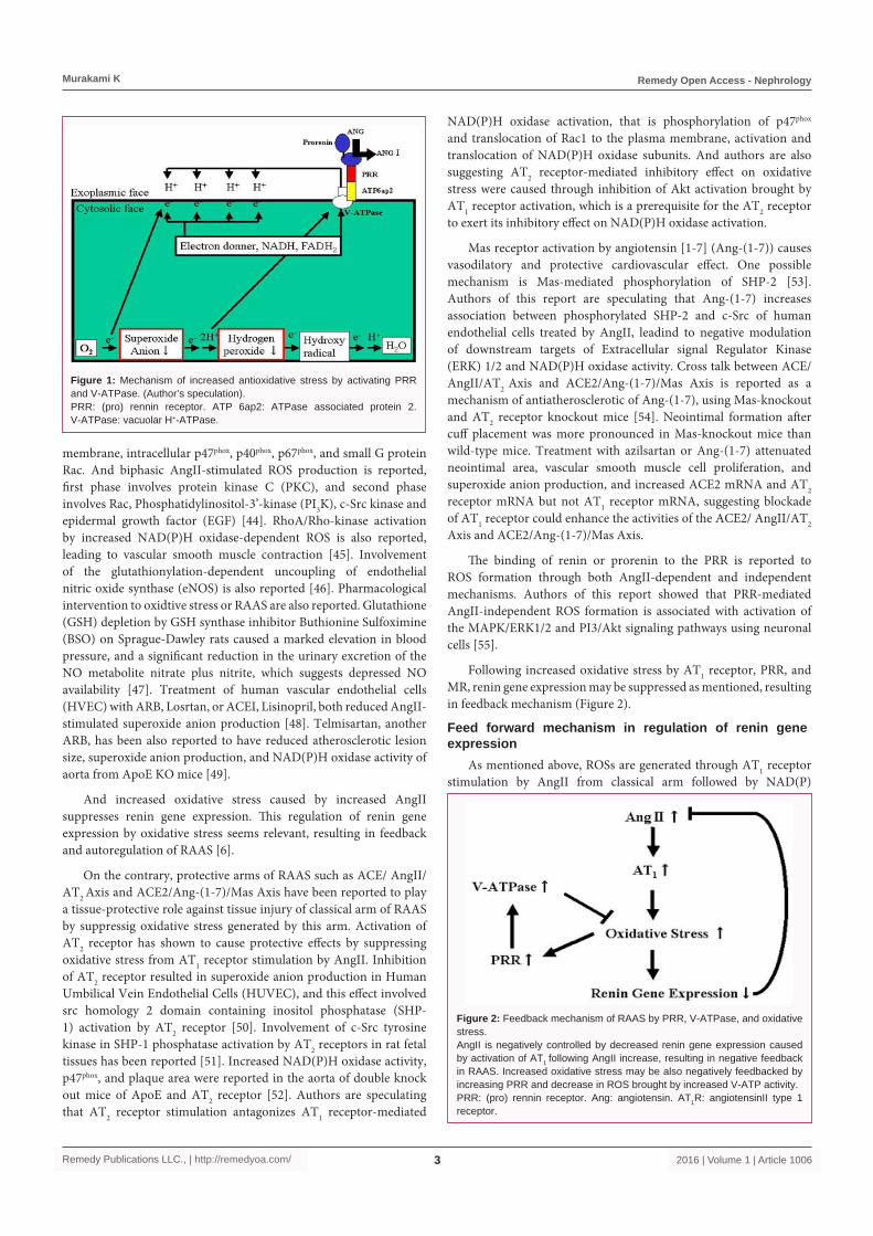
But some reports show that PRR is regulating the production of intracelular ROS such as superoxide anion. In Yeast, mutants lacking V-ATPase subunits results in increased oxidative stress (may be extra-mitochondrial origin) [39,40]. Possible mechanism may be because positively charged cell membrane attracts intracellular electron (from NADH, FADH2, electron donors) to the cytosolic face of plasma membrane electrically due increased H+ concentration of exoplasmic face of the plasma membrane as V-ATPase associated-protein from PRR activates V-ATPase and facilitate outward flow of H+. Thus decreased intracellular electron cause reduction in generation of intracelular ROS such as superoxide anion from triplet oxygen molecule, because intracellular triplet oxygen molecules interact less frequently with electron donners (Figure 1).
Increased oxidative stress through MR activation by aldosterone in renal tubular cell [41], endothelial cell [42], and endothelial progenital cell [43] has been also reported.
Feed back mechanism in regulation of renin gene expression
Reactive Oxygen Species (ROS) production by AngII through AT1 receptor of classical arm is caused mainly by NAD(P)H oxidase, which is composed of p22phox, gp91phox (Nox2), components of cell
Murakami K Remedy Open Access - Nephrology
Remedy Publications LLC., | http://remedyoa.com/ 2016 | Volume 1 | Article 10063
membrane, intracellular p47phox, p40phox, p67phox, and small G protein Rac. And biphasic AngII-stimulated ROS production is reported, first phase involves protein kinase C (PKC), and second phase involves Rac, Phosphatidylinositol-3’-kinase (PI3K), c-Src kinase and epidermal growth factor (EGF) [44]. RhoA/Rho-kinase activation by increased NAD(P)H oxidase-dependent ROS is also reported, leading to vascular smooth muscle contraction [45]. Involvement of the glutathionylation-dependent uncoupling of endothelial nitric oxide synthase (eNOS) is also reported [46]. Pharmacological intervention to oxidtive stress or RAAS are also reported. Glutathione (GSH) depletion by GSH synthase inhibitor Buthionine Sulfoximine (BSO) on Sprague-Dawley rats caused a marked elevation in blood pressure, and a significant reduction in the urinary excretion of the NO metabolite nitrate plus nitrite, which suggests depressed NO availability [47]. Treatment of human vascular endothelial cells (HVEC) with ARB, Losrtan, or ACEI, Lisinopril, both reduced AngII-stimulated superoxide anion production [48]. Telmisartan, another ARB, has been also reported to have reduced atherosclerotic lesion size, superoxide anion production, and NAD(P)H oxidase activity of aorta from ApoE KO mice [49].
And increased oxidative stress caused by increased AngII suppresses renin gene expression. This regulation of renin gene expression by oxidative stress seems relevant, resulting in feedback and autoregulation of RAAS [6].
On the contrary, protective arms of RAAS such as ACE/ AngII/AT2 Axis and ACE2/Ang-(1-7)/Mas Axis have been reported to play a tissue-protective role against tissue injury of classical arm of RAAS by suppressig oxidative stress generated by this arm. Activation of AT2 receptor has shown to cause protective effects by suppressing oxidative stress from AT1 receptor stimulation by AngII. Inhibition of AT2 receptor resulted in superoxide anion production in Human Umbilical Vein Endothelial Cells (HUVEC), and this effect involved src homology 2 domain containing inositol phosphatase (SHP-1) activation by AT2 receptor [50]. Involvement of c-Src tyrosine kinase in SHP-1 phosphatase activation by AT2 receptors in rat fetal tissues has been reported [51]. Increased NAD(P)H oxidase activity, p47phox, and plaque area were reported in the aorta of double knock out mice of ApoE and AT2 receptor [52]. Authors are speculating that AT2 receptor stimulation antagonizes AT1 receptor-mediated
NAD(P)H oxidase activation, that is phosphorylation of p47phox and translocation of Rac1 to the plasma membrane, activation and translocation of NAD(P)H oxidase subunits. And authors are also suggesting AT2 receptor-mediated inhibitory effect on oxidative stress were caused through inhibition of Akt activation brought by AT1 receptor activation, which is a prerequisite for the AT2 receptor to exert its inhibitory effect on NAD(P)H oxidase activation.
Mas receptor activation by angiotensin [1-7] (Ang-(1-7)) causes vasodilatory and protective cardiovascular effect. One possible mechanism is Mas-mediated phosphorylation of SHP-2 [53]. Authors of this report are speculating that Ang-(1-7) increases association between phosphorylated SHP-2 and c-Src of human endothelial cells treated by AngII, leadind to negative modulation of downstream targets of Extracellular signal Regulator Kinase (ERK) 1/2 and NAD(P)H oxidase activity. Cross talk between ACE/ AngII/AT2 Axis and ACE2/Ang-(1-7)/Mas Axis is reported as a mechanism of antiatherosclerotic of Ang-(1-7), using Mas-knockout and AT2 receptor knockout mice [54]. Neointimal formation after cuff placement was more pronounced in Mas-knockout mice than wild-type mice. Treatment with azilsartan or Ang-(1-7) attenuated neointimal area, vascular smooth muscle cell proliferation, and superoxide anion production, and increased ACE2 mRNA and AT2 receptor mRNA but not AT1 receptor mRNA, suggesting blockade of AT1 receptor could enhance the activities of the ACE2/ AngII/AT2 Axis and ACE2/Ang-(1-7)/Mas Axis.
The binding of renin or prorenin to the PRR is reported to ROS formation through both AngII-dependent and independent mechanisms. Authors of this report showed that PRR-mediated AngII-independent ROS formation is associated with activation of the MAPK/ERK1/2 and PI3/Akt signaling pathways using neuronal cells [55].
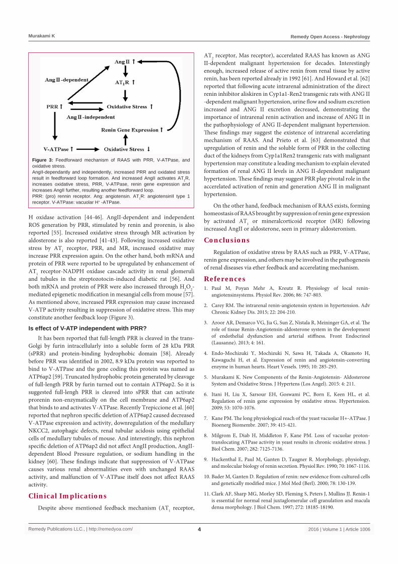
Following increased oxidative stress by AT1 receptor, PRR, and MR, renin gene expression may be suppressed as mentioned, resulting in feedback mechanism (Figure 2).
Feed forward mechanism in regulation of renin gene expression
As mentioned above, ROSs are generated through AT1 receptor stimulation by AngII from classical arm followed by NAD(P)
Figure 1: Mechanism of increased antioxidative stress by activating PRR and V-ATPase. (Author’s speculation).PRR: (pro) rennin receptor. ATP 6ap2: ATPase associated protein 2. V-ATPase: vacuolar H+-ATPase.
Figure 2: Feedback mechanism of RAAS by PRR, V-ATPase, and oxidative stress. AngII is negatively controlled by decreased renin gene expression caused by activation of AT1 following AngII increase, resulting in negative feedback in RAAS. Increased oxidative stress may be also negatively feedbacked by increasing PRR and decrease in ROS brought by increased V-ATP activity.PRR: (pro) rennin receptor. Ang: angiotensin. AT1R: angiotensinII type 1 receptor.
Murakami K Remedy Open Access - Nephrology
Remedy Publications LLC., | http://remedyoa.com/ 2016 | Volume 1 | Article 10064
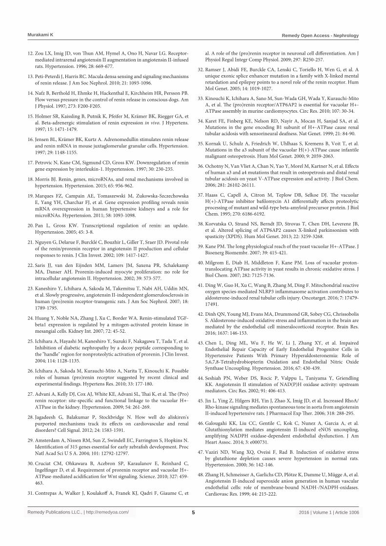
H oxidase activation [44-46]. AngII-dependent and independent ROS generation by PRR, stimulated by renin and prorenin, is also reported [55]. Increased oxidative stress through MR activation by aldosterone is also reported [41-43]. Following increased oxidative stress by AT1 receptor, PRR, and MR, increased oxidative may increase PRR expression again. On the other hand, both mRNA and protein of PRR were reported to be upregulated by enhancement of AT1 receptor-NADPH oxidase cascade activity in renal glomeruli and tubules in the streptozotocin-induced diabetic rat [56]. And both mRNA and protein of PRR were also increased through H2O2-mediated epigenetic modification in mesangial cells from mouse [57]. As mentioned above, increased PRR expression may cause increased V-ATP activity resulting in suppression of oxidative stress. This may constitute another feedback loop (Figure 3).
Is effect of V-ATP independent with PRR?It has been reported that full-length PRR is cleaved in the trans-
Golgi by furin intracellularly into a soluble form of 28 kDa PRR (sPRR) and protein-binding hydrophobic domain [58]. Already before PRR was identified in 2002, 8.9 kDa protein was reported to bind to V-ATPase and the gene coding this protein was named as ATP6ap2 [59]. Truncated hydrophobic protein generated by cleavage of full-length PRR by furin turned out to contain ATP6ap2. So it is suggested full-lengh PRR is cleaved into sPRR that can activate prorenin non-enzymatically on the cell membrane and ATP6ap2 that binds to and activates V-ATPase. Recently Trepiccione et al. [60] reported that nephron specific deletion of ATP6ap2 caused decreased V-ATPase expression and activity, downregulation of the medullary NKCC2, autophagic defects, renal tubular acidosis using epithelial cells of medullary tubules of mouse. And interestingly, this nephron specific deletion of ATP6ap2 did not affect AngII production, AngII-dependent Blood Pressure regulation, or sodium handling in the kidney [60]. These findings indicate that suppression of V-ATPase causes various renal abnormalities even with unchanged RAAS activity, and malfunction of V-ATPase itself does not affect RAAS activity.
Clinical ImplicationsDespite above mentioned feedback mechanism (AT1 receptor,
AT2 receptor, Mas receptor), accerelated RAAS has known as ANG II-dependent malignant hypertension for decades. Interestingly enough, increased release of active renin from renal tissue by active renin, has been reported already in 1992 [61]. And Howard et al. [62] reported that following acute intrarenal administration of the direct renin inhibitor aliskiren in Cyp1a1-Ren2 transgenic rats with ANG II -dependent malignant hypertension, urine flow and sodium excretion increased and ANG II excretion decreased, demonstrating the importance of intrarenal renin activation and increase of ANG II in the pathophysiology of ANG II-dependent malignant hypertension. These findings may suggest the existence of intrarenal accerelating mechanism of RAAS. And Prieto et al. [63] demonstrated that upregulation of renin and the soluble form of PRR in the collecting duct of the kidneys from Cyp1a1Ren2 transgenic rats with malignant hypertension may constitute a leading mechanism to explain elevated formation of renal ANG II levels in ANG II-dependent malignant hypertension. These findings may suggest PRR play pivotal role in the accerelated activation of renin and generation ANG II in malignant hypertension.
On the other hand, feedback mechanism of RAAS exists, forming homeostasis of RAAS brought by suppression of renin gene expression by activated AT1 or mineralcorticoid receptor (MR) following increased AngII or aldosterone, seen in primary aldosteronism.
ConclusionsRegulation of oxidative stress by RAAS such as PRR, V-ATPase,
renin gene expression, and others may be involved in the pathogenesis of renal diseases via ether feedback and accerelating mechanism.
References1. Paul M, Poyan Mehr A, Kreutz R. Physiology of local renin-
angiotensinsystems. Physiol Rev. 2006; 86: 747-803.
2. Carey RM. The intrarenal renin-angiotensin system in hypertension. Adv Chronic Kidney Dis. 2015; 22: 204-210.
3. Aroor AR, Demarco VG, Jia G, Sun Z, Nistala R, Meininger GA, et al. The role of tissue Renin-Angiotensin-aldosterone system in the development of endothelial dysfunction and arterial stiffness. Front Endocrinol (Lausanne). 2013; 4: 161.
4. Endo-Mochizuki Y, Mochizuki N, Sawa H, Takada A, Okamoto H, Kawaguchi H, et al. Expression of renin and angiotensin-converting enzyme in human hearts. Heart Vessels. 1995; 10: 285-293.
5. Murakami K. New Components of the Renin-Angiotensin- Aldosterone System and Oxidative Stress. J Hypertens (Los Angel). 2015: 4: 211.
6. Itani H, Liu X, Sarsour EH, Goswami PC, Born E, Keen HL, et al. Regulation of renin gene expression by oxidative stress. Hypertension. 2009; 53: 1070-1076.
7. Kane PM. The long physiological reach of the yeast vacuolar H+-ATPase. J Bioenerg Biomembr. 2007; 39: 415-421.
8. Milgrom E, Diab H, Middleton F, Kane PM. Loss of vacuolar proton-translocating ATPase activity in yeast results in chronic oxidative stress. J Biol Chem. 2007; 282: 7125-7136.
9. Hackenthal E, Paul M, Ganten D, Taugner R. Morphology, physiology, and molecular biology of renin secretion. Physiol Rev. 1990; 70: 1067-1116.
10. Bader M, Ganten D. Regulation of renin: new evidence from cultured cells and genetically modified mice. J Mol Med (Berl). 2000; 78: 130-139.
11. Clark AF, Sharp MG, Morley SD, Fleming S, Peters J, Mullins JJ. Renin-1 is essential for normal renal juxtaglomerular cell granulation and macula densa morphology. J Biol Chem. 1997; 272: 18185-18190.
Figure 3: Feedforward mechanism of RAAS with PRR, V-ATPase, and oxidative stress.AngII-dependantly and independently, increased PRR and oxidated stress result in feedforward loop formation. And increased AngII activates AT1R, increases oxidative stress, PRR, V-ATPase, renin gene expression and increases AngII further, resulting another feedforward loop. PRR: (pro) rennin receptor. Ang: angiotensin. AT1R: angiotensinII type 1 receptor. V-ATPase: vacuolar H+ -ATPase.
Murakami K Remedy Open Access - Nephrology
Remedy Publications LLC., | http://remedyoa.com/ 2016 | Volume 1 | Article 10065
12. Zou LX, Imig JD, von Thun AM, Hymel A, Ono H, Navar LG. Receptor-mediated intrarenal angiotensin II augmentation in angiotensin II-infused rats. Hypertension. 1996; 28: 669-677.
13. Peti-Peterdi J, Harris RC. Macula densa sensing and signaling mechanisms of renin release. J Am Soc Nephrol. 2010; 21: 1093-1096.
14. Nafz B, Berthold H, Ehmke H, Hackenthal E, Kirchheim HR, Persson PB. Flow versus pressure in the control of renin release in conscious dogs. Am J Physiol. 1997; 273: F200-F205.
15. Holmer SR, Kaissling B, Putnik K, Pfeifer M, Krämer BK, Riegger GA, et al. Beta-adrenergic stimulation of renin expression in vivo. J Hypertens. 1997; 15: 1471-1479.
16. Jensen BL, Krämer BK, Kurtz A. Adrenomedullin stimulates renin release and renin mRNA in mouse juxtaglomerular granular cells. Hypertension. 1997; 29: 1148-1155.
17. Petrovic N, Kane CM, Sigmund CD, Gross KW. Downregulation of renin gene expression by interleukin-1. Hypertension. 1997; 30: 230-235.
18. Morris BJ. Renin, genes, microRNAs, and renal mechanisms involved in hypertension. Hypertension. 2015; 65: 956-962.
19. Marques FZ, Campain AE, Tomaszewski M, Zukowska-Szczechowska E, Yang YH, Charchar FJ, et al. Gene expression profiling reveals renin mRNA overexpression in human hypertensive kidneys and a role for microRNAs. Hypertension. 2011; 58: 1093-1098.
20. Pan L, Gross KW. Transcriptional regulation of renin: an update. Hypertension. 2005; 45: 3-8.
21. Nguyen G, Delarue F, Burcklé C, Bouzhir L, Giller T, Sraer JD. Pivotal role of the renin/prorenin receptor in angiotensin II production and cellular responses to renin. J Clin Invest. 2002; 109: 1417-1427.
22. Saris JJ, van den Eijnden MM, Lamers JM, Saxena PR, Schalekamp MA, Danser AH. Prorenin-induced myocyte proliferation: no role for intracellular angiotensin II. Hypertension. 2002; 39: 573-577.
23. Kaneshiro Y, Ichihara A, Sakoda M, Takemitsu T, Nabi AH, Uddin MN, et al. Slowly progressive, angiotensin II-independent glomerulosclerosis in human (pro)renin receptor-transgenic rats. J Am Soc Nephrol. 2007; 18: 1789-1795.
24. Huang Y, Noble NA, Zhang J, Xu C, Border WA. Renin-stimulated TGF-beta1 expression is regulated by a mitogen-activated protein kinase in mesangial cells. Kidney Int. 2007; 72: 45-52.
25. Ichihara A, Hayashi M, Kaneshiro Y, Suzuki F, Nakagawa T, Tada Y, et al. Inhibition of diabetic nephropathy by a decoy peptide corresponding to the "handle" region for nonproteolytic activation of prorenin. J Clin Invest. 2004; 114: 1128-1135.
26. Ichihara A, Sakoda M, Kurauchi-Mito A, Narita T, Kinouchi K. Possible roles of human (pro)renin receptor suggested by recent clinical and experimental findings. Hypertens Res. 2010; 33: 177-180.
27. Advani A, Kelly DJ, Cox AJ, White KE, Advani SL, Thai K, et al. The (Pro)renin receptor: site-specific and functional linkage to the vacuolar H+-ATPase in the kidney. Hypertension. 2009; 54: 261-269.
28. Jagadeesh G, Balakumar P, Stockbridge N. How well do aliskiren's purported mechanisms track its effects on cardiovascular and renal disorders? Cell Signal. 2012; 24: 1583-1591.
29. Amsterdam A, Nissen RM, Sun Z, Swindell EC, Farrington S, Hopkins N. Identification of 315 genes essential for early zebrafish development. Proc Natl Acad Sci U S A. 2004; 101: 12792-12797.
30. Cruciat CM, Ohkawara B, Acebron SP, Karaulanov E, Reinhard C, Ingelfinger D, et al. Requirement of prorenin receptor and vacuolar H+-ATPase-mediated acidification for Wnt signaling. Science. 2010; 327: 459-463.
31. Contrepas A, Walker J, Koulakoff A, Franek KJ, Qadri F, Giaume C, et
al. A role of the (pro)renin receptor in neuronal cell differentiation. Am J Physiol Regul Integr Comp Physiol. 2009; 297: R250-257.
32. Ramser J, Abidi FE, Burckle CA, Lenski C, Toriello H, Wen G, et al. A unique exonic splice enhancer mutation in a family with X-linked mental retardation and epilepsy points to a novel role of the renin receptor. Hum Mol Genet. 2005; 14: 1019-1027.
33. Kinouchi K, Ichihara A, Sano M, Sun-Wada GH, Wada Y, Kurauchi-Mito A, et al. The (pro)renin receptor/ATP6AP2 is essential for vacuolar H+-ATPase assembly in murine cardiomyocytes. Circ Res. 2010; 107: 30-34.
34. Karet FE, Finberg KE, Nelson RD, Nayir A, Mocan H, Sanjad SA, et al. Mutations in the gene encoding B1 subunit of H+-ATPase cause renal tubular acidosis with sensorineural deafness. Nat Genet. 1999; 21: 84-90.
35. Kornak U, Schulz A, Friedrich W, Uhlhaas S, Kremens B, Voit T, et al. Mutations in the a3 subunit of the vacuolar H(+)-ATPase cause infantile malignant osteopetrosis. Hum Mol Genet. 2000; 9: 2059-2063.
36. Ochotny N, Van Vliet A, Chan N, Yao Y, Morel M, Kartner N, et al. Effects of human a3 and a4 mutations that result in osteopetrosis and distal renal tubular acidosis on yeast V-ATPase expression and activity. J Biol Chem. 2006; 281: 26102-26111.
37. Haass C, Capell A, Citron M, Teplow DB, Selkoe DJ. The vacuolar H(+)-ATPase inhibitor bafilomycin A1 differentially affects proteolytic processing of mutant and wild-type beta-amyloid precursor protein. J Biol Chem. 1995; 270: 6186-6192.
38. Korvatska O, Strand NS, Berndt JD, Strovas T, Chen DH, Leverenz JB, et al. Altered splicing of ATP6AP2 causes X-linked parkinsonism with spasticity (XPDS). Hum Mol Genet. 2013; 22: 3259-3268.
39. Kane PM. The long physiological reach of the yeast vacuolar H+-ATPase. J Bioenerg Biomembr. 2007; 39: 415-421.
40. Milgrom E, Diab H, Middleton F, Kane PM. Loss of vacuolar proton-translocating ATPase activity in yeast results in chronic oxidative stress. J Biol Chem. 2007; 282: 7125-7136.
41. Ding W, Guo H, Xu C, Wang B, Zhang M, Ding F. Mitochondrial reactive oxygen species-mediated NLRP3 inflammasome activation contributes to aldosterone-induced renal tubular cells injury. Oncotarget. 2016; 7: 17479-17491.
42. Dinh QN, Young MJ, Evans MA, Drummond GR, Sobey CG, Chrissobolis S. Aldosterone-induced oxidative stress and inflammation in the brain are mediated by the endothelial cell mineralocorticoid receptor. Brain Res. 2016; 1637: 146-153.
43. Chen L, Ding ML, Wu F, He W, Li J, Zhang XY, et al. Impaired Endothelial Repair Capacity of Early Endothelial Progenitor Cells in Hypertensive Patients With Primary Hyperaldosteronemia: Role of 5,6,7,8-Tetrahydrobiopterin Oxidation and Endothelial Nitric Oxide Synthase Uncoupling. Hypertension. 2016; 67: 430-439.
44. Seshiah PN, Weber DS, Rocic P, Valppu L, Taniyama Y, Griendling KK. Angiotensin II stimulation of NAD(P)H oxidase activity: upstream mediators. Circ Res. 2002; 91: 406-413.
45. Jin L, Ying Z, Hilgers RH, Yin J, Zhao X, Imig JD, et al. Increased RhoA/Rho-kinase signaling mediates spontaneous tone in aorta from angiotensin II-induced hypertensive rats. J Pharmacol Exp Ther. 2006; 318: 288-295.
46. Galougahi KK, Liu CC, Gentile C, Kok C, Nunez A, Garcia A, et al. Glutathionylation mediates angiotensin II-induced eNOS uncoupling, amplifying NADPH oxidase-dependent endothelial dysfunction. J Am Heart Assoc. 2014; 3: e000731.
47. Vaziri ND, Wang XQ, Oveisi F, Rad B. Induction of oxidative stress by glutathione depletion causes severe hypertension in normal rats. Hypertension. 2000; 36: 142-146.
48. Zhang H, Schmeisser A, Garlichs CD, Plötze K, Damme U, Mügge A, et al. Angiotensin II-induced superoxide anion generation in human vascular endothelial cells: role of membrane-bound NADH-/NADPH-oxidases. Cardiovasc Res. 1999; 44: 215-222.
Murakami K Remedy Open Access - Nephrology
Remedy Publications LLC., | http://remedyoa.com/ 2016 | Volume 1 | Article 10066
49. Takaya T, Kawashima S, Shinohara M, Yamashita T, Toh R, Sasaki N, et al. Angiotensin II type 1 receptor blocker telmisartan suppresses superoxide production and reduces atherosclerotic lesion formation in apolipoprotein E-deficient mice. Atherosclerosis. 2006; 186: 402-410.
50. Sohn HY, Raff U, Hoffmann A, Gloe T, Heermeier K, Galle J, et al. Differential role of angiotensin II receptor subtypes on endothelial superoxide formation. Br J Pharmacol. 2000; 131: 667-672.
51. Alvarez SE, Seguin LR, Villarreal RS, Nahmias C, Ciuffo GM. Involvement of c-Src tyrosine kinase in SHP-1 phosphatase activation by Ang II AT2 receptors in rat fetal tissues. J Cell Biochem. 2008; 105: 703-711.
52. Iwai M, Chen R, Li Z, Shiuchi T, Suzuki J, Ide A, et al. Deletion of angiotensin II type 2 receptor exaggerated atherosclerosis in apolipoprotein E-null mice. Circulation. 2005; 112: 1636-1643.
53. Sampaio WO, Henrique de Castro C, Santos RA, Schiffrin EL, Touyz RM. Angiotensin-(1-7) counterregulates angiotensin II signaling in human endothelial cells. Hypertension. 2007; 50: 1093-1098.
54. Ohshima K, Mogi M, Nakaoka H, Iwanami J, Min LJ, Kanno H, et al. Possible role of angiotensin-converting enzyme 2 and activation of angiotensin II type 2 receptor by angiotensin-(1-7) in improvement of vascular remodeling by angiotensin II type 1 receptor blockade. Hypertension. 2014; 63: e53-e59.
55. Peng H, Li W, Seth DM, Nair AR, Francis J, Feng Y. (Pro)renin receptor mediates both angiotensin II-dependent and -independent oxidative stress in neuronal cells. PLoS One. 2013; 8: e58339.
56. Siragy HM, Huang J. Renal (pro)renin receptor upregulation in diabetic rats through enhanced angiotensin AT1 receptor and NADPH oxidase activity. Exp Physiol. 2008; 93: 709-714.
57. Lee DY, Kim HS, Won KJ, Lee KP, Jung SH, Park ES, et al. DJ-1 regulates the expression of renal (pro)renin receptor via reactive oxygen species-mediated epigenetic modification. Biochim Biophys Acta. 2015; 1850: 426-434.
58. Cousin C, Bracquart D, Contrepas A, Corvol P, Muller L, Nguyen G. Soluble form of the (pro)renin receptor generated by intracellular cleavage by furin is secreted in plasma. Hypertension. 2009; 53: 1077-1082.
59. Ludwig J, Kerscher S, Brandt U, Pfeiffer K, Getlawi F, Apps DK, et al. Identification and characterization of a novel 9.2-kDa membrane sector-associated protein of vacuolar proton-ATPase from chromaffin granules. J Biol Chem. 1998; 273: 10939-10947.
60. Trepiccione F, Gerber SD, Grahammer F, López-Cayuqueo KI, Baudrie V, Păunescu TG, et al. Renal Atp6ap2/(Pro)renin Receptor Is Required for Normal Vacuolar H+-ATPase Function but Not for the Renin-Angiotensin System. J Am Soc Nephrol. 2016; 27.
61. Hosoi M, Kim S, Takada T, Suzuki F, Murakami K, Yamamoto K. Effects of prorenin on blood pressure and plasma renin concentrations in stroke-prone spontaneously hypertensive rats. Am J Physiol. 1992; 262: E234-E239.
62. Howard CG, Mitchell KD. Renal functional responses to selective intrarenal renin inhibition in Cyp1a1-Ren2 transgenic rats with ANG II-dependent malignant hypertension. Am J Physiol Renal Physiol. 2012; 302: F52-59.
63. Prieto MC, Williams DE, Liu L, Kavanagh KL, Mullins JJ, Mitchell KD. Enhancement of renin and prorenin receptor in collecting duct of Cyp1a1-Ren2 ratsmay contribute to development and progression of malignant hypertension. Am J Physiol Renal Physiol. 2011; 300: F581-588.